User login
STS: Lactate dehydrogenase of 1,150 IU/L flags LVAD thrombosis
PHOENIX – A blood lactate dehydrogenase level of at least 1,150 IU/L may be the best cutoff for identifying pump thrombosis in patients who have received a left ventricular assist device based on findings from a combined series of 502 pump recipients.
Using this cutoff correlated with a positive predictive value of 74% and a negative predictive value of 92%, Dr. Vikram Sood said at the annual meeting of the Society of Thoracic Surgeons.
Results from several prior studies had established an elevated blood level of lactate dehydrogenase (LDH) as perhaps the best early flag of significant thrombus inside of a left ventricular assist device (LVAD), a condition called pump thrombosis. But prior studies had not identified a specific cutoff value to use that could confidently diagnose pump thrombosis. A level of 1,150 IU/L is about five times the upper limit of normal, noted Dr. Francis D. Pagani, professor of surgery and surgical director of adult heart transplantation at the University of Michigan, Ann Arbor, and a senior author on this report.
Dr. Sood and his associates reviewed records for 502 patients who received a HeartMate II LVAD at either the University of Michigan or at the Mayo Clinic in Rochester, Minn., during 2004-2014. During that time, 58 of the patients required a total of 70 LVAD explants. Fifty-two of the explants were for suspected pump thrombosis, 12 for infection, and 6 for lead fracture. The average time from LVAD placement to explant was 12 months.
Among the 52 LVADs explanted for suspected pump thrombosis, 47 actually had thrombosis. In addition, six of the LVADs that were explanted for other reasons also had a significant amount of thrombus, resulting in 53 total explanted LVAD with confirmed pump thrombosis.
The researchers analyzed blood levels of LDH during the 6 months prior to explant. Among the patients with the 53 LVADs explanted with confirmed pump thrombosis, the median of all LDH levels during the period was 1,061 IU/L and the median maximum LDH level for each patient was 1,940 IU/L, reported Dr. Sood, a cardiac surgeon at the University of Michigan. In contrast, among the patients who had a total of 17 LVADs explanted without pump thrombosis, the median value for all LDH measures during the 6 months prior to explant was 533 IU/L, and their medium maximum level was 504 IU/L.
Analysis of LDH levels prior to explant and subsequent confirmation of pump thrombosis identified a cut off of 1,150 IU/L as able to identify pump thrombosis with a sensitivity of 78% and a specificity of 90%, Dr. Sood reported.
On Twitter @mitchelzoler
AT THE STS ANNUAL MEETING
Key clinical point: In patients who have received a left ventricular assist device, a blood level of lactate dehydrogenase of at least 1,150 IU/L had a good positive and negative predictive value for pump thrombosis.
Major finding: A 1,150 IU/L cutoff for LDH had a positive predictive value of 74% and a negative predictive value of 92%.
Data source: Retrospective review of 502 patients who received a HeartMate II LVAD at either of two U.S. centers during 2004-2014.
Disclosures: Dr. Sood and Dr. Pagani had no disclosures.
STS: Minimizing LVAD pump thrombosis poses new challenges
PHOENIX – Cardiothoracic surgeons who implant left ventricular assist devices in patients with failing hearts remain at a loss to fully explain why they started seeing a sharp increase in thrombus clogging in these devices in 2012, but nevertheless they are gaining a better sense of how to minimize the risk.
Three key principles for minimizing thrombosis risk are selecting the right patients to receive left ventricular assist devices (LVAD), applying optimal management strategies once patients receive a LVAD, and maintaining adequate flow of blood through the pump, Dr. Francis D. Pagani said in a talk at a session devoted to pump thrombosis at the annual meeting of the Society of Thoracic Surgeons.
Other critical aspects include optimal implantation technique, quick work-up of patients to rule out reversible LVAD inflow or outflow problems once pump thrombosis is suspected, and ceasing medical therapy of the thrombosis if it proves ineffective and instead progress to surgical pump exchange, pump explantation, or heart transplant when necessary, said Dr. Ahmet Kilic, a cardiothoracic surgeon at the Ohio State University, Columbus.
Another key issue is that, now that the pump thrombosis incidence is averaging about 10% of LVAD recipients, with an incidence rate during 2-year follow-up as high as 24% reported from one series, surgeons and physicians who care for LVAD patients must have a high index of suspicion and routinely screen LVAD recipients for early signs of pump thrombosis. The best way to catch pump thrombosis early seems to be by regularly measuring patients’ serum level of lactate dehydrogenase (LDH), said Dr. Robert L. Kormos, professor of surgery and director of the artificial heart program at the University of Pittsburgh.
“We measure LDH on most clinic visits, whether or not the patient has an indication of pump thrombosis. We need to screen [LDH levels] much more routinely than we used to,” he said during the session. “Elevated LDH is probably the first and most reliable early sign, but you need to also assess LDH isoenzymes because we’ve had patients with an elevation but no sign of pump thrombosis, and their isoenzymes showed that the increased LDH was coming from their liver,” Dr. Kormos said in an interview.
Although serial measurements and isoenzyme analysis can establish a sharp rise in heart-specific LDH in an individual patient, a report at the meeting documented that in a series of 53 patients with pump thrombosis treated at either of two U.S. centers, an LDH level of at least 1,155 IU/L flagged pump thrombosis with a fairly high sensitivity and specificity. This LDH level is roughly five times the upper limit of normal, noted Dr. Pagani, professor of surgery and surgical director of adult heart transplantation at the University of Michigan, Ann Arbor, and a senior author on this report.
But prior to this report Dr. Kormos said that he regarded a LDH level of 600-800 IU/L as enough of an elevation above normal to prompt concern and investigation. And he criticized some LVAD programs that allow LDH levels to rise much higher.
“I know of clinicians who see a LDH of 1,500-2,000 IU/L but the patient seems okay and they wonder if they should change out the pump. For me, it’s a no brainer. Others try to list a patient like this for a heart transplant so they can avoid doing a pump exchange. I think that’s dangerous; it risks liver failure or renal failure. I would not sit on any LVAD that is starting to produce signs of hemolysis syndrome, but some places do this,” Dr. Kormos said in an interview.
“Pump thrombosis probably did not get addressed in as timely a fashion as it should have been” when it was first seen on the rise in 2012, noted Dr. James K. Kirklin, professor of surgery and director of cardiothoracic surgery at the University of Alabama, Birmingham. “It is now being addressed, and we realize that this is not just a pump problem but also involves patient factors and management factors that we need to learn more about. We are quite ignorant of the patient factors and understanding their contributions to bleeding and thrombosis,” said Dr. Kirklin. He also acknowledged that whatever role the current generation of LVAD pumps play in causing thrombosis will not quickly resolve.
“I’m looking forward to a new generation of pumps, but the pumps we have today will probably remain for another 3-5 years.”
The issue of LVAD pump thrombosis first came into clear focus with publication at the start of 2014 of a report that tracked its incidence from 2004 to mid-2013 at three U.S. centers that had placed a total of 895 LVADs in 837 patients. The annual rate of new episodes of pump thrombosis jumped from about 1%-2% of LVAD recipients throughout the first part of the study period through the end of 2011, to an annual rate of about 10% by mid 2013 (N Engl J Med. 2014 Jan 2;370[1]:33-40).
“The inflection occurred in about 2012,” noted Dr. Nicholas G. Smedira, a cardiothoracic surgeon at the Cleveland Clinic. “No one has figured out why” the incidence suddenly spiked starting in 2012 and intensified in 2013, he said. This epidemic of pump thrombosis has produced “devastating complications” that have led to multiple readmissions and reduced cost-effectiveness of LVADs and has affected how the heart transplant community allocates hearts, Dr. Smedira said during his talk at the session. He noted that once the surge in pump thrombosis started, the timing of the appearance of significant thrombus shifted earlier, often occurring within 2-3 months after LVAD placement. There now is “increasing device-related pessimism” and increasing demoralization among clinicians because of this recurring complication, he said.
More recent data show the trend toward increasingly higher rates of pump thrombosis continuing through the end of 2013, with the situation during 2014 a bit less clear. Late last year, data from 9,808 U.S. patients who received an LVAD and entered the Interagency Registry for Mechanically Assisted Circulatory Support (INTERMACS) showed that the incidence of pump thrombosis during the first 6 months following an implant rose from 1% in 2008 to 2% in 2009 and in 2010, 4% in 2011, 7% in 2012, 8% in 2013, and then eased back to 5% in the first half of 2014 (J Heart Lung Transplant. 2015 Dec;34[12]:1515-26). The annual rate rose from 2% in 2008 to a peak of 11% in 2013, with 12-month data from 2014 not yet available at the time of this report.
“The modest reduction of observed pump thrombosis at 6 months during 2014 has occurred in a milieu of heightened intensity of anti-coagulation management, greater surgical awareness of optimal pump implantation and positioning and pump speed management. Thus, one may speculate that current thrombosis risk-mitigation strategies have contributed to reducing but not eliminating the increased thrombosis risk observed since 2011,” concluded the authors of the report.
Surgeons and cardiologists must now have a high index of suspicion for pump thrombosis in LVAD recipients, and be especially on the lookout for four key flags of a problem, said Dr. Kormos. The first is a rising LDH level, but additional flags include an isolated power elevation that doesn’t correlate with anything else, evidence of hemolysis, and new-onset heart failure symptoms. These can occur individually or in some combination. He recommended following a diagnostic algorithm first presented in 2013 that remains very valid today (J Heart Lung Transplant. 2013 July;32[7]:667-70).
Dr. Kormos also highlighted that the presentation of pump thrombosis can differ between the two LVADs most commonly used in U.S. practice, the HeartMate II and the HeartWare devices. A LDH elevation is primarily an indicator for HeartMate II, while both that model and the HeartWare device show sustained, isolated power elevations when thrombosis occurs.
Dr. Pagani, Dr. Kirklin, and Dr. Smedira had no disclosures. Dr. Kormos has received travel support from HeartWare. Dr. Kilic has been a consultant to Thoratec and a speaker on behalf of Baxter International.
On Twitter @mitchelzoler
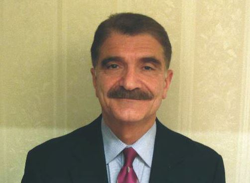
Dr. Hossein Almassi, FCCP, comments: With improvements in technology and development of rotary pumps, there has been a significant growth in the use of mechanical circulatory support (MCS) for treatment of end stage heart failure with a parallel improvement in patients’ survival and the quality of life.
|
| Dr. Hossein Almassi |
The authors of this report presented at the 2016 annual meeting of the STS, are authorities in the field of MCS outlining the observed increase in pump thrombosis noted in 2012. The sharp increase in the thrombosis rate is different from the lower incidence seen in the preapproval stage of the pump trial.
It should be noted that the report is related mainly to the HeatMate II left ventricular assist device (LVAD) and not the more recently implanted HeartWare device.
The diagnostic algorithm outlined in the accompanying reference (J Heart Lung Transplant. 2013 July;32[7]:667-70) regarding the diagnosis and management of suspected pump thrombosis is worth reading with the main criteria heralding a potential pump thrombosis being 1)sustained pump power elevation, 2) elevation of cardiac LDH or plasma-free hemoglobin, 3) hemolysis, and 4) symptoms of heart failure.
With further refinements in technology, the field of MCS is awaiting the development of newer LVAD devices that would mitigate the serious problem of pump thrombosis.
Dr. Hossein Almassi, FCCP, comments: With improvements in technology and development of rotary pumps, there has been a significant growth in the use of mechanical circulatory support (MCS) for treatment of end stage heart failure with a parallel improvement in patients’ survival and the quality of life.

|
| Dr. Hossein Almassi |
The authors of this report presented at the 2016 annual meeting of the STS, are authorities in the field of MCS outlining the observed increase in pump thrombosis noted in 2012. The sharp increase in the thrombosis rate is different from the lower incidence seen in the preapproval stage of the pump trial.
It should be noted that the report is related mainly to the HeatMate II left ventricular assist device (LVAD) and not the more recently implanted HeartWare device.
The diagnostic algorithm outlined in the accompanying reference (J Heart Lung Transplant. 2013 July;32[7]:667-70) regarding the diagnosis and management of suspected pump thrombosis is worth reading with the main criteria heralding a potential pump thrombosis being 1)sustained pump power elevation, 2) elevation of cardiac LDH or plasma-free hemoglobin, 3) hemolysis, and 4) symptoms of heart failure.
With further refinements in technology, the field of MCS is awaiting the development of newer LVAD devices that would mitigate the serious problem of pump thrombosis.
Dr. Hossein Almassi, FCCP, comments: With improvements in technology and development of rotary pumps, there has been a significant growth in the use of mechanical circulatory support (MCS) for treatment of end stage heart failure with a parallel improvement in patients’ survival and the quality of life.

|
| Dr. Hossein Almassi |
The authors of this report presented at the 2016 annual meeting of the STS, are authorities in the field of MCS outlining the observed increase in pump thrombosis noted in 2012. The sharp increase in the thrombosis rate is different from the lower incidence seen in the preapproval stage of the pump trial.
It should be noted that the report is related mainly to the HeatMate II left ventricular assist device (LVAD) and not the more recently implanted HeartWare device.
The diagnostic algorithm outlined in the accompanying reference (J Heart Lung Transplant. 2013 July;32[7]:667-70) regarding the diagnosis and management of suspected pump thrombosis is worth reading with the main criteria heralding a potential pump thrombosis being 1)sustained pump power elevation, 2) elevation of cardiac LDH or plasma-free hemoglobin, 3) hemolysis, and 4) symptoms of heart failure.
With further refinements in technology, the field of MCS is awaiting the development of newer LVAD devices that would mitigate the serious problem of pump thrombosis.
PHOENIX – Cardiothoracic surgeons who implant left ventricular assist devices in patients with failing hearts remain at a loss to fully explain why they started seeing a sharp increase in thrombus clogging in these devices in 2012, but nevertheless they are gaining a better sense of how to minimize the risk.
Three key principles for minimizing thrombosis risk are selecting the right patients to receive left ventricular assist devices (LVAD), applying optimal management strategies once patients receive a LVAD, and maintaining adequate flow of blood through the pump, Dr. Francis D. Pagani said in a talk at a session devoted to pump thrombosis at the annual meeting of the Society of Thoracic Surgeons.
Other critical aspects include optimal implantation technique, quick work-up of patients to rule out reversible LVAD inflow or outflow problems once pump thrombosis is suspected, and ceasing medical therapy of the thrombosis if it proves ineffective and instead progress to surgical pump exchange, pump explantation, or heart transplant when necessary, said Dr. Ahmet Kilic, a cardiothoracic surgeon at the Ohio State University, Columbus.
Another key issue is that, now that the pump thrombosis incidence is averaging about 10% of LVAD recipients, with an incidence rate during 2-year follow-up as high as 24% reported from one series, surgeons and physicians who care for LVAD patients must have a high index of suspicion and routinely screen LVAD recipients for early signs of pump thrombosis. The best way to catch pump thrombosis early seems to be by regularly measuring patients’ serum level of lactate dehydrogenase (LDH), said Dr. Robert L. Kormos, professor of surgery and director of the artificial heart program at the University of Pittsburgh.
“We measure LDH on most clinic visits, whether or not the patient has an indication of pump thrombosis. We need to screen [LDH levels] much more routinely than we used to,” he said during the session. “Elevated LDH is probably the first and most reliable early sign, but you need to also assess LDH isoenzymes because we’ve had patients with an elevation but no sign of pump thrombosis, and their isoenzymes showed that the increased LDH was coming from their liver,” Dr. Kormos said in an interview.
Although serial measurements and isoenzyme analysis can establish a sharp rise in heart-specific LDH in an individual patient, a report at the meeting documented that in a series of 53 patients with pump thrombosis treated at either of two U.S. centers, an LDH level of at least 1,155 IU/L flagged pump thrombosis with a fairly high sensitivity and specificity. This LDH level is roughly five times the upper limit of normal, noted Dr. Pagani, professor of surgery and surgical director of adult heart transplantation at the University of Michigan, Ann Arbor, and a senior author on this report.
But prior to this report Dr. Kormos said that he regarded a LDH level of 600-800 IU/L as enough of an elevation above normal to prompt concern and investigation. And he criticized some LVAD programs that allow LDH levels to rise much higher.
“I know of clinicians who see a LDH of 1,500-2,000 IU/L but the patient seems okay and they wonder if they should change out the pump. For me, it’s a no brainer. Others try to list a patient like this for a heart transplant so they can avoid doing a pump exchange. I think that’s dangerous; it risks liver failure or renal failure. I would not sit on any LVAD that is starting to produce signs of hemolysis syndrome, but some places do this,” Dr. Kormos said in an interview.
“Pump thrombosis probably did not get addressed in as timely a fashion as it should have been” when it was first seen on the rise in 2012, noted Dr. James K. Kirklin, professor of surgery and director of cardiothoracic surgery at the University of Alabama, Birmingham. “It is now being addressed, and we realize that this is not just a pump problem but also involves patient factors and management factors that we need to learn more about. We are quite ignorant of the patient factors and understanding their contributions to bleeding and thrombosis,” said Dr. Kirklin. He also acknowledged that whatever role the current generation of LVAD pumps play in causing thrombosis will not quickly resolve.
“I’m looking forward to a new generation of pumps, but the pumps we have today will probably remain for another 3-5 years.”
The issue of LVAD pump thrombosis first came into clear focus with publication at the start of 2014 of a report that tracked its incidence from 2004 to mid-2013 at three U.S. centers that had placed a total of 895 LVADs in 837 patients. The annual rate of new episodes of pump thrombosis jumped from about 1%-2% of LVAD recipients throughout the first part of the study period through the end of 2011, to an annual rate of about 10% by mid 2013 (N Engl J Med. 2014 Jan 2;370[1]:33-40).
“The inflection occurred in about 2012,” noted Dr. Nicholas G. Smedira, a cardiothoracic surgeon at the Cleveland Clinic. “No one has figured out why” the incidence suddenly spiked starting in 2012 and intensified in 2013, he said. This epidemic of pump thrombosis has produced “devastating complications” that have led to multiple readmissions and reduced cost-effectiveness of LVADs and has affected how the heart transplant community allocates hearts, Dr. Smedira said during his talk at the session. He noted that once the surge in pump thrombosis started, the timing of the appearance of significant thrombus shifted earlier, often occurring within 2-3 months after LVAD placement. There now is “increasing device-related pessimism” and increasing demoralization among clinicians because of this recurring complication, he said.
More recent data show the trend toward increasingly higher rates of pump thrombosis continuing through the end of 2013, with the situation during 2014 a bit less clear. Late last year, data from 9,808 U.S. patients who received an LVAD and entered the Interagency Registry for Mechanically Assisted Circulatory Support (INTERMACS) showed that the incidence of pump thrombosis during the first 6 months following an implant rose from 1% in 2008 to 2% in 2009 and in 2010, 4% in 2011, 7% in 2012, 8% in 2013, and then eased back to 5% in the first half of 2014 (J Heart Lung Transplant. 2015 Dec;34[12]:1515-26). The annual rate rose from 2% in 2008 to a peak of 11% in 2013, with 12-month data from 2014 not yet available at the time of this report.
“The modest reduction of observed pump thrombosis at 6 months during 2014 has occurred in a milieu of heightened intensity of anti-coagulation management, greater surgical awareness of optimal pump implantation and positioning and pump speed management. Thus, one may speculate that current thrombosis risk-mitigation strategies have contributed to reducing but not eliminating the increased thrombosis risk observed since 2011,” concluded the authors of the report.
Surgeons and cardiologists must now have a high index of suspicion for pump thrombosis in LVAD recipients, and be especially on the lookout for four key flags of a problem, said Dr. Kormos. The first is a rising LDH level, but additional flags include an isolated power elevation that doesn’t correlate with anything else, evidence of hemolysis, and new-onset heart failure symptoms. These can occur individually or in some combination. He recommended following a diagnostic algorithm first presented in 2013 that remains very valid today (J Heart Lung Transplant. 2013 July;32[7]:667-70).
Dr. Kormos also highlighted that the presentation of pump thrombosis can differ between the two LVADs most commonly used in U.S. practice, the HeartMate II and the HeartWare devices. A LDH elevation is primarily an indicator for HeartMate II, while both that model and the HeartWare device show sustained, isolated power elevations when thrombosis occurs.
Dr. Pagani, Dr. Kirklin, and Dr. Smedira had no disclosures. Dr. Kormos has received travel support from HeartWare. Dr. Kilic has been a consultant to Thoratec and a speaker on behalf of Baxter International.
On Twitter @mitchelzoler
EXPERT ANALYSIS FROM THE STS ANNUAL MEETING
STS: Valved conduit shows right ventricular outflow durability
PHOENIX – A prosthetic conduit that contains a porcine valve showed excellent intermediate-term durability for repairing the right ventricular outflow tract in 100 teenagers and young adults at a single U.S. center.
“The Carpentier-Edwards xenograft for right ventricular outflow tract [RVOT] reconstruction provides excellent freedom from reoperation and valve dysfunction, as well as sustained improvement in right-ventricular chamber size at intermediate-term follow-up,” Dr. Heidi B. Schubmehl said at the Society of Thoracic Surgeons annual meeting.
Dr. Schubmehl reported a 92% rate of freedom from valve dysfunction with follow-up out to about 10 years, and significant reductions in right ventricular size at follow-up, compared with baseline, as measured by both echocardiography and by MRI.
The Carpentier-Edwards porcine valve and conduit “seemed to hold up better than a lot of other [prosthetic] valves,” said Dr. George M. Alfieris, director of pediatric cardiac surgery at the University of Rochester (N.Y.), and senior author for the study. In addition to the valve’s durability over approximately the first 10 years following placement, the results also showed the positive impact the valve had on right ventricular size, an important result of the repair’s efficacy, Dr. Alfieris said.
“It’s a mistake to allow the right ventricle to be under high pressure or to reach a large volume. We now focus on preserving the right ventricle,” he said in an interview. “I’ve become very concerned about preventing right ventricular dilation and preserving right ventricular function.”
Dr. Alfieris noted that his prior experience using other types of valves in the pulmonary valve and RVOT position showed those valves “did great for the first 10 years and then failed. What’s different in this series is that after 10 years, we have not seen the same dysfunction as with the prior generation of valves. I will be very interested to see what happens to them” as follow-up continues beyond 10 years. He also expressed dismay that recently the company that had been marketing the valve and conduit used in the current study, the Carpentier-Edwards, stopped selling them. He expects that as his supply of conduits runs out he’ll have to start using a different commercial valve and conduit that he believes will not perform as well or create his own conduits with a porcine valve from a different supplier.
The series of 100 patients comprised individuals aged 17 or older who received a pulmonary artery and had RVOT reconstruction at the University of Rochester during 2000-2010, Dr. Schubmehl reported. The series included 78 patients with a history of tetralogy of Fallot, 8 patients born with transposition of their great arteries, 8 patients with truncus arteriosus, and 6 patients with other congenital heart diseases. Their median age at the time they received the RVOT conduit was 24 years, 59% were men, and 99 had undergone a prior sternotomy. At the time they received the conduit, 55 had pulmonary valve insufficiency, 30 had valve stenosis, and 15 had both. Follow-up occurred an average of 7 years after conduit placement.
Two recipients died: One death occurred perioperatively in a 41-year old who had a massive cerebrovascular event, and the second death was in a 39-year old who died 2.6 years after conduit placement from respiratory failure. Two additional patients required a reintervention during follow-up, said Dr. Schubmehl, a general surgeon at the University of Rochester. One reintervention occurred after 11 years to treat endocarditis, and the second after 11 years to perform balloon valvuloplasty because of valve stenosis.
The results reported by Dr. Schubmehl for echocardiography examinations showed that the patients had a statistically significant reduction in their RVOT pressure gradient from baseline to 1-year follow-up that was sustained through their intermediate-term follow-up. Seventy-seven patients had pulmonary valve insufficiency at baseline that resolved in all patients at 1-year follow-up and remained resolved in all but one patient at extended follow-up. Nineteen patients underwent additional imaging with MRI at an average follow-up of 7 years, and these findings confirmed the echo results.
On Twitter @mitchelzoler
The intermediate-term results reported by Dr. Schubmehl using a Carpentier-Edwards conduit in the right-ventricular outflow tract are clearly better than what we have seen using other types of valves and conduits in this position. If the valve and conduit they used persists with similar performance beyond 10 years, it would be a very good option. However, what typically happens is that replacement valves look good for about 10 years and then start to fail, often with a steep failure curve. I suspect that during the next 10 years of follow-up many more of the valves they placed will start to fail. The 10- to 20-year follow-up period is critical for demonstrating long-term durability of this valve and conduit.
|
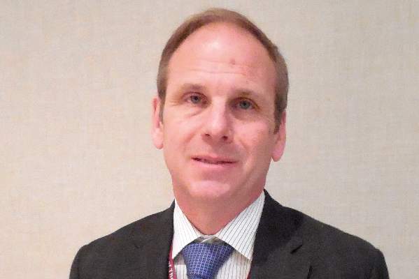
Dr. James Jaggers |
One additional potential advantage of the Carpentier-Edwards prosthesis is that the valve it contains is larger than the usual valve placed in the right ventricular outflow tract (RVOT). Failed valves increasingly are replaced by a transcatheter approach that puts a new valve inside the old, failed valve. As patients who received these replacement valves continue to survive we anticipate their need over time for a series of valve-in-valve procedures. The larger the valve at the outset, the more feasible it will be to have multiple episodes of valve-in-valve replacement.
At one time, we regarded early surgical repair of a tetralogy of Fallot defect as curative. We now know that as children with a repaired tetralogy of Fallot grow into teens and adults they require additional repairs, most often replacement of their RVOTs. This has made pulmonary valve replacement the most common surgery for adult survivors of congenital heart disease. The numbers of teen or adult patients who require a new RVOT will steadily increase as more of these children survive.
Dr. James Jaggers, professor of surgery at the University of Colorado and chief of cardiothoracic surgery at Children’s Hospital Colorado in Denver, made these comments in an interview. He had no disclosures.
The intermediate-term results reported by Dr. Schubmehl using a Carpentier-Edwards conduit in the right-ventricular outflow tract are clearly better than what we have seen using other types of valves and conduits in this position. If the valve and conduit they used persists with similar performance beyond 10 years, it would be a very good option. However, what typically happens is that replacement valves look good for about 10 years and then start to fail, often with a steep failure curve. I suspect that during the next 10 years of follow-up many more of the valves they placed will start to fail. The 10- to 20-year follow-up period is critical for demonstrating long-term durability of this valve and conduit.

|
Dr. James Jaggers |
One additional potential advantage of the Carpentier-Edwards prosthesis is that the valve it contains is larger than the usual valve placed in the right ventricular outflow tract (RVOT). Failed valves increasingly are replaced by a transcatheter approach that puts a new valve inside the old, failed valve. As patients who received these replacement valves continue to survive we anticipate their need over time for a series of valve-in-valve procedures. The larger the valve at the outset, the more feasible it will be to have multiple episodes of valve-in-valve replacement.
At one time, we regarded early surgical repair of a tetralogy of Fallot defect as curative. We now know that as children with a repaired tetralogy of Fallot grow into teens and adults they require additional repairs, most often replacement of their RVOTs. This has made pulmonary valve replacement the most common surgery for adult survivors of congenital heart disease. The numbers of teen or adult patients who require a new RVOT will steadily increase as more of these children survive.
Dr. James Jaggers, professor of surgery at the University of Colorado and chief of cardiothoracic surgery at Children’s Hospital Colorado in Denver, made these comments in an interview. He had no disclosures.
The intermediate-term results reported by Dr. Schubmehl using a Carpentier-Edwards conduit in the right-ventricular outflow tract are clearly better than what we have seen using other types of valves and conduits in this position. If the valve and conduit they used persists with similar performance beyond 10 years, it would be a very good option. However, what typically happens is that replacement valves look good for about 10 years and then start to fail, often with a steep failure curve. I suspect that during the next 10 years of follow-up many more of the valves they placed will start to fail. The 10- to 20-year follow-up period is critical for demonstrating long-term durability of this valve and conduit.

|
Dr. James Jaggers |
One additional potential advantage of the Carpentier-Edwards prosthesis is that the valve it contains is larger than the usual valve placed in the right ventricular outflow tract (RVOT). Failed valves increasingly are replaced by a transcatheter approach that puts a new valve inside the old, failed valve. As patients who received these replacement valves continue to survive we anticipate their need over time for a series of valve-in-valve procedures. The larger the valve at the outset, the more feasible it will be to have multiple episodes of valve-in-valve replacement.
At one time, we regarded early surgical repair of a tetralogy of Fallot defect as curative. We now know that as children with a repaired tetralogy of Fallot grow into teens and adults they require additional repairs, most often replacement of their RVOTs. This has made pulmonary valve replacement the most common surgery for adult survivors of congenital heart disease. The numbers of teen or adult patients who require a new RVOT will steadily increase as more of these children survive.
Dr. James Jaggers, professor of surgery at the University of Colorado and chief of cardiothoracic surgery at Children’s Hospital Colorado in Denver, made these comments in an interview. He had no disclosures.
PHOENIX – A prosthetic conduit that contains a porcine valve showed excellent intermediate-term durability for repairing the right ventricular outflow tract in 100 teenagers and young adults at a single U.S. center.
“The Carpentier-Edwards xenograft for right ventricular outflow tract [RVOT] reconstruction provides excellent freedom from reoperation and valve dysfunction, as well as sustained improvement in right-ventricular chamber size at intermediate-term follow-up,” Dr. Heidi B. Schubmehl said at the Society of Thoracic Surgeons annual meeting.
Dr. Schubmehl reported a 92% rate of freedom from valve dysfunction with follow-up out to about 10 years, and significant reductions in right ventricular size at follow-up, compared with baseline, as measured by both echocardiography and by MRI.
The Carpentier-Edwards porcine valve and conduit “seemed to hold up better than a lot of other [prosthetic] valves,” said Dr. George M. Alfieris, director of pediatric cardiac surgery at the University of Rochester (N.Y.), and senior author for the study. In addition to the valve’s durability over approximately the first 10 years following placement, the results also showed the positive impact the valve had on right ventricular size, an important result of the repair’s efficacy, Dr. Alfieris said.
“It’s a mistake to allow the right ventricle to be under high pressure or to reach a large volume. We now focus on preserving the right ventricle,” he said in an interview. “I’ve become very concerned about preventing right ventricular dilation and preserving right ventricular function.”
Dr. Alfieris noted that his prior experience using other types of valves in the pulmonary valve and RVOT position showed those valves “did great for the first 10 years and then failed. What’s different in this series is that after 10 years, we have not seen the same dysfunction as with the prior generation of valves. I will be very interested to see what happens to them” as follow-up continues beyond 10 years. He also expressed dismay that recently the company that had been marketing the valve and conduit used in the current study, the Carpentier-Edwards, stopped selling them. He expects that as his supply of conduits runs out he’ll have to start using a different commercial valve and conduit that he believes will not perform as well or create his own conduits with a porcine valve from a different supplier.
The series of 100 patients comprised individuals aged 17 or older who received a pulmonary artery and had RVOT reconstruction at the University of Rochester during 2000-2010, Dr. Schubmehl reported. The series included 78 patients with a history of tetralogy of Fallot, 8 patients born with transposition of their great arteries, 8 patients with truncus arteriosus, and 6 patients with other congenital heart diseases. Their median age at the time they received the RVOT conduit was 24 years, 59% were men, and 99 had undergone a prior sternotomy. At the time they received the conduit, 55 had pulmonary valve insufficiency, 30 had valve stenosis, and 15 had both. Follow-up occurred an average of 7 years after conduit placement.
Two recipients died: One death occurred perioperatively in a 41-year old who had a massive cerebrovascular event, and the second death was in a 39-year old who died 2.6 years after conduit placement from respiratory failure. Two additional patients required a reintervention during follow-up, said Dr. Schubmehl, a general surgeon at the University of Rochester. One reintervention occurred after 11 years to treat endocarditis, and the second after 11 years to perform balloon valvuloplasty because of valve stenosis.
The results reported by Dr. Schubmehl for echocardiography examinations showed that the patients had a statistically significant reduction in their RVOT pressure gradient from baseline to 1-year follow-up that was sustained through their intermediate-term follow-up. Seventy-seven patients had pulmonary valve insufficiency at baseline that resolved in all patients at 1-year follow-up and remained resolved in all but one patient at extended follow-up. Nineteen patients underwent additional imaging with MRI at an average follow-up of 7 years, and these findings confirmed the echo results.
On Twitter @mitchelzoler
AT THE STS ANNUAL MEETING
Key clinical point: A prosthetic conduit with a porcine valve showed excellent durability for congenital heart defect repairs at intermediate-term follow-up.
Major finding: After an average 7-year follow-up, the replacement valve and conduit had a 92% rate of freedom from valve dysfunction.
Data source: Single-center series of 100 patients.
Disclosures: Dr. Schubmehl and Dr. Alfieris had no disclosures.
Hemodynamic principles key to managing right ventricular heart failure
PHOENIX – In the clinical opinion of Dr. Nevin M. Katz, caring for critical care patients after cardiothoracic surgery requires a multidisciplinary team.
“It used to be just the surgeons and the residents and anesthesiologists, but in this era, it’s a broad team,” Dr. Katz said at the annual meeting of the Society of Thoracic Surgeons. “Coordination of the team, which includes surgeons, anesthesiologists, physician assistants, bedside nurses, nurse practitioners, perfusionists, pharmacists, respiratory therapists, and nutritionists is very important, and it’s important that members of the team be on the same page.”
Dr. Katz, a cardiovascular surgeon/intensivist at Johns Hopkins University, Baltimore, went on to offer tips for managing right ventricular failure in cardiac surgical patients. He recommends that clinicians consider five basic parameters of hemodynamic management: the heart rate and rhythm; the preload; the afterload; contractility; and the surgical result, including the potential for an anatomic problem and the risk of cardiac tamponade. “One must also consider a cardiac assist device,” he said.
He recommended that the cardiac index goal for cardiovascular patients in the ICU be in the range of 2.2-4.4 L per min/m2. The recommended hemodynamic goals also included systemic blood pressure ranges with a systolic pressure of 90-140 mm Hg and a mean arterial pressure of 70-90 mm Hg; a left arterial pressure or pulmonary capillary wedge pressure of 5-18 mm Hg, a right arterial pressure or central venous pressure of 5-15 mm Hg, and a systemic vascular resistance of 800-1,200 dynes per sec/cm5. “When treating right ventricular failure or complex patients, I think advanced PA [pulmonary artery] catheters are valuable, although they’re not absolutely necessary,” he said.
Complementary technologies available in most ICUs can help clinicians manage these patients, particularly ultrasound. With ultrasound, “we can determine where the patient is on the ventricular function curve, regional versus global dysfunction, right ventricular versus left ventricular dysfunction, valve dysfunction, and cardiac tamponade,” said Dr. Katz, who also created the Foundation for the Advancement of Cardiothoracic Care, also known as FACTS-Care. “It’s a very important monitoring modality.”
An important goal in managing patients with right ventricular failure is to establish an optimal heart rate and rhythm. “We have modalities to treat bradycardia and heart block,” he said. “Loss of atrial contraction is very important. If we can avoid atrial fibrillation, that’s good. Ventricular arrhythmias can be a problem, and nowadays we can treat atrioventricular and ventricular dyssynchrony.”
Optimal preload requires a focus on volume responsiveness, Dr. Katz continued. “Where is the patient on that ventricular function curve?” he asked. “With the advanced PA catheters, there are ways to look at that. You would like to be on the ascending part of that curve.” Clinicians can also use pulsus paradoxus, a variation of systemic arterial pulse volume. “That will indicate that perhaps you’re low in volume, but you can use stroke volume variation with an advanced PA catheter,” he said. “If your stroke volume variation is greater than 15% you’re on the ascending part of the curve. But if your stroke volume variation is less than 15%, your volume is probably optimal and you’re not going to be volume responsive.”
Clinicians who lack the benefit of an advanced PA catheter can assess volume responsiveness with passive leg raising.
Low preload causes of RV failure include hypovolemia, bleeding, third-spacing, high urine output, and cardiac tamponade. High preload can be a problem, too, from excess fluid administration, tricuspid or pulmonary valve regurgitation, or from left to right shunting.
Overall, optimal management of RV failure depends on the coordination of the multidisciplinary critical care team.
Dr. Katz reported having no financial disclosures.
EXPERT ANALYSIS AT THE STS ANNUAL MEETING
Study evaluates which prior cancers pose a risk for developing NSCLC
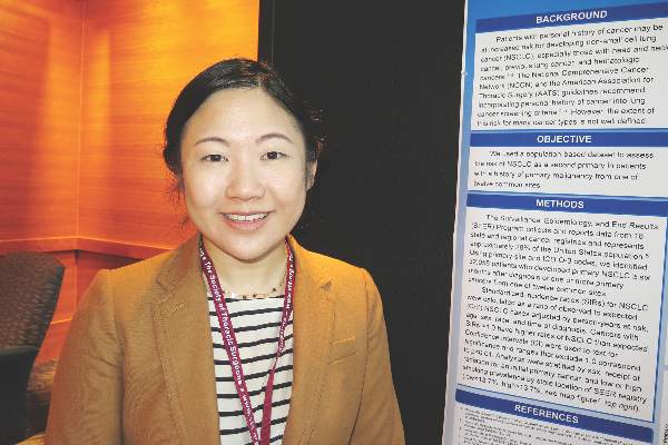
PHOENIX – Patients with a history of head and neck, lung, bladder, and hematologic malignancies had increased rates of subsequent non–small cell lung cancer (NSCLC), a large analysis of national data found.
“It is unclear to what extent the higher rate of primary NSCLC in these patients may be attributed to smoking, previous cancer history, or other lung cancer risk factors,” researchers led by Dr. Geena Wu wrote in an abstract presented during a poster session at the annual meeting of the Society of Thoracic Surgeons. “Further research using individual smoking data may better delineate who is at increased risk of NSCLC based on prior cancer site and smoking history.”
In a study that Dr. Wu led during a research fellowship at the City of Hope National Medical Center, Duarte, Calif., she and her associates used the Surveillance, Epidemiology, and End Results (SEER) 1992-2007 dataset to identify 32,058 patients with a prior malignancy who subsequently developed primary lung cancer at 6 months or more after their initial cancer. They calculated standardized incidence ratios (SIRs) for NSCLC as a rate of observed to expected NSCLC cases adjusted by person-years at risk, age, gender, and time of diagnosis.
The researchers found that patients with a history of the following cancers had higher rates of second primary NSCLC than expected: head and neck (SIR, 4.00), colon and rectum (SIR, 1.16), pancreas (SIR, 1.44), lung (SIR, 4.88), bladder (SIR, 1.97), kidney (SIR, 1.21), breast (SIR, 1.09), and leukemia or lymphoma (SIR, 1.40).
At the same time, patients with a history of pancreatic or breast cancer who were treated with radiation had a higher incidence of second primary NSCLC (SIR of 2.54 and SIR of 1.14, respectively), while those who were not treated with radiation did not.
Although the SEER database does not contain information about patient smoking history, the researchers evaluated adult smoking rates by state by using a national map from the Centers for Disease Control and Prevention’s 2013 Behavioral Risk Factor Surveillance System. Smoking rates were low (defined as 13.7% or less) in California, Hawaii, and Utah, and were higher in all other states, especially in Southeastern states. Dr. Wu and her associates found that patients from high smoking areas who had previous cancers of the colon and rectum, pancreas, kidney, thyroid, and breast had higher rates of a primary NSCLC, while those from low smoking areas did not. Interestingly, patients from high smoking areas who previously had uterine cancer, prostate, or melanoma had did not have higher rates of a primary NSCLC.
“Just because someone has a previous history of cancer, they’re not necessarily at increased risk of a second lung cancer,” Dr. Wu, who is now a fourth-year general surgery resident at Maricopa County Hospital in Phoenix said in an interview at the meeting. “You have to look at what kind of cancer they had and what their smoking history is – whether or not they continue to smoke, because smoking is such an important risk factor.”
Another limitation of the SEER database is that it lacks details about the type of chemotherapy patients receive, “so whether or not chemotherapy plays an impact in the elevated risk of lung cancer we can’t say.”
Dr. Wu reported having no financial disclosures.
AT THE STS ANNUAL MEETING
Key clinical point: Not all patients with history of cancer face an increased risk of developing subsequent NSCLC.
Major finding: Patients with a history of the following cancers had higher rates of second primary NSCLC than expected: head and neck (SIR, 4.00), colon and rectum (SIR, 1.16), pancreas (SIR, 1.44), lung (SIR, 4.88), bladder (SIR, 1.97), kidney (SIR, 1.21), breast (SIR, 1.09), and leukemia or lymphoma (SIR, 1.40).
Data source: An analysis of 32,058 patients with a prior malignancy that subsequently developed primary lung cancer at 6 months or more after their initial cancer.
Disclosures: Dr. Wu reported having no financial disclosures.
Cadaveric allograft system used to reconstruct anterior chest wall
PHOENIX – Cadaveric allograft sternal replacement has proven to be safe, providing optimal stability to the chest wall and protection of surrounding organs, an analysis of 18 cases demonstrated.
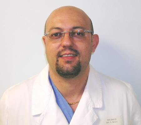
“The allograft was biologically well tolerated, allowing a perfect integration into the host,” Dr. Giuseppe Marulli said at the annual meeting of the Society of Thoracic Surgeons. “Donor cryopreserved sternochondral allograft may become the ideal way for anterior chest wall reconstruction, particularly for wide resections.”
Dr. Marulli, a thoracic surgeon at the University of Padova, Italy, noted that prior experimental studies have demonstrated that cryopreserved bone allografts preserve osteoconduction and osteoinduction capacity (Eur Spine J. 2001 Oct;10:S96-101). “Therefore, they form the basis for new bone tissue formation, allowing for the capillary and perivascular blood supply,” he said.
Limitations of current materials used for sternal reconstruction include “excessive rigidity with risk of erosion and insufficient support for large chest wall defects,” he said. Perceived advantages of using cadaveric bone allograft include easy incorporation, no risk of rejection, and a low risk of infection. For each procedure used in the current analysis, cadaveric allograft sternums with costal cartilages were harvested with an aseptic method and treated with an antibiotic solution for 72 hours. Next, they were cryopreserved at –80º C and underwent microbiologic testing for at least 1 month to ensure sterility and absence of immunogenic capacity.
Dr. Marulli reported results from 18 patients who underwent the procedure between January 2009 and January 2015, 13 of whom were female. Their median age was 59 years, their median tumor diameter was 4.75 cm, most (88%) had undergone preoperative needle biopsy, and 50% had undergone induction therapy. The main indication for sternectomy was a single-site sternal metastasis (nine patients), primary chondrosarcoma (four cases), sternal dehiscence after cardiac surgery (two cases), malignant fibrous tumor (one case), radioinduced soft-tissue sarcoma (one case), and a thymic carcinoma invading the sternum (one case).
All patients were extubated in the OR, and one patient died in the hospital from a pulmonary embolism. Two patients (11%) developed postoperative complications: one case of Candida urinary infection and one case of bleeding at the site of the muscle flap. The median postoperative length of stay was 11 days.
To date, no infections or rejections of the grafts have occurred, Dr. Marulli said. After a median of 36 months, 13 patients are alive and 4 are dead (3 from a metastatic recurrence and 1 from an unrelated cause). One patient required removal of a clavicular screw for dislocation 4 months after the operation.
Dr. Marulli reported having no financial disclosures.
AT THE STS ANNUAL MEETING
Key clinical point: Cadaveric allograft sternal replacement appears to be an effective option for reconstructing the anterior chest wall.
Major finding: To date, no infections or rejections of the grafts have occurred in patients who underwent cadaveric allograft sternal replacement.
Data source: An analysis of 18 patients who underwent the procedure between January 2009 and January 2015.
Disclosures: Dr. Marulli reported having no financial disclosures.
Fewer general surgery residents doing thoracic surgery cases
PHOENIX – Over the past 11 years, fewer general surgery residents have participated in important types of general thoracic surgery cases, a retrospective review found.
“These findings may be the result of the work-hours reduction causing less exposure to general thoracic surgery and/or a reluctance to allow general surgery residents to perform the increasingly common minimally invasive procedures,” researchers led by Dr. William S. Ragalie wrote in an abstract presented during a poster session at the annual meeting of the Society of Thoracic Surgeons.
Dr. Ragalie of the Medical College of Wisconsin, Milwaukee, and his associates retrospectively reviewed the Accreditation Council for Graduate Medical Education resident case log database for the most recent 11 years in an effort to quantify and trend the operative experience among general surgery residents. They categorized cases by year, level of resident participation, and level of complexity. Major general thoracic cases were defined as esophagectomy, pneumonectomy, and lobectomy, while cases that did not involve hilar dissection were classified as “other thoracic.”
The researchers found that the 90th percentile of first assist thoracic surgery cases decreased significantly over the study period by an average of 1.46 cases per year (P = .0012). Decreased case volumes in pneumonectomy were also noted at the junior level (–0.012 cases per year; P less than .0001) and at the chief resident level (–0.31 cases per year; P less than .001). This was also true of open lobectomy cases (–0.14 cases per year at the junior level; P less than .001, and –3.41 cases per year at the chief resident level; P less than .0001).
As for video-assisted thoracoscopic surgery (VATS) lobectomy, the researchers observed an increase in average case volume at the junior surgeon level of .13 cases per year, but a decrease at the chief resident level of one case per year (P less than .001 for both).
Dr. Ragalie and his associates also observed a decrease in the following procedures performed by chief residents: open exploratory thoracoscopy (–3.17 cases per year; P less than .001), VATS exploratory thoracoscopy (–2.95 cases per year; P less than .0001), open wedge resection (–1.52 cases per year; P less than .0227), VATS wedge resection (–2.72 cases per year; P less than .0002), “other thoracic” (–6.3 cases per year; P = .0001), and thoracoscopic pleurodesis (–2.09 cases per year; P less than .0001).
At the same time, a significant trend of decreased case volume at the junior surgeon level was noted for open exploratory thoracoscopy (–0.10 cases per year; P less than .0001) and open wedge resection (–0.22 cases per year; P = . 0115).
The researchers reported having no financial disclosures.
AT THE STS ANNUAL MEETING
Key clinical point: Fewer general surgery residents are participating in important types of general thoracic surgery cases during their residency.
Major finding: The 90th percentile of first-assist thoracic surgery cases decreased significantly over the study period by an average of 1.46 cases per year (P = .0012).
Data source: A retrospective analysis of the Accreditation Council for Graduate Medical Education resident case log database for the most recent 11 years.
Disclosures: The researchers reported having no financial disclosures.
STS: Risk Score Predicts Rehospitalization After Heart Failure
PHOENIX – A simple, five-element formula can help identify the patients undergoing heart surgery who face the greatest risk for a hospital readmission within 30 days following discharge from their index hospitalization.
The surgeons who developed this formula hope to use it in an investigational program that will target intensified management resources in postsurgical patients who face the highest readmission risk, to cut rehospitalizations and better improve their clinical status and quality of life.
The analysis that produced this formula also documented that the worst offender for triggering rehospitalizations following heart surgery is fluid overload, the proximate readmission cause for 23% of postsurgical patients, Dr. Arman Kilic said at the annual meeting of the Society of Thoracic Surgeons. The next most common cause was infection, which led to 20% of readmissions, followed by arrhythmias, responsible for 8% of readmissions, said Dr. Kilic, a thoracic surgeon at the University of Pennsylvania in Philadelphia.
Because fluid overload, often in the form of pleural effusion, is such an important driver of rehospitalizations, a more targeted management program would include better titration of diuretic treatment to patients following heart surgery, thoracentesis, and closer monitoring of clinical features that flag fluid overload such as weight.
“The volume overload issue is where the money is. If we can reduce that, it could really impact readmissions,” Dr. Kilic said in an interview.
An investigational program to target rehospitalization risk in heart surgery patients is planned at Johns Hopkins Hospital in Baltimore, where Dr. Kilic worked when he performed this analysis. Surgeons at Johns Hopkins are now in the process of getting funding for this pilot program, said Dr. John V. Conte Jr., professor of surgery and director of mechanical circulatory support at Johns Hopkins and a collaborator with Dr. Kilic on developing the risk formula.
“We’ll tailor postoperative follow-up. We’ll get high-risk patients back to the clinic sooner, and we’ll send nurse practitioners to see them to make sure they’re taking their medications and are getting weighed daily,” Dr. Conte said in an interview. “When a patient has heart surgery, they typically retain about 5-10 pounds of fluid. Patients with good renal function give up that fluid easily, but others are difficult to diurese. Many patients go home before they have been fully diuresed, and we need to follow these patients and transition them better to out-of-hospital care.”
He noted that other situations also come up that unnecessarily drive patients back to the hospital when an alternative and less expensive intervention might be equally effective. For example, some patients return to the hospital out of concern for how their chest wound is healing. Instead of being rehospitalized, such patients could be reassured by having them send a nurse a photo of their wound or by coming to an outpatient clinic.
“We need to engage more often with recently discharged patients,” Dr. Conte said in an interview. “Discharging them doesn’t mean separating them from the health care system; it should mean interacting with patients in a different way” that produces better outcomes and patient satisfaction for less money. Developing improved ways to manage recent heart surgery patients following discharge becomes even more critical later this year when, in July, the Centers for Medicare & Medicaid Services adds 30-day readmissions following coronary artery bypass grafting (CABG) to its list of procedures that can generate a penalty to hospitals if they exceed U.S. norms for readmission rates.
The risk model developed by Dr. Kilic, Dr. Conte, and their associates used data collected from 5,360 heart surgery patients treated at Johns Hopkins during 2008-2013. Nearly half the patients underwent isolated CABG, and 20% had isolated valve surgery. Overall, 585 patients (11%) had a hospital readmission within 30 days of their index discharge. One limitation of the analysis was it used data only on readmissions back to Johns Hopkins Hospital.
The researchers used data from three-quarters of the database to derive the risk formula, and from the remaining 25% of the database to validate the formula. A multivariate analysis of demographic and clinical characteristics that significantly linked with an elevated risk for readmissions identified five factors that independently made a significant contribution to readmission risk. The researchers assigned each of these five factors points depending on its relative contribution to readmission risk in the adjusted model: Severe chronic lung disease received 6 points; placement of a ventricular assist device received 5 points, while other types of heart surgery that was not CABG or valve surgery received 4 points (isolated CABG, isolated valve, or combined CABG and valve surgery received 0 points); development of acute renal failure postoperatively but before index discharge received 4 points; an index length of stay beyond 7 days received 4 points; and African American race received 3 points. The maximum number of points a patient could receive was 22.
Patients with a score of 0 had a 6% rate of a 30-day readmission; those with a score of 22 had a 63% readmission rate. For simplicity, Dr. Kilic suggested dividing patients into three categories based on their readmission risk score: Low-risk patients with a score of 0 had a readmission risk of 6%, medium-risk patients with a score of 1-10 had a readmission risk of 12%, and high-risk patients with a score of 11 or more had a readmission risk of 31%. The researchers found a 96% correlation when comparing these predicted readmission risk rates based on the derivation-subgroup analysis with the actual readmission rates seen in the validation subgroup of their database. The targeted risk-management program planned by Dr. Conte would primarily focus on high-risk patients.
Dr. Kilic and Dr. Conte said they had no relevant financial disclosures.
Identifying the factors that determine whether a patient will need rehospitalization following discharge after heart surgery is a huge and unresolved problem. Risk models for the rate of hospital readmission following cardiothoracic surgery have historically performed poorly. Perhaps that’s because the models often fail to include factors with the strongest impact on readmissions. Most of the factors that appear to drive readmissions seem to be out of the direct control of hospital staffs, such as a lack of support for patients once they leave the hospital. Socioeconomic factors like this have usually not been included in risk models.
|
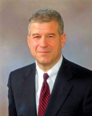
Dr. David M. Shahian |
The C statistic (area under the receiver operating characteristic curve) for the model reported by Dr. Kilic was 0.66, very close to the 0.648 that my colleagues and I reported in 2014 for a risk model of 30-day hospital readmission following isolated coronary artery bypass grafting that used data from more than 162,000 Medicare patients who underwent surgery during 2008-2010 (Circulation. 2014 July 29;130[5]:399-409). This means that both models accounted for roughly two-thirds of the variability in readmission rates, which makes our model as well as Dr. Kilic’s model marginal in its ability to identify patients at greatest risk. Similar limitations exist for the other reported models for assessing the readmission risk following heart surgery.
One strength of the model reported by Dr. Kilic was its inclusion of patient factors that developed following the start of the index admission, such as postoperative acute renal failure.
Dr. David M. Shahian is a professor of surgery at Harvard Medical School and associate director of the Codman Center for Clinical Effectiveness in Surgery at the Massachusetts General Hospital, both in Boston. He made these comments during the discussion of Dr. Kilic’s report. Dr. Shahian had no relevant financial disclosures.
Identifying the factors that determine whether a patient will need rehospitalization following discharge after heart surgery is a huge and unresolved problem. Risk models for the rate of hospital readmission following cardiothoracic surgery have historically performed poorly. Perhaps that’s because the models often fail to include factors with the strongest impact on readmissions. Most of the factors that appear to drive readmissions seem to be out of the direct control of hospital staffs, such as a lack of support for patients once they leave the hospital. Socioeconomic factors like this have usually not been included in risk models.

|
Dr. David M. Shahian |
The C statistic (area under the receiver operating characteristic curve) for the model reported by Dr. Kilic was 0.66, very close to the 0.648 that my colleagues and I reported in 2014 for a risk model of 30-day hospital readmission following isolated coronary artery bypass grafting that used data from more than 162,000 Medicare patients who underwent surgery during 2008-2010 (Circulation. 2014 July 29;130[5]:399-409). This means that both models accounted for roughly two-thirds of the variability in readmission rates, which makes our model as well as Dr. Kilic’s model marginal in its ability to identify patients at greatest risk. Similar limitations exist for the other reported models for assessing the readmission risk following heart surgery.
One strength of the model reported by Dr. Kilic was its inclusion of patient factors that developed following the start of the index admission, such as postoperative acute renal failure.
Dr. David M. Shahian is a professor of surgery at Harvard Medical School and associate director of the Codman Center for Clinical Effectiveness in Surgery at the Massachusetts General Hospital, both in Boston. He made these comments during the discussion of Dr. Kilic’s report. Dr. Shahian had no relevant financial disclosures.
Identifying the factors that determine whether a patient will need rehospitalization following discharge after heart surgery is a huge and unresolved problem. Risk models for the rate of hospital readmission following cardiothoracic surgery have historically performed poorly. Perhaps that’s because the models often fail to include factors with the strongest impact on readmissions. Most of the factors that appear to drive readmissions seem to be out of the direct control of hospital staffs, such as a lack of support for patients once they leave the hospital. Socioeconomic factors like this have usually not been included in risk models.

|
Dr. David M. Shahian |
The C statistic (area under the receiver operating characteristic curve) for the model reported by Dr. Kilic was 0.66, very close to the 0.648 that my colleagues and I reported in 2014 for a risk model of 30-day hospital readmission following isolated coronary artery bypass grafting that used data from more than 162,000 Medicare patients who underwent surgery during 2008-2010 (Circulation. 2014 July 29;130[5]:399-409). This means that both models accounted for roughly two-thirds of the variability in readmission rates, which makes our model as well as Dr. Kilic’s model marginal in its ability to identify patients at greatest risk. Similar limitations exist for the other reported models for assessing the readmission risk following heart surgery.
One strength of the model reported by Dr. Kilic was its inclusion of patient factors that developed following the start of the index admission, such as postoperative acute renal failure.
Dr. David M. Shahian is a professor of surgery at Harvard Medical School and associate director of the Codman Center for Clinical Effectiveness in Surgery at the Massachusetts General Hospital, both in Boston. He made these comments during the discussion of Dr. Kilic’s report. Dr. Shahian had no relevant financial disclosures.
PHOENIX – A simple, five-element formula can help identify the patients undergoing heart surgery who face the greatest risk for a hospital readmission within 30 days following discharge from their index hospitalization.
The surgeons who developed this formula hope to use it in an investigational program that will target intensified management resources in postsurgical patients who face the highest readmission risk, to cut rehospitalizations and better improve their clinical status and quality of life.
The analysis that produced this formula also documented that the worst offender for triggering rehospitalizations following heart surgery is fluid overload, the proximate readmission cause for 23% of postsurgical patients, Dr. Arman Kilic said at the annual meeting of the Society of Thoracic Surgeons. The next most common cause was infection, which led to 20% of readmissions, followed by arrhythmias, responsible for 8% of readmissions, said Dr. Kilic, a thoracic surgeon at the University of Pennsylvania in Philadelphia.
Because fluid overload, often in the form of pleural effusion, is such an important driver of rehospitalizations, a more targeted management program would include better titration of diuretic treatment to patients following heart surgery, thoracentesis, and closer monitoring of clinical features that flag fluid overload such as weight.
“The volume overload issue is where the money is. If we can reduce that, it could really impact readmissions,” Dr. Kilic said in an interview.
An investigational program to target rehospitalization risk in heart surgery patients is planned at Johns Hopkins Hospital in Baltimore, where Dr. Kilic worked when he performed this analysis. Surgeons at Johns Hopkins are now in the process of getting funding for this pilot program, said Dr. John V. Conte Jr., professor of surgery and director of mechanical circulatory support at Johns Hopkins and a collaborator with Dr. Kilic on developing the risk formula.
“We’ll tailor postoperative follow-up. We’ll get high-risk patients back to the clinic sooner, and we’ll send nurse practitioners to see them to make sure they’re taking their medications and are getting weighed daily,” Dr. Conte said in an interview. “When a patient has heart surgery, they typically retain about 5-10 pounds of fluid. Patients with good renal function give up that fluid easily, but others are difficult to diurese. Many patients go home before they have been fully diuresed, and we need to follow these patients and transition them better to out-of-hospital care.”
He noted that other situations also come up that unnecessarily drive patients back to the hospital when an alternative and less expensive intervention might be equally effective. For example, some patients return to the hospital out of concern for how their chest wound is healing. Instead of being rehospitalized, such patients could be reassured by having them send a nurse a photo of their wound or by coming to an outpatient clinic.
“We need to engage more often with recently discharged patients,” Dr. Conte said in an interview. “Discharging them doesn’t mean separating them from the health care system; it should mean interacting with patients in a different way” that produces better outcomes and patient satisfaction for less money. Developing improved ways to manage recent heart surgery patients following discharge becomes even more critical later this year when, in July, the Centers for Medicare & Medicaid Services adds 30-day readmissions following coronary artery bypass grafting (CABG) to its list of procedures that can generate a penalty to hospitals if they exceed U.S. norms for readmission rates.
The risk model developed by Dr. Kilic, Dr. Conte, and their associates used data collected from 5,360 heart surgery patients treated at Johns Hopkins during 2008-2013. Nearly half the patients underwent isolated CABG, and 20% had isolated valve surgery. Overall, 585 patients (11%) had a hospital readmission within 30 days of their index discharge. One limitation of the analysis was it used data only on readmissions back to Johns Hopkins Hospital.
The researchers used data from three-quarters of the database to derive the risk formula, and from the remaining 25% of the database to validate the formula. A multivariate analysis of demographic and clinical characteristics that significantly linked with an elevated risk for readmissions identified five factors that independently made a significant contribution to readmission risk. The researchers assigned each of these five factors points depending on its relative contribution to readmission risk in the adjusted model: Severe chronic lung disease received 6 points; placement of a ventricular assist device received 5 points, while other types of heart surgery that was not CABG or valve surgery received 4 points (isolated CABG, isolated valve, or combined CABG and valve surgery received 0 points); development of acute renal failure postoperatively but before index discharge received 4 points; an index length of stay beyond 7 days received 4 points; and African American race received 3 points. The maximum number of points a patient could receive was 22.
Patients with a score of 0 had a 6% rate of a 30-day readmission; those with a score of 22 had a 63% readmission rate. For simplicity, Dr. Kilic suggested dividing patients into three categories based on their readmission risk score: Low-risk patients with a score of 0 had a readmission risk of 6%, medium-risk patients with a score of 1-10 had a readmission risk of 12%, and high-risk patients with a score of 11 or more had a readmission risk of 31%. The researchers found a 96% correlation when comparing these predicted readmission risk rates based on the derivation-subgroup analysis with the actual readmission rates seen in the validation subgroup of their database. The targeted risk-management program planned by Dr. Conte would primarily focus on high-risk patients.
Dr. Kilic and Dr. Conte said they had no relevant financial disclosures.
AT THE STS ANNUAL MEETING
STS: Risk score predicts rehospitalization after heart surgery
PHOENIX – A simple, five-element formula can help identify the patients undergoing heart surgery who face the greatest risk for a hospital readmission within 30 days following discharge from their index hospitalization.
The surgeons who developed this formula hope to use it in an investigational program that will target intensified management resources in postsurgical patients who face the highest readmission risk, to cut rehospitalizations and better improve their clinical status and quality of life.
The analysis that produced this formula also documented that the worst offender for triggering rehospitalizations following heart surgery is fluid overload, the proximate readmission cause for 23% of postsurgical patients, Dr. Arman Kilic said at the annual meeting of the Society of Thoracic Surgeons. The next most common cause was infection, which led to 20% of readmissions, followed by arrhythmias, responsible for 8% of readmissions, said Dr. Kilic, a thoracic surgeon at the University of Pennsylvania in Philadelphia.
Because fluid overload, often in the form of pleural effusion, is such an important driver of rehospitalizations, a more targeted management program would include better titration of diuretic treatment to patients following heart surgery, thoracentesis, and closer monitoring of clinical features that flag fluid overload such as weight.
“The volume overload issue is where the money is. If we can reduce that, it could really impact readmissions,” Dr. Kilic said in an interview.
An investigational program to target rehospitalization risk in heart surgery patients is planned at Johns Hopkins Hospital in Baltimore, where Dr. Kilic worked when he performed this analysis. Surgeons at Johns Hopkins are now in the process of getting funding for this pilot program, said Dr. John V. Conte Jr., professor of surgery and director of mechanical circulatory support at Johns Hopkins and a collaborator with Dr. Kilic on developing the risk formula.
“We’ll tailor postoperative follow-up. We’ll get high-risk patients back to the clinic sooner, and we’ll send nurse practitioners to see them to make sure they’re taking their medications and are getting weighed daily,” Dr. Conte said in an interview. “When a patient has heart surgery, they typically retain about 5-10 pounds of fluid. Patients with good renal function give up that fluid easily, but others are difficult to diurese. Many patients go home before they have been fully diuresed, and we need to follow these patients and transition them better to out-of-hospital care.”
He noted that other situations also come up that unnecessarily drive patients back to the hospital when an alternative and less expensive intervention might be equally effective. For example, some patients return to the hospital out of concern for how their chest wound is healing. Instead of being rehospitalized, such patients could be reassured by having them send a nurse a photo of their wound or by coming to an outpatient clinic.
“We need to engage more often with recently discharged patients,” Dr. Conte said in an interview. “Discharging them doesn’t mean separating them from the health care system; it should mean interacting with patients in a different way” that produces better outcomes and patient satisfaction for less money. Developing improved ways to manage recent heart surgery patients following discharge becomes even more critical later this year when, in July, the Centers for Medicare & Medicaid Services adds 30-day readmissions following coronary artery bypass grafting (CABG) to its list of procedures that can generate a penalty to hospitals if they exceed U.S. norms for readmission rates.
The risk model developed by Dr. Kilic, Dr. Conte, and their associates used data collected from 5,360 heart surgery patients treated at Johns Hopkins during 2008-2013. Nearly half the patients underwent isolated CABG, and 20% had isolated valve surgery. Overall, 585 patients (11%) had a hospital readmission within 30 days of their index discharge. One limitation of the analysis was it used data only on readmissions back to Johns Hopkins Hospital.
The researchers used data from three-quarters of the database to derive the risk formula, and from the remaining 25% of the database to validate the formula. A multivariate analysis of demographic and clinical characteristics that significantly linked with an elevated risk for readmissions identified five factors that independently made a significant contribution to readmission risk. The researchers assigned each of these five factors points depending on its relative contribution to readmission risk in the adjusted model: Severe chronic lung disease received 6 points; placement of a ventricular assist device received 5 points, while other types of heart surgery that was not CABG or valve surgery received 4 points (isolated CABG, isolated valve, or combined CABG and valve surgery received 0 points); development of acute renal failure postoperatively but before index discharge received 4 points; an index length of stay beyond 7 days received 4 points; and African American race received 3 points. The maximum number of points a patient could receive was 22.
Patients with a score of 0 had a 6% rate of a 30-day readmission; those with a score of 22 had a 63% readmission rate. For simplicity, Dr. Kilic suggested dividing patients into three categories based on their readmission risk score: Low-risk patients with a score of 0 had a readmission risk of 6%, medium-risk patients with a score of 1-10 had a readmission risk of 12%, and high-risk patients with a score of 11 or more had a readmission risk of 31%. The researchers found a 96% correlation when comparing these predicted readmission risk rates based on the derivation-subgroup analysis with the actual readmission rates seen in the validation subgroup of their database. The targeted risk-management program planned by Dr. Conte would primarily focus on high-risk patients.
Dr. Kilic and Dr. Conte said they had no relevant financial disclosures.
On Twitter@mitchelzoler
Identifying the factors that determine whether a patient will need rehospitalization following discharge after heart surgery is a huge and unresolved problem. Risk models for the rate of hospital readmission following cardiothoracic surgery have historically performed poorly. Perhaps that’s because the models often fail to include factors with the strongest impact on readmissions. Most of the factors that appear to drive readmissions seem to be out of the direct control of hospital staffs, such as a lack of support for patients once they leave the hospital. Socioeconomic factors like this have usually not been included in risk models.

|
Dr. David M. Shahian |
The C statistic (area under the receiver operating characteristic curve) for the model reported by Dr. Kilic was 0.66, very close to the 0.648 that my colleagues and I reported in 2014 for a risk model of 30-day hospital readmission following isolated coronary artery bypass grafting that used data from more than 162,000 Medicare patients who underwent surgery during 2008-2010 (Circulation. 2014 July 29;130[5]:399-409). This means that both models accounted for roughly two-thirds of the variability in readmission rates, which makes our model as well as Dr. Kilic’s model marginal in its ability to identify patients at greatest risk. Similar limitations exist for the other reported models for assessing the readmission risk following heart surgery.
One strength of the model reported by Dr. Kilic was its inclusion of patient factors that developed following the start of the index admission, such as postoperative acute renal failure.
Dr. David M. Shahian is a professor of surgery at Harvard Medical School and associate director of the Codman Center for Clinical Effectiveness in Surgery at the Massachusetts General Hospital, both in Boston. He made these comments during the discussion of Dr. Kilic’s report. Dr. Shahian had no relevant financial disclosures.
Identifying the factors that determine whether a patient will need rehospitalization following discharge after heart surgery is a huge and unresolved problem. Risk models for the rate of hospital readmission following cardiothoracic surgery have historically performed poorly. Perhaps that’s because the models often fail to include factors with the strongest impact on readmissions. Most of the factors that appear to drive readmissions seem to be out of the direct control of hospital staffs, such as a lack of support for patients once they leave the hospital. Socioeconomic factors like this have usually not been included in risk models.

|
Dr. David M. Shahian |
The C statistic (area under the receiver operating characteristic curve) for the model reported by Dr. Kilic was 0.66, very close to the 0.648 that my colleagues and I reported in 2014 for a risk model of 30-day hospital readmission following isolated coronary artery bypass grafting that used data from more than 162,000 Medicare patients who underwent surgery during 2008-2010 (Circulation. 2014 July 29;130[5]:399-409). This means that both models accounted for roughly two-thirds of the variability in readmission rates, which makes our model as well as Dr. Kilic’s model marginal in its ability to identify patients at greatest risk. Similar limitations exist for the other reported models for assessing the readmission risk following heart surgery.
One strength of the model reported by Dr. Kilic was its inclusion of patient factors that developed following the start of the index admission, such as postoperative acute renal failure.
Dr. David M. Shahian is a professor of surgery at Harvard Medical School and associate director of the Codman Center for Clinical Effectiveness in Surgery at the Massachusetts General Hospital, both in Boston. He made these comments during the discussion of Dr. Kilic’s report. Dr. Shahian had no relevant financial disclosures.
Identifying the factors that determine whether a patient will need rehospitalization following discharge after heart surgery is a huge and unresolved problem. Risk models for the rate of hospital readmission following cardiothoracic surgery have historically performed poorly. Perhaps that’s because the models often fail to include factors with the strongest impact on readmissions. Most of the factors that appear to drive readmissions seem to be out of the direct control of hospital staffs, such as a lack of support for patients once they leave the hospital. Socioeconomic factors like this have usually not been included in risk models.

|
Dr. David M. Shahian |
The C statistic (area under the receiver operating characteristic curve) for the model reported by Dr. Kilic was 0.66, very close to the 0.648 that my colleagues and I reported in 2014 for a risk model of 30-day hospital readmission following isolated coronary artery bypass grafting that used data from more than 162,000 Medicare patients who underwent surgery during 2008-2010 (Circulation. 2014 July 29;130[5]:399-409). This means that both models accounted for roughly two-thirds of the variability in readmission rates, which makes our model as well as Dr. Kilic’s model marginal in its ability to identify patients at greatest risk. Similar limitations exist for the other reported models for assessing the readmission risk following heart surgery.
One strength of the model reported by Dr. Kilic was its inclusion of patient factors that developed following the start of the index admission, such as postoperative acute renal failure.
Dr. David M. Shahian is a professor of surgery at Harvard Medical School and associate director of the Codman Center for Clinical Effectiveness in Surgery at the Massachusetts General Hospital, both in Boston. He made these comments during the discussion of Dr. Kilic’s report. Dr. Shahian had no relevant financial disclosures.
PHOENIX – A simple, five-element formula can help identify the patients undergoing heart surgery who face the greatest risk for a hospital readmission within 30 days following discharge from their index hospitalization.
The surgeons who developed this formula hope to use it in an investigational program that will target intensified management resources in postsurgical patients who face the highest readmission risk, to cut rehospitalizations and better improve their clinical status and quality of life.
The analysis that produced this formula also documented that the worst offender for triggering rehospitalizations following heart surgery is fluid overload, the proximate readmission cause for 23% of postsurgical patients, Dr. Arman Kilic said at the annual meeting of the Society of Thoracic Surgeons. The next most common cause was infection, which led to 20% of readmissions, followed by arrhythmias, responsible for 8% of readmissions, said Dr. Kilic, a thoracic surgeon at the University of Pennsylvania in Philadelphia.
Because fluid overload, often in the form of pleural effusion, is such an important driver of rehospitalizations, a more targeted management program would include better titration of diuretic treatment to patients following heart surgery, thoracentesis, and closer monitoring of clinical features that flag fluid overload such as weight.
“The volume overload issue is where the money is. If we can reduce that, it could really impact readmissions,” Dr. Kilic said in an interview.
An investigational program to target rehospitalization risk in heart surgery patients is planned at Johns Hopkins Hospital in Baltimore, where Dr. Kilic worked when he performed this analysis. Surgeons at Johns Hopkins are now in the process of getting funding for this pilot program, said Dr. John V. Conte Jr., professor of surgery and director of mechanical circulatory support at Johns Hopkins and a collaborator with Dr. Kilic on developing the risk formula.
“We’ll tailor postoperative follow-up. We’ll get high-risk patients back to the clinic sooner, and we’ll send nurse practitioners to see them to make sure they’re taking their medications and are getting weighed daily,” Dr. Conte said in an interview. “When a patient has heart surgery, they typically retain about 5-10 pounds of fluid. Patients with good renal function give up that fluid easily, but others are difficult to diurese. Many patients go home before they have been fully diuresed, and we need to follow these patients and transition them better to out-of-hospital care.”
He noted that other situations also come up that unnecessarily drive patients back to the hospital when an alternative and less expensive intervention might be equally effective. For example, some patients return to the hospital out of concern for how their chest wound is healing. Instead of being rehospitalized, such patients could be reassured by having them send a nurse a photo of their wound or by coming to an outpatient clinic.
“We need to engage more often with recently discharged patients,” Dr. Conte said in an interview. “Discharging them doesn’t mean separating them from the health care system; it should mean interacting with patients in a different way” that produces better outcomes and patient satisfaction for less money. Developing improved ways to manage recent heart surgery patients following discharge becomes even more critical later this year when, in July, the Centers for Medicare & Medicaid Services adds 30-day readmissions following coronary artery bypass grafting (CABG) to its list of procedures that can generate a penalty to hospitals if they exceed U.S. norms for readmission rates.
The risk model developed by Dr. Kilic, Dr. Conte, and their associates used data collected from 5,360 heart surgery patients treated at Johns Hopkins during 2008-2013. Nearly half the patients underwent isolated CABG, and 20% had isolated valve surgery. Overall, 585 patients (11%) had a hospital readmission within 30 days of their index discharge. One limitation of the analysis was it used data only on readmissions back to Johns Hopkins Hospital.
The researchers used data from three-quarters of the database to derive the risk formula, and from the remaining 25% of the database to validate the formula. A multivariate analysis of demographic and clinical characteristics that significantly linked with an elevated risk for readmissions identified five factors that independently made a significant contribution to readmission risk. The researchers assigned each of these five factors points depending on its relative contribution to readmission risk in the adjusted model: Severe chronic lung disease received 6 points; placement of a ventricular assist device received 5 points, while other types of heart surgery that was not CABG or valve surgery received 4 points (isolated CABG, isolated valve, or combined CABG and valve surgery received 0 points); development of acute renal failure postoperatively but before index discharge received 4 points; an index length of stay beyond 7 days received 4 points; and African American race received 3 points. The maximum number of points a patient could receive was 22.
Patients with a score of 0 had a 6% rate of a 30-day readmission; those with a score of 22 had a 63% readmission rate. For simplicity, Dr. Kilic suggested dividing patients into three categories based on their readmission risk score: Low-risk patients with a score of 0 had a readmission risk of 6%, medium-risk patients with a score of 1-10 had a readmission risk of 12%, and high-risk patients with a score of 11 or more had a readmission risk of 31%. The researchers found a 96% correlation when comparing these predicted readmission risk rates based on the derivation-subgroup analysis with the actual readmission rates seen in the validation subgroup of their database. The targeted risk-management program planned by Dr. Conte would primarily focus on high-risk patients.
Dr. Kilic and Dr. Conte said they had no relevant financial disclosures.
On Twitter@mitchelzoler
AT THE STS ANNUAL MEETING
Key clinical point: A risk score predicted which heart surgery patients faced the greatest risk for hospital readmission within 30 days of their index discharge.
Major finding: Patients with a 0 score had a 6% 30-day readmission rate; a high score of 22 linked with a 63% rate.
Data source: A review of 5,360 heart surgery patients treated at one U.S. center.
Disclosures: Dr. Kilic and Dr. Conte said they had no relevant financial disclosures.
VIDEO: U.S. TAVR growth continues, mostly among octogenarians
PHOENIX – Use of transcatheter aortic valve replacement continued to expand through the first half of 2015, but the procedure remained primarily targeted to patients at least 80 years old, according to data collected in a U.S. postmarketing registry.
When the Food and Drug Administration first approved a transcatheter aortic valve replacement (TAVR) system for routine U.S. use in late 2011, the patients who underwent TAVR “were either at very high risk or inoperable, and we’ve seen that move into high-risk patients – and I’m sure we’ll see more introduction of this into patients who are at medium risk,” said Dr. Frederick L. Grover in a video interview at the annual meeting of the Society of Thoracic Surgeons.
Despite this downward trend in risk level, the median and average ages of TAVR patients remain above 80 years.
In 2015, U.S. TAVR recipients had a median age of 83 years and a mean age of 81 years, virtually unchanged from the 84-year median and 82-year mean during routine U.S. practice in 2012, the first year for data collection by the STS and American College of Cardiology Transcatheter Valve Therapy (TVT) Registry. Dr. Grover reported the latest data from the registry at the meeting, through roughly the first half of 2015.
“There has been some movement downward” from 2012 to 2014 in the predicted 30-day mortality rate of patients as measured by their preprocedural STS risk score. The rate declined from an average predicted mortality rate of 7.05% in 2012 to an average of 6.69% among patients treated during 2014.
Despite this shift, TAVR patients remain highly vulnerable to surgical complications because of their advanced age and frailty, said Dr. Grover, a professor of cardiothoracic surgery at the University of Colorado in Aurora and vice chairman of the registry steering committee.
STS encourages surgeons and cardiologists who collaborate on the heart teams that judge patient suitability for TAVR to measure frailty with the 5-meter walk test, run sequentially three times. Patients who take an average of 6 seconds or more to complete the test are deemed frail and eligible for TAVR. Registry data show that during 2012-2014, 81% of TAVR patients met this frailty criterion.
Perhaps the most notable statistics in the registry are the snowballing numbers of procedures performed, which have come close to doubling each year.
In the first full year of commercial use, 2012, 4,601 patients underwent TAVR, which jumped to 9,128 patients in 2013, 16,314 patients in 2014, and 23,002 patients during just the first part of 2015, Dr. Grover reported.
Dr. Grover had no relevant disclosures.
The video associated with this article is no longer available on this site. Please view all of our videos on the MDedge YouTube channel
On Twitter @mitchelzoler
AT THE STS ANNUAL MEETING
Key clinical point: The U.S. postmarketing database for TAVR showed steadily increasing growth in use from 2012 to 2015, with an ongoing focus on treating octogenarian patients.
Major finding: U.S. TAVR use jumped from 4,600 procedures in 2012 to 23,000 procedures in roughly the first half of 2015.
Data source: The STS/ACC TVT registry, which included 53,045 U.S. TAVR patients through mid 2015.
Disclosures: Dr. Grover had no relevant disclosures.

