User login
FMT Could Prevent Recurrence of Hepatic Encephalopathy in Patients With Cirrhosis
MILAN — , results of a phase 2 randomized controlled trial show.
“Not only was FMT more beneficial, but also it didn’t matter which route of administration was used — oral or enema — which is good because people don’t really like enemas,” said Jasmohan S. Bajaj, MD, AGAF, professor, School of Medicine, Virginia Commonwealth University, Richmond, and hepatologist at Richmond VA Medical Center.

Donor background (including vegan or omnivore) and dose range also did not affect the efficacy of FMT, Dr. Bajaj said.
Dr. Bajaj presented the findings (Abstract GS-001) at the opening session of the annual European Association for the Study of the Liver (EASL) Congress 2024.
Hepatic encephalopathy is a complication of advanced liver disease that causes a dementia-like state. Standard treatment with lactulose and rifaximin often results in a lack of patient response, meaning the patient is constantly being readmitted to the hospital, Dr. Bajaj said.
“This is a burden for the family as well as the patients,” and is very difficult to manage from a clinical and psychosocial perspective, he said in an interview.
With FMT, “we are transferring an ecosystem of good microbes,” which modifies the gut microbiome in patients with advanced liver disease and reduces associated brain toxicity, Dr. Bajaj explained.
Resetting the Gut
The double-blind, randomized, placebo-controlled trial enrolled a total of 60 patients with cirrhosis who had experienced hepatic encephalopathy. Aged 61-65 years, participants had Model for End-Stage Liver Disease (MELD) scores of 12-13, all were taking lactulose and rifaximin, and all had experienced their last hepatic encephalopathy episode 8-13 months prior.
Participants had similar baseline cognition, Sickness Impact Profile (SIP), and cirrhosis severity. Those with recent infections, taking other antibiotics, with a MELD score > 22, had received a transplant, or were immunosuppressed were excluded.
Study participants were divided into four dose administration groups (n = 15 each): oral and enema active FMT therapy (group 1), oral active FMT and enema placebo (group 2), oral placebo and enema active FMT (group 3), and oral and enema placebo (group 4).
The range of FMT dose frequency was zero (all placebo), or one, two, or three FMT administrations, each given 1 month apart.
Two thirds of those receiving active FMT were given omnivore-donor FMT, and one third were given vegan-donor FMT, in addition to receiving standard of care.
“Colony-forming units were standard and the same whether given via oral capsule or enema,” Dr. Bajaj said. This is “similar to what we used in our phase 1 study.”
Intent-to-treat (ITT) analysis was performed with 6-month data. The primary outcomes were safety and hepatic encephalopathy recurrence defined as ≥ grade 2 on West-Haven criteria. Secondary outcomes included other adverse events, changes in infections, severity of cirrhosis and cognition, and patient-reported outcomes. A statistical regression for hepatic encephalopathy recurrence was also performed. Patients were followed for 6 months or until death.
One Dose of FMT Better Than None
Hepatic encephalopathy recurrence was highest (40%) in group 4 patients, compared with those in group 1 (13%), group 2 (13%), and group 3 (0%), as were liver-related hospitalizations (47% vs 7%-20%).
SIP total/physical and psych scores improved with FMT (P = .003).
When all patients were included in the analysis, the hepatic encephalopathy recurrence was related to dose number (odds radio [OR], 0.27; 95% CI, 0.10-0.79; P = .02), male sex (OR, 0.16; 95% CI, 0.03-0.89; P = .04), and physical SIP (OR, 1.05; 95% CI, 1.01-1.10, P = .05). However, when analyzing results from FMT recipients only, FMT dose, route of administration, and donor source were not found to affect recurrence.
Of those on placebo alone, six patients (40%) had a recurrence, compared with four on FMT (8.8%) in the combined FMT groups.
“As long as a patient received at least one FMT dose, they had a better response than a patient who had none,” Dr. Bajaj said.
Six patients dropped out; two in group 1 died after hepatic encephalopathy and falls, and one in group 2 died after a seizure. Three others did not return for follow-up visits. Four patients developed infections, including spontaneous bacterial peritonitis, cholecystitis, and cellulitis, all unrelated to FMT.
“I think many patients in Western countries are underserved because apart from lactulose and rifaximin, there is little else to give them,” Dr. Bajaj said. “The assumption is because rifaximin kills everything, we shouldn’t give FMT. But here, we administered it to a harsh and hostile wasteland of microbiota, and it still got a toehold and generated a reduction in hepatic encephalopathy.”
He pointed out that in smaller prior studies, the effects lasted up to 1 year.
Setting the Stage for Phase 3 Trials
Dr. Bajaj noted that this phase 2 study sets the stage for larger phase 3 trials in patients not responding to first-line therapy.
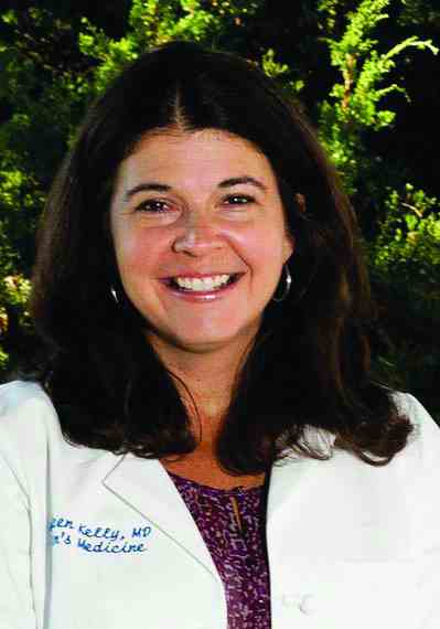
“Given how well-tolerated and effective FMT appears to be in these patients, if the larger phase 3 trial shows similar results, I can imagine FMT becoming a standard therapy,” said Colleen R. Kelly, MD, AGAF, gastroenterologist at Brigham and Women’s Hospital and Harvard Medical School, Boston, who was not involved in the study.
This study was built on Dr. Bajaj’s prior work that established the safety of FMT by enema, she added, stressing that this new research was incredibly important in these immunocompromised patients who are at higher risk for infection transmission.
That the administration route doesn’t matter is also an important finding as oral administration is much more feasible than enema, said Dr. Kelly, who went on to point out the importance of finding an alternative to rifaximin and lactulose, which are often poorly tolerated.
The study highlights the central role played by the gut microbiota in dysbiosis in the pathophysiology of hepatic encephalopathy, Dr. Kelly said. “It is another exciting example of how gut microbiota can be manipulated to treat disease.”
Dr. Bajaj and Dr. Kelly report no relevant financial relationships to this study.
A version of this article appeared on Medscape.com.
MILAN — , results of a phase 2 randomized controlled trial show.
“Not only was FMT more beneficial, but also it didn’t matter which route of administration was used — oral or enema — which is good because people don’t really like enemas,” said Jasmohan S. Bajaj, MD, AGAF, professor, School of Medicine, Virginia Commonwealth University, Richmond, and hepatologist at Richmond VA Medical Center.
Donor background (including vegan or omnivore) and dose range also did not affect the efficacy of FMT, Dr. Bajaj said.
Dr. Bajaj presented the findings (Abstract GS-001) at the opening session of the annual European Association for the Study of the Liver (EASL) Congress 2024.
Hepatic encephalopathy is a complication of advanced liver disease that causes a dementia-like state. Standard treatment with lactulose and rifaximin often results in a lack of patient response, meaning the patient is constantly being readmitted to the hospital, Dr. Bajaj said.
“This is a burden for the family as well as the patients,” and is very difficult to manage from a clinical and psychosocial perspective, he said in an interview.
With FMT, “we are transferring an ecosystem of good microbes,” which modifies the gut microbiome in patients with advanced liver disease and reduces associated brain toxicity, Dr. Bajaj explained.
Resetting the Gut
The double-blind, randomized, placebo-controlled trial enrolled a total of 60 patients with cirrhosis who had experienced hepatic encephalopathy. Aged 61-65 years, participants had Model for End-Stage Liver Disease (MELD) scores of 12-13, all were taking lactulose and rifaximin, and all had experienced their last hepatic encephalopathy episode 8-13 months prior.
Participants had similar baseline cognition, Sickness Impact Profile (SIP), and cirrhosis severity. Those with recent infections, taking other antibiotics, with a MELD score > 22, had received a transplant, or were immunosuppressed were excluded.
Study participants were divided into four dose administration groups (n = 15 each): oral and enema active FMT therapy (group 1), oral active FMT and enema placebo (group 2), oral placebo and enema active FMT (group 3), and oral and enema placebo (group 4).
The range of FMT dose frequency was zero (all placebo), or one, two, or three FMT administrations, each given 1 month apart.
Two thirds of those receiving active FMT were given omnivore-donor FMT, and one third were given vegan-donor FMT, in addition to receiving standard of care.
“Colony-forming units were standard and the same whether given via oral capsule or enema,” Dr. Bajaj said. This is “similar to what we used in our phase 1 study.”
Intent-to-treat (ITT) analysis was performed with 6-month data. The primary outcomes were safety and hepatic encephalopathy recurrence defined as ≥ grade 2 on West-Haven criteria. Secondary outcomes included other adverse events, changes in infections, severity of cirrhosis and cognition, and patient-reported outcomes. A statistical regression for hepatic encephalopathy recurrence was also performed. Patients were followed for 6 months or until death.
One Dose of FMT Better Than None
Hepatic encephalopathy recurrence was highest (40%) in group 4 patients, compared with those in group 1 (13%), group 2 (13%), and group 3 (0%), as were liver-related hospitalizations (47% vs 7%-20%).
SIP total/physical and psych scores improved with FMT (P = .003).
When all patients were included in the analysis, the hepatic encephalopathy recurrence was related to dose number (odds radio [OR], 0.27; 95% CI, 0.10-0.79; P = .02), male sex (OR, 0.16; 95% CI, 0.03-0.89; P = .04), and physical SIP (OR, 1.05; 95% CI, 1.01-1.10, P = .05). However, when analyzing results from FMT recipients only, FMT dose, route of administration, and donor source were not found to affect recurrence.
Of those on placebo alone, six patients (40%) had a recurrence, compared with four on FMT (8.8%) in the combined FMT groups.
“As long as a patient received at least one FMT dose, they had a better response than a patient who had none,” Dr. Bajaj said.
Six patients dropped out; two in group 1 died after hepatic encephalopathy and falls, and one in group 2 died after a seizure. Three others did not return for follow-up visits. Four patients developed infections, including spontaneous bacterial peritonitis, cholecystitis, and cellulitis, all unrelated to FMT.
“I think many patients in Western countries are underserved because apart from lactulose and rifaximin, there is little else to give them,” Dr. Bajaj said. “The assumption is because rifaximin kills everything, we shouldn’t give FMT. But here, we administered it to a harsh and hostile wasteland of microbiota, and it still got a toehold and generated a reduction in hepatic encephalopathy.”
He pointed out that in smaller prior studies, the effects lasted up to 1 year.
Setting the Stage for Phase 3 Trials
Dr. Bajaj noted that this phase 2 study sets the stage for larger phase 3 trials in patients not responding to first-line therapy.
“Given how well-tolerated and effective FMT appears to be in these patients, if the larger phase 3 trial shows similar results, I can imagine FMT becoming a standard therapy,” said Colleen R. Kelly, MD, AGAF, gastroenterologist at Brigham and Women’s Hospital and Harvard Medical School, Boston, who was not involved in the study.
This study was built on Dr. Bajaj’s prior work that established the safety of FMT by enema, she added, stressing that this new research was incredibly important in these immunocompromised patients who are at higher risk for infection transmission.
That the administration route doesn’t matter is also an important finding as oral administration is much more feasible than enema, said Dr. Kelly, who went on to point out the importance of finding an alternative to rifaximin and lactulose, which are often poorly tolerated.
The study highlights the central role played by the gut microbiota in dysbiosis in the pathophysiology of hepatic encephalopathy, Dr. Kelly said. “It is another exciting example of how gut microbiota can be manipulated to treat disease.”
Dr. Bajaj and Dr. Kelly report no relevant financial relationships to this study.
A version of this article appeared on Medscape.com.
FROM EASL 2024
The Smartphone Problem
I am going to guess that if we asked 500,000 adults in this country if they felt that children and adolescents were spending too much time on their smartphones, we would elicit almost uniform agreement that, yes indeed, smartphone use is gobbling up too much time from our young people. And, the adults would volunteer a long laundry list of all the bad consequences this overuse was generating. If you ask this same sample of adults if they too were spending too much time on their smartphones they would answer yes and, again, give you a list of the problems they feel are the result of this overuse.
We might begin to find a scattering of responses if we ask the adults when a child is too young to have his/her own cell phone. But, they would all agree that “young children” weren’t ready to be trusted with a cell phone. The “when” they were ready would be up for discussion. However, I suspect we might see a clustering around age 10 years. The reality is that despite what the majority may believe, a 2022 survey found that 42% of children have a cell phone by age 10, 71% by age 12, and 91% by age 14.

So, it would appear that, while we believe there can be significant downsides to having a cell phone, we are having great difficulty in policing ourselves and creating limits for our children. Does cell phone use qualify as an addiction, or is it just another example of how adults have lost the ability to say “no” to themselves and to their children?
When it comes to cell phones in school, the situation gets increasingly murky. The teachers I speak with are very clear that cell phones are creating problems for both the academic and the social experiences of their students. One teacher referred me to an article from the Norwegian Institute of Public Health, which found that banning cell phones in school decreased the incidence of psychological symptoms and diseases in girls. Bullying decreased in both genders and the girls’ GPA scores improved. In schools with cell phone bans, girls were more likely to choose and attend academic track programs, an effect which was more pronounced in young women with lower socioeconomic backgrounds. But, the if, when, and how to institute smartphone bans in school is complicated.
On one front, the movement toward cell phone bans in school has been given a major boost with the publication and publicity of a new book titled The Anxious Generation by social psychologist Jonathan Haidt, PhD. The New York University professor sees the GenZ’ers as experiencing a tsunami of mental health challenges including anxiety, self-harm, and suicide. And, he lays much of the blame for this situation on cell phone use.
He is optimistic about turning the tide because he claims that everywhere he speaks about the problem he says “I feel that I’m pushing on open doors.” Comparing the phenomenon to the collapse of the Berlin Wall, Dr. Haidt says “When you have a system that everyone hates, and then you have a way to escape it, it can change in a year.”
I wish I could share in his optimism, although I did just encounter a news story in the Portland paper describing a national program called “Wait Until 8th,” which is being considered by a parents’ group here in Maine.
The usual suspects have their own predictable take on the issue. The House and Senate have proposed a study on the use of cell phones in elementary and secondary schools and a pilot program awarding grants to some schools to create mobile device–free environments. Sounds like a momentum killer to me.
Not surprisingly, the issue of cell phone bans in school has taken on a bit of a political odor. The National Parents Union reports in a very small and inadequately described sample that 56% of parents are against total school bans. In the accompanying press release, the organizations offers an extensive list of concerns parents have reported — many cite the need to remain in contact with their children throughout the day. One has to wonder how often these concerns are a reflection of unresolved separation anxiety.
The American Academy of Pediatrics has rolled out a “5 Cs” framework that pediatricians can use to discuss media use with families. As usual, the thought is that talking about a problem is going to somehow convince parents to do what they already know is the correct action. And, of course, pediatricians have plenty of time to initiate this discussion of the obvious.
A recent study from the Department of Pediatrics at University of California, San Francisco, has found that parental monitoring, limit setting, and modeling good screen use behavior (my bolding) are the most effective strategies for reducing adolescent screen time. Using screen time allowances as a reward or punishment does not seem to be effective.
So there you have it. It looks like However, despite Dr. Haidt’s optimism about a seismic turnaround, I suspect it will more likely be guerrilla warfare — one family, one school, or one school district at a time.
Dr. Wilkoff practiced primary care pediatrics in Brunswick, Maine, for nearly 40 years. He has authored several books on behavioral pediatrics, including “How to Say No to Your Toddler.” Other than a Littman stethoscope he accepted as a first-year medical student in 1966, Dr. Wilkoff reports having nothing to disclose. Email him at [email protected].
I am going to guess that if we asked 500,000 adults in this country if they felt that children and adolescents were spending too much time on their smartphones, we would elicit almost uniform agreement that, yes indeed, smartphone use is gobbling up too much time from our young people. And, the adults would volunteer a long laundry list of all the bad consequences this overuse was generating. If you ask this same sample of adults if they too were spending too much time on their smartphones they would answer yes and, again, give you a list of the problems they feel are the result of this overuse.
We might begin to find a scattering of responses if we ask the adults when a child is too young to have his/her own cell phone. But, they would all agree that “young children” weren’t ready to be trusted with a cell phone. The “when” they were ready would be up for discussion. However, I suspect we might see a clustering around age 10 years. The reality is that despite what the majority may believe, a 2022 survey found that 42% of children have a cell phone by age 10, 71% by age 12, and 91% by age 14.
So, it would appear that, while we believe there can be significant downsides to having a cell phone, we are having great difficulty in policing ourselves and creating limits for our children. Does cell phone use qualify as an addiction, or is it just another example of how adults have lost the ability to say “no” to themselves and to their children?
When it comes to cell phones in school, the situation gets increasingly murky. The teachers I speak with are very clear that cell phones are creating problems for both the academic and the social experiences of their students. One teacher referred me to an article from the Norwegian Institute of Public Health, which found that banning cell phones in school decreased the incidence of psychological symptoms and diseases in girls. Bullying decreased in both genders and the girls’ GPA scores improved. In schools with cell phone bans, girls were more likely to choose and attend academic track programs, an effect which was more pronounced in young women with lower socioeconomic backgrounds. But, the if, when, and how to institute smartphone bans in school is complicated.
On one front, the movement toward cell phone bans in school has been given a major boost with the publication and publicity of a new book titled The Anxious Generation by social psychologist Jonathan Haidt, PhD. The New York University professor sees the GenZ’ers as experiencing a tsunami of mental health challenges including anxiety, self-harm, and suicide. And, he lays much of the blame for this situation on cell phone use.
He is optimistic about turning the tide because he claims that everywhere he speaks about the problem he says “I feel that I’m pushing on open doors.” Comparing the phenomenon to the collapse of the Berlin Wall, Dr. Haidt says “When you have a system that everyone hates, and then you have a way to escape it, it can change in a year.”
I wish I could share in his optimism, although I did just encounter a news story in the Portland paper describing a national program called “Wait Until 8th,” which is being considered by a parents’ group here in Maine.
The usual suspects have their own predictable take on the issue. The House and Senate have proposed a study on the use of cell phones in elementary and secondary schools and a pilot program awarding grants to some schools to create mobile device–free environments. Sounds like a momentum killer to me.
Not surprisingly, the issue of cell phone bans in school has taken on a bit of a political odor. The National Parents Union reports in a very small and inadequately described sample that 56% of parents are against total school bans. In the accompanying press release, the organizations offers an extensive list of concerns parents have reported — many cite the need to remain in contact with their children throughout the day. One has to wonder how often these concerns are a reflection of unresolved separation anxiety.
The American Academy of Pediatrics has rolled out a “5 Cs” framework that pediatricians can use to discuss media use with families. As usual, the thought is that talking about a problem is going to somehow convince parents to do what they already know is the correct action. And, of course, pediatricians have plenty of time to initiate this discussion of the obvious.
A recent study from the Department of Pediatrics at University of California, San Francisco, has found that parental monitoring, limit setting, and modeling good screen use behavior (my bolding) are the most effective strategies for reducing adolescent screen time. Using screen time allowances as a reward or punishment does not seem to be effective.
So there you have it. It looks like However, despite Dr. Haidt’s optimism about a seismic turnaround, I suspect it will more likely be guerrilla warfare — one family, one school, or one school district at a time.
Dr. Wilkoff practiced primary care pediatrics in Brunswick, Maine, for nearly 40 years. He has authored several books on behavioral pediatrics, including “How to Say No to Your Toddler.” Other than a Littman stethoscope he accepted as a first-year medical student in 1966, Dr. Wilkoff reports having nothing to disclose. Email him at [email protected].
The Positive Effects of Exercise in MS
NASHVILLE, TENNESSEE — Exercise has a long history in multiple sclerosis (MS). In 1838, the Scottish physician John Abercrombie reported that a patient with “a diminution of muscular power,” who could walk but only unsteadily, decided after various failed treatments like “evacuations and spare diet” to try “violent exercise.” He walked 5-6 miles on a warm evening, as quickly as he was able, and returned home “much fatigued, and considerably heated. Next morning he had severe pains in the calves of his legs, but his other complaints were much diminished, and in a few days disappeared. He has ever since enjoyed good health,” Dr. Abercrombie was quoted in Multiple Sclerosis: The History of a Disease by T. Jock Murray.
The first randomized, controlled trial of an exercise intervention for MS didn’t appear in the literature until 1988, but more than 200 have been published in the years since, according to Robert Motl, PhD, who spoke about exercise interventions for MS at the annual meeting of the Consortium of Multiple Sclerosis Centers.

In fact, the evidence shows that exercise can improve walking performance and quality of life. “When we look at what we might call the unseen symptoms, we can see the exercise training is very effective at reducing fatigue in people with MS. It’s very effective at reducing depressive mood in individuals living with MS. There is moderate evidence that it can improve mobility, particularly lower extremity mobility and walking performance in individuals living with multiple sclerosis, as well as balance. And lastly, we see consistent evidence that exercise training can improve quality of life,” said Dr. Motl, who is a professor of kinesiology and nutrition at University of Illinois, Chicago.
There is less evidence that exercise training helps mobility, anxiety, pain, and participation, he said.
Dr. Motl showed the results of various meta-analyses that he co-authored of randomized, controlled trials (RCTs) of exercise training. One meta-analysis of 20 trials that examined the effect on fitness found an effect size of 0.47, which was about one-half of a standard deviation, and is considered to be a clinically meaningful effect. There was also about a 20% improvement in aerobic capacity, and this improves the capacity for maintaining independence, according to Dr. Motl. “That’s huge as individuals who are living with MS over a long-term period of time are aging with this chronic disease and independence does become an issue later in life. We maybe can forestall some of that,” he said.
Another meta-analysis of 17 RCTs examining exercise training and fatigue found a similar effect size of 0.452. When the authors limited the analysis to studies that used the Fatigue Severity Score and its benchmark of clinically significant fatigue of 4.0, “they were able to reduce the mean fatigue severity score below 4.0, meaning you’re taking individuals who have severe fatigue and reducing their fatigue below a threshold of severity that impacts everyday life. So this is something that is clinically meaningful and relevant to the lives of individuals with MS,” he said.
With respect to depression, a meta-analysis of 14 randomized, controlled trials found an effect size of 0.55 standard deviations. The researchers found that the effect size was associated with the number of days per week: The effect was size was doubled among individuals who exercised 3 or more times per week. Another meta-analysis of walking found an average 2-second improvement in walking speed and about a 40-meter improvement in walking endurance. “I believe that’s pretty comparable to what you see with Ampyra (dalfampridine) and its effects on walking speeds, so we’re seeing something that’s as good as a pharmacological agent for managing walking in MS,” said Dr. Motl.
Another meta-analysis of health-related quality of life found that the effect on the physical domain was about twice as large as the effect on mental health–related quality of life. “I think that makes sense because when you are engaging in exercise, it’s a physically invoking stimulus. As you see adaptations, your perceptions of your physical health improve,” said Dr. Motl.
Dr. Motl also addressed safety. There have been some concerns that exercise could lead to temporary worsening of symptoms, “but it was blown up into a major, major problem when it is only 5% of individuals who have these sorts of severe problems,” said Dr. Motl. A systematic review in 2023 found an adverse event rate of 1.2% in the control groups and 2.0% in the exercise groups. This was about the same rates that are seen in the general population, according to Dr. Motl. A consistent adverse event was lower back pain, but further analysis showed it was only reported with resistance training. “The beauty of that is that we have incredible people in the field of MS, who know how to deliver resistance training more safely. And if we do that more effectively, we can avoid this very common injury with exercise training,” said Dr. Motl.
The review also found a 25% reduction in relapses. “It was very interesting. I don’t know if we want to say exercise is a disease-modifying behavior yet, but that effect at the time that these studies were done was about the same as some of the early disease-modifying therapies, showing the same degree of reduction of relapse rate,” said Dr. Motl.
Dr. Motl also discussed updated guidelines for exercise in patients with mild to moderate MS, as well as Parkinson’s disease and stroke survivors. The general advice is for 2-3 days of moderate aerobic exercise per week, beginning at 10 minutes and gradually increasing to 30 minutes per session. The newer guidelines added an option for advanced aerobic exercise, which can be up to 5 times per week and up to 40 minutes per session. Activities include ergometry, walking, aquatics, and elliptical machines for general aerobic exercise, while advanced exercise can also include running or road cycling. Resistance exercise can be done 2-3 times per week with 1-3 sets of 8-15 repetitions, with a total of 5-10 exercises. The authors recommend weight machines, free weights, or resistance bands.
Dr. Motl has received funding from the Department of Defense, National Institutes of Health, Patient-Centered Outcomes Research Institute, National Multiple Sclerosis Society, and Bristol Myers Squibb Foundation.
NASHVILLE, TENNESSEE — Exercise has a long history in multiple sclerosis (MS). In 1838, the Scottish physician John Abercrombie reported that a patient with “a diminution of muscular power,” who could walk but only unsteadily, decided after various failed treatments like “evacuations and spare diet” to try “violent exercise.” He walked 5-6 miles on a warm evening, as quickly as he was able, and returned home “much fatigued, and considerably heated. Next morning he had severe pains in the calves of his legs, but his other complaints were much diminished, and in a few days disappeared. He has ever since enjoyed good health,” Dr. Abercrombie was quoted in Multiple Sclerosis: The History of a Disease by T. Jock Murray.
The first randomized, controlled trial of an exercise intervention for MS didn’t appear in the literature until 1988, but more than 200 have been published in the years since, according to Robert Motl, PhD, who spoke about exercise interventions for MS at the annual meeting of the Consortium of Multiple Sclerosis Centers.
In fact, the evidence shows that exercise can improve walking performance and quality of life. “When we look at what we might call the unseen symptoms, we can see the exercise training is very effective at reducing fatigue in people with MS. It’s very effective at reducing depressive mood in individuals living with MS. There is moderate evidence that it can improve mobility, particularly lower extremity mobility and walking performance in individuals living with multiple sclerosis, as well as balance. And lastly, we see consistent evidence that exercise training can improve quality of life,” said Dr. Motl, who is a professor of kinesiology and nutrition at University of Illinois, Chicago.
There is less evidence that exercise training helps mobility, anxiety, pain, and participation, he said.
Dr. Motl showed the results of various meta-analyses that he co-authored of randomized, controlled trials (RCTs) of exercise training. One meta-analysis of 20 trials that examined the effect on fitness found an effect size of 0.47, which was about one-half of a standard deviation, and is considered to be a clinically meaningful effect. There was also about a 20% improvement in aerobic capacity, and this improves the capacity for maintaining independence, according to Dr. Motl. “That’s huge as individuals who are living with MS over a long-term period of time are aging with this chronic disease and independence does become an issue later in life. We maybe can forestall some of that,” he said.
Another meta-analysis of 17 RCTs examining exercise training and fatigue found a similar effect size of 0.452. When the authors limited the analysis to studies that used the Fatigue Severity Score and its benchmark of clinically significant fatigue of 4.0, “they were able to reduce the mean fatigue severity score below 4.0, meaning you’re taking individuals who have severe fatigue and reducing their fatigue below a threshold of severity that impacts everyday life. So this is something that is clinically meaningful and relevant to the lives of individuals with MS,” he said.
With respect to depression, a meta-analysis of 14 randomized, controlled trials found an effect size of 0.55 standard deviations. The researchers found that the effect size was associated with the number of days per week: The effect was size was doubled among individuals who exercised 3 or more times per week. Another meta-analysis of walking found an average 2-second improvement in walking speed and about a 40-meter improvement in walking endurance. “I believe that’s pretty comparable to what you see with Ampyra (dalfampridine) and its effects on walking speeds, so we’re seeing something that’s as good as a pharmacological agent for managing walking in MS,” said Dr. Motl.
Another meta-analysis of health-related quality of life found that the effect on the physical domain was about twice as large as the effect on mental health–related quality of life. “I think that makes sense because when you are engaging in exercise, it’s a physically invoking stimulus. As you see adaptations, your perceptions of your physical health improve,” said Dr. Motl.
Dr. Motl also addressed safety. There have been some concerns that exercise could lead to temporary worsening of symptoms, “but it was blown up into a major, major problem when it is only 5% of individuals who have these sorts of severe problems,” said Dr. Motl. A systematic review in 2023 found an adverse event rate of 1.2% in the control groups and 2.0% in the exercise groups. This was about the same rates that are seen in the general population, according to Dr. Motl. A consistent adverse event was lower back pain, but further analysis showed it was only reported with resistance training. “The beauty of that is that we have incredible people in the field of MS, who know how to deliver resistance training more safely. And if we do that more effectively, we can avoid this very common injury with exercise training,” said Dr. Motl.
The review also found a 25% reduction in relapses. “It was very interesting. I don’t know if we want to say exercise is a disease-modifying behavior yet, but that effect at the time that these studies were done was about the same as some of the early disease-modifying therapies, showing the same degree of reduction of relapse rate,” said Dr. Motl.
Dr. Motl also discussed updated guidelines for exercise in patients with mild to moderate MS, as well as Parkinson’s disease and stroke survivors. The general advice is for 2-3 days of moderate aerobic exercise per week, beginning at 10 minutes and gradually increasing to 30 minutes per session. The newer guidelines added an option for advanced aerobic exercise, which can be up to 5 times per week and up to 40 minutes per session. Activities include ergometry, walking, aquatics, and elliptical machines for general aerobic exercise, while advanced exercise can also include running or road cycling. Resistance exercise can be done 2-3 times per week with 1-3 sets of 8-15 repetitions, with a total of 5-10 exercises. The authors recommend weight machines, free weights, or resistance bands.
Dr. Motl has received funding from the Department of Defense, National Institutes of Health, Patient-Centered Outcomes Research Institute, National Multiple Sclerosis Society, and Bristol Myers Squibb Foundation.
FROM CMSC 2024
Delivery of Care: The Ethical Imperative in Healthcare
The ethical imperative in healthcare necessitates equitable delivery of care to all individuals, regardless of their socio-economic status or insurance coverage. This principle is rooted in the concept of justice and is crucial to achieving health equity.
As gastroenterologists, despite our various practice settings, we have seen the harmful effects of economic and social disparities on health outcomes. We must therefore ensure that we acknowledge the existence of these disparities, and then begin to provide a framework that allows us to ethically and successfully navigate these complexities for our patients and our affiliated structures.

The following cases illustrate the complexities and ethical dilemmas that gastroenterology and hepatology healthcare professionals encounter in delivering care within the traditional healthcare system.
- Case 1: A 44-year-old male presents to the hospital with intermittent rectal bleeding every few weeks without associated abdominal pain or weight loss and not associated with straining. He has bowel movements every 2-3 days. There is no family history of underlying gastrointestinal disease or associated neoplasm. He is accompanied at the time of the interview by his coworker who offered to drive him to the hospital as he is having personal car trouble. Physical examination reveals normal hemodynamics, abdomen is benign, a digital rectal exam reveals small internal hemorrhoids without pain. Hemoglobin is 10, MCV 85. There is scant blood on the glove. He is uninsured. A GI consult is placed to determine the disposition of the patient. The resident on service suggests outpatient follow-up given low risk of clinical deterioration.
- Case 2: A 28-year-old woman postpartum 6 weeks presents in the office with a history of ulcerative colitis which was diagnosed 2 years prior. She was initially placed on steroid therapy. She underwent a colonoscopy at the time of her diagnosis and was following with a gastroenterologist at which time she was found to have moderate left-sided disease with a modified Mayo score of 9. She complains of urgency and rectal bleeding. She saw a gastroenterologist during her pregnancy and was placed on oral mesalamine, which she remains on at the time of evaluation. Once her physical examination is completed and laboratory values are reviewed, you begin to discuss advanced therapies including biologics as she has failed conventional therapies.
- Case 3: You receive a phone call from an outside hospital about a potential transfer for a 46-year-old male who is an immigrant of unknown citizenship status with fulminant liver failure. He meets all criteria including encephalopathy and coagulopathy. He drinks only socially. His secondary liver workup for extensive disease including ceruloplasmin remains pending. Viral hepatology serologies and autoimmune serologies are negative.
Challenges to the Delivery of Equitable Care
These cases underscore the challenges of delivering equitable care within a system that often fails to address the social determinants of health (SDOH). The disparity in the evaluation and treatment of patients based on insurance status not only affects patient outcomes, but also emphasizes the ethical dilemma of balancing cost with population health management.
The introduction of measures SDOH-1 and SDOH-2 by the Centers for Medicare & Medicaid Services in the 2023 IPPS Final Rule is a step towards requiring hospitals to systematically collect patient-level SDOH data, aiming to establish meaningful collaborations between healthcare providers and community-based organizations for whole-person care.1 The primary goal is to allow ecosystems to collect patient-level social risk factors followed by the creation of meaningful collaboration between healthcare providers and the community-based organizations.
The office settings may or may not implement the SDOH and the current electronic medical record systems. However, from a social history standpoint and certainly from a decision standpoint, the impact of SDOH is realized in all settings.
Interplay of SDOH and Ethical Considerations
The recognition of social determinants of health is crucial for ethical healthcare delivery. In the first case, considering the patient’s identified social determinants of health — including lack of insurance and transportation, combined with the rising incidence of colorectal cancer in individuals under 55 — an argument could be made for admitting the patient under observation for inpatient colonoscopy.
Data have shown disparities in treatment and referrals in emergency care setting for Black patients with rectal bleeding.2 It is imperative that we recognize these existing disparities in diagnosis and outcomes, along with determining SDOH to appropriately come to a final disposition. This approach aligns with the principle of justice and the imperative to deliver equitable care.
In the third case study, we have a patient facing the life-or-death situation of fulminant liver failure. He requires an expeditious decision to be made about transfer candidacy for liver transplant evaluation by the hepatology team.
Impact of Insurance Status on Healthcare Access
Insurance status significantly influences access to healthcare and disparities in treatment outcomes. As seen in case 2 and case 3, our therapies often hinge upon access.
In the inflammatory bowel disease (IBD) case, the therapy that we will choose for our IBD patient may be more influenced by access than efficacy. In a national sample of children with Crohn’s disease, publicly insured children were more likely to receive a biologic within 18 months of diagnosis compared to children with private insurance.3 This would suggest that those with private insurance perhaps experience increased barriers.
In the IBD case that we presented here, we do have a publicly insured woman who will face a potential loss of her Medicaid coverage. Our therapeutic decision will therefore not just rely on risk stratification and individualized approach, but rather the programs that are put in place by our pharmaceutical partners to support a future self-pay patient. This may or may not be favorable to her outcome. This discrepancy points to systemic inequalities in healthcare access and the need for policies that ensure equitable treatment for all, regardless of insurance status.
Conclusion
The delivery of care in healthcare is an ethical imperative that demands equity and justice. The cases discussed above illustrate the complex interplay between socioeconomic factors, insurance status, and the ethical challenges in providing equitable care.
Systematic efforts to address social determinants of health, as mandated by recent CMS measures, along with a commitment to ethical principles, are essential steps toward reducing disparities and ensuring that all individuals receive the care they need. As healthcare expenditures continue to rise, particularly in areas like gastrointestinal health, addressing these ethical and systemic challenges becomes even more critical for the sustainability of the healthcare system and the well-being of the population it serves.
Gastrointestinal healthcare expenditures totaled $119.6 billion in 2018. Annually there were more than 36.8 million ambulatory visits for GI symptoms and 43.4 million ambulatory visits with primary GI diagnosis.4 The use of higher-acuity settings and lack of continuity of care, and the under-recognition and lack of longitudinal framework to follow those families at risk continue to compromise our healthcare system.
Dr. McCutchen is a gastroenterologist at United Digestive, Atlanta, Georgia. She is vice chair of the AGA Research Foundation. Dr. Boules is vice president of global medical and scientific affairs at Ironwood Pharmaceuticals, Cleveland, Ohio.
References
1. www.govinfo.gov/content/pkg/FR-2022-08-10/pdf/2022-16472.pdf.
2. Shields HM et al. Disparities in evaluation of patients with rectal bleeding 40 years and older. Clin Gastroenterol Hepatol. 2014 Apr. doi: 10.1016/j.cgh.2013.07.008.
3. Quiros JA et al. Insurance type influences access to biologics and healthcare utilization in pediatric Crohn’s disease. Crohns Colitis 360. 2021 Aug. doi: 10.1093/crocol/otab057.
4. Peery AF et al. Burden and cost of gastrointestinal, liver, and pancreatic diseases in the United States: Update 2021. Gastroenterology. 2022 Feb. doi: 10.1053/j.gastro.2021.10.017.
The ethical imperative in healthcare necessitates equitable delivery of care to all individuals, regardless of their socio-economic status or insurance coverage. This principle is rooted in the concept of justice and is crucial to achieving health equity.
As gastroenterologists, despite our various practice settings, we have seen the harmful effects of economic and social disparities on health outcomes. We must therefore ensure that we acknowledge the existence of these disparities, and then begin to provide a framework that allows us to ethically and successfully navigate these complexities for our patients and our affiliated structures.
The following cases illustrate the complexities and ethical dilemmas that gastroenterology and hepatology healthcare professionals encounter in delivering care within the traditional healthcare system.
- Case 1: A 44-year-old male presents to the hospital with intermittent rectal bleeding every few weeks without associated abdominal pain or weight loss and not associated with straining. He has bowel movements every 2-3 days. There is no family history of underlying gastrointestinal disease or associated neoplasm. He is accompanied at the time of the interview by his coworker who offered to drive him to the hospital as he is having personal car trouble. Physical examination reveals normal hemodynamics, abdomen is benign, a digital rectal exam reveals small internal hemorrhoids without pain. Hemoglobin is 10, MCV 85. There is scant blood on the glove. He is uninsured. A GI consult is placed to determine the disposition of the patient. The resident on service suggests outpatient follow-up given low risk of clinical deterioration.
- Case 2: A 28-year-old woman postpartum 6 weeks presents in the office with a history of ulcerative colitis which was diagnosed 2 years prior. She was initially placed on steroid therapy. She underwent a colonoscopy at the time of her diagnosis and was following with a gastroenterologist at which time she was found to have moderate left-sided disease with a modified Mayo score of 9. She complains of urgency and rectal bleeding. She saw a gastroenterologist during her pregnancy and was placed on oral mesalamine, which she remains on at the time of evaluation. Once her physical examination is completed and laboratory values are reviewed, you begin to discuss advanced therapies including biologics as she has failed conventional therapies.
- Case 3: You receive a phone call from an outside hospital about a potential transfer for a 46-year-old male who is an immigrant of unknown citizenship status with fulminant liver failure. He meets all criteria including encephalopathy and coagulopathy. He drinks only socially. His secondary liver workup for extensive disease including ceruloplasmin remains pending. Viral hepatology serologies and autoimmune serologies are negative.
Challenges to the Delivery of Equitable Care
These cases underscore the challenges of delivering equitable care within a system that often fails to address the social determinants of health (SDOH). The disparity in the evaluation and treatment of patients based on insurance status not only affects patient outcomes, but also emphasizes the ethical dilemma of balancing cost with population health management.
The introduction of measures SDOH-1 and SDOH-2 by the Centers for Medicare & Medicaid Services in the 2023 IPPS Final Rule is a step towards requiring hospitals to systematically collect patient-level SDOH data, aiming to establish meaningful collaborations between healthcare providers and community-based organizations for whole-person care.1 The primary goal is to allow ecosystems to collect patient-level social risk factors followed by the creation of meaningful collaboration between healthcare providers and the community-based organizations.
The office settings may or may not implement the SDOH and the current electronic medical record systems. However, from a social history standpoint and certainly from a decision standpoint, the impact of SDOH is realized in all settings.
Interplay of SDOH and Ethical Considerations
The recognition of social determinants of health is crucial for ethical healthcare delivery. In the first case, considering the patient’s identified social determinants of health — including lack of insurance and transportation, combined with the rising incidence of colorectal cancer in individuals under 55 — an argument could be made for admitting the patient under observation for inpatient colonoscopy.
Data have shown disparities in treatment and referrals in emergency care setting for Black patients with rectal bleeding.2 It is imperative that we recognize these existing disparities in diagnosis and outcomes, along with determining SDOH to appropriately come to a final disposition. This approach aligns with the principle of justice and the imperative to deliver equitable care.
In the third case study, we have a patient facing the life-or-death situation of fulminant liver failure. He requires an expeditious decision to be made about transfer candidacy for liver transplant evaluation by the hepatology team.
Impact of Insurance Status on Healthcare Access
Insurance status significantly influences access to healthcare and disparities in treatment outcomes. As seen in case 2 and case 3, our therapies often hinge upon access.
In the inflammatory bowel disease (IBD) case, the therapy that we will choose for our IBD patient may be more influenced by access than efficacy. In a national sample of children with Crohn’s disease, publicly insured children were more likely to receive a biologic within 18 months of diagnosis compared to children with private insurance.3 This would suggest that those with private insurance perhaps experience increased barriers.
In the IBD case that we presented here, we do have a publicly insured woman who will face a potential loss of her Medicaid coverage. Our therapeutic decision will therefore not just rely on risk stratification and individualized approach, but rather the programs that are put in place by our pharmaceutical partners to support a future self-pay patient. This may or may not be favorable to her outcome. This discrepancy points to systemic inequalities in healthcare access and the need for policies that ensure equitable treatment for all, regardless of insurance status.
Conclusion
The delivery of care in healthcare is an ethical imperative that demands equity and justice. The cases discussed above illustrate the complex interplay between socioeconomic factors, insurance status, and the ethical challenges in providing equitable care.
Systematic efforts to address social determinants of health, as mandated by recent CMS measures, along with a commitment to ethical principles, are essential steps toward reducing disparities and ensuring that all individuals receive the care they need. As healthcare expenditures continue to rise, particularly in areas like gastrointestinal health, addressing these ethical and systemic challenges becomes even more critical for the sustainability of the healthcare system and the well-being of the population it serves.
Gastrointestinal healthcare expenditures totaled $119.6 billion in 2018. Annually there were more than 36.8 million ambulatory visits for GI symptoms and 43.4 million ambulatory visits with primary GI diagnosis.4 The use of higher-acuity settings and lack of continuity of care, and the under-recognition and lack of longitudinal framework to follow those families at risk continue to compromise our healthcare system.
Dr. McCutchen is a gastroenterologist at United Digestive, Atlanta, Georgia. She is vice chair of the AGA Research Foundation. Dr. Boules is vice president of global medical and scientific affairs at Ironwood Pharmaceuticals, Cleveland, Ohio.
References
1. www.govinfo.gov/content/pkg/FR-2022-08-10/pdf/2022-16472.pdf.
2. Shields HM et al. Disparities in evaluation of patients with rectal bleeding 40 years and older. Clin Gastroenterol Hepatol. 2014 Apr. doi: 10.1016/j.cgh.2013.07.008.
3. Quiros JA et al. Insurance type influences access to biologics and healthcare utilization in pediatric Crohn’s disease. Crohns Colitis 360. 2021 Aug. doi: 10.1093/crocol/otab057.
4. Peery AF et al. Burden and cost of gastrointestinal, liver, and pancreatic diseases in the United States: Update 2021. Gastroenterology. 2022 Feb. doi: 10.1053/j.gastro.2021.10.017.
Congratulations to the 2024 AGA Research Foundation awardees!
The program serves as a catalyst for discovery and career growth among the most promising researchers in gastroenterology and hepatology.
“This year’s awardees are an exceptional group of investigators who are committed to furthering patient care through research,” said Michael Camilleri, MD, AGAF, chair, AGA Research Foundation. “The AGA Research Foundation is proud to fund these investigators and their ongoing efforts to advance GI research at a critical time in their careers. We believe the Foundation’s investment will ultimately enable new discoveries in gastroenterology and hepatology that will benefit patients.”
Treatment options for digestive diseases begin with vigorous research. The AGA Research Foundation supports medical investigators as they advance our understanding of gastrointestinal and liver conditions. Here are this year’s award recipients:
RESEARCH SCHOLAR AWARDS
AGA Research Scholar Award
- Karen Jane Dunbar, PhD, Columbia University, New York, New York
- Aaron Hecht, MD, PhD, Hospital of the University of Pennsylvania, Philadelphia, Pennsylvania
- Sarah Maxwell, MD, University of California, San Francisco
- Chung Sang Tse, MD, University of Pennsylvania, Philadelphia, Pennsylvania
- Jason (Yanjia) Zhang, MD, PhD, Boston Children’s Hospital, Massachusetts
AGA-Bristol Myers Squibb Research Scholar Award in Inflammatory Bowel Disease
- Joseph R. Burclaff, PhD, University of North Carolina at Chapel Hill
SPECIALTY AWARDS
AGA-Caroline Craig Augustyn & Damian Augustyn Award in Digestive Cancer
- Swathi Eluri, MD, MSCR, Mayo Clinic, Rochester, Minnesota
AGA-R. Robert & Sally Funderburg Research Award in Gastric Cancer
- Jianwen Que, MD, PhD, Columbia University, New York, New York
AGA-Pfizer Fellowship-to-Faculty Transition Award
- Lianna Wood, MD, PhD, Boston Children’s Hospital, Massachusetts
AGA-Ironwood Fellowship-to-Faculty Transition Award
- ZeNan Li Chang, MD, PhD, Washington University School of Medicine, St. Louis, Missouri
PILOT AWARDS
AGA Pilot Research Award
- Linda C. Cummings, MD, MS, University Hospitals Cleveland Medical Center, Cleveland, Ohio
- Pooja Mehta, MD, MSCS, University of Colorado Denver
- Guilherme Piovezani Ramos, MD, Boston Children’s Hospital
- Simon Schwoerer, PhD, University of Chicago, Illinois
- Yankai Wen, PhD, University of Texas Health Science Center at Houston
AGA-Pfizer Pilot Research Award in Non-Alcoholic Steatohepatitis
- Alice Cheng, PhD, Stanford University, California
- Petra Hirsova, PhD, PharmD, Mayo Clinic, Rochester, Minnesota
- Sarah Maxwell, MD, University of California, San Francisco
AGA-Pfizer Pilot Research Award in Inflammatory Bowel Disease
- David Boone, PhD, Indiana University, Indianapolis, Indiana
- Sara Chloe Di Rienzi, PhD, Baylor College of Medicine, Houston, Texas
- Jared Andrew Sninsky, MD, MSCR, Baylor College of Medicine, Houston, Texas
UNDERGRADUATE RESEARCH AWARDS
AGA-Aman Armaan Ahmed Family Surf for Success Program
- Eli Burstein, Yeshiva University, New York, New York
- Chloe Carlisle, University of Florida, Gainesville, Florida
- Adna Hassan, University of Minnesota Rochester
- Nicole Rodriguez Hilario, Barry University, Miami, Florida
- Maryam Jimoh, College of Wooster, Wooster, Ohio
- Viktoriya Kalinina, Brandeis University, Waltham, Massachusetts
AGA-Dr. Harvey Young Education & Development Foundation’s Young Guts Scholar Program
- Rafaella Lavalle Lacerda de Almeida, Michigan State University, East Lansing, Michigan
- Lara Cheesman, John’s Hopkins University School of Medicine, Baltimore, Maryland
- Cass Condray, University of Oklahoma, Norman, Oklahoma
- Daniel Juarez, Columbia University, New York, New York
- Jason Lin, University of Michigan, Ann Arbor, Michigan
- Riya Malhotra, Case Western Reserve University, Cleveland, Ohio
- Brian Nguyen, Brown University, Providence, Rhode Island
- Mahmoud (Moudy) Salem, Stony Brook University, Stony Brook, New York
ABSTRACT AWARDS
AGA Fellow Abstract of the Year Award
- Andrea Tou, MD Children’s Hospital of Philadelphia, Pennsylvania
AGA Fellow Abstract Awards
- Manik Aggarwal, MBBS, Mayo Clinic, Rochester, Minnesota
- Kole Buckley, PhD, University of Pennsylvania, Philadelphia, Pennsylvania
- Jane Ha, MD, Massachusetts General Hospital, Boston, Massachusetts
- Brent Hiramoto, MD, Brigham and Women’s Hospital, Boston, Massachusetts
- Md Obaidul Islam, PhD, University of Miami, Coral Gables, Florida
- Kanak Kennedy, MD, MPH, Children’s Hospital of Philadelphia, Pennsylvania
- Hanseul Kim, PhD, MS, Massachusetts General Hospital, Boston, Massachusetts
- Chiraag Kulkarni, MD, Stanford University, Stanford, California
- Su-Hyung Lee, PhD, DVM, Vanderbilt University Medical Center, Nashville, Tennessee
- Caroline Muiler, PhD, Yale School of Medicine, New Haven, Connecticut
- Sarah Najjar, PhD, New York University, New York, New York
- Ronaldo Panganiban, MD, PhD Penn State Hershey Medical Center, Hershey, Pennsylvania
- Perseus Patel, MD, Stanford University, California
- Hassan Sinan, MD, Johns Hopkins University, Baltimore, Maryland
- Patricia Snarski, PhD, Tulane University, New Orleans, Louisiana
- Fernando Vicentini, PhD, MS, McMaster University, Hamilton, Ontario, Canada
- Remington Winter, MD, University of Manitoba – Health Sciences Centre, Winnipeg, Manitoba, Canada
- Tiaosi Xing, PhD, MBBS, MS, Penn State College of Medicine, Hershey, Pennsylvania
AGA Student Abstract of the Year Award
- Jazmyne Jackson, Temple University, Philadelphia, Pennsylvania
AGA Student Abstract Award
- Valentina Alvarez, University of Washington School of Medicine, Seattle, Washington
- Yasaman Bahojb Habibyan, MS, University of Calgary, Alberta, Canada
- Tessa Herman, MD, University of Minnesota, Minneapolis-Saint Paul, Minnesota
- Jason Jin, Yale School of Medicine, New Haven, Connecticut
- Frederikke Larsen, Western University, London, Ontario, Canada
- Kara McNamara, Vanderbilt University, Nashville, Tennessee
- Julia Sessions, MD, University of Virginia, Charlottesville, Virginia
- Scott Silvey, MS, Virginia Commonwealth University, Richmond, Virginia
- Vijaya Sundaram, Marshall University School of Medicine, Huntington, West Virginia
- Kafayat Yusuf, MS, University of Kansas Medical Center, Kansas City, Kansas
AGA–Eric Esrailian Student Abstract Prize
- Brent Gawey, MD, MS, Mayo Clinic, Rochester, Minnesota
- Fei Li, MBBS, MS, University of Michigan, Ann Arbor, Michigan
- Emily Wong, University of Toronto, Ontario, Canada
- Jordan Woodard, MD, Prisma Health – Upstate, Greenville, South Carolina
AGA–Radhika Srinivasan Student Abstract Prize
- Raz Abdulqadir, MS, Penn State College of Medicine, Hershey, Pennsylvania
- Rebecca Ekeanyanwu, MHS, Meharry Medical College, Nashville, Tennessee
- Jared Morris, MD, University of Manitoba, Winnipeg City, Manitoba, Canada
- Rachel Stubler, Medical University of South Carolina, Charleston, South Carolina
AGA Abstract Award for Health Disparities Research
- Saqr Alsakarneh, MD University of Missouri-Kansas City
- Marco Noriega, MD, Beth Israel Deaconess Medical Center, Boston, Massachusetts
- Temitope Olasehinde, MD, University Hospitals/Case Western Reserve University, Cleveland, Ohio
- Gabrielle Waclawik, MD, MPH, University of Wisconsin, Madison, Wisconsin
AGA-Moti L. & Kamla Rustgi International Travel Award
- W. Keith Tan, MBChB, University of Cambridge, Cambridge, England
- Elsa van Liere, MD Amsterdam Universitair Medische Centra, Amsterdam, Netherlands
FIB-4 Index Misclassifies Many Patients
, potentially impacting clinical decisions, according to investigators.
These findings call for a cautious interpretation of low-risk FIB-4 results among patients at greatest risk of misclassification, and/or use of alternative assessment strategies, reported Mazen Noureddin, MD, MHSc, of Houston Methodist Hospital, and coauthors.

“Currently, the AGA/AASLD Pathways recommends identifying patients at risk for metabolic dysfunction-associated steatotic liver disease (MASLD), then using sequential testing with FIB-4 followed by FibroScan to risk-stratify patients,” the investigators wrote in Clinical Gastroenterology and Hepatology.
Yet the performance of the FIB-4 index in this context remains unclear.
“Previous studies have shown FIB-4 to have low accuracy for screening liver fibrosis, especially among obese and diabetic patients,” the investigators wrote. “Thus, there is a concern that classifying patients with FIB-4 can lead to misclassification and missed diagnosis.”
To explore this concern, Dr. Noureddin and colleagues turned to data from the 2017-2020 National Health and Nutrition Examination Surveys, including 5285 subjects at risk for MASLD. Exclusions were made for those with excessive alcohol intake or other liver diseases, resulting in a final cohort of 3741 individuals.
All subjects were classified as low-, indeterminate-, or high-risk for advanced liver fibrosis based on FIB-4 scores. These scores were then compared with liver stiffness measurements (LSM) obtained through transient elastography (FibroScan).
Out of 2776 subjects classified as low-risk by FIB-4, 277 (10%) were reclassified as higher risk by LSM, including 75 (2.7%) who were found to be at high risk. Out of 879 subjects with indeterminate FIB-4 scores, 37 (4.2%) were at high risk according to LSM. Finally, among the 86 subjects classified as high risk by FIB-4, 68 (79.1%) were reclassified as lower risk by LSM, including 54 (62.8%) who were deemed low risk.
Subjects misclassified as low risk by FIB-4 were typically older and had higher waist circumferences, body mass indices, glycohemoglobin A1c levels, fasting glucose levels, liver enzyme levels, diastolic blood pressures, controlled attenuation parameter scores, white blood cell counts, and alkaline phosphatase levels, but lower high-density lipoprotein and albumin levels (all P less than .05). They were also more likely to have prediabetes or diabetes.
“[I]t is important to acknowledge that 10% of the subjects were misclassified as low risk by FIB-4,” Dr. Noureddin and colleagues wrote, including 2.7% of patients who were actually high risk. “This misclassification of high-risk patients can lead to missed diagnoses, delaying crucial medical treatments or lifestyle interventions.”
They therefore suggested cautious interpretation of low-risk FIB-4 results among patients with factors predicting misclassification, or even use of alternative diagnostic strategies.
“Some possible alternatives to FIB-4 include new serum tests such NIS-2+, MASEF, SAFE score, and machine learning methods,” Dr. Noureddin and colleagues wrote. “However, additional confirmatory and cost-effective studies are required to validate the effectiveness of these tests, including studies conducted on the general population.”
The investigators disclosed relationships with AbbVie, Corcept, Galectin, and others.
, potentially impacting clinical decisions, according to investigators.
These findings call for a cautious interpretation of low-risk FIB-4 results among patients at greatest risk of misclassification, and/or use of alternative assessment strategies, reported Mazen Noureddin, MD, MHSc, of Houston Methodist Hospital, and coauthors.
“Currently, the AGA/AASLD Pathways recommends identifying patients at risk for metabolic dysfunction-associated steatotic liver disease (MASLD), then using sequential testing with FIB-4 followed by FibroScan to risk-stratify patients,” the investigators wrote in Clinical Gastroenterology and Hepatology.
Yet the performance of the FIB-4 index in this context remains unclear.
“Previous studies have shown FIB-4 to have low accuracy for screening liver fibrosis, especially among obese and diabetic patients,” the investigators wrote. “Thus, there is a concern that classifying patients with FIB-4 can lead to misclassification and missed diagnosis.”
To explore this concern, Dr. Noureddin and colleagues turned to data from the 2017-2020 National Health and Nutrition Examination Surveys, including 5285 subjects at risk for MASLD. Exclusions were made for those with excessive alcohol intake or other liver diseases, resulting in a final cohort of 3741 individuals.
All subjects were classified as low-, indeterminate-, or high-risk for advanced liver fibrosis based on FIB-4 scores. These scores were then compared with liver stiffness measurements (LSM) obtained through transient elastography (FibroScan).
Out of 2776 subjects classified as low-risk by FIB-4, 277 (10%) were reclassified as higher risk by LSM, including 75 (2.7%) who were found to be at high risk. Out of 879 subjects with indeterminate FIB-4 scores, 37 (4.2%) were at high risk according to LSM. Finally, among the 86 subjects classified as high risk by FIB-4, 68 (79.1%) were reclassified as lower risk by LSM, including 54 (62.8%) who were deemed low risk.
Subjects misclassified as low risk by FIB-4 were typically older and had higher waist circumferences, body mass indices, glycohemoglobin A1c levels, fasting glucose levels, liver enzyme levels, diastolic blood pressures, controlled attenuation parameter scores, white blood cell counts, and alkaline phosphatase levels, but lower high-density lipoprotein and albumin levels (all P less than .05). They were also more likely to have prediabetes or diabetes.
“[I]t is important to acknowledge that 10% of the subjects were misclassified as low risk by FIB-4,” Dr. Noureddin and colleagues wrote, including 2.7% of patients who were actually high risk. “This misclassification of high-risk patients can lead to missed diagnoses, delaying crucial medical treatments or lifestyle interventions.”
They therefore suggested cautious interpretation of low-risk FIB-4 results among patients with factors predicting misclassification, or even use of alternative diagnostic strategies.
“Some possible alternatives to FIB-4 include new serum tests such NIS-2+, MASEF, SAFE score, and machine learning methods,” Dr. Noureddin and colleagues wrote. “However, additional confirmatory and cost-effective studies are required to validate the effectiveness of these tests, including studies conducted on the general population.”
The investigators disclosed relationships with AbbVie, Corcept, Galectin, and others.
FROM CLINICAL GASTROENTEROLOGY AND HEPATOLOGY
Introducing the 119th AGA President: Dr. Maria T. Abreu
She currently serves as the Martin Kalser Endowed Chair of Gastroenterology; professor of medicine, microbiology, and immunology; and director of the Crohn’s and Colitis Center at the University of Miami. Dr. Abreu is the fifth woman to lead AGA as president.
Born in New York and raised in New Jersey, Dr. Abreu grew up surrounded by a strong, tight-knit Cuban community. Her family moved to Miami when she was in the ninth grade. She later entered the 6-year medical program at the University of Miami, which was the beginning of her unparalleled academic and professional excellence in medicine.
Dr. Abreu is a leader in inflammatory bowel disease patient care, and she was honored by the prestigious Sherman Prize in 2019. Her service to AGA is lengthy and begins when she took on the role of fellow representative for the research grant committee. She has since sat on both the government advocacy and diversity committees. She also served as the chair of the Immunology, Microbiology and Inflammatory Bowel Diseases Section of the AGA Council, and later as chair of the full AGA Council. While chair she developed a more streamlined in-person planning committee meeting to better organize DDW.
When asked about goals for her presidency, Dr. Abreu wants to make DDW a better experience for the modern gastroenterologist. This includes finding that perfect balance between digesting the latest education and science with networking and socializing. She plans to collaborate with the presidents of the other societies to make this come to fruition.
Perhaps the area that Dr. Abreu is most passionate about is welcoming and fostering the growth of women in gastroenterology. She wants to support women who want to succeed in academics and in practice, who want ergonomics to match their work needs, and who want to have families.
“Maria is the ultimate ‘triple threat’: master scientist, master clinician, and devoted mentor. She has not only been a major player advancing knowledge in IBD, but also motivating and pushing others to develop successful careers,” said Andres Yarur, MD, AGAF, associate professor of medicine at Cedars-Sinai Medical Center. “Her work, brilliance, passion, and charm inspire all of us and will continue to inspire many generations to come.”
Chemo May Benefit Some Older Patients With Metastatic Pancreatic Cancer
TOPLINE:
METHODOLOGY:
Pancreatic cancer is most often diagnosed in adults aged 65 years or older. Providing cancer treatment for this older, often vulnerable, population comes with significant challenges and can lead to worse survival.
To examine real-world outcomes of older adults with untreated metastatic pancreatic cancer, researchers recruited patients aged 70 years or older and performed a geriatric assessment to identify comorbidities, cognitive issues, and other geriatric abnormalities.
Those who were deemed “fit” (ie, with no geriatric abnormalities) were assigned to receive off-study standard-of-care treatment, whereas those classified as “frail” (ie, with severe abnormalities) received off-study supportive care.
The remaining 176 “vulnerable” patients with mild to moderate geriatric abnormalities completed a geriatric and quality-of-life assessment and were then randomly assigned to receive either dose-reduced 5-fluorouracil (5-FU), leucovorin plus liposomal irinotecan (n = 88) or modified gemcitabine plus nab-paclitaxel (n = 88) every 2 weeks. Ultimately, 79 patients started the 5-FU combination and 75 received gemcitabine plus nab-paclitaxel. Patients were assessed every 8 weeks until disease progression or intolerance.
Overall, patients had a median age of 77 years; 61.9% were aged 75 years or older. About half were female, and 81.5% were White. The majority (87.5%) had a performance status of 0 or 1.
TAKEAWAY:
- Median overall survival was 4.7 months in the gemcitabine plus nab-paclitaxel arm and 4.4 months in the 5-FU combination group, with no significant survival difference observed between the two arms (P = .72).
- When the overall survival analysis was restricted to patients who received at least 4 weeks, or two cycles, of treatment (about 62% of patients), the median overall survival across the two treatment arms reached 8.0 months, in line with expectations for these regimens.
- Patient stratification revealed that those with a performance status of 2 had significantly worse overall survival than those with a status of 0: 1.4 months vs 6.9 months, respectively (hazard ratio [HR], 2.77; P < .001). A similar divide was seen when patients were stratified by physical/functional status and well-being. Age, however, did not significantly influence the results.
- Overall, more than half of patients experienced grade 3 or higher adverse events. Just over 38% of patients received only one to three cycles of therapy, whereas 26% remained on treatment for 12 or more cycles. The adverse event rates were similar between the two regimens, but the toxicity profile was slightly different — the researchers, for instance, observed more peripheral neuropathy with gemcitabine plus nab-paclitaxel and more diarrhea in the 5-FU combination arm.
IN PRACTICE:
- Overall, the “survival outcomes among vulnerable older patients were lower than expected, with high percentage of patients not able to start treatment, or complete one month of therapy due to clinical deterioration,” said study presenter Efrat Dotan, MD, chief, Division of Gastrointestinal Medical Oncology, Fox Chase Cancer Center, Philadelphia.
- “For vulnerable older adults who can tolerate treatment, these two regimens provide clinicians with options for tailoring therapy based on toxicity profile,” Dr. Dotan added. But “tools are needed to better identify patients who can benefit from treatment.”
- The results underline the need to perform geriatric assessments, as opposed to merely looking at performance status, commented David F. Chang, PhD, MS, MBBS, professor of Surgical Oncology, University of Glasgow, Scotland, who was not involved in the study.
SOURCE:
The research, presented at the 2024 annual meeting of the American Society of Clinical Oncology, was funded by the National Cancer Institute and the Eastern Cooperative Oncology Group.
LIMITATIONS:
Dr. Chang noted that the study did not reveal which treatment regimen was more effective.
DISCLOSURES:
Dr. Dotan declared relationships with Agenus, Amgen, G1 Therapeutics, Incyte, Olympus, and Taiho Pharmaceutical and institutional relationships with Dragonfly Therapeutics, Gilead Sciences, Ipsen, Kinnate Biopharma, Leap Therapeutics, Lilly, Lutris, NGM Biopharmaceuticals, Relay Therapeutics, and Zymeworks. Dr. Chang declared relationships with Immodulon Therapeutics and Mylan and institutional relationships with AstraZeneca, BMS GmbH & Co. KG, Immodulon Therapeutics, and Merck.
A version of this article appeared on Medscape.com.
HPV Vaccine Offers Cancer Protection Beyond Cervical Cancer
The analysis, featured at a press briefing ahead of the presentation at the American Society of Clinical Oncology (ASCO) 2024 annual meeting, notably found that men who received the HPV vaccine had a 56% lower risk for head and neck cancers.
“We’ve known for a long time that having the HPV vaccine can prevent the development of HPV infection, yes, but importantly, cancer,” primarily cervical cancer, said briefing moderator and ASCO president Lynn Schuchter, MD, Abramson Cancer Center, University of Pennsylvania, Philadelphia. “This is a really important study that extends the information about the impact.”
Using the US TriNetX database, lead investigator Jefferson DeKloe, BS, a research fellow with Thomas Jefferson University, Philadelphia, and colleagues created a matched cohort of 760,540 HPV-vaccinated and unvaccinated men and 945,999 HPV-vaccinated and unvaccinated women.
HPV-vaccinated men had a 54% lower risk for all HPV-related cancers (odds ratio [OR], 0.46; P < .001) and a 56% lower risk for head and neck cancers (OR, 0.44; P < .001) than unvaccinated men. There were not enough cases of anal and penile cancers for analysis.
HPV-vaccinated women had a 27% lower risk for all HPV-related cancers (OR, 0.73; P < .05), a 54% lower risk for cervical cancer (OR, 0.46; P < .05), and a 33% lower risk for head and neck cancers (OR, 0.67; 95% CI, 0.42-1.08) than HPV-unvaccinated women, but this finding was not significant. There were not enough cases of anal cancers for analysis, and the odds of developing vulvar or vaginal cancer was not significantly different in HPV-vaccinated vs unvaccinated women.
Vaccinated women, however, were less likely than unvaccinated women to develop high-grade squamous intraepithelial lesions (OR, 0.44), cervical carcinoma in situ (OR, 0.42), or abnormal Pap findings (OR, 0.87), and were less likely to undergo cone biopsy and loop electrosurgical excision (OR, 0.45).
“This study really highlights the importance of getting the HPV vaccine,” Dr. Schuchter said at the briefing.
“HPV vaccination is cancer prevention,” Glenn Hanna, MD, with Dana-Farber Cancer Institute, Boston, said in an ASCO statement.
Still, HPV vaccination rates in the United States remain relatively low. According to the National Cancer Institute, in 2022, only about 58% of adolescents aged 13-15 years had received two or three doses of HPV vaccine as recommended.
“The goal,” Dr. Schuchter said at the briefing, “is that younger girls and young boys get vaccinated to prevent development of HPV infection, and that should decrease the risk of cancer, which is what we’ve seen.”
Mr. DeKloe agreed and highlighted the importance of improving vaccination rates. “Identifying effective interventions that increase HPV vaccination rates is critical in reducing undue cancer burden in the United States,” Mr. DeKloe said in a statement.
The study had no funding source. Mr. DeKloe had no relevant disclosures. Dr. Hanna has disclosed relationships with Bicara Therapeutics, Bristol Myers Squibb, Coherus BioSciences, and others. Dr. Schuchter had no relevant disclosures.
A version of this article appeared on Medscape.com .
FROM ASCO 2024
EULAR 2024 Preview: Therapeutics in Development Take Center Stage
The European Alliance of Associations for Rheumatology (EULAR) 2024 European Congress of Rheumatology annual meeting is about to take place in Vienna, Austria. From June 12 to 15, some of the world’s leading researchers and clinicians will convene to present and learn about data on some of the new and innovative treatments for people with rheumatic and musculoskeletal diseases (RMDs) as well as to discuss how to use and optimize existing approaches.
Ahead of the Congress, this news organization asked the Congress Committee’s Scientific Programme Chair Caroline Ospelt, MD, PhD, and Abstract Chair Christian Dejaco, MD, PhD, MBA, to discuss some of their highlights of this year’s meeting.
From Bench to Bedside
“For me, the beauty at EULAR is really that you have the latest on basic research, how this can be translated in clinical trials, and then the last step would be how EULAR recommends it to be used in clinical practice,” Dr. Ospelt, professor of experimental rheumatology at University Hospital Zurich, said in an interview.

“So, if you go to EULAR continuously, you can actually follow the whole story of how novelty comes into clinical practice,” she added.
In a separate interview, Dr. Dejaco, a consultant rheumatologist and associate professor at the Medical University of Graz in Austria, said: “There are several new drug trials that are going to be presented.”
One of his highlights on the use of new drugs for the treatment of giant cell arteritis will be the phase 3 SELECT-GCA trial of the Janus kinase (JAK) inhibitor upadacitinib (LBA0001).
“It’s a trial that hopefully will lead to the approval of this drug in this indication,” Dr. Dejaco said.

Late-Breaking Abstracts
Dr. Ospelt noted: “We had a lot of good late-breaking abstracts this year.”
Some of these include:
- Real-world data on the comparative effectiveness of five different classes of drugs used to treat psoriatic arthritis (PsA; LBA0002)
- The 16-week results of a phase 2b/3 study with the novel interleukin (IL)–17A inhibitor izokibep in people with PsA (LBA0005)
- Data from the COSPIRIT-JIA trial on the efficacy and safety of ixekizumab (Taltz) in juvenile idiopathic arthritis (LBA0009)
- Phase 2 data on the safety and efficacy of the CD38-targeting monoclonal antibody daratumumab in systemic lupus erythematosus (LBA0007)
- Results of the phase 2 DAHLIAS study of the anti–neonatal Fc receptor monoclonal antibody nipocalimab in people with primary Sjögren disease (LBA0010)
- Safety and immunogenicity data from a phase 1 study of an active anti–IL-6 immunotherapy in people with knee osteoarthritis (LBA0011)
The latter is “really interesting,” Dr. Ospelt said. As of now, there is no approved treatment for osteoarthritis, and there is no immunotherapy, “so this would be the first.”
But it’s not just the late-breaker abstracts to look out for. Dr. Dejaco highlighted two abstracts that will be presented during the Abstract Plenary:
- A phase 3 study of a new selective JAK1 inhibitor, SHR0302, in rheumatoid arthritis (OP0037)
- A multi-omics analysis and targeted gene-editing study in people with , which causes inflammatory and hematologic changes (OP0073)
Of the latter, he said, “this disease is still incompletely understood, and this abstract really helps to better understand the mechanisms underlying this disease.”
One to Watch: CAR T-Cell Therapy
Dr. Ospelt said that the scientific program is about 80% clinical and 20% basic science overall. However, more sessions are being held jointly because data are starting to move from the bench to bedside.
One of the basic science areas that has had “a real buzz” around it and is now producing results in the clinic is the use of chimeric antigen receptor (CAR) T cells. In one of the first, and perhaps aptly titled What Is New, or WIN, sessions of the congress, Georg Schett, MD, vice president of research at Friedrich-Alexander-Universität Erlangen-Nüremberg in Germany, will discuss the use of CAR T-cell therapy for inflammatory RMDs. There are also multiple abstract presentations on this topic.
In-depth tissue analysis and prediction of treatment response is another interesting approach, Dr. Ospelt said. “I think that’s the way to go, that we come from the blood, we go into the tissue.” A “very nice” example of this approach will be presented during the Abstract Plenary session on Wednesday, June 12, looking at how synovial tissue macrophages may be able to give information on likely treatment response in treatment-naive rheumatoid arthritis (OP0062). There are also some further findings related to the tissue biopsy–driven treatment trial R4RA that are being presented at the meeting (OP0218, OP0242, and POS0351).
EULAR Highlighted Sessions
Among the highlighted sessions on the EULAR 2024 website is one on axial involvement in PsA and spondyloarthritis (SpA).
“Axial involvement in psoriatic arthritis and peripheral involvement in axial spondyloarthritis is quite a hot topic at the moment,” Dr. Ospelt said. There are lots of questions: “How connected are they? How different are they? Do we need different treatment for axial involvement compared to peripheral involvement?”
Another EULAR highlighted session is the 75th anniversary of glucocorticoid treatment, during which Past President of EULAR and Emeritus Professor of Rheumatology Josef S. Smolen, MD, will overview the “past, present, and future” of glucocorticoids in RMDs. Consultant rheumatologist Frank Buttgereit, MD, from the German Rheumatism Research Center in Berlin, will discuss the practicalities of using these drugs in clinical practice.
Dr. Dejaco noted: “Glucocorticoids have been one of the most important treatments for a very long time, and they’re still the most important treatment for the acute treatment of systemic inflammatory diseases.”
For a long time, there was no alternative to using steroids, he added, but steroid-sparing options now exist, and there will be data presented on a new type of drug that could potentially be used to control cortisol levels in the body (OP0335).
Recommendations and More
Dr. Ospelt and Dr. Dejaco both pointed out other sessions that are likely to be very popular, such as the first and second EULAR Recommendations sessions, a session on rheumatoid arthritis prevention, as well as the many presentations and sessions on digital health and nonpharmacologic interventions such as exercise.
With over 5242 submitted abstracts, there is going to be no shortage of data being presented at EULAR 2024. Alongside the traditional abstract submission categories, this year there is a new clinical case reports category.
“We had about 578 submissions for that category,” Dr. Dejaco said. There were 3315 abstracts submitted for the clinical research category, 812 for the basic and translational research category, 283 from health professionals in rheumatology, 152 from patient groups, and 102 in the field of pediatric rheumatology.
Join in On-Site, Watch on Demand
EULAR 2024 reverts to an on-site–only meeting this year. Some of the more lighthearted yet educational elements of the program for those attending include the second edition of the EMEUNET Rheumatology Quiz and, new for this year, two escape rooms. These rooms will provide an interactive experience where small teams will have to solve rheumatologic conundrums in order to escape the room within the hour, Dr. Dejaco explained. There will also be a morning run on Friday, June 14. “It’s not a race, it’s simply to meet and run together,” Dr. Dejaco said.
But if you cannot make the congress in person, the EULAR 2024 Livestream will be broadcasting throughout the congress. Anyone registered by June 30 will have on-demand access to the recorded content from June 17 until December 31, 2024.
Abstracts for the meeting will be published as a supplement to Annals of the Rheumatic Diseases, the official journal of EULAR.
Dr. Ospelt reported no relevant financial relationships. Dr. Dejaco has received consulting/speaker fees from AbbVie, Eli Lilly, Janssen, Sparrow, Novartis, Pfizer, Roche, Galapagos, and Sanofi.
A version of this article appeared on Medscape.com.
The European Alliance of Associations for Rheumatology (EULAR) 2024 European Congress of Rheumatology annual meeting is about to take place in Vienna, Austria. From June 12 to 15, some of the world’s leading researchers and clinicians will convene to present and learn about data on some of the new and innovative treatments for people with rheumatic and musculoskeletal diseases (RMDs) as well as to discuss how to use and optimize existing approaches.
Ahead of the Congress, this news organization asked the Congress Committee’s Scientific Programme Chair Caroline Ospelt, MD, PhD, and Abstract Chair Christian Dejaco, MD, PhD, MBA, to discuss some of their highlights of this year’s meeting.
From Bench to Bedside
“For me, the beauty at EULAR is really that you have the latest on basic research, how this can be translated in clinical trials, and then the last step would be how EULAR recommends it to be used in clinical practice,” Dr. Ospelt, professor of experimental rheumatology at University Hospital Zurich, said in an interview.
“So, if you go to EULAR continuously, you can actually follow the whole story of how novelty comes into clinical practice,” she added.
In a separate interview, Dr. Dejaco, a consultant rheumatologist and associate professor at the Medical University of Graz in Austria, said: “There are several new drug trials that are going to be presented.”
One of his highlights on the use of new drugs for the treatment of giant cell arteritis will be the phase 3 SELECT-GCA trial of the Janus kinase (JAK) inhibitor upadacitinib (LBA0001).
“It’s a trial that hopefully will lead to the approval of this drug in this indication,” Dr. Dejaco said.
Late-Breaking Abstracts
Dr. Ospelt noted: “We had a lot of good late-breaking abstracts this year.”
Some of these include:
- Real-world data on the comparative effectiveness of five different classes of drugs used to treat psoriatic arthritis (PsA; LBA0002)
- The 16-week results of a phase 2b/3 study with the novel interleukin (IL)–17A inhibitor izokibep in people with PsA (LBA0005)
- Data from the COSPIRIT-JIA trial on the efficacy and safety of ixekizumab (Taltz) in juvenile idiopathic arthritis (LBA0009)
- Phase 2 data on the safety and efficacy of the CD38-targeting monoclonal antibody daratumumab in systemic lupus erythematosus (LBA0007)
- Results of the phase 2 DAHLIAS study of the anti–neonatal Fc receptor monoclonal antibody nipocalimab in people with primary Sjögren disease (LBA0010)
- Safety and immunogenicity data from a phase 1 study of an active anti–IL-6 immunotherapy in people with knee osteoarthritis (LBA0011)
The latter is “really interesting,” Dr. Ospelt said. As of now, there is no approved treatment for osteoarthritis, and there is no immunotherapy, “so this would be the first.”
But it’s not just the late-breaker abstracts to look out for. Dr. Dejaco highlighted two abstracts that will be presented during the Abstract Plenary:
- A phase 3 study of a new selective JAK1 inhibitor, SHR0302, in rheumatoid arthritis (OP0037)
- A multi-omics analysis and targeted gene-editing study in people with , which causes inflammatory and hematologic changes (OP0073)
Of the latter, he said, “this disease is still incompletely understood, and this abstract really helps to better understand the mechanisms underlying this disease.”
One to Watch: CAR T-Cell Therapy
Dr. Ospelt said that the scientific program is about 80% clinical and 20% basic science overall. However, more sessions are being held jointly because data are starting to move from the bench to bedside.
One of the basic science areas that has had “a real buzz” around it and is now producing results in the clinic is the use of chimeric antigen receptor (CAR) T cells. In one of the first, and perhaps aptly titled What Is New, or WIN, sessions of the congress, Georg Schett, MD, vice president of research at Friedrich-Alexander-Universität Erlangen-Nüremberg in Germany, will discuss the use of CAR T-cell therapy for inflammatory RMDs. There are also multiple abstract presentations on this topic.
In-depth tissue analysis and prediction of treatment response is another interesting approach, Dr. Ospelt said. “I think that’s the way to go, that we come from the blood, we go into the tissue.” A “very nice” example of this approach will be presented during the Abstract Plenary session on Wednesday, June 12, looking at how synovial tissue macrophages may be able to give information on likely treatment response in treatment-naive rheumatoid arthritis (OP0062). There are also some further findings related to the tissue biopsy–driven treatment trial R4RA that are being presented at the meeting (OP0218, OP0242, and POS0351).
EULAR Highlighted Sessions
Among the highlighted sessions on the EULAR 2024 website is one on axial involvement in PsA and spondyloarthritis (SpA).
“Axial involvement in psoriatic arthritis and peripheral involvement in axial spondyloarthritis is quite a hot topic at the moment,” Dr. Ospelt said. There are lots of questions: “How connected are they? How different are they? Do we need different treatment for axial involvement compared to peripheral involvement?”
Another EULAR highlighted session is the 75th anniversary of glucocorticoid treatment, during which Past President of EULAR and Emeritus Professor of Rheumatology Josef S. Smolen, MD, will overview the “past, present, and future” of glucocorticoids in RMDs. Consultant rheumatologist Frank Buttgereit, MD, from the German Rheumatism Research Center in Berlin, will discuss the practicalities of using these drugs in clinical practice.
Dr. Dejaco noted: “Glucocorticoids have been one of the most important treatments for a very long time, and they’re still the most important treatment for the acute treatment of systemic inflammatory diseases.”
For a long time, there was no alternative to using steroids, he added, but steroid-sparing options now exist, and there will be data presented on a new type of drug that could potentially be used to control cortisol levels in the body (OP0335).
Recommendations and More
Dr. Ospelt and Dr. Dejaco both pointed out other sessions that are likely to be very popular, such as the first and second EULAR Recommendations sessions, a session on rheumatoid arthritis prevention, as well as the many presentations and sessions on digital health and nonpharmacologic interventions such as exercise.
With over 5242 submitted abstracts, there is going to be no shortage of data being presented at EULAR 2024. Alongside the traditional abstract submission categories, this year there is a new clinical case reports category.
“We had about 578 submissions for that category,” Dr. Dejaco said. There were 3315 abstracts submitted for the clinical research category, 812 for the basic and translational research category, 283 from health professionals in rheumatology, 152 from patient groups, and 102 in the field of pediatric rheumatology.
Join in On-Site, Watch on Demand
EULAR 2024 reverts to an on-site–only meeting this year. Some of the more lighthearted yet educational elements of the program for those attending include the second edition of the EMEUNET Rheumatology Quiz and, new for this year, two escape rooms. These rooms will provide an interactive experience where small teams will have to solve rheumatologic conundrums in order to escape the room within the hour, Dr. Dejaco explained. There will also be a morning run on Friday, June 14. “It’s not a race, it’s simply to meet and run together,” Dr. Dejaco said.
But if you cannot make the congress in person, the EULAR 2024 Livestream will be broadcasting throughout the congress. Anyone registered by June 30 will have on-demand access to the recorded content from June 17 until December 31, 2024.
Abstracts for the meeting will be published as a supplement to Annals of the Rheumatic Diseases, the official journal of EULAR.
Dr. Ospelt reported no relevant financial relationships. Dr. Dejaco has received consulting/speaker fees from AbbVie, Eli Lilly, Janssen, Sparrow, Novartis, Pfizer, Roche, Galapagos, and Sanofi.
A version of this article appeared on Medscape.com.