User login
Higher blood pressure after thrombectomy links with bad stroke outcomes
HONOLULU – Acute ischemic stroke patients who underwent endovascular thrombectomy and then had a peak systolic blood pressure of greater than 158 mm Hg during the next 24 hours had worse 90-day outcomes than did patients whose peak systolic pressure remained at or below 158 mm Hg in a prospective, multicenter, observational study with 485 patients.
The results hint that maintaining a lower systolic blood pressure after thrombectomy in acute ischemic stroke patients may improve outcomes, but because the current study was observational, the hypothesis that patients benefit when treatment keeps their systolic pressure at or below 158 mm Hg must undergo testing in a prospective, randomized trial, Eva A. Mistry, MBBS, said at the International Stroke Conference, sponsored by the American Heart Association.
The finding from this study that 158 mm Hg provided the best dichotomous division between systolic blood pressures linked with good or bad outcomes is a first step toward trying to devise a more systematic and evidence-based approach to blood pressure management in acute ischemic stroke patients following endovascular thrombectomy, said Dr. Mistry, a neurologist at Vanderbilt University in Nashville, Tenn.
Neither Vanderbilt nor any of the other 11 major U.S. stroke centers that participated in the current study currently have an established protocol for blood pressure management after thrombectomy, Dr. Mistry said in an interview.
“We usually treat to reduce blood pressure, but we don’t have a [broadly agreed on] threshold” to trigger treatment. “It depends on a collective decision” by the various medical specialists who care for an individual acute stroke patient. In addition, no consensus yet exists for the best treatment strategy for blood pressure lowering in acute ischemic stroke patients. Intravenous nicardipine is often the top choice because it is fast-acting and easy to administer and control as an intravenous agent. Those same properties make the beta blocker labetalol a frequently used second drug, she said.
The BEST (Blood Pressure After Endovascular Stroke Therapy) study ran at 12 U.S. centers and enrolled 485 patients who underwent endovascular thrombectomy to treat an acute ischemic stroke. The patients averaged 69 years old, and 48% also underwent thrombolytic treatment. The study’s primary outcome was the percentage of patients with a modified Rankin Scale score of 0-2 at 90 days after their stroke, an outcome reached by 39% of all patients in the study.
Statistical analysis of the collected data showed that a peak systolic blood pressure of 158 mm Hg reached during the 24 hours following thrombectomy best divided patients with good 90-day outcomes from those with worse outcomes. Patients with a postthrombectomy peak systolic pressure above 158 mm Hg had a 2.2-fold increased rate of having a modified Rankin Scale score of 3 or higher after 90 days, a statistically significant relationship, Dr. Mistry reported. However, in an analysis that also adjusted for age, baseline stroke severity, glucose level, time to reperfusion, ASPECTS score, history of hypertension, and recanalization status, the elevated risk for a bad outcome linked with higher systolic pressure dropped to 39% greater than that for patients with systolic pressures that did not rise above 158 mm Hg, a difference that was not statistically significant. This suggests that these adjustments were unable to account for all confounders and further highlighted the need for a prospective, randomized trial to test the value of controlling blood pressure following thrombectomy, Dr. Mistry said. The unadjusted results confirmed a prior report from Dr. Mistry and her associates that found a link between higher blood pressure after stroke thrombectomy and worse outcomes (J Am Heart Assoc. 2017 May 18. doi: 10.1161/JAHA.117.006167).
The analysis also showed that patients who were successfully recanalized by thrombectomy, achieving a thrombolysis in cerebral infarction (TICI) score of 2b or 3, had lower peak systolic blood pressures than did patients who failed to get this level of restored cerebral blood flow from thrombectomy.
BEST received no commercial funding. Dr. Mistry had no disclosures.
SOURCE: Mistry EA et al. Stroke. 2019 Feb;50(Suppl_1): Abstract 94.
REPORTING FROM ISC 2019
Eisenmenger syndrome is a minefield for unwary physicians
SNOWMASS, COLO. – Interventions that are simple and straightforward for other patients – such as therapeutic phlebotomy, bronchoscopy, anticoagulation, administration of IV antibiotics – can quickly have catastrophic results in patients with Eisenmenger syndrome, Carole A. Warnes, MD, cautioned at the Annual Cardiovascular Conference at Snowmass sponsored by the American College of Cardiology.

This is why the 2018 ACC/American Heart Association guidelines on the management of patients with adult congenital heart disease recommend as a Class I indication that most patients with ACHD, including all those with Eisenmenger syndrome, be managed in collaboration with a cardiologist at a specialized ACHD center (Circulation. 2018 Aug 16. doi: 10.1161/CIR.0000000000000603), noted Dr. Warnes, professor of medicine at the Mayo Clinic in Rochester, Minn., and director of the Snowmass conference.
“There are lots of mistakes in diagnosis and management because so many physicians are unfamiliar with the kinds of problems these patients face,” the cardiologist said.
She cited as a real-world example a patient with Eisenmenger syndrome admitted for pneumonia. Twenty minutes after being placed on intravenous antibiotics she had a stroke. Why?
“She didn’t have an air filter on her IV line. Any air going into a cyanotic’s blood stream will go to the head and give them a stroke. Something that is simple – routine for other patients – may kill a patient with cyanotic heart disease,” Dr. Warnes said.
Another illustrative case: a patient with Eisenmenger syndrome presents with hemoptysis, an infiltrate in her right lung, a hemoglobin of 19.8 g/dL, and anemia. Should she undergo therapeutic phlebotomy? How about urgent bronchoscopy?
No and no, Dr. Warnes emphasized.
“You do not phlebotomize cyanotic patients unless they have symptoms of hyperviscosity, meaning terrible headache or poor concentration, along with a hemoglobin greater than 20 g/dL. They need that high hemoglobin. They may be blue, and you need those red cells to carry the oxygen around. Otherwise you may make them worse. And if you give them iron for their anemia, you will increase their risk of stroke,” she explained.
Hemoptysis in patients with Eisenmenger syndrome is a life-threatening warning sign. By the time Eisenmenger syndrome patients reach age 40 years, nearly all of them have experienced episodes of hemoptysis. And more and more patients with the syndrome are moving well beyond that milestone and surviving into their 60s and beyond.
“Many Eisenmenger syndrome patients are not dying when they’re 20 or 30. We can get them to a good old age,” according to Dr. Warnes, who founded the ACHD center at the Mayo Clinic.
Most of the time the cause of hemoptysis in patients with Eisenmenger syndrome is an intrapulmonary hemorrhage. Anticoagulation can turn that hemorrhage catastrophic. So can bronchoscopy. Indeed, the cause of death in roughly 30% of patients with this form of congenital heart disease is catastrophic hemoptysis.
“You bar the door from your friendly pulmonologist who wants to bronchoscope these patients. Bronchoscopy never does any good unless the patient is so hypoxic that you need to suck the blood out of their lungs,” Dr. Warnes emphasized.
Anticoagulation is ordinarily to be avoided in patients with Eisenmenger syndrome because of their bleeding risk, even when pulmonary thrombosis is suspected, she added.
So, to summarize: These patients basically need stabilizing, with no anticoagulation, no bronchoscopy, and no phlebotomy, she said.
Another danger zone for patients with Eisenmenger syndrome is pregnancy. The condition is associated with a 30% maternal mortality rate.
Dr. Warnes noted that, even at the Mayo Clinic, which has had a pioneering ACHD center for decades, awareness of key elements of management of affected patients is low among nonspecialists.
“As I’ve done my hospital services in the last couple of months, I sort of felt I was on patrol, snatching these patients from problems,” she recalled.
Dr. Warnes reported having no financial conflicts regarding her presentation.
SNOWMASS, COLO. – Interventions that are simple and straightforward for other patients – such as therapeutic phlebotomy, bronchoscopy, anticoagulation, administration of IV antibiotics – can quickly have catastrophic results in patients with Eisenmenger syndrome, Carole A. Warnes, MD, cautioned at the Annual Cardiovascular Conference at Snowmass sponsored by the American College of Cardiology.

This is why the 2018 ACC/American Heart Association guidelines on the management of patients with adult congenital heart disease recommend as a Class I indication that most patients with ACHD, including all those with Eisenmenger syndrome, be managed in collaboration with a cardiologist at a specialized ACHD center (Circulation. 2018 Aug 16. doi: 10.1161/CIR.0000000000000603), noted Dr. Warnes, professor of medicine at the Mayo Clinic in Rochester, Minn., and director of the Snowmass conference.
“There are lots of mistakes in diagnosis and management because so many physicians are unfamiliar with the kinds of problems these patients face,” the cardiologist said.
She cited as a real-world example a patient with Eisenmenger syndrome admitted for pneumonia. Twenty minutes after being placed on intravenous antibiotics she had a stroke. Why?
“She didn’t have an air filter on her IV line. Any air going into a cyanotic’s blood stream will go to the head and give them a stroke. Something that is simple – routine for other patients – may kill a patient with cyanotic heart disease,” Dr. Warnes said.
Another illustrative case: a patient with Eisenmenger syndrome presents with hemoptysis, an infiltrate in her right lung, a hemoglobin of 19.8 g/dL, and anemia. Should she undergo therapeutic phlebotomy? How about urgent bronchoscopy?
No and no, Dr. Warnes emphasized.
“You do not phlebotomize cyanotic patients unless they have symptoms of hyperviscosity, meaning terrible headache or poor concentration, along with a hemoglobin greater than 20 g/dL. They need that high hemoglobin. They may be blue, and you need those red cells to carry the oxygen around. Otherwise you may make them worse. And if you give them iron for their anemia, you will increase their risk of stroke,” she explained.
Hemoptysis in patients with Eisenmenger syndrome is a life-threatening warning sign. By the time Eisenmenger syndrome patients reach age 40 years, nearly all of them have experienced episodes of hemoptysis. And more and more patients with the syndrome are moving well beyond that milestone and surviving into their 60s and beyond.
“Many Eisenmenger syndrome patients are not dying when they’re 20 or 30. We can get them to a good old age,” according to Dr. Warnes, who founded the ACHD center at the Mayo Clinic.
Most of the time the cause of hemoptysis in patients with Eisenmenger syndrome is an intrapulmonary hemorrhage. Anticoagulation can turn that hemorrhage catastrophic. So can bronchoscopy. Indeed, the cause of death in roughly 30% of patients with this form of congenital heart disease is catastrophic hemoptysis.
“You bar the door from your friendly pulmonologist who wants to bronchoscope these patients. Bronchoscopy never does any good unless the patient is so hypoxic that you need to suck the blood out of their lungs,” Dr. Warnes emphasized.
Anticoagulation is ordinarily to be avoided in patients with Eisenmenger syndrome because of their bleeding risk, even when pulmonary thrombosis is suspected, she added.
So, to summarize: These patients basically need stabilizing, with no anticoagulation, no bronchoscopy, and no phlebotomy, she said.
Another danger zone for patients with Eisenmenger syndrome is pregnancy. The condition is associated with a 30% maternal mortality rate.
Dr. Warnes noted that, even at the Mayo Clinic, which has had a pioneering ACHD center for decades, awareness of key elements of management of affected patients is low among nonspecialists.
“As I’ve done my hospital services in the last couple of months, I sort of felt I was on patrol, snatching these patients from problems,” she recalled.
Dr. Warnes reported having no financial conflicts regarding her presentation.
SNOWMASS, COLO. – Interventions that are simple and straightforward for other patients – such as therapeutic phlebotomy, bronchoscopy, anticoagulation, administration of IV antibiotics – can quickly have catastrophic results in patients with Eisenmenger syndrome, Carole A. Warnes, MD, cautioned at the Annual Cardiovascular Conference at Snowmass sponsored by the American College of Cardiology.

This is why the 2018 ACC/American Heart Association guidelines on the management of patients with adult congenital heart disease recommend as a Class I indication that most patients with ACHD, including all those with Eisenmenger syndrome, be managed in collaboration with a cardiologist at a specialized ACHD center (Circulation. 2018 Aug 16. doi: 10.1161/CIR.0000000000000603), noted Dr. Warnes, professor of medicine at the Mayo Clinic in Rochester, Minn., and director of the Snowmass conference.
“There are lots of mistakes in diagnosis and management because so many physicians are unfamiliar with the kinds of problems these patients face,” the cardiologist said.
She cited as a real-world example a patient with Eisenmenger syndrome admitted for pneumonia. Twenty minutes after being placed on intravenous antibiotics she had a stroke. Why?
“She didn’t have an air filter on her IV line. Any air going into a cyanotic’s blood stream will go to the head and give them a stroke. Something that is simple – routine for other patients – may kill a patient with cyanotic heart disease,” Dr. Warnes said.
Another illustrative case: a patient with Eisenmenger syndrome presents with hemoptysis, an infiltrate in her right lung, a hemoglobin of 19.8 g/dL, and anemia. Should she undergo therapeutic phlebotomy? How about urgent bronchoscopy?
No and no, Dr. Warnes emphasized.
“You do not phlebotomize cyanotic patients unless they have symptoms of hyperviscosity, meaning terrible headache or poor concentration, along with a hemoglobin greater than 20 g/dL. They need that high hemoglobin. They may be blue, and you need those red cells to carry the oxygen around. Otherwise you may make them worse. And if you give them iron for their anemia, you will increase their risk of stroke,” she explained.
Hemoptysis in patients with Eisenmenger syndrome is a life-threatening warning sign. By the time Eisenmenger syndrome patients reach age 40 years, nearly all of them have experienced episodes of hemoptysis. And more and more patients with the syndrome are moving well beyond that milestone and surviving into their 60s and beyond.
“Many Eisenmenger syndrome patients are not dying when they’re 20 or 30. We can get them to a good old age,” according to Dr. Warnes, who founded the ACHD center at the Mayo Clinic.
Most of the time the cause of hemoptysis in patients with Eisenmenger syndrome is an intrapulmonary hemorrhage. Anticoagulation can turn that hemorrhage catastrophic. So can bronchoscopy. Indeed, the cause of death in roughly 30% of patients with this form of congenital heart disease is catastrophic hemoptysis.
“You bar the door from your friendly pulmonologist who wants to bronchoscope these patients. Bronchoscopy never does any good unless the patient is so hypoxic that you need to suck the blood out of their lungs,” Dr. Warnes emphasized.
Anticoagulation is ordinarily to be avoided in patients with Eisenmenger syndrome because of their bleeding risk, even when pulmonary thrombosis is suspected, she added.
So, to summarize: These patients basically need stabilizing, with no anticoagulation, no bronchoscopy, and no phlebotomy, she said.
Another danger zone for patients with Eisenmenger syndrome is pregnancy. The condition is associated with a 30% maternal mortality rate.
Dr. Warnes noted that, even at the Mayo Clinic, which has had a pioneering ACHD center for decades, awareness of key elements of management of affected patients is low among nonspecialists.
“As I’ve done my hospital services in the last couple of months, I sort of felt I was on patrol, snatching these patients from problems,” she recalled.
Dr. Warnes reported having no financial conflicts regarding her presentation.
REPORTING FROM ACC SNOWMASS 2019
Newer antihyperglycemic drugs have distinctive CV, kidney benefits
The two newer classes of antihyperglycemic drugs that lower cardiovascular risk have different effects on specific cardiovascular and kidney disease outcomes in patients with type 2 diabetes, results of a meta-analysis suggest. Sodium-glucose contransporter-2 (SGLT2) inhibitors significantly reduced hospitalization from heart failure, whereas glucagon-like peptide-1 receptor agonists (GLP-1 RAs) did not, according to the reported results.
The GLP-1–RA class reduced risk of kidney disease progression, largely driven by a reduction in macroalbuminuria, according to the authors, whereas only the SGLT2 inhibitors reduced adverse kidney disease outcomes in a composite excluding that biomarker.
“The prevention of heart failure and progression of kidney disease by SGLT2 [inhibitors] should be considered in the decision-making process when treating patients with type 2 diabetes,” study senior author Marc S. Sabatine, MD, MPH, of Brigham and Women’s Hospital, Boston, and his coauthors wrote in a report on the study appearing in Circulation.
Both GLP-1 RAs and SGLT2 inhibitors significantly reduced major adverse cardiovascular events (MACE) and, as shown in other recent findings, their benefits were confined to patients with established atherosclerotic cardiovascular disease, Dr. Sabatine and his colleagues wrote.
The systematic review and meta-analysis of eight cardiovascular outcomes trials included 77,242 patients, of whom about 56% participated in GLP-1–RA studies and 44% in SGLT2-inhibitor trials. Just under three-quarters of the patients had established atherosclerotic cardiovascular disease, while the remainder had multiple risk factors for it.
Relative risk of hospitalization for heart failure was reduced by 31% with SGLT2 inhibitors, but it was not significantly reduced by GLP-1 RAs, the authors noted.
Risk of kidney disease progression was reduced by 38% with SGLT2 inhibitors and by 18% with GLP-1 RAs when the researchers used a broad composite endpoint including macroalbuminuria, estimated glomerular filtration rate (eGFR), end-stage kidney disease, and death due to renal causes.
By contrast, SGLT2 inhibitors reduced by 45% the relative risk of a narrower kidney outcome that excluded macroalbuminuria, whereas GLP-1 RAs had only a nonsignificant effect on the risk of doubling serum creatinine. That suggests the relative risk reduction of the kidney composite with GLP-1 RAs was driven mainly by a reduction in macroalbuminuria, the authors wrote.
Although albuminuria is an established biomarker for kidney and cardiovascular disease, it is a surrogate marker and can even be absent in patients with reduced eGFR, they said.
“Reduction in eGFR has emerged as a more meaningful endpoint of greater importance and is used in ongoing diabetes trials for kidney outcomes,” the authors said in a discussion of their results.
Relative risk of the composite MACE endpoint, including myocardial infarction, stroke, and cardiovascular death, was reduced by 12% for GLP-1 RAs and by 11% for SGLT2 [inhibitors], according to results of the analysis. However, the benefit was confined to patients with established cardiovascular disease, who had a 14% reduction of risk, compared with no treatment effect in patients who had multiple risk factors only.
Looking at individual MACE components, investigators found that both drug classes significantly reduced relative risk of myocardial infarction and of cardiovascular death, whereas only GLP-1 RAs significantly reduced relative risk of stroke.
Study authors provided disclosures related to AstraZeneca, Amgen, Daiichi-Sankyo, Eisai, GlaxoSmithKline, Intarcia, Janssen Research and Development, and Medimmune, among others.
SOURCE: Zelniker TA et al. Circulation. 2019 Feb 21. doi: 10.1161/CIRCULATIONAHA.118.038868.
FROM CIRCULATION
Case report may link gluteal implants to lymphoma
Patients with textured silicone gluteal implants could be at risk of anaplastic large cell lymphoma, based on a possible case of ALCL in a patient diagnosed 1 year after implant placement.
The 49-year-old woman was initially diagnosed with anaplastic lymphoma kinase–negative ALCL via a lung mass and pleural fluid before bilateral gluteal ulceration occurred 1 month later, reported Orr Shauly of the University of Southern California in Los Angeles, and his colleagues.
Soft-tissue disease and fluid accumulation around the gluteal implants suggested that the lung mass had metastasized from primary neoplasia in the gluteal region. If ALCL did originate at the site of the gluteal implants, it would represent a first for silicone implant–associated ALCL, which has historically been associated exclusively with breast implants.
“As many as 200 cases of [breast implant-associated ALCL] have been described worldwide, with a majority in the context of cosmetic primary breast augmentation or cancer-related breast reconstruction with the use of a textured implant (57% of all cases),” the investigators wrote in Aesthetic Surgery Journal. “Recently however, it has been hypothesized that the relationship of ALCL with the placement of textured silicone implants may not [be] limited to the breast due to its multifactorial nature and association with texturization of the implant surface.”
During the initial work-up, a CT showed fluid collection and enhancement around the gluteal implants. Following ALCL diagnosis via lung mass biopsy and histopathology, the patient was transferred to a different facility for chemotherapy. When the patient presented 1 month later to the original facility with gluteal ulceration, the oncology team suspected infection; however, all cultures from fluid around the implants were negative.
Because of the possibility of false-negative tests, the patient was started on a regimen of acyclovir, vancomycin, metronidazole, and isavuconazole. Explantation was planned, but before this could occur, the patient deteriorated rapidly and died of respiratory and renal failure.
ALCL was not confirmed via cytology or histopathology in the gluteal region, and the patient’s family did not consent to autopsy, so a definitive diagnosis of gluteal implant–associated ALCL remained elusive.
“In this instance, it can only be concluded that the patient’s condition may have been associated with placement of textured silicone gluteal implants, but [we] still lack evidence of causation,” the investigators wrote. “It should also be noted that ALCL does not typically present with skin ulceration, and this may be a unique disease process in this patient or as a result of her bedridden state given the late stage of her disease. Furthermore, this presentation was uniquely aggressive and presented extremely quickly after placement of the gluteal implants. In most patients, ALCL develops and presents approximately 10 years after implantation.”
The investigators cautioned that “care should be taken to avoid sensationalizing all implant-associated ALCL.”
The authors reported having no conflicts of interest and the study did not receive funding.
SOURCE: Shauly O et al. Aesthet Surg J. 2019 Feb 15. doi: 10.1093/asj/sjz044.
FROM AESTHETIC SURGERY JOURNAL
Weak evidence surrounds use of mesenchymal stem cells
Weak evidence suggests that prophylactic use of mesenchymal stem cells (MSCs) may reduce the risk of chronic graft-versus-host disease (GVHD) in patients undergoing allogeneic hematopoietic stem cell transplant (HSCT), according to a recent Cochrane review.
However, other uses of MSCs with HSCT – such as prophylaxis with MSCs for acute GVHD or treatment of GVHD – lack evidence of efficacy, reported lead author Sheila A. Fisher, PhD, of National Health Service Blood and Transplant in Oxford (England), and her colleagues.
The investigators noted that most studies included in the review had low-quality evidence.
“[R]andomization methods were poorly reported and several of the included studies were subject to a high risk of performance bias and reporting bias,” the investigators wrote in the Cochrane Database of Systematic Reviews. “One trial which started in 2008 has not been published and the progress of this trial is unknown, leading to potential publication bias. The quality of evidence was therefore low or very low for all outcomes due to a high risk of bias as well as imprecision due to the low number of overall participants, and in some cases, evidence based on a single study.”
The investigators identified 25 randomized, controlled trials (RCTs) involving MSCs for GVHD, of which 12 were completed (879 participants) and 13 were ongoing (1,532 planned participants). Out of the 12 completed trials, 2 compared different doses of MSCs, while 10 compared MSCs with no MSCs. Five trials investigated treatment with MSCs and seven investigated prophylactic use.
The one finding that was supportive of MSCs, albeit based on low-quality evidence, showed that prophylactic use reduced the risk of chronic GVHD, compared with no prophylaxis (risk ratio, 0.66). Other findings were less supportive or unsupportive of MSCs.
Prophylactic use of MSCs had little or no impact on risk of acute GVHD (RR, 0.86; low-quality evidence), risk of relapse of malignant disease (RR, 1.08; low-quality evidence), or all-cause mortality (hazard ratio, 0.85; low-quality evidence).
Treatment with MSCs was disappointing across the board. Risk of all-cause mortality was unaffected (HR, 1.12; very low–quality evidence) and a minimal impact was found for complete responses in acute GVHD (RR, 1.16; very low–quality evidence).
Although treatment of chronic GVHD with MSCs was associated with an improved complete response rate (RR, 5.00), the investigators noted that this finding came from a single trial with 40 participants that was deemed to have very low–quality evidence. The two trials comparing doses of MSCs found no differences between treatment groups.
“Despite a number of reports of positive outcomes from the use of MSCs for treating acute GVHD, the evidence to date from RCTs has not supported the conclusion that they are an effective therapy,” the investigators wrote.
The study was funded by NHS Blood and Transplant. Dr. Fischer reported having no financial disclosures. One coauthor reported research funding from the Leukemia and Lymphoma Research charity and the National Institute for Health Research.
SOURCE: Fisher SA et al. Cochrane Database Syst Rev. 2019 Jan 30. doi: 10.1002/14651858.CD009768.pub2.
FROM THE COCHRANE DATABASE OF SYSTEMATIC REVIEWS
Semaglutide plus SGLT2 inhibitors improves glycemic control in type 2 diabetes
Adding once-weekly treatment with the glucagon-like peptide-1 receptor agonist (GLP-1 RA) semaglutide helped most patients with type 2 diabetes already being treated with sodium-glucose cotransporter-2 (SGLT2) inhibitors meet their glycemic targets and lose weight, researchers reported.
“Combining the distinct modes of action of these two drug classes has beneficial effects on glucose and weight outcomes,” wrote Bernard Zinman, MD, of Mount Sinai Hospital, Toronto, and his coauthors. Their report is in Lancet Diabetes and Endocrinology.
In the international, double-blind, phase 3 SUSTAIN 9 trial, 302 patients with type 2 diabetes whose hemoglobin A1c (HbA1c) levels were 7.0% to 10.0% (53 to 86 mmol/mol) despite at least 90 days of SGLT2-inhibitor therapy (alone or with metformin or sulfonylurea) were randomly assigned to add either once-weekly semaglutide (1-mg injection) or placebo to their regimen.
A total of 294 patients completed the trial. After 30 weeks, those who had received adjunctive semaglutide had significantly greater reductions in their HbA1c levels (estimated treatment difference, –1.42%; P less than .0001). They also lost about 3.81 kg more bodyweight than did patients in the placebo group and they had significantly greater reductions in mean body mass index, waist circumference, fasting and self-measured blood glucose, systolic blood pressure, pulse rate, total cholesterol, and low-density lipoprotein and triglyceride levels.
The most commonly reported adverse effects of semaglutide were nausea, diarrhea, vomiting, and constipation, which were usually mild in severity. Severe or blood-glucose–confirmed hypoglycemia occurred in 2.7% patients on semaglutide and none on placebo. However, most patients who received semaglutide achieved an HbA1c of 7.0% (53 mmol/mol) or less without weight gain or severe or blood-glucose–confirmed hypoglycemia (P less than .0001 vs. placebo).
Novo Nordisk funded the study. Three of the authors were employees of Novo Nordisk at the time of the study, and the remaining authors reported that they and/or their institutions had received funding from the company.
SOURCE: Zinman B et al. Lancet Diabetes Endocrinol. 2019 Mar 1. doi: 10.1016/S2213-8587(19)30066-X.
FROM LANCET DIABETES AND ENDOCRINOLOGY
Key clinical point: Adding once-weekly treatment with the glucagon-like peptide-1 receptor agonist (GLP-1 RA) semaglutide helped most patients with type 2 diabetes on sodium-glucose contransporter-2 (SGLT2) inhibitors meet glycemic targets and lose weight.
Major finding: After 30 weeks, the estimated treatment difference for hemoglobin A1c (HbA1c) favored semaglutide over placebo (–1.42; P less than .0001). Patients also lost about 3.81 kg more bodyweight with adjunctive semaglutide, compared with placebo (P less than .0001)
Study details: International, double-blind, phase 3 trial of 302 patients with type 2 diabetes with HbA1c levels of 7.0% to 10.0% despite having received at least 90 days of SGLT2-inhibitor therapy (SUSTAIN 9).
Disclosures: Novo Nordisk funded the study. Three of the authors were employees of Novo Nordisk at the time of the study, and the remaining authors reported that they and/or their institutions had received funding from the company.
Source: Zinman B et al. Lancet Diabetes Endocrinol. 2019 Mar 1. doi: 10.1016/S2213-8587(19)30066-X.
What’s driving burnout?
Working fewer hours but still struggling
According to a new survey report released by The Physicians Foundation, 80% of physicians across all specialties report being at full capacity or overextended, and 78% reported sometimes, often, or always experiencing feelings of burnout.
Sixty-two percent of U.S. doctors are pessimistic about the future of medicine, and 49% wouldn’t recommend medicine as a career to their children. This paints a pretty grim picture of medical practice in the United States in 2018.
The survey is conducted every other year by The Physicians Foundation with the assistance of Merritt Hawkins, and I wrote a blog post about the 2016 survey results, which showed alarming levels of disengagement and burnout. So, I thought it would be worthwhile looking over the 2018 report to see if anything has improved.
It appears that things haven’t changed much for doctors since 2016 regarding their attitudes toward their work. The biggest take-away from this year’s survey is that doctors overall are working fewer hours and seeing fewer patients but still struggling with morale and burnout. One important trend that was highlighted is the move toward employment by hospitals or integrated delivery systems; only 31% of physicians are independent practice owners or partners, vs. 49% in the first such survey conducted in 2012. Interestingly, employed doctors tend to work longer hours but see fewer patients compared with their practice-owner colleagues.
The 39-question survey is sent out via e-mail to more than 700,000 physicians (everyone the AMA or Merritt Hawkins has in their databases), and this year 8,774 physicians responded; the statistics geniuses at the University of Tennessee say the survey results have a margin of error of +/– 1.057%. Interestingly, that’s less than half of the 17,236 physicians who responded to the survey in 2016, and I wonder if the reduction in response rate itself indicates an increased level of disengagement among doctors.
Doctors expressed similar frustrations with specific aspects of their work this year, compared with 2016. The single biggest frustration cited by doctors was EHRs (39% this year vs. 27% in 2016), followed by regulatory/insurance requirements (down to 38% from 58% in 2016) and loss of clinical autonomy (37% vs. 32% in 2016). Survey respondents reported working an average of 51.4 hours per week, of which 11.4 hours (22%) are spent on nonclinical (paperwork) duties.
Read the full post at hospitalleader.org.
Also on The Hospital Leader
- I don’t want someone like you caring for me by Gopi Astik, MD
- CMS added care transition codes a few years back. How’s that goin’? by Brad Flansbaum, DO, MPH, MHM
- Sleepless in the hospital no more: Lessons learned during the SIESTA Study by Vineet Arora, MD, MAPP, MHM
- Are you committing malpractice by not treating opioid use disorder in the hospital? by Chris Moriates, MD
Working fewer hours but still struggling
Working fewer hours but still struggling
According to a new survey report released by The Physicians Foundation, 80% of physicians across all specialties report being at full capacity or overextended, and 78% reported sometimes, often, or always experiencing feelings of burnout.
Sixty-two percent of U.S. doctors are pessimistic about the future of medicine, and 49% wouldn’t recommend medicine as a career to their children. This paints a pretty grim picture of medical practice in the United States in 2018.
The survey is conducted every other year by The Physicians Foundation with the assistance of Merritt Hawkins, and I wrote a blog post about the 2016 survey results, which showed alarming levels of disengagement and burnout. So, I thought it would be worthwhile looking over the 2018 report to see if anything has improved.
It appears that things haven’t changed much for doctors since 2016 regarding their attitudes toward their work. The biggest take-away from this year’s survey is that doctors overall are working fewer hours and seeing fewer patients but still struggling with morale and burnout. One important trend that was highlighted is the move toward employment by hospitals or integrated delivery systems; only 31% of physicians are independent practice owners or partners, vs. 49% in the first such survey conducted in 2012. Interestingly, employed doctors tend to work longer hours but see fewer patients compared with their practice-owner colleagues.
The 39-question survey is sent out via e-mail to more than 700,000 physicians (everyone the AMA or Merritt Hawkins has in their databases), and this year 8,774 physicians responded; the statistics geniuses at the University of Tennessee say the survey results have a margin of error of +/– 1.057%. Interestingly, that’s less than half of the 17,236 physicians who responded to the survey in 2016, and I wonder if the reduction in response rate itself indicates an increased level of disengagement among doctors.
Doctors expressed similar frustrations with specific aspects of their work this year, compared with 2016. The single biggest frustration cited by doctors was EHRs (39% this year vs. 27% in 2016), followed by regulatory/insurance requirements (down to 38% from 58% in 2016) and loss of clinical autonomy (37% vs. 32% in 2016). Survey respondents reported working an average of 51.4 hours per week, of which 11.4 hours (22%) are spent on nonclinical (paperwork) duties.
Read the full post at hospitalleader.org.
Also on The Hospital Leader
- I don’t want someone like you caring for me by Gopi Astik, MD
- CMS added care transition codes a few years back. How’s that goin’? by Brad Flansbaum, DO, MPH, MHM
- Sleepless in the hospital no more: Lessons learned during the SIESTA Study by Vineet Arora, MD, MAPP, MHM
- Are you committing malpractice by not treating opioid use disorder in the hospital? by Chris Moriates, MD
How much difference will Eli Lilly’s half-price insulin make?
When Erin Gilmer filled her insulin prescription at a Denver-area Walgreens in January, she paid $8.50. U.S. taxpayers paid another $280.51.
“It eats at me to know that taxpayer money is being wasted,” said Gilmer, who has Medicare and was diagnosed with type 1 diabetes while a sophomore at the University of Colorado in 2002.
The diagnosis meant that for the rest of her life she’d require daily insulin shots to stay alive. But the price of that insulin is skyrocketing.
Between 2009 and 2017 the wholesale price of a single vial of Humalog, the Eli Lilly and Co.–manufactured insulin Gilmer uses, nearly tripled – rising from $92.70 to $274.70, according to data from IBM Watson Health.
Six years ago, Gilmer qualified for Social Security Disability Insurance – and thus, Medicare – because of a range of health issues. At the time, the insulin she needed cost $167.70 per vial, according to IBM Watson Health.
“When it’s taxpayer money paying for medication for someone like me, it makes it a national issue, not just a diabetic issue,” Gilmer said.
Stories about people with type 1 diabetes dying when they couldn’t afford insulin have made headlines. Patient activists like Gilmer have protested high prices outside Lilly’s headquarters in Indianapolis.
Last October in Minnesota, State Attorney General Lori Swanson sued insulin manufacturers, alleging price gouging. Pharmaceutical executives were grilled about high drug prices by the Senate Finance Committee on Feb. 26.
This is the backdrop for Lilly’s announcement March 4 that it is rolling out a half-priced, generic version of Humalog called “insulin lispro.” The list price: $137.35 per vial.
“Patients, doctors and policymakers are demanding lower list prices for medicines and lower patient costs at the pharmacy counter,” Eli Lilly CEO David Ricks wrote in a blog post about the move. “You might be surprised to hear that we agree – it’s time for change in our system and for consumer prices to come down.”
No panacea
When Lilly’s Humalog, the first short-acting insulin, came to market in 1996, the list price was about $21 per vial. The price didn’t reach $275 overnight, but yearly price increases added up.
In February 2009, for example, the wholesale price was $92.70, according to IBM Watson Health. It rose to $99.65 in December 2009, then to $107.60 in September 2010, $115.70 in May 2011, and so on.
“There’s no justification for why prices should keep increasing at an average rate of 10% every year,” said Inmaculada Hernandez of the University of Pittsburgh School of Pharmacy, who was lead author of a January report in Health Affairs attributing the skyrocketing cost of prescription drugs to accumulated yearly price hikes.
“The public perception that we have in general is that drugs are so expensive because we need to pay for research and development, and that’s true,” Hernandez said. “However, usually research and development is paid for in the first years of life of a drug.”
At $137.35 per vial, Lilly’s generic insulin is priced at about the same level as Humalog was in 2012, 16 years after it came to market.
“We want to recognize that this is not a panacea,” said company spokesman Greg Kueterman. “This is an option that we hope can help people in the current system that we work with.”
It’s worth noting that Humalog is a rapid-acting insulin, but that’s only one of the two types of insulin most people with type 1 diabetes use every day. The second kind is long-lasting. Lilly makes one called Basaglar. The most popular long-lasting insulin is Lantus, produced by Sanofi. Neither has a lower-cost alternative.
Still, Lilly’s move on Humalog could put pressure on the other two big makers of insulin to act.
Novo Nordisk called Lilly’s lower-priced generic insulin “an important development,” in an emailed statement.
“Bringing affordable insulin to the market requires ideas from all stakeholders,” Novo Nordisk’s Ken Inchausti said in an email, which also listed steps the company has taken, such as a patient assistance program. The statement didn’t say whether Novo Nordisk is considering offering a lower-priced version of its popular insulin Novolog, a rival of Humalog.
A statement from Sanofi, the third major insulin maker, also didn’t say whether the company would offer lower-priced versions of its insulins.
“Sanofi supports any actions that increase access to insulins for patients living with diabetes at an affordable price,” spokeswoman Ashleigh Koss said in the email, which also touted the company’s patient assistance program.
A different kind of generic
One twist in this story is that Lilly’s new insulin is just a repackaged version of Humalog, minus the brand name. It’s called an “authorized generic.”
“Whoever came up with the term ‘authorized generic’?” Dr. Vincent Rajkumar said, laughing. Rajkumar is a hematologist at the Mayo Clinic in Rochester, Minn.
“It’s the same exact drug” as the brand name, he continued.
Typically, Rajkumar said, authorized generics are introduced by brand-name drugmakers to compete with generic versions of their drugs made by rival companies.
But in the case of Humalog and other insulins, there are no generics made by competitors, as there are for, say, the cholesterol medicine Lipitor or even other diabetes drugs, such as metformin.
So when Lilly’s authorized generic comes to market, the company will have both Humalog insulin and the authorized generic version of that medicine on the market.
Rajkumar said it’s a public relations move.
“There’s outrage over the price of insulin that is being discussed in Congress and elsewhere. And so the company basically says, ‘Hey, we will make the identical product available at half price.’ On the surface that sounds great,” Rajkumar said.
“But you look at the problems and you think, ‘OK, how crazy is this that someone is actually going to be buying the brand-name drug?’ ”
In fact, it’s possible that Lilly could break even or profit off its authorized generic compared to the name-brand Humalog, according to University of Pittsburgh’s Hernandez.
The profit margin would depend on the rebates paid by the company to insurers and pharmacy benefit managers. Rebates are getting a lot of attention these days as one factor that pushes drug prices higher. They’re usually not disclosed and increase as a drug’s price increases, providing an incentive to some companies to raise prices.
“Doing an authorized generic is nothing else than giving insurers two options,” Hernandez said: Pay the full list price for a brand-name drug and receive a higher rebate, or pay the lower price for the authorized generic and receive a presumably smaller rebate.
“What we really need to get insulin prices down is to get generics into the market, and we need more than one,” Hernandez said, adding that previous research has shown that prices begin to go down when two or three generics are competing in the marketplace.
Even so, Lillly’s Kueterman said the authorized generic insulin “is going to help hopefully move the system toward a more sustainable model.”
“I can guarantee you the reason that we’re doing this is to help people,” Kueterman said, noting the company’s Diabetes Solution Center has also helped “10,000 people each month pay significantly less for their insulin” since it opened in August.
For Erin Gilmer, the news about an authorized generic insulin from Lilly has left her mildly encouraged.
“It sounds really good, and it will help some people, which is great,” Gilmer said. “It’s Eli Lilly and pharma starting to understand that grassroots activism has to be taken seriously, and we are at a tipping point.”
This story is part of a partnership that includes NPR and Kaiser Health News. Kaiser Health News is a nonprofit national health policy news service. It is an editorially independent program of the Henry J. Kaiser Family Foundation that is not affiliated with Kaiser Permanente.
Possible mortality risk seen with tramadol in osteoarthritis
Tramadol appears to be associated with higher mortality risk among older patients with osteoarthritis when compared against common NSAIDs, according to findings from a study published online March 12 in JAMA.
The findings from the retrospective cohort study are worth noting despite their susceptibility to confounding by indication because “tramadol is a weak opioid agonist and has been considered a potential alternative to NSAIDs and traditional opioids because of its assumed relatively lower risk of serious cardiovascular and gastrointestinal adverse effects than NSAIDs, as well as a lower risk of addiction and respiratory depression compared with other opioids,” wrote Chao Zeng, MD, PhD, of Xiangya Hospital of Central South University, Changsha, China, and his coauthors.
The investigators analyzed data from a combined total of 88,902 individuals aged 50 years and older with knee, hip, or hand osteoarthritis who were seen during 2000-2015 and had visits recorded in the United Kingdom’s The Health Improvement Network (THIN) electronic medical records database. Participants were matched on sociodemographic and lifestyle factors, as well as osteoarthritis duration, comorbidities, other prescriptions, and health care utilization prior to the index date of the study.
Over 1 year of follow-up, researchers saw a 71% higher risk of all-cause mortality in patients taking tramadol than that in seen in those taking naproxen, 88% higher than in those taking diclofenac, 70% higher than in those taking celecoxib, and about twice as high as in patients taking etoricoxib.
However, there was no significant difference in risk of all-cause mortality between tramadol and codeine, the researchers found.
The authors suggested that tramadol may have adverse effects on the neurologic system by inhibiting central serotonin and norepinephrine uptake, which could potentially lead to serotonin syndrome. They also speculated that it could increase the risk of postoperative delirium, cause fatal poisoning or respiratory depression if taken in conjunction with alcohol or other drugs, or increase the risk of hypoglycemia, hyponatremia, fractures, or falls.
The numbers of deaths from cardiovascular, gastrointestinal, infection, cancer, and respiratory diseases were all higher in the tramadol group, compared with patients taking NSAIDs, but the differences were not statistically significant because of the relatively small number of deaths, the authors said.
Overall, 44,451 patients were taking tramadol, 12,397 were taking naproxen, 6,512 were taking diclofenac, 5,674 were taking celecoxib, 2,946 were taking etoricoxib, and 16,922 were taking codeine.
Patients in the tramadol cohort were generally older, with higher body mass index, a longer duration of osteoarthritis, and had a higher prevalence of comorbidities, higher health care utilization, and more prescriptions of other medications.
The authors noted that, while the patients from each medication cohort were matched on propensity score, the results were still susceptible to confounding by indication and should be interpreted with caution.
The study was supported by grants from the National Institute of Arthritis and Musculoskeletal and Skin Diseases and the National Natural Science Foundation of China. One author declared funding from the National Institute on Drug Abuse during the conduct of the study and grants from Optum Labs outside the study. No other conflicts of interest were declared.
SOURCE: Zeng C et al. JAMA. 2019;321:969-82.
FROM JAMA
Survey of MS patients reveals numerous pregnancy-related concerns
DALLAS – When it comes to family planning and pregnancy-related decisions such as breastfeeding and medication management, patients with multiple sclerosis (MS) receive a wide variety of advice, guidance, and engagement from their health care providers, results from a single-center survey demonstrated.
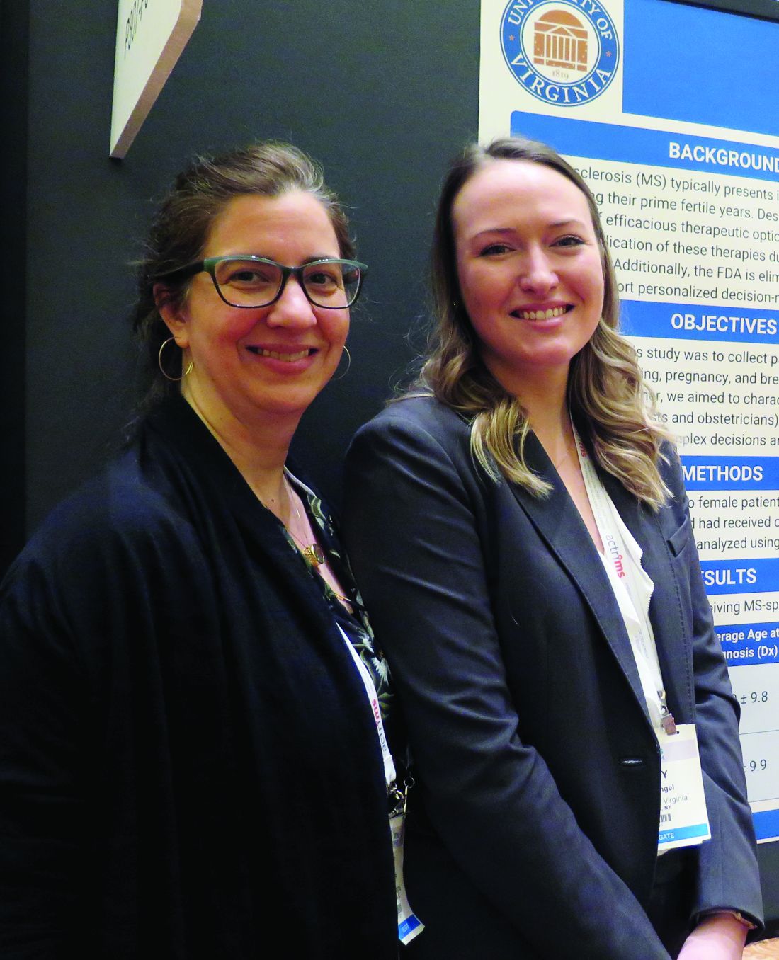
“We want our patients to feel comfortable when they come to us in their 20s or 30s and they get diagnosed, they’re scared, and it’s all new to them,” one of the study authors, Casey E. Engel said in an interview at the meeting held by the Americas Committee for Treatment and Research in Multiple Sclerosis. “We want them to know that family planning is something to consider and that they can proceed with having a family with our help and guidance.”
In an effort to collect patient-experience data around family planning, pregnancy, and breastfeeding post-MS diagnosis, Ms. Engel and senior author Myla D. Goldman, MD, mailed a survey to 1,000 women with confirmed MS diagnosis who had received care at the University of Virginia Medical Center in Charlottesville. The researchers reported findings from 173 respondents, of whom 70% were receiving specialty care for MS. Most of the survey participants (137) did not become pregnant following their diagnosis, while 36 did.
Of the 137 respondents who did not become pregnant following diagnosis, 22 (16%) indicated that their decision was driven by MS-related concerns, including MS worsening with pregnancy (64%), ability to care for child secondary to MS (46%), lack of knowledge about options for pregnancy and MS (18%), passing MS onto child (18%), and stopping disease-modifying therapy (DMT) to attempt pregnancy (9%).
Of the 36 women who had a pregnancy following diagnosis, 20% reported postpartum depression or anxiety, higher than the national average of 10%-15%. In addition, 79% reported not being on DMT at the time of conception, 9% were on either glatiramer acetate injection or interferon beta-1a at time of conception, and 3% were on fingolimod (Gilenya) at time of conception. The majority reported receiving inconsistent advice about when to discontinue DMT before attempting pregnancy (a range from 0 to 6 months).
“It’s also noteworthy that 20% took a year to achieve pregnancy,” said Dr. Goldman, a neurologist who directs the university’s MS clinic. “If these women stop [their DMT] 6 months in advance and they take a year to achieve pregnancy, that’s 18 months without therapeutic coverage. That’s a concern to bring to light.”
Breastfeeding was reported in 71% of mothers in postdiagnosis pregnancy with a range between 1 week and 10 months, driven in part by variable guidelines regarding DMT reinitiation. In the meantime, respondents who did not breastfeed made this decision due to fear of relapse, glucocorticoids, or desire to reinitiate medication.
“Though our study was limited by low survey response, we hope that our work may highlight the difficulty our patients face and foster discussions within the MS community around these issues to improve the individual patient experience,” the researchers wrote in their poster.
Ms. Engel worked on the study while an undergraduate at the University of Virginia. The study was supported by the ziMS Foundation.
SOURCE: Engel CE et al. ACTRIMS Forum 2019, Poster 307.
REPORTING FROM ACTRIMS FORUM 2019
