User login
Mycobiome much more diverse in children than in adults
The normal fungal communities that inhabit healthy skin are much more diverse in children than adults, a new study has discovered.
That diversity dwindles, however, around puberty, when the lipophilic taxa Malassezia surges in abundance. This is probably mediated by the increase in sebaceous gland activation and sebum composition that occurs around sexual maturity, Jay-Hyun Jo, PhD, wrote (J Invest Dermatol. 2016 Jul 28; doi: 10.1016/j.jid.2016.05.130).
The diversity of the childhood mycobiome may also play into the larger prevalence of fungal skin diseases in children, wrote Dr. Jo of the National Cancer Institute.
“Several fungal skin infections (dermatophytoses), such as tinea capitis and tinea corporis, are more frequently seen in children. This epidemiological dichotomy in fungal infections may relate to the physiologic characteristics of younger skin, which appears more permissive to colonization by diverse fungi.”
The researchers used the fungal internal transcribed spacer–1 (ITS1) sequence to pinpoint the taxonomic details of the mycobiome of 14 healthy children and 19 healthy adults. They looked at samples from 10 sites on each subject: the external auditory canal, forehead, occiput, retroauricular crease, back, manubrium, antecubital fossa, inguinal crease, volar forearm, and nares.
Malassezia monopolized the adult samples, constituting 80%-99% of the communities on each skin site. In children, however, Malassezia was much less common, comprising 35%-76% of the samples of each site.
However, children boasted a much more diverse mycobiome. Other constituents included members of the Ascomycota, Aspergillus, Epicoccum, and Phoma taxae. Ascomycota species were found on 40% of samples from children, compared with 9.5% of samples from adults. Children also played host to communities of Epicoccum, Cladosporium, and Cryptococcus.
There were individual variations in diversity, however, the authors noted. “For clinical samples from children, decreased diversity was correlated with increased relative abundance of Malassezia, especially on sebaceous sites. Given the predominance of Malassezia on sebaceous skin, it is possible that reduction in diversity was attributed to relative overexpansion of Malassezia.”
The team also discovered gender differences in the mycobiome of children. The sebaceous skin sites of boys were much more likely to host species of Epicoccum and Cryptococcus. Girls showed an early enrichment of Malassezia. “These results suggested that gender may affect mycobiome structures during sexual maturation.”
“Since Malassezia is an obligatory lipophilic fungus, differential Malassezia abundance might be due to the full activation of sebaceous glands during puberty,” they theorized. “Therefore, it would be intriguing to identify the sebaceous gland activity and sebum signatures during childhood in conjunction with sequence-based mycobiome analysis.”
The National Institutes of Health funded the study. Dr. Jo had no financial disclosures.
On Twitter @Alz_Gal
FROM THE JOURNAL OF INVESTIGATIVE DERMATOLOGY
Key clinical point: The mycobiome of children is much more diverse than that of adults.
Major finding: Malassezia species comprised 80%-99% the adult mycobiome, while numerous other taxae were found on children’s skin.
Data source: The taxonomic analysis comprised 19 healthy adults and 14 healthy children.
Disclosures: The National Institutes of Health funded the study. Dr. Jo had no financial disclosures.
Candida auris in Venezuela outbreak is triazole-resistant, opportunistic
BOSTON – An investigation into 18 nosocomial Candida auris infections at a tertiary care center in Venezuela showed that isolates of the emerging fungal pathogen obtained during the outbreak were resistant to fluconazole and voriconazole. However, the isolates were intermediately susceptible to amphotericin B and susceptible to 5-fluorocitosine, and demonstrated high susceptibility to the candin antifungal anidulafungin.
Dr. Belinda Calvo, an infectious disease specialist at the University of Maracaibo, Venezuela, and her collaborators reported these findings, related to a 2012-2013 C. auris outbreak at the hospital. Dr. Calvo and her coinvestigators noted that other invasive C. auris outbreaks have been reported in India, Korea, and South Africa, but that “the real prevalence of this organism may be underestimated,” since common rapid microbial identification techniques may misidentify the species.
In a poster session at the annual meeting of the American Society of Microbiology, Dr. Calvo and her collaborators reported that the 18 patients involved in the Venezuelan outbreak were critically ill, of whom 11 were pediatric, and all had central venous catheter placement. All but two of the pediatric patients were neonates, and all had serious underlying morbidities; several had significant congenital anomalies. The median patient age was 26 days (range, 2 days to 72 years), reflecting the high number of neonates affected. One of the adult patients had esophageal carcinoma. Overall, 10/18 patients (56%) had undergone surgical procedures, and all had received antibiotics.
As has been reported in other C. auris outbreaks, isolates from blood cultures of affected individuals were initially reported as C. haemulonii by the Vitek 2 C automated microbial identification system. Molecular identification was completed by sequencing the internal transcribed spacer (ITS) of the rDNA gene, with analysis aided by the National Institutes of Health’s GenBank and the Netherland’s CBS Fungal Diversity Centre , in order to confirm the identity of the fungal isolates as C. auris. Dr. Calvo and her associates were able to generate a dendrogram of the 18 isolates, showing high clonality, a trait shared with other nosocomial C. auris outbreaks.
Susceptibility testing of the C. auris cultured from blood samples of the affected patients showed that fluconazole had a minimum inhibitory concentration to inhibit the growth of 50% of the organisms (MIC50) of greater than 64 mcg/mL. For fluconazole, the MIC90, range, and geometric mean were all also above 64 mcg/mL, indicating a high level of resistance. For voriconazole, the MICs, range, and mean were all 4 mcg/mL. For amphotericin B, the MIC50 was 1 mcg/mL, the MIC90 was 2 mcg/mL, the range was 1-2, and the geometric mean was 1.414 mcg/mL.
The high number of pediatric patients affected, as well as early pathogen identification with speedy and appropriate antifungal therapy and prompt removal of central venous catheters, likely contributed to the relatively low 30-day crude mortality rate of 28%, said Dr. Calvo and her coauthors.
“C. auris should be considered an emergent multiresistant species,” wrote Dr. Calbo and her collaborators, noting that the opportunistic pathogen has a “high potential for nosocomial horizontal transmission.”
In June 2016, the Centers for Disease Control issued a clinical alert to U.S. healthcare facilities regarding the global emergence of invasive infections caused by C. auris.
The study authors reported no external sources of funding and no conflicts of interest.
On Twitter @karioakes
AT ASM 2016
Key clinical point: Isolates in an outbreak of nosocomially acquired Candida auris were fluconazole-resistant.
Major finding: All C. auris isolates were resistant to fluconazole, with geometric mean minimum inhibitory concentrations greater than 64 mcg/mL.
Data source: Retrospective, single-center study of 18 pediatric and adult patients with C. auris infections at a tertiary care center in Venezuela.
Disclosures: The study investigators reported no outside sources of funding and no disclosures.
Former chief of Endocrine Society: Send HbA1c packing
SAN DIEGO – The former president of the Endocrine Society told diabetes educators that it’s time to replace the much-used hemoglobin A1c level with a measurement that better reflects how diabetes patients are faring.
The problem is that the HbA1c is “woefully inadequate,” said Robert Vigersky, MD, the medical director of Medtronic Diabetes and president of the Endocrine Society in 2009-2010. “It doesn’t tell you about time-in-range or the frequency, duration, and severity of hypoglycemia or hyperglycemia. And there’s nothing about glycemic variability.”
These measurements are important, he told an audience at the annual meeting of the American Association of Diabetes Educators. For example, “there are several new classes of medication and new technologies. Some decrease HbA1c with no effect on hypoglycemia. Some affect hypoglycemia with no effect on HbA1c. How do we think about these globally and compare them to one another?”
To shed more light on the true condition of patients, he said, it’s time to “change the conversation from HbA1c alone to one that is more glucose-centric. It’s about thinking about glucose as a vital sign, not HbA1c. This may help health care providers, regulators, and payers better understand what is best for patients.”
He is also thinking about going beyond sugar levels. “Maybe a future composite metric will have more than just glucose numbers,” he said.
Indeed, last hear Dr. Vigersky proposed a composite metric known as “the hypoglycemia-A1C score” that can also take factors like weight, quality of life and costs into account (J Diabetes Sci Technol. 2015 Feb 19;9[5]:1148-51).
But he acknowledged there are challenges. For one, there are at least a dozen different ways to measure hypoglycemia, he said, “and every paper cherry-picks the method they want to show their data in the best light.”
It’s also not clear how best to represent the data once researchers figure out which statistics should be included. Should the overall measurement be a single number? Or should there be multiple numbers? In that case, should the numbers be represented graphically?
Dr. Vigersky said he is working on an approach that illustrates various measurements through a pentagon shape. Its appearance reflects measurements such as mean glucose and duration of high glucose.
However, he predicted the future will produce a simpler measurement: “a multicomponent single value.”
What’s next? “We need to educate the payers about how we can’t stay with HbA1c. This will take an effort with professional societies. Once everyone agrees, we need to get some consensus about what the elements of the composite metric are.”
Dr. Vigersky is hopeful that the HbA1c is on its way out, although he acknowledges that it won’t be a rapid process. “It took 20 or more years for everyone to buy into A1C and understand what it represented,” he said. “Changing the conversation isn’t going to happen overnight. But unless we start to address it, it will never happen.”
Dr. Vigersky reported having no relevant financial disclosures.
EXPERT ANALYSIS FROM AADE 16
Tofacitinib clears pediatric alopecia areata in small study
MINNEAPOLIS – The first study to evaluate tofacitinib’s effectiveness at treating severe alopecia areata in the pediatric population found that the janus kinase inhibitor was effective for more than half of the patients, and well tolerated by all.
Of a case series of 13 pediatric patients who had alopecia areata (AA) and were treated with tofacitinib, 9 (68%) experienced “clinically significant” regrowth of hair, with mean improvement in the Severity of Alopecia Tool (SALT) score of 88% for these responders. The nonresponding group, all of whom had alopecia universalis or totalis, saw essentially no response, with a 1% reduction in SALT score.
Lucy Y. Liu, a medical student at Yale University, New Haven, Conn., presented the findings at the annual meeting of the Society for Pediatric Dermatology.
Ms. Liu and her coinvestigators reported that all of the patients had severe AA by SALT scoring, with an overall mean pretreatment SALT score of 74. Eight of the patients (62%) had alopecia universalis, and two (15%) had alopecia totalis.
The patients ranged in age from 12 to 17 years, with a median age of 15. All but three were male, and patients were an average 9 years old at onset of AA. For patients with alopecia totalis or universalis, the duration of the current episode was a median 1.75 years.
Five patients (38%) had atopic dermatitis, while 1 (8%) had thyroid disease. Three patients (23%) had family members with AA; all but one patient, however, had a family history of autoimmune disease of some sort.
Patients were given tofacitinib 5 mg orally twice daily for 5 months. One patient developed new patches of alopecia during treatment, so the dosing for that patient was increased to 10 mg/5 mg daily.
Adverse events for participants included headaches, upper respiratory infections, and “mild, transient increases in transaminases,” wrote Dr. Lieu and her collaborators. No serious adverse events were reported.
Previous work at Yale had shown that tofacitinib reversed alopecia universalis in a patient who received the medication for plaque psoriasis, and that topical treatment with ruxolitinib, another janus kinase inhibitor, was effective in treating alopecia universalis.
Study limitations included the small sample size and the relatively short duration of follow-up, an important consideration because relapse has been observed after tofacitinib treatment in AA. Still, “Tofacitinib is a promising therapy for the treatment of severe AA in adolescents,” wrote Ms. Liu and her colleagues, recommending randomized clinical trials for further exploration of efficacy and safety in the pediatric population.
On Twitter @karioakes
AT THE SPD ANNUAL MEETING
Key clinical point: The JAK inhibitor tofacitinib resolved alopecia areata in 68% of pediatric patients.
Major finding: Among the responders, the Severity of Alopecia Tool score improved by a mean 88% over 5 months.
Data source: Case series of 13 pediatric patients with severe alopecia areata treated with tofacitinib at a single site.
Disclosures: Ms. Liu and her collaborators reported no conflicts of interest.
NIAID to test new yellow fever vaccine
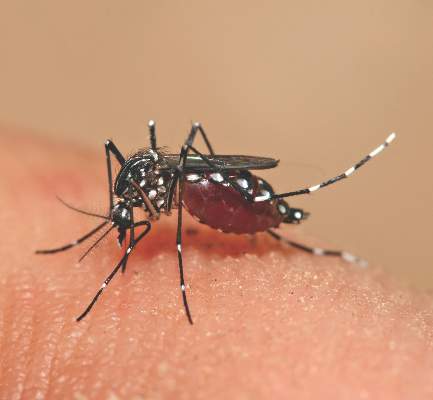
The National Institute of Allergy and Infectious Diseases (NIAID) has initiated a phase I clinical trial of an investigational vaccine designed to protect against yellow fever virus.
According to an NIAID statement, the study will evaluate whether an experimental vaccine developed by the pharmaceutical manufacturer Bavarian Nordic is “safe, tolerable and has the potential to prevent yellow fever virus infection.”
Bavarian Nordic’s experimental yellow fever vaccine, called MVA-BN-YF, is based on the company’s proprietary MVA-BN platform, which uses an attenuated version of the Modified Vaccinia Ankara (MVA) virus as a vaccine vector to carry yellow fever virus genes into the body. Bavarian Nordic says that more than 7,600 people, including 1,000 who are immunocompromised, have been safely vaccinated with MVA-BN–based vaccines.
The NIAID statement noted that prior studies have suggested that combining MVA-BN with ISA 720, an experimental immune-boosting adjuvant, induces a strong immune response after a single dose of vaccine. One goal of the study will be to assess whether two doses of unadjuvanted vaccine or a single dose of ISA 720 adjuvanted vaccine could provide protection against yellow fever.
NIAID said the placebo-controlled, double-blinded clinical trial will enroll 90 healthy men and women aged 18-45 years who have never been infected with a flavivirus. Participants will be divided into six groups: One will receive the currently licensed yellow fever vaccine (15 participants) and five groups (15 participants each) will receive the investigational Bavarian Nordic vaccine, either with or without an adjuvant. The investigational vaccine will be administered intramuscularly while the licensed yellow fever vaccine will be administered subcutaneously. Trial participants will receive one or two doses of vaccine or placebo, separated by a month.
According to NIAID’s statement, the multisite trial will be conducted by NIAID-funded Vaccine and Treatment Evaluation Units at the University of Iowa, Iowa City, and Saint Louis (Mo.) University. The Emory Vaccine Center in Decatur, Ga., will assist in evaluating data.
Yellow fever infection usually causes fever, back pain, headache, nausea, vomiting, fatigue, and weakness, but roughly 15% of infected patients develop severe disease manifested as jaundice, hemorrhage, and shock, resulting in potentially fatal kidney, liver, or heart conditions.
The current yellow fever vaccine can produce severe adverse complications, such as neurologic side effects, multiple organ system dysfunction and death, and thus cannot be given to infants, the elderly, pregnant women, and those with compromised immune systems. More than 105 million people in Africa have been vaccinated against yellow fever since 2006, according to the World Health Organization (WHO), but a new outbreak of the disease has caused an estimated 84,000-170,000 severe illnesses and 29,000 to 60,000 deaths in 2013.
For more details about the trial, visit the National Institutes of Health Clinical Trials website.
On Twitter @richpizzi

Portable device may underestimate FEV1 in children
The PiKo-1 device (nSpire Health) has limited utility in determining forced expiratory volume in 1 second (FEV1) in children with asthma, according to Jonathan M. Gaffin, MD, and his associates.
In a study of 242 children, spirometry and PiKo-1 devices were used to test FEV1. In the Bland-Altman analysis, it reported a mean difference between FEV1 measured by spirometry and PiKo-1 of 0.14 L. The PiKo-1 FEV1 was found to be moderately biased to underestimate FEV1 with increasing volumes, for every 1-liter increase in spirometry FEV1, having the difference between spirometry and PiKo-1 increased by 0.19 L (P < .001).
Researchers also used the pulmonary function test (PFT) and t showed variability was 0.4 L for spirometry at 2 SDs, a significant smaller range than seen in the PFT-PiKo confidence intervals (1.1 L). It is noted that this indicates that differences are credited to distinctions in the devices themselves and not within the techniques of the person using them. There was no effect on the order of PFT or PiKo-1 performance (P = .88).
“The findings from this study suggest that the PiKo-1 device has limited utility in assessing FEV1 in clinical or research settings in children with asthma,” researchers concluded. “Further investigation of its use in this respect and with different populations may prove the device more valuable.”
Find the full study in the Annals of Allergy, Asthma and Immunology (doi: 10.1016/j.anai.2016.06.022).
FROM THE ANNALS OF ALLERGY, ASTHMA & IMMUNOLOGY
Intensified rifampicin boosts outcomes in TB/HIV coinfection
DURBAN, SOUTH AFRICA – Prescribing high-dose rifampicin in addition to antiretroviral therapy reduces 12-month all-cause mortality in patients who are coinfected with tuberculosis and HIV and who have a low CD4 cell count, Corinne S. Merle, MD, reported at the 21st International AIDS Conference.
“Current strategies to reduce TB/HIV mortality rely largely on optimal management of HIV disease with early ART [antiretroviral therapy]. We wanted to look at whether there is value in focusing on the TB side of the problem. This is the first study to look at more intensive TB therapy for reducing mortality; and we think that, at least in patients who are immunosuppressed, there might be some benefit in a more aggressive TB treatment from the start,” said Dr. Merle of the London School of Hygiene and Tropical Medicine.
She presented the results of the open-label, multicenter trial of 747 ART-naive adults from West Africa. All were coinfected with TB/HIV and had a CD4 count of at least 50 cells/mm3 at enrollment. They were randomized to one of three treatment arms: ART starting at 2 weeks combined with standard TB treatment; ART starting at 8 weeks plus standard TB therapy; or ART initiation at 8 weeks coupled with 2 months of high-dose rifampicin (Rifadin) at 15 mg/kg daily, followed by standard TB therapy. None of the participants had multidrug-resistant TB. More than one-quarter of them were undernourished as evidenced by a baseline body mass index below 16 kg/m2.
The primary outcome was all-cause mortality at 12 months. There was no significant difference between the study arms, with a 10% rate in the intensified TB treatment arm and mortality rates of 11% and 14% with standard TB therapy and ART starting after 2 and 8 weeks, respectively.
However, a prespecified secondary analysis restricted to the 159 subjects with a baseline CD4 count below 100 cells/mm3 struck gold. Overall 12-month mortality was 4% in the intensified TB treatment subgroup, compared with 19% in patients on standard TB therapy with ART starting at 2 weeks and 28% with ART starting at 8 weeks. In a Cox regression analysis, severely immunosuppressed patients in the high-dose rifampicin group were an adjusted 88% less likely to die within 12 months than those on standard TB treatment with ART starting at 8 weeks and 80% less likely to die than those starting ART at 2 weeks.
At 18 months after randomization, roughly three-quarters of patients in each study arm had undetectable HIV viral loads.
There was no evidence of an increased risk of hepatotoxicity with 2 months of high-dose rifampicin. Only 4 of nearly 3,800 aspartate aminotransferase measurements obtained during the trial showed grade 3 or 4 hepatotoxicity, Dr. Merle noted.
In a plenary lecture on TB/HIV coinfection at the AIDS 2016 conference, Anton Pozniak, MD, singled out the trial as a sterling example of how to optimize available clinical management tools while awaiting a desperately needed new TB vaccine and better drugs.
More than 1 million new TB cases occur annually in HIV-infected persons, roughly 80% of them in sub-Saharan Africa. There are now 400,000 deaths per year worldwide in coinfected TB/HIV patients. Indeed, TB has become the No. 1 cause of death among people living with HIV infection, said Dr. Pozniak, director of HIV services at Chelsea and Westminster Hospital in London.
He offered a road map to eliminating TB by the year 2035. At present, the global trend is a 2% per year decline in new cases. Optimizing TB case finding, treatment, and preventive therapy could achieve a 10% per year decrease in new cases. That rate still wouldn’t reach the goal by 2035. But more than a dozen candidate TB vaccines are in the developmental pipeline, including a mycobacterial whole cell extract in phase III testing in China. If a new vaccine can be introduced by 2025, that would be a game changer.
“A new vaccine that could prevent adolescents and adults from developing and transmitting TB would be the single most cost-effective tool in mitigating the epidemic,” he said. “Even if we had only a 60% efficacious vaccine and delivered it to 20% of the target population, it could potentially avert 30-50 million incident cases of TB by 2050.”
A new vaccine plus effective alternatives to the standard 6 months of isoniazid for latency prophylaxis by 2025 are estimated to reduce new cases of TB by an average of 17% per year. That circumstance would mean the end of TB by 2035, Dr. Pozniak declared.
The trial was funded by the European and Developing Countries Clinical Trials Partnership. Dr. Merle reported having no financial conflicts of interest.
AT AIDS 2016
Key clinical point: High-dose rifampicin improves survival in patients who are coinfected with tuberculosis and HIV and have low CD4 counts.
Major finding: Overall 12-month mortality was 4% in the intensified TB treatment subgroup, 19% in patients on standard TB therapy with ART starting at 2 weeks, and 28% with standard TB therapy and ART starting at 8 weeks.
Data source: This was a randomized, prospective, three-arm, open-label trial including 747 patients coinfected with tuberculosis and HIV.
Disclosures: The trial was funded by the European and Developing Countries Clinical Trials Partnership. The presenter reported having no financial conflicts of interest.

HIV-related lymphoma rate remains sky-high despite ART
DURBAN, SOUTH AFRICA – The good news about non-Hodgkin lymphoma in the setting of HIV infection is that the risk drops dramatically after several years of antiretroviral therapy. The bad news? The risk still remains extraordinarily high, compared with the risk seen in the general population, Mathias Egger, MD, reported at the 21st International AIDS Conference.
That was a key finding in a new analysis of lymphoma trends in more than 210,000 HIV-positive adults on combination antiretroviral therapy (ART) during more than 1.1 million person-years of follow-up in North America, Europe, Latin America, and South Africa.

The non-Hodgkin lymphoma (NHL) incidence rate standardized to 40 years of age was 287 cases per 100,000 person-years. From a pre-ART baseline of about 500 cases per 100,000 person-years, it dropped “massively” within a year after going on ART. Even after 5 years of ART, however, the rate remained in the range of 60-200 cases per 100,000 person-years, depending upon geographic location and HIV transmission route. In contrast, the incidence rate among the general population of the U.S. and Canada, which is among the world’s highest, is less than 10 per 100,000 person-years, according to Dr. Egger, professor of epidemiology and public health at the University of Bern, Switzerland.
The risk of developing NHL in the setting of HIV infection varied by continent. It was slightly higher in HIV-infected patients in North America than in Europe or South Africa, although the South African data are considered unreliable due to underascertainment of cancers.
In Latin American HIV-infected adults the NHL rate was lowest of all, fully 54% lower than in Europe after adjustment for current CD4 cell count, ART regimen and duration, and transmission risk group. The low NHL rate in Latin America was driven by a very low risk in HIV-infected women.
Across the world, NHL rates in patients on ART were consistently lowest in women, intermediate in heterosexual men, and highest in men who have sex with men.
The explanation for the regional variation in NHL trends might plausibly involve differing prevalences of Epstein-Barr virus–2 and other oncogenic viruses as well as differences in the completeness of cancer ascertainment, Dr. Egger said.
While NHL is categorized as an AIDS-defining condition, Hodgkin lymphoma is not. Nonetheless, the risk of Hodgkin lymphoma is markedly increased in the setting of HIV infection. In one classic meta-analysis, it was increased by 11-fold, compared with that seen in the general population (Lancet. 2007 Jul 7;370(9581):59-67).
In a study of more than 41,000 HIV-infected European adults by Dr. Egger and his coworkers, the incidence of Hodgkin lymphoma was 49 cases per 100,000 person-years. Importantly, unlike in NHL, the cumulative incidence and mortality of Hodgkin lymphoma were unaffected by ART (Blood. 2011 Jun 9;117(23):6100-8).
The clinical implication of these trends in HIV-related lymphomas is clear: with more than 2 million new cases of HIV infection occurring annually worldwide, and with infected patients living far longer as ART transforms HIV infection into a chronic manageable condition, physicians can anticipate encountering a steadily growing number of patients with NHL and Hodgkin lymphoma, he said.
The NHL study was funded by the European Union and the U.S. National Institutes of Health. The analysis utilized data collected by the Collaboration of Observational HIV Epidemiology and Research Europe (COHERE) and the International Epidemiologic Databases to Evaluate AIDS (IeDEA). Dr. Egger reported having no financial conflicts of interest.
DURBAN, SOUTH AFRICA – The good news about non-Hodgkin lymphoma in the setting of HIV infection is that the risk drops dramatically after several years of antiretroviral therapy. The bad news? The risk still remains extraordinarily high, compared with the risk seen in the general population, Mathias Egger, MD, reported at the 21st International AIDS Conference.
That was a key finding in a new analysis of lymphoma trends in more than 210,000 HIV-positive adults on combination antiretroviral therapy (ART) during more than 1.1 million person-years of follow-up in North America, Europe, Latin America, and South Africa.
The non-Hodgkin lymphoma (NHL) incidence rate standardized to 40 years of age was 287 cases per 100,000 person-years. From a pre-ART baseline of about 500 cases per 100,000 person-years, it dropped “massively” within a year after going on ART. Even after 5 years of ART, however, the rate remained in the range of 60-200 cases per 100,000 person-years, depending upon geographic location and HIV transmission route. In contrast, the incidence rate among the general population of the U.S. and Canada, which is among the world’s highest, is less than 10 per 100,000 person-years, according to Dr. Egger, professor of epidemiology and public health at the University of Bern, Switzerland.
The risk of developing NHL in the setting of HIV infection varied by continent. It was slightly higher in HIV-infected patients in North America than in Europe or South Africa, although the South African data are considered unreliable due to underascertainment of cancers.
In Latin American HIV-infected adults the NHL rate was lowest of all, fully 54% lower than in Europe after adjustment for current CD4 cell count, ART regimen and duration, and transmission risk group. The low NHL rate in Latin America was driven by a very low risk in HIV-infected women.
Across the world, NHL rates in patients on ART were consistently lowest in women, intermediate in heterosexual men, and highest in men who have sex with men.
The explanation for the regional variation in NHL trends might plausibly involve differing prevalences of Epstein-Barr virus–2 and other oncogenic viruses as well as differences in the completeness of cancer ascertainment, Dr. Egger said.
While NHL is categorized as an AIDS-defining condition, Hodgkin lymphoma is not. Nonetheless, the risk of Hodgkin lymphoma is markedly increased in the setting of HIV infection. In one classic meta-analysis, it was increased by 11-fold, compared with that seen in the general population (Lancet. 2007 Jul 7;370(9581):59-67).
In a study of more than 41,000 HIV-infected European adults by Dr. Egger and his coworkers, the incidence of Hodgkin lymphoma was 49 cases per 100,000 person-years. Importantly, unlike in NHL, the cumulative incidence and mortality of Hodgkin lymphoma were unaffected by ART (Blood. 2011 Jun 9;117(23):6100-8).
The clinical implication of these trends in HIV-related lymphomas is clear: with more than 2 million new cases of HIV infection occurring annually worldwide, and with infected patients living far longer as ART transforms HIV infection into a chronic manageable condition, physicians can anticipate encountering a steadily growing number of patients with NHL and Hodgkin lymphoma, he said.
The NHL study was funded by the European Union and the U.S. National Institutes of Health. The analysis utilized data collected by the Collaboration of Observational HIV Epidemiology and Research Europe (COHERE) and the International Epidemiologic Databases to Evaluate AIDS (IeDEA). Dr. Egger reported having no financial conflicts of interest.
AT AIDS 2016
Key clinical point: Antiretroviral therapy has had a major impact upon the incidence of HIV-related non-Hodgkin lymphoma but no effect on Hodgkin lymphoma.
Major finding: The overall incidence of non-Hodgkin lymphoma in HIV-positive adults on antiretroviral therapy is 287 cases per 100,000 person-years, varying by location and route of HIV acquisition.
Data source: This was a longitudinal analysis of non-Hodgkin lymphoma incidence in more than 210,000 HIV-infected adults on combination antiretroviral therapy on four continents.
Disclosures: The study was funded by the European Union and the U.S. National Institutes of Health. The presenter reported having no financial conflicts of interest.
Monitoring renal function during daily oral HIV PrEP
DURBAN, SOUTH AFRICA – The optimal frequency of kidney safety monitoring in patients using oral daily tenofovir/emtricitabine for pre-exposure prophylaxis against HIV infection is every 6 months, but less frequent monitoring may be reasonable in most low-risk patients, Renee Heffron, PhD, said at the 21st International AIDS Conference.
The occurrence and pattern of detection of a drop in creatinine clearance to less than 60 mL/min during the first 12 months of therapy didn’t differ significantly regardless of whether monitoring was done at 3- or 6-month intervals. The risk of a clinically relevant decline in creatinine clearance during the first 12 months of therapy appears to be largely confined to the subgroup of patients on tenofovir/emtricitabine (Truvada) for pre-exposure prophylaxis (PrEP) who weigh 55 kg or less, have a baseline creatinine clearance rate of 60-90 mL/min, or are at least 45 years old, according to Dr. Heffron of the University of Washington, Seattle.
The question of how frequently to monitor renal function is a key issue as PrEP with tenofovir/emtricitabine is ramped up to scale in sub-Saharan Africa and other parts of the developing world where the majority of new HIV infections occur – and where laboratory resources are often limited. The randomized clinical trials that led to marketing approval of tenofovir/emtricitabine for PrEP in the United States and elsewhere monitored creatinine clearance every 3 months. But the confirmatory demonstration projects used a range of kidney monitoring schedules, she explained.
She presented an analysis of clinically relevant kidney toxicity in 4,404 initially HIV-negative subjects on tenofovir/emtricitabine in the Partners PrEP Study, in which creatinine clearance was measured every 3 months, and in 955 participants in the Partners Demonstration Study, in which monitoring was performed every 6 months. All participants were at high risk for HIV acquisition because they were members of serodiscordant couples.
The occurrence and pattern of detection of a drop in creatinine clearance to less than 60 mL/min during the first 12 months of therapy didn’t differ significantly regardless of whether monitoring was done at 3- or 6-month intervals. The cumulative rate in the randomized trial was 0.4%, 0.5%, and 0.7% at 3, 6, and 12 months, and it was 0.2% at both 6 and 12 months in the demonstration project, Dr. Heffron reported.
These renal events were not only rare, they were reassuringly nonprogressive and resolved within a few weeks of PrEP discontinuation, she added.
Her analysis of the combined 5,359 subjects in the two Partners studies identified three independent predictors of a fall in creatinine clearance to below 60 mL/min during the first 12 months of therapy. A baseline age of 45 years or more was associated with an adjusted 2.5-fold increase, compared with younger patients. Subjects with a creatinine clearance of 60-90 mL/min at enrollment were 74 times more likely to experience a significant drop in creatinine clearance than those who started on PrEP with a creatinine clearance rate in excess of 90 mL/min. And patients weighing 55 kg or less had a 2.7-fold greater risk than those weighing more. But fewer than 5% of patients with any of these three predictors actually experienced a drop in creatinine clearance to below 60 mL/min.
The data from the two Partners studies support guidelines from the Centers for Disease Control and Prevention recommending creatinine monitoring every 6 months for people on oral daily PrEP. Still, patients with one of the defined risk factors might logically be candidates for targeted monitoring, Dr. Heffron observed.
The Partners studies were funded by the National Institutes of Health, the Bill and Melinda Gates Foundation, and the U.S. Agency for International Development. Dr. Heffron reported having no financial conflicts of interest.
AT AIDS 2016
Key clinical point: Monitoring creatinine clearance every 6 months is optimal in patients taking tenofovir/emtricitabine for pre-exposure prophylaxis against HIV infection.
Major finding: Fewer than 1% of patients experienced a decline in creatinine clearance to below 60 mL/min during their first 12 months on oral daily tenofovir/emtricitabine for pre-exposure prophylaxis against HIV infection.
Data source: This was a secondary analysis of 5,359 adults whose creatinine clearance was measured every 3 or 6 months while on oral daily tenofovir/emtricitabine for pre-exposure prophylaxis against HIV infection in a randomized trial or open-label demonstration project.
Disclosures: The studies were funded by NIH, the Bill and Melinda Gates Foundation, and the U.S. Agency for International Development. The presenter reported having no financial conflicts of interest.

Don’t delay treatment for patients with TB and HIV
Clinicians should treat patients diagnosed with HIV and tuberculosis for both conditions immediately, according to new guidelines on the treatment of drug-susceptible tuberculosis.
The clinical practice guidelines were issued collectively by three organizations: the American Thoracic Society (ATS), the U.S. Centers for Disease Control and Prevention (CDC), and the Infectious Diseases Society of America (IDSA), and published online in Clinical Infectious Diseases.

The guidelines recommend starting TB treatment for all patients as soon as an infection is suspected, rather than waiting for test results, and focusing on daily therapy to reduce the risk of relapse. In addition, all TB patients should receive comprehensive care, including direct observed therapy (DOT) when appropriate (Clin Infect Dis. 2016 Aug 10. doi: 10.1093/cid/ciw376).
“Treatment of tuberculosis is focused on both curing the individual patient and minimizing the transmission,” wrote Payam Nahid, MD, professor of medicine at the University of California, San Francisco, and his colleagues on the guidelines committee.
The guidelines’ section on treatment of tuberculosis in special situations addresses management of TB in patients with conditions including HIV, extrapulmonary TB, culture-negative pulmonary TB, pregnancy, renal disease, and hepatic disease, as well as treatment of children and the elderly.
With regard to HIV, the guidelines recommend the standard 6-month daily TB treatment for HIV patients on antiretroviral therapy. This treatment includes 2 months of isoniazid (INH), rifampin (RIF), pyrazinamide (PZA), and ethambutol (EMB), followed by a continuation phase of 4 months of INH and RIF.
“Patients with HIV infection and tuberculosis are at an increased risk of developing paradoxical worsening of symptoms, signs, or clinical manifestations of tuberculosis after beginning antituberculosis and antiretroviral treatments,” according to the guidelines. These responses are defined as Immune Reconstitution Inflammatory Syndrome (IRIS). However, IRIS does not appear to impact the simultaneous treatment of TB and HIV, and the condition can be managed symptomatically if it occurs, the researchers noted.
The guidelines identified several areas in need of further study, including new TB drugs and treatment plans; the effects of biomarkers to help design individual therapy; TB in special populations including HIV patients, pregnant women, and children; and treatment delivery strategies.
The guidelines also are endorsed by the European Respiratory Society (ERS) and the U.S. National Tuberculosis Controllers Association (NCTA).
The American Thoracic Society, the Infections Diseases Society of America, and the Centers for Disease Control and Prevention provided financial support. Lead author Dr. Nahid had no financial conflicts to disclose.
Clinicians should treat patients diagnosed with HIV and tuberculosis for both conditions immediately, according to new guidelines on the treatment of drug-susceptible tuberculosis.
The clinical practice guidelines were issued collectively by three organizations: the American Thoracic Society (ATS), the U.S. Centers for Disease Control and Prevention (CDC), and the Infectious Diseases Society of America (IDSA), and published online in Clinical Infectious Diseases.
The guidelines recommend starting TB treatment for all patients as soon as an infection is suspected, rather than waiting for test results, and focusing on daily therapy to reduce the risk of relapse. In addition, all TB patients should receive comprehensive care, including direct observed therapy (DOT) when appropriate (Clin Infect Dis. 2016 Aug 10. doi: 10.1093/cid/ciw376).
“Treatment of tuberculosis is focused on both curing the individual patient and minimizing the transmission,” wrote Payam Nahid, MD, professor of medicine at the University of California, San Francisco, and his colleagues on the guidelines committee.
The guidelines’ section on treatment of tuberculosis in special situations addresses management of TB in patients with conditions including HIV, extrapulmonary TB, culture-negative pulmonary TB, pregnancy, renal disease, and hepatic disease, as well as treatment of children and the elderly.
With regard to HIV, the guidelines recommend the standard 6-month daily TB treatment for HIV patients on antiretroviral therapy. This treatment includes 2 months of isoniazid (INH), rifampin (RIF), pyrazinamide (PZA), and ethambutol (EMB), followed by a continuation phase of 4 months of INH and RIF.
“Patients with HIV infection and tuberculosis are at an increased risk of developing paradoxical worsening of symptoms, signs, or clinical manifestations of tuberculosis after beginning antituberculosis and antiretroviral treatments,” according to the guidelines. These responses are defined as Immune Reconstitution Inflammatory Syndrome (IRIS). However, IRIS does not appear to impact the simultaneous treatment of TB and HIV, and the condition can be managed symptomatically if it occurs, the researchers noted.
The guidelines identified several areas in need of further study, including new TB drugs and treatment plans; the effects of biomarkers to help design individual therapy; TB in special populations including HIV patients, pregnant women, and children; and treatment delivery strategies.
The guidelines also are endorsed by the European Respiratory Society (ERS) and the U.S. National Tuberculosis Controllers Association (NCTA).
The American Thoracic Society, the Infections Diseases Society of America, and the Centers for Disease Control and Prevention provided financial support. Lead author Dr. Nahid had no financial conflicts to disclose.
FROM CLINICAL INFECTIOUS DISEASES
Key clinical point: Clinicians should treat patients diagnosed with HIV and tuberculosis for both conditions immediately.
Major finding: A four-drug regimen of INH, RIF, PZA, and EMB remains the preferred initial treatment for drug-susceptible pulmonary tuberculosis. Treatment should be initiated promptly even before diagnostic test results are known in patients with high likelihood of having tuberculosis.
Data source: Nine PICO (population, intervention, comparators, outcomes) questions and associated recommendations for the treatment of patients diagnosed with both HIV and TB, developed based on the evidence appraised using GRADE (Grading of Recommendations Assessment, Development, and Evaluation) methodology.
Disclosures: The American Thoracic Society, the Infections Diseases Society of America, and the Centers for Disease Control and Prevention provided financial support. Lead author Dr. Nahid had no financial conflicts to disclose.
