User login
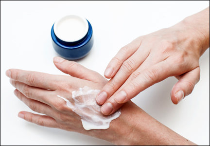
Cosmetic Corner: Dermatologists Weigh in on Hand Creams
To improve patient care and outcomes, leading dermatologists offered their recommendations on hand creams. Consideration must be given to:
- CeraVe Therapeutic Hand Cream
- Maximum Body Repair
- Neutrogena Norwegian Formula Hand Cream
- O’Keeffe’s Working Hands
Cutis invites readers to send us their recommendations. Scar treatments, body scrubs, and OTC acne treatments will be featured in upcoming editions of Cosmetic Corner. Please e-mail your recommendation(s) to the Editorial Office.
Disclaimer: Opinions expressed herein do not necessarily reflect those of Cutis or Frontline Medical Communications Inc. and shall not be used for product endorsement purposes. Any reference made to a specific commercial product does not indicate or imply that Cutis or Frontline Medical Communications Inc. endorses, recommends, or favors the product mentioned. No guarantee is given to the effects of recommended products.
Core outcomes needed for endometriosis research
The clinical usefulness of endometriosis research is being hindered by a lack of a uniform core outcomes set in published trials, according to the findings of a systematic review.
Martin Hirsch of Barts and The London School of Medicine and Dentistry and his colleagues reviewed 54 randomized controlled trials with 5,427 participants evaluating a surgical intervention – with or without medical adjuvant therapy – for the treatment of endometriosis symptoms. They included all randomized, controlled trials in the Cochrane Central Register of Controlled Trials, Embase, and MEDLINE from inception to November 2014 and found a wide variation in the outcomes reported.

Across all trials, there were 164 outcomes and 113 outcomes measures reported. The three most commonly reported primary outcomes were dysmenorrhea (23 trials), dyspareunia (21 trials), and pregnancy (26 trials).
This level of variation among trials makes it difficult to make comparisons and synthesize data, according to the researchers. “This limits the usefulness of research to inform clinical practice, enhance patient care, and improve patient outcomes.”
Mr. Hirsch and his colleagues called for an international consensus on a core outcome set for endometriosis trials. Until then, they suggested the use of the three most common pain-related outcomes – dysmenorrhea, dyspareunia, and pelvic pain – as well as subfertility outcomes, measured by pregnancy, miscarriage, and live birth.
Read the full study in the American Journal of Obstetrics & Gynecology (doi: 10.1016/j.ajog.2015.12.039).
On Twitter @maryellenny
The clinical usefulness of endometriosis research is being hindered by a lack of a uniform core outcomes set in published trials, according to the findings of a systematic review.
Martin Hirsch of Barts and The London School of Medicine and Dentistry and his colleagues reviewed 54 randomized controlled trials with 5,427 participants evaluating a surgical intervention – with or without medical adjuvant therapy – for the treatment of endometriosis symptoms. They included all randomized, controlled trials in the Cochrane Central Register of Controlled Trials, Embase, and MEDLINE from inception to November 2014 and found a wide variation in the outcomes reported.
Across all trials, there were 164 outcomes and 113 outcomes measures reported. The three most commonly reported primary outcomes were dysmenorrhea (23 trials), dyspareunia (21 trials), and pregnancy (26 trials).
This level of variation among trials makes it difficult to make comparisons and synthesize data, according to the researchers. “This limits the usefulness of research to inform clinical practice, enhance patient care, and improve patient outcomes.”
Mr. Hirsch and his colleagues called for an international consensus on a core outcome set for endometriosis trials. Until then, they suggested the use of the three most common pain-related outcomes – dysmenorrhea, dyspareunia, and pelvic pain – as well as subfertility outcomes, measured by pregnancy, miscarriage, and live birth.
Read the full study in the American Journal of Obstetrics & Gynecology (doi: 10.1016/j.ajog.2015.12.039).
On Twitter @maryellenny

Society of Hospital Medicine Awards Three with Exclusive 'Masters in Hospital Medicine' Designation
SAN DIEGO—The Society of Hospital Medicine (SHM) today named three new Masters in Hospital Medicine at HM16, the largest meeting dedicated to the hospital medicine specialty. The Master in Hospital Medicine (MHM) designation was introduced in 2010 to honor those who have uniquely distinguished themselves in hospital medicine through excellence in the field and healthcare as a whole.
“As hospital medicine continues to evolve, we look to experts in the field to lead us into the future with their innovation and vision,” SHM President Brian Harte, MD, SFHM, says. “These accomplished individuals have played foundational roles in hospital medicine’s growth and success and continue to ensure hospitalists are equipped with the tools and resources they need to provide the highest quality of patient care.”
SHM is proud to announce the following MHMs for their outstanding contributions:
- Tina L. Budnitz, MPH, MHM, for her leadership in advancing the hospital medicine movement and SHM. Throughout her time with SHM, Budnitz has led the development of the Core Competencies for Hospital Medicine to define the skills of a practicing hospitalist, launched Project BOOST, a mentored implementation program to improve care transitions that was recognized with the 2012 John M. Eisenberg Patient Safety and Quality Award and assisted in the development of SHM’s Leadership Academy and Certificate of Leadership Program, designed to build the healthcare leaders of tomorrow.
- Eric E. Howell, MD, MHM, Chief of the Division of Hospital Medicine at Johns Hopkins Bayview Medical Center in Baltimore, MD, in recognition of his foundational leadership in the hospital medicine movement and SHM. Dr. Howell has been instrumental in managing and implementing change in the hospital, including conducting research on the relationship between the emergency department and medicine floors as well as improving communication, throughput and patient outcomes. A past President of SHM, Dr. Howell serves as the Senior Physician Advisor to SHM’s Center for Hospital Innovation and Improvement and has mentored numerous hospitals and hospital medicine programs in more than six countries.
- Gregory Maynard, MD, MSc, MHM, Chief Quality Officer at the University of California Davis Medical Center in Sacramento, CA, in honor of his leadership in local, regional and national quality improvement efforts and related contributions to SHM’s quality improvement programs. A longtime member of the SHM Hospital Quality and Patient Safety Committee, Dr. Maynard played an integral role in creating and leading SHM’s mentored implementation programs to enhance care transitions, improve glycemic control and prevent venous thromboembolism (VTE). Dr. Maynard is nationally recognized in each of these areas, making significant contributions to the medical community.
For details on SHM’s fellowship designations, visit www.hospitalmedicine.org/fellows.
The problem of office theft
Why do people steal stuff from my office?
I’m not talking about pens. I’ve unintentionally walked off with more pens then I can count over the years, and never realized that I did until later. I figure others do the same with mine.

In the last few years, I’ve had a Far Side cartoon book stolen from the lobby, a stapler off my secretary’s desk, a roll of medical tape from my EMG cart, and a few other items.
Most recently, my secretary bought a candy dish at the store. It was nothing fancy, just a few bucks, but she liked it. She set it out on the front desk with some Jolly Ranchers.
A few days later, she left her desk to refill her coffee cup. While in back she heard the front door of the office open and close. When she returned up front, the dish (and candy) were gone.
None of these are a major financial loss, maybe adding up to $15-$20 a year at most. But it’s irritating to have someone steal something minor from my office.
Taking pens, or even magazines, is perhaps understandable, at times unintentional. But to reach over a desk and grab a stapler, or to walk in, grab a candy dish, and leave, are volitional and just wrong. I don’t understand this. Do people feel that, because I’m a doctor (and therefore stereotyped as rich), I can afford it? Do they do it because, since they’re giving me a copay and letting me bill their insurance, they feel entitled to something back? Or are they angry at me over something, and this is a passive-aggressive way to get even?
I don’t know. Admittedly, it’s a tiny minority who do such things. The vast majority of people wouldn’t dream of stealing a $3 candy dish from an office. But still, it points to a sad level of dishonesty among a few of the routine people I see day in and day out. I’m pretty sure they aren’t so hard up that they need to steal such petty items, either. I imagine the black market value of a used stapler is pretty low.
P.S. If someone out there is willing to return the candy dish or the Beyond The Far Side cartoon book, no questions will be asked.
Dr. Block has a solo neurology practice in Scottsdale, Ariz.
Why do people steal stuff from my office?
I’m not talking about pens. I’ve unintentionally walked off with more pens then I can count over the years, and never realized that I did until later. I figure others do the same with mine.
In the last few years, I’ve had a Far Side cartoon book stolen from the lobby, a stapler off my secretary’s desk, a roll of medical tape from my EMG cart, and a few other items.
Most recently, my secretary bought a candy dish at the store. It was nothing fancy, just a few bucks, but she liked it. She set it out on the front desk with some Jolly Ranchers.
A few days later, she left her desk to refill her coffee cup. While in back she heard the front door of the office open and close. When she returned up front, the dish (and candy) were gone.
None of these are a major financial loss, maybe adding up to $15-$20 a year at most. But it’s irritating to have someone steal something minor from my office.
Taking pens, or even magazines, is perhaps understandable, at times unintentional. But to reach over a desk and grab a stapler, or to walk in, grab a candy dish, and leave, are volitional and just wrong. I don’t understand this. Do people feel that, because I’m a doctor (and therefore stereotyped as rich), I can afford it? Do they do it because, since they’re giving me a copay and letting me bill their insurance, they feel entitled to something back? Or are they angry at me over something, and this is a passive-aggressive way to get even?
I don’t know. Admittedly, it’s a tiny minority who do such things. The vast majority of people wouldn’t dream of stealing a $3 candy dish from an office. But still, it points to a sad level of dishonesty among a few of the routine people I see day in and day out. I’m pretty sure they aren’t so hard up that they need to steal such petty items, either. I imagine the black market value of a used stapler is pretty low.
P.S. If someone out there is willing to return the candy dish or the Beyond The Far Side cartoon book, no questions will be asked.
Dr. Block has a solo neurology practice in Scottsdale, Ariz.
HM16 Session Analysis: Physician Engagement in Quality Improvement
Presenter: Jordan Messler, MD, SHFM
Summary: The main objective of this lecture was to understand the culture that often limits physician engagement. It also offered insights on how to best understand motivators for engagement, and tried to focus on strategies to improve and create an environment for physician engagement.
Despite strong evidence, there remains a refusal to confront healthcare provider’s severe quality problems. There is a high rate of failure, considering that 80% of major initiatives don’t meet their objectives. Dr. Messler pointed out that the second principle of the "Key Characteristics of an Effective Hospital Medicine Group" is an engaged hospitalist. To make this more complicated, 40% of hospitalists report inpatient census that exceed safe levels at least once a month; and 52% of hospitalists have signs of burnout.
Dr. Messler explained are intrinsic and extrinsic motivators: the culture of the group will impact the extrinsic motivation factors when it tries to encourage physician to do their work because they expected of themselves, not because some else is looking over their shoulders. Among intrinsic motivators: a sense of autonomy, mastery and purpose drives a culture of not only do the work but also improve it. Some hospitalist groups are trying to explore new approaches, including protective time for physicians to allow them to get involved in committee and QI projects.
Applying behavioral economics concepts (science of human motivation) can help HMGs design incentives among such domains as inertia (by simplifying processes), immediacy (giving bonus right after achieving goals), mental accounting (using paper checks for rewards).
HM Takeaways:
- There is lack of awareness of physician disengagement.
- Burn out is the opposite of engagement and affects patient quality.
- There are intrinsic and extrinsic factors that drives engagement.
- By creating a culture of ownership, mastery, autonomy, and rediscovery of purpose and right mix of incentives physicians can engage more.
- SHM has an Engagement Survey that can help get to know baseline motivators driving among specific groups.
Dr. Villagra is a hospitalist in Batesville, Ark., and a member of Team Hospitalist.
Data don’t support use of induction chemotherapy in head and neck cancer
SCOTTSDALE, ARIZ. – Patients with head and neck cancer undergoing radiation therapy do not have better survival if given induction chemotherapy instead of concurrent chemotherapy, finds a cohort study reported at the Multidisciplinary Head and Neck Cancer Symposium.

Using the National Cancer Data Base, Dr. Daniel W. Bowles and his colleagues analyzed outcomes for 8,003 patients treated for nonmetastatic but more advanced disease with one of these two approaches.
Results reported in a poster session and related press briefing showed that the patients given induction chemotherapy were more likely to receive radiation doses lower than those recommended in guidelines and lived, on average, about 13 months less.
“The use of induction chemotherapy is not supported by this analysis,” commented Dr. Bowles, director of cancer research at the University of Colorado at Denver, Aurora, and staff physician at the Denver VA Medical Center.
The study – the largest yet to compare the two approaches and specifically in a cohort of patients with more advanced disease – adds to others that have hinted at inferior outcomes with induction chemotherapy as compared with concurrent chemotherapy, the standard of care.
“Recently, there have been several randomized controlled studies that have looked at induction chemotherapy followed by concurrent chemoradiation versus concurrent chemoradiation alone,” he elaborated. “These studies have had somewhat varied results, but have been critiqued as being underpowered and [the possibility] that no survival benefit was seen in the induction chemotherapy arm perhaps because there were too many low-risk cancers that were included in these studies, including patients who have stage III cancer or patients who had N0 to N2a disease.”
Press briefing moderator Dr. Randall J. Kimple of the University of Wisconsin–Madison, commented, “I think this study adds to the growing data that I would say is now nearly overwhelming that induction chemotherapy, other than in maybe very selected cases, has essentially no role in the treatment of head and neck cancer patients in a routine setting and outside of the setting of a clinical trial.”
The investigators included in their analysis patients with stage Tis-T4,N2b-3,M0 squamous cell carcinoma of the oropharynx, hypopharynx, or larynx diagnosed between 2003 and 2011 and treated with external-beam radiation therapy, without surgery.
Analyses were based on 1,907 patients given induction chemotherapy (starting 43 to 98 days before radiation therapy) and 6,086 patients given concurrent chemotherapy (starting within 7 days of radiation therapy).
In univariate analyses, median overall survival was 52.1 months with induction chemotherapy versus 64.9 months with concurrent chemotherapy, reported Dr. Bowles, who disclosed that he had no relevant conflicts of interest. The difference translated to a 14% higher risk of death with the former (hazard ratio, 1.14; P less than .01).
In multivariate analysis, survival did not differ significantly between the two chemotherapy approaches in the cohort as a whole or in various subgroups of patients having especially advanced disease: those with T4 or N3 disease, with N3 disease only, or with T4N3 disease. Repeating analyses after propensity score matching yielded essentially the same results.
“We couldn’t identify any specific subgroups that appeared to benefit with regards to overall survival,” Dr. Bowles commented, while also noting some caveats.
“One potential limitation from looking at the National Cancer Data Base is that they only provide information about overall survival. We don’t know about cancer-specific survival, so you can’t say based on these data whether there is a progression-free survival benefit. The other question is how this affects quality of life,” he elaborated. “Those are important questions, [whether] induction chemotherapy would benefit anyone with regards to those outcomes. We just don’t have that data from the National Cancer Data Base.”
In other study findings, compared with peers given concurrent chemotherapy, patients given induction chemotherapy were more likely to receive a radiation dose less than the minimum of 66 Gy recommended by the National Comprehensive Cancer Network and the American Society for Radiation Oncology (20.9% vs. 14.9%, P less than .01).
In multivariate analyses, patients given induction chemotherapy were still less likely to receive guideline-concordant doses of radiation (odds ratio, 1.42; P less than .01), and receipt of such doses was associated with an increased risk of death (HR, 1.76; P less than .01).
SCOTTSDALE, ARIZ. – Patients with head and neck cancer undergoing radiation therapy do not have better survival if given induction chemotherapy instead of concurrent chemotherapy, finds a cohort study reported at the Multidisciplinary Head and Neck Cancer Symposium.
Using the National Cancer Data Base, Dr. Daniel W. Bowles and his colleagues analyzed outcomes for 8,003 patients treated for nonmetastatic but more advanced disease with one of these two approaches.
Results reported in a poster session and related press briefing showed that the patients given induction chemotherapy were more likely to receive radiation doses lower than those recommended in guidelines and lived, on average, about 13 months less.
“The use of induction chemotherapy is not supported by this analysis,” commented Dr. Bowles, director of cancer research at the University of Colorado at Denver, Aurora, and staff physician at the Denver VA Medical Center.
The study – the largest yet to compare the two approaches and specifically in a cohort of patients with more advanced disease – adds to others that have hinted at inferior outcomes with induction chemotherapy as compared with concurrent chemotherapy, the standard of care.
“Recently, there have been several randomized controlled studies that have looked at induction chemotherapy followed by concurrent chemoradiation versus concurrent chemoradiation alone,” he elaborated. “These studies have had somewhat varied results, but have been critiqued as being underpowered and [the possibility] that no survival benefit was seen in the induction chemotherapy arm perhaps because there were too many low-risk cancers that were included in these studies, including patients who have stage III cancer or patients who had N0 to N2a disease.”
Press briefing moderator Dr. Randall J. Kimple of the University of Wisconsin–Madison, commented, “I think this study adds to the growing data that I would say is now nearly overwhelming that induction chemotherapy, other than in maybe very selected cases, has essentially no role in the treatment of head and neck cancer patients in a routine setting and outside of the setting of a clinical trial.”
The investigators included in their analysis patients with stage Tis-T4,N2b-3,M0 squamous cell carcinoma of the oropharynx, hypopharynx, or larynx diagnosed between 2003 and 2011 and treated with external-beam radiation therapy, without surgery.
Analyses were based on 1,907 patients given induction chemotherapy (starting 43 to 98 days before radiation therapy) and 6,086 patients given concurrent chemotherapy (starting within 7 days of radiation therapy).
In univariate analyses, median overall survival was 52.1 months with induction chemotherapy versus 64.9 months with concurrent chemotherapy, reported Dr. Bowles, who disclosed that he had no relevant conflicts of interest. The difference translated to a 14% higher risk of death with the former (hazard ratio, 1.14; P less than .01).
In multivariate analysis, survival did not differ significantly between the two chemotherapy approaches in the cohort as a whole or in various subgroups of patients having especially advanced disease: those with T4 or N3 disease, with N3 disease only, or with T4N3 disease. Repeating analyses after propensity score matching yielded essentially the same results.
“We couldn’t identify any specific subgroups that appeared to benefit with regards to overall survival,” Dr. Bowles commented, while also noting some caveats.
“One potential limitation from looking at the National Cancer Data Base is that they only provide information about overall survival. We don’t know about cancer-specific survival, so you can’t say based on these data whether there is a progression-free survival benefit. The other question is how this affects quality of life,” he elaborated. “Those are important questions, [whether] induction chemotherapy would benefit anyone with regards to those outcomes. We just don’t have that data from the National Cancer Data Base.”
In other study findings, compared with peers given concurrent chemotherapy, patients given induction chemotherapy were more likely to receive a radiation dose less than the minimum of 66 Gy recommended by the National Comprehensive Cancer Network and the American Society for Radiation Oncology (20.9% vs. 14.9%, P less than .01).
In multivariate analyses, patients given induction chemotherapy were still less likely to receive guideline-concordant doses of radiation (odds ratio, 1.42; P less than .01), and receipt of such doses was associated with an increased risk of death (HR, 1.76; P less than .01).
AT THE MULTIDISCIPLINARY HEAD AND NECK CANCER SYMPOSIUM
Key clinical point: Induction chemotherapy has no survival advantage, compared with concurrent chemotherapy in patients with head and neck cancer.
Major finding: Median overall survival was 52.1 months with induction chemotherapy versus 64.9 months with concurrent chemotherapy.
Data source: A cohort study of 8,003 patients with more advanced head and neck cancer given radiation therapy plus either induction or concurrent chemotherapy (National Cancer Data Base).
Disclosures: Dr. Bowles disclosed that he had no relevant conflicts of interest.

AAD: Transgender patients: Isotretinoin regs can be a challenge
WASHINGTON – Enrolling a transgender man in the iPLEDGE pregnancy prevention program is among the issues dermatologists can face when caring for transgender patients, Dr. Brian Ginsberg said at the annual meeting of the American Academy of Dermatology.*
iPLEDGE requirements are based on an individual’s gender, not only on their potential childbearing potential, “making it a challenge to appropriately classify our transgender patients,” said Dr. Ginsberg, a dermatologist in private practice in New York. Enrolling a transgender man who is experiencing severe acne vulgaris as a result of hormone therapy is one of the hurdles in caring for this patient population, he added.
Dr. Ginsberg provided advice on how to manage adverse effects of hormone therapy on skin and hair, and other dermatologic issues that transgender individuals may experience. Because there is not much information on this topic in the medical literature, he pointed out that his recommendations are based largely on personal and anecdotal experiences.
Transgender men taking testosterone experience significant increases in sebum production, and there are many reports of transgender men with severe acne vulgaris, he noted.
Transgender men may still be of childbearing potential, even if they are on testosterone, and he typically keeps his transgender male patients classified as females of childbearing potential, for the sake of iPLEDGE, and has “a very important and honest conversation with them” about having to register as females.
“It’s unfortunate that for now, we have to have that conversation,” but it must be done, he added.
A member of the audience said he has a transgender male patient who is preparing for reduction mammoplasty, is on testosterone, has severe acne, and was previously registered in the iPLEDGE program as a female. “So what’s my next step?” he asked.
Dr. Ginsberg said he has had patients in the same situation, and after having an honest conversation with the patient, “we realized the priority was getting the patient on isotretinoin and the patient was comfortable in maintaining the registration as a female of childbearing potential.”
This is not an issue for female transgender patients, who do not have a uterus and are not of childbearing potential. These patients are, however, at an increased risk of hormone-associated dermatoses.
“Transgender women taking estrogens experience rapid and prolonged low sebum production, resulting in xerosis and asteatotic eczema,” he said.
Another issue is when to prescribe finasteride for transgender men on testosterone who experience male pattern hair loss. He advised avoiding finasteride until body hair and other desired secondary sex characteristics are fully developed, which could be up to 2 years, Dr. Ginsberg said.
To make transgender patients more comfortable in the office, Dr. Ginsberg recommended the following:
• Modify intake forms. Allow for patients to write in their gender, instead of offering the option of male or female.
• Respect the use of correct pronouns. “If you have a transgender woman sitting in front of you, don’t refer to her as him,” he commented. Consider asking the patient which pronoun is preferred.
• Make no assumptions. “Gender identity has nothing to do with sexual orientation,” he said. “A person’s gender is however they self-identify, period.” It has nothing to do with clothing, hormones, surgery “or any other aspect of transitioning,” he explained. “If a patient sitting in front of you says that they are a man, it doesn’t matter what they look like or what they’ve had done, that person is a man.”
• Be comfortable about being uncomfortable. “The community is coming to understand that we don’t know everything. … and we may have questions, we may not understand the details of the surgery or medications that they’re on. So rather than ignore the issue altogether, ask them about it,” Dr. Ginsberg said. “They will be more excited that you care … and want to help, rather than ignore the issue altogether.” Dr. Ginsberg reported no relevant disclosures.
This article was updated March 8, 2016.
*Correction, 03/15/2016: An earlier version of this article misstated Dr. Brian Ginsberg's name.
EXPERT ANALYSIS AT AAD 16
Test All Kidney Transplant Patients for Hepatitis E
All kidney transplant recipients with abnormal liver function test results should be tested for hepatitis E virus RNA, according to a recent finding by French investigators.
Hepatitis E virus (HEV) is most often transmitted through the fecal-oral route in contaminated drinking water or food, but it also can be transmitted by blood and blood products. A research letter from Dr. Vincent Mallett of the Université Paris Descartes Sorbonne Paris Cité and colleagues published in Annals of Internal Medicine reported a case of HEV transmission to a kidney transplant recipient through plasma exchange.

Nineteen months after a 48-year-old patient received a kidney transplant, Dr. Mallett and his colleagues detected HEV RNA genotype 3f in the patient’s blood based on sequencing, and the patient tested positive for anti-HEV IgG and negative for anti-HEV IgM. The physicians confirmed that the patient had been infected for more than 1 year by finding HEV RNA in a frozen plasma sample drawn 5 months after transplantation. The kidney donor had tested negative for HEV RNA, and the patient’s stored blood samples tested negative for HEV markers before transplantation.
The investigators said that the method of HEV transmission remained undetermined until the investigators tested for HEV RNA in stored samples of all 18 blood products used during the peritransplantation period. From a single sample of fresh frozen plasma from a donor who had tested negative on multiple occasions for hepatitis C virus, HIV-1 and -2, and hepatitis B virus before the plasma was used, the researchers recovered a strain of HEV identical to the one infecting the patient. This plasma had been used during a plasma exchange for treating acute humoral rejection.
Plasma exchange typically involves the removal of 2-5 L of plasma several times a week, Dr. Mallett and associates said, which often is replaced with donor plasma. If replacement involves 2.5 L (10 bags) of donor plasma, which is a typical amount, then the risk for HEV is 10 times greater than the risk involving a single bag.
“In some circumstances, replacement procedures use plasma that has been pooled from many donors and then treated with solvents and detergents to inactivate infectious agents,” they wrote. “However, HEV is not affected by this treatment, so pooling multiplies the risk for infection.”
On the basis of these findings, the coauthors said that “all kidney transplant recipients with abnormal liver function test results, especially those treated with plasma exchange, should be tested for HEV RNA.”
Read the letter in Annals of Internal Medicine (2016 Mar 1. doi: 10.7326/L15-0502).
All kidney transplant recipients with abnormal liver function test results should be tested for hepatitis E virus RNA, according to a recent finding by French investigators.
Hepatitis E virus (HEV) is most often transmitted through the fecal-oral route in contaminated drinking water or food, but it also can be transmitted by blood and blood products. A research letter from Dr. Vincent Mallett of the Université Paris Descartes Sorbonne Paris Cité and colleagues published in Annals of Internal Medicine reported a case of HEV transmission to a kidney transplant recipient through plasma exchange.
Nineteen months after a 48-year-old patient received a kidney transplant, Dr. Mallett and his colleagues detected HEV RNA genotype 3f in the patient’s blood based on sequencing, and the patient tested positive for anti-HEV IgG and negative for anti-HEV IgM. The physicians confirmed that the patient had been infected for more than 1 year by finding HEV RNA in a frozen plasma sample drawn 5 months after transplantation. The kidney donor had tested negative for HEV RNA, and the patient’s stored blood samples tested negative for HEV markers before transplantation.
The investigators said that the method of HEV transmission remained undetermined until the investigators tested for HEV RNA in stored samples of all 18 blood products used during the peritransplantation period. From a single sample of fresh frozen plasma from a donor who had tested negative on multiple occasions for hepatitis C virus, HIV-1 and -2, and hepatitis B virus before the plasma was used, the researchers recovered a strain of HEV identical to the one infecting the patient. This plasma had been used during a plasma exchange for treating acute humoral rejection.
Plasma exchange typically involves the removal of 2-5 L of plasma several times a week, Dr. Mallett and associates said, which often is replaced with donor plasma. If replacement involves 2.5 L (10 bags) of donor plasma, which is a typical amount, then the risk for HEV is 10 times greater than the risk involving a single bag.
“In some circumstances, replacement procedures use plasma that has been pooled from many donors and then treated with solvents and detergents to inactivate infectious agents,” they wrote. “However, HEV is not affected by this treatment, so pooling multiplies the risk for infection.”
On the basis of these findings, the coauthors said that “all kidney transplant recipients with abnormal liver function test results, especially those treated with plasma exchange, should be tested for HEV RNA.”
Read the letter in Annals of Internal Medicine (2016 Mar 1. doi: 10.7326/L15-0502).
FROM ANNALS OF INTERNAL MEDICINE

Test all kidney transplant patients for hepatitis E
All kidney transplant recipients with abnormal liver function test results should be tested for hepatitis E virus RNA, according to a recent finding by French investigators.
Hepatitis E virus (HEV) is most often transmitted through the fecal-oral route in contaminated drinking water or food, but it also can be transmitted by blood and blood products. A research letter from Dr. Vincent Mallett of the Université Paris Descartes Sorbonne Paris Cité and colleagues published in Annals of Internal Medicine reported a case of HEV transmission to a kidney transplant recipient through plasma exchange.

Nineteen months after a 48-year-old patient received a kidney transplant, Dr. Mallett and his colleagues detected HEV RNA genotype 3f in the patient’s blood based on sequencing, and the patient tested positive for anti-HEV IgG and negative for anti-HEV IgM. The physicians confirmed that the patient had been infected for more than 1 year by finding HEV RNA in a frozen plasma sample drawn 5 months after transplantation. The kidney donor had tested negative for HEV RNA, and the patient’s stored blood samples tested negative for HEV markers before transplantation.
The investigators said that the method of HEV transmission remained undetermined until the investigators tested for HEV RNA in stored samples of all 18 blood products used during the peritransplantation period. From a single sample of fresh frozen plasma from a donor who had tested negative on multiple occasions for hepatitis C virus, HIV-1 and -2, and hepatitis B virus before the plasma was used, the researchers recovered a strain of HEV identical to the one infecting the patient. This plasma had been used during a plasma exchange for treating acute humoral rejection.
Plasma exchange typically involves the removal of 2-5 L of plasma several times a week, Dr. Mallett and associates said, which often is replaced with donor plasma. If replacement involves 2.5 L (10 bags) of donor plasma, which is a typical amount, then the risk for HEV is 10 times greater than the risk involving a single bag.
“In some circumstances, replacement procedures use plasma that has been pooled from many donors and then treated with solvents and detergents to inactivate infectious agents,” they wrote. “However, HEV is not affected by this treatment, so pooling multiplies the risk for infection.”
On the basis of these findings, the coauthors said that “all kidney transplant recipients with abnormal liver function test results, especially those treated with plasma exchange, should be tested for HEV RNA.”
Read the letter in Annals of Internal Medicine (2016 Mar 1. doi: 10.7326/L15-0502).
On Twitter @richpizzi
All kidney transplant recipients with abnormal liver function test results should be tested for hepatitis E virus RNA, according to a recent finding by French investigators.
Hepatitis E virus (HEV) is most often transmitted through the fecal-oral route in contaminated drinking water or food, but it also can be transmitted by blood and blood products. A research letter from Dr. Vincent Mallett of the Université Paris Descartes Sorbonne Paris Cité and colleagues published in Annals of Internal Medicine reported a case of HEV transmission to a kidney transplant recipient through plasma exchange.
Nineteen months after a 48-year-old patient received a kidney transplant, Dr. Mallett and his colleagues detected HEV RNA genotype 3f in the patient’s blood based on sequencing, and the patient tested positive for anti-HEV IgG and negative for anti-HEV IgM. The physicians confirmed that the patient had been infected for more than 1 year by finding HEV RNA in a frozen plasma sample drawn 5 months after transplantation. The kidney donor had tested negative for HEV RNA, and the patient’s stored blood samples tested negative for HEV markers before transplantation.
The investigators said that the method of HEV transmission remained undetermined until the investigators tested for HEV RNA in stored samples of all 18 blood products used during the peritransplantation period. From a single sample of fresh frozen plasma from a donor who had tested negative on multiple occasions for hepatitis C virus, HIV-1 and -2, and hepatitis B virus before the plasma was used, the researchers recovered a strain of HEV identical to the one infecting the patient. This plasma had been used during a plasma exchange for treating acute humoral rejection.
Plasma exchange typically involves the removal of 2-5 L of plasma several times a week, Dr. Mallett and associates said, which often is replaced with donor plasma. If replacement involves 2.5 L (10 bags) of donor plasma, which is a typical amount, then the risk for HEV is 10 times greater than the risk involving a single bag.
“In some circumstances, replacement procedures use plasma that has been pooled from many donors and then treated with solvents and detergents to inactivate infectious agents,” they wrote. “However, HEV is not affected by this treatment, so pooling multiplies the risk for infection.”
On the basis of these findings, the coauthors said that “all kidney transplant recipients with abnormal liver function test results, especially those treated with plasma exchange, should be tested for HEV RNA.”
Read the letter in Annals of Internal Medicine (2016 Mar 1. doi: 10.7326/L15-0502).
On Twitter @richpizzi
FROM ANNALS OF INTERNAL MEDICINE

HM16 Session Analysis: Infectious Disease Emergencies: Three Diagnoses You Can’t Afford to Miss
Presenter: Jim Pile, MD, Cleveland Clinic
Summary: The following three infectious diagnoses are relatively uncommon but important not to miss as they are associated with high mortality, especially when diagnosis and treatment are delayed. Remembering these key points can help you make the diagnosis:
- Bacterial meningitis: Many patients do not have the classic triad—fever, nuchal rigidity, and altered mental status—but nearly all have at least one of these signs, and most have headache. The jolt accentuation test—horizontal movement of the head causing exacerbation of the headache—is more sensitive than nuchal rigidity in these cases. Diagnosis is confirmed by lumbar puncture. It appears safe to not to perform head CT in patients
- Spinal epidural abscess: Risk factors include DM, IV drug use, hemodialysis, UTI, trauma, epidural anesthesia, trauma/surgery. Presentation is acute to indolent and usually consists of four stages: central back pain, radicular pain, neurologic deficits, paralysis; fever variable. Checking ESR can be helpful as it is elevated in most cases. MRI is imaging study of choice. Initial management includes antibiotics to coverage Staph Aureus and gram negative rods and surgery consultation.
- Necrotizing soft tissue infection: Risk factors include DM, IV drug use, trauma/surgery, ETOH, immunosuppression (Type I); muscle trauma, skin integrity deficits (Type II). Clinical suspicion is paramount. Specific clues include: pain out of proportion, anesthesia, systemic toxicity, rapid progression, bullae/crepitus, and failure to respond to antibiotics. Initial management includes initiation of B-lactam/lactamase inhibitor or carbapenem plus clindamycin and MRSA coverage, imaging and prompt surgical consultation (as delayed/inadequate surgery associated with poor prognosis.
Key Takeaway
Clinical suspicion is key to diagnosis of bacterial meningitis, spinal epidural abscesses, and necrotizing soft tissue infections, and delays in diagnosis and treatment are associated with increased mortality.TH