User login
Uveitis in juvenile idiopathic arthritis may be preventable
MAUI, HAWAII – Uveitis is a common, highly destructive manifestation of juvenile idiopathic arthritis that is readily treatable when caught early, Dr. Anne M. Stevens said at the 2016 Rheumatology Winter Clinical Symposium.
Moreover, recent evidence from Germany suggests that uveitis may actually be preventable through early aggressive anti-inflammatory therapy, noted Dr. Stevens, a pediatric rheumatologist at Seattle Children’s Hospital and the University of Washington.

She cited a report from investigators participating in the National Pediatric Rheumatological Database in Germany. This large study included 3,512 juvenile idiopathic arthritis (JIA) patients with a mean age at arthritis onset of 7.8 years, a disease duration of less than 12 months at enrollment, and a mean follow-up of 3.6 years.
Uveitis occurred in 5.1% of patients within the first year after onset of JIA, and in another 7.1% following the first year. The key finding in the German study was that aggressive disease-modifying antirheumatic drug (DMARD) therapy during the year prior to uveitis significantly reduced the likelihood of developing this complication. Children on methotrexate during that time period were 37% less likely to develop uveitis than were those not on a DMARD. Moreover, patients placed on methotrexate within the first year after being diagnosed with JIA had a 71% relative risk reduction.
Patients on a tumor necrosis factor inhibitor during the year prior to uveitis had a 44% reduction in the risk of developing the eye complication. Most impressive of all, children on both methotrexate and a TNF inhibitor during the year prior to uveitis had a whopping 90% reduction in the risk of developing uveitis, compared with those not on a DMARD (Arthritis Care Res [Hoboken]. 2016 Jan;68[1]:46-54).
“I was fascinated by this study showing that treatment with two types of therapy may be preventive,” Dr. Stevens commented. “If this is substantiated in another large population-based cohort, it will be interesting to see if practice moves to treating the ANA [antinuclear antibody]–positive oligo JIA patients very early with TNF [tumor necrosis factor] inhibitors and methotrexate to prevent uveitis.”
The logic behind this aggressive preventive therapy lies in the fact that while uveitis occurs in about 20% of patients with oligoarticular JIA overall, roughly 90% of cases involve ANA-positive patients. For this reason, guidelines recommend slit lamp examinations every 3 months for a year in patients with young-onset, ANA-positive JIA.
“Imagine trying to do a slit lamp exam on a 2-year-old. It really helps to send these kids to a pediatric ophthalmologist who’s done a lot of them,” she advised.
The uveitis of JIA is typically anterior, asymptomatic, and low grade. It’s also a leading cause of blindness in childhood. Complications include band keratopathy, glaucomatous optic neuropathy, cataracts, and maculopathy.
“If we catch uveitis early, we can treat this disease really well now,” according to Dr. Stevens.
The initial therapy is short-term topical steroids. It’s important to keep in touch with the ophthalmologist regarding the slit lamp findings, however, because ophthalmologists generally tend to favor longer-term topical steroid therapy, while rheumatologists are appropriately primed to push on to more aggressive systemic therapy very quickly.
The first-line systemic agent for treatment of uveitis in patients with JIA is methotrexate at 1 mg/kg/week. If that doesn’t achieve satisfactory results, pediatric rheumatologists are quick to move on to second-line therapy with cyclosporine, azathioprine, or mycophenolate (CellCept).
“We move on fairly quickly if need be to TNF inhibitors, and we go with very high doses. The literature for infliximab [Remicade] is supportive of 20 mg/kg every 4 weeks. That’s what I use,” she continued.
High-dose adalimumab (Humira) is another option (J Rheumatol. 2013 Jan;40[1]:74-9). However, etanercept (Enbrel) is not effective for this condition. The use of abatacept (Orencia) or rituximab (Rituxan) in refractory patients is supported by favorable case reports.
Dr. Stevens reported having no financial interests relevant to her presentation.
MAUI, HAWAII – Uveitis is a common, highly destructive manifestation of juvenile idiopathic arthritis that is readily treatable when caught early, Dr. Anne M. Stevens said at the 2016 Rheumatology Winter Clinical Symposium.
Moreover, recent evidence from Germany suggests that uveitis may actually be preventable through early aggressive anti-inflammatory therapy, noted Dr. Stevens, a pediatric rheumatologist at Seattle Children’s Hospital and the University of Washington.
She cited a report from investigators participating in the National Pediatric Rheumatological Database in Germany. This large study included 3,512 juvenile idiopathic arthritis (JIA) patients with a mean age at arthritis onset of 7.8 years, a disease duration of less than 12 months at enrollment, and a mean follow-up of 3.6 years.
Uveitis occurred in 5.1% of patients within the first year after onset of JIA, and in another 7.1% following the first year. The key finding in the German study was that aggressive disease-modifying antirheumatic drug (DMARD) therapy during the year prior to uveitis significantly reduced the likelihood of developing this complication. Children on methotrexate during that time period were 37% less likely to develop uveitis than were those not on a DMARD. Moreover, patients placed on methotrexate within the first year after being diagnosed with JIA had a 71% relative risk reduction.
Patients on a tumor necrosis factor inhibitor during the year prior to uveitis had a 44% reduction in the risk of developing the eye complication. Most impressive of all, children on both methotrexate and a TNF inhibitor during the year prior to uveitis had a whopping 90% reduction in the risk of developing uveitis, compared with those not on a DMARD (Arthritis Care Res [Hoboken]. 2016 Jan;68[1]:46-54).
“I was fascinated by this study showing that treatment with two types of therapy may be preventive,” Dr. Stevens commented. “If this is substantiated in another large population-based cohort, it will be interesting to see if practice moves to treating the ANA [antinuclear antibody]–positive oligo JIA patients very early with TNF [tumor necrosis factor] inhibitors and methotrexate to prevent uveitis.”
The logic behind this aggressive preventive therapy lies in the fact that while uveitis occurs in about 20% of patients with oligoarticular JIA overall, roughly 90% of cases involve ANA-positive patients. For this reason, guidelines recommend slit lamp examinations every 3 months for a year in patients with young-onset, ANA-positive JIA.
“Imagine trying to do a slit lamp exam on a 2-year-old. It really helps to send these kids to a pediatric ophthalmologist who’s done a lot of them,” she advised.
The uveitis of JIA is typically anterior, asymptomatic, and low grade. It’s also a leading cause of blindness in childhood. Complications include band keratopathy, glaucomatous optic neuropathy, cataracts, and maculopathy.
“If we catch uveitis early, we can treat this disease really well now,” according to Dr. Stevens.
The initial therapy is short-term topical steroids. It’s important to keep in touch with the ophthalmologist regarding the slit lamp findings, however, because ophthalmologists generally tend to favor longer-term topical steroid therapy, while rheumatologists are appropriately primed to push on to more aggressive systemic therapy very quickly.
The first-line systemic agent for treatment of uveitis in patients with JIA is methotrexate at 1 mg/kg/week. If that doesn’t achieve satisfactory results, pediatric rheumatologists are quick to move on to second-line therapy with cyclosporine, azathioprine, or mycophenolate (CellCept).
“We move on fairly quickly if need be to TNF inhibitors, and we go with very high doses. The literature for infliximab [Remicade] is supportive of 20 mg/kg every 4 weeks. That’s what I use,” she continued.
High-dose adalimumab (Humira) is another option (J Rheumatol. 2013 Jan;40[1]:74-9). However, etanercept (Enbrel) is not effective for this condition. The use of abatacept (Orencia) or rituximab (Rituxan) in refractory patients is supported by favorable case reports.
Dr. Stevens reported having no financial interests relevant to her presentation.
EXPERT ANALYSIS FROM RWCS 2016

Utah Health Sciences Center Realizes Significant Savings from Cost-Analysis Tool
Most healthcare systems don’t know or understand their cost of care at the unit that matters, i.e., the actual cost of care for an individual patient. Knowing this, University of Utah Health Sciences Center (UHSC) sought to develop methodologies using clinical data that would enable it to determine cost of care. Ultimately, a team led by systems, data, and analytics personnel, and including hospitalists, created an analytics framework known as value driven outcomes (VDO) using an agile methodology. Evaluation consisted of measurement against project objectives, including implementation timeliness, system performance, completeness, accuracy, extensibility, adoption, satisfaction, and the ability to support value improvement.1

“The initial results of employing the cost-savings tool have been exciting. For example, a 30% reduction in the cost of an orthopedic joint replacement—an inpatient hospital procedure—was realized,” says Robert C. Pendleton, MD, chief medical quality officer and professor of medicine at the University of Utah in Salt Lake City.
In another instance, hospitalists at UHSC led an initiative to reduce the use of unnecessary laboratory tests. That resulted in $400,000 in cost savings in the first year.
Another UHSC hospitalist used data to reduce unnecessary telemetry utilization by 60% on the hospitalist service.
“The accumulative impact of these initiatives, among others, will be a savings of millions of dollars annually by making process changes,” says Dr. Pendleton, a hospitalist who served on the executive steering committee that designed the web-based tool. “Our early experience shows that there is a lot of waste in the healthcare system.”
Along with the costing tool, quality and outcome measures were integrated so that the actual value of care (best outcomes at the lowest cost) could be assessed and incrementally improved.
Widespread Use?
Dr. Pendleton believes the tool could become popular among hospitalists, explaining that the field is known as the “leading system thinkers in healthcare.”
“By their nature, if you give them meaningful data and they can compare themselves to their peers, they are highly engaged to determine where waste in their hospitals exists and to lead improvement efforts,” he says.
UHSC is currently in the process of rolling out the VDO tool systemwide to its 1,200 faculty members. The rollout will take six to 12 months. In the next year, it’s also working to define and integrate outcome measures for the top 50 medical conditions it treats, which will allow it to robustly assess the value of care delivery.
“Success will equally be to increase quality without an increase in cost,” he says.
What’s more, Dr. Pendleton says efforts are in the works to make the self-intuitive system available to institutions nationwide. For example, the center is working to address technical complexities resulting from the use of multiple electronic health record vendors.
“The goal is to make it usable in every environment so we can support other organizations in the most effective ways,” he says.
Although the tool is very easy to use, stakeholders are providing feedback that allows for continuous improvements in how data are presented.
“Anyone with basic computer skills and the ability to interpret bar charts and basic graphs can use it,” Dr. Pendleton says. The center also offers a 15-minute, confidential, training module and a one-hour, one-on-one training session with an analyst. TH
Karen Appold is a freelance author in Pennsylvania.
Reference
- Kawamoto K, Martin CJ, Williams K, et al. Value driven outcomes (VDO): a pragmatic, modular, and extensible software framework for understanding and improving health care costs and outcomes. J Am Med Inform Assoc. 2015;22(1):223-235.
Most healthcare systems don’t know or understand their cost of care at the unit that matters, i.e., the actual cost of care for an individual patient. Knowing this, University of Utah Health Sciences Center (UHSC) sought to develop methodologies using clinical data that would enable it to determine cost of care. Ultimately, a team led by systems, data, and analytics personnel, and including hospitalists, created an analytics framework known as value driven outcomes (VDO) using an agile methodology. Evaluation consisted of measurement against project objectives, including implementation timeliness, system performance, completeness, accuracy, extensibility, adoption, satisfaction, and the ability to support value improvement.1
“The initial results of employing the cost-savings tool have been exciting. For example, a 30% reduction in the cost of an orthopedic joint replacement—an inpatient hospital procedure—was realized,” says Robert C. Pendleton, MD, chief medical quality officer and professor of medicine at the University of Utah in Salt Lake City.
In another instance, hospitalists at UHSC led an initiative to reduce the use of unnecessary laboratory tests. That resulted in $400,000 in cost savings in the first year.
Another UHSC hospitalist used data to reduce unnecessary telemetry utilization by 60% on the hospitalist service.
“The accumulative impact of these initiatives, among others, will be a savings of millions of dollars annually by making process changes,” says Dr. Pendleton, a hospitalist who served on the executive steering committee that designed the web-based tool. “Our early experience shows that there is a lot of waste in the healthcare system.”
Along with the costing tool, quality and outcome measures were integrated so that the actual value of care (best outcomes at the lowest cost) could be assessed and incrementally improved.
Widespread Use?
Dr. Pendleton believes the tool could become popular among hospitalists, explaining that the field is known as the “leading system thinkers in healthcare.”
“By their nature, if you give them meaningful data and they can compare themselves to their peers, they are highly engaged to determine where waste in their hospitals exists and to lead improvement efforts,” he says.
UHSC is currently in the process of rolling out the VDO tool systemwide to its 1,200 faculty members. The rollout will take six to 12 months. In the next year, it’s also working to define and integrate outcome measures for the top 50 medical conditions it treats, which will allow it to robustly assess the value of care delivery.
“Success will equally be to increase quality without an increase in cost,” he says.
What’s more, Dr. Pendleton says efforts are in the works to make the self-intuitive system available to institutions nationwide. For example, the center is working to address technical complexities resulting from the use of multiple electronic health record vendors.
“The goal is to make it usable in every environment so we can support other organizations in the most effective ways,” he says.
Although the tool is very easy to use, stakeholders are providing feedback that allows for continuous improvements in how data are presented.
“Anyone with basic computer skills and the ability to interpret bar charts and basic graphs can use it,” Dr. Pendleton says. The center also offers a 15-minute, confidential, training module and a one-hour, one-on-one training session with an analyst. TH
Karen Appold is a freelance author in Pennsylvania.
Reference
- Kawamoto K, Martin CJ, Williams K, et al. Value driven outcomes (VDO): a pragmatic, modular, and extensible software framework for understanding and improving health care costs and outcomes. J Am Med Inform Assoc. 2015;22(1):223-235.
Risk of Chronic Kidney Disease is Increased If You Have Sleep Apnea
(Reuters Health) - Having sleep apnea may increase the risk of chronic kidney disease, according to a report from Taiwan.
Researchers analyzed data from 2000 through 2010 on 8,600 adults diagnosed with sleep apnea and four times as many adults of similar age, sex and monthly income without sleep apnea, using Taiwan's National Health Insurance Research Database.
They found 157 new cases of chronic kidney disease among people with sleep apnea and 298 cases in the comparison group, according to Yung-Tai Chen of Taipei City Hospital Heping Fuyou Branch in Taiwan and coauthors.
After taking other health factors into account, sleep apnea increased the risk of kidney disease by 58%. By comparison, hypertension (a known risk factor for kidney disease) increased the risk by 17%. Diabetes was a stronger predictor than both other factors, more than doubling the risk of kidney disease, the research team reported online February 1 in Respirology.
Intermittent low oxygen levels during the night and fragmented sleep patterns may activate higher blood pressure, which would damage the kidneys and could make individuals more susceptible to chronic kidney disease, said Tetyana Kendzerska of the University of Toronto Institute for Clinical Evaluative Sciences, who was not part of the study in Taiwan.
But, "the findings from this study are limited by lack of information on sleep apnea and chronic kidney disease severity given that these conditions were defined through the health administrative data," Kendzerska said.
Factors like obesity and smoking status are also important for kidney risk but were not included in the assessment, she told Reuters Health by email.
"So, instead of concluding that sleep apnea has the same impact as high blood pressure on the kidney, I would rather conclude that this study suggests that the association between sleep apnea and chronic kidney disease may exist," and should be validated with more powerful studies, she said.
Moderate to severe obstructive sleep apnea can be treated with continuous positive airway pressure (CPAP) at night which may decrease high blood pressure and mitigate kidney risk, Kendzerska said.
"These findings raise the issue of whether the relationship between sleep apnea and chronic kidney disease is unidirectional or bidirectional," she said. "If the importance of sleep apnea and preventive effect of treatment will be confirmed in further studies, sleep apnea should be added to the list of modifiable risk factors considered in (chronic kidney disease) risk assessment."
The authors of the study did not respond to a request for comment.
FDA issues new guidance on Zika virus transmission

Photo courtesy of NHS
The US Food and Drug Administration (FDA) has issued a guidance document intended to help reduce the risk of Zika virus transmission via human cells, tissues, and cellular and tissue-based products (HCT/Ps).
The guidance addresses donation of HCT/Ps from both living and deceased donors, including donors of umbilical cord blood and hematopoietic stem/progenitor cells.
In this document, the FDA recommends a 6-month deferral period for most HCT/P donors who may have been exposed to the Zika virus.
The exception is donors of gestational tissues, who should be considered ineligible if they were at risk of contracting the virus at any time during their pregnancy.
The new guidance is a part of the FDA’s ongoing efforts to protect HCT/Ps and blood products from Zika virus transmission. Last month, the agency issued recommendations for reducing the risk of Zika virus transmission via blood transfusion.
There is a potential risk that Zika virus can be transmitted by HCT/Ps used as part of a medical, surgical, or reproductive procedure. HCT/Ps include products such as corneas, bone, skin, heart valves, hematopoietic stem/progenitor cells, gestational tissues such as amniotic membrane, and reproductive tissues such as semen and oocytes.
According to the Centers for Disease Control and Prevention, Zika virus can be spread by a man to his sexual partners. To date, there have been several cases of sexual transmission in the US.
Current information about Zika virus detection in semen suggests the period of deferral for HCT/P donors should be longer than the period recommended for donors of whole blood and blood components.
Therefore, the FDA has recommended that living donors of HCT/Ps be considered ineligible if they were diagnosed with Zika virus infection, were in an area with active Zika virus transmission, or had sex with a male with either of those risk factors within the past 6 months.
Donors of umbilical cord blood, placenta, or other gestational tissues should be considered ineligible if they have had any of the above risk factors at any point during their pregnancy.
Deceased donors should be considered ineligible if they were diagnosed with Zika virus infection in the past 6 months.
A deferral period of 6 months was chosen because of the limited data available on the length of time the virus can persist in all tissues. Zika virus has been detected in tissues and body fluids after the virus is no longer detectable in the blood stream and has been detected in semen possibly up to 10 weeks after the onset of symptoms.
Less evidence exists regarding the potential for transmission of Zika virus by HCT/Ps typically recovered from deceased donors. As more information becomes available, the understanding of the risks to recipients of HCT/Ps, including HCT/Ps recovered from deceased donors, may evolve.
The FDA said it will continue to monitor the situation and evaluate new information regarding the associated risks as it becomes available.
In addition, the FDA said it is prioritizing the development of blood donor screening and diagnostic tests that may be useful for identifying the presence of or recent infection with the Zika virus. The agency is prepared to evaluate investigational vaccines and therapeutics that might be developed and review technology that may help suppress populations of the mosquitoes that can spread the virus. ![]()
Factors driving blood cell development

Researchers believe they have identified key factors that drive hematopoietic specification and differentiation.
The team performed detailed analyses on cells at 6 consecutive stages of development—starting with embryonic stem cells and ending with macrophages.
They said this revealed the complete set of regulatory elements driving differential gene expression during these stages of development.
The researchers described this work in Developmental Cell.
The team studied hematopoietic specification and differentiation by looking at 6 stages of cell development—embryonic stem cells, mesoderm cells, hemangioblasts, hemogenic endothelium, hematopoietic precursors, and macrophages.
“We examined how embryonic cells develop towards blood cells by collecting multi-omics data from measuring gene activity, changes in chromosome structure, and the interaction of regulatory factors with the genes themselves,” explained study author Constanze Bonifer, PhD, of the University of Birmingham in the UK.
“Our research shows, in unprecedented detail, how a vast network of interacting genes control blood cell development. It also shows how we can use such data to enhance our knowledge of this process.”
The researchers said their findings help explain how regulatory elements in the DNA work together, driving gene expression and the switch from one developmental stage to another.
The team believes the work also revealed the minimum requirements for generating blood cells from an unrelated, cultured cell type.
The researchers have made their findings available to the public on the following website: http://www.haemopoiesis.leeds.ac.uk/data_analysis/. ![]()
Study results support screening of childhood HL survivors

A new study indicates that early screening can reduce the risk of death from breast cancer among female survivors of childhood Hodgkin lymphoma (HL) who received chest radiation.
Researchers found evidence to suggest that starting mammograms at age 25 can reduce the risk of breast cancer death among these patients, but using MRI can reduce the risk further.
Unfortunately, both methods come with a risk of false-positive results.
David Hodgson, MD, of the University of Toronto in Canada, and his colleagues reported these findings in the Journal of the National Cancer Institute.
Dr Hodgson estimates there are thousands of HL survivors in North America treated throughout the 1990s and later who received chest radiation and are unaware they are at risk of breast cancer and eligible for early screening.
“Many of these are women who received radiotherapy to more normal tissue or at higher doses than are used currently,” he said. “But even for more recently treated patients, screening should reduce the risk of breast cancer death.”
For this study, Dr Hodgson and his colleagues gathered published information from dozens of studies about the risk of developing breast cancer in childhood HL survivors, the accuracy of different forms of breast cancer screening, and the rates at which women agree to be screened when asked.
Using mathematical models, the researchers used these data to quantify the effectiveness of starting screening early—at age 25.
The team found that, using mammography, about 260 survivors of childhood HL would need to be invited to have early breast cancer screening to prevent 1 breast cancer death.
However, the use of MRI for screening improved the effectiveness considerably. It reduced the number of women needing screening to prevent 1 breast cancer death to less than 80.
For HL survivors treated at age 15, the absolute risk of breast cancer mortality by age 75 was predicted to decrease from 16.65% with no early screening to 16.28% with annual mammography, 15.40% with annual MRI, 15.38% with same-day annual mammography and MRI, and 15.37% with alternating mammography and MRI every 6 months.
Dr Hodgson cautioned that there is a risk of false-positive screening results, particuarly with MRI, given that the method can detect many changes in breast tissue, most of which are not cancer.
The data suggested that, from age 25 to 75, at least one false-positive result would occur in 48% of women screened with mammography, 74% screened with MRI alone, and 79% screened with both methods.
The number of false-positives per 1000 screens would be 29.98 with mammography, 71.71 with MRI, and 99.52 with either same-day mammography and MRI or alternating mammography and MRI.
“So this is important for patients to know and for physicians to counsel patients about because it’s stressful for a patient to be called back about suspicious findings,” Dr Hodgson said. ![]()
NICE recommends device for managing SCD
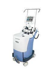
Image courtesy of Terumo BCT
The National Institute for Health and Care Excellence (NICE) has issued a guidance recommending a new device for managing sickle cell disease (SCD).
The device is the Spectra Optia Apheresis System for automated red blood cell (RBC) exchange in patients with SCD who need regular blood transfusions.
The Spectra Optia Apheresis System automatically replaces sickled RBCs with healthy RBCs.
The system is made up of 3 components: an apheresis machine, embedded software, and a single-use, disposable blood tubing set.
NICE said the Spectra Optia system is faster than manual RBC exchange, and patients need RBC exchange less often with this device. In addition, the system could provide considerable savings to the National Health Service (NHS) in England.
“The device could save the NHS in England an estimated £13 million each year—around £18,000 per patient—with the size of the saving depending on the patient’s condition and the equipment already owned by the NHS,” said Carole Longson, director of the NICE Centre for Health Technology Evaluation.
“We also recommend that specialists collaborate to collect and publish data on some outcomes of treatment with Spectra Optia to provide further clinical evidence. It would be particularly helpful to have long-term data on how automated and manual exchange affects the amount of iron in the body and the need to treat this complication.”
Treatment with the Spectra Optia system is intended to be iron-neutral, meaning that patients who are already iron-overloaded can have their condition managed effectively.
The Spectra Optia Apheresis System is manufactured by Terumo BCT.
Costs and savings
The list prices (excluding tax) for the components of the Spectra Optia Apheresis System are as follows:
- Spectra Optia device: £45,350
- RBC exchange software: £6700
- Spectra Optia exchange set: £1007 per 6
- Astotube with injection port: £218 per 50
- ACD-A anticoagulant (750 ml): £57 per 12
- Service charge: £4572 per year.
Bulk order discounts are available.
Based on current evidence and expert advice on the anticipated benefits of the technology when used in patients with iron overload, cost modelling shows that, in most cases, using Spectra Optia is cost-saving compared with manual RBC exchange or top-up transfusion.
The savings depend on the iron overload status of the patient and are more likely to be achieved if devices already owned by the NHS can be used to treat SCD.
The estimated cost saving for adopting Spectra Optia is £18,100 per patient per year, which has the potential to save the NHS £12.9 million each year. ![]()
ISC: Pick up extra AF with extended Holter monitoring
LOS ANGELES – Atrial fibrillation is three times more likely to be detected within 6 months of an ischemic stroke if, instead of the usual 24 or so hours of Holter ECG monitoring, patients are monitored for 10 days poststroke, and then again for 10 days at 3 and 6 months, according to German investigators.
“Enhanced and prolonged monitoring should be considered for all stroke patients to improve the detection of atrial fibrillation,” said investigator Dr. Rolf Wachter, a cardiologist at the University of Göttingen (Germany).
Patients in the trial had no history of atrial fibrillation (AF) and were at least 60 years old; 200 were randomized to the extended-monitoring protocol, and 198 to standard of care, which included a median of 24 hours of Holter monitoring. The median time from symptom onset to randomization was 3 days. All patients were enrolled by day 5.
The study wasn’t limited to cryptogenic strokes; although the first stroke may not have been related to atrial fibrillation, a recurrent stroke could be, so “our approach was to look for AF in all stroke patients irrespective of etiology,” Dr. Wachter said at the International Stroke Conference.
By 6 months, AF was detected in 27 patients (13.5 %) in the extended-monitoring group versus 9 (4.5 %) in the control group (absolute difference, 9%; 95% confidence interval, 3.5%-14.6%; P = .002). The findings were largely the same when results were broken down by age, sex, National Institutes of Health Stroke Scale scores, and other metrics. AF was detected in the majority of both groups within a month.
Dr. Wachter did not elaborate on the frequency and duration of AF, but inclusion criteria required at least one episode lasting 30 seconds or longer.
The work touches on key issues facing neurologists and cardiologists today: How aggressive should post-stroke AF monitoring be, and when should treatment start?
Every AF patient in the study was orally anticoagulated, and almost all remained on their anticoagulant at 1 year. Their oral anticoagulant wasn’t reported, and the study wasn’t powered to detect differences in clinical outcomes. Even so, “we saw very positive trends in the right direction” for prolonged monitoring, he said.
Five patients in the intervention arm (2.5%) had a recurrent stroke and three (1.5%) had transient ischemic events at 1 year, versus nine (4.5%) recurrent strokes and five (2.5%) TIAs in the control arm. Six intervention patients (3%) and nine (4.5%) in the control group, had died.
A total of 60% of the subjects were men, and the mean age in the study was 73 years. The mean NIH Stroke Scale score was 3 in the intervention group and 2 in the control group. The mean CHADS2 (heart failure, hypertension, age, diabetes, stroke) score – a marker of thromboembolic risk – was 3.5 in both. Patients in the extended-monitoring group wore their Holter monitors for a median of 9.5 days at all three time points, but the initial compliance of 100% dropped to about 75% by the third session.
The work was funded by Boehringer Ingelheim, maker of the oral anticoagulant dabigatran (Pradaxa). Dr. Wachter is a speaker and consultant for the company.
There is widespread belief among most stroke neurologists that when you have a stroke that looks embolic and you don’t have a source, you need to monitor for longer than we have done traditionally. The nuances are really how to do that. This is a great approach; it’s a lot easier than wearing a 30-day Holter monitor or having an implanted device, but we have to compare it to other approaches and see what the heart endpoints really are to know if it’s the best approach.
I think everybody in the stroke community appreciates that the more you monitor, the more you are likely to find AF. How long you should monitor, by what technique, and whether monitoring should be continuous or intermittent are unanswered questions. The other question is how much AF is significant. Is a 30-second run significant, or does it need to be minutes at a time? These questions need to be answered before there’s a wholesale buy-in for this kind of monitoring for every patient.
Dr. Kyra Becker is a professor in the departments of neurology and neurological surgery, and director of Vascular Neurology Services at the University of Washington’s Comprehensive Stroke Center in Seattle. She was not involved in the study.
LOS ANGELES – Atrial fibrillation is three times more likely to be detected within 6 months of an ischemic stroke if, instead of the usual 24 or so hours of Holter ECG monitoring, patients are monitored for 10 days poststroke, and then again for 10 days at 3 and 6 months, according to German investigators.
“Enhanced and prolonged monitoring should be considered for all stroke patients to improve the detection of atrial fibrillation,” said investigator Dr. Rolf Wachter, a cardiologist at the University of Göttingen (Germany).
Patients in the trial had no history of atrial fibrillation (AF) and were at least 60 years old; 200 were randomized to the extended-monitoring protocol, and 198 to standard of care, which included a median of 24 hours of Holter monitoring. The median time from symptom onset to randomization was 3 days. All patients were enrolled by day 5.
The study wasn’t limited to cryptogenic strokes; although the first stroke may not have been related to atrial fibrillation, a recurrent stroke could be, so “our approach was to look for AF in all stroke patients irrespective of etiology,” Dr. Wachter said at the International Stroke Conference.
By 6 months, AF was detected in 27 patients (13.5 %) in the extended-monitoring group versus 9 (4.5 %) in the control group (absolute difference, 9%; 95% confidence interval, 3.5%-14.6%; P = .002). The findings were largely the same when results were broken down by age, sex, National Institutes of Health Stroke Scale scores, and other metrics. AF was detected in the majority of both groups within a month.
Dr. Wachter did not elaborate on the frequency and duration of AF, but inclusion criteria required at least one episode lasting 30 seconds or longer.
The work touches on key issues facing neurologists and cardiologists today: How aggressive should post-stroke AF monitoring be, and when should treatment start?
Every AF patient in the study was orally anticoagulated, and almost all remained on their anticoagulant at 1 year. Their oral anticoagulant wasn’t reported, and the study wasn’t powered to detect differences in clinical outcomes. Even so, “we saw very positive trends in the right direction” for prolonged monitoring, he said.
Five patients in the intervention arm (2.5%) had a recurrent stroke and three (1.5%) had transient ischemic events at 1 year, versus nine (4.5%) recurrent strokes and five (2.5%) TIAs in the control arm. Six intervention patients (3%) and nine (4.5%) in the control group, had died.
A total of 60% of the subjects were men, and the mean age in the study was 73 years. The mean NIH Stroke Scale score was 3 in the intervention group and 2 in the control group. The mean CHADS2 (heart failure, hypertension, age, diabetes, stroke) score – a marker of thromboembolic risk – was 3.5 in both. Patients in the extended-monitoring group wore their Holter monitors for a median of 9.5 days at all three time points, but the initial compliance of 100% dropped to about 75% by the third session.
The work was funded by Boehringer Ingelheim, maker of the oral anticoagulant dabigatran (Pradaxa). Dr. Wachter is a speaker and consultant for the company.
AT THE INTERNATIONAL STROKE CONFERENCE
Key clinical point: Repeated Holter monitoring over 6 months catches more atrial fibrillation than one-time, poststroke monitoring.
Major finding: By 6 months, AF was detected in 27 patients (13.5 %) in the extended-monitoring group, versus 9 (4.5 %) in the control group (absolute difference, 9%; 95% confidence interval, 3.5%-14.6%; P = .002).
Data source: Randomized trial with 398 ischemic stroke patients.
Disclosures: The work was funded by Boehringer Ingelheim, maker of the oral anticoagulant dabigatran (Pradaxa). The presenter is a speaker and consultant for the company.

BENEFIT 11: No new safety signals with interferon beta-1b
NEW ORLEANS – The long-term safety of interferon beta-1b for the treatment of multiple sclerosis is well established, and 11-year outcomes from the randomized, placebo-controlled BENEFIT trial (BENEFIT 11) provide further support for the existing data.
Eleven years after patients with clinically isolated syndrome were randomized to receive 250 mcg every other day or placebo (with an option at 2 years to switch to active treatment), no new safety signals were found, and the safety profile remained favorable, Dr. Mark S. Freedman of the University of Ottawa and his colleagues reported in a poster at the meeting sponsored by the Americas Committee for Treatment and Research in Multiple Sclerosis.

In the 278 of the 468 patients originally enrolled in BENEFIT who were evaluated at 11 years, 107 (38.5%) reported one or more safety events (total, 278). Interferon beta-1b was used continuously during the 2 years prior to the 11-year evaluation in 82 patients (29.5%), and 28 (34.1%) of those reported an adverse event or medical history event (total, 72 events). Of 196 patients not treated continuously with interferon beta-1b in the 2 years prior to BENEFIT 11, 79 (40.3%) reported an adverse event or medical history event (total, 206 events), the investigators said.
The events reported in BENEFIT 11 were consistent with those seen previously in interferon beta-1b–treated patients; back pain was the most commonly reported musculoskeletal/connective tissue adverse event (reported by 2.4% of patients in the continuous treatment group and 2.6% in the remaining patients, and headache was the most common neurologic adverse event (reported by 1.2% and 2.0% of patients in the groups, respectively).
Neoplasms occurred in 3.7% and 3.6%; most were benign.
“Overall, the safety results from BENEFIT 11 supported the already well-established long-term safety profile of interferon beta-1b,” the investigators wrote.
The higher rates of some events in patients without continuous interferon beta-1b treatment may be due to escalation to second-line therapy in some patients who required agents that may be associated with greater frequency of safety events, they explained.
This study was supported by Bayer. Dr. Freedman has received compensation from Actelion, Bayer, Biogen Idec, Chugai, EMD Canada, Genzyme, Merck Serono, Novartis, Roche Canada, Sanofi-Aventis, and Teva Canada Innovation for consulting services. He also participates in a Genzyme-sponsored speakers bureau.
NEW ORLEANS – The long-term safety of interferon beta-1b for the treatment of multiple sclerosis is well established, and 11-year outcomes from the randomized, placebo-controlled BENEFIT trial (BENEFIT 11) provide further support for the existing data.
Eleven years after patients with clinically isolated syndrome were randomized to receive 250 mcg every other day or placebo (with an option at 2 years to switch to active treatment), no new safety signals were found, and the safety profile remained favorable, Dr. Mark S. Freedman of the University of Ottawa and his colleagues reported in a poster at the meeting sponsored by the Americas Committee for Treatment and Research in Multiple Sclerosis.
In the 278 of the 468 patients originally enrolled in BENEFIT who were evaluated at 11 years, 107 (38.5%) reported one or more safety events (total, 278). Interferon beta-1b was used continuously during the 2 years prior to the 11-year evaluation in 82 patients (29.5%), and 28 (34.1%) of those reported an adverse event or medical history event (total, 72 events). Of 196 patients not treated continuously with interferon beta-1b in the 2 years prior to BENEFIT 11, 79 (40.3%) reported an adverse event or medical history event (total, 206 events), the investigators said.
The events reported in BENEFIT 11 were consistent with those seen previously in interferon beta-1b–treated patients; back pain was the most commonly reported musculoskeletal/connective tissue adverse event (reported by 2.4% of patients in the continuous treatment group and 2.6% in the remaining patients, and headache was the most common neurologic adverse event (reported by 1.2% and 2.0% of patients in the groups, respectively).
Neoplasms occurred in 3.7% and 3.6%; most were benign.
“Overall, the safety results from BENEFIT 11 supported the already well-established long-term safety profile of interferon beta-1b,” the investigators wrote.
The higher rates of some events in patients without continuous interferon beta-1b treatment may be due to escalation to second-line therapy in some patients who required agents that may be associated with greater frequency of safety events, they explained.
This study was supported by Bayer. Dr. Freedman has received compensation from Actelion, Bayer, Biogen Idec, Chugai, EMD Canada, Genzyme, Merck Serono, Novartis, Roche Canada, Sanofi-Aventis, and Teva Canada Innovation for consulting services. He also participates in a Genzyme-sponsored speakers bureau.
AT ACTRIMS FORUM 2016
Key clinical point: The long-term safety of interferon beta-1b for the treatment of multiple sclerosis is well established, and 11-year outcomes from the randomized, placebo-controlled BENEFIT trial (BENEFIT 11) provide further support for the existing data.
Major finding: 107 of 278 patients reported at least one safety event; no new safety signals were found.
Data source: The 11-year follow-up of 278 patients from the randomized controlled BENEFIT Trial.
Disclosures: This study was supported by Bayer. Dr. Freedman has received compensation from Actelion, Bayer, Biogen Idec, Chugai, EMD Canada, Genzyme, Merck Serono, Novartis, Roche Canada, Sanofi-Aventis, and Teva Canada Innovation for consulting services. He also participates in a Genzyme-sponsored speakers bureau.

Unintended pregnancies fall to 30-year low
The rate of unintended pregnancies in the United States declined 18% between 2008 and 2011 after a long interval of minimal change, and it is now at the lowest level in 30 years, according to a report published online March 2 in the New England Journal of Medicine.
This “substantial” reduction occurred across all ages, ethnicities, income levels, education levels, and religious affiliations. However, large disparities among demographic groups were still present in 2011, the most recent year for which national data are available for analysis.

“In particular, poor, black, and Hispanic women and girls continued to have much higher rates of unintended pregnancy than did whites and those with higher incomes. Much more progress can be made in eliminating these disparities,” wrote Lawrence B. Finer, Ph.D., and Mia R. Zolna of the Guttmacher Institute, New York.
National rates of unintended pregnancies haven’t been examined since 2008. To update these figures, the investigators analyzed information from the National Center for Health Statistics, the National Survey of Family Growth, and others.
They calculated that there were 6.1 million pregnancies in the United States in 2011, of which 45% (2.8 million) were unintended. The rate was 45 unintended pregnancies for every 1,000 women of childbearing age in 2011, compared with 54/1,000 in 2008, which corresponds to an 18% decline (N Engl J Med.2016;374:843-52. doi: 10.1056/NEJMsa1506575).
“This was the first substantial decline since at least 1981,” the investigators wrote.
Abortion rates remained steady during the study period. In 2011, the percentage of unintended pregnancies that ended in abortion was 42%, compared with 40% in 2008.
Although the study was not designed to determine the reasons for the decline, several possible factors deserve consideration, according to the investigators. Changes in sexual behavior are unlikely to have driven this reduction, since the incidence of sexual activity tends to remain static over time; changes in the composition of the population also aren’t likely causes, since the demographic subgroups who have higher rates of unintended pregnancy actually increased during this period, the investigators said.
Changes in the desire for pregnancy may have contributed a small amount to the decline, as surveys showed that many women said they would reduce or delay their childbearing during the recent economic recession.
The most likely explanation for the decline, the investigators wrote, is a change in the frequency and type of contraceptive use. Several studies have reported that women at high risk of unintended pregnancy increased both their use of any contraception and their use of highly effective long-acting methods, particularly IUDs.
This study was supported chiefly by the Susan Thompson Buffet Foundation, with additional support from the Guttmacher Center through a grant from the National Institutes of Health. The investigators reported having no other relevant financial disclosures.
The rate of unintended pregnancies in the United States declined 18% between 2008 and 2011 after a long interval of minimal change, and it is now at the lowest level in 30 years, according to a report published online March 2 in the New England Journal of Medicine.
This “substantial” reduction occurred across all ages, ethnicities, income levels, education levels, and religious affiliations. However, large disparities among demographic groups were still present in 2011, the most recent year for which national data are available for analysis.
“In particular, poor, black, and Hispanic women and girls continued to have much higher rates of unintended pregnancy than did whites and those with higher incomes. Much more progress can be made in eliminating these disparities,” wrote Lawrence B. Finer, Ph.D., and Mia R. Zolna of the Guttmacher Institute, New York.
National rates of unintended pregnancies haven’t been examined since 2008. To update these figures, the investigators analyzed information from the National Center for Health Statistics, the National Survey of Family Growth, and others.
They calculated that there were 6.1 million pregnancies in the United States in 2011, of which 45% (2.8 million) were unintended. The rate was 45 unintended pregnancies for every 1,000 women of childbearing age in 2011, compared with 54/1,000 in 2008, which corresponds to an 18% decline (N Engl J Med.2016;374:843-52. doi: 10.1056/NEJMsa1506575).
“This was the first substantial decline since at least 1981,” the investigators wrote.
Abortion rates remained steady during the study period. In 2011, the percentage of unintended pregnancies that ended in abortion was 42%, compared with 40% in 2008.
Although the study was not designed to determine the reasons for the decline, several possible factors deserve consideration, according to the investigators. Changes in sexual behavior are unlikely to have driven this reduction, since the incidence of sexual activity tends to remain static over time; changes in the composition of the population also aren’t likely causes, since the demographic subgroups who have higher rates of unintended pregnancy actually increased during this period, the investigators said.
Changes in the desire for pregnancy may have contributed a small amount to the decline, as surveys showed that many women said they would reduce or delay their childbearing during the recent economic recession.
The most likely explanation for the decline, the investigators wrote, is a change in the frequency and type of contraceptive use. Several studies have reported that women at high risk of unintended pregnancy increased both their use of any contraception and their use of highly effective long-acting methods, particularly IUDs.
This study was supported chiefly by the Susan Thompson Buffet Foundation, with additional support from the Guttmacher Center through a grant from the National Institutes of Health. The investigators reported having no other relevant financial disclosures.
FROM THE NEW ENGLAND JOURNAL OF MEDICINE
Key clinical point: Unintended pregnancies fell by 18% between 2008 and 2011.
Major finding: The rate of unintended pregnancies was 45 for every 1,000 women of childbearing age in 2011, compared with 54/1,000 in 2008.
Data source: An analysis of data from numerous nationally representative surveys and databases regarding pregnancy intentions and outcomes in 2011.
Disclosures: The study was supported chiefly by the Susan Thompson Buffet Foundation, with additional support from the Guttmacher Center through a grant from the National Institutes of Health. The investigators reported having no other relevant financial disclosures.