User login
Experts in the Elderly
The average young or middle-age person probably finds a hospital stay stressful, uncomfortable, and inconvenient. The experience can be strikingly more disruptive for a geriatric patient.
A frail elderly person can easily succumb to delirium, a fall, dehydration, polypharmacy, and deterioration in basic life skills, quickly turning even a routine hospitalization into a catastrophic downhill slide. But if a patient is lucky, she will be treated by a geriatric hospitalist—a physician who by training and temperament is uniquely suited to care for her.
Geriatric hospitalists bring heightened sensitivity and experience to treating and preventing the common syndromes that can overwhelm the elderly during hospitalizations. Fredrick Sherman, MD, FACP, medical director of Senior Health Partners and professor of geriatrics at the Mount Sinai School of Medicine in Manhattan, has been a geriatrician since the 1980s. He sees a great opportunity for hospitalist geriatricians to improve the metrics by which hospitalist programs are judged: reduced length of stay, bounceback, and morbidity. They do this, he says, with a unique blend of skills, mindset, and temperament.
“In one or two minutes at the bedside, a geriatric hospitalist can do a basic functional assessment of an elderly patient,” he says. “We can understand their ADL [activities of daily living] skills, mental status, and what resources we have to mobilize during the hospital stay and for a safe discharge plan. We don’t want to turn hospitalists into social workers, but geriatricians can teach them a body of knowledge to help them better understand and develop a holistic approach to elderly patients.”
How geriatricians work best, though, can be somewhat out of synch with hospital medicine’s fast pace. “Many hospitalists are younger and have been trained very recently,” Dr. Sherman says. “They quickly learn to take care of a 55-year-old with a [myocardial infarction], but they sometimes lack a global view of geriatric patients that is more a frame of mind than about the physician’s technical skills.”
Challenges Ahead
As hospital medicine groups integrate geriatricians into their ranks, they will have recruited major players invested in improving the care of hospitalized elderly patients. There’s a lot at stake in caring for them.
The Healthcare Cost and Utilization Project’s (HCUP) most recent figures of what hospitalizations of the elderly cost is staggering. Medicare patients account for 76% of public spending on hospital care. The costliest diagnoses for Medicare-paid hospitalizations are coronary arteriosclerosis ($44 billion), acute myocardial infarction ($31 billion), and heart failure ($29 billion). Further, 90% of elderly patients with osteoarthritis are hospitalized for elective hip or knee joint replacement therapy.
The expertise of board-certified hospitalist geriatricians will be hard to disseminate throughout the corps of hospitalists. Only a tiny fraction of the nation’s hospitalist programs claim special expertise in geriatrics. Researchers from the University of Colorado Health Sciences Center and the Mayo Clinic College of Medicine conducted a cross-sectional survey of the hospitalist community in 2003-2004 to determine the impact of the hospitalist movement on acute care geriatrics. They found:
- Out of 1,415 hospitalist programs, 11 reported geriatric innovations.
- Four developed core clinical activities, four used geriatric QI measures, three used comprehensive geriatric assessments, and two had specific protocols for elderly patients discharged to nursing facilities; and
- In terms of staffing, four had hospitalists with no special geriatric training, four employed fellowship-trained geriatricians, two had general hospitalists and geriatricians, and four used advanced practice nurses with and without geriatric training.1
Adding to the difficulty of building a cadre of geriatric hospitalists is the national paucity of geriatricians. According to the American Geriatrics Society (AGS), there were 9,000 board-certified geriatricians in 1998. A decade later there are 7,600—and the pipeline is narrow. Of 9,780 medical school graduates in 2004, only 321 were geriatricians. The AGS estimates that the United States needs 14,000 geriatricians now and 36,000 in 2030, when there will be an estimated 70 million adults 65 years and older.
But there’s hope. Hospital medicine programs, growing by leaps and bounds, offer a new career path for physicians interested in geriatrics. As the number of hospitalists continues to grow, there’s room for physicians to have an impact by staying tuned in to the special clinical, psychosocial, emotional, spiritual, and environmental needs of elderly patients.
The Breed
According to Leslie Libow, MD, distinguished clinical professor at the Jewish Home and Hospital of New York in Manhattan, physicians who pursue a career in geriatrics do so because they have the right psychological make-up to work with elderly people.
He should know. In 1968, Dr. Libow petitioned the American Board of Internal Medicine (ABIM) to recognize geriatrics as a sub-specialty of internal medicine. Shortly after ABIM recognition, Dr. Libow established a geriatric residency/fellowship at Mount Sinai—still a national leader in geriatric education.
Being a geriatric hospitalist allows physicians with a simpatico set of personality traits to thrive. One study of geriatricians who had been practicing for up to 25 years found that they shared these traits:
- Highly value enduring relationships;
- Enjoy making small but potent changes in their patients’ lives;
- Like to make a difference personally and for society;
- Prefer working in a multidisciplinary team;
- Prefer democratic, not autocratic, decision-making;
- Desire the intellectual challenges of geriatric medicine and like to teach; and
- Perceive that they have a distinct and different career path than other physicians.2
That essentially describes Purnima Joshi, chief of medicine, at Kaiser-Permanente Mid-Atlantic States at Washington Hospital Center in Washington, D.C. She directs a group of 12 hospitalists, is the group’s only geriatrician at the 800-bed tertiary care facility, and enjoys teaching residents about geriatric medicine. A family physician by trade, she was grandfathered into geriatrics in 1992 and recertified in 2002.
“I love working with the frail elderly and practicing Kaiser’s brand of medicine because I don’t do billing—I just treat patients,” Dr. Joshi says. Additionally, Kaiser simplifies record-keeping on inpatient and outpatient treatments and makes communicating with Kaiser’s outpatient doctors about post-discharge care smooth and efficient.
Dr. Joshi explains that being a geriatric hospitalist is different than specializing in general internal medicine because she customizes the approach for each patient—including tailoring therapies to life expectancy. “Guidelines and evidence-based medicine are fine and very important, and we use geriatric guidelines on our teaching service,” she says. “But geriatrics liberates your thinking as a doctor. You treat the whole person—a diabetic with three days to live, and a 90-year-old with delirium and decubitus ulcers.”
She calls herself a surrogate primary care physician, seeing 12 patients most days. She consults frequently with other hospitalists on their toughest geriatric issues and makes daily multidisciplinary rounds—with discharge planner, pharmacist, physical therapist, palliative care specialist, nurses, and resident in tow.
“We keep length of stay and guidelines in mind, but the patient is the center of my universe,” Dr. Joshi asserts. “I deliver holistic, patient-centered care and use gentle teaching tools for our residents. I have the luxury of taking time to see the patient and talk to them and their families. It’s wonderful.”
Dr. Joshi’s attitude toward her profession reflects a consistent national finding: Geriatricians rank No. 1 in nearly every study of physician career satisfaction, from the American Medical Association to the American Geriatrics Society.
Across the country, Alpesh Amin, MD, MBA, FACP, professor and chief of general internal medicine and executive director of the University of California at Irvine’s School of Medicine Hospitalist Service, is making the most of the two geriatricians on his 15-hospitalist team. Starting about eight years ago, Dr. Amin—also a member of SHM’s board of directors—turned to his hospitalist geriatricians for a host of services: geriatric assessments, co-management of psychiatric problems, perioperative consults, critical care, and palliative care consults.
“Geriatricians have such knowledge and insight into elderly patients to share with the other hospitalists,” says Dr. Amin. “That’s why they work well side by side with internal medicine and family medicine hospitalists. They keep us aware of issues in geriatrics and the literature on what works best with these patients.”
Knowing that geriatricians are scarce, Dr. Amin accesses their expertise by using a system that focuses team members’ attention on their knowledge. There are journal clubs, frequent consults, monthly meetings, teaching rounds, geriatric fellowships, and other opportunities that keep the geriatrician’s unique perspective front and center for other team members. “They are so in tune with issues related to delirium, polypharmacy, falls risk, etc.,” he says. “Our model incorporates that expertise, and it works very well. We truly work as a multidisciplinary team with ownership and accountability of the special needs of our geriatric patients.”
—Fredrick Sherman, MD, FACP, medical director of Senior Health Partners and professor of geriatrics at the Mount Sinai School of Medicine in Manhattan
A New Generation
The rapid growth of hospital medicine has encouraged new physicians to choose this career path.
Claudene George, MD, recently completed a two-year geriatric fellowship at Mount Sinai Hospital in New York City and is starting as a geriatric hospitalist at Montefiore Hospital in the Bronx. “Becoming a geriatrician sort of surprised me because I thought I’d go into internal medicine,” she says. “But I love the approach to caring for the whole person and communicating with their families.”
As part of her contract at Montefiore, she negotiated a half-day-per-week rotation at the hospital’s outpatient clinic—part of her commitment to being a well-rounded physician.
“The geriatric assessment up front is essential to find out what the patient’s and family’s goals of care are,” says Dr. George. “If they’re 80 years old and want to stay at home, we need to help them do that safely. That may mean linking them to the [visiting nurse service], a home aide, or adult day care.”
She points out that an inpatient stay also offers seniors the opportunity to be seen by subspecialists and do a lot in a short period of time. “As a hospitalist geriatrician you can see change almost immediately; you can have an impact,” she concludes.
As the hospitalist movement affords career opportunities to geriatricians, young physicians can obtain financial incentives to pursue a career in geriatrics. For instance, in 2006 South Carolina enacted a Geriatrician Loan Forgiveness program, helping physicians to repay up to $35,000 of medical school loans if they complete a geriatrics fellowship and practice in South Carolina for five years after completing medical training.
Victor Hirth, MD, medical director for the Division of Geriatric Services of Palmetto Health of Columbia, S.C., has recruited eight geriatric fellows, two of whom will be hospitalists. A recent recruit, Andres Leone, MD, went to medical school in Ecuador, recently completed a geriatric fellowship in South Carolina, and works as a hospitalist half time and at a free clinic for Hispanics half-time. “The flexibility to work as a hospitalist and in an outpatient clinic feels right to me,” says Dr. Leone.
While the number of geriatric hospitalists today is small, some predict their growing presence is inevitable.
“The baby boomers will deluge us, and they will demand so much more of hospitalists in the near future,” Dr. Sherman says. “They will have complicated issues and be very inquisitive geriatric patients.” TH
Marlene Piturro is a frequent contributor to The Hospitalist.
References
- Wald H, Huddleston J, Kramer A. Is there a geriatrician in the house? Geriatric care approaches in hospitalist programs. J Hosp Med. 2006 Jan;1(1):29-35.
- Shah U, Aung M, Chan S, et al. Do geriatricians stay in geriatrics? Gerontol Geriatr Educ. 2006;27(1):57-65.
Diagnose Misdiagnosis
This is the first of a two-part series examining medical errors. This article addresses thought processes hospitalists use that may lead to mistaken diagnoses. Part 2 will look at what healthcare corporations are doing to improve diagnoses and reduce errors.
When talking about tough diagnoses, academic hospitalist David Feinbloom, MD, recalls the story of a female patient seen by his hospitalist group whose diagnosis took some time to nail down.
This woman had been in and out of the hospital for several years with nonspecific abdominal pain and intermittent diarrhea. She had been seen by numerous doctors and tested extensively. Increasingly her doctors concluded that there was some psychiatric overlay—she was depressed or somatic.
“Patients like these are very common and often end up on the hospitalist service,” says Dr. Feinbloom, who works at Beth Israel Deaconess Medical Center in Boston.
But to Joseph Li, MD, director of the hospital medicine program at Beth Israel, this patient seemed normal. There was something about the symptoms she described that reminded him of a patient he had seen who had been diagnosed with a metastatic neuroendocrine tumor.
Although this patient’s past MRI had been negative, Dr. Li remembered that if you don’t perform the right MRI protocol, you’ll miss something. He asked the team to obtain a panel looking for specific markers and to repeat the MRI with the correct protocol. It was accepted as fact that there was no pathology to explain her symptoms but that she had had every test. He requested another gastrointestinal (GI) consult.
“It seemed so far out there, and then everything he said was completely correct,” says Dr. Feinbloom. “She had Zollinger-Ellison syndrome.”
Clues from Sherlock
In his book How Doctors Think, Jerome Groopman, MD, discusses Sir Arthur Conan Doyle, physician and creator of the brilliant detective Sherlock Holmes. When it comes to solving crimes, Holmes’ superior observation and logic, intellectual prowess, and pristine reasoning help him observe and interpret the most obscure and arcane clues. He is, in the end, a consummate diagnostician.
One of the first rules a great diagnostician must follow is to not get boxed into one way of thinking, says Dr. Groopman, the Dina and Raphael Recanati chair of medicine at the Harvard Medical School and chief of experimental medicine at the Beth Israel Deaconess Medical Center, Boston. That is one of the downsides of a too-easy attachment to using clinical practice guidelines, he says.
“Guidelines are valuable reference points, but in order to use a guideline effectively, you have to have the correct diagnosis,” he says. “Studies over decades with hospitalized patients show that the misdiagnosis rate is at least 15% and hasn’t changed.1 A great deal of effort needs to be put into improving our accuracy in making diagnoses.”
Compared with other kinds of medical errors, diagnostic errors have not gotten a great deal of attention. The hospital patient safety movement has been more focused on preventing medication errors, surgical errors, handoff communications, nosocomial infections, falls, and blood clots.2 There have been few studies pertaining exclusively to diagnostic errors—but the topic is gaining headway.3
Think about Thinking
Diagnostic errors are usually multifactorial in origin and typically involve system-related and individual factors. The systems-based piece includes environmental and organizational factors. Medical researchers conclude the majority of diagnostic errors arise from flaws in physician thinking, not technical mistakes.
Cognitive errors involve instances where knowledge, data gathering, data processing, or verification (such as by lab testing) are faulty. Improving diagnostics will require better accountability by institutions and individuals. To do the latter, experts say, physicians would do well to familiarize themselves with their diagnostic weaknesses.
Thinking about thinking is the science of cognitive psychology and addresses the cognitive aspects of clinical reasoning underlying diagnostic decision-making. It is an area of study in which few medical professionals are versed. “Except for a few of these guys who trained in psych or were voices in the wilderness that have been largely ignored,” most physicians are unaware of the cognitive psychology literature, Dr. Groopman says.
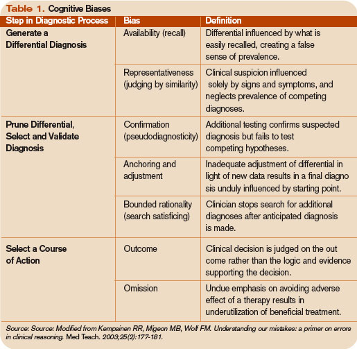
Common biases and errors in clinical reasoning are presented in Table 1 (right).4,5 These are largely individual mistakes for which physicians traditionally have been accountable.

Patterns and Heuristics
The following factors contribute to how shortcuts are used: the pressures of working in medicine, the degrees of uncertainty a physician may feel, and the fact that hospitalists rarely have all the information they need about a patient.
“That’s just the nature of medicine,” says Dr. Groopman. “These shortcuts are natural ways of thinking under those conditions. They succeed about 85% of the time; they fail up to 10-20% of the time. The first thing we need to educate ourselves about is that this is how our minds work as doctors.”
Dr. Groopman and those he interviewed for his book have a razor-sharp overview of clinical practice within hospitals throughout the U.S. and Canada, including academic centers, community centers, affluent areas, suburbs, inner cities, and Native American reservations. But except for Pat Croskerry, MD, PhD, in the department of emergency medicine at Dalhousie University’s Queen Elizabeth II Health Sciences Center in Halifax, Nova Scotia, none of the experts he interviewed had rigorous training in cognitive science.
Although how to think is a priority in physicians’ training, how to think about one’s thinking is not.
“We are not given a vocabulary during medical training, or later through CME courses, in this emerging science—and yet this science involves how our mind works successfully and when we make mistakes,” Dr. Groopman says.
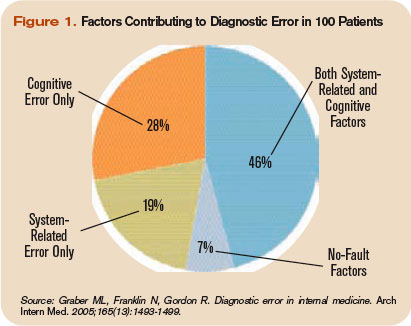
The data back this assertion. In a study of 100 cases of diagnostic error, 90 involved injury, including 33 deaths; 74% were attributed to errors in cognitive reasoning (see Figure 1, right).1 Failure to consider reasonable alternatives after an initial diagnosis was the most common cause. Other common causes included faulty context generation, misjudging the salience of findings, faulty perception, and errors connected with the use of heuristics. In this study, faulty or inadequate knowledge was uncommon.
Underlying contributions to error fell into three categories: “no fault,” system-related, and cognitive. Only seven cases reflected no-fault errors alone. In the remaining 93 cases, 548 errors were identified as system-related or cognitive factors (5.9 per case). System-related factors contributed to the diagnostic error in 65% of the cases and cognitive factors in 74%. The most common system-related factors involved problems with policies and procedures, inefficient processes, teamwork, and communication. The most common cognitive problems involved faulty synthesis.
Dr. Groopman believes it is important for physicians to be more introspective about the thinking patterns they employ and learn the traps to which they are susceptible. He also feels it is imperative to develop curricula at different stages of medical training so this new knowledge can be used to reduce error rates. Because the names for these traps can vary, the development of a universal and comprehensive taxonomy for classifying diagnostic errors is also needed.
“It’s impossible to be perfect; we’re never going to be 100%,” Dr. Groopman says. “But I deeply believe that it is quite feasible to think about your thinking and to assess how your mind came to a conclusion about a diagnosis and treatment plan.”
When phy-sicians think about errors in cognitive reasoning, they often focus on the “don’t-miss diagnoses” or the uncommon variant missed by recall or anchoring errors.
“When I reflect on the errors I have made, they mostly fall into the categories that Dr. Groopman describes in his book,” Dr. Feinbloom says. “Interestingly, the errors that I see most often stem from the fear of making an error of omission.”
It is paradoxical, but in order to ensure that no possible diagnosis is missed, doctors often feel the need to rule out all possible diagnoses.
“While it makes us feel that we are doing the best for our patients, this approach leads to an inordinate amount of unnecessary testing and potentially harmful interventions,” says Dr. Feinbloom. “Understanding how cognitive errors occur should allow us to be judicious in our approach, with the confidence to hold back when the diagnosis is clear, and push harder when we know that something does not fit.”
Emotional Dimension
Although many diagnostic errors are attributable to mistakes in thinking, emotions, and feelings—which may not be easy to detect or admit—also contribute to decision-making.
As hypothesized by noted neurologist and author Antonio Damasio in Descartes’ Error: Emotion, Reason, and the Human Brain, some feelings—visceral signals he calls somatic markers—deter us from or attract us to certain images and decisions.6 Remaining cognizant of those feelings helps clarify how they may inform a medical decision—for good and bad.
The emotional dimension of decision-making cannot be disregarded, says Dr. Groopman. “We need to take our emotional temperature; there are patients we like more and patients we like less,” he says. “There are times when we are tremendously motivated to succeed with a very complicated and daunting patient in the hospital, and there are times when we retreat from that for whatever psychological reason. Sometimes it’s fear of failure, sometimes it’s stereotyping. Regardless, we need to have a level of self-awareness.”
The stressful atmosphere of hospital-based medicine contributes to a high level of anxiety. “Physicians use a telegraphic language full of sound bytes with each other that may contribute to the way heuristics are passed from one generation of doctors to the next,” Dr. Groopman says. “That language is enormously powerful in guiding our thinking and the kinds of shortcuts that we use.”
Pitfalls in Reasoning
Of all the bias errors in clinical reasoning, two of the most influential on physicians are anchoring and attribution. Bound-ed rationality—the failure to continue considering reasonable alternatives after an initial diagnosis is reached—is also a pitfall. The difference between the latter and anchoring is whether the clinician adjusts the diagnosis when new data emerge.
Anchoring errors may arise from seizing the first bits of data and allowing them to guide all future questioning. “It happens every day,” says Dr. Feinbloom. “The diagnosis kind of feels right. There is something about the speed with which it comes to mind, the familiarity with the diagnosis in question, [that] reinforces your confidence.”
Dr. Feinbloom teaches his young trainees to trust no one. “I mean that in a good-hearted way,” he says. “Never assume what you’re told is accurate. You have to review everything yourself, interview the patient again; skepticism is a powerful tool.”
With the woman who was ultimately diagnosed with Zollinger-Ellison syndrome, Dr. Li’s skepticism paid off—and the hospitalist team benefited from deconstructing its clinical thinking to see where it went awry.
“If someone had gotten the gastrin level earlier,” says Dr. Feinbloom, “they would have caught it, but it was not on anyone’s radar. When imaging was negative, the team assumed it wasn’t a tumor.”
Lessons Learned
There are lots of lessons here, says Dr. Feinbloom. “You could spin it any one of five different ways with heuristic lessons, but what jumped out at me was that if you don’t know it, you don’t know it, and you can’t diagnose it,’’ he says. “And that gives you a sense of confidence that you’ve covered everything.”
No one had a familiarity with the subtle manifestations of that diagnosis until Dr. Li stepped in. “One lesson is that if you think the patient is on the up and up, and you haven’t yet made a diagnosis,” says Dr. Feinbloom, “it doesn’t mean there’s no diagnosis to be made.”
Dr. Li gives this lesson to his students this way: You may not have seen diagnosis X, but has diagnosis X seen you?
Andrea Sattinger is a frequent contributor to The Hospitalist.
References
- Graber ML, Franklin N, Gordon R. Diagnostic error in internal medicine. Arch Intern Med. 2005;165(13):1493-1499.
- Wachter RM. Is ambulatory patient safety just like hospital safety, only without the ‘‘stat’’? Ann Intern Med. 2006;145(7):547-549.
- Schiff GD, Kim S, Abrams R, et al. Diagnosing diagnosis errors: lessons from a multi-institutional collaborative project. Rockville, MD: Agency for Healthcare Research and Quality (AHRQ Publication No.050021-2.); 2005.
- Kempainen RR, Migeon MB, Wolf FM. Understanding our mistakes: a primer on errors in clinical reasoning. Med Teach. 2003;25(2):177-181.
- Redelmeier DA. Improving patient care. The cognitive psychology of missed diagnoses. Ann Intern Med. 2005;142(2):115-120.
- Damasio AR. Descartes’ Error: Emotion, Reason, and the Human Brain. New York: GP Putnam’s Sons; 1995.
This is the first of a two-part series examining medical errors. This article addresses thought processes hospitalists use that may lead to mistaken diagnoses. Part 2 will look at what healthcare corporations are doing to improve diagnoses and reduce errors.
When talking about tough diagnoses, academic hospitalist David Feinbloom, MD, recalls the story of a female patient seen by his hospitalist group whose diagnosis took some time to nail down.
This woman had been in and out of the hospital for several years with nonspecific abdominal pain and intermittent diarrhea. She had been seen by numerous doctors and tested extensively. Increasingly her doctors concluded that there was some psychiatric overlay—she was depressed or somatic.
“Patients like these are very common and often end up on the hospitalist service,” says Dr. Feinbloom, who works at Beth Israel Deaconess Medical Center in Boston.
But to Joseph Li, MD, director of the hospital medicine program at Beth Israel, this patient seemed normal. There was something about the symptoms she described that reminded him of a patient he had seen who had been diagnosed with a metastatic neuroendocrine tumor.
Although this patient’s past MRI had been negative, Dr. Li remembered that if you don’t perform the right MRI protocol, you’ll miss something. He asked the team to obtain a panel looking for specific markers and to repeat the MRI with the correct protocol. It was accepted as fact that there was no pathology to explain her symptoms but that she had had every test. He requested another gastrointestinal (GI) consult.
“It seemed so far out there, and then everything he said was completely correct,” says Dr. Feinbloom. “She had Zollinger-Ellison syndrome.”
Clues from Sherlock
In his book How Doctors Think, Jerome Groopman, MD, discusses Sir Arthur Conan Doyle, physician and creator of the brilliant detective Sherlock Holmes. When it comes to solving crimes, Holmes’ superior observation and logic, intellectual prowess, and pristine reasoning help him observe and interpret the most obscure and arcane clues. He is, in the end, a consummate diagnostician.
One of the first rules a great diagnostician must follow is to not get boxed into one way of thinking, says Dr. Groopman, the Dina and Raphael Recanati chair of medicine at the Harvard Medical School and chief of experimental medicine at the Beth Israel Deaconess Medical Center, Boston. That is one of the downsides of a too-easy attachment to using clinical practice guidelines, he says.
“Guidelines are valuable reference points, but in order to use a guideline effectively, you have to have the correct diagnosis,” he says. “Studies over decades with hospitalized patients show that the misdiagnosis rate is at least 15% and hasn’t changed.1 A great deal of effort needs to be put into improving our accuracy in making diagnoses.”
Compared with other kinds of medical errors, diagnostic errors have not gotten a great deal of attention. The hospital patient safety movement has been more focused on preventing medication errors, surgical errors, handoff communications, nosocomial infections, falls, and blood clots.2 There have been few studies pertaining exclusively to diagnostic errors—but the topic is gaining headway.3
Think about Thinking
Diagnostic errors are usually multifactorial in origin and typically involve system-related and individual factors. The systems-based piece includes environmental and organizational factors. Medical researchers conclude the majority of diagnostic errors arise from flaws in physician thinking, not technical mistakes.
Cognitive errors involve instances where knowledge, data gathering, data processing, or verification (such as by lab testing) are faulty. Improving diagnostics will require better accountability by institutions and individuals. To do the latter, experts say, physicians would do well to familiarize themselves with their diagnostic weaknesses.
Thinking about thinking is the science of cognitive psychology and addresses the cognitive aspects of clinical reasoning underlying diagnostic decision-making. It is an area of study in which few medical professionals are versed. “Except for a few of these guys who trained in psych or were voices in the wilderness that have been largely ignored,” most physicians are unaware of the cognitive psychology literature, Dr. Groopman says.
Common biases and errors in clinical reasoning are presented in Table 1 (right).4,5 These are largely individual mistakes for which physicians traditionally have been accountable.
Patterns and Heuristics
The following factors contribute to how shortcuts are used: the pressures of working in medicine, the degrees of uncertainty a physician may feel, and the fact that hospitalists rarely have all the information they need about a patient.
“That’s just the nature of medicine,” says Dr. Groopman. “These shortcuts are natural ways of thinking under those conditions. They succeed about 85% of the time; they fail up to 10-20% of the time. The first thing we need to educate ourselves about is that this is how our minds work as doctors.”
Dr. Groopman and those he interviewed for his book have a razor-sharp overview of clinical practice within hospitals throughout the U.S. and Canada, including academic centers, community centers, affluent areas, suburbs, inner cities, and Native American reservations. But except for Pat Croskerry, MD, PhD, in the department of emergency medicine at Dalhousie University’s Queen Elizabeth II Health Sciences Center in Halifax, Nova Scotia, none of the experts he interviewed had rigorous training in cognitive science.
Although how to think is a priority in physicians’ training, how to think about one’s thinking is not.
“We are not given a vocabulary during medical training, or later through CME courses, in this emerging science—and yet this science involves how our mind works successfully and when we make mistakes,” Dr. Groopman says.
The data back this assertion. In a study of 100 cases of diagnostic error, 90 involved injury, including 33 deaths; 74% were attributed to errors in cognitive reasoning (see Figure 1, right).1 Failure to consider reasonable alternatives after an initial diagnosis was the most common cause. Other common causes included faulty context generation, misjudging the salience of findings, faulty perception, and errors connected with the use of heuristics. In this study, faulty or inadequate knowledge was uncommon.
Underlying contributions to error fell into three categories: “no fault,” system-related, and cognitive. Only seven cases reflected no-fault errors alone. In the remaining 93 cases, 548 errors were identified as system-related or cognitive factors (5.9 per case). System-related factors contributed to the diagnostic error in 65% of the cases and cognitive factors in 74%. The most common system-related factors involved problems with policies and procedures, inefficient processes, teamwork, and communication. The most common cognitive problems involved faulty synthesis.
Dr. Groopman believes it is important for physicians to be more introspective about the thinking patterns they employ and learn the traps to which they are susceptible. He also feels it is imperative to develop curricula at different stages of medical training so this new knowledge can be used to reduce error rates. Because the names for these traps can vary, the development of a universal and comprehensive taxonomy for classifying diagnostic errors is also needed.
“It’s impossible to be perfect; we’re never going to be 100%,” Dr. Groopman says. “But I deeply believe that it is quite feasible to think about your thinking and to assess how your mind came to a conclusion about a diagnosis and treatment plan.”
When phy-sicians think about errors in cognitive reasoning, they often focus on the “don’t-miss diagnoses” or the uncommon variant missed by recall or anchoring errors.
“When I reflect on the errors I have made, they mostly fall into the categories that Dr. Groopman describes in his book,” Dr. Feinbloom says. “Interestingly, the errors that I see most often stem from the fear of making an error of omission.”
It is paradoxical, but in order to ensure that no possible diagnosis is missed, doctors often feel the need to rule out all possible diagnoses.
“While it makes us feel that we are doing the best for our patients, this approach leads to an inordinate amount of unnecessary testing and potentially harmful interventions,” says Dr. Feinbloom. “Understanding how cognitive errors occur should allow us to be judicious in our approach, with the confidence to hold back when the diagnosis is clear, and push harder when we know that something does not fit.”
Emotional Dimension
Although many diagnostic errors are attributable to mistakes in thinking, emotions, and feelings—which may not be easy to detect or admit—also contribute to decision-making.
As hypothesized by noted neurologist and author Antonio Damasio in Descartes’ Error: Emotion, Reason, and the Human Brain, some feelings—visceral signals he calls somatic markers—deter us from or attract us to certain images and decisions.6 Remaining cognizant of those feelings helps clarify how they may inform a medical decision—for good and bad.
The emotional dimension of decision-making cannot be disregarded, says Dr. Groopman. “We need to take our emotional temperature; there are patients we like more and patients we like less,” he says. “There are times when we are tremendously motivated to succeed with a very complicated and daunting patient in the hospital, and there are times when we retreat from that for whatever psychological reason. Sometimes it’s fear of failure, sometimes it’s stereotyping. Regardless, we need to have a level of self-awareness.”
The stressful atmosphere of hospital-based medicine contributes to a high level of anxiety. “Physicians use a telegraphic language full of sound bytes with each other that may contribute to the way heuristics are passed from one generation of doctors to the next,” Dr. Groopman says. “That language is enormously powerful in guiding our thinking and the kinds of shortcuts that we use.”
Pitfalls in Reasoning
Of all the bias errors in clinical reasoning, two of the most influential on physicians are anchoring and attribution. Bound-ed rationality—the failure to continue considering reasonable alternatives after an initial diagnosis is reached—is also a pitfall. The difference between the latter and anchoring is whether the clinician adjusts the diagnosis when new data emerge.
Anchoring errors may arise from seizing the first bits of data and allowing them to guide all future questioning. “It happens every day,” says Dr. Feinbloom. “The diagnosis kind of feels right. There is something about the speed with which it comes to mind, the familiarity with the diagnosis in question, [that] reinforces your confidence.”
Dr. Feinbloom teaches his young trainees to trust no one. “I mean that in a good-hearted way,” he says. “Never assume what you’re told is accurate. You have to review everything yourself, interview the patient again; skepticism is a powerful tool.”
With the woman who was ultimately diagnosed with Zollinger-Ellison syndrome, Dr. Li’s skepticism paid off—and the hospitalist team benefited from deconstructing its clinical thinking to see where it went awry.
“If someone had gotten the gastrin level earlier,” says Dr. Feinbloom, “they would have caught it, but it was not on anyone’s radar. When imaging was negative, the team assumed it wasn’t a tumor.”
Lessons Learned
There are lots of lessons here, says Dr. Feinbloom. “You could spin it any one of five different ways with heuristic lessons, but what jumped out at me was that if you don’t know it, you don’t know it, and you can’t diagnose it,’’ he says. “And that gives you a sense of confidence that you’ve covered everything.”
No one had a familiarity with the subtle manifestations of that diagnosis until Dr. Li stepped in. “One lesson is that if you think the patient is on the up and up, and you haven’t yet made a diagnosis,” says Dr. Feinbloom, “it doesn’t mean there’s no diagnosis to be made.”
Dr. Li gives this lesson to his students this way: You may not have seen diagnosis X, but has diagnosis X seen you?
Andrea Sattinger is a frequent contributor to The Hospitalist.
References
- Graber ML, Franklin N, Gordon R. Diagnostic error in internal medicine. Arch Intern Med. 2005;165(13):1493-1499.
- Wachter RM. Is ambulatory patient safety just like hospital safety, only without the ‘‘stat’’? Ann Intern Med. 2006;145(7):547-549.
- Schiff GD, Kim S, Abrams R, et al. Diagnosing diagnosis errors: lessons from a multi-institutional collaborative project. Rockville, MD: Agency for Healthcare Research and Quality (AHRQ Publication No.050021-2.); 2005.
- Kempainen RR, Migeon MB, Wolf FM. Understanding our mistakes: a primer on errors in clinical reasoning. Med Teach. 2003;25(2):177-181.
- Redelmeier DA. Improving patient care. The cognitive psychology of missed diagnoses. Ann Intern Med. 2005;142(2):115-120.
- Damasio AR. Descartes’ Error: Emotion, Reason, and the Human Brain. New York: GP Putnam’s Sons; 1995.
What is the best medical therapy for the secondary prevention of stroke?
The Case
A 62-year-old obese woman with prior history of type 2 diabetes, hypertension, and a pack-a-day smoking habit presented to the emergency department (ED) for acute onset of right-side weakness and sensory loss noted on awakening from sleep.
She reports taking a baby aspirin daily to “prevent heart attacks” prior to her stroke. Her electrocardiogram demonstrates a left bundle branch block and frequent premature atrial contractions. She recovers with mild hemiparesis and is ready for discharge. What is the best medical therapy for secondary prevention of stroke?
Overview
Cerebrovascular accident (CVA) represents an important diagnosis for the hospitalist, with 700,000 people suffering a stroke in the U.S. each year.1 This translates to a stroke every 45 seconds. About 200,000 of these strokes are recurrent events.
Cardioembolism is the largest cause of ischemic strokes, representing 29% of all infarcts.2 Stasis from impaired contractile function, atrial fibrillation, or mechanical valves are significant risk factors. More rarely, a paradoxical embolus arising in the venous system may pass through a patent foramen ovale.
Large-artery atherosclerosis and lacunar infarcts each account for 16% of strokes. Risk factors for these forms of strokes are the same as those for atherosclerosis and include hypertension and diabetes. Rarer causes such as vasculitis, dissection, hypercoagulability, or hematological disorders account for 3% of strokes. Work-up for these should be driven by historical and atypical features such as young age, family history, or unusual distribution of ischemic zones. Despite appropriate work-up, the mechanism remains uncertain in 36% of strokes.
Regardless of the manifestation and residua of the index event, the hospitalist must initiate appropriate therapy to prevent a disabling CVA. While antithrombotic drugs are the mainstay of secondary prevention, it is a mistake to miss other opportunities for risk modification. Optimal management requires a tailored evaluation for etiology, identification of modifiable risk factors, and initiation of antiplatelet or anticoagulant therapy.
Cardioembolic Stroke
Treatment of stroke depends on the etiology of the original infarct. Evidence is strong that the optimal therapy for cardioembolic stroke is anticoagulation with warfarin.
The European Atrial Fibrillation Trial found that warfarin reduces the risk for second strokes in patients with atrial fibrillation by two-thirds and is superior to antiplatelet agents for preventing cardioembolic strokes.3 Warfarin increases the risk of extracranial bleeding, but not severely enough to negate the benefit of reducing stroke death and disability. The target international normalized ratio (INR) for non-valvular atrial fibrillation is generally two to three, although this may be higher for certain prosthetic valves.
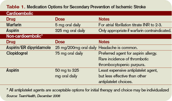
Noncardioembolic Stroke
For large-vessel atherosclerotic and lacunar cerebral ischemia, the oldest—and still effective—treatment for recurrent stroke is aspirin. The use of low-dose aspirin after transient ischemic attack (TIA) or stroke reduces second strokes or death by approximately 15%-18%.4-5 Larger doses do not appear to be more effective, although the rate of gastrointestinal complaints is greater with increased dosage. The use of either 325 mg or 1,200 mg of aspirin produced the same 15% reduction in second ischemic events. Similar efficacy has been seen in comparisons between 30 mg and 283 mg dosing.6
While a subset of patients may experience aspirin resistance, reliable assays in clinical practice are not commonly available to guide management. Current recommendations suggest that use of between 50 mg and 325 mg of aspirin is appropriate for secondary prevention.7
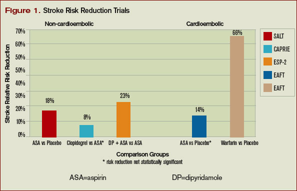
Clopidogrel is another antiplatelet agent that can be given daily at 75 mg as alternate therapy for secondary prevention of non-cardioembolic stroke. The Clopidogrel versus Aspirin in Patients at Risk of Ischemic Events trial comparing clopidogrel with aspirin in patients at risk of ischemic events demonstrated significant reduction in the annual rate of combined endpoint of stroke, myocardial infarction, and vascular death—from 5.83% with aspirin to 5.32% with clopidogrel.8 This study’s applicability to secondary prevention of stroke is limited by the fact that only 19% of the patients in this trial were included because of prior stroke, and the results were not significant for reduction of stroke as a lone endpoint. Clopidogrel is recommended as an acceptable agent for CVA secondary prevention and is preferred for patients with stroke and an aspirin allergy or with recent coronary stent.
The combination of a low-dose aspirin and extended-release dipyridamole has proved superior to aspirin monotherapy in multiple trials. Over two years, the European Stroke Prevention 2 trial found an 18% reduction with aspirin alone compared with 37% reduction with the combination therapy, and the European/Australasian Stroke Prevention in Reversible Ischaemia trial confirmed that the combination reduced the absolute rate of second ischemic events by 1% annually.9-10 Headache is a common side effect of dipyridamole and may limit use. Dypridamole/aspirin is recommended as another acceptable option for secondary prevention of non-cardioembolic stroke.
Evidence suggests that aspirin/dipyridamole and clopidogrel—although significantly more expensive—are more effective than aspirin monotherapy for preventing second cerebral ischemic events. Direct comparison between aspirin/dipyridamole and clopidogrel is ongoing in the Prevention Regimen for Effectively Avoiding Second Stroke trial, with results anticipated in 2008.
Things That Don’t Work
The Warfarin-Aspirin Recurrent Stroke Study trial demonstrated that warfarin was not better than aspirin for prevention of non-cardioembolic stroke, and the Warfarin–Aspirin Symptomatic Intracranial Disease trial found the same result for patients with intracranial stenosis.11-12 There is little evidence that warfarin should have a role in the treatment of most non-cardioembolic strokes. The MATCH trial failed to show benefit to adding aspirin to clopidogrel over clopidogrel monotherapy for secondary preventions of non-cardioembolic cerebral ischemia.13 Despite efficacy following coronary stenting, the combination of clopidogrel and aspirin can not be recommended for stroke prevention.

What To Do
Aggressive risk factor modification is key in the prevention of second ischemic events. One of the most promising therapies is the use of statins following a CVA. Maintaining low-density lipoprotein (LDL) at less than 100 mg/dL (or less than 70 mg/dL in the highest-risk patients) is recommended despite a relatively weak association between stroke and hyperlipidemia.
This stands in contrast to the strong relationship between elevated LDL and coronary disease. However, the Stroke Prevention by Aggressive Reduction in Cholesterol Levels trial utilized high-dose atorvastatin after acute CVA and was able to create an absolute risk reduction for second stroke of 2.2% over the next five years.14 It is possible that the findings of this trial may reflect actions of statin therapy on the endothelium independent of the lipid lowering effect.
Blood pressure commonly has a transient elevation following cerebral ischemia. This is managed permissively to preserve perfusion to the ischemic penumbra. Once the hyperacute period is over, reduction of blood pressure to less than 140/90 mm/Hg (130/80 mm/Hg for diabetics) is recommended.
Interventions to treat chronic hypertension have been demonstrated to reduce the rate of strokes by approximately 30% to 40% over four to five years.15-16 An optimal agent has not been determined, but therapy with angiotensin converting enzyme inhibitor (ACE-I) or angiotensin II receptor blocker (ARB), possibly in combination with a diuretic, have been effective. Close follow-up for titration to goal in the outpatient setting should be arranged. Diabetics should have optimization of glycemic control, and lifestyle counseling should occur regarding recognized risk factors for stroke such as smoking, inactivity, and alcohol abuse.
While antithrombotic therapy is the mainstay of what we think of in secondary prevention of stroke, treatment of these other modifiable risk factors have been shown to affect mortality and second strokes of a similar magnitude and should not be neglected.
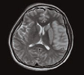
How to Treat This Case
The patient described should undergo an MRI with diffusion (to define the area of ischemia) and targeted evaluation for etiology with cardiac monitoring, echocardiogram, and carotid ultrasound.
Assuming atrial fibrillation or intracardiac thrombus is ruled out, this likely represents atherosclerotic disease. MRI will help distinguish between large-vessel atherosclerotic etiology and lacunar infarct. If carotid stenosis of greater than 70% is found in the setting of large vessel atherosclerotic stroke, then she should be referred for carotid endarterectomy. At 50% to 69% stenosis, carotid endarterectomy would still be a consideration. Antithrombotic agent of choice for non-cardioembolic CVA is an anti-platelet agent. With a stroke occurring on a reasonable dose of aspirin, I would not recommend increasing the dose as there is little evidence that 325 mg is more effective than 81mg. The most appropriate step would be to change to an alternate anti-platelet agent such as combination dipyridamole/aspirin or clopidogrel.
In the absence of a direct comparison trial, either choice is acceptable. The evidence supporting dipyridamole/aspirin is stronger for secondary stroke prevention. Atorvastatin 80 mg daily is an evidence-based therapy after acute stroke and can be started immediately. Her hypertension should be managed permissively for the first few days after the acute event, but then an ACE-I or ARB—possibly in combination with a diuretic—would be appropriate. This patient’s goal blood pressure as a diabetic would be at least less than 130/80 mm/Hg.
Finally we would be remiss if we did not stress the importance of smoking cessation, exercise, and weight loss. TH
Dr. Cumbler is an assistant professor in the Section of Hospital Medicine at the University of Colorado, where he is a member of the Acute Stroke Service and serves on the Stroke Council.
References
- Rosamond W, Flegal K, Friday G, et al. Heart disease and stroke statistics-2007 update: a report from the American Heart Association Statistics Committee and Stroke Statistics Subcommittee. Circulation. 2007 Feb 6;115(5):e69-e171.
- Petty GW, Brown RD, Whisnant JP, et al. Ischemic stroke subtypes: a population-based study of incidence and risk factors. Stroke. 1999;30:2513-2516.
- European Atrial Fibrillation Trial Study Group. Secondary prevention in non-rheumatic atrial fibrillation after transient ischaemic attack or minor stroke. Lancet. 1993;342:1255-1262.
- Swedish Aspirin Low-Dose Trial Collaborative Group. Swedish aspirin low-dose aspirin trial (SALT) of 775 mg aspirin as secondary prophylaxis after cerebrovascular ischaemic events. Lancet. 1991;338(8779):1345-1349.
- Farrell B, Godwin J, Richards S, et al. The United Kingdom Transient Ischaemic Attack (UK-TIA) aspirin trial: final results (abstract). J Neurol Neurosurg. Psychiatry 1991;54:1044-1054.
- Dutch TIA Trial Study Group. A comparison of two doses of aspirin (30 mg versus 283 mg a day) in patients after a transient ischemic attack or minor ischemic stroke. N Engl J Med. 1991 Oct 31;325(18):1261-1266.
- Sacco RL, Adams R, Albers G, et al. Guidelines for prevention of stroke in patients with ischemic stroke or transient ischemic attack. Stroke. 2006 Feb;37(2):577-617.
- CAPRIE Steering Committee. A randomized, blinded, trial of clopidogrel versus aspirin in patients at risk of ischaemic events. Lancet 1996 Jan;348:1329-1339.
- Diener H, Cunha L, Forbes C, et al. European stroke prevention study 2: dipyridamole and acetylsalicylic acid in the secondary prevention of stroke. J Neurol Sci. 1996;143:1-13.
- ESPRIT Study Group. Aspirin plus dipyridamole versus aspirin alone after cerebral ischaemia of arterial origin (ESPRIT): randomised controlled trial. Lancet. 2006;367:1665-1673.
- Mohr JP, Thompson JLP, Lazar RM, et al. A comparison of warfarin and aspirin for the prevention of recurrent ischemic stroke. N Engl J Med. 2001 Nov 15; 345(20):1444-1451.
- Chimowitz MI, Lynn MJ, Howlett-Smith H, et al. Comparison of warfarin and aspirin for symptomatic intracranial arterial stenosis. N Engl J Med. 2005 Mar 31;352(13):1305-1316.
- Diener HC, Bogousslavsky J, Brass LM, et al. Aspirin and clopidogrel compared with clopidogrel alone after recent ischaemic stroke or transient ischaemic attack in high-risk patients (MATCH): randomised, double-blind, placebo controlled trial. Lancet. 2004 Jul 24-30;36499431):331-337.
- Stroke Prevention by Aggressive Reduction in Cholesterol Levels Investigators. High-dose atorvastatin after stroke or transient ischemic attack. N Engl J Med. 2006;355(6):549-559.
- The PROGRESS Collaborative Group. Randomised trial of a perindopril-based blood-pressure-lowering regimen among 6,105 individuals with previous stroke or transient ischaemic attack. Lancet 2001;358(9287):1033-1041.
- The Heart Outcomes Prevention Evaluation Study Investigators. Effects of an angiotensin-converting-enzyme inhibitor, ramipril, on cardiovascular events in high-risk patients. N Engl J Med. 2000 Jan 20;342:145-153.
The Case
A 62-year-old obese woman with prior history of type 2 diabetes, hypertension, and a pack-a-day smoking habit presented to the emergency department (ED) for acute onset of right-side weakness and sensory loss noted on awakening from sleep.
She reports taking a baby aspirin daily to “prevent heart attacks” prior to her stroke. Her electrocardiogram demonstrates a left bundle branch block and frequent premature atrial contractions. She recovers with mild hemiparesis and is ready for discharge. What is the best medical therapy for secondary prevention of stroke?
Overview
Cerebrovascular accident (CVA) represents an important diagnosis for the hospitalist, with 700,000 people suffering a stroke in the U.S. each year.1 This translates to a stroke every 45 seconds. About 200,000 of these strokes are recurrent events.
Cardioembolism is the largest cause of ischemic strokes, representing 29% of all infarcts.2 Stasis from impaired contractile function, atrial fibrillation, or mechanical valves are significant risk factors. More rarely, a paradoxical embolus arising in the venous system may pass through a patent foramen ovale.
Large-artery atherosclerosis and lacunar infarcts each account for 16% of strokes. Risk factors for these forms of strokes are the same as those for atherosclerosis and include hypertension and diabetes. Rarer causes such as vasculitis, dissection, hypercoagulability, or hematological disorders account for 3% of strokes. Work-up for these should be driven by historical and atypical features such as young age, family history, or unusual distribution of ischemic zones. Despite appropriate work-up, the mechanism remains uncertain in 36% of strokes.
Regardless of the manifestation and residua of the index event, the hospitalist must initiate appropriate therapy to prevent a disabling CVA. While antithrombotic drugs are the mainstay of secondary prevention, it is a mistake to miss other opportunities for risk modification. Optimal management requires a tailored evaluation for etiology, identification of modifiable risk factors, and initiation of antiplatelet or anticoagulant therapy.
Cardioembolic Stroke
Treatment of stroke depends on the etiology of the original infarct. Evidence is strong that the optimal therapy for cardioembolic stroke is anticoagulation with warfarin.
The European Atrial Fibrillation Trial found that warfarin reduces the risk for second strokes in patients with atrial fibrillation by two-thirds and is superior to antiplatelet agents for preventing cardioembolic strokes.3 Warfarin increases the risk of extracranial bleeding, but not severely enough to negate the benefit of reducing stroke death and disability. The target international normalized ratio (INR) for non-valvular atrial fibrillation is generally two to three, although this may be higher for certain prosthetic valves.
Noncardioembolic Stroke
For large-vessel atherosclerotic and lacunar cerebral ischemia, the oldest—and still effective—treatment for recurrent stroke is aspirin. The use of low-dose aspirin after transient ischemic attack (TIA) or stroke reduces second strokes or death by approximately 15%-18%.4-5 Larger doses do not appear to be more effective, although the rate of gastrointestinal complaints is greater with increased dosage. The use of either 325 mg or 1,200 mg of aspirin produced the same 15% reduction in second ischemic events. Similar efficacy has been seen in comparisons between 30 mg and 283 mg dosing.6
While a subset of patients may experience aspirin resistance, reliable assays in clinical practice are not commonly available to guide management. Current recommendations suggest that use of between 50 mg and 325 mg of aspirin is appropriate for secondary prevention.7
Clopidogrel is another antiplatelet agent that can be given daily at 75 mg as alternate therapy for secondary prevention of non-cardioembolic stroke. The Clopidogrel versus Aspirin in Patients at Risk of Ischemic Events trial comparing clopidogrel with aspirin in patients at risk of ischemic events demonstrated significant reduction in the annual rate of combined endpoint of stroke, myocardial infarction, and vascular death—from 5.83% with aspirin to 5.32% with clopidogrel.8 This study’s applicability to secondary prevention of stroke is limited by the fact that only 19% of the patients in this trial were included because of prior stroke, and the results were not significant for reduction of stroke as a lone endpoint. Clopidogrel is recommended as an acceptable agent for CVA secondary prevention and is preferred for patients with stroke and an aspirin allergy or with recent coronary stent.
The combination of a low-dose aspirin and extended-release dipyridamole has proved superior to aspirin monotherapy in multiple trials. Over two years, the European Stroke Prevention 2 trial found an 18% reduction with aspirin alone compared with 37% reduction with the combination therapy, and the European/Australasian Stroke Prevention in Reversible Ischaemia trial confirmed that the combination reduced the absolute rate of second ischemic events by 1% annually.9-10 Headache is a common side effect of dipyridamole and may limit use. Dypridamole/aspirin is recommended as another acceptable option for secondary prevention of non-cardioembolic stroke.
Evidence suggests that aspirin/dipyridamole and clopidogrel—although significantly more expensive—are more effective than aspirin monotherapy for preventing second cerebral ischemic events. Direct comparison between aspirin/dipyridamole and clopidogrel is ongoing in the Prevention Regimen for Effectively Avoiding Second Stroke trial, with results anticipated in 2008.
Things That Don’t Work
The Warfarin-Aspirin Recurrent Stroke Study trial demonstrated that warfarin was not better than aspirin for prevention of non-cardioembolic stroke, and the Warfarin–Aspirin Symptomatic Intracranial Disease trial found the same result for patients with intracranial stenosis.11-12 There is little evidence that warfarin should have a role in the treatment of most non-cardioembolic strokes. The MATCH trial failed to show benefit to adding aspirin to clopidogrel over clopidogrel monotherapy for secondary preventions of non-cardioembolic cerebral ischemia.13 Despite efficacy following coronary stenting, the combination of clopidogrel and aspirin can not be recommended for stroke prevention.
What To Do
Aggressive risk factor modification is key in the prevention of second ischemic events. One of the most promising therapies is the use of statins following a CVA. Maintaining low-density lipoprotein (LDL) at less than 100 mg/dL (or less than 70 mg/dL in the highest-risk patients) is recommended despite a relatively weak association between stroke and hyperlipidemia.
This stands in contrast to the strong relationship between elevated LDL and coronary disease. However, the Stroke Prevention by Aggressive Reduction in Cholesterol Levels trial utilized high-dose atorvastatin after acute CVA and was able to create an absolute risk reduction for second stroke of 2.2% over the next five years.14 It is possible that the findings of this trial may reflect actions of statin therapy on the endothelium independent of the lipid lowering effect.
Blood pressure commonly has a transient elevation following cerebral ischemia. This is managed permissively to preserve perfusion to the ischemic penumbra. Once the hyperacute period is over, reduction of blood pressure to less than 140/90 mm/Hg (130/80 mm/Hg for diabetics) is recommended.
Interventions to treat chronic hypertension have been demonstrated to reduce the rate of strokes by approximately 30% to 40% over four to five years.15-16 An optimal agent has not been determined, but therapy with angiotensin converting enzyme inhibitor (ACE-I) or angiotensin II receptor blocker (ARB), possibly in combination with a diuretic, have been effective. Close follow-up for titration to goal in the outpatient setting should be arranged. Diabetics should have optimization of glycemic control, and lifestyle counseling should occur regarding recognized risk factors for stroke such as smoking, inactivity, and alcohol abuse.
While antithrombotic therapy is the mainstay of what we think of in secondary prevention of stroke, treatment of these other modifiable risk factors have been shown to affect mortality and second strokes of a similar magnitude and should not be neglected.
How to Treat This Case
The patient described should undergo an MRI with diffusion (to define the area of ischemia) and targeted evaluation for etiology with cardiac monitoring, echocardiogram, and carotid ultrasound.
Assuming atrial fibrillation or intracardiac thrombus is ruled out, this likely represents atherosclerotic disease. MRI will help distinguish between large-vessel atherosclerotic etiology and lacunar infarct. If carotid stenosis of greater than 70% is found in the setting of large vessel atherosclerotic stroke, then she should be referred for carotid endarterectomy. At 50% to 69% stenosis, carotid endarterectomy would still be a consideration. Antithrombotic agent of choice for non-cardioembolic CVA is an anti-platelet agent. With a stroke occurring on a reasonable dose of aspirin, I would not recommend increasing the dose as there is little evidence that 325 mg is more effective than 81mg. The most appropriate step would be to change to an alternate anti-platelet agent such as combination dipyridamole/aspirin or clopidogrel.
In the absence of a direct comparison trial, either choice is acceptable. The evidence supporting dipyridamole/aspirin is stronger for secondary stroke prevention. Atorvastatin 80 mg daily is an evidence-based therapy after acute stroke and can be started immediately. Her hypertension should be managed permissively for the first few days after the acute event, but then an ACE-I or ARB—possibly in combination with a diuretic—would be appropriate. This patient’s goal blood pressure as a diabetic would be at least less than 130/80 mm/Hg.
Finally we would be remiss if we did not stress the importance of smoking cessation, exercise, and weight loss. TH
Dr. Cumbler is an assistant professor in the Section of Hospital Medicine at the University of Colorado, where he is a member of the Acute Stroke Service and serves on the Stroke Council.
References
- Rosamond W, Flegal K, Friday G, et al. Heart disease and stroke statistics-2007 update: a report from the American Heart Association Statistics Committee and Stroke Statistics Subcommittee. Circulation. 2007 Feb 6;115(5):e69-e171.
- Petty GW, Brown RD, Whisnant JP, et al. Ischemic stroke subtypes: a population-based study of incidence and risk factors. Stroke. 1999;30:2513-2516.
- European Atrial Fibrillation Trial Study Group. Secondary prevention in non-rheumatic atrial fibrillation after transient ischaemic attack or minor stroke. Lancet. 1993;342:1255-1262.
- Swedish Aspirin Low-Dose Trial Collaborative Group. Swedish aspirin low-dose aspirin trial (SALT) of 775 mg aspirin as secondary prophylaxis after cerebrovascular ischaemic events. Lancet. 1991;338(8779):1345-1349.
- Farrell B, Godwin J, Richards S, et al. The United Kingdom Transient Ischaemic Attack (UK-TIA) aspirin trial: final results (abstract). J Neurol Neurosurg. Psychiatry 1991;54:1044-1054.
- Dutch TIA Trial Study Group. A comparison of two doses of aspirin (30 mg versus 283 mg a day) in patients after a transient ischemic attack or minor ischemic stroke. N Engl J Med. 1991 Oct 31;325(18):1261-1266.
- Sacco RL, Adams R, Albers G, et al. Guidelines for prevention of stroke in patients with ischemic stroke or transient ischemic attack. Stroke. 2006 Feb;37(2):577-617.
- CAPRIE Steering Committee. A randomized, blinded, trial of clopidogrel versus aspirin in patients at risk of ischaemic events. Lancet 1996 Jan;348:1329-1339.
- Diener H, Cunha L, Forbes C, et al. European stroke prevention study 2: dipyridamole and acetylsalicylic acid in the secondary prevention of stroke. J Neurol Sci. 1996;143:1-13.
- ESPRIT Study Group. Aspirin plus dipyridamole versus aspirin alone after cerebral ischaemia of arterial origin (ESPRIT): randomised controlled trial. Lancet. 2006;367:1665-1673.
- Mohr JP, Thompson JLP, Lazar RM, et al. A comparison of warfarin and aspirin for the prevention of recurrent ischemic stroke. N Engl J Med. 2001 Nov 15; 345(20):1444-1451.
- Chimowitz MI, Lynn MJ, Howlett-Smith H, et al. Comparison of warfarin and aspirin for symptomatic intracranial arterial stenosis. N Engl J Med. 2005 Mar 31;352(13):1305-1316.
- Diener HC, Bogousslavsky J, Brass LM, et al. Aspirin and clopidogrel compared with clopidogrel alone after recent ischaemic stroke or transient ischaemic attack in high-risk patients (MATCH): randomised, double-blind, placebo controlled trial. Lancet. 2004 Jul 24-30;36499431):331-337.
- Stroke Prevention by Aggressive Reduction in Cholesterol Levels Investigators. High-dose atorvastatin after stroke or transient ischemic attack. N Engl J Med. 2006;355(6):549-559.
- The PROGRESS Collaborative Group. Randomised trial of a perindopril-based blood-pressure-lowering regimen among 6,105 individuals with previous stroke or transient ischaemic attack. Lancet 2001;358(9287):1033-1041.
- The Heart Outcomes Prevention Evaluation Study Investigators. Effects of an angiotensin-converting-enzyme inhibitor, ramipril, on cardiovascular events in high-risk patients. N Engl J Med. 2000 Jan 20;342:145-153.
Transfer Training
When attendings at Denver Health Medical Center (DHMC) were asked to be available to help supervise the teams of residents and interns with the hand-off process, Eugene Chu, MD, director of the hospital medicine program, quickly knew there was a problem.
“They didn’t really know what they were teaching,” says Dr. Chu. “They had an idea of how to do a hand-off, but they had never explicitly learned what a good hand-off was because it had never been described before. Some of our attendings fell back on what they did best—teach medicine. But that was not necessarily what the house staff wanted at that time of day.”
The house staff did want to learn to give safe, effective, and efficient hand-offs. “Giving a lecture on renal failure was not really the point of the hand-off,” says Dr. Chu.
Time for Training
“Sign outs serve a lot of purposes, not just information [transfer],” says Leora Horwitz, MD, an assistant professor in the division of general internal medicine at Yale School of Medicine, New Haven, Conn. “Signout is also a time for training. It is a time for socialization in terms of how we talk about patients and what is expected. And it is a time for catching errors and for rethinking plans and diagnoses because as you are describing something to someone, they might pick up on gaps or inconsistencies or things that should be done differently.”
Dr. Horwitz, who is also associate medical director of the Center for Outcomes Research and Evaluation at Yale-New Haven Hospital, has researched training for transfer of care over the past three years.1
“The most important thing is that hospitalists should not assume that residents have any skills [pertaining to transfer of care],” says Dr. Horwitz. “In medical schools, students are taught over and over how to present a patient for the first time. There’s a rigid order in which the information is supposed to flow, and there is a rigid list of categories of information that should be conveyed. People are taught that same order and that same flow and that same list of categories at all med schools. Consequently, as residents, everybody has the same sense of how to represent an initial history and physical. There is no such thing for hand-offs.”
Resident duty-hour limitations have increased the number of hand-offs, which creates greater risks for discontinuity of care and patient safety.2, 3 “Hand-offs occur two or three times a day and a patient presentation only occurs once—when the patient shows up,” says Dr. Horwitz. On top of that, when residents appear in their clinical duties, the attendings tend to forget residents don’t have the skills to execute a comprehensive and well-communicated hand-off. “The first thing to remember is that people need to be trained,” she says.
In a study in the Archives of Internal Medicine in 2006 (for which Dr. Horwitz is first author), the investigators asked internal medicine chief residents whether their program provides direct training in how to perform sign outs. Sign-out training varied considerably, and fewer than half the 202 programs that responded (62% of all U.S. residency programs) provided formal sign-out skill training: 40% of the programs taught sign-out skills through a lecture or workshop, 45% supervised oral sign outs, and 38% reviewed written sign outs.4 Residents in 27% of the programs received no training or supervision. Further, in more than one-third of training programs they found hand-offs were left to interns. “Residency programs need to recognize the problem and address it in some way,” Dr. Horwitz says.
Be Explicit, Create a Model
“Many residency programs have a standardized form that residents use to sign out to the cross-covering physician,” says Sunil Kripalani, MD, MSc, director of the Section of Hospital Medicine at Vanderbilt University in Nashville, Tenn. “However, there is often not much attention given to the actual process of transferring patient information to another physician.”
For example, residents may tack the form up to a wall or leave it on a computer, he says, because this may be more convenient than meeting for a verbal, face-to-face sign out. “It is important that residents receive training about how to best sign out patients, so it is viewed as a priority area,” he says.
The initial training should cover best practices for hand-offs, says Dr. Kripalani. “It may not be intuitive, especially to new residents, that poorly executed hand-offs can be perilous,” he says.
It is also important to teach trainees how to best convey that information. “Sometimes you’ll think more is better,” says Dr. Horwitz, “but that’s not the case; people turn off or get distracted. There is a tension between providing enough information to take care of the person overnight versus providing too much information.”
Modeling best behaviors is also an important part of training, says Jay Routson, MD, a teaching hospitalist and clinical assistant professor of medicine in the Idaho State University Department of Family Medicine in Pocatello, Idaho. Dr. Routson, trained in internal medicine, thinks opportunities to train residents and students in transfer of care are also a chance to model what you expect of them.
This is particularly important in Dr. Routson’s circumstances because of the nature of his university’s family medicine residency: It is conducted at a number of locations. At morning report on the first day of a block, residents who have been on the previous rotation are to transfer patient care to the incoming residents. But they may have already left for their new pediatrics or NICU assignments, for instance, not only elsewhere in Pocatello but perhaps in Boise or Logan.
Another problem in his program’s training for transfer of care, says Dr. Routson, is that less-experienced residents are not always aware of the important things to check. What one resident thinks is important to follow up on the next resident may put at the bottom of his or her list.
“I think that you have to model the importance you place on [hand-offs],” says Dr. Routson. “You have to set aside time during the day and make it a priority. Model the behavior when you’re checking out to a new attending, make sure the residents and interns know it’s a priority, especially early in the academic year.”
Supervision and Feedback
Supervised evaluation of performance and feedback are key aspects of training for transfer of care.
“Training residents means supervising them,” says Dr. Horwitz. “Are they getting the concepts? Are they incorporating the key points into everyday communication and actions?” Having more senior and experienced doctors present during sign out or at the very least prepare people for sign out is crucial, she says, because an issue such as anticipatory guidance/contingency planning is difficult for less-experienced residents.
“As a chief resident I made a point of periodically going around and observing sign outs,” she says. “Nobody ever watches sign out because it happens at the odd hours of the day; nobody gets feedback or evaluation. Ideally an attending should, at least now and again, be involved.”
Questions to ask include:
- Do the residents meet face to face in a dedicated time and place?
- Do they fill out a standardized template and updating it appropriately, especially with regard to medications?
- Do residents measure aspects of hand-offs to ensure they are being done?
Two-Way Street
Just as the DHMC team recognized that communication for hand-offs is a two-way street, the same is true for performing a discharge communication exchange. Are primary care providers considering best practices to train themselves in this integral aspect of patient care and safety?5, 6
“I don’t think the primary care community has gotten together and come up with a consensus of what they want to know and how much interaction they want,” Dr. Horwitz says. “There should be a standard for that.”
That is a first step to encourage greater training all around. She urges collaboration between SHM and a national organization, such as the American Academy on Family Practice, and hopes SHM will open that dialogue.
In that regard, some steps have been taken.7 Dr. Kripalani is a member of the SHM Hand-off and Communication Standards Task Force, which is developing national standards (The Hospitalist, August 2007, p. 17). There is a working list of nine best practices for hand-offs at shift changes or rotation ends, and the group plans to publish a final list in the upcoming year. The list is designed primarily for practicing hospitalists but will also be suitable for residents. The task force hopes other groups, such as internal medicine program directors and chief residents, will disseminate the best practices to trainees. National best practices are also likely to affect providers in the community.
“My suspicion is that a lot of community-based programs are interested in improving hand-offs, but each program may develop a different set of procedures,” says Dr. Kripalani. “One of the main reasons for developing national standards is so that both academic and community groups can refer to a list of evidence-based best practices.”
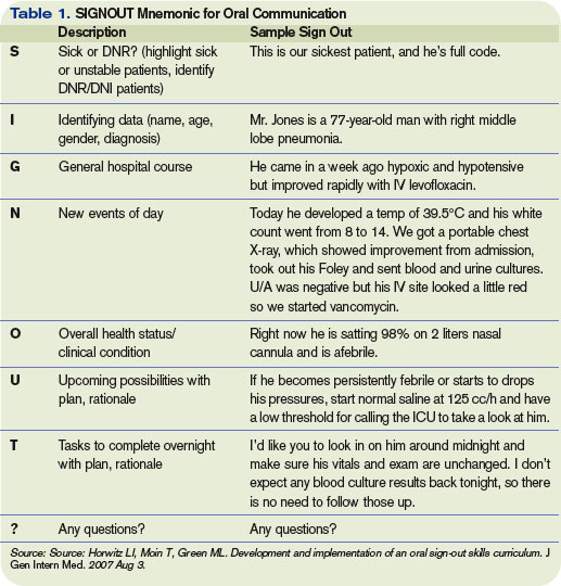
Evolution of Training
“Training for transfer of care and transfer of care are very different things, says Dr. Chu, “but they’re interrelated.” He and his colleague Gregory Misky, MD, a hospitalist and instructor with the University of Colorado Health Sciences Center (UCHSC), have been disseminating that training program throughout the UCHSC internal medicine residency program for the past three years.
In order to develop a structured, standard method for patient hand-offs, they took on teaching and supervising interns during their internal medicine ward rotations. Although much of the literature advocates the SBAR (situation, background, assessment, and recommendation) communication technique, the UCHSC team has developed a verbal structure specifically for hand-offs that differs from the SBAR model.8
Having learned many lessons their first year of training the trainers and the trainees, the Colorado hospitalists regrouped the next year. Because there were two interns coming on duty each evening, both taking cross cover, one attending could supervise only one of the interns. And, those hospitalists were getting called away to handle their clinical responsibilities.
“We felt it was important that attendings were available to guide the process safely,” says Dr. Chu. Therefore, because the evening hand-offs were conducted at 6 p.m. and a hospitalist was in house daily at that time, they began having the evening person, who was on each night until about 11, supervise the interns.
To improve the quality and consistency of the teaching, only four of the Division of Hospital Medicine’s eight attendings focused on supervising intern hand-offs. Also, in a series of focus-group meetings with attendings and house staff, they discussed supervision and identified several structures and standards for teaching hand-offs.
In addition to determining a consistent time and place for hand-off exchanges, they developed a consistent written template and a standard for verbal communication that provides an order of thinking and presenting, just as they use for presenting patient history and physical or daily patient progress updates. “The written template and the verbal are not necessarily the same thing,” says Dr. Chu. “We distinguished them as being two separate standards, just as they are with the written and verbal of morning rounds.”
With these protocols in place, attendings now had distinctions to watch for and a clear-cut means by which to supervise. In the third year of training at UCHSC, the hospitalist attendings provided training and feedback on the first night of call for the first four months beginning in July and in small-group sessions during the first week of the ward month.
The Reviews Are In
In response to a survey, the vast majority of UCHSC internal medicine residents said the new protocols were useful or extremely useful. Responses also revealed that training increased interns’ self-perceived hand-off skills and knowledge. The common denominator of a same time and place for hand-offs was judged the most useful element of the program; lectures were considered the least useful element.
Word got around of the program’s success. Eva Aagaard, MD, associate chair for education in the department of medicine, approached the originators to incorporate the program into the medical school’s interclinical curriculum, given to students between the third and fourth years as additional skills they would need in their sub-internships. Originally offered as an elective, demand for the course exceeded the class limit. Plans are under way to make this course mandatory.
“This past year, for the first time, we specifically taught our medical students hand-offs,” says Dr. Chu. He and his colleagues presented an abstract at SHM’s annual meeting in Dallas and are preparing a manuscript for publication.
Until now, all physicians have managed to do hand-offs without formal training. “Most residents figure out their own way of doing things after a while, but what we’re trying to do is not let it be a random evolution of learning,” says Dr. Chu. “It’s like learning to drive or ski with no lesson s: If you point your skis downhill, eventually you’ll learn how to ski, but you’ll have a lot of crashes. In hand-offs, that means communication failures, and a patient may suffer morbidity. We want to train and supervise [residents] so they learn in a progressive fashion and have fewer crashes.” TH
Andrea Sattinger is a frequent contributor to The Hospitalist.
References
- Horwitz LI, Moin T, Green ML. Development and implementation of an oral sign-out skills curriculum. J Gen Intern Med. 2007 Aug 3.
- Kripalani S, LeFevre F, Phillips CO, et al. Deficits in communication and information transfer between hospital-based and primary care physicians: implications for patient safety and continuity of care. JAMA. 2007;297(8):831-841.
- Vidyarthi AR, Arora V, Schnipper JL, et al. Managing discontinuity in academic medical centers: strategies for a safe and effective resident sign-out. J Hosp Med. 2006;1(4):257-266.
- Horwitz LI, Krumholz HM, Green ML,. Transfers of patient care between house staff on internal medicine wards: a national survey. Arch Intern Med. 2006 Jun;166(11):1173-1177.
- Coleman EA, Berenson RA. Lost in transition: challenges and opportunities for improving the quality of transitional care. Ann Intern Med. 2004 Oct;141(7):533-536.
- Halasyamani L, Kripalani S, Coleman E, et al. Transition of care for hospitalized elderly patients—development of a discharge checklist for hospitalists. J Hosp Med. 2006 Nov;1(6):354-360.
- Pantilat SZ, Lindenauer PK, Katz PP, et al. Primary care physician attitudes regarding communication with hospitalists. Am J Med. 2001;111(9, Supplement 2):15-20.
- SBAR initiative to improve staff communication. Healthcare Benchmarks Qual Improv. 2005;12(4):40-41.
When attendings at Denver Health Medical Center (DHMC) were asked to be available to help supervise the teams of residents and interns with the hand-off process, Eugene Chu, MD, director of the hospital medicine program, quickly knew there was a problem.
“They didn’t really know what they were teaching,” says Dr. Chu. “They had an idea of how to do a hand-off, but they had never explicitly learned what a good hand-off was because it had never been described before. Some of our attendings fell back on what they did best—teach medicine. But that was not necessarily what the house staff wanted at that time of day.”
The house staff did want to learn to give safe, effective, and efficient hand-offs. “Giving a lecture on renal failure was not really the point of the hand-off,” says Dr. Chu.
Time for Training
“Sign outs serve a lot of purposes, not just information [transfer],” says Leora Horwitz, MD, an assistant professor in the division of general internal medicine at Yale School of Medicine, New Haven, Conn. “Signout is also a time for training. It is a time for socialization in terms of how we talk about patients and what is expected. And it is a time for catching errors and for rethinking plans and diagnoses because as you are describing something to someone, they might pick up on gaps or inconsistencies or things that should be done differently.”
Dr. Horwitz, who is also associate medical director of the Center for Outcomes Research and Evaluation at Yale-New Haven Hospital, has researched training for transfer of care over the past three years.1
“The most important thing is that hospitalists should not assume that residents have any skills [pertaining to transfer of care],” says Dr. Horwitz. “In medical schools, students are taught over and over how to present a patient for the first time. There’s a rigid order in which the information is supposed to flow, and there is a rigid list of categories of information that should be conveyed. People are taught that same order and that same flow and that same list of categories at all med schools. Consequently, as residents, everybody has the same sense of how to represent an initial history and physical. There is no such thing for hand-offs.”
Resident duty-hour limitations have increased the number of hand-offs, which creates greater risks for discontinuity of care and patient safety.2, 3 “Hand-offs occur two or three times a day and a patient presentation only occurs once—when the patient shows up,” says Dr. Horwitz. On top of that, when residents appear in their clinical duties, the attendings tend to forget residents don’t have the skills to execute a comprehensive and well-communicated hand-off. “The first thing to remember is that people need to be trained,” she says.
In a study in the Archives of Internal Medicine in 2006 (for which Dr. Horwitz is first author), the investigators asked internal medicine chief residents whether their program provides direct training in how to perform sign outs. Sign-out training varied considerably, and fewer than half the 202 programs that responded (62% of all U.S. residency programs) provided formal sign-out skill training: 40% of the programs taught sign-out skills through a lecture or workshop, 45% supervised oral sign outs, and 38% reviewed written sign outs.4 Residents in 27% of the programs received no training or supervision. Further, in more than one-third of training programs they found hand-offs were left to interns. “Residency programs need to recognize the problem and address it in some way,” Dr. Horwitz says.
Be Explicit, Create a Model
“Many residency programs have a standardized form that residents use to sign out to the cross-covering physician,” says Sunil Kripalani, MD, MSc, director of the Section of Hospital Medicine at Vanderbilt University in Nashville, Tenn. “However, there is often not much attention given to the actual process of transferring patient information to another physician.”
For example, residents may tack the form up to a wall or leave it on a computer, he says, because this may be more convenient than meeting for a verbal, face-to-face sign out. “It is important that residents receive training about how to best sign out patients, so it is viewed as a priority area,” he says.
The initial training should cover best practices for hand-offs, says Dr. Kripalani. “It may not be intuitive, especially to new residents, that poorly executed hand-offs can be perilous,” he says.
It is also important to teach trainees how to best convey that information. “Sometimes you’ll think more is better,” says Dr. Horwitz, “but that’s not the case; people turn off or get distracted. There is a tension between providing enough information to take care of the person overnight versus providing too much information.”
Modeling best behaviors is also an important part of training, says Jay Routson, MD, a teaching hospitalist and clinical assistant professor of medicine in the Idaho State University Department of Family Medicine in Pocatello, Idaho. Dr. Routson, trained in internal medicine, thinks opportunities to train residents and students in transfer of care are also a chance to model what you expect of them.
This is particularly important in Dr. Routson’s circumstances because of the nature of his university’s family medicine residency: It is conducted at a number of locations. At morning report on the first day of a block, residents who have been on the previous rotation are to transfer patient care to the incoming residents. But they may have already left for their new pediatrics or NICU assignments, for instance, not only elsewhere in Pocatello but perhaps in Boise or Logan.
Another problem in his program’s training for transfer of care, says Dr. Routson, is that less-experienced residents are not always aware of the important things to check. What one resident thinks is important to follow up on the next resident may put at the bottom of his or her list.
“I think that you have to model the importance you place on [hand-offs],” says Dr. Routson. “You have to set aside time during the day and make it a priority. Model the behavior when you’re checking out to a new attending, make sure the residents and interns know it’s a priority, especially early in the academic year.”
Supervision and Feedback
Supervised evaluation of performance and feedback are key aspects of training for transfer of care.
“Training residents means supervising them,” says Dr. Horwitz. “Are they getting the concepts? Are they incorporating the key points into everyday communication and actions?” Having more senior and experienced doctors present during sign out or at the very least prepare people for sign out is crucial, she says, because an issue such as anticipatory guidance/contingency planning is difficult for less-experienced residents.
“As a chief resident I made a point of periodically going around and observing sign outs,” she says. “Nobody ever watches sign out because it happens at the odd hours of the day; nobody gets feedback or evaluation. Ideally an attending should, at least now and again, be involved.”
Questions to ask include:
- Do the residents meet face to face in a dedicated time and place?
- Do they fill out a standardized template and updating it appropriately, especially with regard to medications?
- Do residents measure aspects of hand-offs to ensure they are being done?
Two-Way Street
Just as the DHMC team recognized that communication for hand-offs is a two-way street, the same is true for performing a discharge communication exchange. Are primary care providers considering best practices to train themselves in this integral aspect of patient care and safety?5, 6
“I don’t think the primary care community has gotten together and come up with a consensus of what they want to know and how much interaction they want,” Dr. Horwitz says. “There should be a standard for that.”
That is a first step to encourage greater training all around. She urges collaboration between SHM and a national organization, such as the American Academy on Family Practice, and hopes SHM will open that dialogue.
In that regard, some steps have been taken.7 Dr. Kripalani is a member of the SHM Hand-off and Communication Standards Task Force, which is developing national standards (The Hospitalist, August 2007, p. 17). There is a working list of nine best practices for hand-offs at shift changes or rotation ends, and the group plans to publish a final list in the upcoming year. The list is designed primarily for practicing hospitalists but will also be suitable for residents. The task force hopes other groups, such as internal medicine program directors and chief residents, will disseminate the best practices to trainees. National best practices are also likely to affect providers in the community.
“My suspicion is that a lot of community-based programs are interested in improving hand-offs, but each program may develop a different set of procedures,” says Dr. Kripalani. “One of the main reasons for developing national standards is so that both academic and community groups can refer to a list of evidence-based best practices.”
Evolution of Training
“Training for transfer of care and transfer of care are very different things, says Dr. Chu, “but they’re interrelated.” He and his colleague Gregory Misky, MD, a hospitalist and instructor with the University of Colorado Health Sciences Center (UCHSC), have been disseminating that training program throughout the UCHSC internal medicine residency program for the past three years.
In order to develop a structured, standard method for patient hand-offs, they took on teaching and supervising interns during their internal medicine ward rotations. Although much of the literature advocates the SBAR (situation, background, assessment, and recommendation) communication technique, the UCHSC team has developed a verbal structure specifically for hand-offs that differs from the SBAR model.8
Having learned many lessons their first year of training the trainers and the trainees, the Colorado hospitalists regrouped the next year. Because there were two interns coming on duty each evening, both taking cross cover, one attending could supervise only one of the interns. And, those hospitalists were getting called away to handle their clinical responsibilities.
“We felt it was important that attendings were available to guide the process safely,” says Dr. Chu. Therefore, because the evening hand-offs were conducted at 6 p.m. and a hospitalist was in house daily at that time, they began having the evening person, who was on each night until about 11, supervise the interns.
To improve the quality and consistency of the teaching, only four of the Division of Hospital Medicine’s eight attendings focused on supervising intern hand-offs. Also, in a series of focus-group meetings with attendings and house staff, they discussed supervision and identified several structures and standards for teaching hand-offs.
In addition to determining a consistent time and place for hand-off exchanges, they developed a consistent written template and a standard for verbal communication that provides an order of thinking and presenting, just as they use for presenting patient history and physical or daily patient progress updates. “The written template and the verbal are not necessarily the same thing,” says Dr. Chu. “We distinguished them as being two separate standards, just as they are with the written and verbal of morning rounds.”
With these protocols in place, attendings now had distinctions to watch for and a clear-cut means by which to supervise. In the third year of training at UCHSC, the hospitalist attendings provided training and feedback on the first night of call for the first four months beginning in July and in small-group sessions during the first week of the ward month.
The Reviews Are In
In response to a survey, the vast majority of UCHSC internal medicine residents said the new protocols were useful or extremely useful. Responses also revealed that training increased interns’ self-perceived hand-off skills and knowledge. The common denominator of a same time and place for hand-offs was judged the most useful element of the program; lectures were considered the least useful element.
Word got around of the program’s success. Eva Aagaard, MD, associate chair for education in the department of medicine, approached the originators to incorporate the program into the medical school’s interclinical curriculum, given to students between the third and fourth years as additional skills they would need in their sub-internships. Originally offered as an elective, demand for the course exceeded the class limit. Plans are under way to make this course mandatory.
“This past year, for the first time, we specifically taught our medical students hand-offs,” says Dr. Chu. He and his colleagues presented an abstract at SHM’s annual meeting in Dallas and are preparing a manuscript for publication.
Until now, all physicians have managed to do hand-offs without formal training. “Most residents figure out their own way of doing things after a while, but what we’re trying to do is not let it be a random evolution of learning,” says Dr. Chu. “It’s like learning to drive or ski with no lesson s: If you point your skis downhill, eventually you’ll learn how to ski, but you’ll have a lot of crashes. In hand-offs, that means communication failures, and a patient may suffer morbidity. We want to train and supervise [residents] so they learn in a progressive fashion and have fewer crashes.” TH
Andrea Sattinger is a frequent contributor to The Hospitalist.
References
- Horwitz LI, Moin T, Green ML. Development and implementation of an oral sign-out skills curriculum. J Gen Intern Med. 2007 Aug 3.
- Kripalani S, LeFevre F, Phillips CO, et al. Deficits in communication and information transfer between hospital-based and primary care physicians: implications for patient safety and continuity of care. JAMA. 2007;297(8):831-841.
- Vidyarthi AR, Arora V, Schnipper JL, et al. Managing discontinuity in academic medical centers: strategies for a safe and effective resident sign-out. J Hosp Med. 2006;1(4):257-266.
- Horwitz LI, Krumholz HM, Green ML,. Transfers of patient care between house staff on internal medicine wards: a national survey. Arch Intern Med. 2006 Jun;166(11):1173-1177.
- Coleman EA, Berenson RA. Lost in transition: challenges and opportunities for improving the quality of transitional care. Ann Intern Med. 2004 Oct;141(7):533-536.
- Halasyamani L, Kripalani S, Coleman E, et al. Transition of care for hospitalized elderly patients—development of a discharge checklist for hospitalists. J Hosp Med. 2006 Nov;1(6):354-360.
- Pantilat SZ, Lindenauer PK, Katz PP, et al. Primary care physician attitudes regarding communication with hospitalists. Am J Med. 2001;111(9, Supplement 2):15-20.
- SBAR initiative to improve staff communication. Healthcare Benchmarks Qual Improv. 2005;12(4):40-41.
Fly Solo
Lone hospitalist. It sounds adventurous. It might mean having the chance to set the stage for a hospital medicine program—working exactly the way one wants to and enjoying the feeling of indispensability.
But it’s not for the faint of heart, as those who’d done it attest. There’s the potential for having no starting patient base, or being overwhelmed because there are few to no physicians to share coverage. Having the chance to educate the hospital and its staff about hospital medicine can be a blessing—and a curse.
An example of a lone hospitalist who has experienced the joys and pains of her position is Patricia M. Hopkins-Braddock, MD, an assistant professor of pediatrics at Albany Medical College in N.Y. She was hired as the only pediatric hospitalist in the pediatrics intensive care unit (PICU) at Albany Medical Center. This is her fourth year in that position. Having become the residency program director at her institution in January, she works every day plus one weekend a month, alternating weekly between a pediatrics floor and a long-term care facility and sedation service for children.
“I like the fact that I have turned into the go-to person for problems within the hospital continuum,” Dr. Hopkins-Braddock says. “I also have to say that that is probably one of the things I like least. I have that presence in the hospital and the understanding of the way the floor works. I also do pediatrics sedation. Somehow I become the solution for every problem. It’s good in its own sense, but it can also become very overwhelming.”
A Perfect World?
“If we could figure out a way for one person to function productively and efficiently by themselves, it would be wonderful,” says Cary Ward, MD, who works with hospitalist programs that are part of Catholic Health Initiatives, based in Denver, and is the chief medical officer at St. Elizabeth Regional Medicine Center in Lincoln, Neb. “There is a large group of hospitals that wants to have a hospitalist program, and those are the critical access hospitals.” At these hospitals, which never have more than 25 patients, several community physicians round at the hospital in the morning, finishing by 7 or 7:30 a.m., and return to their primary office bases. “These hospitals are often clamoring for someone to be in the hospital the majority of the day,” he says.
“You’ve heard the saying, ‘You’ve seen one hospitalist program, you’ve seen one hospitalist program,’ ” continues Dr. Ward. “I’ve been amazed at all the hybrid programs out there. At most small hospitalist groups, even those programs under the smallest census of 12 or 13 patients, hospitals still often bring in two doctors with alternate week rotations. “Many consider this the most feasible way to try to cover one hospital census at all times; however, some worry that this ‘feast or famine’ schedule may lead to burnout and this can be expensive for the hospital. To get only one physician to cover that kind of responsibility is a real challenge.”
And yet, some hospitals and hospitalists manage to do it. Of the 362 hospitalist groups that responded to SHM’s 2005-2006 survey “Bi-Annual Survey on the State of the Hospital Medicine Movement,” only nine groups (2.5%) consisted of one physician. Joseph A. Miller, MD, who staffs SHM’s Benchmarks Task Force and has been helping SHM build a national hospitalist database, estimates that of the 2,500 hospitalist groups in the U.S., 62 groups might have just one hospitalist.
Pluses and Minuses
Martin C. Johns, MD, a rural internal medicine and pediatrics hospitalist at Gifford Medical Center in Randolph, Vt., sees some definite advantages to being a lone hospitalist, a post he has held for the 1 1/2 years. “It allowed me the time to interact with all the other modalities and to establish with physician therapy, occupational therapy, care management, pharmacy, what was lacking in the previous model with a variety of docs covering,” Dr. Johns says. “I was trying to create standards that made sense for everyone. The establishment of my being the only hospitalist was determinant primarily on my ability to create those relationships and ensure that they were solid, and also to have the support of all the primary care doctors.”
Gifford’s administration was also supportive. “Because we are a critical access hospital, there are certain restrictions and requirements that we have to take into consideration with Medicare and Medicaid,” Dr. Johns says. “Being the sole hospitalist as we’re expanding allowed me to set the stage: what was lacking, what was missing, what we could improve on, what was already working quite well. [I incorporated] the help of the administration to fill in the gaps of what we needed.”
The primary nonclinical challenge for the lone hospitalist is finding patients to care for and doctors to share coverage. Christopher Farrar, MD, lead hospitalist at Anderson Hospital in Maryville, Ill., began the program there. His employer, the hospitalist company Inpatient Management Inc., based in St. Louis, Mo., manages 18 hospitalist programs in 12 states. “As I was ending my primary care role,” says Dr. Farrar, “this opportunity came available. I think that they weren’t expecting someone to jump in so quickly. They didn’t have the luxury of time to find another physician right away.”
There are now two physicians in the program there, and they are on the verge of getting a third. Dr. Farrar said he doesn’t think there are a lot of pluses to being the lone hospitalist. The most difficult part is avoiding burnout, he warns. Although there were plenty of patients and work for him when he first started at his institution, he wasn’t overwhelmed. But he was relieved when the company brought in a second physician with whom to share call. “I could have easily become overwhelmed quickly had it gone down that road,” he says.
Finding a patient base and physician call backup were not issues for J. Stewart Fulton, DO, medical director of the hospitalist program at Southern New Hampshire Medical Center in Nashua, when he began five years ago as the lone hospitalist. Foundation Medical Partners, a multispecialty group of 27 doctors directly affiliated with the hospital, recruited Dr. Fulton to start a hospitalist program. “I walked into an ideal situation,” he says. “I stepped into a group that had a patient base, that knew they needed hospital coverage, and were willing to support me as I grew to provide 24/7 coverage for myself.”
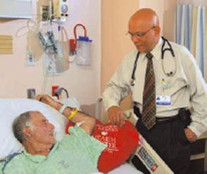
Culture Shock
In July 1998, Joseph Li, MD, director of the hospital medicine program at Beth Israel Deaconess Medical Center, Boston, was the chief medical resident at that institution when his chief of medicine approached him about starting a hospitalist program.
In the late 1990s there was no model of that kind in Boston. “He hired me as our first full-time hospitalist, and I was referred to as a hospitalist—but nobody really understood what that meant,” says Dr. Li. “Looking back now, I didn’t even really understand what that meant.”
Tin M. Oo, MD, medical director for the hospitalist program at St. Mary’s Health Center in Jefferson City, Mo., practiced for seven months as a lone hospitalist but was hardly a new physician. He has been practicing medicine for 34 years in the U.S. and four other countries: Burma (now Myanmar, his native country), Sri Lanka, Malaysia, and Brunei Darussalam. Since he emigrated to the U.S. 15 years ago, he has served as an epidemiologist with the state Department of Health in Minnesota, matriculated into a three-year internal medicine residency program in New York and practiced there, and was in private practice in Chattanooga, Tenn.
When Dr. Oo first came to St. Mary’s, he taught his co-workers, the patients, and their families. “The hospital [and nurses] didn’t have any experience with hospitalists so they didn’t know when to call me and when not to call me,” he says. Dr. Oo got his first hospitalist experience in Chattanooga, Tenn., when he worked with outpatients and some inpatients. His prior experience moonlighting as a hospitalist as well as an emergency physician has helped him greatly as a lone hospitalist. “It was a good thing that I wasn’t just a hospitalist; that I had been in private practice and worked in the ER and as a hospitalist. I knew what the private doctors were facing, and what was coming across from the ER.”
For those who have not yet practiced medicine, Dr. Oo would dissuade them from taking a position as a lone hospitalist. “You have to have a feel for what the ER physicians or what the family practice/ internists, and what the specialists do,” he says. “You also have to be in the hospitalist’s shoes, at least from time to time.”

Be Flexible
While all hospitalists need to be flexible, this may be particularly true for the lone hospitalist. Dr. Johns finds his dual internal medicine/pediatrics training serves him well. “I assist with patients, especially pediatrics, in the emergency department [ED], go up to [resuscitate the infant in] C sections, deal with all the pediatric issues and ER consultations during the day plus do all the general internal medicine care and adult care, which does make up the majority of what I do during the day,” he says. Two physician assistants have been brought on board to improve continuity on nights and weekends and decrease the workload of all physician providers.
Dr. Johns, whose title is associate medical director in charge of hospitalist services, finds the biggest challenge has been attaining and maintaining a commitment to quality on opposite shifts. Sharing coverage with several primary care physicians, he says, means there are differences in concepts of protocols for admissions, commitment to caring for the extra patients, and the physician’s comfort in his or her inpatient knowledge base.
Taking over after a previous night’s coverage, Dr. Johns is unsure which orders were carried out and which patients received what therapy, for instance. “The covering providers take care and make sure patients make it through to next day but often hesitate to alter the plan too much because they are not covering the following day as the inpatient provider.” A newly instated Thursday-through-Monday hospitalist service schedule has helped improve continuity of care and transfer of information through the weekend. But it is still not a perfect situation.
After 15 months as the lone hospitalist, Dr. Johns’ position has changed. He cares for patients during the day and during the opposite shift takes on administrative responsibilities, such as deciding on protocols and expanding services. That kind of juggling—without hospitalist colleagues—has required flexibility as well.
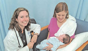
Plan and Set Boundaries
Educating the hospital taught Dr. Fulton a good deal as well, especially about his and their expectations. “The group that grasps the whole concept the quickest is the ancillary services (case management) and the nursing staff,” he says. He likens their receptivity to having a hospitalist to the workings of a pendulum: “There is nothing before this better way, and then they want more of it and they want it all the time. It really means needing to create boundaries”
Dr. Oo agrees that as a lone hospitalist it is important to set boundaries for your accountabilities. The administration of his hospital asked him whether he would take charge of rapid response calls. He declined, leaving that traditional role to the ED.
The year Dr. Fulton practiced as a lone hospitalist gave him a window of time to plan. “It really allowed me to hit the bumps in the road and figure things out so when I added partners, I was able to get them up to speed more quickly,” he says. “Hospitals continue to get busier and busier and that requires planning. I was tracking and trending the volume [of patients that] these 27 doctors were generating in the hospital so that I could anticipate how many doctors I needed. Hospitals continue to increase in volume. The intensity of medicine continues to increase. You always need to plan for one or two more docs than you think you need.”
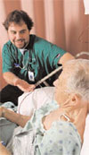
Coverage
For many lone hospitalists, sharing schedule coverage is a dilemma. Dr. Li, an assistant professor of medicine at Harvard Medical School in Boston and a board member of SHM, spent one year as a lone hospitalist before another full-timer joined him. “Everybody understands that as sole physician you really can’t be expected to see patients 24/7/365,” says Dr. Li.
Dr. Li’s recommendation for any hospitalist group of any size—but particularly early on when the hospitalist is alone or in a small group—is to match volume with staffing. “With every program that I have seen or been involved with, early on there is always a massive shift of primary care providers who want to refer patients to you after the program is up and running, Dr. Li says.
Another recommendation Dr. Li has for the lone hospitalist is to identify how you will quickly get help in urgent circumstances. “There is a real benefit to the whole service for having more than one physician on any given day,” he says. It takes only one critically ill patient to cause an upheaval in the schedule.
Committee Work
As recent SHM data show, a large proportion of hospitalists serve on hospital committees. But during his year as a lone hospitalist, Dr. Li focused on taking care of patients, relying upon consultants, and getting through the day.
“I certainly had very little insight at that point of the hospitalist model in terms of communications and leading a team and being the leader of quality in the institution,” he says. “Those were the furthest things from my mind.”
Dr. Fulton participated on committees even though he was a lone hospitalist. “I was involved because I was willing to do the extra work and to use that opportunity to educate and establish who we were in the hospital,” he says. When he was joined by his first two partners, he protected them from committee work because that wasn’t their initial responsibility,” he says.
For lone hospitalists, “you either need to anticipate being on committees and protecting time for it, or you need to anticipate protecting yourself from the committees in order to provide your service,” says Dr. Fulton. “That’s sort of a slippery slope because if you lose the opportunity to become involved in committees, someone else will do it and then they’re making decisions for you that affect your practice of medicine in the hospital.”
Because Dr. Fulton would not sit in hourlong meetings, he took an indirect route—discussing issues with case managers on the floors in between patients or when they shared a patient.
Dr. Fulton advises the lone hospitalist to consider “who is boss” when he or she considers allocating time for committee involvement. Employment by a hospital versus a multispecialty group versus going out as a solo practitioner or working in a private group will determine whose agenda you have to fulfill. “If you are owned by the hospital, the hospital calls the shots; and you have to negotiate … where you put your efforts and your service. If they want you to be on committees, you need to negotiate less patient interaction. You can’t do both; you’ll begin to lose your mind.” TH
Andrea Sattinger is a frequent contributor to The Hospitalist.
Lone hospitalist. It sounds adventurous. It might mean having the chance to set the stage for a hospital medicine program—working exactly the way one wants to and enjoying the feeling of indispensability.
But it’s not for the faint of heart, as those who’d done it attest. There’s the potential for having no starting patient base, or being overwhelmed because there are few to no physicians to share coverage. Having the chance to educate the hospital and its staff about hospital medicine can be a blessing—and a curse.
An example of a lone hospitalist who has experienced the joys and pains of her position is Patricia M. Hopkins-Braddock, MD, an assistant professor of pediatrics at Albany Medical College in N.Y. She was hired as the only pediatric hospitalist in the pediatrics intensive care unit (PICU) at Albany Medical Center. This is her fourth year in that position. Having become the residency program director at her institution in January, she works every day plus one weekend a month, alternating weekly between a pediatrics floor and a long-term care facility and sedation service for children.
“I like the fact that I have turned into the go-to person for problems within the hospital continuum,” Dr. Hopkins-Braddock says. “I also have to say that that is probably one of the things I like least. I have that presence in the hospital and the understanding of the way the floor works. I also do pediatrics sedation. Somehow I become the solution for every problem. It’s good in its own sense, but it can also become very overwhelming.”
A Perfect World?
“If we could figure out a way for one person to function productively and efficiently by themselves, it would be wonderful,” says Cary Ward, MD, who works with hospitalist programs that are part of Catholic Health Initiatives, based in Denver, and is the chief medical officer at St. Elizabeth Regional Medicine Center in Lincoln, Neb. “There is a large group of hospitals that wants to have a hospitalist program, and those are the critical access hospitals.” At these hospitals, which never have more than 25 patients, several community physicians round at the hospital in the morning, finishing by 7 or 7:30 a.m., and return to their primary office bases. “These hospitals are often clamoring for someone to be in the hospital the majority of the day,” he says.
“You’ve heard the saying, ‘You’ve seen one hospitalist program, you’ve seen one hospitalist program,’ ” continues Dr. Ward. “I’ve been amazed at all the hybrid programs out there. At most small hospitalist groups, even those programs under the smallest census of 12 or 13 patients, hospitals still often bring in two doctors with alternate week rotations. “Many consider this the most feasible way to try to cover one hospital census at all times; however, some worry that this ‘feast or famine’ schedule may lead to burnout and this can be expensive for the hospital. To get only one physician to cover that kind of responsibility is a real challenge.”
And yet, some hospitals and hospitalists manage to do it. Of the 362 hospitalist groups that responded to SHM’s 2005-2006 survey “Bi-Annual Survey on the State of the Hospital Medicine Movement,” only nine groups (2.5%) consisted of one physician. Joseph A. Miller, MD, who staffs SHM’s Benchmarks Task Force and has been helping SHM build a national hospitalist database, estimates that of the 2,500 hospitalist groups in the U.S., 62 groups might have just one hospitalist.
Pluses and Minuses
Martin C. Johns, MD, a rural internal medicine and pediatrics hospitalist at Gifford Medical Center in Randolph, Vt., sees some definite advantages to being a lone hospitalist, a post he has held for the 1 1/2 years. “It allowed me the time to interact with all the other modalities and to establish with physician therapy, occupational therapy, care management, pharmacy, what was lacking in the previous model with a variety of docs covering,” Dr. Johns says. “I was trying to create standards that made sense for everyone. The establishment of my being the only hospitalist was determinant primarily on my ability to create those relationships and ensure that they were solid, and also to have the support of all the primary care doctors.”
Gifford’s administration was also supportive. “Because we are a critical access hospital, there are certain restrictions and requirements that we have to take into consideration with Medicare and Medicaid,” Dr. Johns says. “Being the sole hospitalist as we’re expanding allowed me to set the stage: what was lacking, what was missing, what we could improve on, what was already working quite well. [I incorporated] the help of the administration to fill in the gaps of what we needed.”
The primary nonclinical challenge for the lone hospitalist is finding patients to care for and doctors to share coverage. Christopher Farrar, MD, lead hospitalist at Anderson Hospital in Maryville, Ill., began the program there. His employer, the hospitalist company Inpatient Management Inc., based in St. Louis, Mo., manages 18 hospitalist programs in 12 states. “As I was ending my primary care role,” says Dr. Farrar, “this opportunity came available. I think that they weren’t expecting someone to jump in so quickly. They didn’t have the luxury of time to find another physician right away.”
There are now two physicians in the program there, and they are on the verge of getting a third. Dr. Farrar said he doesn’t think there are a lot of pluses to being the lone hospitalist. The most difficult part is avoiding burnout, he warns. Although there were plenty of patients and work for him when he first started at his institution, he wasn’t overwhelmed. But he was relieved when the company brought in a second physician with whom to share call. “I could have easily become overwhelmed quickly had it gone down that road,” he says.
Finding a patient base and physician call backup were not issues for J. Stewart Fulton, DO, medical director of the hospitalist program at Southern New Hampshire Medical Center in Nashua, when he began five years ago as the lone hospitalist. Foundation Medical Partners, a multispecialty group of 27 doctors directly affiliated with the hospital, recruited Dr. Fulton to start a hospitalist program. “I walked into an ideal situation,” he says. “I stepped into a group that had a patient base, that knew they needed hospital coverage, and were willing to support me as I grew to provide 24/7 coverage for myself.”
Culture Shock
In July 1998, Joseph Li, MD, director of the hospital medicine program at Beth Israel Deaconess Medical Center, Boston, was the chief medical resident at that institution when his chief of medicine approached him about starting a hospitalist program.
In the late 1990s there was no model of that kind in Boston. “He hired me as our first full-time hospitalist, and I was referred to as a hospitalist—but nobody really understood what that meant,” says Dr. Li. “Looking back now, I didn’t even really understand what that meant.”
Tin M. Oo, MD, medical director for the hospitalist program at St. Mary’s Health Center in Jefferson City, Mo., practiced for seven months as a lone hospitalist but was hardly a new physician. He has been practicing medicine for 34 years in the U.S. and four other countries: Burma (now Myanmar, his native country), Sri Lanka, Malaysia, and Brunei Darussalam. Since he emigrated to the U.S. 15 years ago, he has served as an epidemiologist with the state Department of Health in Minnesota, matriculated into a three-year internal medicine residency program in New York and practiced there, and was in private practice in Chattanooga, Tenn.
When Dr. Oo first came to St. Mary’s, he taught his co-workers, the patients, and their families. “The hospital [and nurses] didn’t have any experience with hospitalists so they didn’t know when to call me and when not to call me,” he says. Dr. Oo got his first hospitalist experience in Chattanooga, Tenn., when he worked with outpatients and some inpatients. His prior experience moonlighting as a hospitalist as well as an emergency physician has helped him greatly as a lone hospitalist. “It was a good thing that I wasn’t just a hospitalist; that I had been in private practice and worked in the ER and as a hospitalist. I knew what the private doctors were facing, and what was coming across from the ER.”
For those who have not yet practiced medicine, Dr. Oo would dissuade them from taking a position as a lone hospitalist. “You have to have a feel for what the ER physicians or what the family practice/ internists, and what the specialists do,” he says. “You also have to be in the hospitalist’s shoes, at least from time to time.”
Be Flexible
While all hospitalists need to be flexible, this may be particularly true for the lone hospitalist. Dr. Johns finds his dual internal medicine/pediatrics training serves him well. “I assist with patients, especially pediatrics, in the emergency department [ED], go up to [resuscitate the infant in] C sections, deal with all the pediatric issues and ER consultations during the day plus do all the general internal medicine care and adult care, which does make up the majority of what I do during the day,” he says. Two physician assistants have been brought on board to improve continuity on nights and weekends and decrease the workload of all physician providers.
Dr. Johns, whose title is associate medical director in charge of hospitalist services, finds the biggest challenge has been attaining and maintaining a commitment to quality on opposite shifts. Sharing coverage with several primary care physicians, he says, means there are differences in concepts of protocols for admissions, commitment to caring for the extra patients, and the physician’s comfort in his or her inpatient knowledge base.
Taking over after a previous night’s coverage, Dr. Johns is unsure which orders were carried out and which patients received what therapy, for instance. “The covering providers take care and make sure patients make it through to next day but often hesitate to alter the plan too much because they are not covering the following day as the inpatient provider.” A newly instated Thursday-through-Monday hospitalist service schedule has helped improve continuity of care and transfer of information through the weekend. But it is still not a perfect situation.
After 15 months as the lone hospitalist, Dr. Johns’ position has changed. He cares for patients during the day and during the opposite shift takes on administrative responsibilities, such as deciding on protocols and expanding services. That kind of juggling—without hospitalist colleagues—has required flexibility as well.
Plan and Set Boundaries
Educating the hospital taught Dr. Fulton a good deal as well, especially about his and their expectations. “The group that grasps the whole concept the quickest is the ancillary services (case management) and the nursing staff,” he says. He likens their receptivity to having a hospitalist to the workings of a pendulum: “There is nothing before this better way, and then they want more of it and they want it all the time. It really means needing to create boundaries”
Dr. Oo agrees that as a lone hospitalist it is important to set boundaries for your accountabilities. The administration of his hospital asked him whether he would take charge of rapid response calls. He declined, leaving that traditional role to the ED.
The year Dr. Fulton practiced as a lone hospitalist gave him a window of time to plan. “It really allowed me to hit the bumps in the road and figure things out so when I added partners, I was able to get them up to speed more quickly,” he says. “Hospitals continue to get busier and busier and that requires planning. I was tracking and trending the volume [of patients that] these 27 doctors were generating in the hospital so that I could anticipate how many doctors I needed. Hospitals continue to increase in volume. The intensity of medicine continues to increase. You always need to plan for one or two more docs than you think you need.”
Coverage
For many lone hospitalists, sharing schedule coverage is a dilemma. Dr. Li, an assistant professor of medicine at Harvard Medical School in Boston and a board member of SHM, spent one year as a lone hospitalist before another full-timer joined him. “Everybody understands that as sole physician you really can’t be expected to see patients 24/7/365,” says Dr. Li.
Dr. Li’s recommendation for any hospitalist group of any size—but particularly early on when the hospitalist is alone or in a small group—is to match volume with staffing. “With every program that I have seen or been involved with, early on there is always a massive shift of primary care providers who want to refer patients to you after the program is up and running, Dr. Li says.
Another recommendation Dr. Li has for the lone hospitalist is to identify how you will quickly get help in urgent circumstances. “There is a real benefit to the whole service for having more than one physician on any given day,” he says. It takes only one critically ill patient to cause an upheaval in the schedule.
Committee Work
As recent SHM data show, a large proportion of hospitalists serve on hospital committees. But during his year as a lone hospitalist, Dr. Li focused on taking care of patients, relying upon consultants, and getting through the day.
“I certainly had very little insight at that point of the hospitalist model in terms of communications and leading a team and being the leader of quality in the institution,” he says. “Those were the furthest things from my mind.”
Dr. Fulton participated on committees even though he was a lone hospitalist. “I was involved because I was willing to do the extra work and to use that opportunity to educate and establish who we were in the hospital,” he says. When he was joined by his first two partners, he protected them from committee work because that wasn’t their initial responsibility,” he says.
For lone hospitalists, “you either need to anticipate being on committees and protecting time for it, or you need to anticipate protecting yourself from the committees in order to provide your service,” says Dr. Fulton. “That’s sort of a slippery slope because if you lose the opportunity to become involved in committees, someone else will do it and then they’re making decisions for you that affect your practice of medicine in the hospital.”
Because Dr. Fulton would not sit in hourlong meetings, he took an indirect route—discussing issues with case managers on the floors in between patients or when they shared a patient.
Dr. Fulton advises the lone hospitalist to consider “who is boss” when he or she considers allocating time for committee involvement. Employment by a hospital versus a multispecialty group versus going out as a solo practitioner or working in a private group will determine whose agenda you have to fulfill. “If you are owned by the hospital, the hospital calls the shots; and you have to negotiate … where you put your efforts and your service. If they want you to be on committees, you need to negotiate less patient interaction. You can’t do both; you’ll begin to lose your mind.” TH
Andrea Sattinger is a frequent contributor to The Hospitalist.
The Bad Hire
Hospital medicine groups depend on camaraderie and expertise to carry them through long days and heavy workloads. Group cohesiveness—often fragile—depends on recruiting and keeping hard-working doctors who pull their weight professionally and boost the group’s chemistry.
In a field with five job openings for every qualified candidate, and average annual turnover at 12%, hospital medicine groups can ill afford a bad hire. Whether that person is a practice killer, a cipher who blends into the wallpaper while collecting a paycheck, or a doctor marking time until a fellowship or something better comes along, the group leader must quickly limit a bad hire’s negative impact.
Recognizing that competition to hire hospitalists is fierce, it may seem that avoiding or axing a bad hire—the physician who either doesn’t mesh with your team, is a professional and/or personal train wreck, or has a blue-ribbon pedigree, but performs poorly—is a luxury hospitalist groups can’t afford.
But as Per Danielsson, MD, medical director of Seattle-based Swedish Medical Center’s adult hospitalist program has learned the hard way: “No doctor is better than the wrong doctor. I don’t sugarcoat the demands of our program with prospects. We’re a seasoned hospitalist program, we work hard, and, if we have a position vacant, we’ll work even harder for short periods of time until we find the right person.”
With a hospitalist group of 25 providers, Dr. Danielsson spends more time than he’d like recruiting and interviewing candidates, but he considers it time well spent. “The CV and interview are important, but I’ve devised a list of 12 personality traits that I consider important,” he says. “I share the list with candidates to see if we have a good fit.”
Chris Nussbaum, MD, CEO of Synergy Medical Group, based in Brandon, Fla., says: “They don’t make doctors the way they used to. I don’t see why some hospitalists think seeing 20 to 25 patients a day is such a big deal. I’ve had several tell me that 20 patients a day is no problem—and then they only last one day.” When that happens, Synergy cuts its losses, not allowing a bad hire to linger.
Dr. Nussbaum didn’t think twice about firing one new hire—a physician with an impressive resume who, while writing chart notes at a nursing station, watched a nurse have a seizure, gathered his notes and left the room. “He expected an endocrinologist standing nearby to help out, but it’s outrageous that any hospitalist wouldn’t respond appropriately,” he says. Such callous behavior would send shock waves through any group, and that physician was fired on the spot.
Another organizational disrupter, briefly employed by IPC-the Hospitalist Company (North Hollywood, Calif.) made inflammatory remarks about a hospital’s pre-eminent specialist and other referring physicians. He was fired. Several hospitalist leaders report hiring physicians with stellar pedigrees whose hands consistently strayed to nurses’ derrieres. Those doctors were quickly shown the door.
Robin Ryan, a career coach from Newcastle, Wash., who has prepared office-based physicians for professional moves to hospitalist careers, says the new career path can be confusing. When a physician and a hospitalist group have made a mistake, Ryan says most groups cut their losses by terminating someone who doesn’t fit. “Contracts often require a hefty severance fee, but it’s often the road that groups take,” she says.
Probing Personality
To weed out potential bad hires, employees long have used personality tests. Such tests also help job candidates clarify what matters most to them professionally. The SHM’s Career Satisfaction Task Force has developed a framework for hospitalists to do that. The self-test rests on four pillars of job satisfaction: reward/recognition, workload/schedule, autonomy/control, and community/environment (to view, go to www.hospitalmedicine.org and click “Career Satisfaction White Paper”).
Sylvia McKean, MD, medical director, the Brigham & Women’s Hospital/Faulkner Hospitalist Program in Boston and the task force’s co-chair, urges hospitalists to complete the self-test to maximize a potential job fit.
“All jobs have unpleasant side effects,” says Dr. McKean. “People get sick at bad times. There is high stress and sometimes high error rates. It’s important for a hospitalist to analyze what your needs are and to find an environment that best suits them.”
Dr. McKean also offers wisdom from the other side of desk, having interviewed candidates for coveted spots at Brigham & Women’s hospitalist program. “I’ve interviewed doctors who aren’t interested in hospitalist medicine but view our program as a stepping stone to the job they really want here,’’ she says. “We hired and fired someone who wanted her own way all the time. She left for another prestigious hospital. Then there are others who don’t want to teach, but choose a teaching hospital.”
Dr. McKean hopes SHM’s self-assessment tools will help job candidates focus on what they want from a hospital medicine group and avoid the “pebbles” that erode job satisfaction.
IPC, which employs 600 physicians in 100 practices in 24 markets, tried personality testing then discarded it. IPC hired a psychometric firm to devise a psychological profile of “best” and “worst” performing hospitalists. The testers created a test measuring seven key characteristics relating to temperament, intelligence, and clinical skills.
IPC’s CEO Adam Singer, MD, says: “We tested all candidates but found the test ineffective because nearly everyone, including me, got five or better.” He dropped the test, relying instead on extensive interviews. Dr. Singer reviews 2,500 to 3,000 physician resumes annually and spends significant resources on avoiding bad hires. All that hard work doesn’t avoid the occasional mistake.
“I’ve seen everything—the brilliant doctor who can’t function on a team, aloofness, temper tantrums, rudeness, and always pushing responsibility on someone else,” says Dr. Singer. “When something’s wrong, 90% of the time we terminate them ASAP. The other 10% we salvage by finding what’s stressing them, relieving the pressure, and mentoring them into proper behavior.”
Cynthia Stamer, a Dallas-based attorney at Glast, Phillips & Murray, P.C., works extensively with physicians and hospitals and sees young physicians straight from residency joining hospitalist programs “just looking for a job and not focused on whether or not there’s a good personality fit.” She urges job candidates and hirers to better probe the fit.
Stamer finds good hospitalists to be stress jockeys who thrive on the intensity of hospital work. “I think they’re born and not bred,” she says. “They tend to be bored or disruptive in office practices, and to enjoy a pattern of work hard, play hard. The ability to throw the ‘on’ switch and be intense for block scheduling, then be ‘off’ for a block suits them,” she says.
Not That Bad
In a field where an extra pair of hands can make the difference between taking night call or the freedom to take several days off for emergencies, a mediocre team member might seem better than none. Some hospitalist groups would rather pull a bigger load temporarily than tolerate a laggard; others stomach imperfection.
Spotting problems during the hiring process can turn a bad hire into a proper fit. For example, offering a permanent part-time position to someone with young children who can’t commit to full-time employment avoids potential problems. Or, asking enough probing questions might help you discover a physician has a year before a coveted fellowship begins; tailoring a one-year contract for that person optimizes fit. Eliminating managerial tasks for a pure clinician who eschews the leadership fast track works, too.
What to do with the mediocre performer rather than the egregious misfit? Perhaps she consistently arrives to work late, doesn’t complete her charts, and tries to avoid admissions or challenging assignments. A group leader may salvage the situation through mentoring and tying pay to performance. Dr. Singer says: “Underperformers usually don’t understand their impact on the group. We teach them healthcare economics and the flow of dollars. We train them to get the relationship between pay and performance, and hope for results.”
Stamer urges hospitalist leaders to build termination procedures into employment contracts, to document poor performance, and to give severance pay or buy out a contract with a bad hire. “People get testy around disengagement, but if you can take the heat out of the process, it’s better in the long run,” she concludes.
As hospitalist supply approaches demand, avoiding bad hires should be easier. For now, most groups prefer pulling together and working harder rather than abide an outlier. It comes with the territory. TH
Marlene Piturro is a frequent contributor to The Hospitalist.
New Take on Elder Care
Families often note that after older relatives return home from the hospital, something is wrong with them.
While the acute condition that brought a relative into the hospital has been remedied, major functional and cognitive deficits such as confusion, falls, and difficulty with basic activities of daily living remain.
This post-hospital decline may not be appreciated by hospital clinicians, perhaps because the problems do not become visible until the patient is home. However, these problems place significant burdens on patients and families.
Following discharge for an acute hospitalization, about a third of older patients will have a major new disability that threatens their ability to live independently.1 Among community-dwelling elders, half of all new disability occurs within a month of hospitalization.2 It isn’t surprising that nursing home placement has grown more common for medical hospitalizations, even for seemingly reversible medical problems.
Older adults will make up an increasing number of the patients cared for by hospitalists. The Acute Care for Elders (ACE) unit model of care focuses on preventing functional decline and increasing discharges to home.
In 2005, leadership of the San Francisco General Hospital Medical Center (SFGHMC) committed to improving care for hospitalized elders by adopting the ACE model.3
The ACE model combats hospital-acquired disability by improving care processes for older patients.
Major motivating factors for the change included demographic and quality-of-care imperatives. After a nine-month planning process, the SFGHMC ACE unit opened in February.
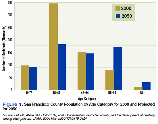
Rationale
Largely driven by the baby boom, the number of California seniors older than 65 will double from 3.5 million to 7 million over the next 40 years, and those older than 85 will triple from about 500,000 to 1.5 million. In San Francisco, changes will be even more dramatic as the number of residents over 65 increases from 14% of the population to 32%.4 (See Figure 1, right).
In California, people 65 to 84 are almost three times as likely to be hospitalized as those between 45 and 64. If rates of hospitalization do not change, an increase in hospitalized older adults will occur as the baby boom generation ages.
In addition, hospitalization can be hazardous for older adults, with increased risk for functional and cognitive disability and adverse events.5-7 As a result, hospitalization-associated disability represents a growing threat to the independence of the older population. A variety of changes to usual care have been adopted in an effort to reduce the hazards of hospitalization in the elderly.8-11
ACE Model
The ACE unit model, proven to reduce the risk of hospital-acquired disability in the elderly, is based on the Model of Dysfunction for Hospitalized Elders.12 (See Figure 2, p. 23)
This model outlines how processes of hospital care for the elderly promote physical impairment and depressed mood, leading to dysfunction. Counterproduc-tive factors in older adults include a hostile environment (lack of natural sunlight, high-glare floors, poor way-finding cues, high noise levels), depersonalization (lack of personal effects, clothing, and usual daily routines), bed rest through multiple tethers or inattention, medicines inappropriate in the elderly or given at inappropriate doses, procedures, and negative expectations (usually that the patient will require nursing home placement after admission).
These processes are the targets of the ACE intervention. The idea is to improve quality of care for the elderly by promoting rehabilitation and preventing disability.
The ACE unit addresses these issues through a “prehabilitation program.” The ACE unit is a physical location in the hospital with 10 beds. Care is redesigned by placing the patient at the center of restorative efforts of an interdisciplinary team consisting of an advanced practice nurse, a social worker, an occupational and a physical therapist, a nutritionist, a pharmacist, and a medical director. This ACE Unit team meets daily to review and plan care for all patients. Recommendations for nursing care and rehabilitation evaluation are implemented by the ACE team directly. Other recommendations, such as changing medications or considering alternative approaches to common geriatric syndromes are communicated to the primary team, which maintains overall responsibility for the care of the patient.
Each patient’s assessment is multidimensional, with an emphasis on nonpharmacologic interventions where practical. For example, an emphasis is placed on after-dinner exercise such as walking and socializing to promote sleep and reduce medication use. Nursing-care plans were revised to promote mobility, discourage inappropriate Foley use, and encourage adequate hydration and nutrition.
Recommendations are communicated to the primary team via a recommendation form placed in the physician-order section and text pages. The unit’s medical director and pharmacist review medications. Recommendations that involve medication changes are discussed with the primary team, which write all medication orders. Home planning begins on the day of admission.
Prior to opening the unit, the ACE unit social worker met with key city and county agencies including Aging and Adult Services, the Public Guardian, In-Home Supportive Services, and community nursing homes to introduce the unit and plan for an effective and safe transition.
All staff expect patients to maintain prehospital physical functioning. When possible, patients are expected to wear their own clothes, eat all their meals in a common dining room, and ambulate or exercise daily.
We considered establishing criteria for admission.13 We have not adopted formal criteria for patients 65 or older, presence of medical non-surgical condition(s) that require(s) acute hospitalization, and no need for telemetry or chemotherapy. As we learn how best to serve our hospitalized older adult population with the resources of the unit, we will re-evaluate targeting criteria. Most of our admissions are from the emergency department (ED), and the remainder are from other units in the hospital.
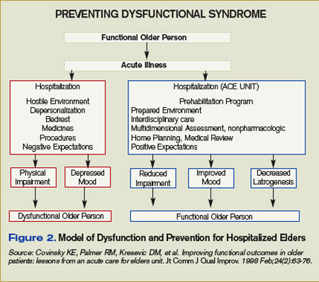
Challenges
Key challenges in opening the new service include securing commitment and resources from organizational leaders and key stakeholders; incorporation of the ACE unit concept in an academic training center; hiring key staff, especially the geriatric clinical nurse specialist and pharmacist positions; and completing the environmental rehabilitation on a limited budget.
While gaps in the care of the geriatric patient population were well identified at SFGHMC as far back as 1996 by a multidisciplinary task force, no actions on recommendations were taken, for several key reasons.
First, an executive level administrator or physician champion was not a member of the task force. Second, the organization did not have a department or regulatory mandate to address the gaps in the care of the elderly patients. Third, there was no link between the hospital strategic plan and the recommendations.
By 2004, these issues were largely addressed. A new chief nursing officer with a background in quality improvement understood the demographic and quality imperatives to improve care for hospitalized older adults.
That same year, the Hospital Executive Committee incorporated patient safety into the hospital strategic plan. This resulted in a successful business plan for an ACE Unit and geriatric consultation service linked to organizational strategy.
Funding was allocated for a medical director and a clinical nurse specialist in fiscal year 2005-2006. In addition, a grant was obtained from the SFGH Foundation to fund equipment, renovations, and staff education/training.
The original ACE unit concept involved expert, interdisciplinary geriatric assessment and communication of suggestions via a paper-based chart.
Initially, we felt the primary medical team should round with the ACE team, preferably at the bedside. However, informal focus groups held with the residents suggested this would happen infrequently.
The demands on the medical teams of completing patient rounds before morning attending rounds were cited as the main reason that model wouldn’t work.
We have implemented the following methods to promote communication between the ACE unit team and the primary medical team:
- Medical teams are encouraged to attend ACE unit rounds while on bedside rounds. This provides an opportunity to model the team-delivered care for house staff and medical students, an ACGME requirement;
- Suggestions to change medications are directly text-paged to the house staff; and
- Recommendations are summarized in a communication sheet left in the chart (this not a permanent part of the medical record).
We plan on using the text-paging more widely once the unit has wireless computer capability. Despite this, there are occasions where a team is not aware of a recommendation or new emphasis in the care plan. We are considering additional ways to improve communication such as attending the primary team attending rounds.
In California and other states there is a shortage of clinical pharmacists and masters-prepared nurses with expertise in geriatrics.14-16 The advanced practice nurse performs a vital role in raising the level of knowledge, skills, and attitudes for the nursing staff on the ACE unit.
In addition, we see the ACE unit as a drop in the pond; we feel a responsibility to expand nursing geriatric competency throughout appropriate hospital areas. Thus, this nursing role is at the center of preparing the hospital to care for an older patient population.
This position remained unfilled for almost a year despite an intensive national search. This prompted us to incorporate the geriatric resource nurse model into our unit while we continued our recruitment.
Although we have successfully concluded our search for a nursing leader for the ACE unit, we have yet to hire a clinical pharmacist.
The rehabilitation of the unit would not have been possible without foundation support. As a public hospital with many competing demands, monies are limited for the rehabilitation required to create a more welcoming, safer environment for the older patient. In this case, the hospital foundation and a local foundation made grants to the ACE unit to allow us to change the environment.
These grants have allowed for significant changes to the unit, including elevated toilet seats, high-backed chairs, handrails, unit-based physical therapy equipment, and activities to promote non-pharmacological approaches to agitation.
Readiness for Change
All levels of hospital staff embraced the ACE unit concept. Department leaders in rehabilitation, nutrition, social work, and pharmacy felt ACE unit principles would improve care delivery over usual care.
Early on, department leaders and medical staff enthusiastically participated on a steering committee to help guide implementation efforts. In addition, when we offered geriatric resource nurse training to our nursing staff, more than 20 out of 50 unit nurses expressed interest.
Staff from all departments represented on the ACE unit team also expressed interest in and attended the three days of training. This provided baseline knowledge of common geriatric syndromes in hospitalized older patients for all team members. This been helpful during ACE team discussions.
Although medical residents felt they could not consistently attend ACE rounds, they appreciated potential benefits of the unit:
- Different perspectives could provide a wider range of evaluation and treatment recommendations for their patients;
- The co-location of key disciplines could result in overall time savings in calling for and ordering evaluations;
- The reduced likelihood that key interventions such as mobilization, feeding, catheter removal, and medication review would be missed; and
- The opportunity to learn principles of geriatric care through attending ACE rounds (when possible).
Medical staff in other departments immediately accepted the rationale for the unit.
Many expressed interest in expanding the concept to their departments, especially orthopedics, general surgery, and the ED. Although unfamiliar with specific interventions to improve care for hospitalized elders, the underlying concepts of patient-centered, team-delivered care with a focus on function resonated with most medical staff.
Next Steps
The unit is still in startup mode. Our major areas of focus are:
- Evaluating and improving team dynamics: We have engaged with researchers to evaluate our team dynamics and intervene where necessary to promote a high-functioning team.17
- Developing a culture of performance improvement: One of the hallmarks of high-functioning teams are measures of performance that are team-derived and reflect work product that the team can control.17
We are putting into place processes to measure key quality parameters including length of stay; nursing home placement; readmission rate, inappropriate catheter use; inappropriate medication prescribing; incidence of delirium, falls, and pressure ulcers; functional and cognitive status at admission and discharge; and patient satisfaction.
The orders set used by admitting residents are the standard general medical ward admission set and need revision for the ACE unit.
- Developing a research program: Our goal is to develop a research program evaluating interventions to prevent post-hospital degeneration of elders’ health. There is a dearth of research on improving hospital care for older, vulnerable adults.
- Expanding philanthropy support: The unit has benefited tremendously from philanthropy. In a relatively resource-poor setting, this allowed for rapid engagement with designers and vendors to remake the environment. We plan to expand our outreach efforts to interested philanthropists.
Summary
The ACE unit model can improve care for hospitalized older adults.
It requires a sustained level of commitment from hospital leaders, a focus on patient-centered, team-delivered care, sensitivity to communication modalities with primary caregivers, an awareness of the market for key professionals required, and flexibility to respond effectively to the many challenges that will emerge in implementing this model locally. TH
Dr. Pierluissi is medical director of the ACE unit at the San Francisco General Hospital. Susan Currin is the hospital’s chief nursing officer.
References
- Covinsky KE, Palmer RM, Fortinsky RH, et al. Loss of independence in activities of daily living in older adults hospitalized with medical illnesses: increased vulnerability with age. J Am Geriatr Soc. 2003;51(4):451-458.
- Gill TM, Allore HG, Holford TR, et al. Hospitalization, restricted activity, and the development of disability among older persons. JAMA. 2004 Nov 3;292(17):2115-2124
- Landefeld CS, Palmer RM, Kresevic DM, et al. A randomized trial of care in a hospital medical unit especially designed to improve the functional outcomes of acutely ill older patients. N Engl J Med. 1995 May 18;332(20):1338-1344.
- State of California, Department of Finance. Race/Ethnic Population with Age and Sex Detail, 2000–2050; 2004: Sacramento, Calif.
- Creditor MC. Hazards of hospitalization of the elderly. Ann Intern Med. 1993 Feb 1;118(3):219-223.
- Inouye SK. Delirium in older persons. N Engl J Med. 2006 Jun 8;354(23):2509-2511; author reply 2509-11. Comment on: N Engl J Med. 2006 Mar 16;354(11):1157-1165.
- Brennan TA, Leape LL, Laird NM, et al. Incidence of adverse events and negligence in hospitalized patients. Results of the Harvard Medical Practice Study I. N Engl J Med. 1991 Feb 7;324(6):370-376. Comment in: N Engl J Med. 1991 Jul 18;325(3):210.
- Cohen HJ, Feussner JR, Weinberger M, et al. A controlled trial of inpatient and outpatient geriatric evaluation and management. N Engl J Med. 2002 Mar 21;346(12):905-912. Comment in: Curr Surg. 2004 May-Jun;61(3):266-74. N Engl J Med. 2002 Aug 1;347(5):371-373; author reply 371-373; author reply 371-3: N Engl J Med. 2002 Mar 21;346(12):874.
- Inouye SK, Bogardus ST Jr, Charpentier PA, et al. A multicomponent intervention to prevent delirium in hospitalized older patients. N Engl J Med. 1999 Mar 4;340(9):669-676. Comment in: N Engl J Med. 1999 Jul 29;341(5):369-370; author reply 370. N Engl J Med. 1999 Mar 4;340(9):720-721.
- Reuben DB, Borok GM, Wolde-Tsadik G, et al. A randomized trial of comprehensive geriatric assessment in the care of hospitalized patients. N Engl J Med. 1995 May 18;332(20):1345-1350. Comment in: N Engl J Med. 1995 May 18;332(20):1376-1378.
- Leff B, Burton L, Mader SL, et al. Hospital at home: feasibility and outcomes of a program to provide hospital-level care at home for acutely ill older patients. Ann Intern Med. 2005 Dec 6;143(11):798-808. Comment in: Ann Intern Med. 2005 Dec 6;143(11):840-1. Ann Intern Med. 2006 Mar 21;144(6):456. Summary for patients in:Ann Intern Med. 2005 Dec 6;143(11):I56.
- Covinsky KE, Palmer RM, Kresevic DM, et. al. Improving functional outcomes in older patients: lessons from an acute care for elders unit. Jt Comm J Qual Improv. 1998 Feb;24(2):63-76.
- Wieland D, Rubenstein LZ. What do we know about patient targeting in geriatric evaluation and management (GEM) programs? Aging (Milano). 1996 Oct; 8(5):297-310.
- Spetz J, Dyer W. Forecasts of the Registered Nurse Workforce in California. 2005, University of California, San Francisco: San Francisco.
- Fleming KC, Evans JM, Chutka DS. Caregiver and clinician shortages in an aging nation. Mayo Clin Proc. 2003 Aug;78(8):1026-1040.
- Knapp KK, Quist RM, Walton SM, et al. Update on the pharmacist shortage: National and state data through 2003. Am J Health Syst Pharm. 2005 Mar 1;62(5):492-499.
- Katzenbach JR, Smith DK. The Discipline of Teams. Harv Bus Rev. July-August 2005:1-9.
Families often note that after older relatives return home from the hospital, something is wrong with them.
While the acute condition that brought a relative into the hospital has been remedied, major functional and cognitive deficits such as confusion, falls, and difficulty with basic activities of daily living remain.
This post-hospital decline may not be appreciated by hospital clinicians, perhaps because the problems do not become visible until the patient is home. However, these problems place significant burdens on patients and families.
Following discharge for an acute hospitalization, about a third of older patients will have a major new disability that threatens their ability to live independently.1 Among community-dwelling elders, half of all new disability occurs within a month of hospitalization.2 It isn’t surprising that nursing home placement has grown more common for medical hospitalizations, even for seemingly reversible medical problems.
Older adults will make up an increasing number of the patients cared for by hospitalists. The Acute Care for Elders (ACE) unit model of care focuses on preventing functional decline and increasing discharges to home.
In 2005, leadership of the San Francisco General Hospital Medical Center (SFGHMC) committed to improving care for hospitalized elders by adopting the ACE model.3
The ACE model combats hospital-acquired disability by improving care processes for older patients.
Major motivating factors for the change included demographic and quality-of-care imperatives. After a nine-month planning process, the SFGHMC ACE unit opened in February.
Rationale
Largely driven by the baby boom, the number of California seniors older than 65 will double from 3.5 million to 7 million over the next 40 years, and those older than 85 will triple from about 500,000 to 1.5 million. In San Francisco, changes will be even more dramatic as the number of residents over 65 increases from 14% of the population to 32%.4 (See Figure 1, right).
In California, people 65 to 84 are almost three times as likely to be hospitalized as those between 45 and 64. If rates of hospitalization do not change, an increase in hospitalized older adults will occur as the baby boom generation ages.
In addition, hospitalization can be hazardous for older adults, with increased risk for functional and cognitive disability and adverse events.5-7 As a result, hospitalization-associated disability represents a growing threat to the independence of the older population. A variety of changes to usual care have been adopted in an effort to reduce the hazards of hospitalization in the elderly.8-11
ACE Model
The ACE unit model, proven to reduce the risk of hospital-acquired disability in the elderly, is based on the Model of Dysfunction for Hospitalized Elders.12 (See Figure 2, p. 23)
This model outlines how processes of hospital care for the elderly promote physical impairment and depressed mood, leading to dysfunction. Counterproduc-tive factors in older adults include a hostile environment (lack of natural sunlight, high-glare floors, poor way-finding cues, high noise levels), depersonalization (lack of personal effects, clothing, and usual daily routines), bed rest through multiple tethers or inattention, medicines inappropriate in the elderly or given at inappropriate doses, procedures, and negative expectations (usually that the patient will require nursing home placement after admission).
These processes are the targets of the ACE intervention. The idea is to improve quality of care for the elderly by promoting rehabilitation and preventing disability.
The ACE unit addresses these issues through a “prehabilitation program.” The ACE unit is a physical location in the hospital with 10 beds. Care is redesigned by placing the patient at the center of restorative efforts of an interdisciplinary team consisting of an advanced practice nurse, a social worker, an occupational and a physical therapist, a nutritionist, a pharmacist, and a medical director. This ACE Unit team meets daily to review and plan care for all patients. Recommendations for nursing care and rehabilitation evaluation are implemented by the ACE team directly. Other recommendations, such as changing medications or considering alternative approaches to common geriatric syndromes are communicated to the primary team, which maintains overall responsibility for the care of the patient.
Each patient’s assessment is multidimensional, with an emphasis on nonpharmacologic interventions where practical. For example, an emphasis is placed on after-dinner exercise such as walking and socializing to promote sleep and reduce medication use. Nursing-care plans were revised to promote mobility, discourage inappropriate Foley use, and encourage adequate hydration and nutrition.
Recommendations are communicated to the primary team via a recommendation form placed in the physician-order section and text pages. The unit’s medical director and pharmacist review medications. Recommendations that involve medication changes are discussed with the primary team, which write all medication orders. Home planning begins on the day of admission.
Prior to opening the unit, the ACE unit social worker met with key city and county agencies including Aging and Adult Services, the Public Guardian, In-Home Supportive Services, and community nursing homes to introduce the unit and plan for an effective and safe transition.
All staff expect patients to maintain prehospital physical functioning. When possible, patients are expected to wear their own clothes, eat all their meals in a common dining room, and ambulate or exercise daily.
We considered establishing criteria for admission.13 We have not adopted formal criteria for patients 65 or older, presence of medical non-surgical condition(s) that require(s) acute hospitalization, and no need for telemetry or chemotherapy. As we learn how best to serve our hospitalized older adult population with the resources of the unit, we will re-evaluate targeting criteria. Most of our admissions are from the emergency department (ED), and the remainder are from other units in the hospital.
Challenges
Key challenges in opening the new service include securing commitment and resources from organizational leaders and key stakeholders; incorporation of the ACE unit concept in an academic training center; hiring key staff, especially the geriatric clinical nurse specialist and pharmacist positions; and completing the environmental rehabilitation on a limited budget.
While gaps in the care of the geriatric patient population were well identified at SFGHMC as far back as 1996 by a multidisciplinary task force, no actions on recommendations were taken, for several key reasons.
First, an executive level administrator or physician champion was not a member of the task force. Second, the organization did not have a department or regulatory mandate to address the gaps in the care of the elderly patients. Third, there was no link between the hospital strategic plan and the recommendations.
By 2004, these issues were largely addressed. A new chief nursing officer with a background in quality improvement understood the demographic and quality imperatives to improve care for hospitalized older adults.
That same year, the Hospital Executive Committee incorporated patient safety into the hospital strategic plan. This resulted in a successful business plan for an ACE Unit and geriatric consultation service linked to organizational strategy.
Funding was allocated for a medical director and a clinical nurse specialist in fiscal year 2005-2006. In addition, a grant was obtained from the SFGH Foundation to fund equipment, renovations, and staff education/training.
The original ACE unit concept involved expert, interdisciplinary geriatric assessment and communication of suggestions via a paper-based chart.
Initially, we felt the primary medical team should round with the ACE team, preferably at the bedside. However, informal focus groups held with the residents suggested this would happen infrequently.
The demands on the medical teams of completing patient rounds before morning attending rounds were cited as the main reason that model wouldn’t work.
We have implemented the following methods to promote communication between the ACE unit team and the primary medical team:
- Medical teams are encouraged to attend ACE unit rounds while on bedside rounds. This provides an opportunity to model the team-delivered care for house staff and medical students, an ACGME requirement;
- Suggestions to change medications are directly text-paged to the house staff; and
- Recommendations are summarized in a communication sheet left in the chart (this not a permanent part of the medical record).
We plan on using the text-paging more widely once the unit has wireless computer capability. Despite this, there are occasions where a team is not aware of a recommendation or new emphasis in the care plan. We are considering additional ways to improve communication such as attending the primary team attending rounds.
In California and other states there is a shortage of clinical pharmacists and masters-prepared nurses with expertise in geriatrics.14-16 The advanced practice nurse performs a vital role in raising the level of knowledge, skills, and attitudes for the nursing staff on the ACE unit.
In addition, we see the ACE unit as a drop in the pond; we feel a responsibility to expand nursing geriatric competency throughout appropriate hospital areas. Thus, this nursing role is at the center of preparing the hospital to care for an older patient population.
This position remained unfilled for almost a year despite an intensive national search. This prompted us to incorporate the geriatric resource nurse model into our unit while we continued our recruitment.
Although we have successfully concluded our search for a nursing leader for the ACE unit, we have yet to hire a clinical pharmacist.
The rehabilitation of the unit would not have been possible without foundation support. As a public hospital with many competing demands, monies are limited for the rehabilitation required to create a more welcoming, safer environment for the older patient. In this case, the hospital foundation and a local foundation made grants to the ACE unit to allow us to change the environment.
These grants have allowed for significant changes to the unit, including elevated toilet seats, high-backed chairs, handrails, unit-based physical therapy equipment, and activities to promote non-pharmacological approaches to agitation.
Readiness for Change
All levels of hospital staff embraced the ACE unit concept. Department leaders in rehabilitation, nutrition, social work, and pharmacy felt ACE unit principles would improve care delivery over usual care.
Early on, department leaders and medical staff enthusiastically participated on a steering committee to help guide implementation efforts. In addition, when we offered geriatric resource nurse training to our nursing staff, more than 20 out of 50 unit nurses expressed interest.
Staff from all departments represented on the ACE unit team also expressed interest in and attended the three days of training. This provided baseline knowledge of common geriatric syndromes in hospitalized older patients for all team members. This been helpful during ACE team discussions.
Although medical residents felt they could not consistently attend ACE rounds, they appreciated potential benefits of the unit:
- Different perspectives could provide a wider range of evaluation and treatment recommendations for their patients;
- The co-location of key disciplines could result in overall time savings in calling for and ordering evaluations;
- The reduced likelihood that key interventions such as mobilization, feeding, catheter removal, and medication review would be missed; and
- The opportunity to learn principles of geriatric care through attending ACE rounds (when possible).
Medical staff in other departments immediately accepted the rationale for the unit.
Many expressed interest in expanding the concept to their departments, especially orthopedics, general surgery, and the ED. Although unfamiliar with specific interventions to improve care for hospitalized elders, the underlying concepts of patient-centered, team-delivered care with a focus on function resonated with most medical staff.
Next Steps
The unit is still in startup mode. Our major areas of focus are:
- Evaluating and improving team dynamics: We have engaged with researchers to evaluate our team dynamics and intervene where necessary to promote a high-functioning team.17
- Developing a culture of performance improvement: One of the hallmarks of high-functioning teams are measures of performance that are team-derived and reflect work product that the team can control.17
We are putting into place processes to measure key quality parameters including length of stay; nursing home placement; readmission rate, inappropriate catheter use; inappropriate medication prescribing; incidence of delirium, falls, and pressure ulcers; functional and cognitive status at admission and discharge; and patient satisfaction.
The orders set used by admitting residents are the standard general medical ward admission set and need revision for the ACE unit.
- Developing a research program: Our goal is to develop a research program evaluating interventions to prevent post-hospital degeneration of elders’ health. There is a dearth of research on improving hospital care for older, vulnerable adults.
- Expanding philanthropy support: The unit has benefited tremendously from philanthropy. In a relatively resource-poor setting, this allowed for rapid engagement with designers and vendors to remake the environment. We plan to expand our outreach efforts to interested philanthropists.
Summary
The ACE unit model can improve care for hospitalized older adults.
It requires a sustained level of commitment from hospital leaders, a focus on patient-centered, team-delivered care, sensitivity to communication modalities with primary caregivers, an awareness of the market for key professionals required, and flexibility to respond effectively to the many challenges that will emerge in implementing this model locally. TH
Dr. Pierluissi is medical director of the ACE unit at the San Francisco General Hospital. Susan Currin is the hospital’s chief nursing officer.
References
- Covinsky KE, Palmer RM, Fortinsky RH, et al. Loss of independence in activities of daily living in older adults hospitalized with medical illnesses: increased vulnerability with age. J Am Geriatr Soc. 2003;51(4):451-458.
- Gill TM, Allore HG, Holford TR, et al. Hospitalization, restricted activity, and the development of disability among older persons. JAMA. 2004 Nov 3;292(17):2115-2124
- Landefeld CS, Palmer RM, Kresevic DM, et al. A randomized trial of care in a hospital medical unit especially designed to improve the functional outcomes of acutely ill older patients. N Engl J Med. 1995 May 18;332(20):1338-1344.
- State of California, Department of Finance. Race/Ethnic Population with Age and Sex Detail, 2000–2050; 2004: Sacramento, Calif.
- Creditor MC. Hazards of hospitalization of the elderly. Ann Intern Med. 1993 Feb 1;118(3):219-223.
- Inouye SK. Delirium in older persons. N Engl J Med. 2006 Jun 8;354(23):2509-2511; author reply 2509-11. Comment on: N Engl J Med. 2006 Mar 16;354(11):1157-1165.
- Brennan TA, Leape LL, Laird NM, et al. Incidence of adverse events and negligence in hospitalized patients. Results of the Harvard Medical Practice Study I. N Engl J Med. 1991 Feb 7;324(6):370-376. Comment in: N Engl J Med. 1991 Jul 18;325(3):210.
- Cohen HJ, Feussner JR, Weinberger M, et al. A controlled trial of inpatient and outpatient geriatric evaluation and management. N Engl J Med. 2002 Mar 21;346(12):905-912. Comment in: Curr Surg. 2004 May-Jun;61(3):266-74. N Engl J Med. 2002 Aug 1;347(5):371-373; author reply 371-373; author reply 371-3: N Engl J Med. 2002 Mar 21;346(12):874.
- Inouye SK, Bogardus ST Jr, Charpentier PA, et al. A multicomponent intervention to prevent delirium in hospitalized older patients. N Engl J Med. 1999 Mar 4;340(9):669-676. Comment in: N Engl J Med. 1999 Jul 29;341(5):369-370; author reply 370. N Engl J Med. 1999 Mar 4;340(9):720-721.
- Reuben DB, Borok GM, Wolde-Tsadik G, et al. A randomized trial of comprehensive geriatric assessment in the care of hospitalized patients. N Engl J Med. 1995 May 18;332(20):1345-1350. Comment in: N Engl J Med. 1995 May 18;332(20):1376-1378.
- Leff B, Burton L, Mader SL, et al. Hospital at home: feasibility and outcomes of a program to provide hospital-level care at home for acutely ill older patients. Ann Intern Med. 2005 Dec 6;143(11):798-808. Comment in: Ann Intern Med. 2005 Dec 6;143(11):840-1. Ann Intern Med. 2006 Mar 21;144(6):456. Summary for patients in:Ann Intern Med. 2005 Dec 6;143(11):I56.
- Covinsky KE, Palmer RM, Kresevic DM, et. al. Improving functional outcomes in older patients: lessons from an acute care for elders unit. Jt Comm J Qual Improv. 1998 Feb;24(2):63-76.
- Wieland D, Rubenstein LZ. What do we know about patient targeting in geriatric evaluation and management (GEM) programs? Aging (Milano). 1996 Oct; 8(5):297-310.
- Spetz J, Dyer W. Forecasts of the Registered Nurse Workforce in California. 2005, University of California, San Francisco: San Francisco.
- Fleming KC, Evans JM, Chutka DS. Caregiver and clinician shortages in an aging nation. Mayo Clin Proc. 2003 Aug;78(8):1026-1040.
- Knapp KK, Quist RM, Walton SM, et al. Update on the pharmacist shortage: National and state data through 2003. Am J Health Syst Pharm. 2005 Mar 1;62(5):492-499.
- Katzenbach JR, Smith DK. The Discipline of Teams. Harv Bus Rev. July-August 2005:1-9.
The Power of “Sorry”
Like many people, we like to sing while secure in the anonymity of our cars. This morning, one of us was wailing along with Elton John as he sang “Sorry Seems to Be the Hardest Word”:
It’s sad, so sad
Why can’t we talk it over
Oh, it seems to me
That sorry seems to be the hardest word.
That verse frames a critical legal question physicians regularly encounter: how to communicate with patients after an unexpected outcome. More precisely, should a physician apologize to a patient who suffers complications because of that physician’s treatment?
Traditionally, after a patient suffered a complication, defense lawyers were reluctant to allow the physician to express apologies or regret. The defense lawyer feared the apology would be treated as an “admission against interest.” In other words, the defense lawyer wanted to prevent a plaintiff’s lawyer from someday arguing that the physician’s apology was an admission of negligence or wrongdoing.
But the lawyer’s strategy fails. The patient wants the physician to apologize for an error. In fact, the patient distrusts a physician who does not admit errors.
‘‘Although a physician may wish to tell a patient when he has made a mistake, lawyers often order doctors to say nothing,’’ wrote University of Florida law professor Jonathan R. Cohen in the Southern California Law Review.1 “The physician’s silence may then trigger the patient’s anger. This alienation may then prompt the patient to sue.”
These observations are consistent with studies demonstrating that patients are far less to sue when provided with a full explanation and apology.2
Certainly no physician wants to make a statement that a plaintiff’s lawyer will use against him in court. But the same physician rationally wants to take any steps that might prevent the patient from feeling as though he or she needs to consult with a plaintiff’s lawyer. So, what’s a physician to do when caught between the hospital’s lawsuit-fearing attorney and a patient who expects his doctor to communicate with her honestly and forthrightly?
Fortunately, several state legislatures have recognized this tension and passed legislation that encourages physicians to apologize without facing the prospect that a plaintiff’s lawyer will argue that the physician apologized only because he knew he did something wrong. An example best illustrates how such “I’m sorry” statutes work.
Dr. Smith is treating a 22-year-old patient, John Elway, for a fractured fibula. Dr. Smith sees no signs of neurological compromise while the patient is in a cast. After the cast is removed, it appears the patient has lost function in the leg because the cast was too tight. The patient was a star college athlete who was expected to be drafted into the NFL, but now likely won’t be drafted. Dr. Smith tells the patient: “It’s my fault this happened. I’m really sorry that I didn’t pick up on this sooner.”
Does Dr. Smith’s statement come into evidence in court? Does part of it? The answers probably depend upon which state’s apology statute is applied. Massachusetts was one of the first states to pass an apology statute. It reads:
Statements, writings, or benevolent gestures expressing sympathy or a general sense of benevolence relating to the pain, suffering or death of a person involved in an accident and made to the person or to the family of such a person shall be inadmissible as evidence of an admission of liability in a civil action.
Significantly, the Massachusetts statute applies to people “involved in an accident,” which might imply that it is limited to automobile accidents or workplace accidents. The Massachusetts statute prevents this limited construction by providing a broad definition of “accident,” including any “occurrence resulting in injury or death to one or more persons which is not the result of a willful action by a party.” This definition would encompass ordinary medical negligence.
It would seem clear that the statute would protect Dr. Smith if he simply stated: “I want you to know how sorry I am this happened. I feel awful that you experienced this complication.”
But if Dr. Smith said, “It’s my fault this happened,” would the Massachusetts statute protect Dr. Smith? That’s a much harder call. Saying “It’s my fault” is technically not an expression of “sympathy or a general act of benevolence.” There no clear answer under Massachusetts law. But we believe the result would probably depend on whether the judge hearing the case thought this statement occurred during an overall act of apology.
The answer is clearer in California. That state’s apology statute reads:
The portion of statements, writings, or benevolent gestures expressing sympathy or a general sense of benevolence relating to the pain, suffering or death of a person involved in an accident and made to that person or to the family of that person shall be inadmissible as evidence of an admission of liability in a civil action. A statement of fault, however, which is part of, or in addition to, any of the above shall not be inadmissible pursuant to this section.
California draws a clear distinction between “the portion of statements ... expressing sympathy or a general sense of benevolence” and “a statement of fault.”
In our scenario, the jury would almost certainly be able to hear Dr. Smith’s statement, “It’s my fault this happened.” Critics of California’s law believe it creates too narrow a window for physicians to believe that plaintiff’s lawyers will not use their apology against them in a lawsuit.3
While Dr. Smith’s statement is likely to come into evidence in California, it’s also clear the opposite would occur in Colorado. Colorado’s apology statute, which specifically applies to medical malpractice actions, reads:
In any civil action brought by an alleged victim of an unanticipated outcome of medical care ... any and all statements … expressing apology, fault, sympathy, commiseration, condolence, compassion or a general sense of benevolence ... shall be inadmissible as evidence of an admission of liability or as evidence of an admission against interest.
Because Colorado’s statute specifically renders statements of “fault” inadmissible, a jury would not be able to consider any of Dr. Smith’s statements made during the course of his apology. Colorado’s law provides the physician with the most protection. Critics of Colorado’s law believe it’s unfair for physicians to admit fault to their patients in the hospital, then deny liability after the patient files a lawsuit.
Twenty-six other states have passed apology statutes; each works a bit differently. The choice of words matters. Legally, there is a big difference between a physician telling a patient, “I’m sorry about your pain” or saying, “It’s my fault you’re in pain.”
While apologies are valuable and important in relationships of trust—including the relationship between physicians and patients—we suggest you consult an experienced lawyer when crafting an apology to make sure it conveys your sympathies without opening a door to liability. TH
Patrick O’Rourke works in the Office of University Counsel, Department of Litigation, Denver.
References
- Cohen JR. Advising clients to apologize. Southern California Law Review. 1999;72:1009-1131.
- Hickson GB, Clayton EW, Githens PB, et al. Factors that prompted families to file medical malpractice claims following perinatal injuries. JAMA. 1992 Mar;267(10):1359-1363.
- Eisenberg D. When doctors say, “We’re sorry.” Time. 2005 Aug 15;166(7):50-52.
Is P4P Paying off?
Pay for performance (P4P) has been the hottest topic among physicians for quite a while. Perhaps the time has come to ask: Is it worth the hype?
“In terms of organized pay-for-performance programs, we’re at the very beginning of seeing pay for performance in action,” says Patrick J. Torcson, MD, MMM, FACP, member of SHM’s Public Policy Committee and director of hospital medicine at St. Tammany Parish Hospital in Covington, La.
Although P4P is still in its infancy, one major demonstration trial is complete, and researchers have begun to mine results for indications of success.
The largest national P4P trial to date is the Centers for Medicare and Medicaid (CMS)/Premier Hospital Quality Incentive Demonstration Project, which involved more than 260 hospitals reporting on 34 quality measures from October 2003 through September 2006. The measures were grouped in five clinical areas: acute myocardial infarction, heart failure, coronary artery bypass graft, pneumonia, and hip and knee replacement.
Hospitals in the top 10% for each of the quality measures received a 2% bonus of their Medicare payments for the measured condition; hospitals in the top 20% received a 1% bonus; and hospitals in the bottom 20% returned 1% to 2% of their diagnosis-related group (DRG) payments.
CMS has paid $17.55 million in incentives to the top-tier, participating hospitals and reported savings of $1.4 billion in terms of avoidable deaths, complications and readmissions prevented, and shortened lengths of stay.
As for quality improvements, results from the first two years of the demonstration project show proven improvement across all five clinical focus areas. The average improvement of the composite quality scores (CQS), an aggregate of all quality measures within each clinical area, in the project’s second year was 6.7%, for total gains of 11.8% over the project’s first two years.
The CQS improved significantly between the start date and the end of the second year in all five clinical focus areas:
- From 87.5% to 94.4% for patients with acute myocardial infarction;
- From 64.5% to 82.4% for patients with heart failure;
- From 69.3% to 85.8% for patients with community acquired pneumonia;
- From 84.8% to 93.8% for patients with coronary artery bypass graft; and
- From 84.6% to 93.4% for patients with hip and knee replacement.
“In many circles, this is proof positive that pay for performance works,” Dr. Torcson says of the results of the Premier demo. “However, this was hospital-level P4P and involves a different methodology than physician-level P4P. I don’t think it’s safe or accurate to extrapolate these results.”
What the Research Says
Various researchers have examined available P4P data to see if incentives improve care.
Recent studies include one led by hospitalist Peter Lindenauer, MD, MSc, FACP, medical director, clinical and quality informatics, Baystate Health in Springfield, Mass., and assistant professor of medicine at Tufts University School of Medicine in Boston.1 Dr. Lindenauer and his colleagues examined data from the CMS data warehouse gathered as part of the Hospital Quality Alliance (Hospital Compare) project. Specifically, they compared P4P CMS Premier hospitals with 408 hospitals that participated only in public reporting, with no compensation.
They found that the P4P hospitals showed modestly greater improvement in all composite measures of quality than hospitals that simply reported on measures. Specifically, improvements in the P4P hospitals ranged from 2.6% to 4.1% over two years. “The small gains in process of care measures observed in the study are unlikely to have translated into meaningful improvements in outcomes,” Dr. Lindenauer says.
Other studies of P4P are inconclusive. A literature review on the subject finds “little evidence to support the effectiveness of paying for quality.”2
A second literature review out of the Baylor College of Medicine in Houston based on 17 studies “suggests some positive effects of financial incentives at the physician level, the provider group level, and the healthcare payment system level. The findings also suggest that ongoing monitoring of incentive programs is critical to determine whether incentives are having unintended effects on quality of care.”3
Finally, a study of P4P programs for family practices in the United Kingdom revealed that serious financial incentives for physicians resulted in 83.4% achieving goals for 10 chronic diseases in a year.4
“There is no conclusive evidence that physician-level P4P works to improve quality of care and reduce cost of care,” concludes Dr. Torcson. “The U.K. experience demonstrates that given a sufficient incentive, physicians will adhere to and report on performance measures. Further study is being done to see if this translates into quality improvement for patients.”
The next phase of P4P is pay for reporting—which may help pin down the true value of P4P.
The Physician Quality Reporting Initiative (PQRI), now well under way, “is the first nationwide pay-for-performance program, and one of the first to include hospitalists,” says Dr. Torcson. “This is the first taste we’re all having of physician-level pay-for-performance since the PQRI started on July 1.”
The incentives for participating in the trial aren’t high. “Based on projections of PQRI reporting, hospitalists can earn a bonus of $807,” says Dr. Torcson. “This may not be a strong motivator to participate in PQRI. However, it’s a beginning. If you’re going to fail [at reporting], this is the time to do it.”
The PQRI trial is short; it will end Dec. 31. And early next year, it’s guaranteed that all eyes will be on outcomes from this program. “Private payers are watching this very closely; they’re ready to jump into the game,” says Dr. Torcson. Healthcare organizations and professionals should be ready to jump as well, because next steps for P4P and other payment factors are still unknown.
“What happens after Dec. 31 is wide open,” Dr. Torcson says. “We don’t know what to expect from Congress. Right now we’re looking at a proposed 9.8% cut to physician fees. Will this cut be made up by pay-for-performance bonuses? Congress determines what will happen, and the [2008] election could change everything.”
To date, P4P has not lived up to its hype; however, the use of incentives to improve quality is in the early stages. Time will tell if P4P pays off in improved care—but CMS and many physicians seem committed to the idea.
“I think there’s a lot to be said for the concept of providing incentives that encourage hospitals to invest in quality of care,” says Dr. Lindenauer. “Our current system of healthcare hasn’t done that.”
Dr. Lindenauer’s advice for moving ahead with P4P? “We need to proceed cautiously and be mindful of some of the unintended consequences,” he concludes. TH
Jane Jerrard has been writing for The Hospitalist since 2005.
References
- Lindenauer PK, Remus D, Roman S, et al. Public reporting and pay for performance in hospital quality improvement. N Engl J Med. 2007 Feb 1;356(5):486-496.
- Rosenthal MB, Frank RG. What is the empirical basis for paying for quality in health care? Med Care Res Rev. 2006;63(2):135-137.
- Petersen LA, Woodard LD, Urech T, et al. Does pay-for-performance improve the quality of health care? Ann Intern Med. 2006;145(4):265-272.
- Doran T, Fullwood C, Gravelle H, et al. Pay-for-performance programs in family practices in the United Kingdom. N Engl J Med. 2006;355(4):375-384.
Group Growth
Ambitious hospitalists may be eager to add an MBA or a PhD to their credentials, in the belief those magic letters will open doors to leadership positions or higher compensation. But before you fork over tuition for an advanced degree program, consider whether that degree will pay off.
Choose Your Career Path
If you’re considering pursuing a Master of Business Administration (MBA), Master of Public Health (MPH), Master of Health Administration (MPH), or even a doctorate degree, the first thing you should consider is which career path within hospital medicine you’re interested in. What position would you ultimately like to hold? And which, if any, advanced degree can help you get there?
“Explore the idea [of earning an advanced degree], but the most important steps are to try to get some work experience and set some goals,” says Mary Jo Gorman, MD, MBA, the CEO of Advanced ICU Care, St. Louis, Mo. “Along the way, find out what you have an aptitude for.” Once you know your general or specific career goals, you can consider whether to earn an advanced degree.
“It’s a significant monetary and time commitment, so make sure it makes sense for where you want to go,” advises Dr. Gorman. “I’d also advise career counseling to help with this. Great people to talk to are recruiters. They’ll tell you what you need in order to apply for certain positions.”
It should be obvious that some positions will require certain degrees beyond an MD or a DO. Look at the next—or final—job you want. Is the job held by someone with an MBA, a PhD, or another degree? Is that person’s successor likely to need specific education?
“If you want to be the chief operating officer of a hospital, or the CEO of a large medical group, you’re not getting that without an MBA,” Dr. Gorman says. “In fact, if you’re planning to apply for a position that requires strong financial expertise, they’re not going to accept you without [an MBA] unless you’re of a certain age and have a great track record that shows you can do the job.”
On the other hand, many experienced hospitalist leaders don’t have an MBA and won’t need one. “A lot of community-based hospitalists are already doing these things and don’t need the degree,” Dr. Gorman points out. “They created the job, or they created the group.”
A New Way of Thinking
Perhaps the most valuable aspect of any higher degree is the training one receives, which can provide new ways to approach one’s work, problem solving and general thought processes.
“The degree alone won’t help if you haven’t learned while getting it,” explains Sanjay Saint, MD, MPH, hospitalist and professor of internal medicine, Ann Arbor Veterans Affairs (VA) Medical Center and University of Michigan Medical School. “That’s the real value: Learn the material and it will alter how you approach things.”
Fred A. McCurdy, MD, who holds a PhD and an MBA, was recently promoted from pediatric department chair at Texas Tech University Health Sciences Center at Amarillo to associate dean for faculty development. He earned his MBA with an eye on becoming department chair and says that the MBA program “gave me a background in thought process. From there, I could build on that foundation.”
As for his PhD, Dr. McCurdy says the degree “has its place. The program taught me methodology and scientific process. It taught me how to break down a problem into researchable questions, and I can apply that to areas like education. If your job calls for thinking logically and critically, a PhD gives experience in using scientific methods.”
Earning an MPH also bears fruit.
“Having an MPH is helpful,” says Dr. Saint. “In addition to helping you learn how to research, how to be a better user of literature, it helps prepare someone for taking a leadership role.”

—Sanjay Saint, MD, MPH, hospitalist and professor of internal medicine, Ann Arbor VA Medical Center and University of Michigan Medical School, Ann Arbor
Your Dream Job
While an additional degree can improve your knowledge and skills, it’s no guarantee you’ll move to the top of a list for a promotion or new job.
“It’s not a given that it will necessarily help your career,” warns Dr. Gorman. “You need to first do an analysis about what you want to achieve, then work toward that goal. A lot of doctors don’t really realize that they need to think in terms of their total career plan.”
Dr. Saint agrees, saying of an MPH, “It may open the door, but you still have to walk through it. You still have to do the work yourself. You cannot hide behind the MPH. You have to be productive and even be an overperformer. But it does give you the tools you need, and it can help you get that first job.”
Dr. McCurdy believes a degree such as an MBA can be helpful for today’s hospitalists: “For a hospitalist with a strong interest in rising up through the hospital administrative ranks, having an MBA early in their career could definitely be beneficial,” speculates Dr. McCurdy. “Holding an MBA [in academia] is becoming the norm rather than the exception. There’s an increasing awareness in academics that this is a business.”
Does an advanced degree make a new hospitalist more hirable? “That depends,” says Dr. McCurdy. “For hospitalists working in a large hospital system, it becomes a matter of choice. I don’t think you’d be hired based on an advanced degree [such as a PhD] unless the job has something to do with a scholarly pursuit such as research or teaching. If you’re competing for a job in an academic health science center, a PhD degree can help if it has to do with scholarship.”
The Final Answer
Follow this sound advice: Chart your hospital medicine career path, and then work backward to see whether you’ll benefit from obtaining a specific degree.
“It has to do with what you intend to do in a five- or 10-year timeframe, with the course direction of your career,” says Dr. McCurdy. “If you plan to pursue academic scholarship, a PhD can be very helpful. If you aspire to become medical director at Maryland Shock Trauma, an MBA is the ticket you’re definitely going to need to punch.” TH
Jane Jerrard has been writing for The Hospitalist since 2005.
Ambitious hospitalists may be eager to add an MBA or a PhD to their credentials, in the belief those magic letters will open doors to leadership positions or higher compensation. But before you fork over tuition for an advanced degree program, consider whether that degree will pay off.
Choose Your Career Path
If you’re considering pursuing a Master of Business Administration (MBA), Master of Public Health (MPH), Master of Health Administration (MPH), or even a doctorate degree, the first thing you should consider is which career path within hospital medicine you’re interested in. What position would you ultimately like to hold? And which, if any, advanced degree can help you get there?
“Explore the idea [of earning an advanced degree], but the most important steps are to try to get some work experience and set some goals,” says Mary Jo Gorman, MD, MBA, the CEO of Advanced ICU Care, St. Louis, Mo. “Along the way, find out what you have an aptitude for.” Once you know your general or specific career goals, you can consider whether to earn an advanced degree.
“It’s a significant monetary and time commitment, so make sure it makes sense for where you want to go,” advises Dr. Gorman. “I’d also advise career counseling to help with this. Great people to talk to are recruiters. They’ll tell you what you need in order to apply for certain positions.”
It should be obvious that some positions will require certain degrees beyond an MD or a DO. Look at the next—or final—job you want. Is the job held by someone with an MBA, a PhD, or another degree? Is that person’s successor likely to need specific education?
“If you want to be the chief operating officer of a hospital, or the CEO of a large medical group, you’re not getting that without an MBA,” Dr. Gorman says. “In fact, if you’re planning to apply for a position that requires strong financial expertise, they’re not going to accept you without [an MBA] unless you’re of a certain age and have a great track record that shows you can do the job.”
On the other hand, many experienced hospitalist leaders don’t have an MBA and won’t need one. “A lot of community-based hospitalists are already doing these things and don’t need the degree,” Dr. Gorman points out. “They created the job, or they created the group.”
A New Way of Thinking
Perhaps the most valuable aspect of any higher degree is the training one receives, which can provide new ways to approach one’s work, problem solving and general thought processes.
“The degree alone won’t help if you haven’t learned while getting it,” explains Sanjay Saint, MD, MPH, hospitalist and professor of internal medicine, Ann Arbor Veterans Affairs (VA) Medical Center and University of Michigan Medical School. “That’s the real value: Learn the material and it will alter how you approach things.”
Fred A. McCurdy, MD, who holds a PhD and an MBA, was recently promoted from pediatric department chair at Texas Tech University Health Sciences Center at Amarillo to associate dean for faculty development. He earned his MBA with an eye on becoming department chair and says that the MBA program “gave me a background in thought process. From there, I could build on that foundation.”
As for his PhD, Dr. McCurdy says the degree “has its place. The program taught me methodology and scientific process. It taught me how to break down a problem into researchable questions, and I can apply that to areas like education. If your job calls for thinking logically and critically, a PhD gives experience in using scientific methods.”
Earning an MPH also bears fruit.
“Having an MPH is helpful,” says Dr. Saint. “In addition to helping you learn how to research, how to be a better user of literature, it helps prepare someone for taking a leadership role.”
—Sanjay Saint, MD, MPH, hospitalist and professor of internal medicine, Ann Arbor VA Medical Center and University of Michigan Medical School, Ann Arbor
Your Dream Job
While an additional degree can improve your knowledge and skills, it’s no guarantee you’ll move to the top of a list for a promotion or new job.
“It’s not a given that it will necessarily help your career,” warns Dr. Gorman. “You need to first do an analysis about what you want to achieve, then work toward that goal. A lot of doctors don’t really realize that they need to think in terms of their total career plan.”
Dr. Saint agrees, saying of an MPH, “It may open the door, but you still have to walk through it. You still have to do the work yourself. You cannot hide behind the MPH. You have to be productive and even be an overperformer. But it does give you the tools you need, and it can help you get that first job.”
Dr. McCurdy believes a degree such as an MBA can be helpful for today’s hospitalists: “For a hospitalist with a strong interest in rising up through the hospital administrative ranks, having an MBA early in their career could definitely be beneficial,” speculates Dr. McCurdy. “Holding an MBA [in academia] is becoming the norm rather than the exception. There’s an increasing awareness in academics that this is a business.”
Does an advanced degree make a new hospitalist more hirable? “That depends,” says Dr. McCurdy. “For hospitalists working in a large hospital system, it becomes a matter of choice. I don’t think you’d be hired based on an advanced degree [such as a PhD] unless the job has something to do with a scholarly pursuit such as research or teaching. If you’re competing for a job in an academic health science center, a PhD degree can help if it has to do with scholarship.”
The Final Answer
Follow this sound advice: Chart your hospital medicine career path, and then work backward to see whether you’ll benefit from obtaining a specific degree.
“It has to do with what you intend to do in a five- or 10-year timeframe, with the course direction of your career,” says Dr. McCurdy. “If you plan to pursue academic scholarship, a PhD can be very helpful. If you aspire to become medical director at Maryland Shock Trauma, an MBA is the ticket you’re definitely going to need to punch.” TH
Jane Jerrard has been writing for The Hospitalist since 2005.