User login
Sepsis versus SIRS blood test shows high sensitivity
A molecular host response assay, called SeptiCyte Lab, holds promise as a tool to distinguish between sepsis and noninfectious systemic inflammation (SIRS), reported researchers in an industry-funded study.
Sepsis is a complex and hard-to-diagnose condition, noted two members of the editorial advisory board of CHEST Physician® in interviews. To make things more complicated, there’s not even a standard definition of sepsis, explained board member Nirmal S. Sharma, MD, of the University of South Florida, Tampa.

“Although newer sepsis definitions have been proposed, all of them have pitfalls and are not used universally. Additionally, the presence of inflammatory response leading to suspicion of sepsis can be due to a new infection or underlying disease processes, thus making it difficult to identify the possible cause,” said Dr. Sharma. “Culture-negative cases due to the use of antibiotics prior to suspicion/onset of sepsis can further muddle the picture. Finally, in certain subsets of patients, such as the immunocompromised and elderly, the signs of sepsis may be delayed due to inadequate/dampened immune response, thus making early diagnosis difficult.”
Blood testing can provide information about germs that are causing an infection, but “they often take several days, and we need to start the antibiotics before we have those results,” added Daniel R. Ouellette, MD, FCCP, the other board member interviewed.
The SeptiCyte Lab assay, which was approved by the FDA for use in diagnosing sepsis in 2017, was developed to help physicians distinguish sepsis from SIRS in patients during their first day of ICU treatment, noted the authors of the new study in the American Journal of Respiratory and Critical Care Medicine.
This new tool seems to overcome some of the obstacles encountered when other diagnostic methods are used to determine if a patient has sepsis.
Russell R. Miller III, MD, FCCM, and his colleagues performed their SeptiCyte Lab assay on patients’ blood samples; this involved real-time, reverse-transcription, quantitative polymerase chain reaction screening designed to analyze the relative expression levels of four genes. The testing procedure took approximately 6 hours from the draw of the blood sample, according to the study, which was recently published online.
comprising three members. Negative predictive values were at least 0.89, according to the researchers.
Overall, the findings show “good reliability,” wrote Dr. Miller of the Intermountain Medical Center in Murray, Utah, and the University of Utah, Salt Lake City, and his colleagues.

The test produced scores in four bands, with scores at or above 3.1 considered to be evidence of infection. Lower levels were considered to be evidence of noninfection.
Dr. Miller and his coauthors reported that 86% of patients unanimously considered to have sepsis had scores above 3.1. In contrast, only 30% of those considered to have SIRS had such high scores.
In addition, the study authors determined that the test was more reliable than were the clinical signs and laboratory variables that are commonly used to diagnose sepsis within 24 hours of arrival at the ICU.
Reaching a definitive sepsis diagnosis is challenging based on clinical signs alone, since various conditions mimic the signs of sepsis, noted Dr. Ouellette of Henry Ford Hospital and Wayne State University School of Medicine in Detroit.
In some cases, physicians simply assume that a patient has sepsis and begin antibiotics, he said, “but that’s not a free ride. Each [antibiotic] may produce side effects with consequences for patients. The other problem is that overuse of antibiotics leads to resistance.”
The study by Dr. Miller and his colleagues combined the results of three trials conducted from during 2011-2016 in the United States and the Netherlands in 447 subjects.
One trial analyzed the experiences of 198 consecutive subjects, all critically ill, who met various criteria. (They were part of a consortium trial of 7,500 patients). The second trial had 129 participants, and the third had 120. Of the total participants, 71% were white and 20% were black.
Inclusion of procalcitonin levels in the laboratory variables didn’t appear to make a significant difference. The study authors wrote that the test “differs from, and is complementary to that of procalcitonin. The latter test is cleared for predicting progression from severe sepsis to septic shock, for predicting 28-day mortality, and for managing antibiotic de-escalation.”
According to the researchers, differences in age, sex, and race/ethnicity did not significantly affect the test.
The study concludes by noting that “future studies are warranted to determine how host gene expression could most effectively be integrated into clinical decision making to ensure susceptible patients are accurately managed early in the course of disease.”
The test is “promising new technology, but I don’t think you could say it’s definitive,” noted Dr. Ouellette.“Like any test, it’s not perfect,” he explained. “That’s important because physicians wouldn’t want to guess wrong. We might err on the side of choosing to treat with antibiotics even in the face of a test that suggested they might not have infection.”
Immunexpress and the Australian Government funded the study. Fourteen authors disclosed being current or former employees of Immunexpress and/or shareholders; others reported receiving funding from the company via their institutions. Four authors declared having filed patent applications related to the study or to the diagnosis of community-acquired pneumonia upon ICU admission. Some authors reported various other disclosures.
Dr. Ouellette and Dr. Sharma said they did not have any disclosures.
SOURCE: Miller RR et al. Am J Respir Crit Care Med. 2018 Apr 6. doi: 10.1164/rccm.201712-2472OC.
A molecular host response assay, called SeptiCyte Lab, holds promise as a tool to distinguish between sepsis and noninfectious systemic inflammation (SIRS), reported researchers in an industry-funded study.
Sepsis is a complex and hard-to-diagnose condition, noted two members of the editorial advisory board of CHEST Physician® in interviews. To make things more complicated, there’s not even a standard definition of sepsis, explained board member Nirmal S. Sharma, MD, of the University of South Florida, Tampa.
“Although newer sepsis definitions have been proposed, all of them have pitfalls and are not used universally. Additionally, the presence of inflammatory response leading to suspicion of sepsis can be due to a new infection or underlying disease processes, thus making it difficult to identify the possible cause,” said Dr. Sharma. “Culture-negative cases due to the use of antibiotics prior to suspicion/onset of sepsis can further muddle the picture. Finally, in certain subsets of patients, such as the immunocompromised and elderly, the signs of sepsis may be delayed due to inadequate/dampened immune response, thus making early diagnosis difficult.”
Blood testing can provide information about germs that are causing an infection, but “they often take several days, and we need to start the antibiotics before we have those results,” added Daniel R. Ouellette, MD, FCCP, the other board member interviewed.
The SeptiCyte Lab assay, which was approved by the FDA for use in diagnosing sepsis in 2017, was developed to help physicians distinguish sepsis from SIRS in patients during their first day of ICU treatment, noted the authors of the new study in the American Journal of Respiratory and Critical Care Medicine.
This new tool seems to overcome some of the obstacles encountered when other diagnostic methods are used to determine if a patient has sepsis.
Russell R. Miller III, MD, FCCM, and his colleagues performed their SeptiCyte Lab assay on patients’ blood samples; this involved real-time, reverse-transcription, quantitative polymerase chain reaction screening designed to analyze the relative expression levels of four genes. The testing procedure took approximately 6 hours from the draw of the blood sample, according to the study, which was recently published online.
comprising three members. Negative predictive values were at least 0.89, according to the researchers.
Overall, the findings show “good reliability,” wrote Dr. Miller of the Intermountain Medical Center in Murray, Utah, and the University of Utah, Salt Lake City, and his colleagues.
The test produced scores in four bands, with scores at or above 3.1 considered to be evidence of infection. Lower levels were considered to be evidence of noninfection.
Dr. Miller and his coauthors reported that 86% of patients unanimously considered to have sepsis had scores above 3.1. In contrast, only 30% of those considered to have SIRS had such high scores.
In addition, the study authors determined that the test was more reliable than were the clinical signs and laboratory variables that are commonly used to diagnose sepsis within 24 hours of arrival at the ICU.
Reaching a definitive sepsis diagnosis is challenging based on clinical signs alone, since various conditions mimic the signs of sepsis, noted Dr. Ouellette of Henry Ford Hospital and Wayne State University School of Medicine in Detroit.
In some cases, physicians simply assume that a patient has sepsis and begin antibiotics, he said, “but that’s not a free ride. Each [antibiotic] may produce side effects with consequences for patients. The other problem is that overuse of antibiotics leads to resistance.”
The study by Dr. Miller and his colleagues combined the results of three trials conducted from during 2011-2016 in the United States and the Netherlands in 447 subjects.
One trial analyzed the experiences of 198 consecutive subjects, all critically ill, who met various criteria. (They were part of a consortium trial of 7,500 patients). The second trial had 129 participants, and the third had 120. Of the total participants, 71% were white and 20% were black.
Inclusion of procalcitonin levels in the laboratory variables didn’t appear to make a significant difference. The study authors wrote that the test “differs from, and is complementary to that of procalcitonin. The latter test is cleared for predicting progression from severe sepsis to septic shock, for predicting 28-day mortality, and for managing antibiotic de-escalation.”
According to the researchers, differences in age, sex, and race/ethnicity did not significantly affect the test.
The study concludes by noting that “future studies are warranted to determine how host gene expression could most effectively be integrated into clinical decision making to ensure susceptible patients are accurately managed early in the course of disease.”
The test is “promising new technology, but I don’t think you could say it’s definitive,” noted Dr. Ouellette.“Like any test, it’s not perfect,” he explained. “That’s important because physicians wouldn’t want to guess wrong. We might err on the side of choosing to treat with antibiotics even in the face of a test that suggested they might not have infection.”
Immunexpress and the Australian Government funded the study. Fourteen authors disclosed being current or former employees of Immunexpress and/or shareholders; others reported receiving funding from the company via their institutions. Four authors declared having filed patent applications related to the study or to the diagnosis of community-acquired pneumonia upon ICU admission. Some authors reported various other disclosures.
Dr. Ouellette and Dr. Sharma said they did not have any disclosures.
SOURCE: Miller RR et al. Am J Respir Crit Care Med. 2018 Apr 6. doi: 10.1164/rccm.201712-2472OC.
FROM AJRCCM
Key clinical point: A blood test to distinguish sepsis from noninfectious systemic inflammation showed high sensitivity.
Major finding: The sensitivity at detecting sepsis was 0.97 in patients unanimously believed by expert panelists to have the condition.
Study details: Prospective, observational, noninterventional analysis of 447 critically ill patients in three trials.
Disclosures: Immunexpress and the Australian Government funded the study. Fourteen authors disclose they are current or were former employees of Immunexpress and/or shareholders, and others disclosed receiving funding from the company.
Source: Miller III RR et al. Am J Respir Crit Care Med. 2018 Apr 6. doi: 10.1164/rccm.201712-2472OC.
Don’t delay hip-fracture surgery
Background: Guidelines from the American College of Surgeons and Canadian Institute for Health recommend hip fracture surgery within 48 hours. However, a time-to-surgery threshold after which mortality and complications are increased has not been determined. This study aims to determine a time to surgery threshold for hip-fracture surgery.
Study design: Retrospective cohort trial.
Setting: 72 hospitals in Ontario, Ca., during April 1, 2009-March 31, 2014.
Synopsis: Of the 42,230 adult patients in this study, 14,174 (33.6%) received hip-fracture surgery within 24 hours of emergency department arrival. A matched patient analysis of early surgery (within 24 hours of ED arrival) vs. delayed surgery determined that patients undergoing early operation experienced lower 30-day mortality (5.8% vs 6.5%) and fewer complications (myocardial infarction, deep vein thrombosis, pulmonary embolism, and pneumonia). Major bleeding was not assessed as a complication. Also omitted from analysis were patients undergoing nonoperative hip-fracture management.
These findings suggest a time to surgery of 24 hours may represent a threshold defining higher risk. Two-thirds of patients in this study surpassed this threshold. Hospitalists seeing patients with hip fracture should balance time delay risks with the need for medical optimization.
Bottom line: Wait time greater than 24 hours for adults undergoing hip fracture surgery is associated with an increased risk of 30-day mortality and complications.
Citation: Pincus D et al. Association between wait time and 30-day mortality in adults undergoing hip fracture surgery. JAMA. 2017 Nov 28;318(20):1994-2003.

Dr. Moulder is assistant professor, University of Virginia Health System.
Background: Guidelines from the American College of Surgeons and Canadian Institute for Health recommend hip fracture surgery within 48 hours. However, a time-to-surgery threshold after which mortality and complications are increased has not been determined. This study aims to determine a time to surgery threshold for hip-fracture surgery.
Study design: Retrospective cohort trial.
Setting: 72 hospitals in Ontario, Ca., during April 1, 2009-March 31, 2014.
Synopsis: Of the 42,230 adult patients in this study, 14,174 (33.6%) received hip-fracture surgery within 24 hours of emergency department arrival. A matched patient analysis of early surgery (within 24 hours of ED arrival) vs. delayed surgery determined that patients undergoing early operation experienced lower 30-day mortality (5.8% vs 6.5%) and fewer complications (myocardial infarction, deep vein thrombosis, pulmonary embolism, and pneumonia). Major bleeding was not assessed as a complication. Also omitted from analysis were patients undergoing nonoperative hip-fracture management.
These findings suggest a time to surgery of 24 hours may represent a threshold defining higher risk. Two-thirds of patients in this study surpassed this threshold. Hospitalists seeing patients with hip fracture should balance time delay risks with the need for medical optimization.
Bottom line: Wait time greater than 24 hours for adults undergoing hip fracture surgery is associated with an increased risk of 30-day mortality and complications.
Citation: Pincus D et al. Association between wait time and 30-day mortality in adults undergoing hip fracture surgery. JAMA. 2017 Nov 28;318(20):1994-2003.
Dr. Moulder is assistant professor, University of Virginia Health System.
Lower risk of ICH with dabigatran as compared to warfarin in atrial fibrillation
Clinical question: Is dabigatran superior to warfarin with regards to the risk of intracranial hemorrhage and myocardial infarction in patients with atrial fibrillation?
Background: Several studies – including the RELY trial – have revealed a lower rate of intracranial hemorrhage with dabigatran, compared with warfarin, in patients with atrial fibrillation; however, few of these have been on populations generalizable to clinical practice. Furthermore, there are conflicting data on the association of dabigatran with higher rates of myocardial infarction in patients with atrial fibrillation versus warfarin.
Study design: Retrospective cohort study.
Setting: The Sentinel Program (national surveillance system) including 17 collaborating institutions and health care delivery systems.
Synopsis: In 25,289 propensity score–matched pairs of commercially insured patients aged 21 years or older starting dabigatran or warfarin therapy for atrial fibrillation, dabigatran was associated with a lower rate of intracranial hemorrhage (hazard ratio, 0.89; 95% confidence interval, 0.72-1.09), similar rates of ischemic stroke and extracranial hemorrhage, and a potentially higher rate of myocardial infarction (HR, 1.88; 95% CI, 1.22-2.90). However, the association between dabigatran use and myocardial infarction was smaller and not statistically significant in sensitivity analyses. Also, subgroup analyses demonstrated higher rates of gastrointestinal bleeding with dabigatran than did warfarin in patients aged 75 years or older and those with kidney dysfunction.
Although providing further reassuring evidence about bleeding risks – particularly intracranial hemorrhage – associated with dabigatran use, this observational study fails to clarify the association between dabigatran and myocardial infarction.
Bottom line: Dabigatran, compared with warfarin, in adults with atrial fibrillation is associated with a lower risk of intracranial hemorrhage and similar risk of ischemic stroke and extracranial hemorrhage.
Citation: Go AS et al. Outcomes of dabigatran and warfarin for atrial fibrillation in contemporary practice. Ann Intern Med. 2017 Dec.19;167:845-54.
Dr. Mehta is assistant professor, division of hospital medicine, University of Virginia.
Beware nonopiate meds with high street value
NEW ORLEANS – Gabapentin heads the short list of prescription drugs other than opioids and benzodiazepines with substantial black-market abuse potential, according to Alexander Y. Walley, MD.
“At least in Massachusetts, where I see patients, these are the pills that people are using and trading on the street. Your part of the country might have others,” noted Dr. Walley, director of the addiction medicine fellowship program at Boston Medical Center.

“I’m not telling you to never prescribe these medications – they are clinically indicated in certain cases and should certainly be used,” Dr. Walley said at the annual meeting of the American College of Physicians. “But now that you know that they might be misused, you should use safeguards.
“A lot of these medications – gabapentin is an example – are a problem primarily in people with other substance use disorders,” he added. “That’s really where I think you need to have the greatest caution.”
Gabapentinoids
Gabapentin and pregabalin are not addictive in the sense that it’s easy to get laboratory animals or healthy volunteers to self-administer them. However, gabapentinoid use disorder is extremely common among people with opioid use disorder, who report that the combination boosts the euphoric effects of opioids and reduces opioid withdrawal symptoms without causing side effects.
Indeed, a recent systematic review of 106 studies found that gabapentinoid use disorder was present in up to 26% of opioid users (Eur Neuropsychopharmacol. 2017 Dec;27[12]:1185-1215).
Overdoses involving gabapentinoids alone are uncommon because they require consumption at up to 25 times the maximum recommended dose. Moreover, these overdoses are rarely fatal.
“You almost can’t overdose on a gabapentinoid, because you have to take lots and lots of it to do so, and you’ll usually survive. But if you add it to an opioid or other sedative, then you’re really in dangerous territory. That’s where all the deaths are clustered,” Dr. Walley explained.
A recent Canadian/Dutch population-based, nested, case-control study concluded that concomitant prescription of opioids and gabapentin was associated with a 49% greater chance of fatal overdose, compared with opioid prescription alone, in an analysis extensively adjusted for potential confounders. A dose-response effect was noted, such that coprescription of high-dose gabapentin was linked to an adjusted 58% increased risk (PLoS Med. 2017 Oct 3;14[10]:e1002396).
Complicating the picture, however, is solid evidence from a randomized, placebo-controlled, crossover trial that gabapentin and opioids are synergistic for relief from neuropathic pain, which can be notoriously difficult to control (N Engl J Med. 2005 Mar 31;352[13]:1324-34).
“It’s tricky, because you can potentially spare having to use high-dose opioids by adding gabapentin,” Dr. Walley observed. “So, as prescribers, you’re in a difficult position, because there’s a mixed message here: When gabapentin is combined with opioids, that’s when it’s dangerous – but that’s also when they’re potentially more effective for pain.”
Promethazine
This drug has a host of neurobiologic actions, which collectively provide sedative and antiemetic effects. Promethazine jacks up opioid-induced euphoria and alleviates withdrawal symptoms. It’s commonly detected in toxicology testing of patients on prescription opioids for chronic pain or on methadone therapy for opioid use disorder.
National Poison Data System figures show an unwelcome trend: A sharp uptick in promethazine abuse/misuse beginning in 2008, even while the total number of poisoning events of all kinds reported to the system began a steady decline (J Addict Med. 2015 May-Jun;9[3]:233-7).
Clonidine
This centrally acting alpha2-adrenoreceptor and imidazoline-receptor agonist is indicated for treatment of hypertension. However, it’s also extensively used off-label to treat anxiety, as well as for alcohol and opioid withdrawal symptoms. The problem is, clonidine boosts opioid-induced euphoria.
A retrospective study of clonidine-overdose patients characterized the clonidine overdose syndrome as marked by sedation, hypotension, bradycardia, and excessive pupillary constriction (Clin Toxicol [Phila]. 2017 Mar;55[3]:187-92).
“The combination of clonidine and opioids is particularly dangerous,” according to Dr. Walley. “Even though it mimics what an opioid overdose looks like, a clonidine overdose is not responsive to naloxone.”
Stimulants
One-quarter of patients who are prescribed methylphenidate or amphetamine for ADHD report being asked to divert their medication, and 11%-29% sell or give it to others seeking to use it recreationally or as a performance aid.
It’s common for prescription seekers to misrepresent symptoms of ADHD, and this play-acting is often tough to detect. In contrast, the nonstimulant atomoxetine (Strattera) and the alpha-adrenergic agonists prescribed for ADHD aren’t linked to misuse or diversion (Postgrad Med. 2014 Sep;126[5]:64-81).
Bupropion
This norepinephrine and dopamine reuptake inhibitor is generally assumed to have low abuse potential. That’s usually true – except in jail and prisons.
“In my patient population, where I have a keen eye to what’s being used on the street, bupropion is not one of the medications that I see very often in my patients who are not incarcerated,” Dr. Walley said. “But in incarcerated settings, it does have a street value.”
Consider safeguards
None of the prescription drugs on Dr. Walley’s problem list is included in prescription monitoring programs, nor are they detectable with standard toxicology testing. This poses a challenge for prescribing physicians.
Before prescribing any of these potentially abusable medications for a given patient, therefore, Dr. Walley considers the underlying risks. For example, an addiction history is a big red flag. So is coprescription of an opioid or another drug that might have synergistic adverse effects. Dr. Walley makes sure there is a solid indication for the medication, and, having prescribed the drug, he wants to see and document clear functional benefit.
Drug-specific toxicology screening is worthy of consideration as a means of confirming the presence of the prescribed medication, along with the absence of opioids or other drugs that shouldn’t be on board.
“I do this with gabapentin, because I see a lot of diversion,” he explained. “If gabapentin doesn’t show up in the toxicology screen, I stop prescribing it. Or if I detect it and it hasn’t been prescribed, that allows me to have a safety discussion with the patient.”
Dr. Walley reported no financial conflicts of interest regarding his presentation.
NEW ORLEANS – Gabapentin heads the short list of prescription drugs other than opioids and benzodiazepines with substantial black-market abuse potential, according to Alexander Y. Walley, MD.
“At least in Massachusetts, where I see patients, these are the pills that people are using and trading on the street. Your part of the country might have others,” noted Dr. Walley, director of the addiction medicine fellowship program at Boston Medical Center.
“I’m not telling you to never prescribe these medications – they are clinically indicated in certain cases and should certainly be used,” Dr. Walley said at the annual meeting of the American College of Physicians. “But now that you know that they might be misused, you should use safeguards.
“A lot of these medications – gabapentin is an example – are a problem primarily in people with other substance use disorders,” he added. “That’s really where I think you need to have the greatest caution.”
Gabapentinoids
Gabapentin and pregabalin are not addictive in the sense that it’s easy to get laboratory animals or healthy volunteers to self-administer them. However, gabapentinoid use disorder is extremely common among people with opioid use disorder, who report that the combination boosts the euphoric effects of opioids and reduces opioid withdrawal symptoms without causing side effects.
Indeed, a recent systematic review of 106 studies found that gabapentinoid use disorder was present in up to 26% of opioid users (Eur Neuropsychopharmacol. 2017 Dec;27[12]:1185-1215).
Overdoses involving gabapentinoids alone are uncommon because they require consumption at up to 25 times the maximum recommended dose. Moreover, these overdoses are rarely fatal.
“You almost can’t overdose on a gabapentinoid, because you have to take lots and lots of it to do so, and you’ll usually survive. But if you add it to an opioid or other sedative, then you’re really in dangerous territory. That’s where all the deaths are clustered,” Dr. Walley explained.
A recent Canadian/Dutch population-based, nested, case-control study concluded that concomitant prescription of opioids and gabapentin was associated with a 49% greater chance of fatal overdose, compared with opioid prescription alone, in an analysis extensively adjusted for potential confounders. A dose-response effect was noted, such that coprescription of high-dose gabapentin was linked to an adjusted 58% increased risk (PLoS Med. 2017 Oct 3;14[10]:e1002396).
Complicating the picture, however, is solid evidence from a randomized, placebo-controlled, crossover trial that gabapentin and opioids are synergistic for relief from neuropathic pain, which can be notoriously difficult to control (N Engl J Med. 2005 Mar 31;352[13]:1324-34).
“It’s tricky, because you can potentially spare having to use high-dose opioids by adding gabapentin,” Dr. Walley observed. “So, as prescribers, you’re in a difficult position, because there’s a mixed message here: When gabapentin is combined with opioids, that’s when it’s dangerous – but that’s also when they’re potentially more effective for pain.”
Promethazine
This drug has a host of neurobiologic actions, which collectively provide sedative and antiemetic effects. Promethazine jacks up opioid-induced euphoria and alleviates withdrawal symptoms. It’s commonly detected in toxicology testing of patients on prescription opioids for chronic pain or on methadone therapy for opioid use disorder.
National Poison Data System figures show an unwelcome trend: A sharp uptick in promethazine abuse/misuse beginning in 2008, even while the total number of poisoning events of all kinds reported to the system began a steady decline (J Addict Med. 2015 May-Jun;9[3]:233-7).
Clonidine
This centrally acting alpha2-adrenoreceptor and imidazoline-receptor agonist is indicated for treatment of hypertension. However, it’s also extensively used off-label to treat anxiety, as well as for alcohol and opioid withdrawal symptoms. The problem is, clonidine boosts opioid-induced euphoria.
A retrospective study of clonidine-overdose patients characterized the clonidine overdose syndrome as marked by sedation, hypotension, bradycardia, and excessive pupillary constriction (Clin Toxicol [Phila]. 2017 Mar;55[3]:187-92).
“The combination of clonidine and opioids is particularly dangerous,” according to Dr. Walley. “Even though it mimics what an opioid overdose looks like, a clonidine overdose is not responsive to naloxone.”
Stimulants
One-quarter of patients who are prescribed methylphenidate or amphetamine for ADHD report being asked to divert their medication, and 11%-29% sell or give it to others seeking to use it recreationally or as a performance aid.
It’s common for prescription seekers to misrepresent symptoms of ADHD, and this play-acting is often tough to detect. In contrast, the nonstimulant atomoxetine (Strattera) and the alpha-adrenergic agonists prescribed for ADHD aren’t linked to misuse or diversion (Postgrad Med. 2014 Sep;126[5]:64-81).
Bupropion
This norepinephrine and dopamine reuptake inhibitor is generally assumed to have low abuse potential. That’s usually true – except in jail and prisons.
“In my patient population, where I have a keen eye to what’s being used on the street, bupropion is not one of the medications that I see very often in my patients who are not incarcerated,” Dr. Walley said. “But in incarcerated settings, it does have a street value.”
Consider safeguards
None of the prescription drugs on Dr. Walley’s problem list is included in prescription monitoring programs, nor are they detectable with standard toxicology testing. This poses a challenge for prescribing physicians.
Before prescribing any of these potentially abusable medications for a given patient, therefore, Dr. Walley considers the underlying risks. For example, an addiction history is a big red flag. So is coprescription of an opioid or another drug that might have synergistic adverse effects. Dr. Walley makes sure there is a solid indication for the medication, and, having prescribed the drug, he wants to see and document clear functional benefit.
Drug-specific toxicology screening is worthy of consideration as a means of confirming the presence of the prescribed medication, along with the absence of opioids or other drugs that shouldn’t be on board.
“I do this with gabapentin, because I see a lot of diversion,” he explained. “If gabapentin doesn’t show up in the toxicology screen, I stop prescribing it. Or if I detect it and it hasn’t been prescribed, that allows me to have a safety discussion with the patient.”
Dr. Walley reported no financial conflicts of interest regarding his presentation.
EXPERT ANALYSIS FROM ACP INTERNAL MEDICINE
Fecal microbiota transplantation by capsule effective in preventing recurrent C. difficile
Clinical question: Is fecal microbiota transplantation (FMT) by oral capsule noninferior to administration via colonoscopy in preventing recurrent Clostridium difficile infection (RCDI)?
Background: Approximately 20% of patients with an initial episode of C. difficile develop recurrent disease. FMT is the most effective treatment for RCDI. Currently, it is believed that there is a higher rate of success with FMT by colonoscopy, but this is based on studies lacking a control group. The cost of administering FMT by colonoscopy is more than double the cost via oral capsule, and efficacy between the two routes has not been studied in a randomized fashion. If oral capsule delivery is noninferior, then wait times, cost, and procedure risk would be reduced.
Study design: Randomized, unblinded, noninferiority trial.
Setting: Three academic medical centers in Alberta, Ca.

Synopsis: Patients with at least three documented episodes of C. difficile infection were randomized to receive FMT by either oral capsule or colonoscopy. Exclusion criteria included complicated C. difficile infections, cancer undergoing therapy, and conditions requiring antibiotics. The primary outcome was RCDI within 12 weeks after FMT. A total of 105 patients completed the trial, with 96.2% (51/53) of patients in the capsule group and 96.2% (50/52) of patients in the colonoscopy group remaining free of RCDI at the 12-week follow-up. This met the –15% noninferiority margin and suggests that oral capsule may be an effective route of delivery for FMT. Limitations of the study are exclusion of complicated RCDI patients, lack of blinding, and no placebo control (which would have been helpful since the prevention rates were so high and recurrent diarrhea was self-reported among participants, leading to a subjective outcome).
Bottom line: FMT by oral capsule may be noninferior to FMT by colonoscopy in preventing RCDI at 12 weeks.
Citation: Kao D et al. Effect of oral capsule- vs. colonoscopy-delivered fecal microbiota transplantation on recurrent Clostridium difficile infection: A randomized clinical trial. JAMA. 2017 Nov 28;318(20):1985-93.
Dr. Mehra is assistant professor of medicine, division of hospital medicine, University of Virginia.
Clinical question: Is fecal microbiota transplantation (FMT) by oral capsule noninferior to administration via colonoscopy in preventing recurrent Clostridium difficile infection (RCDI)?
Background: Approximately 20% of patients with an initial episode of C. difficile develop recurrent disease. FMT is the most effective treatment for RCDI. Currently, it is believed that there is a higher rate of success with FMT by colonoscopy, but this is based on studies lacking a control group. The cost of administering FMT by colonoscopy is more than double the cost via oral capsule, and efficacy between the two routes has not been studied in a randomized fashion. If oral capsule delivery is noninferior, then wait times, cost, and procedure risk would be reduced.
Study design: Randomized, unblinded, noninferiority trial.
Setting: Three academic medical centers in Alberta, Ca.
Synopsis: Patients with at least three documented episodes of C. difficile infection were randomized to receive FMT by either oral capsule or colonoscopy. Exclusion criteria included complicated C. difficile infections, cancer undergoing therapy, and conditions requiring antibiotics. The primary outcome was RCDI within 12 weeks after FMT. A total of 105 patients completed the trial, with 96.2% (51/53) of patients in the capsule group and 96.2% (50/52) of patients in the colonoscopy group remaining free of RCDI at the 12-week follow-up. This met the –15% noninferiority margin and suggests that oral capsule may be an effective route of delivery for FMT. Limitations of the study are exclusion of complicated RCDI patients, lack of blinding, and no placebo control (which would have been helpful since the prevention rates were so high and recurrent diarrhea was self-reported among participants, leading to a subjective outcome).
Bottom line: FMT by oral capsule may be noninferior to FMT by colonoscopy in preventing RCDI at 12 weeks.
Citation: Kao D et al. Effect of oral capsule- vs. colonoscopy-delivered fecal microbiota transplantation on recurrent Clostridium difficile infection: A randomized clinical trial. JAMA. 2017 Nov 28;318(20):1985-93.
Dr. Mehra is assistant professor of medicine, division of hospital medicine, University of Virginia.
Inpatient care by PCPs associated with lower mortality than care by hospitalists
Clinical question: Are there differences in mortality and health care resource utilization in patients treated by hospitalists, primary care physicians, or other generalists?
Background: Most hospitalized patients now are being cared for by hospitalists rather than their primary care physicians (PCP). Covering generalists, who lack a prior relationship with the patient, also care for hospitalized patients when their PCP is unavailable. Although past studies have found some differences in outcomes in patients when care was provided by hospitalists vs. PCPs, those studies have grouped covering generalists with PCPs, which could affect the data.

Setting: Medicare admissions to acute care hospitals in all 50 states from January 2013 to December 2013.
Synopsis: Researchers analyzed data from 560,651 patients admitted with the 20 most common diagnoses looking for differences in health care utilization, length of stay, mortality, and discharge disposition depending on the type of provider: PCP, hospitalist, or other covering generalist. PCPs and other generalists consulted specialists more often than hospitalists. Length of stay was shorter in the hospitalist group. PCPs discharged patients to home more often than the other groups (68.5%, compared with 64% for hospitalists and 62% for other generalists). Readmission rates at 7 days were the same between hospitalists and PCPs but were higher in the other generalist group. PCPs also had lower 30-day mortality, compared with hospitalists (8.6% vs. 10.8%), while other generalists had higher mortality at 11%. Limitations include the use of administrative data and including only Medicare patients.
Bottom line: Inpatient care by PCP decreases mortality and increases likelihood of discharging home compared to care by hospitalists or other generalists.
Citation: Stevens JP et al. Comparison of hospital resource use and outcomes among hospitalists, primary care physicians, and other generalists. JAMA Intern Med. 2017 Dec 1;177(12):1781-7.
Dr. Mathew is assistant professor of medicine, division of hospital medicine, University of Virginia.
Clinical question: Are there differences in mortality and health care resource utilization in patients treated by hospitalists, primary care physicians, or other generalists?
Background: Most hospitalized patients now are being cared for by hospitalists rather than their primary care physicians (PCP). Covering generalists, who lack a prior relationship with the patient, also care for hospitalized patients when their PCP is unavailable. Although past studies have found some differences in outcomes in patients when care was provided by hospitalists vs. PCPs, those studies have grouped covering generalists with PCPs, which could affect the data.
Setting: Medicare admissions to acute care hospitals in all 50 states from January 2013 to December 2013.
Synopsis: Researchers analyzed data from 560,651 patients admitted with the 20 most common diagnoses looking for differences in health care utilization, length of stay, mortality, and discharge disposition depending on the type of provider: PCP, hospitalist, or other covering generalist. PCPs and other generalists consulted specialists more often than hospitalists. Length of stay was shorter in the hospitalist group. PCPs discharged patients to home more often than the other groups (68.5%, compared with 64% for hospitalists and 62% for other generalists). Readmission rates at 7 days were the same between hospitalists and PCPs but were higher in the other generalist group. PCPs also had lower 30-day mortality, compared with hospitalists (8.6% vs. 10.8%), while other generalists had higher mortality at 11%. Limitations include the use of administrative data and including only Medicare patients.
Bottom line: Inpatient care by PCP decreases mortality and increases likelihood of discharging home compared to care by hospitalists or other generalists.
Citation: Stevens JP et al. Comparison of hospital resource use and outcomes among hospitalists, primary care physicians, and other generalists. JAMA Intern Med. 2017 Dec 1;177(12):1781-7.
Dr. Mathew is assistant professor of medicine, division of hospital medicine, University of Virginia.
Impaired kidney function no problem for dabigatran reversal
ORLANDO – Idarucizumab, the reversal agent for the anticoagulant dabigatran, appeared as effective in quickly reversing dabigatran’s effects in patients with severe renal dysfunction as in patients with normally working kidneys, in a post hoc analysis of data collected in the drug’s pivotal trial.
A standard dose of idarucizumab “works just as well in patients with bad kidney function as it does in patients with preserved kidney function,” John W. Eikelboom, MD, said at the annual meeting of the American College of Cardiology. “The time to cessation of bleeding and the degree of normal hemostasis achieved was consistent” across the entire range of renal function examined, from severe renal dysfunction, with a creatinine clearance rate of less than 30 mL/min, to normal function, with an estimated rate of 80 mL/min or greater.
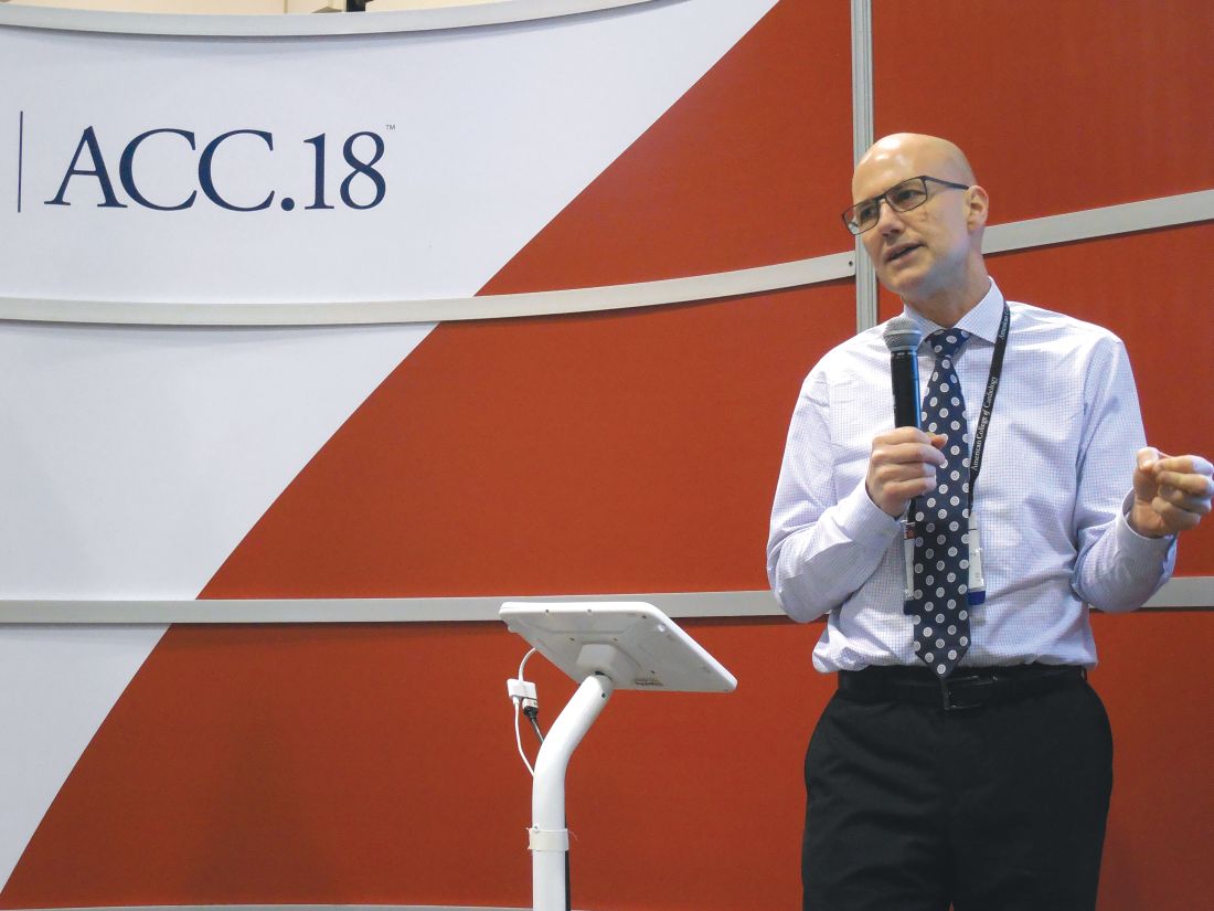
The ability of idarucizumab (Praxbind), conditionally approved by the Food and Drug Administration in 2015 and then fully approved in April 2018, to work in patients with impaired renal function has been an open question and concern because dabigatran (Pradaxa) is excreted renally, so it builds to unusually high levels in patients with poor kidney function. “Plasma dabigatran levels might be sky high, so a standard dose of idarucizumab might not work. That’s been a fear of clinicians,” explained Dr. Eikelboom, a hematologist at McMaster University in Hamilton, Ont.
To examine whether idarucizumab’s activity varied by renal function he used data from the patients enrolled in the RE-VERSE AD (Reversal Effects of Idarucizumab on Active Dabigatran) study, the pivotal dataset that led to idarucizumab’s U.S. approval. The new, post hoc analysis divided patients into four subgroups based on their kidney function, and focused on the 489 patients for whom renal data were available out of the 503 patients in the study (N Engl J Med. 2017 Aug 3;377[5]:431-41). The subgroups included 91 patients with severe dysfunction with a creatinine clearance rate of less than 30 mL/min; 127 with moderate dysfunction and a clearance rate of 30-49 mL/min; 163 with mild dysfunction and a clearance rate of 50-79 mL/min; and 108 with normal function and a creatinine clearance of at least 80 mL/min.
Patients in the subgroup with severe renal dysfunction had the worst clinical profile overall, and as predicted, had a markedly elevated average plasma level of dabigatran, 231 ng/mL, nearly five times higher than the 47-ng/mL average level in patients with normal renal function.
The ability of a single, standard dose of idarucizumab to reverse the anticoagulant effects of dabigatran were essentially identical across the four strata of renal activity, with 98% of patients in both the severely impaired subgroup and the normal subgroup having 100% reversal within 4 hours of treatment, Dr. Eikelboom reported. Every patient included in the analysis had more than 50% reversal.
The study followed patients to 12-24 hours after they received idarucizumab, and 55% of patients with severe renal dysfunction showed a plasma dabigatran level that crept back toward a clinically meaningful level and so might need a second idarucizumab dose. In contrast, this happened in 8% of patients with normal renal function.
In patients with severe renal dysfunction given idarucizumab, “be alert for a recurrent bleed,” which could require a second dose of idarucizumab, Dr. Eikelboom suggested.
SOURCE: Eikelboom JW et al. ACC 18, Abstract 1231M-11.
ORLANDO – Idarucizumab, the reversal agent for the anticoagulant dabigatran, appeared as effective in quickly reversing dabigatran’s effects in patients with severe renal dysfunction as in patients with normally working kidneys, in a post hoc analysis of data collected in the drug’s pivotal trial.
A standard dose of idarucizumab “works just as well in patients with bad kidney function as it does in patients with preserved kidney function,” John W. Eikelboom, MD, said at the annual meeting of the American College of Cardiology. “The time to cessation of bleeding and the degree of normal hemostasis achieved was consistent” across the entire range of renal function examined, from severe renal dysfunction, with a creatinine clearance rate of less than 30 mL/min, to normal function, with an estimated rate of 80 mL/min or greater.
The ability of idarucizumab (Praxbind), conditionally approved by the Food and Drug Administration in 2015 and then fully approved in April 2018, to work in patients with impaired renal function has been an open question and concern because dabigatran (Pradaxa) is excreted renally, so it builds to unusually high levels in patients with poor kidney function. “Plasma dabigatran levels might be sky high, so a standard dose of idarucizumab might not work. That’s been a fear of clinicians,” explained Dr. Eikelboom, a hematologist at McMaster University in Hamilton, Ont.
To examine whether idarucizumab’s activity varied by renal function he used data from the patients enrolled in the RE-VERSE AD (Reversal Effects of Idarucizumab on Active Dabigatran) study, the pivotal dataset that led to idarucizumab’s U.S. approval. The new, post hoc analysis divided patients into four subgroups based on their kidney function, and focused on the 489 patients for whom renal data were available out of the 503 patients in the study (N Engl J Med. 2017 Aug 3;377[5]:431-41). The subgroups included 91 patients with severe dysfunction with a creatinine clearance rate of less than 30 mL/min; 127 with moderate dysfunction and a clearance rate of 30-49 mL/min; 163 with mild dysfunction and a clearance rate of 50-79 mL/min; and 108 with normal function and a creatinine clearance of at least 80 mL/min.
Patients in the subgroup with severe renal dysfunction had the worst clinical profile overall, and as predicted, had a markedly elevated average plasma level of dabigatran, 231 ng/mL, nearly five times higher than the 47-ng/mL average level in patients with normal renal function.
The ability of a single, standard dose of idarucizumab to reverse the anticoagulant effects of dabigatran were essentially identical across the four strata of renal activity, with 98% of patients in both the severely impaired subgroup and the normal subgroup having 100% reversal within 4 hours of treatment, Dr. Eikelboom reported. Every patient included in the analysis had more than 50% reversal.
The study followed patients to 12-24 hours after they received idarucizumab, and 55% of patients with severe renal dysfunction showed a plasma dabigatran level that crept back toward a clinically meaningful level and so might need a second idarucizumab dose. In contrast, this happened in 8% of patients with normal renal function.
In patients with severe renal dysfunction given idarucizumab, “be alert for a recurrent bleed,” which could require a second dose of idarucizumab, Dr. Eikelboom suggested.
SOURCE: Eikelboom JW et al. ACC 18, Abstract 1231M-11.
REPORTING FROM ACC 18
Key clinical point: Renal function had no impact on idarucizumab’s efficacy for dabigatran reversal.
Major finding: Complete dabigatran reversal occurred in 98% of patients with severe renal dysfunction who received idarucizumab.
Study details: Post hoc analysis of data from RE-VERSE AD, idarucizumab’s pivotal trial with 503 patients.
Disclosures: RE-VERSE AD was funded by Boehringer Ingelheim, the company that markets idarucizumab (Praxbind) and dabigatran (Pradaxa). Dr. Eikelboom has been a consultant to and has received research support from Boehringer Ingelheim, as well as from Bayer, Bristol-Myers Squibb, Daiichi-Sankyo, Janssen, and Pfizer.
Source: Eikelboom JW et al. ACC 18, Abstract 1231M-11.
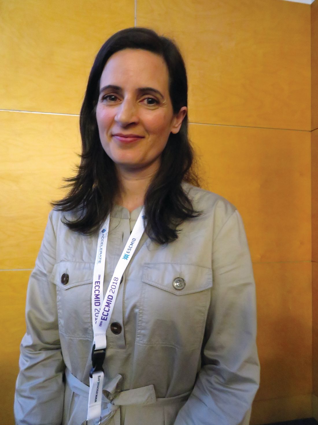
Nitrofurantoin beats fosfomycin for uncomplicated UTI
MADRID – (UTIs), a randomized study has determined.
By 28 days, clinical resolution had occurred in 70% of those who took nitrofurantoin and 58% of those who took fosfomycin – a statistically significant 12% difference, Angela Huttner, MD, said at the European Society of Clinical Microbiology and Infectious Diseases annual congress.
But the benefit was even more pronounced in women whose infections were caused by Escherichia coli, with a 28% spread in clinical resolution, (78% vs. 50%) and a 14-point spread in microbiological cure (72% vs. 58%), said Dr. Huttner of Geneva University, Switzerland.
The results were simultaneously published online in JAMA (2018 Apr 22. doi: 10.1001/jama.2018.3627).
Despite its success, nitrofurantoin did not live up to its purported 96% UTI cure rate – an established number based on study data from the 1950s-1970s.
Such efficacy was probably a false finding, she said. Studies of that era were much less rigorous than they are today, Dr. Huttner pointed out. The primary endpoint in those studies was typically defined not as complete resolution of symptoms – as it was in her study – but as resolution or improvement.
“Also, improvement was often defined microbiologically, often something like a decrease from 105 colony-forming units to 104, which is never something we would use today.”
The study was conducted in Geneva, Poland, and Israel. It randomized 512 women with an uncomplicated lower UTI to either 5 days of thrice-daily nitrofurantoin 100 mg or to a single 3-gram dose of fosfomycin. The women returned for clinical exam and urine culture at 14 and 28 days after they completed their treatment.
The primary outcome was 28-day clinical response. Success was defined as complete resolution of symptoms, a characterization that Dr. Huttner and her colleagues chose carefully. Many UTI studies include “improvement” in the clinical picture as part of a successful response. Dr. Huttner disagreed with that. “Our patients don’t want a partial response. They don’t want just an improvement. They want complete resolution of their symptoms.”
Failure was defined as the need for additional antibiotics or a change in antibiotic treatment. There was also an “indeterminate” category, for the small minority of patients who still felt some mild symptoms but were without microbiological signs of infection.
The mean age of the women was 44 years. All had an uncomplicated UTI characterized by dysuria, urgency, frequency, or suprapubic tenderness; 73% had a positive baseline urine culture. E. coli was the most common infective organism (about 60%) followed by different Klebsiella species, Proteus, and Enterococci. A few women had mixed pathogen infections. Only six patients had infective pathogens that were resistant to either of the study drugs.
After 28 days of treatment, a clinical cure was determined in 70% of those taking nitrofurantoin and 58% of those taking fosfomycin – an absolute difference of 12 points.
“The difference was obvious at 14 days,” Dr. Huttner noted. At that point, 75% of those taking nitrofurantoin and 66% of those taking fosfomycin reported resolution of their symptoms.
Pathology reflected the improving clinical picture: Microbiologic resolution occurred in 74% of the nitrofurantoin group and 63% of the fosfomycin group.
A post hoc analysis looked at results among the 214 women with confirmed E. coli infections.
The difference in clinical response was “even more pronounced” in these patients, Dr. Huttner said. Through day 28, clinical resolution occurred in 78% of those taking nitrofurantoin and 50% of those taking fosfomycin – a significant difference of 28 points.
Patients with E. coli infections were 4.48 times more likely to fail treatment if they received fosfomycin than if they received nitrofurantoin.
Adverse events were few and primarily gastrointestinal. The most common were mild to moderate nausea and diarrhea (less than 4% in each group).
Both of the antibiotics were popular from the 1950s on, but gradually fell out of favor as more powerful therapies were developed. However, as antibiotic resistance began to develop, infectious disease specialists began to support bringing nitrofurantoin and fosfomycin out of mothballs. In 2011, a panel of international experts convened by the Infectious Diseases Society of America (IDSA) and the European Society of Clinical Microbiology and Infectious Diseases (ESCMID) recommended both of the medications as first-line therapy for women with acute uncomplicated cystitis and pyelonephritis.
The group recommended fosfomycin in a single 3-gram dose and a nitrofurantoin regimen of 100 mg twice daily for 5 days. The fosfomycin recommendation is clearly inadequate, Dr. Huttner said.
“Fosfomycin is not a bad drug. I just think it’s underdosed in this setting,” she said.
Dr. Huttner had no financial disclosures.
SOURCE: Huttner A et al. ECCMID 2018. Abstract O0479.
REPORTING FROM ECCMID 2018
Key clinical point: Nitrofurantoin was significantly more effective than was fosfomycin for a clinical and microbiological cure of uncomplicated UTI in women.
Major finding: By 28 days, clinical resolution had occurred in 70% of those who took nitrofurantoin and 58% of those who took fosfomycin .
Study details: The prospective study randomized 512 women.
Disclosures: Dr. Huttner had no financial disclosures.
Source: Huttner A et al. ECCMID 2018. Abstract O0479.
Nonopioids as effective as opioids in reducing acute pain in the ED
Clinical question: What is the most effective analgesic combination, opioid vs. nonopioid, for treating acute extremity pain in the emergency department?
Background: Patients often are prescribed opioids for acute pain in the ED while awaiting work-up. With the current opioid epidemic in the United States, it is important to examine the appropriate use of opioids and look for alternative medications for acute pain.
Setting: Two urban New York City emergency departments from July 2015 to August 2016.
Synopsis: 411 patients aged 21-65 years were randomized to four groups for treatment of acute extremity pain. Each received one oral analgesic combination: ibuprofen 400 mg and acetaminophen 1,000 mg; oxycodone 5 mg and acetaminophen 325 mg; hydrocodone 5 mg and acetaminophen 300 mg; or codeine 30 mg and acetaminophen 300 mg. Their pain was scored on presentation using a standard 0-10 numerical rating scale (NRS) and then at 2 hours after medication. The primary outcome was difference in NRS among groups. All patients had improvement in pain scores. Pain score improved by 4.4 in the oxycodone group, 4.3 in the ibuprofen group, 3.5 in the hydrocodone group, and 3.9 in the codeine group. There were no statistically significant differences among groups. Limitations to the study included short follow-up time, no reported data on adverse effects, and lack of uniform acetaminophen doses in each group.
Bottom line: There was no statistically significant difference in pain control among patients given a combination of acetaminophen and ibuprofen vs. three different opioids with acetaminophen when treating acute extremity pain in the ED.
Citation: Chang AK et al. Effect of a single dose of oral opioid and nonopioid analgesics on acute extremity pain in the emergency department: a randomized clinical trial. JAMA. 2017 Nov 7;318(17):1661-7.
Dr. Mathew is assistant professor of medicine, division of hospital medicine, University of Virginia.
VTE risk after bariatric surgery should be assessed
SEATTLE – Preop thromboelastometry can identify patients who need extra according to a prospective investigation of 40 patients at Conemaugh Memorial Medical Center in Johnstown, Pa.
Enoxaparin 40 mg twice daily just wasn’t enough for people who were hypercoagulable before surgery. The goal of the study was to find the best way to prevent venous thromboembolism (VTE) after weight loss surgery. At present, there’s no consensus on prophylaxis dosing, timing, duration, or even what agent to use for these patients. Conemaugh uses postop enoxaparin, a low-molecular-weight heparin. Among many other options, some hospitals opt for preop dosing with traditional heparin, which is less expensive.

The Conemaugh team turned to thromboelastometry (TEM) to look at the question of VTE risk in bariatric surgery patients. The test gauges coagulation status by measuring elasticity as a small blood sample clots over a few minutes. The investigators found that patients who were hypercoagulable before surgery were likely to be hypercoagulable afterwards. The finding argues for baseline TEM testing to guide postop anticoagulation.
The problem is that bariatric services don’t often have access to TEM equipment, and insurance doesn’t cover the $60 test. In this instance, the Lake Erie College of Osteopathic Medicine in Erie, Pa., had the equipment and covered the testing for the study.
The patients had TEM at baseline and then received 40 mg of enoxaparin about 4 hours after surgery – mostly laparoscopic gastric bypasses – and a second dose about 12 hours after the first. TEM was repeated about 2 hours after the second dose.
At baseline, 2 (5%) of the patients were hypocoagulable, 15 (37.5%) were normal, and 23 (57.5%) were hypercoagulable. On postop TEM, 17 patients (42.5%) were normal and 23 (57.5%) were hypercoagulable: “These 23 were inadequately anticoagulated,” said lead investigator Daniel Urias, MD, a general surgery resident at the medical center.
“There was an association between being normal at baseline and being normal postop, and being hypercoagulable at baseline and hypercoagulable postop. We didn’t anticipate finding such similarity between the numbers. Our suspicion that baseline status plays a major role is holding true,” Dr. Urias said at the World Congress of Endoscopic Surgery hosted by SAGES & CAGS.
When patients test hypercoagulable at baseline, “we are [now] leaning towards [enoxaparin] 60 mg twice daily,” he said.
Ultimately, anticoagulation TEM could be used to titrate patients into the normal range. For best outcomes, it’s likely that “obese patients require goal-directed therapy instead of weight-based or fixed dosing,” he said, but nothing is going to happen until insurance steps up.
The patients did not have underlying coagulopathies, and 33 (82.5%) were women; the average age was 44 years and average body mass index was 43.6 kg/m2. The mean preop Caprini score was 4, indicating moderate VTE risk. Surgery lasted about 200 minutes. Patients were out of bed and walking on postop day 0.
The investigators had no relevant disclosures.
SOURCE: Urias D et al. World Congress of Endoscopic Surgery hosted by SAGES & CAGS abstract S023.
SEATTLE – Preop thromboelastometry can identify patients who need extra according to a prospective investigation of 40 patients at Conemaugh Memorial Medical Center in Johnstown, Pa.
Enoxaparin 40 mg twice daily just wasn’t enough for people who were hypercoagulable before surgery. The goal of the study was to find the best way to prevent venous thromboembolism (VTE) after weight loss surgery. At present, there’s no consensus on prophylaxis dosing, timing, duration, or even what agent to use for these patients. Conemaugh uses postop enoxaparin, a low-molecular-weight heparin. Among many other options, some hospitals opt for preop dosing with traditional heparin, which is less expensive.
The Conemaugh team turned to thromboelastometry (TEM) to look at the question of VTE risk in bariatric surgery patients. The test gauges coagulation status by measuring elasticity as a small blood sample clots over a few minutes. The investigators found that patients who were hypercoagulable before surgery were likely to be hypercoagulable afterwards. The finding argues for baseline TEM testing to guide postop anticoagulation.
The problem is that bariatric services don’t often have access to TEM equipment, and insurance doesn’t cover the $60 test. In this instance, the Lake Erie College of Osteopathic Medicine in Erie, Pa., had the equipment and covered the testing for the study.
The patients had TEM at baseline and then received 40 mg of enoxaparin about 4 hours after surgery – mostly laparoscopic gastric bypasses – and a second dose about 12 hours after the first. TEM was repeated about 2 hours after the second dose.
At baseline, 2 (5%) of the patients were hypocoagulable, 15 (37.5%) were normal, and 23 (57.5%) were hypercoagulable. On postop TEM, 17 patients (42.5%) were normal and 23 (57.5%) were hypercoagulable: “These 23 were inadequately anticoagulated,” said lead investigator Daniel Urias, MD, a general surgery resident at the medical center.
“There was an association between being normal at baseline and being normal postop, and being hypercoagulable at baseline and hypercoagulable postop. We didn’t anticipate finding such similarity between the numbers. Our suspicion that baseline status plays a major role is holding true,” Dr. Urias said at the World Congress of Endoscopic Surgery hosted by SAGES & CAGS.
When patients test hypercoagulable at baseline, “we are [now] leaning towards [enoxaparin] 60 mg twice daily,” he said.
Ultimately, anticoagulation TEM could be used to titrate patients into the normal range. For best outcomes, it’s likely that “obese patients require goal-directed therapy instead of weight-based or fixed dosing,” he said, but nothing is going to happen until insurance steps up.
The patients did not have underlying coagulopathies, and 33 (82.5%) were women; the average age was 44 years and average body mass index was 43.6 kg/m2. The mean preop Caprini score was 4, indicating moderate VTE risk. Surgery lasted about 200 minutes. Patients were out of bed and walking on postop day 0.
The investigators had no relevant disclosures.
SOURCE: Urias D et al. World Congress of Endoscopic Surgery hosted by SAGES & CAGS abstract S023.
REPORTING FROM SAGES 2018
Key clinical point: Preoperative thromboelastometry identifies patients who need extra anticoagulation against venous thromboembolism following bariatric surgery.
Major finding: Baseline and postop coagulation were similar: 37.5% vs. 42.5% were normal and 57.5% vs 57.5% were hypercoagulable.
Study details: Prospective study of 40 bariatric surgery patients.
Disclosures: The investigators did not have any relevant disclosures. The Lake Erie College of Osteopathic Medicine paid for the testing.
Source: Urias D et al. World Congress of Endoscopic Surgery hosted by SAGES & CAGS abstract S023.