User login
Communicate with Millennials using their preferred methods
ORLANDO – Pediatricians can learn from how Millennials use services and purchase products from popular companies, such as Amazon Alexa, Warby Parker, Instagram, and Snapchat, and use that information to connect with Millennial parents and children in their practice, Kristopher Jones, JD, MS, said.
In a video interview, Mr. Jones explained how Amazon Alexa uses structured data as recommendations when users make queries of the service; for example, a Millennial might make a choice to book an appointment with a pediatrician based on the results Amazon Alexa displays, such as an office’s available hours. An office that is not optimized to appear in those results would be missed by those potential patients.
Warby Parker has a digital-first strategy focused on convenience, ease of use, and an emphasis on mission; one opportunity for pediatricians in this area is to be more overt about which causes they are supporting, Mr. Jones said. In the case of Instagram and Snapchat, pediatricians should consider creating a presence on these platforms and focusing on less text-heavy content to appeal to Millennials.
“It’s a really, really important opportunity to be able to meet the Millennials where they want to be met, and that means developing strategies to leveraging pictures and videos and other forms of Millennial content to communicate with them,” he said.
Mr. Jones reported no relevant conflicts of interest.
REPORTING FROM AAP 2018
Emergency physician describes how to prepare for disasters
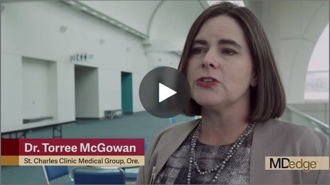
, according to Dr. Toree McGowan, an emergency physician who works in a critical care facility in rural Oregon.

In our video interview at the annual meeting of the American College of Emergency Physicians, she outlined key strategies for obtaining resources and delegating care when managing mass casualties from disasters.
Dr. McGowan of the St. Charles Medical Group, Culver, Ore., said that, although she is the only physician at her rural critical care center about 70% of the time, she has established plans in place for obtaining additional staff and resources in the event of disasters. During her time in the military, she was among a team that implemented a disaster plan after a toxic chemical release at a nearby factory. The response was effective because the threat had been anticipated and a plan was in place. To develop the skills and strategies she describes in this interview, Dr. McGowan recommends free training that is available from the nonprofit National Disaster Life Support Foundation.
, according to Dr. Toree McGowan, an emergency physician who works in a critical care facility in rural Oregon.

In our video interview at the annual meeting of the American College of Emergency Physicians, she outlined key strategies for obtaining resources and delegating care when managing mass casualties from disasters.
Dr. McGowan of the St. Charles Medical Group, Culver, Ore., said that, although she is the only physician at her rural critical care center about 70% of the time, she has established plans in place for obtaining additional staff and resources in the event of disasters. During her time in the military, she was among a team that implemented a disaster plan after a toxic chemical release at a nearby factory. The response was effective because the threat had been anticipated and a plan was in place. To develop the skills and strategies she describes in this interview, Dr. McGowan recommends free training that is available from the nonprofit National Disaster Life Support Foundation.
, according to Dr. Toree McGowan, an emergency physician who works in a critical care facility in rural Oregon.

In our video interview at the annual meeting of the American College of Emergency Physicians, she outlined key strategies for obtaining resources and delegating care when managing mass casualties from disasters.
Dr. McGowan of the St. Charles Medical Group, Culver, Ore., said that, although she is the only physician at her rural critical care center about 70% of the time, she has established plans in place for obtaining additional staff and resources in the event of disasters. During her time in the military, she was among a team that implemented a disaster plan after a toxic chemical release at a nearby factory. The response was effective because the threat had been anticipated and a plan was in place. To develop the skills and strategies she describes in this interview, Dr. McGowan recommends free training that is available from the nonprofit National Disaster Life Support Foundation.
REPORTING FROM ACEP18
AAP renews public health approach to gun injury prevention
ORLANDO – The American Academy of Pediatrics created the Gun Safety and Injury Prevention Research Initiative to study and implement gun safety interventions to prevent homicide, suicide and unintentional injuries caused by firearms.
In an interview at the annual meeting of the American Academy of Pediatrics, Colleen A. Kraft, MD, FAAP, current AAP president, explained how AAP has renewed its efforts to protect children from firearm injuries. Black children are more likely to die in a homicide, while white children are more likely to die in a suicide through use of a firearm, Dr. Kraft said. The AAP seeks to find a nonpolitical way to discuss gun safety “with a lens on children and a lens on safety,” she said.
“What we are looking to do is to bring together partners who have the research expertise in gun safety and injury prevention, find out what we know, decide what we don’t know yet, and begin to bring together focus groups of parents and families and legislators and doctors, and people to talk about … gun safety in a way that resonates with everyone,” Dr. Kraft said.
Visit AAP’s website for more information on the Gun Safety and Injury Prevention Research Initiative.
Dr. Kraft reports no relevant conflicts of interest.
REPORTING FROM AAP 2018
AAP president affirms academy’s support for transgender children
ORLANDO – Many Colleen A. Kraft, MD, president of the American Academy of Pediatrics, said in an interview.
The AAP’s policy on caring and supporting these patients is evidence-based, and includes recommendations on providing appropriate health care services for transgender individuals, as well as respect and understanding for families. In the interview, Dr. Kraft, discussed the role pediatricians have in providing a safe and supportive environment for transgender and gender-diverse individuals and their families.
“[These children] need to be listened to, they need to be respected for who they are, and they need access to the appropriate health services,” Dr. Kraft said.
The AAP’s policy statement on ensuring care and support for transgender children and adolescents is available here.
Dr. Kraft reported no relevant conflicts of interest.
REPORTING FROM AAP 2018
Childhood abuse linked with tripled adult SLE incidence
CHICAGO – , in a study of more than 67,000 American nurses.
The results also suggested that development of depression and post-traumatic stress disorder (PTSD) may have been intermediary steps between episodes of childhood abuse and later development of systemic lupus erythematosus (SLE), Candace H. Feldman, MD, said at the annual meeting of the American College of Rheumatology.
These findings suggest the “importance of screening for childhood abuse exposures as well as for depression and PTSD in routine practice,” although Dr. Feldman acknowledged that interventions aimed at treating depression and PTSD have as of now no proven role for mitigating SLE.
The analysis Dr. Feldman and her associates ran on data collected in the Nurses Health Study II also documented a “striking” number of the enrolled women who completed the survey in 2001 and reported a history of abuse when they were 11 years old or younger: 30% of the 67,516 respondents reported a moderate level of abuse, and 24% reported a high level of abuse. An additional 22% reported either no or a very low level of abuse. These numbers suggest that abuse of girls “is very common and probably underreported,” she said in a video interview.
The Nurses Health Study II enrolled more than 116,429 U.S. women in 1989 who were 25-42 years old and had no history of SLE. Recording of incident SLE cases began in 1991 and for this analysis continued for 24 years, through 2015, during which time 94 women developed SLE that was confirmed in a review by two rheumatologists applying the 1997 SLE classification criteria (Arthritis Rheum. 1997 Sept;40[9]:1725. The incidence of SLE was 2.57-fold more common among women who reported a high level of abuse, compared with those who had no or very low abuse, after adjustment for several demographic and clinical confounders, reported Dr. Feldman, a rheumatologist at Brigham and Women’s Hospital in Boston.
“To our knowledge this is the first study to prospectively look at exposure to different forms of childhood abuse and SLE incidence in a general population of women,” she said.
To make the analysis more prospective the researchers also ran a calculation that considered only SLE cases that appeared after completion of the 2001 abuse survey. Using this criterion the incidence was 3.11-fold higher among women who reported a high level of childhood abuse. Further analyses showed that statistically a diagnosis of PTSD accounted for about 23% of the risk for developing SLE, and depression appeared responsible for about 17% of the risk. The analysis also showed no statistically significant link between sexual abuse in childhood or as a teenager and later onset of SLE.
The findings are consistent with prior reports that linked stress to development of various autoimmune diseases, Dr. Feldman noted. She speculated that high childhood stress could cause changes in inflammation, immune function, epigenetics, the autonomic nervous system, and endocrine pathways that could play a role in triggering depression or PTSD, and eventually SLE.
[email protected]
On Twitter @mitchelzoler
SOURCE:Feldman C et al. Arthritis Rheumatol. 2018;70(suppl 10) Abstract 2807.
REPORTING FROM THE ACR ANNUAL MEETING
Key clinical point: A history of high childhood abuse linked with a nearly three-fold higher incidence of systemic lupus erythematosus during adulthood.
Major finding: The incidence of systemic lupus erythematosus was 2.57-fold higher among women with high childhood abuse compared with unabused women.
Study details: Data from 67,516 women enrolled in the Nurses Health Study II.
Disclosures: Dr. Feldman had no disclosures.
Source: Feldman C et al. Arthritis Rheumatol. 2018;70(suppl 10) Abstract 2807.
Appendix linked to Parkinson’s disease in series of unexpected findings
Appendectomy has been associated with a reduced risk of Parkinson’s disease (PD), which supports the potential for a reservoir of aggregated alpha-synuclein in the appendix to affect risk of the condition, according to new epidemiologic and translational evidence from two data sets that promotes a new and emerging theory for PD etiology.
When placed into the context of other recent studies, these epidemiologic data “point to the appendix as a site of origin for Parkinson’s and provide a path forward for devising new treatment strategies,” reported senior author Viviane Labrie, PhD, of the Van Andel Research Institute (VARI) in Grand Rapids, Mich.
The epidemiologic data was the most recent step in a series of findings summarized in a newly published paper in Science Translational Medicine. As the researchers explained, it is relevant to a separate body of evidence that alpha-synuclein, a protein that serves as the hallmark of PD when it appears in Lewy bodies, can be isolated in the nerve fibers and nerve cells of the appendix.
“We have shown that alpha-synuclein proteins, including the truncated forms observed in Lewy bodies, are abundant in the appendix,” reported first author Bryan A. Killinger, PhD, also at VARI, in a press teleconference. He said this finding is likely to explain the reduced risk of PD from appendectomy.
In the largest of the epidemiologic studies, the effect of appendectomy on subsequent risk of PD was evaluated through the health records from more than 1.6 million individuals in Sweden. The incidence of PD was found to be 19.3% lower among 551,647 patients who had an appendectomy, compared with controls.
In addition, the data showed that when PD did occur after appendectomy, it was delayed on average by 3.6 years. It is notable that appendectomy was not associated with protection from PD in patients with a familial link to PD, a group they said comprises less than 10% of cases.
In patients with PD, nonmotor symptoms often include GI tract dysfunction, which can, in some cases, be part of a prodromal presentation that precedes the onset of classical PD symptoms by several years, the authors reported. However, the new research upends previous conceptions of disease. The demonstration of abundant alpha-synuclein in the appendix coupled with the protective effect of appendectomy, suggests that PD may originate in the GI tract and then spread to the central nervous system (CNS) rather than the other way around.
“The vermiform appendix was once considered to be an unnecessary organ. Although there is now good evidence that the appendix plays a major role in the regulation of the immune system, including the regulation of gut bacteria, our work suggests it is also mediates risk of Parkinson’s,” Dr. Labrie said in the teleconference.
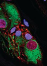
In the paper, numerous pieces of the puzzle are brought together to suggest that alpha-synuclein in the appendix is linked to alpha-synuclein in the CNS. Many of the findings along this investigative pathway were described as surprising. For example, immunohistochemistry studies revealed high amounts of alpha-synuclein in nearly every sample of appendiceal tissue examined, including normal and inflamed tissue, tissue from individuals with PD and those without, and tissues from young and old individuals.
“The normal tissue, as well as appendiceal tissue from PD patients, contained high levels of alpha-synuclein in the truncated forms analogous to those seen in Lewy body pathology,” Dr. Killinger said. Based on these and other findings, he believes that alpha-synuclein in the appendix forms a reservoir for seeding the aggregates involved in the pathology of PD, although he acknowledged that it is not yet clear how the proteins in the appendix find their way to the brain.
From these data, it appears that most individuals with an intact appendix have alpha-synuclein in the nerve fibers, but Dr. Labrie pointed out that the only about 1% of the population develops PD. She speculated that there is “some confluence of events,” such as an environmental trigger altering the GI microbiome, that mediates ultimate risk of PD, but she noted that these events may take place decades before signs and symptoms of PD develop. The data appear to be a substantial reorientation in understanding PD.
“We have shown that the appendix is a hub for the accumulation of clumped forms of alpha-synuclein proteins, which are implicated in Parkinson’s,” Dr. Killinger said. “This knowledge will be invaluable as we explore new prevention and treatment strategies.”
The research was funded by a variety of governmental and private grants to individual authors. Dr. Killinger and Dr. Labrie report no financial relationships relevant to this study.
SOURCE: Killinger BA et al. Sci Transl Med. 2018;10:eaar5380.
FROM SCIENCE TRANSLATIONAL MEDICINE
Key clinical point:
Major finding: A 19.3% reduction in risk of PD from appendectomy may relate to alpha-synuclein in the appendix.
Study details: Series of related epidemiologic and translational studies.
Disclosures: The research was funded by a variety of governmental and private grants to individual authors. Dr. Killinger and Dr. Labrie report no financial relationships relevant to this study.
Source: Killinger BA et al. Sci Transl Med. 2018;10:eaar5380.
Brisk walking may decrease TKR risk in OA
CHICAGO – according to a new analysis of data from the National Institutes of Health-sponsored Osteoarthritis Initiative.

Whether walking increases or decreases the risk of structural deterioration and total knee replacement (TKR) in patients with knee osteoarthritis has been a controversial topic marked by conflicting data. That’s probably because prior studies haven’t taken into account walking intensity, Hiral Master said at the annual meeting of the American College of Rheumatology.
Ms. Master, a PhD candidate in physical therapy at the University of Delaware, Newark, presented a study of 1,854 patients with knee osteoarthritis who participated in the Osteoarthritis Initiative, all of whom had worn an accelerometer. This permitted calculation of time spent walking at various intensities. Subjects spent an average of 459 minutes per day not walking and 8 minutes walking at moderate to vigorous intensity, defined as a cadence of more than 100 steps per minute.
During 5 years of follow-up, the incidence of TKR was 6%. In this video interview, Ms. Master explains that patients who replaced 5 minutes of not walking with 5 minutes of brisk walking daily had an adjusted 14% reduction in the risk of TKR. A dose-response was evident, with more minutes of moderate to vigorous walking being associated with progressively larger reductions in the risk of this major surgery. Walking at a cadence of less than 100 steps per minute, regardless of duration, was nonprotective.
SOURCE: Master H et al. Arthritis Rheumatol. 2018;70(Suppl 10), Abstract 1166.
CHICAGO – according to a new analysis of data from the National Institutes of Health-sponsored Osteoarthritis Initiative.

Whether walking increases or decreases the risk of structural deterioration and total knee replacement (TKR) in patients with knee osteoarthritis has been a controversial topic marked by conflicting data. That’s probably because prior studies haven’t taken into account walking intensity, Hiral Master said at the annual meeting of the American College of Rheumatology.
Ms. Master, a PhD candidate in physical therapy at the University of Delaware, Newark, presented a study of 1,854 patients with knee osteoarthritis who participated in the Osteoarthritis Initiative, all of whom had worn an accelerometer. This permitted calculation of time spent walking at various intensities. Subjects spent an average of 459 minutes per day not walking and 8 minutes walking at moderate to vigorous intensity, defined as a cadence of more than 100 steps per minute.
During 5 years of follow-up, the incidence of TKR was 6%. In this video interview, Ms. Master explains that patients who replaced 5 minutes of not walking with 5 minutes of brisk walking daily had an adjusted 14% reduction in the risk of TKR. A dose-response was evident, with more minutes of moderate to vigorous walking being associated with progressively larger reductions in the risk of this major surgery. Walking at a cadence of less than 100 steps per minute, regardless of duration, was nonprotective.
SOURCE: Master H et al. Arthritis Rheumatol. 2018;70(Suppl 10), Abstract 1166.
CHICAGO – according to a new analysis of data from the National Institutes of Health-sponsored Osteoarthritis Initiative.

Whether walking increases or decreases the risk of structural deterioration and total knee replacement (TKR) in patients with knee osteoarthritis has been a controversial topic marked by conflicting data. That’s probably because prior studies haven’t taken into account walking intensity, Hiral Master said at the annual meeting of the American College of Rheumatology.
Ms. Master, a PhD candidate in physical therapy at the University of Delaware, Newark, presented a study of 1,854 patients with knee osteoarthritis who participated in the Osteoarthritis Initiative, all of whom had worn an accelerometer. This permitted calculation of time spent walking at various intensities. Subjects spent an average of 459 minutes per day not walking and 8 minutes walking at moderate to vigorous intensity, defined as a cadence of more than 100 steps per minute.
During 5 years of follow-up, the incidence of TKR was 6%. In this video interview, Ms. Master explains that patients who replaced 5 minutes of not walking with 5 minutes of brisk walking daily had an adjusted 14% reduction in the risk of TKR. A dose-response was evident, with more minutes of moderate to vigorous walking being associated with progressively larger reductions in the risk of this major surgery. Walking at a cadence of less than 100 steps per minute, regardless of duration, was nonprotective.
SOURCE: Master H et al. Arthritis Rheumatol. 2018;70(Suppl 10), Abstract 1166.
REPORTING FROM THE ACR ANNUAL MEETING
Antiphospholipid antibodies are surprisingly common in first-MI patients
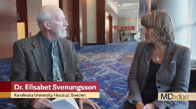
CHICAGO – Patients with a first MI were nearly nine times more likely to have detectable IgG antiphospholipid antibodies than were matched controls in a cross-sectional cohort study, Elisabet Svenungsson, MD, PhD, reported at the annual meeting of the American College of Rheumatology.
Her case-control study included 805 Swedish patients tested for antiphospholipid antibodies 6-10 weeks after experiencing their first MI and an equal number of age-, sex-, and location-matched controls. Prior to their MIs, none of the patients had been diagnosed with antiphospholipid syndrome, which requires both positive antiphospholipid antibodies and a vascular thrombotic event or obstetric morbidity.
A positive test for IgG anti-cardiolipin antibody was present in 10.9% of the first-MI patients, compared with 0.9% of controls. Similarly, 10.4% of acute MI patients and 0.9% of controls were positive for anti-beta2-glycoprotein-1 antibodies. Most patients who tested positive for one were positive for both. Thus, it’s possible that IgG antiphospholipid antibody positivity is an important silent risk factor that’s present in 1 in 10 MI patients, according to Dr. Svenungsson, professor of rheumatology at the Karolinska Institute in Stockholm.
If these results are confirmed and expanded upon in additional studies, testing for antiphospholipid antibodies could become part of the routine care in patients with an acute MI. Those who test positive would meet the criteria for antiphospholipid syndrome and qualify for long-term oral anticoagulation to reduce their elevated risk of further vascular events, she explained in this video interview.
The study was published in Annals of Internal Medicine simultaneously with the presentation at the ACR annual meeting (Ann Int Med. 2018 Oct 23. doi: 10.7326/M18-2130).
SOURCE: Grosso G et al. Arthritis Rheumatol. 2018;70(Suppl 10): Abstract 855.
REPORTING FROM THE ACR ANNUAL MEETING
Technology offers new tools, challenges for rare-disease patients
WASHINGTON – Genetic developments may create a new medical model for patients with rare diseases and the doctors who treat them, according to Marshall Summar, MD, chief of genetics and metabolism at Children’s National Medical Center in Washington, D.C.
In an interview at the NORD Rare Summit, held by the National Organization for Rare Disorders, Dr. Summar and Peter L. Saltonstall, president and CEO of NORD, discussed hot topics in the rare disease field. Those include new knowledge of the natural history of rare diseases, made possible by the creation of patient databases and the expansion of genetic technology. In addition, some DNA therapies “are finally crossing the finish line,” said Dr. Summar. That means clinicians will be looking at some rare diseases as acute conditions rather than chronic.
However, patients with rare diseases continue to face challenges in terms of the need for prior authorization and for drug access. One of NORD’s missions is to help patients access treatment. “We are seeing these prior authorizations take weeks or even longer,” Mr. Saltonstall said – and meanwhile, patients aren’t receiving therapy.
Visit rarediseases.org for more information about NORD’s ongoing research and advocacy efforts.
Dr. Summar and Mr. Saltonstall had no financial conflicts to disclose.
REPORTING FROM NORD SUMMIT 2018
Education and support enhance care for rare-disease patients
WASHINGTON – Physicians in primary and specialty care can provide guidance and support to patients with rare diseases by educating themselves about the resources available, according to Tim Boyd, director of state policy for the National Organization for Rare Disorders (NORD).
In an interview at the NORD Rare Summit, held by the National Organization for Rare Disorders, Mr. Boyd and Melinda Burnworth, PharmD, a pharmacist and NORD state volunteer from Arizona, discussed challenges faced by patients with rare diseases, including securing a correct diagnosis, accessing medication, and managing treatment going forward.
Physicians who understand some of the barriers to medication access can help advocate for their patients, explained Mr. Boyd, and those who know about resources for rare disorders can help make a diagnosis.
“All health care providers have an opportunity to enhance care for patients with rare disorders,” said Dr. Burnworth, author of the Rare Disease eResource Guide, available through the American Society of Health-System Pharmacists. Visit rarediseases.org for more information about NORD’s ongoing research and advocacy efforts.
Mr. Boyd and Dr. Burnworth had no financial conflicts to disclose.
REPORTING FROM NORD SUMMIT 2018

