User login
Does congenital cardiac surgery training need a makeover?
Trainees in congenital cardiac surgery fellowship programs are doing more operations since the programs became accredited in 2007, but no clear parameters have emerged to determine if certification has improved the quality of training, according to an evaluation of fellowship training programs published in the June issue of the Journal of Thoracic and Cardiovascular Surgery (2016 Jun;151:1488-95).
Overall, the training has become standardized, the fellows’ operative experience is “robust,” and fellows are mostly satisfied since the Accreditation Council of Graduate Medical Education (ACGME) recognized congenital cardiac surgery as a fellowship in 2007, lead study author Dr. Brian Kogon of Emory University, Atlanta, said.
However, Dr. Kogon and his colleagues also found some shortcomings in fellowship training. They received survey responses from 36 of 44 fellows in 12 accredited programs nationwide. To determine if fellows were meeting minimum case requirements, they also reviewed operative logs of 38 of the 44 fellows. They compared their findings to a study of congenital cardiac surgery fellowship programs they did pre-ACGME accreditation (J Thorac Cardiovasc Surg. 2006 Dec;132:1280). “The number of operations performed by the fellows during their training was underwhelming, and most of the fellows were dissatisfied with their operative experience,” Dr. Kogon and his colleagues wrote in the earlier study.
The study found that all fellows achieved the minimum number of 75 total cases the standards require for graduation, with a median of 136; and the minimum standard of 36 specific qualifying cases with a median of 63. However, seven did not meet the minimum of five complex neonate cases. Among other types of operations for which fellows failed to meet the minimum cases were atrioventricular septal defect repair, arch reconstruction including coarctation procedures and systemic-to-pulmonary artery shunt procedures.
The comparative lack of adult cardiac surgery operations was also considered a potential problem, the authors noted, pointing out that “the number of adults who have congenital heart disease now exceeds the number of children who have the disease, and many of these patients will require an operation.”
Another shortcoming the study found was a drop-off in international fellowships since 2007. “This change places us at risk of becoming intellectually isolated and losing international relationships that are critical to the future of our specialty,” Dr. Kogon and his colleagues wrote. Graduated fellows also acknowledged dissatisfaction with their lack of exposure to neonate surgery.
The study also determined the following demographics of the fellows: 83% are men and the median age at graduation was 40 years, with a range of 35-48 years. Only 25% of graduates participated in nonsurgical rotations such as cardiac catheterization and echocardiography.
“Although the operative experience seems to be much more robust, and this finding has been corroborated in other surgical disciplines after the advent of ACGME accreditation, comparing training before and after the accreditation process came into existence is difficult,” Dr. Kogon and his colleagues said.
The study also noted that the Thoracic Surgery Directors Association developed a congenital curriculum for congenital cardiothoracic surgery fellows, but only 28% used that curriculum and only 61% used any formal curriculum. “Unfortunately, regardless of the curriculum, only 50% of the graduates found it helpful,” Dr. Kogon and his colleagues said.
And regardless of the curriculum, only half of the graduates have passed the written qualifying and oral certifying examinations after completing their fellowship. “Although the curriculum is quite robust, the latter statistic suggests that we need either more emphasis on education by the program directors or a better and/or different curriculum,” Dr. Kogon and his colleagues said. However, they added that “after training, former fellows have adequate case volumes and mixes and seem to be thriving in the field.”
Dr. Kogon and his study coauthors had no financial disclosures.
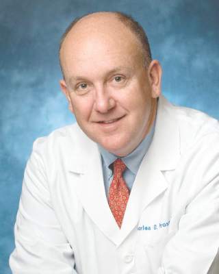
In his invited commentary, Dr. Charles D. Fraser Jr. of Texas Children’s Hospital, Baylor University, Houston, called the study findings that only 50% of congenital cardiac surgery fellowship graduates had passed the congenital examination “quite disturbing” and the demographic data and surgical and nonsurgical experience of the trainees “thought provoking” (J Thorac Cardiovasc Surg. 2016;151:1496-7)
“Is the bar too high or too low?” Dr. Fraser asked. He suggested the fellowship training system for congenital cardiac surgeons may be a work in progress. “For one, having a median age of 40 years for graduates is unacceptable,” he said. For half of trainees to not pass the examination “at this advanced age is tragic.” That 25% of fellows participate in nonsurgical rotations “also is concerning.”
|
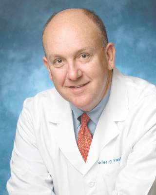
Dr. Charles D. Fraser |
A challenge is that after fellows complete their training in general and cardiothoracic surgery, opportunities to operate on newborns in a new fellowship setting are extremely limited, Dr. Fraser said. “To expect someone to be able to perform complex newborn heart surgery with excellent outcomes in a brand-new environment after just learning how to perform adult cardiac surgery is unrealistic,” he said.
Dr. Fraser said 1 formal year of training for congenital cardiac surgery fellows may not be enough. “Our colleagues in general pediatric surgery have a 2-year fellowship, and our specialty is every bit as complex as theirs,” he said. The basic American Board of Thoracic Surgery thoracic fellowship should have more latitude in its congenital heart surgery rotations, including exposure to pediatrics, neonatal/pediatric critical care, and the nonsurgical rotations the study referred to. Congenital heart surgery fellowships should also embrace adult congenital heart surgery with a more formalized experience requirement, he said.
“As a specialty, we owe it to our fine young surgeon candidates to offer the most robust and fair pathway to success while never compromising on the public trust and patient well-being,” Dr. Fraser said.
Dr. Fraser is chief of the division of congenital heart surgery at Baylor and codirector of the Texas Children’s Heart Center. He had no financial relationships to disclose.
In his invited commentary, Dr. Charles D. Fraser Jr. of Texas Children’s Hospital, Baylor University, Houston, called the study findings that only 50% of congenital cardiac surgery fellowship graduates had passed the congenital examination “quite disturbing” and the demographic data and surgical and nonsurgical experience of the trainees “thought provoking” (J Thorac Cardiovasc Surg. 2016;151:1496-7)
“Is the bar too high or too low?” Dr. Fraser asked. He suggested the fellowship training system for congenital cardiac surgeons may be a work in progress. “For one, having a median age of 40 years for graduates is unacceptable,” he said. For half of trainees to not pass the examination “at this advanced age is tragic.” That 25% of fellows participate in nonsurgical rotations “also is concerning.”

|
Dr. Charles D. Fraser |
A challenge is that after fellows complete their training in general and cardiothoracic surgery, opportunities to operate on newborns in a new fellowship setting are extremely limited, Dr. Fraser said. “To expect someone to be able to perform complex newborn heart surgery with excellent outcomes in a brand-new environment after just learning how to perform adult cardiac surgery is unrealistic,” he said.
Dr. Fraser said 1 formal year of training for congenital cardiac surgery fellows may not be enough. “Our colleagues in general pediatric surgery have a 2-year fellowship, and our specialty is every bit as complex as theirs,” he said. The basic American Board of Thoracic Surgery thoracic fellowship should have more latitude in its congenital heart surgery rotations, including exposure to pediatrics, neonatal/pediatric critical care, and the nonsurgical rotations the study referred to. Congenital heart surgery fellowships should also embrace adult congenital heart surgery with a more formalized experience requirement, he said.
“As a specialty, we owe it to our fine young surgeon candidates to offer the most robust and fair pathway to success while never compromising on the public trust and patient well-being,” Dr. Fraser said.
Dr. Fraser is chief of the division of congenital heart surgery at Baylor and codirector of the Texas Children’s Heart Center. He had no financial relationships to disclose.
In his invited commentary, Dr. Charles D. Fraser Jr. of Texas Children’s Hospital, Baylor University, Houston, called the study findings that only 50% of congenital cardiac surgery fellowship graduates had passed the congenital examination “quite disturbing” and the demographic data and surgical and nonsurgical experience of the trainees “thought provoking” (J Thorac Cardiovasc Surg. 2016;151:1496-7)
“Is the bar too high or too low?” Dr. Fraser asked. He suggested the fellowship training system for congenital cardiac surgeons may be a work in progress. “For one, having a median age of 40 years for graduates is unacceptable,” he said. For half of trainees to not pass the examination “at this advanced age is tragic.” That 25% of fellows participate in nonsurgical rotations “also is concerning.”

|
Dr. Charles D. Fraser |
A challenge is that after fellows complete their training in general and cardiothoracic surgery, opportunities to operate on newborns in a new fellowship setting are extremely limited, Dr. Fraser said. “To expect someone to be able to perform complex newborn heart surgery with excellent outcomes in a brand-new environment after just learning how to perform adult cardiac surgery is unrealistic,” he said.
Dr. Fraser said 1 formal year of training for congenital cardiac surgery fellows may not be enough. “Our colleagues in general pediatric surgery have a 2-year fellowship, and our specialty is every bit as complex as theirs,” he said. The basic American Board of Thoracic Surgery thoracic fellowship should have more latitude in its congenital heart surgery rotations, including exposure to pediatrics, neonatal/pediatric critical care, and the nonsurgical rotations the study referred to. Congenital heart surgery fellowships should also embrace adult congenital heart surgery with a more formalized experience requirement, he said.
“As a specialty, we owe it to our fine young surgeon candidates to offer the most robust and fair pathway to success while never compromising on the public trust and patient well-being,” Dr. Fraser said.
Dr. Fraser is chief of the division of congenital heart surgery at Baylor and codirector of the Texas Children’s Heart Center. He had no financial relationships to disclose.
Trainees in congenital cardiac surgery fellowship programs are doing more operations since the programs became accredited in 2007, but no clear parameters have emerged to determine if certification has improved the quality of training, according to an evaluation of fellowship training programs published in the June issue of the Journal of Thoracic and Cardiovascular Surgery (2016 Jun;151:1488-95).
Overall, the training has become standardized, the fellows’ operative experience is “robust,” and fellows are mostly satisfied since the Accreditation Council of Graduate Medical Education (ACGME) recognized congenital cardiac surgery as a fellowship in 2007, lead study author Dr. Brian Kogon of Emory University, Atlanta, said.
However, Dr. Kogon and his colleagues also found some shortcomings in fellowship training. They received survey responses from 36 of 44 fellows in 12 accredited programs nationwide. To determine if fellows were meeting minimum case requirements, they also reviewed operative logs of 38 of the 44 fellows. They compared their findings to a study of congenital cardiac surgery fellowship programs they did pre-ACGME accreditation (J Thorac Cardiovasc Surg. 2006 Dec;132:1280). “The number of operations performed by the fellows during their training was underwhelming, and most of the fellows were dissatisfied with their operative experience,” Dr. Kogon and his colleagues wrote in the earlier study.
The study found that all fellows achieved the minimum number of 75 total cases the standards require for graduation, with a median of 136; and the minimum standard of 36 specific qualifying cases with a median of 63. However, seven did not meet the minimum of five complex neonate cases. Among other types of operations for which fellows failed to meet the minimum cases were atrioventricular septal defect repair, arch reconstruction including coarctation procedures and systemic-to-pulmonary artery shunt procedures.
The comparative lack of adult cardiac surgery operations was also considered a potential problem, the authors noted, pointing out that “the number of adults who have congenital heart disease now exceeds the number of children who have the disease, and many of these patients will require an operation.”
Another shortcoming the study found was a drop-off in international fellowships since 2007. “This change places us at risk of becoming intellectually isolated and losing international relationships that are critical to the future of our specialty,” Dr. Kogon and his colleagues wrote. Graduated fellows also acknowledged dissatisfaction with their lack of exposure to neonate surgery.
The study also determined the following demographics of the fellows: 83% are men and the median age at graduation was 40 years, with a range of 35-48 years. Only 25% of graduates participated in nonsurgical rotations such as cardiac catheterization and echocardiography.
“Although the operative experience seems to be much more robust, and this finding has been corroborated in other surgical disciplines after the advent of ACGME accreditation, comparing training before and after the accreditation process came into existence is difficult,” Dr. Kogon and his colleagues said.
The study also noted that the Thoracic Surgery Directors Association developed a congenital curriculum for congenital cardiothoracic surgery fellows, but only 28% used that curriculum and only 61% used any formal curriculum. “Unfortunately, regardless of the curriculum, only 50% of the graduates found it helpful,” Dr. Kogon and his colleagues said.
And regardless of the curriculum, only half of the graduates have passed the written qualifying and oral certifying examinations after completing their fellowship. “Although the curriculum is quite robust, the latter statistic suggests that we need either more emphasis on education by the program directors or a better and/or different curriculum,” Dr. Kogon and his colleagues said. However, they added that “after training, former fellows have adequate case volumes and mixes and seem to be thriving in the field.”
Dr. Kogon and his study coauthors had no financial disclosures.
FROM THE JOURNAL OF THORACIC AND CARDIOVASCULAR SURGERY
Key clinical point: Since congenital cardiac fellowship programs became accredited in 2007, training requirements have been standardized and the surgical experience robust.
Major finding: Recent graduates of fellowship programs are thriving in practice, but shortcomings with existing fellowship training exist, including only 50% gaining certification by passing the written and oral exams.
Data source: The study drew on survey responses from 36 of 44 fellows in 12 accredited programs and a review of operative logs of 38 of the 44 fellows.
Disclosures: Dr. Kogon and his study coauthors had no financial disclosures.
LAA excision of no benefit in persistent AF ablation
SAN FRANCISCO – Adding left atrial appendage excision to pulmonary vein isolation does not reduce the rate of recurrence in persistent atrial fibrillation, according to a Russian investigation.
Eighty-eight patients with persistent atrial fibrillation (AF) were randomized to thoracoscopic pulmonary vein isolation (PVI) with bilateral epicardial ganglia ablation and box lesion set of the posterior left atrial wall; 88 others were randomized to that approach plus left atrial appendage (LAA) amputation. After 18 months, 64 out of 87 patients in the LAA-excision group (73.6%) and 61 out of 86 patients (70.9%) in the control group were free from recurrent AF, meaning no episodes greater than 30 seconds (P = .73). Freedom from any atrial arrhythmia after a single procedure with or without follow-up antiarrhythmic drugs (AADs) was also similar, with 70.9% in the control and 74.7% in the treatment groups. “Both approaches had excellent” results with no differences in complication rates, but there “was no reduction in AF recurrence when LAA excision was performed,” said investigator Dr. Alexander Romanov of the State Research Institute of Circulation Pathology, Novosibirsk, Russia.
The results are a bit surprising because some previous studies have suggested that electrical isolation of the LAA improves AF ablation success, and surgical excision might be expected to have a similar effect. In many places in the United States, LAA excisions are routine in open heart surgery when patients have AF, to prevent stroke. Guidelines for AF management from the American Heart Association, American College of Cardiology, and Heart Rhythm Society published in 2014 give a class IIb recommendation, saying “surgical excision of the left atrial appendage may be considered in patients undergoing cardiac surgery,” with an evidence level of C, meaning there are no data to support the recommendation, only expert consensus (J Am Coll Cardiol. 2014;64[21]:2246-80).
There were no significant differences between the groups; patients were about 60 years old, on average, and more than 80% in both groups had baseline CHADS2 scores of 0 or 1. All patients had persistent AF for more than a week but no longer than a year; longer-standing cases were excluded, as were patients with prior heart surgeries or catheter ablations. There were no statistically significant differences in operative times or complications. A few patients in each arm needed sternotomies for hemostasis, and one in each arm had a stroke during follow-up. Patients were followed at regular intervals by ECG and Holter monitoring.
AADs were allowed during the blanking period; patients could continue them afterwards for AF recurrence or have endocardial redo ablations; 10 patients in the control group (12%) and 13 in the LAA group (15%) had repeat procedures (P = .55). Most were for right atrial flutter and a few for left atrial flutter. “Only one redo case was for true AF recurrence,” Dr. Romanov said.
The team did not test for exertion intolerance and other potential LAA excision problems.
Dr. Romanov is a speaker for Medtronic, Biosense Webster, and Boston Scientific.
|
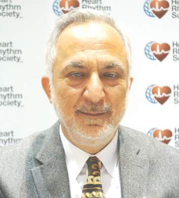
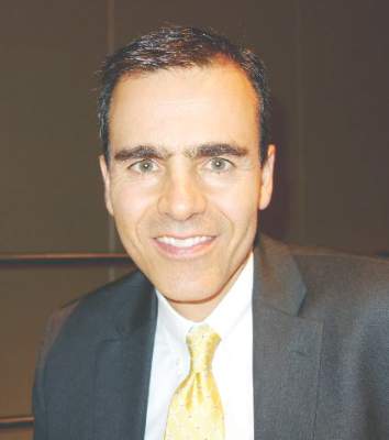
Dr. John Day |
This study is interesting because it goes against what other studies are showing, which is that LAA isolation increases the success rate with AF ablation. What makes me a little suspicious is that the success rates in both arms of this study were unusually high for persistent AF. If they were more in line with previous reports, I would feel a little bit better concluding that LAA isolation doesn’t’ help.
I know anecdotally from having done thousands of these ablations that there are some patients whose AF originates from the LAA, and if you treat it, you improve their outcomes.
Dr. John Day is the director of Intermountain Heart Rhythm Specialists in Murray, Utah, and the current president of the Hearth Rhythm Society. He has no disclosures.
SAN FRANCISCO – Adding left atrial appendage excision to pulmonary vein isolation does not reduce the rate of recurrence in persistent atrial fibrillation, according to a Russian investigation.
Eighty-eight patients with persistent atrial fibrillation (AF) were randomized to thoracoscopic pulmonary vein isolation (PVI) with bilateral epicardial ganglia ablation and box lesion set of the posterior left atrial wall; 88 others were randomized to that approach plus left atrial appendage (LAA) amputation. After 18 months, 64 out of 87 patients in the LAA-excision group (73.6%) and 61 out of 86 patients (70.9%) in the control group were free from recurrent AF, meaning no episodes greater than 30 seconds (P = .73). Freedom from any atrial arrhythmia after a single procedure with or without follow-up antiarrhythmic drugs (AADs) was also similar, with 70.9% in the control and 74.7% in the treatment groups. “Both approaches had excellent” results with no differences in complication rates, but there “was no reduction in AF recurrence when LAA excision was performed,” said investigator Dr. Alexander Romanov of the State Research Institute of Circulation Pathology, Novosibirsk, Russia.
The results are a bit surprising because some previous studies have suggested that electrical isolation of the LAA improves AF ablation success, and surgical excision might be expected to have a similar effect. In many places in the United States, LAA excisions are routine in open heart surgery when patients have AF, to prevent stroke. Guidelines for AF management from the American Heart Association, American College of Cardiology, and Heart Rhythm Society published in 2014 give a class IIb recommendation, saying “surgical excision of the left atrial appendage may be considered in patients undergoing cardiac surgery,” with an evidence level of C, meaning there are no data to support the recommendation, only expert consensus (J Am Coll Cardiol. 2014;64[21]:2246-80).
There were no significant differences between the groups; patients were about 60 years old, on average, and more than 80% in both groups had baseline CHADS2 scores of 0 or 1. All patients had persistent AF for more than a week but no longer than a year; longer-standing cases were excluded, as were patients with prior heart surgeries or catheter ablations. There were no statistically significant differences in operative times or complications. A few patients in each arm needed sternotomies for hemostasis, and one in each arm had a stroke during follow-up. Patients were followed at regular intervals by ECG and Holter monitoring.
AADs were allowed during the blanking period; patients could continue them afterwards for AF recurrence or have endocardial redo ablations; 10 patients in the control group (12%) and 13 in the LAA group (15%) had repeat procedures (P = .55). Most were for right atrial flutter and a few for left atrial flutter. “Only one redo case was for true AF recurrence,” Dr. Romanov said.
The team did not test for exertion intolerance and other potential LAA excision problems.
Dr. Romanov is a speaker for Medtronic, Biosense Webster, and Boston Scientific.
AT HEART RHYTHM 2016
Key clinical point: Adding left atrial appendage excision to pulmonary vein isolation does not reduce the rate of recurrence in persistent atrial fibrillation.
Major finding: After 18 months, 64 out of 87 patients in the LAA-excision group (73.6%) and 61 out of 86 patients (70.9%) in the control group were free from recurrent AF, meaning no episodes greater than 30 seconds (P = 0.73).
Data source: Randomized trial in 176 patients with persistent AF.
Disclosures: The lead investigator is a speaker for Medtronic, Biosense Webster, and Boston Scientific.
ICD same-day discharge safe, but not a money saver
San Francisco – Same day discharge is generally safe after cardioverter defibrillator implantation for primary prevention, but it doesn’t save money.
Furthermore, guidelines are needed to standardize the practice as it becomes increasingly common in the United States, according to a 25-site investigation.
After implantable cardioverter defibrillator (ICD) procedures, patients were monitored for 3-4 hours, and their devices were checked for proper functioning; 129 patients who were stable at that point were randomized to early discharge and 136 to next day discharge (NDD).
The overall 30-day procedural complication rate was 3.1% in the same day discharge (SDD) group and 1.6% in the NDD group, a nonsignificant difference (P = .37). Three patients in the SDD group developed hematomas that resolved on their own, and one had a cardiac perforation. One NDD patient dislodged a lead and another developed an infection. There were no differences in quality of life measures between the two groups at 30 days.
However, there were also no differences in procedural and perioperative direct costs, which was surprising because saving money is a major driver of SDD, and the most expensive part of ICD implantation is the first 24 hours. Direct per-patient medical costs in the study – estimated by applying hospital cost-to-charge ratios to the Medicare-reported charge – were $31,771 for SDD and $30,437 for NDD, but NDD was more expensive than SDD at several sites. The investigators suspect a flaw in their analysis related to the opaque nature of hospital accounting, and plan to look into the matter further with modeling to identify savings opportunities with SDD.
“We can insert ICDs on an outpatient basis, but this study will be difficult to replicate because clinical practice is moving towards SDD. In view of this, we think professional societies should be thinking of standardizing criteria for SDD; guidelines would help with the adoption of this approach. There are clinicians who are astute and have great clinical judgment, but there are others who need a scoring system. We believe that by using the 270,000 patients in the [American College of Cardiology’s ICD Registry], there is the ability to identify patients who have low periprocedural risk,” said lead investigator Dr. Ranjit Suri, a cardiologist at Mt. Sinai Hospital in New York.
The study excluded patients receiving an ICD for secondary prevention, as well as those on periprocedural heparin and patients who were pacemaker dependent. SDD seemed safe otherwise, but it’s unknown “if our concept of low risk is acceptable to all implanting physicians,” Dr. Suri said at the annual scientific sessions of the Heart Rhythm Society.
The study groups were well matched. About 75% in each arm were men, and ischemic cardiomyopathy was the leading ICD indication. Patients were amenable to the idea of SDD; the advent of remote monitoring “adds a certain sense of safety” for both patients and physicians, he said.
Dr. Suri is a speaker for Boehringer Ingelheim and St. Jude Medical. He is also a consultant for Biosense Webster and Zoll, and receives research funding from St. Jude.
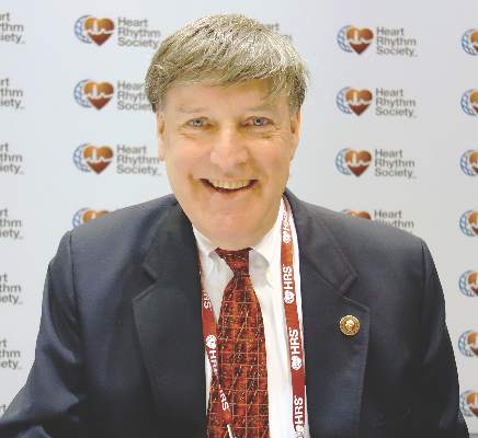
|
Dr. Thomas Deering |
The vast majority of primary prevention patients who are clinically stable enough to come in as outpatients can go home as outpatients if you watch them for a short period of time and make sure they are clinically stable. Most patients don’t want to be in the hospital, and many hospitals are crunched for available beds. It would be great to have guidelines on how to handle this, but we have to allow for clinical judgment.
Dr. Thomas Deering is chief of the Arrhythmia Center at the Piedmont Heart Institute in Atlanta, where he is also chairman of the Executive Council and the Clinical Centers for Excellence. He moderated Dr. Suri’s presentation and was not involved in the work.
San Francisco – Same day discharge is generally safe after cardioverter defibrillator implantation for primary prevention, but it doesn’t save money.
Furthermore, guidelines are needed to standardize the practice as it becomes increasingly common in the United States, according to a 25-site investigation.
After implantable cardioverter defibrillator (ICD) procedures, patients were monitored for 3-4 hours, and their devices were checked for proper functioning; 129 patients who were stable at that point were randomized to early discharge and 136 to next day discharge (NDD).
The overall 30-day procedural complication rate was 3.1% in the same day discharge (SDD) group and 1.6% in the NDD group, a nonsignificant difference (P = .37). Three patients in the SDD group developed hematomas that resolved on their own, and one had a cardiac perforation. One NDD patient dislodged a lead and another developed an infection. There were no differences in quality of life measures between the two groups at 30 days.
However, there were also no differences in procedural and perioperative direct costs, which was surprising because saving money is a major driver of SDD, and the most expensive part of ICD implantation is the first 24 hours. Direct per-patient medical costs in the study – estimated by applying hospital cost-to-charge ratios to the Medicare-reported charge – were $31,771 for SDD and $30,437 for NDD, but NDD was more expensive than SDD at several sites. The investigators suspect a flaw in their analysis related to the opaque nature of hospital accounting, and plan to look into the matter further with modeling to identify savings opportunities with SDD.
“We can insert ICDs on an outpatient basis, but this study will be difficult to replicate because clinical practice is moving towards SDD. In view of this, we think professional societies should be thinking of standardizing criteria for SDD; guidelines would help with the adoption of this approach. There are clinicians who are astute and have great clinical judgment, but there are others who need a scoring system. We believe that by using the 270,000 patients in the [American College of Cardiology’s ICD Registry], there is the ability to identify patients who have low periprocedural risk,” said lead investigator Dr. Ranjit Suri, a cardiologist at Mt. Sinai Hospital in New York.
The study excluded patients receiving an ICD for secondary prevention, as well as those on periprocedural heparin and patients who were pacemaker dependent. SDD seemed safe otherwise, but it’s unknown “if our concept of low risk is acceptable to all implanting physicians,” Dr. Suri said at the annual scientific sessions of the Heart Rhythm Society.
The study groups were well matched. About 75% in each arm were men, and ischemic cardiomyopathy was the leading ICD indication. Patients were amenable to the idea of SDD; the advent of remote monitoring “adds a certain sense of safety” for both patients and physicians, he said.
Dr. Suri is a speaker for Boehringer Ingelheim and St. Jude Medical. He is also a consultant for Biosense Webster and Zoll, and receives research funding from St. Jude.
AT HEART RHYTHM 2016
Key clinical point: Same-day discharge is generally safe after cardioverter defibrillator implantation for primary prevention, but it doesn’t save money and guidelines are needed to standardize the practice as it becomes increasingly common in the United States.
Major finding: The overall 30-day procedural complication rate was 3.1% in the same day discharge (SDD) group and 1.5% in the next-day discharge group, a nonsignificant difference (P = .37).
Data source: Randomized trial of 265 ICD patients.
Disclosures: The lead investigator is a speaker for Boehringer Ingelheim and St. Jude Medical. He is also a consultant for Biosense Webster and Zoll, and receives research funding from St. Jude.
VIDEO: Lobectomy quality requires linking outcomes to process change
BALTIMORE – An “introspective” analysis connecting patient outcomes with process changes may lead to significant surgical quality improvement, according to a study presented at the 2016 annual meeting of the American Association for Thoracic Surgery.
The case study detailed the University of Alabama at Birmingham School of Medicine’s attempt to identify the metrics used for the Society of Thoracic Surgeons lobectomy ranking, and show how the institution used root cause analysis with “lean” and process improvements to improve outcomes from Jan. 2006 until July 2014 in order to achieve a three star STS ranking.
UAB researchers found that their most common root cause analysis was failure to escalate care. The institution implemented process improvements such as increasing pulmonary rehabilitation prior to surgery, adding a respiratory therapist, eliminating (lean) non-valued steps, favoring stereotactic radiotherapy and segmentectomy instead of lobectomy for marginal patients, and using minimally invasive lobectomy. They ultimately achieved a three-star STS ranking.
The video associated with this article is no longer available on this site. Please view all of our videos on the MDedge YouTube channel
Dr. Stephen D. Cassivi, professor of surgery at the Mayo Clinic in Rochester, Minn., and a discussant on the paper at AATS 2016, said in an interview that the research was important because it encourages surgeons to discuss and reevaluate quality improvement measures. He noted that early phases of surgical quality improvement was based on process measures, specifically around the idea that if surgeons were attentive to process measures, their outcome measures would improve. But over time, the emphasis on process measures has dissipated in favor of outcomes-focused analysis.
“Now that we have more robust [outcomes] data... we can examine our practices in a more thoughtful, data-driven, evidence-based way,” Dr. Cassivi said. He added that the shift from process measures to outcome measures is important in that surgeons can easily interpret and compare outcomes data across facilities. But he noted that there is a downside: If an institution’s outcome measures are not up to standard, it is sometimes difficult to determine why.
“The current way that the [outcomes] data are reported and processed is not easily interpretable into which processes we need to adapt,” Dr. Cassivi said. “There is still work that needs to be done, but [this paper] is a first step.”
Dr. Cassivi reported no relevant financial disclosures.
On Twitter @richpizzi
AT THE AATS ANNUAL MEETING
VIDEO: Surgical quality measures boost survival in cancer patients
BALTIMORE – Surgeons’ adherence to select quality measures when treating stage IIIA non–small-cell lung cancer patients led to improved patient survival, according to a study presented at the 2016 annual meeting of the American Association for Thoracic Surgery.
Researchers at Washington University in St. Louis identified 10,323 patients who received surgery for Stage IIIA NSCLC in the National Cancer Data Base from 2006 to 2010, and chose four quality measures that should have been met by surgeons: delivery of neoadjuvant multiagent chemotherapy (with or without radiation therapy); performing a lobectomy or greater resection; obtaining more than 10 lymph nodes, and achieving an R0 resection.
The researchers said 12.8% of patients met all four quality measures. Kaplan-Meier analysis demonstrated improved overall median survival by number of quality measures obtained: 0 quality measures, 12.7 months; 1 quality measure, 25.0 months; 2 quality measures, 31.4 months; 3 quality measures, 36.6 months; and 4 quality measures, 43.5 months.
In an interview, Dr. Mark S. Allen, professor of surgery at the Mayo Clinic in Rochester, Minn., and a discussant on the paper at AATS 2016, said the most striking result of the study was that such a low percentage of patients had all four quality measures performed for stage IIIA cancer. He called that finding “disappointing.”
“In general, [the study] shows there is still some work to be done to improve the quality when we operate on stage IIIA patients,” Dr. Allen said. “I’m not sure we do the greatest job of staging them clinically. When they are staged properly they probably do need preoperative chemotherapy, and I’m not sure we do that all the time.” He added that surgeon education about quality outcomes was critical to process improvement and patient outcomes.
Dr. Allen reported no relevant financial disclosures.
On Twitter @richpizzi
The video associated with this article is no longer available on this site. Please view all of our videos on the MDedge YouTube channel
AT THE AATS ANNUAL MEETING
Low hematocrit in elderly portends increased bleeding post PCI
PARIS – A low hematocrit in an elderly patient who’s going to undergo percutaneous coronary intervention signals a markedly increased risk of major bleeding within 30 days of the procedure, according to Dr. David Marti.
“Analysis of hematocrit in elderly patients can guide important procedural characteristics, such as access site and antithrombotic regimen,” he said at the annual congress of the European Association of Percutaneous Cardiovascular Interventions.
For example, studies have established that transradial artery access percutaneous coronary intervention (PCI) results in significantly less bleeding than the transfemoral route, said Dr. Marti, an interventional cardiologist at the University of Alcalá in Madrid.
He presented a prospective study of 212 consecutive patients aged 75 or older who underwent PCI at a single university hospital. Their mean age was 81.4 years, and slightly over half of them presented with an acute coronary syndrome.
All patients received dual-antiplatelet therapy in accord with current guidelines. Stent type and procedural anticoagulant regimen were left to the discretion of the cardiologist; 80% of the subjects received bivalirudin-based anticoagulation.
The primary study outcome was the 30-day incidence of major bleeding, as defined by a Bleeding Academic Research Consortium (BARC) type 3-5 event. The overall rate in this elderly PCI population was 5.5%. However, the rate varied markedly by baseline hematocrit tertile, in accord with the investigators’ study hypothesis.
Major bleeding occurred in 2.9% of patients with an Hct greater than 42% and 3.1% in those with an Hct of 38%-52%, and jumped to 10.6% in the one-third of subjects whose baseline Hct was below 38%, Dr. Marti reported.
Thus, a preprocedural Hct below 38% was associated with a 4.1-fold increased risk of major bleeding within 30 days following PCI. An Hct in this range was a stronger predictor of BARC type 3-5 bleeding risk than were other factors better known as being important, including advanced age, greater body weight, female sex, or an elevated serum creatinine indicative of chronic kidney disease. Indeed, an Hct below 38% was the only statistically significant predictor of major bleeding in this elderly population.
The likely explanation for the observed results is that a low Hct level in elderly patients usually reflects subclinical blood loss that can be worsened by antithrombotic therapies, the cardiologist explained.
The presenter reported having no financial conflicts regarding this study, conducted without commercial support.
AT EUROPCR 2016
Key clinical point: Elderly patients scheduled for PCI have a fourfold greater risk of major bleeding within 30 days if their Hct is less than 38%.
Major finding: The 30-day incidence of BARC types 3-5 major bleeding was 10.9% in elderly patients with a pre-PCI Hct below 38%, compared with 2.9% in those in the top Hct tertile.
Data source: A prospective study of 212 consecutive patients aged 75 or older who underwent PCI at a single university hospital.
Disclosures: The presenter reported having no financial conflicts regarding this study, conducted without commercial support.
Transcatheter aortic valve implantation equivalent to surgical replacement
Transcatheter aortic valve implantation shows reductions in early and mid-term all-cause mortality similar to those with surgical aortic valve replacement, even in patients with low to intermediate surgical risk, a meta-analysis and systematic review has shown.
Dr. Giuseppe Gargiulo of Federico II University in Naples, Italy, and coauthors analyzed data from five randomized trials and 31 observational matched studies comparing mortality outcomes in 16,638 patients undergoing transcatheter aortic valve implantation (TAVI) or surgical aortic valve replacement (SAVR).
Their analysis found no statistically significant difference between the two procedures in terms of early or midterm all-cause mortality, even among patients judged as being at low to intermediate surgical risk (Ann Intern Med. 2016 Jun 7. doi: 10.7326/M16-0060).
In terms of 2- to 5-year mortality, overall there was a statistically nonsignificant increase in the risk of all-cause mortality with TAVI (odds ratio, 1.28; 95% confidence interval, 0.97-1.69), although the long-term mortality outcomes in patients in the low to intermediate surgical risk subgroup were inconclusive.
However, the authors did note significantly reduced early all-cause mortality in individuals who underwent transfemoral TAVI compared to those who underwent SAVR (OR 0.68, 95%CI, 0.53 to 0.87).
The analysis also showed that individuals who underwent TAVI had a higher incidence of permanent pacemaker implantation, vascular complications, and moderate to severe paravalvular leak, while those who underwent SAVR had more frequent incidence of major bleeding, acute kidney injury, and new-onset atrial fibrillation.
“These findings, which apply to adults with severe aortic stenosis, consolidate the role of TAVI as an alternative to SAVR,” the authors wrote. “Indeed, TAVI techniques continue to improve, newer valves address the issue of paravalvular leak, the percentage of pacemakers is decreasing, and the rate of vascular complications is expected to be lowered as the result of smaller sheaths and improved procedural techniques.”
The researchers noted that elderly patients and those with coronary artery disease showed a greater benefit from TAVI than from SAVR, suggesting that this may be because these groups have a heightened risk that favors less invasive surgical approaches.
They also found greater reductions in early mortality with TAVI when a Sapien valve was implanted, compared to a CoreValve. They noted that this was due mostly to a single large study and the effect did not persist through to the midterm follow-up.
One author reported grants from the CardioPath PhD Program, Federico II University of Naples, and from the European Association of Percutaneous Coronary Interventions, outside the submitted work. Another author declared a consultancy for Edwards Lifesciences. There were no other conflicts of interest declared.
FROM ANNALS OF INTERNAL MEDICINE
Key clinical point: Transcatheter aortic valve implantation shows reductions in early and mid-term all-cause mortality similar to those of surgical aortic valve replacement.
Major finding: Transcatheter aortic valve implantation and surgical aortic valve replacement show similar reductions in mortality, even in patients at low to intermediate surgical risk.
Data source: Systematic review and meta-analysis.
Disclosures: One author reported grants from the CardioPath PhD Program, Federico II University of Naples, and from the European Association of Percutaneous Coronary Interventions, outside the submitted work. Another author declared a consultancy for Edwards Lifesciences. There were no other conflicts of interest declared.
Point/Counterpoint: What’s best for chronic dissection: TEVAR or open?
TEVAR is the best procedure.
At Cedars-Sinai Medical Center, 70% of the aortic operations are performed in open fashion, but in patients with chronic type B aortic dissection, thoracic endovascular repair (TEVAR) is the preferred option. Goals of TEVAR in the setting of chronic type B dissection are to seal off the intimomedial tears, re-route blood to the true lumen, and induce false lumen thrombosis in the descending thoracic aorta, promoting reverse aortic remodeling and reducing future reinterventions.
TEVAR in patients with chronic type B aortic dissection has the best results if the following five rules apply: the patient should be older than 40 years of age and not have connective tissue disorder; there should be a proper proximal landing zone; the distal landing zone at the celiac artery should be smaller than 4 cm to allow for reverse remodeling; most of the large intimomedial tears should be in the descending aorta; and at least three visceral vessels should come off the true lumen. With this clinical scenario, the majority of centers will have excellent results with TEVAR.
TEVAR for chronic type B aortic dissection comes with three usual concerns: short-term outcomes; reverse aortic remodeling; and long-term outcomes.
In evaluating short-term outcomes of TEVAR in chronic type B aortic dissection, many large, single-center studies, including ours, have documented the superior results.1,2 The VIRTUE study reported an operative mortality and 30-day hospitality mortality of zero and spinal cord ischemia rate of 3.8% in a prospective, multi-center review.3 A meta-analysis of TEVAR for chronic dissection that involved 567 patients reported a 30-day mortality rate of 3.2%, paraplegia rate of 0.45%, a stroke rate of 1.5%, and retrograde type A dissection rate of 0.7%.4
These outcomes are far better than those reported in a meta-analysis of open repair for chronic dissection (771 patients): post-1997 mortality of 8.8% (the overall 30-day mortality rate was 12.5%); paraplegia of 6%; and renal failure with hemodialysis of 4%.5 A statewide analysis of elective open repair for thoracoabdominal aortic aneurysm, including chronic dissections, in California had a 30-day mortality rate of 19.7%.6
With regard to reverse aortic remodeling, acceptable results with TEVAR have been reported. The INSTEAD-XL study reported that at 5 years, 73% of patients had reverse aortic remodeling, with absolute risk reduction of 12.4%, compared with optimal medical therapy.7 A systematic review reported an 85.7% median rate of false lumen thrombosis.4 In the past some surgeons were concerned about a thick septum and whether it would give way and allow reverse remodeling; these studies confirmed that it does. The radial force of a stent graft over time will enlarge to the size that you would expect and it will cause the false lumen thrombosis.
Our group has provided anatomical indicators to achieve reverse remodeling in chronic type B dissection, including the location and size of intimomedial tears above the celiac artery.8 A patient with this anatomy has a great chance of not requiring any future reinterventions if the tears are mostly within the thoracic aorta upper fit.
Large multicenter studies also provide answers to the third concern about TEVAR for chronic aortic dissection – long-term outcomes – and found that they are comparable to open surgery. A study of the Medtronic Thoracic Endovascular Registry (MOTHER) database, a prospective, multicenter, adjudicated registry, looked at three types of aortic pathology: chronic aortic dissection (CAD); acute aortic dissection (AAD); and thoracic aortic aneurysm (TAA). The 195 patients with CAD had the best all-cause mortality outcomes: 3.2 per 100 patient years.9 Aortic-related mortality was also lowest in the CAD group: 0.4 per 100 patient years vs. 0.6 for TAA and 1.2 for AAD.
The reintervention rates for patients with aortic dissection were high, compared with TAA in the MOTHER registry. INSTEAD XL revealed all-cause and aortic-related mortality, respectively, at 11.1% and 6.9% in patients with chronic type B dissection treated with TEVAR at 5 years.
Last but not least, TEVAR is the first choice for many elderly or frail patients with type B aortic dissection. Recovery after the open procedure is much more difficult for this population. Our specialty frequently underappreciates quality of life after an aortic operation.
Overall, TEVAR for chronic type B aortic dissection is feasible, reproducible, and less invasive than open repair. It has acceptable early results, rate of reverse aortic modeling, and late mortality, and although its reintervention rate can be significant, that can be reduced with experience and a careful algorithmic approach.
Dr. Ali Khoynezhad is a professor of cardiovascular surgery, director of aortic surgery, and co-director of the atrial fibrillation program at Cedars-Sinai Heart Institute, Los Angeles. He disclosed receiving research grants from Medtronic, Gore, and Vascutek.
1. J Thorac Cardiovasc Surg. 2008 May;135:1103-9.
2. J Thorac Cardiovasc Surg. 2011 Feb;141:322-7.
3. Eur J Vasc Endovasc Surg. 2011 Feb;41:159-66.
4. Eur J Vasc Endovasc Surg. 2011 Nov;42:632-47.
5. J Vasc Surg. 2010 Oct;52:3S-9S.
6. J Vasc Surg. 2006 Feb;43:217-22.
7. Circ Cardiovasc Interv. 2013 Aug;6:407-16.
8. J Vasc Surg. 2010 Sep;52:562-8.
9. Circulation. 2013 Jan;127:24-32.
Open repair is the better procedure.
It’s my contention that open repair is still the gold standard for chronic thoracoabdominal aortic dissection. It has a record of outstanding results in high-volume centers of excellence with low morbidity and mortality and very good long-term survival. It has no anatomical constraints. It’s a durable repair and there are no device- or procedure-related proximal and/or distal aortic complications. Reintervention on the operated segment is very rare.1-6
Thoracic endovascular repair (TEVAR), despite having very good procedural results, has challenges in the successful treatment of chronic aortic dissection. Morbidity is low, as are rates of spinal cord ischemia, stroke, and renal failure. However, thoracic remodeling at the level of the endograft is in the 70%-88% range, which means that 12%-30% of patients do not have protection in the form of reverse aortic remodeling. In the abdominal aorta, remodeling is uncommon, with 11%-23% thrombosis of the false lumen even with advantageous anatomy. Survival in several series is in the 60%-80% range at 3 and 5 years. TEVAR creates new challenges for chronic thoracoabdominal aortic dissection: retrograde type A dissections have been as high as 2%-7%; 15%-30% of cases require intervention; and stent graft-induced new entry (SINE) has been reported as high as 36%.
Specific anatomical features are not suitable for TEVAR. They include multiple visceral vessels off of the false lumen; multiple fenestrations, especially in the abdominal aorta; dissection within the dissection; and pseudocoarctation. The durability of the endovascular graft for chronic aortic dissection is unknown; it’s a relatively new procedure so the long-term data is lacking.
Success of the endovascular approach depends on aortic remodeling, and it’s my contention that the thoracic devices now available cannot effectively treat a chronic thoracoabdominal aortic dissection. There are steps surgeons can take to improve their success, but procedures specifically addressing the false lumen are not time-tested and thoracoabdominal-specific devices are not widely available in the United States. And of course, morbidity and mortality will increase with the complexity of the endovascular repair.
Our in-hospital outcomes with open repair of chronic thoracoabdominal aortic dissection using deep hypothermia and circulatory arrest have been excellent: mortality rate of 3.6%; a stroke rate of 1%; permanent spinal cord ischemia rate 2.6%; and a 0% rate of patients on permanent hemodialysis. Our hospital length of stay is approximately 12 days. Blood product transfusion is reasonable with a mean blood product transfusion of 9 units for the hospital admission. The reintervention rate is 1% for infected grafts, 3.1% for anastomotic pseudoaneurysm and 3.6% for growth of a distal aneurysm. Long-term survival is very good: 93% at one year; 79% at five years; and 57% at 10 years.
There’s no denying that mortality rates for endovascular repair are excellent. There’s no denying that open repair is much more invasive. And if the patient is of advanced age, is frail and has comorbidities, the endovascular repair can have a certain advantage.
But for false lumen obliteration, the advantage goes to open repair. With regard to reintervention, certainly the advantage is with open repair. For durability, from what we know currently, open repair has the advantage. There are no stent-induced new entries in open repair; and open repair can address any and all anatomy. A successful endograft repair requires fixation and seal in the appropriate aortic anatomy; it demands good proximal and distal landing zones. However, in chronic dissection, there is the added complexity of having to address the false lumen flow from an untreated abdominal aortic segment.
For patients with connective tissue disorders, open repair is still the gold standard. As for long-term survival, the advantage goes to open only because the endovascular approach is relatively new. If it’s done in high-volume centers with great experience, open repair has a mortality advantage as well.
Dr. Joel Corvera is an assistant professor of surgery and director of thoracic and vascular surgery at Indiana University, Indianapolis. He had no relationships to disclose.
1. Eur J Vasc Endovasc Surg. 2011 Feb;41:159-66.
2. J Thorac Cardiovasc Surg. 2011 Feb;141:322-7.
3. Ann Cardiothorac Surg. 2014 May;3:264-74.
4. J Thorac Cardiovasc Surg. 2010 Jun;139:1548-53.
5. Ann Thorac Surg. 2013 Mar;95:914-21.
EXPERT ANALYSIS FROM THE AMERICAN ASSOCIATION FOR THORACIC SURGERY AORTIC SYMPOSIUM 2016
Key clinical point: Open repair for chronic thoracoabdominal aortic dissection has been the “gold standard” with good results, but thoracic endovascular repair (TEVAR) may have even lower mortality and complications in selected patients.
Major finding: Open and endovascular repair for thoracoabdominal aortic dissection have comparable results, but the former is a better choice for younger patients while the latter provides an option for elderly and more frail patients.
Data source: The presenters cited several studies to support their positions, including an analysis of 1,010 patients from the California Office of Statewide Health Planning and Development for 1991-2002 and 196-patient series from Indiana University.
Disclosures: Dr. Khoynezhad disclosed receiving research grants from Medtronic, Gore, and Vascutek. Dr. Corvera had no financial relationships to disclose.
Esophageal perforation severity scoring system reliably stratifies patients
The Pittsburgh perforation severity score (PSS) can be used to improve decision making in the management of esophageal perforation, findings from a retrospective, multicenter study have shown.
Dr. Michael Schweigert and his colleagues performed a study of 288 patients with esophageal perforation treated at 11 centers between 1990 and 2014, using them as a completely independent population to validate whether the PSS could be used to stratify such patients into discrete subgroups with differential outcomes.
The PSS was analyzed using logistic regression as a continuous variable and stratified into low, intermediate and high score groups, according to their report published in the Journal of Thoracic and Cardiovascular Surgery (2016 Apr;151:1002-11).
Operative management was more frequent than nonoperative management (200 patients, or 69.4% vs. 30.6%), according to Dr. Schweigert of the Städtisches Klinikum Dresden Friedrichstadt, Germany, and his colleagues. Patients with esophageal cancer (34/43; 79%) and stricture (18/23; 78.3%) mainly were treated operatively. The most common type of surgery was primary repair (83 patients), followed by surgical drainage (38 patients).
Perforation-related morbidity was seen in 180 patients (65%), with sepsis (21%) and pneumonia (19%) being most common. Overall in-hospital mortality was 20%, and the median length of stay was 27 days.
Patients with fatal outcomes had a significantly higher median PSS score (11 vs. 1) and the median PSS was significantly higher in operatively managed cases, compared with nonoperative cases (5 vs. 4, P = .0001). The researchers found that the PSS score predicted morbidity well, with an area under the curve (AUC) of 0.77, as well as mortality (AUC = 0.83). However, prediction of the need for operative management was not as good (AUC = 0.65).
Based upon their analysis, the researchers proposed a treatment decision tree in which group I (low PSS patients) should have a focus of nonoperative management. Group 2 patients (medium PSS) with non–contained leak preferably should be managed by surgery.
They found that the high-risk group (PSS greater than 5) had the worst prognosis and highest mortality, with the odds for mortality being 8 times higher than that the intermediate group and 18 times higher than the low-risk group. “Because these patients are most endangered by esophageal perforation, early and aggressive treatment is mandatory to avoid fatal outcomes,” the authors stated.
They found that nonoperative management was not associated with higher mortality or more unfavorable outcome regarding perforation-related morbidity or length of stay, but they pointed out that nonoperative treatment was only successful in 60% of cases, with 36 out of the 88 nonoperative patients eventually undergoing surgery and 8 undergoing esophagectomy. But patients with a high perforation severity score were 3.37 times more likely to have operative management compared to low-scoring patients. “Better selective criteria for nonoperative management are urgently required,” they stated.
“The Pittsburgh PSS is helpful to assess the severity and potential consequences of esophageal injury and stratifies patients into low-, intermediate-, and high-risk groups with differential morbidity and mortality outcomes. Prospective studies are required to analyze the influence of the Pittsburgh scoring system on the treatment of esophageal perforation,” the researchers concluded.
The authors reported having no disclosures.
A webcast of the AATS Annual Meeting presentation of this paper is available.

|
Dr. Mara B. Antonoff |
Schweigert and his colleagues suggest that the Pittsburgh scoring system may identify patients suitable for nonoperative management. The authors retrospectively found less morbidity/mortality and less-frequent operative management among patients in Group 1, and thus, a recommendation was formulated favoring less-invasive management for these individuals.
The additional step of evaluating the success of nonoperative management in each group, either through further analyses of the current study or with future prospective studies is needed in order to make such recommendations.
Further demonstrating the utility of the Pittsburgh esophageal PSS, this study supports the notion that prospective, large-scale studies are in need, and that such scoring systems will be instrumental in standardizing data across centers.
Dr. Mara B. Antonoff is from the department of thoracic and cardiothoracic surgery at the University of Texas MD Anderson Cancer Center, Houston. Her remarks were made as part of an invited commentary on the article (J Thorac Cardiovasc Surg. 2016 Apr;151:1012-3).
The Pittsburgh perforation severity score (PSS) can be used to improve decision making in the management of esophageal perforation, findings from a retrospective, multicenter study have shown.
Dr. Michael Schweigert and his colleagues performed a study of 288 patients with esophageal perforation treated at 11 centers between 1990 and 2014, using them as a completely independent population to validate whether the PSS could be used to stratify such patients into discrete subgroups with differential outcomes.
The PSS was analyzed using logistic regression as a continuous variable and stratified into low, intermediate and high score groups, according to their report published in the Journal of Thoracic and Cardiovascular Surgery (2016 Apr;151:1002-11).
Operative management was more frequent than nonoperative management (200 patients, or 69.4% vs. 30.6%), according to Dr. Schweigert of the Städtisches Klinikum Dresden Friedrichstadt, Germany, and his colleagues. Patients with esophageal cancer (34/43; 79%) and stricture (18/23; 78.3%) mainly were treated operatively. The most common type of surgery was primary repair (83 patients), followed by surgical drainage (38 patients).
Perforation-related morbidity was seen in 180 patients (65%), with sepsis (21%) and pneumonia (19%) being most common. Overall in-hospital mortality was 20%, and the median length of stay was 27 days.
Patients with fatal outcomes had a significantly higher median PSS score (11 vs. 1) and the median PSS was significantly higher in operatively managed cases, compared with nonoperative cases (5 vs. 4, P = .0001). The researchers found that the PSS score predicted morbidity well, with an area under the curve (AUC) of 0.77, as well as mortality (AUC = 0.83). However, prediction of the need for operative management was not as good (AUC = 0.65).
Based upon their analysis, the researchers proposed a treatment decision tree in which group I (low PSS patients) should have a focus of nonoperative management. Group 2 patients (medium PSS) with non–contained leak preferably should be managed by surgery.
They found that the high-risk group (PSS greater than 5) had the worst prognosis and highest mortality, with the odds for mortality being 8 times higher than that the intermediate group and 18 times higher than the low-risk group. “Because these patients are most endangered by esophageal perforation, early and aggressive treatment is mandatory to avoid fatal outcomes,” the authors stated.
They found that nonoperative management was not associated with higher mortality or more unfavorable outcome regarding perforation-related morbidity or length of stay, but they pointed out that nonoperative treatment was only successful in 60% of cases, with 36 out of the 88 nonoperative patients eventually undergoing surgery and 8 undergoing esophagectomy. But patients with a high perforation severity score were 3.37 times more likely to have operative management compared to low-scoring patients. “Better selective criteria for nonoperative management are urgently required,” they stated.
“The Pittsburgh PSS is helpful to assess the severity and potential consequences of esophageal injury and stratifies patients into low-, intermediate-, and high-risk groups with differential morbidity and mortality outcomes. Prospective studies are required to analyze the influence of the Pittsburgh scoring system on the treatment of esophageal perforation,” the researchers concluded.
The authors reported having no disclosures.
A webcast of the AATS Annual Meeting presentation of this paper is available.
FROM THE JOURNAL OF THORACIC AND CARDIOVASCULAR SURGERY
Key clinical point: Scoring system reliably stratifies patients into low-, intermediate-, and high-risk groups.
Major finding: Patients with a high perforation severity score were 3.37 times more likely to have operative management, compared with low-scoring patients.
Data source: A retrospective study was performed on 288 patients with esophageal perforation at 11 centers since 1990.
Disclosures: The authors presented no relevant disclosures.
Pulmonary function testing adds little to STS risk scores
Routine preoperative pulmonary function tests appear to have only limited utility in predicting outcomes in patients undergoing cardiothoracic surgery when the Society of Thoracic Surgeons risk score is available, according to the results of a retrospective study.
Dr. Alexander Ivanov of New York Methodist Hospital, Brooklyn, and his colleagues conducted a database analysis of 1,685 patients undergoing index cardiac surgery at New York Methodist Hospital between April 2004 and January 2014. They used the STS risk model version 2.73 to estimate postoperative risk of respiratory failure (defined as the need for mechanical ventilation greater than or equal to 72 hours, or reintubation), prolonged postoperative length of stay (defined as greater than 14 days), and 30-day all cause mortality in these patients, according to their report in The Journal of Thoracic and Cardiovascular Surgery (2016;151:1183-9).
They plotted the receiver operating characteristics curve for the STS score for each of these adverse events and compared the resulting area under the curve (AUC) with the AUC after adding pulmonary function testing parameters and COPD classifications.
A total of 1,412 patients had a calculated STS score, of which 751 underwent pulmonary function testing (53%). In general, patients who had pulmonary function testing were older and had higher rates of comorbidities and more complex cardiothoracic surgery compared with their counterparts, according to Dr. Ivanov. These patients also had significantly elevated STS risk for prolonged ventilation (12.4% vs. 10.3%), prolonged postoperative length of stay (8.9% vs. 7.2%), and 30-day mortality (2.7% vs. 2.2%).
The decision to perform pulmonary testing was left to the treating physician. Of those patients tested, 652 had bedside spirometry and 99 had formal laboratory testing. Forced expiratory volume in 1 second (FEV1) and forced volume vital capacity (FVC) values were determined by taking the best of three trials. COPD was diagnosed in cases of an FEV1/FVC ratio of less than 70%.
Among these patients, 4.5% developed postoperative respiratory failure, and there was no statistically significant difference in the respiratory failure rate between patients with and without pulmonary function testing. In addition, there was no significant difference in 30-day mortality between these patients (1.9% vs. 2.1%). However, a total of 6.9% had a prolonged postoperative length of stay, with a significantly higher rate in the patients with pulmonary function testing than without (8.8% vs. 4.7%).
Dr. Ivanov and his colleagues found that the AUC of the STS score was 0.65 (95% confidence interval [CI], 0.55-0.74)for respiratory failure, 0.67 (95% CI, 0.6-0.74) for prolonged postoperative length of stay, and 0.74 (95% CI, 0.6-0.87) for 30-day mortality. Even though the STS score based upon clinical definitions of lung disease afforded only modest discriminatory ability for the three studied outcomes, they found that there was no significant added benefit to the predictive ability of these STS scores obtained by incorporating any of the pulmonary function testing parameters or COPD classifications studied.
“A possible physiological explanation for these findings may be that the examined pulmonary function testing variables do not depend solely on pulmonary parameters such as airway diameter, degree of obstruction, or lung elasticity, but rather on a patient’s effort and muscle “strength,” characteristics that are already well captured and accounted for in the current STS model,” the researchers stated.
“The STS score calculated with clinical information on lung disease status offers modest discriminatory ability for respiratory failure, prolonged postoperative length of stay, and 30-day mortality after CT surgery, which cannot be improved by adding PFT parameters or PFT-derived COPD categorization,” they wrote. “Therefore, routine preoperative PFTs may have only limited clinical utility in patients undergoing CT surgery when the STS score is readily available. Further prospective studies will be helpful in confirming these conclusions,” Dr. Ivanov and his colleagues noted.
The authors reported that they had nothing to disclose.
Chronic lung disease is one of the risk factors included in the STS model for mortality, renal failure, prolonged ventilation, sternal wound infection, reoperation, and length of hospital stay. Mild, moderate, and severe CLD increases the odds ratio for those complications. A total of 20% of almost 1 million patients used in developing the current STS risk model had CLD.

|
Dr. Juan A. Crestanello |
The authors found that none of the pulmonary function testing parameters added to the predictive ability of the STS risk model for operative mortality, prolonged ventilation, or prolonged length of hospital stay. Because CLD is 1 of 40 preoperative and operative variables used in the STS risk model, an improvement in discrimination of only 1 of 40 variables is very unlikely to improve the overall model.
One may be tempted to conclude that it is not worth performing pulmonary function testing before cardiac surgery. However, remember once again the importance of precise and accurate data to support risk stratification. In science, behind each word resides a precise definition; without a pulmonary function test, we cannot define chronic lung disease severity.
Dr. Juan A. Crestanello is in the division of cardiac surgery, Wexner Medical Center, Ohio State University, Columbus. His remarks are from an invited commentary (J Thorac Cardiovasc Surg. 2016;151:1189-90).
Chronic lung disease is one of the risk factors included in the STS model for mortality, renal failure, prolonged ventilation, sternal wound infection, reoperation, and length of hospital stay. Mild, moderate, and severe CLD increases the odds ratio for those complications. A total of 20% of almost 1 million patients used in developing the current STS risk model had CLD.

|
Dr. Juan A. Crestanello |
The authors found that none of the pulmonary function testing parameters added to the predictive ability of the STS risk model for operative mortality, prolonged ventilation, or prolonged length of hospital stay. Because CLD is 1 of 40 preoperative and operative variables used in the STS risk model, an improvement in discrimination of only 1 of 40 variables is very unlikely to improve the overall model.
One may be tempted to conclude that it is not worth performing pulmonary function testing before cardiac surgery. However, remember once again the importance of precise and accurate data to support risk stratification. In science, behind each word resides a precise definition; without a pulmonary function test, we cannot define chronic lung disease severity.
Dr. Juan A. Crestanello is in the division of cardiac surgery, Wexner Medical Center, Ohio State University, Columbus. His remarks are from an invited commentary (J Thorac Cardiovasc Surg. 2016;151:1189-90).
Chronic lung disease is one of the risk factors included in the STS model for mortality, renal failure, prolonged ventilation, sternal wound infection, reoperation, and length of hospital stay. Mild, moderate, and severe CLD increases the odds ratio for those complications. A total of 20% of almost 1 million patients used in developing the current STS risk model had CLD.

|
Dr. Juan A. Crestanello |
The authors found that none of the pulmonary function testing parameters added to the predictive ability of the STS risk model for operative mortality, prolonged ventilation, or prolonged length of hospital stay. Because CLD is 1 of 40 preoperative and operative variables used in the STS risk model, an improvement in discrimination of only 1 of 40 variables is very unlikely to improve the overall model.
One may be tempted to conclude that it is not worth performing pulmonary function testing before cardiac surgery. However, remember once again the importance of precise and accurate data to support risk stratification. In science, behind each word resides a precise definition; without a pulmonary function test, we cannot define chronic lung disease severity.
Dr. Juan A. Crestanello is in the division of cardiac surgery, Wexner Medical Center, Ohio State University, Columbus. His remarks are from an invited commentary (J Thorac Cardiovasc Surg. 2016;151:1189-90).
Routine preoperative pulmonary function tests appear to have only limited utility in predicting outcomes in patients undergoing cardiothoracic surgery when the Society of Thoracic Surgeons risk score is available, according to the results of a retrospective study.
Dr. Alexander Ivanov of New York Methodist Hospital, Brooklyn, and his colleagues conducted a database analysis of 1,685 patients undergoing index cardiac surgery at New York Methodist Hospital between April 2004 and January 2014. They used the STS risk model version 2.73 to estimate postoperative risk of respiratory failure (defined as the need for mechanical ventilation greater than or equal to 72 hours, or reintubation), prolonged postoperative length of stay (defined as greater than 14 days), and 30-day all cause mortality in these patients, according to their report in The Journal of Thoracic and Cardiovascular Surgery (2016;151:1183-9).
They plotted the receiver operating characteristics curve for the STS score for each of these adverse events and compared the resulting area under the curve (AUC) with the AUC after adding pulmonary function testing parameters and COPD classifications.
A total of 1,412 patients had a calculated STS score, of which 751 underwent pulmonary function testing (53%). In general, patients who had pulmonary function testing were older and had higher rates of comorbidities and more complex cardiothoracic surgery compared with their counterparts, according to Dr. Ivanov. These patients also had significantly elevated STS risk for prolonged ventilation (12.4% vs. 10.3%), prolonged postoperative length of stay (8.9% vs. 7.2%), and 30-day mortality (2.7% vs. 2.2%).
The decision to perform pulmonary testing was left to the treating physician. Of those patients tested, 652 had bedside spirometry and 99 had formal laboratory testing. Forced expiratory volume in 1 second (FEV1) and forced volume vital capacity (FVC) values were determined by taking the best of three trials. COPD was diagnosed in cases of an FEV1/FVC ratio of less than 70%.
Among these patients, 4.5% developed postoperative respiratory failure, and there was no statistically significant difference in the respiratory failure rate between patients with and without pulmonary function testing. In addition, there was no significant difference in 30-day mortality between these patients (1.9% vs. 2.1%). However, a total of 6.9% had a prolonged postoperative length of stay, with a significantly higher rate in the patients with pulmonary function testing than without (8.8% vs. 4.7%).
Dr. Ivanov and his colleagues found that the AUC of the STS score was 0.65 (95% confidence interval [CI], 0.55-0.74)for respiratory failure, 0.67 (95% CI, 0.6-0.74) for prolonged postoperative length of stay, and 0.74 (95% CI, 0.6-0.87) for 30-day mortality. Even though the STS score based upon clinical definitions of lung disease afforded only modest discriminatory ability for the three studied outcomes, they found that there was no significant added benefit to the predictive ability of these STS scores obtained by incorporating any of the pulmonary function testing parameters or COPD classifications studied.
“A possible physiological explanation for these findings may be that the examined pulmonary function testing variables do not depend solely on pulmonary parameters such as airway diameter, degree of obstruction, or lung elasticity, but rather on a patient’s effort and muscle “strength,” characteristics that are already well captured and accounted for in the current STS model,” the researchers stated.
“The STS score calculated with clinical information on lung disease status offers modest discriminatory ability for respiratory failure, prolonged postoperative length of stay, and 30-day mortality after CT surgery, which cannot be improved by adding PFT parameters or PFT-derived COPD categorization,” they wrote. “Therefore, routine preoperative PFTs may have only limited clinical utility in patients undergoing CT surgery when the STS score is readily available. Further prospective studies will be helpful in confirming these conclusions,” Dr. Ivanov and his colleagues noted.
The authors reported that they had nothing to disclose.
FROM THE JOURNAL OF THORACIC AND CARDIOVASCULAR SURGERY
Key clinical point: Additional pulmonary function testing adds little predictive value to STS risk scoring when available.
Major finding: There was no significant added benefit to the predictive ability of STS scores obtained by incorporating any of the pulmonary function testing parameters or COPD classifications studied, as determined by AUC analysis.
Data source: A retrospective, database analysis of 1,685 patients undergoing index cardiac surgery at a single center between April 2004 and January 2014.
Disclosures: The authors reported that they had no disclosures.
