User login
TAVR degeneration estimated at 50% after 8 years
PARIS – The first-ever study of the long-term durability of transcatheter bioprosthetic aortic valves has documented a disturbing rise in the valve degeneration rate occurring 5-7 years post implant.
Prior consistently reassuring follow-up studies have been intermediate in length, with a maximum of 5 years. The PARTNER 2A trial, which generated enormous enthusiasm for moving TAVR to intermediate-risk patients on the basis of positive results presented at the 2016 meeting of the American College of Cardiology, reported 2-year results.
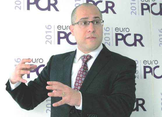
“We found, as have others, that there’s very little degeneration in the first 4 years: 94% freedom from degeneration. But at 6 years, it’s 82%, and we estimate that by 8 years, it’s about 50%,” Dr. Danny Dvir reported at the annual congress of the European Association of Percutaneous Cardiovascular Interventions.
“We need to be cautious: This is our first look at the data. But we have a signal for a problem,” said Dr. Dvir of St. Paul’s Hospital in Vancouver.
He presented a retrospective study of 378 patients who underwent TAVR 5-14 years ago at two pioneering centers for the procedure: St. Paul’s and Charles Nicolle Hospital in Rouen, France. Patients’ average age at the time of TAVR was 82.3 years, with an STS score of 8.3%. The study featured serial echocardiography conducted during house calls in this frail elderly population.
Thirty-five patients developed prosthetic valve degeneration, defined by at least moderate regurgitation and/or a mean gradient of at least 20 mm Hg in 23 cases and stenosis in 12. The risk of degeneration was unrelated to the use of warfarin, a finding that suggests the valve deterioration issue is unrelated to clotting. The strongest risk factor for transcatheter valve degeneration in this study was baseline renal failure at the time of TAVR.
Dr. Dvir’s presentation was the talk of the meeting, and it cast a pall over the proceedings. The red flag raised by the study regarding valve durability has major implications regarding the current enthusiasm among many interventional cardiologists to routinely extend TAVR to intermediate and even lower-risk patients. As one audience member later confessed, “I have felt sick since hearing that presentation.”
Discussant Dr. A. Pieter Kappetein observed that transcatheter heart valve durability was a hot topic of discussion about 4 years ago but subsequently faded below the radar as a concern – until Dr. Dvir’s study.
“This is a very important study that puts transcatheter heart valve implantation in a little bit different light,” said Dr. Kappetein, professor of cardiothoracic surgery at Erasmus University in Rotterdam, the Netherlands.
He noted that the surgical aortic valve replacement (SAVR) literature shows that the rate of structural valve deterioration is age related. It’s higher in 75-year-olds than in 85-year-olds, and higher still in 65-year-olds.
“Valve degeneration didn’t play a major role when we were doing TAVR in 80- and 85-year-olds because of their limited life expectancy, but it will play a role in younger patients. So I think we have to be careful before we move toward lower-risk patients,” the surgeon continued.
Dr. Jean-Francois Obadia, who performs both SAVR and TAVR, noted that the median duration of freedom from valve degeneration for Edwards Lifescience’s Carpentier surgical aortic valve is a hefty 17.9 years.
“This should be the gold standard,” declared Dr. Obadia, head of the department of adult cardiovascular surgery and heart transplantation at the Louis Pradel Cardiothoracic Hospital of Claude Bernard University in Lyon, France.
“Dr. Dvir’s study is one of the key messages we all should take back home: a 50% rate of valve deterioration at 8 years. Valve deterioration is the Achilles’ heel of bioprostheses. There is a lot of improvement left to do for the TAVR,” he said.
Dr. Dvir and others were quick to note that his long-term study was of necessity restricted to early-generation, balloon-expandable devices: the Cribier Edwards, Edwards Sapien, and Sapien XT valves. Contemporary valves, patient selection methods, and procedural techniques are far advanced in comparison.
“The Sapien 3 has much less paravalvular leakage than earlier-generation valves. Maybe with less paravalvular leakage and better hemodynamics there will be a decreased rate of degeneration. It could be. We need to see. We have to wait a few more years to see if later-generation transcatheter heart valves are more durable,” Dr. Dvir said.
To gain a better understanding of the full dimensions of the valve degeneration issue, he and his coinvestigators have formed the VALID (VAlve Long-term Durability International Data) registry. Operators interested in contributing patients to what is hoped will be a very large and informative data base are encouraged to contact Dr. Dvir ([email protected]).
In the meantime, he has reservations about extending TAVR to intermediate-risk patients outside of the rigorous clinical trial setting. He added that he’d feel far more comfortable in performing TAVR in intermediate-risk 70- to 75-year-olds if there was a tried and true valve-in-valve replacement method, something that doesn’t yet exist. The major limitation of current attempts at valve-in-valve replacement is underexpansion because the former valve doesn’t allow sufficient room for the new one to expand fully, resulting in residual stenosis.
“If you tell me that you can implant a platform that will enable a safe valve-in-valve procedure in 5, 7, 10 years – a less invasive bailout for a failed prosthetic valve – if you can do that safely and effectively I would be more keen to do TAVR even in a young patient,” the interventional cardiologist said.
He and others are working on this unmet need. Dr. Dvir’s novel valve, being developed with Edwards Lifesciences, has performed well in valve-in-valve procedures in cadavers and animals. The first clinical trials are being planned.
“We need to think always that a bioprosthetic valve is not a cure, it’s a palliation. We treat the patients, they feel better, but we leave them with some kind of a chronic disease that’s prone to thrombosis, prone to degeneration and failure, prone to many different things,” he reflected.
The study was conducted without commercial support. Dr. Dvir reported serving as a consultant to Edwards Lifesciences, Medtronic, and St. Jude Medical.
AT EUROPCR 2016
Key clinical point: The first study to examine transcatheter aortic bioprosthetic valve performance beyond 5 years has found a 50% rate of valve degeneration 8 years post TAVR.
Major finding: A sharp increase in the incidence of degeneration of these early-generation valves occurred 5-7 years post TAVR.
Data source: This retrospective study featured serial home echocardiography in 378 patients who underwent TAVR 5-14 years ago at two pioneering centers for the procedure.
Disclosures: The presenter of this study, conducted without commercial support, serves as a consultant to Edwards Lifesciences, Medtronic, and St. Jude Medical.
Watchman registry provides reassuring answers on device safety
PARIS – Results of the real-world EWOLUTION registry of recipients of the Watchman device for stroke prevention provide reassuring answers to several key questions which have slowed physician uptake of the left atrial appendage closure device, Dr. Martin W. Bergmann said at the annual congress of the European Association of Percutaneous Cardiovascular Interventions.
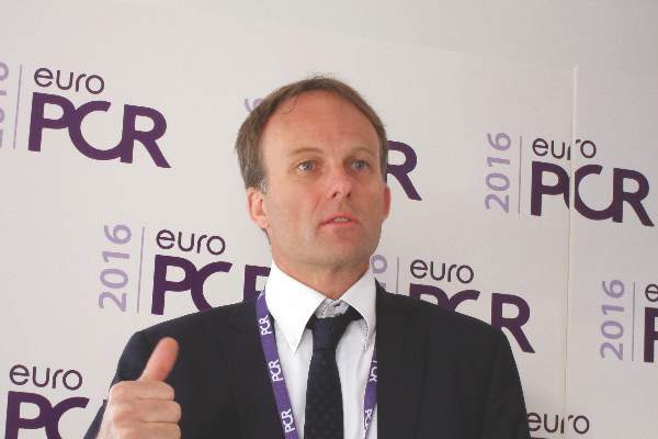
“EWOLUTION, with more than 1,000 patients, is I think the study that we’ve needed to make this intervention happen in routine practice as a procedure for stroke prevention in atrial fibrillation,” declared Dr. Bergmann of Cardiologicum Hamburg, a large German group practice.
EWOLUTION is a prospective, single-arm, multicenter registry including 1,025 recipients of the first-generation version of the Watchman, a transcatheter device for left atrial appendage closure as a means of stroke prevention in patients with atrial fibrillation (AF). These were high-risk patients: 50% of them had a CHA2DS2-VASc score of 5 or higher, and 72% of participants were deemed unsuitable for warfarin. EWOLUTION was conducted at 47 sites in Europe, the Middle East, and Russia. Dr. Bergmann presented the 3-month outcomes, a key period for device-related complications because protective epithelialization of the device isn’t yet complete. Follow-up will continue at the 1- and 2-year marks.
Among the key findings in the domain of procedural success: The rate of successful device implantation was 98.5%. A complete seal or an echocardiographic color jet of 5 mm or less at the time of implantation was achieved in 99.7% of cases, and this remained the case in 98.9% at 3 months.
With regard to safety, the stroke rate was 0.4%, device embolization occurred in 0.4%, and the pericardial effusion/tamponade rate was 0.7%, all of which are lower rates than in earlier clinical trials.
Just over 4% of patients experienced device-related adverse events with full recovery at the 3-month follow-up. These were mostly bleeding events at the groin access site. More importantly, only 0.5% of patients had device-related serious adverse events that weren’t resolved at 3 months.
“This 0.5% is a major step forward,” Dr. Bergmann said. “This includes all pericardial effusions, all device embolizations, all periprocedural strokes – everything involving the procedure that puts a patient at risk. In fact, that 0.5% risk for left atrial appendage closure with the Watchman is pretty much the same risk as if you refer your patient with AF to an electrophysiologist for pulmonary vein isolation.”
One key question answered by EWOLUTION is, do you need to be an expert in the procedure in order to get excellent results? The answer, Dr. Bergmann said, is emphatically “no.” Interventionalists at the 47 participating sites varied widely in their experience with left atrial appendage occlusion, so investigators divided the sites into quartiles based upon experience and patient volumes contributed to the registry. While cardiologists in the most experienced centers had a significantly higher rate of successful device release on the first try, the most- and least-experienced centers did equally well on the endpoint that really counts: a complete or near-complete seal of the left atrial appendage at follow-up. Complication rates didn’t differ by site experience, either.
Another important practical question that’s been holding up wider adoption of the Watchman concerns how best to handle postimplantation antithrombotic therapy. Here the EWOLUTION registry provides a partial answer: 607 patients were on dual antiplatelet therapy, 113 were on a novel anticoagulant (NOAC), 159 were on warfarin, 67 were on nothing at all, and the rest were on a single antiplatelet agent. And there was no significant difference between any of these groups in the 3-month rates of thrombus on the device, bleeding events, stroke, or other adverse events.
“The main message is there is no difference in serious adverse events between any of the drug regimens. So if you’re in a situation where you can’t give any medication to reduce stroke risk because the bleeding risk is so high, you can go for left atrial appendage closure technology and give them nothing post implant,” according to Dr. Bergmann.
He added that he’d personally try to avoid that strategy: “You want to have some coverage.”
Also noteworthy is that 113 Watchman recipients did well with 3 months of NOAC monotherapy. The appeal of that approach is the short half-life of those agents in the event of a bleeding problem.
Discussant Dr. Farrel Hellig commented that EWOLUTION demonstrates that the Watchman procedure is predictable and safe, with an impressively low complication rate even in inexperienced centers. For that, he added, kudos goes to Boston Scientific, which markets the Watchman, for rolling out an effective physician training program.
“The most important topic to discuss is bleeding, because a bleeding rate of 4.1% at 3 months is certainly not insignificant,” he continued. “It appears, firstly, from EWOLUTION that it’s safe to omit warfarin. I think the most important thing we’d like to know now is could a postprocedure NOAC be the best option, and can aspirin be eliminated in the long term?”
“Is it too early to recommend a NOAC-only for a period of time post procedure? Perhaps. But I think this would be an important next clinical trial: a NOAC-only for a period post procedure with no aspirin to follow. This is the next piece of information we need to complete the puzzle and for left atrial appendage closure to reach its full potential,” according to Dr. Hellig of the University of Cape Town (South Africa).
The EWOLUTION registry is sponsored by Boston Scientific. Dr. Bergmann reported serving as a consultant to Boston Scientific and Biosense Webster and receiving honoraria from more than a half-dozen pharmaceutical and device companies.
AT EUROPCR 2016
Key clinical point: You don’t have to be an expert at implanting the Watchman left atrial appendage closure device to achieve the same sort of sky-high procedural success and low complication rates the experts do.
Major finding: The Watchman device successfully sealed off the left atrial appendage in 98.9% of cases at 3 months follow-up, with rates closely similar at the least- and most-experienced centers.
Data source: EWOLUTION is a prospective, multicenter, real-world registry of 1,025 recipients of the Watchman left atrial appendage closure device at 47 sites in Europe, the Middle East, and Russia.
Disclosures: The EWOLUTION registry is sponsored by Boston Scientific. The presenter reported serving as a consultant to Boston Scientific and Biosense Webster and receiving honoraria from more than a half-dozen pharmaceutical and device companies.
Rotor ablation for atrial fibrillation strikes out in first randomized trial
SAN FRANCISCO – Focal impulse and rotor modulation-guided ablation for persistent atrial fibrillation – either alone or in conjunction with other procedures – increased procedural times without improving outcomes, according to the first randomized trial to assess its utility.
In fact, enrollment in the rotor ablation-only (RA) arm was halted early for futility. “There was 100% recurrence” of atrial fibrillation (AF), said senior investigator Dr. Andrea Natale, executive medical director of the Texas Cardiac Arrhythmia Institute, Austin.
“I’m surprised it took this long for a randomized study, because this system has been around for 5 or 6 years,” noted Dr. Natale. “Our community should demand these sorts of studies earlier, because it’s not fair for patients to go on with a procedure for years that has not been proven to be effective.
“For us, unless there is a new version of rotor mapping that I feel is significantly different, this will be the end of rotor ablation in my lab with this system [the Topera Physiologic Rotor Mapping Solution],” Dr. Natale said at the annual scientific sessions of the Heart Rhythm Society.
In the study, his team randomized 29 patients to RA only, 42 to RA plus pulmonary vein antral isolation (PVAI), and 42 to PVAI plus posterior wall and nonpulmonary vein trigger ablation.
At about 1 year, four RA-only patients (14%), 22 RA plus PVAI patients (52%), and 32 patients in the PVAI plus trigger group (76%) were free of AF and atrial tachycardias without antiarrhythmic drugs (P < .0001).
Meanwhile, RA alone and RA plus PVAI cases took about 230 minutes, while the more effective PVAI plus trigger approach took about 130 minutes (P < .001).
There was “a very poor outcome with rotor-only ablation,” Dr. Natale said. “There isn’t a benefit either alone or as an add-on strategy, at least with this mapping software.”
Perhaps “people who think rotors don’t exist are right,” he added. On the other hand, maybe the basket mapping catheter doesn’t touch enough of the left atrium, or the software that makes sense of what the catheter detects needs to be improved, Dr. Natale noted.
All the patients were undergoing their first ablation. They were in their early 60s, on average, and most were men. The mean left atrium diameter was about 47 mm, and mean left ventricle ejection fraction about 55%. There were no statistically significant differences between the study arms, and no significant differences in outcomes between the 70% of patients with persistent AF and the 30% with long-standing persistent AF.
There was no industry funding for the work. Dr. Natale disclosed relationships with Biosense Webster, Boston Scientific, Janssen, Medtronic, and St. Jude Medical.
My gut sense is that there’s something to rotor mapping, but we are not there yet. There are a lot of investment dollars and a lot of bright people working on this. It really is the Holy Grail to find the source of AF.
Dr. John Day is the director of Intermountain Heart Rhythm Specialists in Murray, Utah, and the current president of the Hearth Rhythm Society. He had no disclosures.
SAN FRANCISCO – Focal impulse and rotor modulation-guided ablation for persistent atrial fibrillation – either alone or in conjunction with other procedures – increased procedural times without improving outcomes, according to the first randomized trial to assess its utility.
In fact, enrollment in the rotor ablation-only (RA) arm was halted early for futility. “There was 100% recurrence” of atrial fibrillation (AF), said senior investigator Dr. Andrea Natale, executive medical director of the Texas Cardiac Arrhythmia Institute, Austin.
“I’m surprised it took this long for a randomized study, because this system has been around for 5 or 6 years,” noted Dr. Natale. “Our community should demand these sorts of studies earlier, because it’s not fair for patients to go on with a procedure for years that has not been proven to be effective.
“For us, unless there is a new version of rotor mapping that I feel is significantly different, this will be the end of rotor ablation in my lab with this system [the Topera Physiologic Rotor Mapping Solution],” Dr. Natale said at the annual scientific sessions of the Heart Rhythm Society.
In the study, his team randomized 29 patients to RA only, 42 to RA plus pulmonary vein antral isolation (PVAI), and 42 to PVAI plus posterior wall and nonpulmonary vein trigger ablation.
At about 1 year, four RA-only patients (14%), 22 RA plus PVAI patients (52%), and 32 patients in the PVAI plus trigger group (76%) were free of AF and atrial tachycardias without antiarrhythmic drugs (P < .0001).
Meanwhile, RA alone and RA plus PVAI cases took about 230 minutes, while the more effective PVAI plus trigger approach took about 130 minutes (P < .001).
There was “a very poor outcome with rotor-only ablation,” Dr. Natale said. “There isn’t a benefit either alone or as an add-on strategy, at least with this mapping software.”
Perhaps “people who think rotors don’t exist are right,” he added. On the other hand, maybe the basket mapping catheter doesn’t touch enough of the left atrium, or the software that makes sense of what the catheter detects needs to be improved, Dr. Natale noted.
All the patients were undergoing their first ablation. They were in their early 60s, on average, and most were men. The mean left atrium diameter was about 47 mm, and mean left ventricle ejection fraction about 55%. There were no statistically significant differences between the study arms, and no significant differences in outcomes between the 70% of patients with persistent AF and the 30% with long-standing persistent AF.
There was no industry funding for the work. Dr. Natale disclosed relationships with Biosense Webster, Boston Scientific, Janssen, Medtronic, and St. Jude Medical.
AT HEART RHYTHM 2016
Key clinical point: Focal impulse and rotor modulation-guided ablation for persistent atrial fibrillation – either alone or in conjunction with other procedures – increased procedural times without improving outcomes.
Major finding: At about 1 year, four rotor ablation-only patients (14%), 22 RA plus pulmonary vein antral isolation patients (52.4%), and 32 patients in the PVAI plus trigger group (76%) were free of atrial fibrillation and atrial tachycardias without antiarrhythmic drugs (P < .0001).
Data source: A randomized trial in 113 persistent AF patients.
Disclosures: There was no industry funding for the work. The senior investigator disclosed relationships with Biosense Webster, Boston Scientific, Janssen, Medtronic, and St. Jude Medical.
Drug-coated stent sets new standard in ACS patients at high bleeding risk
PARIS – In patients at high bleeding risk undergoing percutaneous coronary intervention for acute coronary syndrome, a unique drug-coated, polymer-free stent backed by just a single month of dual-antiplatelet therapy (DAPT) trounced a bare metal stent in both efficacy and safety at 1 year in a large randomized trial, Dr. Christoph K. Naber reported at the annual congress of the European Association of Percutaneous Cardiovascular Interventions.
The BioFreedom stent, coated with a proprietary sirolimus analogue called Biolimus-A9, rapidly transfers the drug to the vessel wall, leaving in place a metallic stent. This drug-coated stent (DCS) provides the antirestenotic benefits of a drug-eluting stent with the advantages of a shorter DAPT requirement and no potential polymer-related adverse events. The device’s performance in this prespecified subgroup analysis of the Leaders Free trial will be practice altering, predicted Dr. Naber of Elisabeth University Hospital in Essen, Germany.
“We believe that current guidelines and clinical practice need to change. Bare-metal stents can no longer be recommended for high–bleeding risk patients presenting with ACS. The Biolimus-A9–coated stent currently has the best supporting evidence for this indication,” he said.
“This was the last chance for bare metal stents to show a good indication. They were thought to be good only in high–bleeding risk patients. Now we see that a drug-coated stent provides better efficacy and is even safer,” the cardiologist continued.
Leaders Free was a randomized double-blind clinical trial in which 2,499 patients at high risk of bleeding who underwent PCI received either the DCS or the Gazelle bare-metal stent (BMS) and 1 month of dual-antiplatelet therapy. The main results, in which the DCS showed superior safety and efficacy at 12 months of follow-up, have been published (N Engl J Med. 2015 Nov 19;373[21]:2038-47).
Dr. Naber presented a prespecified subgroup analysis that included the 659 Leaders Free participants who presented with ACS.
The primary efficacy endpoint – the 12-month rate of clinically driven target lesion revascularization – was 3.9% in the DCS group, compared with 9% in patients randomized to the BMS, for an adjusted 59% relative risk reduction.
The composite safety endpoint was composed of the 1-year rate of cardiac death, acute MI, and definite or probable stent thrombosis. This occurred in 9.3% of the DCS- and 18.5% of the BMS-treated patients, for a relative risk reduction of 52%.
The BioFreedom stent has a selectively microstructured surface coated with Biolimus-A9, a drug that’s far more lipophilic than sirolimus, everolimus, or zotarolimus. As a result, 98% of the drug is transferred to the plaque-laden vessel within 30 days, which is why it’s safe to shorten DAPT to 1 month, Dr. Naber explained.
It’s estimated that up to 20% of patients undergoing PCI are at high bleeding risk for various reasons.
The DCS is marketed in Europe and in clinical trials in the United States aimed at getting Food and Drug Administration approval.
Discussant Dr. Thomas Cuisset of Timone Hospital in Marseille, France, agreed that “in 2016, bare-metal stents are no longer the gold standard in patients at high bleeding risk.”
He added that the Leaders Free ACS substudy leaves unanswered two major remaining questions: What’s the optimal duration of DAPT for DCS in non-ACS patients at high bleeding risk – is 1 month of DAPT as safe as 6 months? And how do contemporary drug-eluting, polymer-based stents compare to the BioFreedom stent for PCI in the setting of high bleeding risk?
In an interview, Dr. Naber said that before a head-to-head comparative clinical trial can even be considered, there will need to be evidence that drug-eluting stents are safe with shortened DAPT. That has yet to be shown.
The study was funded by Biosensors. Dr. Naber reported receiving consultant’s fees from that medical device company and several others. Dr. Cuisset serves as a consultant to or paid lecturer for more than a dozen medical companies.
Simultaneous with Dr. Naber’s presentation at EuroPCR in Paris, the Leaders Free ACS substudy results were published online (Eur Heart J. doi:10.1093/eurheartj/ehw203).
AT EUROPCR 2016
Key clinical point: There are no longer any good indications for implanting a bare-metal stent.
Major finding: The cumulative 1-year rate of cardiac death, MI, or definite or probable stent thrombosis in high–bleeding risk patients who underwent PCI for acute coronary syndrome was 9.3% in those randomized to a novel drug-coated stent, compared with 18.5% in those who got a bare-metal stent.
Data source: This was a prespecified subgroup analysis of the Leaders Free trial in which 659 acute coronary syndrome patients with high bleeding risk undergoing PCI for acute coronary syndrome were randomized to a unique drug-coated, polymer-free stent or a bare-metal stent, with 1 month of dual-antiplatelet therapy for all.
Disclosures: The study was funded by Biosensors. The presenter reported receiving consultant fees from that medical device company and several others.
VIDEO: Cardiothoracic surgeon shortage requires action
BALTIMORE – By 2035, U.S. cardiothoracic surgeons will see a 61% increase in the national caseload, and potentially a 121% increase in cases for each surgeon, according to a data analysis presented at the annual meeting of the American Association for Thoracic Surgery.
Using data from the American Board of Thoracic Surgery, a research team at Ohio State University performed case load calculations for 2035 based on cases per surgeon per year in 2010. The researchers estimated that the average caseload per surgeon in 2035 will be 299 cases, compared with a 2010 caseload of 135 per surgeon. This increase is not matched by the number of surgeons currently trained and certified annually.
Dr. John Ikonomidis, chief of the division of cardiothoracic surgery at the Medical University of South Carolina in Charleston, and a discussant on the presentation, said surgeon retirements and an increase in the population needing treatment have put the specialty in a bind.
“We have a bit of a crisis now, honestly, but this particular paper puts it in even further perspective,” Dr. Ikonomidis said in a video interview. “By 2035 we’re looking at a 3,000-surgeon shortage, relative to what would be available.” He noted that approximately 90 medical residents per year are certified as cardiothoracic surgeons, a rate which will not produce enough CT surgeons to meet the projected shortage.
“We need to continue to have this conversation,” he concluded. “It is a reminder that the predictions we made 15 years ago appear to be true, and we probably need to do something about it, at least in the short term.”
Dr. Ikonomidis reported no relevant financial disclosures.
The video associated with this article is no longer available on this site. Please view all of our videos on the MDedge YouTube channel
On Twitter @richpizzi
AT THE AATS ANNUAL MEETING
Epicardial GP ablation of no benefit in advanced atrial fibrillation
San Francisco – Routine ganglionic plexus ablation increases risk and offers no clinical benefit in patients undergoing thoracoscopic surgery for advanced atrial fibrillation, according to a randomized Dutch trial.
“Most surgeons who do epicardial ablation do GP [ganglionic plexus] ablation because of the assumption that they are doing something good; that assumption is wrong. GP ablation should not be performed in patients with advanced AF [atrial fibrillation],” said lead investigator Dr. Joris de Groot, a cardiologist at the University of Amsterdam.
Following pulmonary vein isolation (PVI), 117 patients were randomized to GP ablation, and 123 to no GP ablation. GP ablation eliminated 100% of evoked vagal responses; vagal responses remained intact in nearly all of the control subjects.
At 1 year, 70.9% in the GP group compared with 68.4% in control arm were free of recurrence (P = .7); there were no statistically significant differences when the analysis was limited to the 59% of patients who went into the trial with persistent AF or limited to the rest of the patients with paroxysmal AF. Recurrences constituted significantly more atrial tachycardia in the GP group than in the control group. Even after the researchers controlled for a wide variety of demographic, anatomical, and clinical variables, “GP ablation made no difference in atrial fibrillation recurrence at 1 year,” Dr. de Groot said at the annual scientific sessions of the Hearth Rhythm Society.
Meanwhile, major perioperative bleeding occurred in nine patients, all in the GP group, and one required a sternotomy for hemostatic control. Clinically relevant sinus node dysfunction occurred in 12 of the GP group, but only four control patients; six GP patients – but no one in the control arm – required subsequent pacemakers, three while in the hospital after surgery and three during follow-up. Almost 30 patients in each arm required cardioversion during the 3-month blanking period, and about 20 in each arm afterwards.
“The largest randomized study in thoracoscopic surgery for advanced AF to date demonstrates that GP ablation is associated with significantly more periprocedural major bleeding, sinus node dysfunction, and pacemaker implantation, but not with improved rhythm outcome,” the investigators concluded.
Procedure time was 185 +/– 54 minutes in the GP arm, and 168 +/– 54 minutes in the control arm (P = .015). In the GP group, four major GPs and the ligament of Marshall were ablated.
Patients were 60 years old, on average, and three-quarters were men. AF duration was a median of 4 years. Four patients had died at 1 year, all in the GP arm, but none related to the procedure. All antiarrhythmic drugs were stopped after the blanking period; any atrial arrhythmia lasting 30 seconds or longer thereafter was considered a recurrence.
Dr. de Groot disclosed payments for services from AtriCure, Daiichi, and St. Jude Medical and research funding from AtriCure and St. Jude.
AF ablation is an evolving field, and we are constantly trying to think of new ways to improve our success rates. Some of the things we try turn out to be advantageous and others do not. Negative studies like this have a very important clinical impact; they help us figure out what road to take.
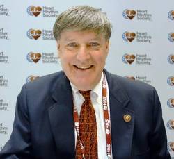
|
Dr. Thomas Deering |
Dr. Thomas Deering is chief of the Arrhythmia Center at the Piedmont Heart Institute in Atlanta, where he is also chairman of the Executive Council and the Clinical Centers for Excellence. He moderated Dr. de Groot’s presentation, and wasn’t involved in the work.
AF ablation is an evolving field, and we are constantly trying to think of new ways to improve our success rates. Some of the things we try turn out to be advantageous and others do not. Negative studies like this have a very important clinical impact; they help us figure out what road to take.

|
Dr. Thomas Deering |
Dr. Thomas Deering is chief of the Arrhythmia Center at the Piedmont Heart Institute in Atlanta, where he is also chairman of the Executive Council and the Clinical Centers for Excellence. He moderated Dr. de Groot’s presentation, and wasn’t involved in the work.
AF ablation is an evolving field, and we are constantly trying to think of new ways to improve our success rates. Some of the things we try turn out to be advantageous and others do not. Negative studies like this have a very important clinical impact; they help us figure out what road to take.

|
Dr. Thomas Deering |
Dr. Thomas Deering is chief of the Arrhythmia Center at the Piedmont Heart Institute in Atlanta, where he is also chairman of the Executive Council and the Clinical Centers for Excellence. He moderated Dr. de Groot’s presentation, and wasn’t involved in the work.
San Francisco – Routine ganglionic plexus ablation increases risk and offers no clinical benefit in patients undergoing thoracoscopic surgery for advanced atrial fibrillation, according to a randomized Dutch trial.
“Most surgeons who do epicardial ablation do GP [ganglionic plexus] ablation because of the assumption that they are doing something good; that assumption is wrong. GP ablation should not be performed in patients with advanced AF [atrial fibrillation],” said lead investigator Dr. Joris de Groot, a cardiologist at the University of Amsterdam.
Following pulmonary vein isolation (PVI), 117 patients were randomized to GP ablation, and 123 to no GP ablation. GP ablation eliminated 100% of evoked vagal responses; vagal responses remained intact in nearly all of the control subjects.
At 1 year, 70.9% in the GP group compared with 68.4% in control arm were free of recurrence (P = .7); there were no statistically significant differences when the analysis was limited to the 59% of patients who went into the trial with persistent AF or limited to the rest of the patients with paroxysmal AF. Recurrences constituted significantly more atrial tachycardia in the GP group than in the control group. Even after the researchers controlled for a wide variety of demographic, anatomical, and clinical variables, “GP ablation made no difference in atrial fibrillation recurrence at 1 year,” Dr. de Groot said at the annual scientific sessions of the Hearth Rhythm Society.
Meanwhile, major perioperative bleeding occurred in nine patients, all in the GP group, and one required a sternotomy for hemostatic control. Clinically relevant sinus node dysfunction occurred in 12 of the GP group, but only four control patients; six GP patients – but no one in the control arm – required subsequent pacemakers, three while in the hospital after surgery and three during follow-up. Almost 30 patients in each arm required cardioversion during the 3-month blanking period, and about 20 in each arm afterwards.
“The largest randomized study in thoracoscopic surgery for advanced AF to date demonstrates that GP ablation is associated with significantly more periprocedural major bleeding, sinus node dysfunction, and pacemaker implantation, but not with improved rhythm outcome,” the investigators concluded.
Procedure time was 185 +/– 54 minutes in the GP arm, and 168 +/– 54 minutes in the control arm (P = .015). In the GP group, four major GPs and the ligament of Marshall were ablated.
Patients were 60 years old, on average, and three-quarters were men. AF duration was a median of 4 years. Four patients had died at 1 year, all in the GP arm, but none related to the procedure. All antiarrhythmic drugs were stopped after the blanking period; any atrial arrhythmia lasting 30 seconds or longer thereafter was considered a recurrence.
Dr. de Groot disclosed payments for services from AtriCure, Daiichi, and St. Jude Medical and research funding from AtriCure and St. Jude.
AT HEART RHYTHM 2016
Key clinical point: Routine ganglionic plexus ablation increases risk and offers no clinical benefit in patients undergoing thoracoscopic surgery for advanced atrial fibrillation.
Major finding: At 1 year, 70.9% in the GP ablation group, but 68.4% in the control arm, were free of recurrence (P = .7)
Data source: Randomized trial of 240 AF patients, almost two-thirds with persistent disease
Disclosures: The lead investigator disclosed payments for services from AtriCure, Daiichi, and St. Jude Medical, and research funding from AtriCure and St. Jude.
Hybrid option ‘reasonable’ for HLHS?
Although the classic Norwood palliation for infants with hypoplastic left heart syndrome (HLHS) has been well established, the procedure has had its drawbacks, namely the need for cardiopulmonary bypass with hypothermia and a because it rules out biventricular correction months later. A hybrid procedure avoids the need for bypass and accommodates short-term biventricular correction, but it has lacked strong evidence.
Researchers from Justus-Liebig University Giessen, Germany, reported on 182 patients with HLHS who had the three-stage Giessen hybrid procedure, noting 10-year survival of almost 80% with almost a third of patients requiring no artery intervention in that time (J Thorac Cardiovasc Surg. 2016 April;151:1112-23).
“In view of the early results and long-term outcome after Giessen hybrid palliation, the hybrid approach has become a reasonable alternative to the conventional strategy to treat neonates with HLHS and variants,” wrote Dr. Can Yerebakan and colleagues. “Further refinements are warranted to decrease patient morbidity.”
The Giessen hybrid procedure uses a technique to control pulmonary blood flow that is different from the Norwood procedure. The hybrid approach involves stenting of the arterial duct or prostaglandin therapy to maintain systemic perfusion combined with off-pump bilateral banding of the pulmonary arteries (bPAB) in the neonatal period. The Giessen hybrid operation defers the Norwood-type palliation using cardiopulmonary bypass that involves an aortic arch reconstruction, including a superior cavopulmonary connection or a biventricular correction, if indicated, until the infant is 4-8 months of age.
“In recent years, hybrid treatment has moved from a myth to an alternative modality in a growing number of institutions globally,” Dr. Yerebakan and colleagues said. The hybrid procedure has been used in high-risk patients. One report claimed higher morbidity in the hybrid procedure due to bPAB (Ann Thorac Surg. 2013;96:1382-8). Another study raised concerns about an adequate pulmonary artery rehabilitation at the time of the Fontan operation, the third stage in the hybrid strategy (J Thorac Cardiovasc Surg. 2014;147:706-12).
But with the hybrid approach, patients retain the potential to receive a biventricular correction up to 8 months later without compromising survival, “postponing an immediate definitive decision in the newborn period in comparison with the classic Norwood palliation,” Dr. Yerebakan and coauthors noted.
The doctors at the Pediatric Heart Center Giessen treat all types and variants of HLHS with the modified Giessen hybrid strategy. Between 1998 and 2015, 182 patients with HLHS had the Giessen hybrid stage I operation, including 126 patients who received univentricular palliation or a heart transplant. The median age of stage I recipients was 6 days, and median weight 3.2 kg. The stage II operation was performed at 4.5 months, with a range of 2.9 to 39.5 months, and Fontan completion was established at 33.7 months, with a range of 21 to 108 months.
Median follow-up after the stage I procedure was 4.6 years, and the death rate was 2.5%. After stage II, mortality was 4.9%; no deaths were reported after Fontan completion. Body weight less than 2.5 kg and aortic atresia had no significant effect on survival. Mortality rates were 8.9% between stages I and II and 5.3% between stage II and Fontan completion. “Cumulative interstage mortality was 14.2%,” Dr. Yerebakan and colleagues noted. “At 10 years, the probability of survival is 77.8%.”
Also at 10 years, 32.2% of patients were free from further pulmonary artery intervention, and 16.7% needed aortic arch reconstruction. Two patients required reoperations for aortic arch reconstruction.
Dr. Yerebakan and colleagues suggested several steps to improve outcomes with the hybrid approach: “intense collaboration” with anesthesiology and pediatric cardiology during and after the procedure to risk stratify individual patients; implementation of standards for management of all stages, including out-of-hospital care, in all departments that participate in a case; and liberalized indications for use of MRI before the stage II and Fontan completion.
Among the limitations of the study the authors noted were its retrospective nature and a median follow-up of only 5 years when the center has some cases with up to 15 years of follow-up. But Dr. Yerebakan and coauthors said they could not determine if the patients benefit from the hybrid treatment in the long-term.
The researchers had no disclosures.
The study by Dr. Yerebakan and colleagues is one of the largest single-center series of patients with HLHS who routinely undergo a hybrid palliation to date, and while the study is open to criticisms, “the authors should be applauded,” Dr. Ralph S. Mosca of New York University said in his invited commentary (J Thorac Cardiovasc Surg. 2016;151:1123-25).
Among the criticisms Dr. Mosca mentioned are that the hybrid approach requires a more extensive stage II reconstruction, “often further complicated by the presence of significant branch PA stenosis and a difficult aortic arch reconstruction”; that there is “appreciable” interstage mortality at 12.2%; and that there is an absence of data on renal or respiratory insufficiency, infection rates, and neurologic outcomes.
Dr. Mosca cited the cause for applause, however: “Through their persistence and collective experience, [the authors] have achieved commendable results in this difficult patient population.”
Yet, Dr. Mosca also noted a number of “potential problems” with the hybrid approach: bilateral banding of the pulmonary artery is a “crude procedure”; arterial duct stenting can lead to retrograde aortic arch reduction; and the interstage mortality “remains significant.”
Results of the hybrid and Norwood procedures are “strikingly similar,” Dr. Mosca said. While the hybrid approach may lower neonatal mortality, it may also carry longer-term consequences “predicated upon the need to closely observe and intervene,” he said. Clinicians need more information on hybrid outcomes, but in time it will likely take its place as an option for HLHS alongside the Norwood procedure, Dr. Mosca said.
Although the classic Norwood palliation for infants with hypoplastic left heart syndrome (HLHS) has been well established, the procedure has had its drawbacks, namely the need for cardiopulmonary bypass with hypothermia and a because it rules out biventricular correction months later. A hybrid procedure avoids the need for bypass and accommodates short-term biventricular correction, but it has lacked strong evidence.
Researchers from Justus-Liebig University Giessen, Germany, reported on 182 patients with HLHS who had the three-stage Giessen hybrid procedure, noting 10-year survival of almost 80% with almost a third of patients requiring no artery intervention in that time (J Thorac Cardiovasc Surg. 2016 April;151:1112-23).
“In view of the early results and long-term outcome after Giessen hybrid palliation, the hybrid approach has become a reasonable alternative to the conventional strategy to treat neonates with HLHS and variants,” wrote Dr. Can Yerebakan and colleagues. “Further refinements are warranted to decrease patient morbidity.”
The Giessen hybrid procedure uses a technique to control pulmonary blood flow that is different from the Norwood procedure. The hybrid approach involves stenting of the arterial duct or prostaglandin therapy to maintain systemic perfusion combined with off-pump bilateral banding of the pulmonary arteries (bPAB) in the neonatal period. The Giessen hybrid operation defers the Norwood-type palliation using cardiopulmonary bypass that involves an aortic arch reconstruction, including a superior cavopulmonary connection or a biventricular correction, if indicated, until the infant is 4-8 months of age.
“In recent years, hybrid treatment has moved from a myth to an alternative modality in a growing number of institutions globally,” Dr. Yerebakan and colleagues said. The hybrid procedure has been used in high-risk patients. One report claimed higher morbidity in the hybrid procedure due to bPAB (Ann Thorac Surg. 2013;96:1382-8). Another study raised concerns about an adequate pulmonary artery rehabilitation at the time of the Fontan operation, the third stage in the hybrid strategy (J Thorac Cardiovasc Surg. 2014;147:706-12).
But with the hybrid approach, patients retain the potential to receive a biventricular correction up to 8 months later without compromising survival, “postponing an immediate definitive decision in the newborn period in comparison with the classic Norwood palliation,” Dr. Yerebakan and coauthors noted.
The doctors at the Pediatric Heart Center Giessen treat all types and variants of HLHS with the modified Giessen hybrid strategy. Between 1998 and 2015, 182 patients with HLHS had the Giessen hybrid stage I operation, including 126 patients who received univentricular palliation or a heart transplant. The median age of stage I recipients was 6 days, and median weight 3.2 kg. The stage II operation was performed at 4.5 months, with a range of 2.9 to 39.5 months, and Fontan completion was established at 33.7 months, with a range of 21 to 108 months.
Median follow-up after the stage I procedure was 4.6 years, and the death rate was 2.5%. After stage II, mortality was 4.9%; no deaths were reported after Fontan completion. Body weight less than 2.5 kg and aortic atresia had no significant effect on survival. Mortality rates were 8.9% between stages I and II and 5.3% between stage II and Fontan completion. “Cumulative interstage mortality was 14.2%,” Dr. Yerebakan and colleagues noted. “At 10 years, the probability of survival is 77.8%.”
Also at 10 years, 32.2% of patients were free from further pulmonary artery intervention, and 16.7% needed aortic arch reconstruction. Two patients required reoperations for aortic arch reconstruction.
Dr. Yerebakan and colleagues suggested several steps to improve outcomes with the hybrid approach: “intense collaboration” with anesthesiology and pediatric cardiology during and after the procedure to risk stratify individual patients; implementation of standards for management of all stages, including out-of-hospital care, in all departments that participate in a case; and liberalized indications for use of MRI before the stage II and Fontan completion.
Among the limitations of the study the authors noted were its retrospective nature and a median follow-up of only 5 years when the center has some cases with up to 15 years of follow-up. But Dr. Yerebakan and coauthors said they could not determine if the patients benefit from the hybrid treatment in the long-term.
The researchers had no disclosures.
FROM THE JOURNAL OF THORACIC AND CARDIOVASCULAR SURGERY
Key clinical point: A hybrid operation for hypoplastic left heart syndrome (HLHS) and variants in neonates is emerging as an alternative to the Norwood palliation.
Major finding: At 10 years, the probability of survival with the hybrid procedure was 77.8%. Low body weight (less than 2.5 kg) and aortic atresia had no significant impact on survival.
Data source: Retrospective study of 182 patients who had the hybrid procedure at a single center between June 1998 and February 2015.
Disclosures: The study investigators had no relationships to disclose.
Angina rates similar across metal stents
WASHINGTON – In patients with coronary artery disease, there is no increased risk of angina 1 year after percutaneous coronary intervention for bare metal stents relative to drug-eluting metal stents, according to data presented at Cardiovascular Research Technologies 2016.
“Angina pectoris in the first year after PCI [percutaneous coronary intervention] is remarkably common, affecting 32.3% of patients,” but “metallic stent type is not independently associated with the occurrence of angina,” reported Dr. Michael A. Gaglia Jr., an interventional cardiologist at MedStar Heart and Vascular Institute in Washington.
This conclusion was based on a study in which 8,804 patients who underwent PCI with metal stents were questioned about angina and its severity. The incidence of angina was compared for bare-metal stents relative to five drug-eluting metal stents: Cypher (sirolimus-eluting, Johnson & Johnson), Taxus Express2 (paclitaxel, Boston Scientific), Xience V (everolimus, Abbott Vascular), Promus Element (everolimus, Boston Scientific), and Resolute Integrity (zotarolimus, Medtronic).
For nearly 3 months, the cumulative incidence of angina remained tightly grouped at 5% or less across stent types. Incidence rates began climbing slowly through the first 9 months of follow-up and then more steeply at about 10 months. When depicted graphically, the incidence of angina appeared higher after placement of the Cypher stent, which was discontinued in 2011, but multivariate analysis found “no significant association between stent type and angina at 1 year after PCI,” Dr. Gaglia reported.
Although risk of angina was not correlated with type of metal stent, angina was highly correlated with risk of a major adverse cardiovascular event (MACE). When angina severity was stratified by the Canadian Cardiovascular Society system, MACE, defined as a composite of all-cause mortality, target vessel revascularization, and Q-wave myocardial infarction, occurred in 6.8% of those without angina, 10.0% of those with class 1 or 2 angina, and 19.7% of those with class 3 or 4 angina (P less than .001 for this trend) over the course of follow-up.
Independent of stent type, angina was more common in patients with a history of severe angina prior to PCI, a prior PCI, or prior coronary artery bypass grafting. A reduced likelihood of angina was independently associated with older age, male sex, a presentation of acute coronary syndrome, and a longer stented length.
Other studies have also shown that angina after PCI is associated with an increased risk of MACE relative to the absence of ischemia, but the contribution of this study is that it is the first set of data to suggest that drug eluting stents provide no advantage over bare metal stents for controlling angina, according to the authors. In the graphic representation of angina incidence for different stent types over 1-year of follow-up, four of the six lines, including the line representing bare metal stents, were essentially superimposable. In addition to the line representing angina incidence in those receiving the Cypher stent, the line representing angina incidence on the Promus Element stent climbed higher at 9 months relative to the remaining four stent types, but this line had rejoined the others at 12 months.
“Metallic coronary stents alter vessel geometry, shear stress, and hemodynamics. Stents also vary in design and architecture,” Dr. Gaglia observed. Although protection from angina is one of the major indications for the placement of stents, Dr. Gaglia emphasized that data comparing different metallic stents in regards to the incidence of angina pectoris at long-term follow-up have until this study “been lacking.”
The meeting was sponsored by the Cardiovascular Research Institute at Washington Hospital Center. Dr. Gaglia reports no relevant financial relationships. Abbott Vascular funded the study.
AT CARDIOVASCULAR RESEARCH TECHNOLOGIES 2016
Key clinical point: One year after stent placement, angina rates are no higher with bare metal stents than with drug-eluting metal stents.
Major finding: One year after stenting, the incidence of angina was 32.3%.
Data source: Observational study with 8,804 patients.
Disclosures: Dr. Gaglia reports no relevant financial relationships. Abbott Vascular funded the study.
Standard incubation can miss P. acnes in infective endocarditis
AMSTERDAM – Accounting for less than 1% of cases, Propionibacterium acnes has been considered an uncommon cause of infective endocarditis.
But data presented at the annual congress of the European Society of Clinical Microbiology and Infectious Diseases suggest that the common anaerobe may be responsible for many more cases than is now believed. The bacteria are difficult to culture and grow very slowly, Dr. Jona Banzon said at the meeting. Incubating it for the standard 5 days may simply not be long enough.
“Due to this slow-growing nature, the standard incubation period may not be enough to detect it,” said Dr. Banzon, an infectious disease fellow at the Cleveland Clinic. “And since P. acnes is part of our commensal flora, it’s frequently a contaminate in culture, and we might be inappropriately dismissing it as such. In fact, we now wonder if this lack of extended incubation may be accounting for a significant proportion of negative blood cultures, and whether more prosthetic valve endocarditis than we think is actually being caused by P. acnes.”
Dr. Banzon presented a series of 23 cases included in the Cleveland Clinic Infective Endocarditis Registry from the period of 2007-2015. All had P. acnes confirmed as the causative organism. The group comprises 3.3% of the institution’s entire infective endocarditis registry, “making infective carditis with P. acnes already much more common than it is said to be in the literature,” she noted.
All of the cases were confirmed by any of the following standards:
• Two or more blood cultures positive for P. acnes.
• Two or more valve cultures positive for P. acnes.
• Two or more valve sequencing rests positive for P. acnes.
• At least two of the following: a positive blood culture, a positive valve culture or valve sequencing, or histopathologic demonstration of microorganism consistent with P. acnes.
Of the cohort, 22 had prosthetic valve endocarditis. One patient had endocarditis on a native valve. This is an important point, Dr. Banzon said.
The organism is being increasingly recognized for causing infections of prosthetic material, including shoulder joint infections and shunts, but it rarely seems to affect native tissue. The patient who had native valve endocarditis had experienced an episode of Staphylococcus aureus endocarditis about 18 months earlier. He was not treated surgically, and ended up with a damaged valve. After the initial illness, he was readmitted several times with symptoms of endocarditis, but all of his blood cultures were negative. This was attributed to the receipt of antibiotics. “When he was finally operated on, there were three valve sequencing specimens and all were positive for P. acnes,” Dr. Banzon said.
The patients in the cohort were a mean of 74 years old; about 75% were men. All of the cases were left-sided endocarditis, with the majority (74%) involving the aortic valve. Other sites included the mitral valve (18%), aortic plus mitral (4%), and aortic plus tricuspid (4%).
The most common predisposing factor was having a prosthetic valve (96%). Other factors included having a cardiac implantable device (17%), and a prior episode of infective endocarditis (13%). None of the patients had indwelling vascular catheters or used injectable drugs.
The cases presented with severe disease, Dr. Banzon said. Almost half (48%) had a perivalvular abscess at presentation. A third (35%) had valve dehiscence, and 35% had severe valvular regurgitation. There was a vegetation of more than 1 cm in 9% of cases.
Emboli were not uncommon; 17% had emboli in the central nervous system, and 17% had peripheral emboli. Two of these patients had kidney and spleen infarcts and two had acute arterial thromboembolism that required thrombectomy.
All of the patients underwent blood cultures; overall, 30% of the cultures were positive for P. acnes. But there were “striking differences” when the tests were broken down by incubation time, Dr. Banzon said.
Most of the cultures (16) were incubated for the standard of 5 days or less. Among these, 12.5% were positive. However, cultures on seven patients were incubated for more than 5 days and in this group, 71.4% were positive for P. acnes. The median time to positivity overall was 7 days, with a range of 3-9 days.
Other diagnostic methods were important in closing this gap, she said. Valve culturing was positive in 57% of the cases, while valve sequencing was positive in 95%. The median time to positive for valve culture was somewhat shorter than for blood culture (5.5 days).
In nine cases, no organism would have been identified without valve sequencing, Dr. Banzon said.
Because they presented with severe disease, almost all of the patients (22) underwent surgery as their intimal treatment. At the time of surgery, everyone was taking an antibiotic that covered P. acnes. Single-agent therapy was the definitive treatment for most, with vancomycin being most commonly employed (59%), followed by ceftriaxone (25%). A few patients had a combination of both drugs or a combination of vancomycin and rifampin. One patient took penicillin.
The single patient who was medically treated received 6 weeks of intravenous ceftriaxone. After 1 month, he was readmitted with blood cultures positive for P. acnes. He underwent surgery and a valve sequencing confirmed P. acnes as the infective agent.
There were two in-hospital deaths, but the rest of the patients were discharged on antibiotic therapy and recovered with no additional deaths or relapses.
The extended time P. acnes required to show in culture was enough for the Cleveland Clinic to reconsider incubation guidelines for the microorganism, Dr. Banzon said.
“There are enough cases taking 9 or 10 days that we now always hold these cultures for at least 10 days when we’re looking for P. acnes.”
She had no financial disclosures.
AT ECCMID 2016
Key clinical point: P. acnes’ slow growth may result in false negative blood cultures in infective endocarditis.
Major finding: Just 12% of blood cultures incubated for 5 days were positive, while 71% of those incubated for more than 5 days were positive.
Data source: The Cleveland Clinic Infective Endocarditis Registry.
Disclosures: Dr. Banzon had no financial disclosures.
DANAMI 3-DEFER: No benefit with delayed stenting for STEMI
CHICAGO – Delaying stent implantation in patients with ST-segment elevation myocardial infarction failed to reduce the rate of mortality, heart failure, myocardial infarction, or repeat revascularization, compared with conventional percutaneous intervention in the randomized, controlled DANAMI 3-DEFER trial.
Among 1,215 patients with ST-segment elevation MI (STEMI) who were randomized to receive either standard primary percutaneous coronary intervention (PCI) with immediate stent implantation or deferred stent implantation 48 hours after the index procedure, the rate of the primary composite endpoint of all-cause mortality, hospital admission for heart failure, recurrent infarction, or any unplanned revascularization of the target vessel within 2 years was 18% in the immediate treatment group and 17% in the deferred stent implantation group, a nonsignificant difference, Dr. Henning Kelbæk reported at the annual meeting of the American College of Cardiology.
Procedure-related myocardial infarction, bleeding requiring transfusion or surgery, contrast-induced nephropathy, or stroke occurred in 5% and 4% of patients in the groups, respectively, he said.
Although some might be relieved to know there won’t be a need for doing a second procedure, the findings are a disappointment in that preliminary findings suggested a benefit when stenting is delayed for several hours to several days after angioplasty, said Dr. Kelbæk of Roskilde Hospital (Denmark).
The thinking was that medication given during the delay might help diminish residual blood clots, thereby reducing the risk of distal embolization, which occurs in 7% of cases, and which can occur despite successful treatment of the culprit artery lesion by primary PCI with stent implantation, he explained, noting that slow- or no-flow occurs in 10% of cases.
It is possible that the study may not have been large enough to detect overall differences in the two treatment groups. It is also possible that patients at the highest risk for developing another arterial blockage could potentially benefit from a delay, especially given that a small but significant improvement in left ventricular function was detected 18 months after treatment among patients who underwent deferred stenting (left ventricular ejection fraction, 60% vs. 57% in the immediate treatment group), but such patients were excluded from DANAMI 3-DEFER (the Third Danish Study of Optimal Acute Treatment of Patients with ST-segment Elevation Myocardial Infarction: Deferred stent implantation in connection with primary PCI), he said.
He added that he and his coinvestigators will “look carefully for possible ‘hypothesis-generating’ findings in subsets of patients – both those who might have benefited from the deferred-treatment strategy and, equally important, those in whom this strategy might have worsened their condition.”
Patients were enrolled into DANAMI 3-DEFER during March 2011–February 2014 at four primary PCI centers in Denmark. All were adults with acute onset symptoms lasting 12 hours or less, and ST-segment elevation of 0.1 mV or more in at least 2 contiguous electrocardiographic leads, or newly developed left bundle branch block. Those in the deferred treatment group were only randomized to that group if stabilized flow could be obtained in the infarct-related artery. Median follow-up was 42 months.
The findings indicate that at this point, deferred stent implantation cannot be recommended as a routine procedure for STEMI patients treated with primary PCI, Dr. Kelbæk concluded. The findings were published online simultaneously with the presentation (Lancet. 2016 Apr 3. doi: 10.1016/S0140-6736[16]30072-1).
The DANAMI-3-DEFER trial was funded by the Danish Agency for Science, Technology and Innovation and Danish Council for Strategic Research. Dr. Kelbæk reported having no disclosures.
AT ACC 16
Key clinical point: Delaying stent implantation in patients with STEMI failed to improve outcomes, compared with conventional percutaneous intervention in the randomized, controlled DANAMI 3-DEFER trial.
Major finding: The rate of the primary composite endpoint was 18% in the immediate treatment group and 17% in the deferred stent implantation group.
Data source: The open-label, randomized, controlled DANAMI 3-DEFER trial of 1,215 patients.
Disclosures: The DANAMI 3-DEFER trial was funded by the Danish Agency for Science, Technology, and Innovation, and the Danish Council for Strategic Research. Dr. Kelbæk reported having no disclosures.
