User login
PEXIVAS trial results will likely change ANCA-associated vasculitis therapy
CHICAGO – Results of the landmark PEXIVAS study – far and away the largest randomized trial ever done in ANCA-associated vasculitis – will likely change treatment in a couple of major ways.
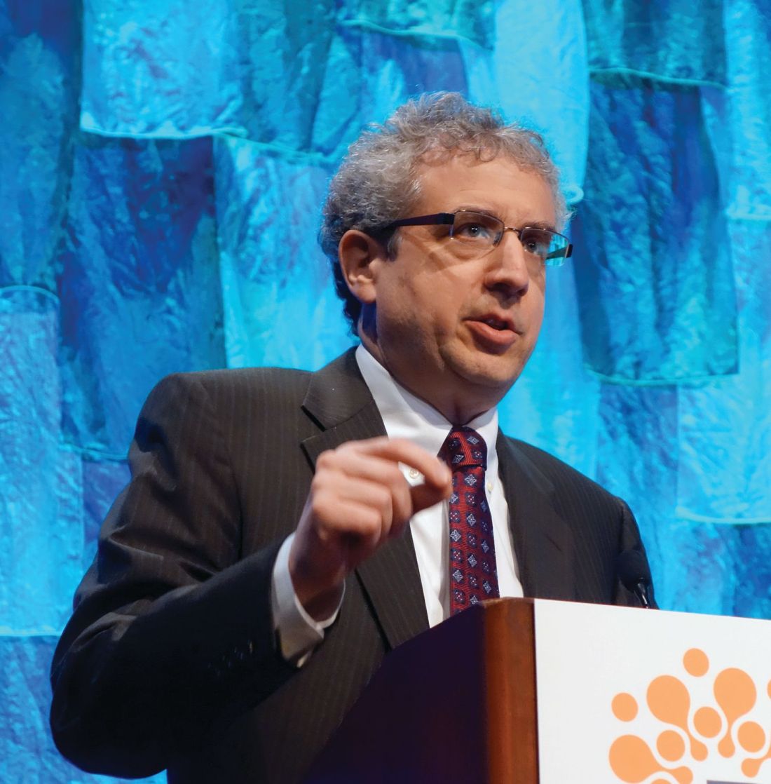
“I think this trial will have an impact on care. Based on these findings, physicians should strongly reconsider the utility of plasma exchange as a treatment for AAV [antineutrophil cytoplasmic antibody–associated vasculitis] patients and should now consider using lower cumulative doses of glucocorticoids for the treatment of severe AAV,” PEXIVAS coprincipal investigator Peter A. Merkel, MD, said at the annual meeting of the American College of Rheumatology.
That’s because the trial demonstrated that plasma exchange neither saved lives nor avoided end-stage renal disease, while utilization of oral glucocorticoids in doses substantially lower than the high-dose current standard significantly reduced the serious infection rate without causing less effective disease control, according to Dr. Merkel, chief of rheumatology and professor of medicine and epidemiology at the University of Pennsylvania in Philadelphia.
PEXIVAS comprised 704 patients with severe granulomatosis with polyangiitis or microscopic polyangiitis, making it more than twice as large as any other trial in AAV. This was a multicenter, international, open-label, randomized trial with a 2-by-2 factorial design. To qualify as having severe AAV, participants had to have an estimated glomerular filtration rate below 50 mL/min per 1.73 m2 and/or lung hemorrhage.
“This was in essence two trials embedded within one protocol in the factorial design,” he explained.
The impetus for this major clinical trial was a recognition that mortality due to AAV remains high, especially in the first year, with a clear unmet need for better, less toxic therapies. Indeed, it’s estimated that only 29% of deaths in the first year after diagnosis are due to the vasculitis disease itself, while over 50% of the mortality is caused by infection, much of it collateral damage from immunosuppressive therapies.
Dr. Merkel, who heads the National Institutes of Health–supported Vasculitis Clinical Research Consortium, said the time was right for a clinical trial aimed at improving patient management: “Clinical equipoise exists for the efficacy of both plasma exchange and reduced-dose glucocorticoids in ANCA-associated vasculitis.”
The patients underwent induction therapy with cyclophosphamide or rituximab (Rituxan) plus IV methylprednisolone. Then they were randomized to seven plasma exchange sessions in 14 days or no plasma exchange, and further randomized to conventional weight-based, high-dose oral glucocorticoids or a lower-dose regimen. Those on the reduced-dose regimen received 54% of the cumulative amount of glucocorticoids used in the standard-dose group through the first 3 months, and 61% over the course of 6 months. By week 4, those on the reduced-dose regimen were on an average of 25 mg/day, while those on standard therapy were on 50 mg/day. Adherence to assigned study arms exceeded 90%. Patients were followed prospectively for 1-7 years.
The primary endpoint, a composite of all-cause mortality or development of end-stage renal disease, occurred in 28% of patients on plasma exchange and 31% of those who did not undergo plasma exchange, a nonsignificant difference indicative of a lack of benefit for the intervention. No differential effect was seen in prespecified subgroups based on age, creatinine clearance, ANCA type, form of immunosuppression, or presence or absence of lung hemorrhage.
Further, the primary endpoint occurred in 28% of patients on reduced-dose glucocorticoids, compared with 26% on full-dose therapy; again, a nonsignificant difference, meaning lower-dose therapy didn’t result in less effective disease control. But it did result in a significant reduction in the prespecified endpoint of serious infections in the first year: 27% versus 33% with full-dose therapy, representing a 30% relative risk reduction.
Audience members wanted to know if there are any circumstances at all in which Dr. Merkel would now consider resorting to plasma exchange, such as maybe in AAV patients at the most extreme end of the severity spectrum.
“I’m not sure I should be the one dictating that; I think the world needs to see the data,” he replied.
That being said, he added, “I think these data are incredibly helpful to physicians and patients as they face this decision. I think plasma exchange is an expensive therapy and somewhat invasive. I think our results indicate that the benefit that we may have thought was there is not there.”
The study was sponsored by the National Institutes of Health, the Food and Drug Administration, the U.K. Medical Research Council and the National Institute for Health Research, the Canadian Institutes of Health Research, and the governments of France, Australia, and New Zealand. The presenter reported receiving research funding from the ACR, EULAR, FDA, NIH, Patient-Centered Outcomes Research Institute, and the Vasculitis Foundation. He also receives research funding from and/or serves as a consultant to more than a dozen pharmaceutical companies.
SOURCE: Merkel PA et al. Arthritis Rheumatol. 2018;70(Suppl 10):Abstract 2788.
CHICAGO – Results of the landmark PEXIVAS study – far and away the largest randomized trial ever done in ANCA-associated vasculitis – will likely change treatment in a couple of major ways.
“I think this trial will have an impact on care. Based on these findings, physicians should strongly reconsider the utility of plasma exchange as a treatment for AAV [antineutrophil cytoplasmic antibody–associated vasculitis] patients and should now consider using lower cumulative doses of glucocorticoids for the treatment of severe AAV,” PEXIVAS coprincipal investigator Peter A. Merkel, MD, said at the annual meeting of the American College of Rheumatology.
That’s because the trial demonstrated that plasma exchange neither saved lives nor avoided end-stage renal disease, while utilization of oral glucocorticoids in doses substantially lower than the high-dose current standard significantly reduced the serious infection rate without causing less effective disease control, according to Dr. Merkel, chief of rheumatology and professor of medicine and epidemiology at the University of Pennsylvania in Philadelphia.
PEXIVAS comprised 704 patients with severe granulomatosis with polyangiitis or microscopic polyangiitis, making it more than twice as large as any other trial in AAV. This was a multicenter, international, open-label, randomized trial with a 2-by-2 factorial design. To qualify as having severe AAV, participants had to have an estimated glomerular filtration rate below 50 mL/min per 1.73 m2 and/or lung hemorrhage.
“This was in essence two trials embedded within one protocol in the factorial design,” he explained.
The impetus for this major clinical trial was a recognition that mortality due to AAV remains high, especially in the first year, with a clear unmet need for better, less toxic therapies. Indeed, it’s estimated that only 29% of deaths in the first year after diagnosis are due to the vasculitis disease itself, while over 50% of the mortality is caused by infection, much of it collateral damage from immunosuppressive therapies.
Dr. Merkel, who heads the National Institutes of Health–supported Vasculitis Clinical Research Consortium, said the time was right for a clinical trial aimed at improving patient management: “Clinical equipoise exists for the efficacy of both plasma exchange and reduced-dose glucocorticoids in ANCA-associated vasculitis.”
The patients underwent induction therapy with cyclophosphamide or rituximab (Rituxan) plus IV methylprednisolone. Then they were randomized to seven plasma exchange sessions in 14 days or no plasma exchange, and further randomized to conventional weight-based, high-dose oral glucocorticoids or a lower-dose regimen. Those on the reduced-dose regimen received 54% of the cumulative amount of glucocorticoids used in the standard-dose group through the first 3 months, and 61% over the course of 6 months. By week 4, those on the reduced-dose regimen were on an average of 25 mg/day, while those on standard therapy were on 50 mg/day. Adherence to assigned study arms exceeded 90%. Patients were followed prospectively for 1-7 years.
The primary endpoint, a composite of all-cause mortality or development of end-stage renal disease, occurred in 28% of patients on plasma exchange and 31% of those who did not undergo plasma exchange, a nonsignificant difference indicative of a lack of benefit for the intervention. No differential effect was seen in prespecified subgroups based on age, creatinine clearance, ANCA type, form of immunosuppression, or presence or absence of lung hemorrhage.
Further, the primary endpoint occurred in 28% of patients on reduced-dose glucocorticoids, compared with 26% on full-dose therapy; again, a nonsignificant difference, meaning lower-dose therapy didn’t result in less effective disease control. But it did result in a significant reduction in the prespecified endpoint of serious infections in the first year: 27% versus 33% with full-dose therapy, representing a 30% relative risk reduction.
Audience members wanted to know if there are any circumstances at all in which Dr. Merkel would now consider resorting to plasma exchange, such as maybe in AAV patients at the most extreme end of the severity spectrum.
“I’m not sure I should be the one dictating that; I think the world needs to see the data,” he replied.
That being said, he added, “I think these data are incredibly helpful to physicians and patients as they face this decision. I think plasma exchange is an expensive therapy and somewhat invasive. I think our results indicate that the benefit that we may have thought was there is not there.”
The study was sponsored by the National Institutes of Health, the Food and Drug Administration, the U.K. Medical Research Council and the National Institute for Health Research, the Canadian Institutes of Health Research, and the governments of France, Australia, and New Zealand. The presenter reported receiving research funding from the ACR, EULAR, FDA, NIH, Patient-Centered Outcomes Research Institute, and the Vasculitis Foundation. He also receives research funding from and/or serves as a consultant to more than a dozen pharmaceutical companies.
SOURCE: Merkel PA et al. Arthritis Rheumatol. 2018;70(Suppl 10):Abstract 2788.
REPORTING FROM THE ACR ANNUAL MEETING
Key clinical point: Plasma exchange was without benefit and reduced-dose oral glucocorticoids safely decreased serious infections in ANCA-associated vasculitis.
Major finding: The rate of serious infections in the first year was 27% in patients on reduced-dose oral glucocorticoids and 33% with standard high-dose therapy, for a significant 30% relative risk reduction.
Study details: PEXIVAS was a multicenter, international, open-label, randomized trial with a 2-by-2 factorial design comprising 704 patients with severe ANCA-associated vasculitis.
Disclosures: The study was sponsored by the National Institutes of Health, the Food and Drug Administration, the U.K. Medical Research Council and the National Institute of Health Research, the Canadian Institutes of Health Research, and the governments of France, Australia, and New Zealand. The presenter reported receiving research funding from the ACR, EULAR, FDA, NIH, Patient-Centered Outcomes Research Institute, and the Vasculitis Foundation. He also receives research funding from and/or serves as a consultant to more than a dozen pharmaceutical companies.
Source: Merkel PA et al. Arthritis Rheumatol. 2018;70(Suppl 10): Abstract 2788.
Drafts of new classification criteria presented for GCA, Takayasu’s arteritis
CHICAGO – Drafts of new classification criteria for giant cell arteritis and Takayasu’s arteritis developed by the American College of Rheumatology and the European League Against Rheumatism (EULAR) reflect the increasingly important role of advanced vascular imaging in the diagnosis and management of large-vessel vasculitis, according to Peter A. Merkel, MD.
The drafts, which are the result of a multiyear collaboration between the ACR and EULAR, were presented at the annual meeting of the ACR and will be submitted to the ACR/EULAR committee overseeing the work for comprehensive review and possible revisions. Once endorsed, the new criteria will replace the “extremely important,” but outdated, existing classification criteria, which were published in 1990.
“What we’ve done is, rather than purely revise the 1990 [criteria], we’ve started again from scratch ... with a great number of cases from a wide variety of centers throughout the world. This was a very large international effort ... really a great community effort in the field of rheumatology,” Dr. Merkel, professor and chief of the division of rheumatology at the University of Pennsylvania, Philadelphia, and one of the chief investigators for the project, said in a video interview.
The new criteria will allow for better classification of patients with giant cell arteritis versus Takayasu’s arteritis versus another form of vasculitis, he said, noting that advances in imaging that allow for “more enriched data with which to make decisions” play a large role.
However, the new criteria are not meant to be used for diagnosis, but to “sort out among the different types of vasculitis,” he said.
“It provides awareness and it provides a tool, especially for research investigation, but that seeps out into the broader community,” he added.
Dr. Merkel reported having no disclosures.
SOURCE: Merkel PA et al. ACR Annual Meeting, Presentation 5T116.
REPORTING FROM THE ACR ANNUAL MEETING
SLE low-disease definition receives prospective validation
CHICAGO – The long path toward a validated definition of low disease activity in patients with systemic lupus erythematosus may be nearing an end as a definition first proposed more than 5 years ago received validation with data from more than 1,700 patients in a prospective, multicenter study.

The next step is to test this definition in treat-to-target intervention studies, and to apply the definition in other clinical trials as well as in routine clinical practice, Vera Golder, MD, said at the annual meeting of the American College of Rheumatology.
The tested definition has five elements that a patient needs to achieve to be considered in a lupus low disease activity state (LLDAS):
• A systemic lupus erythematosus (SLE) disease activity index (SLEDAI) score of 4 or less with no major organ involvement.
• No new disease activity.
• A physician’s global assessment of the patient of 1 or less on a 0-3 scale.
• Maintenance on a prednisolone dosage of 7.5 mg/day or less.
• Maintenance on a standard immunosuppressive regimen.
The Asia Pacific Lupus Collaboration (APLC) first proposed this definition of LLDAS in 2013 (Ann Rheum Dis. 2013 June;72[Suppl 3]:THU0298), and was the organization behind the latest test of its validity. The APLC based its LLDAS definition on recommendations made by an international working party (Ann Rheum Dis. 2014 June;73[6]:958-67).
The APLC prospectively collected data from 1,735 SLE patients at 13 centers in eight countries during May 2013–December 2016, with a median follow-up of 2.2 years. During that time, 78% of the patients achieved the LLDAS at least once. Two-thirds of the patients had at least one sustained period of LLDAS of at least 3 months, and overall the enrolled patients spent 69% of the time in the LLDAS, reported Dr. Golder, a rheumatologist at Monash University in Melbourne.
The validation analysis she described focused on examining the correlation between the amount of time that patients spent in the defined LLDAS and their subsequent clinical outcomes. The analysis showed that when patients were in the LLDAS their rate of subsequent flare or damage accrual was substantially reduced.
Patients in the LLDAS for at least half the time had a 51% reduced rate of subsequent flare and a 47% reduced rate of subsequent damage accrual, she reported. Patients with a LLDAS of 3-6 months had a 57% reduced rate of damage accrual. As time spent in continuous LLDAS continued to increase the rate of subsequent damage accrual continued to drop until the duration reached more than 9 months, at which point the rate of subsequent damage fell to nearly 90% lower than that of patients without this amount of sustained LLDAS. Patients with LLDAS sustained for more than 9 months and as long as 12 months had an 86% reduction in subsequent damage accrual. Periods of sustained LLDAS that extended longer than a year continued to maintain a nearly 90% reduced rate of damage accrual, Dr. Golder said.
The Asia Pacific Lupus Collaboration has received grants from AstraZeneca, Bristol-Myers Squibb, GlaxoSmithKline, Janssen, and UCB. Dr. Golder had no disclosures.
SOURCE: Golder V et al. Arthritis Rheumatol. 2018;70(Suppl 10): Abstract 2786.
Having a validated, formal definition of low disease activity in patients with systemic lupus erythematosus will be very helpful to clinicians and patients with this disease. The lack of such a widely accepted definition of a treatment target until now has been a significant issue that had made it more challenging to advise and treat patients.
The concept of a low disease state has been much easier to define in other rheumatic diseases, but lupus has posed a major challenge because of its very heterogeneous presentation. This heterogeneity has led to the creation of several measures of disease activity as well as multiple serologic parameters that also help define disease activity. It’s unrealistic to expect most lupus patients to be in a low disease activity state all the time.

Validation of a reasonable definition of low activity is a great, pragmatic step forward for our field that will help clinicians better care for their patients with systemic lupus erythematosus.
Lisa R. Sammaritano, MD , is a rheumatologist at the Hospital for Special Surgery and Cornell University in New York. She had no disclosures. She made these comments in an interview.
Having a validated, formal definition of low disease activity in patients with systemic lupus erythematosus will be very helpful to clinicians and patients with this disease. The lack of such a widely accepted definition of a treatment target until now has been a significant issue that had made it more challenging to advise and treat patients.
The concept of a low disease state has been much easier to define in other rheumatic diseases, but lupus has posed a major challenge because of its very heterogeneous presentation. This heterogeneity has led to the creation of several measures of disease activity as well as multiple serologic parameters that also help define disease activity. It’s unrealistic to expect most lupus patients to be in a low disease activity state all the time.
Validation of a reasonable definition of low activity is a great, pragmatic step forward for our field that will help clinicians better care for their patients with systemic lupus erythematosus.
Lisa R. Sammaritano, MD , is a rheumatologist at the Hospital for Special Surgery and Cornell University in New York. She had no disclosures. She made these comments in an interview.
CHICAGO – The long path toward a validated definition of low disease activity in patients with systemic lupus erythematosus may be nearing an end as a definition first proposed more than 5 years ago received validation with data from more than 1,700 patients in a prospective, multicenter study.
The next step is to test this definition in treat-to-target intervention studies, and to apply the definition in other clinical trials as well as in routine clinical practice, Vera Golder, MD, said at the annual meeting of the American College of Rheumatology.
The tested definition has five elements that a patient needs to achieve to be considered in a lupus low disease activity state (LLDAS):
• A systemic lupus erythematosus (SLE) disease activity index (SLEDAI) score of 4 or less with no major organ involvement.
• No new disease activity.
• A physician’s global assessment of the patient of 1 or less on a 0-3 scale.
• Maintenance on a prednisolone dosage of 7.5 mg/day or less.
• Maintenance on a standard immunosuppressive regimen.
The Asia Pacific Lupus Collaboration (APLC) first proposed this definition of LLDAS in 2013 (Ann Rheum Dis. 2013 June;72[Suppl 3]:THU0298), and was the organization behind the latest test of its validity. The APLC based its LLDAS definition on recommendations made by an international working party (Ann Rheum Dis. 2014 June;73[6]:958-67).
The APLC prospectively collected data from 1,735 SLE patients at 13 centers in eight countries during May 2013–December 2016, with a median follow-up of 2.2 years. During that time, 78% of the patients achieved the LLDAS at least once. Two-thirds of the patients had at least one sustained period of LLDAS of at least 3 months, and overall the enrolled patients spent 69% of the time in the LLDAS, reported Dr. Golder, a rheumatologist at Monash University in Melbourne.
The validation analysis she described focused on examining the correlation between the amount of time that patients spent in the defined LLDAS and their subsequent clinical outcomes. The analysis showed that when patients were in the LLDAS their rate of subsequent flare or damage accrual was substantially reduced.
Patients in the LLDAS for at least half the time had a 51% reduced rate of subsequent flare and a 47% reduced rate of subsequent damage accrual, she reported. Patients with a LLDAS of 3-6 months had a 57% reduced rate of damage accrual. As time spent in continuous LLDAS continued to increase the rate of subsequent damage accrual continued to drop until the duration reached more than 9 months, at which point the rate of subsequent damage fell to nearly 90% lower than that of patients without this amount of sustained LLDAS. Patients with LLDAS sustained for more than 9 months and as long as 12 months had an 86% reduction in subsequent damage accrual. Periods of sustained LLDAS that extended longer than a year continued to maintain a nearly 90% reduced rate of damage accrual, Dr. Golder said.
The Asia Pacific Lupus Collaboration has received grants from AstraZeneca, Bristol-Myers Squibb, GlaxoSmithKline, Janssen, and UCB. Dr. Golder had no disclosures.
SOURCE: Golder V et al. Arthritis Rheumatol. 2018;70(Suppl 10): Abstract 2786.
REPORTING FROM THE ACR ANNUAL MEETING
Key clinical point: A proposed definition of low disease activity in systemic lupus erythematosus received prospective validation.
Major finding: Patients meeting the definition for 9-12 months had an 86% reduced rate of subsequent damage accrual.
Study details: A prospective, multicenter study of 1,735 patients.
Disclosures: The Asia Pacific Lupus Collaboration has received grants from AstraZeneca, Bristol-Myers Squibb, GlaxoSmithKline, Janssen, and UCB. Dr. Golder had no disclosures.
Source: Golder V et al. Arthritis Rheumatol. 2018;70(Suppl 10): Abstract 2786.
Case review: Posttransplant lupus nephritis recurrence rates declining
CHICAGO – Lupus nephritis recurrence rates in kidney transplant recipients declined over the past decade, compared with rates seen in earlier studies, according to a review of cases at the University of Tennessee Health Science Center (UTHSC).
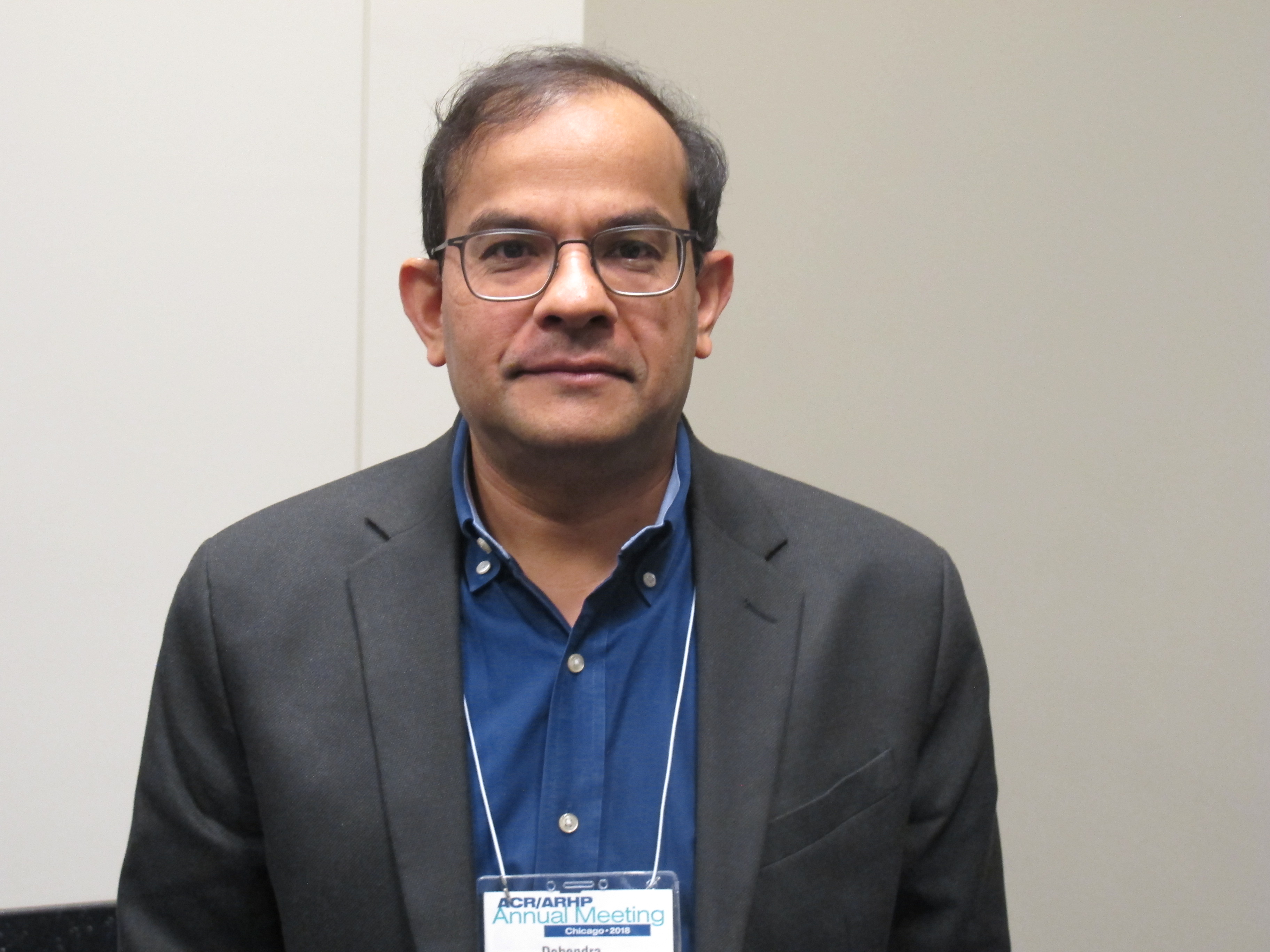
The findings are likely related to improvements in posttransplant immunosuppressive regimens, and may have implications for the timing of transplant going forward, Debendra N. Pattanaik, MD, said at the annual meeting of the American College of Rheumatology.
The biopsy-proven recurrence rate in 38 transplant recipients who received standard immunosuppression with prednisone, tacrolimus, and mycophenolate mofetil was 11%, and graft loss or death occurred in 26% at a median follow-up of 1,230 days, said Dr. Pattanaik, a rheumatologist at UTHSC, Memphis.
Patients with recurrence showed a trend for increased risk for graft loss or death, compared with recipients without recurrence (hazard ratio = 3.14), he noted during a press briefing at the meeting.
Lupus nephritis is a severe complication occurring in more than half of all patients with systemic lupus erythematosus (SLE), and despite a great deal of progress over the years, 10%-30% develop end-stage renal disease and require dialysis and/or transplant, he said, noting that studies have shown that transplant recipients do better over time than do those who remain on dialysis.
“So renal transplant is an important modality of treatment for end-stage renal disease from lupus nephritis,” he added.
However, recurrence of lupus nephritis in the graft is a concern, he said.
In previous eras – prior to improvements in immunosuppressive regimens for transplant recipients – studies showed variable rates of lupus nephritis recurrence, with some reporting rates up to 50% depending on the patient populations and protocols, he noted.
The rates in recent years at UTHSC seemed lower than that, so he and his colleagues looked more closely at the outcomes.
Case patients included all those with end-stage renal disease secondary to lupus nephritis who were transplanted between 2006 and 2017 at the center. Medical records of all 38 were reviewed along with information from the United Network for Organ Sharing Network. The mean age of the patients at baseline was 42 years, 89% were women, 89% were African American, and previous time on dialysis was a median of 4 years. Most (80%) received hemodialysis, and nearly one-third (31%) received living donor transplantation, Dr. Pattanaik said.
The main difference in the past decade compared with those previous eras is the use of posttransplant immunosuppressive regimens consisting of tacrolimus and mycophenolate mofetil rather than cyclosporine and azathioprine in addition to prednisone, he explained.
Previous reports showing higher recurrence rates were from studies in which patients received cyclosporine and azathioprine as part of the posttransplant regimen, he said.
“Our next question is whether patients can be transplanted early,” he said, explaining that transplant is often delayed for many months or years until SLE is in remission, but if the new regimens are reducing recurrence risk, early transplant may be feasible.
Dr. Pattanaik reported having no disclosures.
SOURCE: Pattanaik D et al. Arthritis Rheumatol. 2018;70(Suppl 10): Abstract 711.
CHICAGO – Lupus nephritis recurrence rates in kidney transplant recipients declined over the past decade, compared with rates seen in earlier studies, according to a review of cases at the University of Tennessee Health Science Center (UTHSC).
The findings are likely related to improvements in posttransplant immunosuppressive regimens, and may have implications for the timing of transplant going forward, Debendra N. Pattanaik, MD, said at the annual meeting of the American College of Rheumatology.
The biopsy-proven recurrence rate in 38 transplant recipients who received standard immunosuppression with prednisone, tacrolimus, and mycophenolate mofetil was 11%, and graft loss or death occurred in 26% at a median follow-up of 1,230 days, said Dr. Pattanaik, a rheumatologist at UTHSC, Memphis.
Patients with recurrence showed a trend for increased risk for graft loss or death, compared with recipients without recurrence (hazard ratio = 3.14), he noted during a press briefing at the meeting.
Lupus nephritis is a severe complication occurring in more than half of all patients with systemic lupus erythematosus (SLE), and despite a great deal of progress over the years, 10%-30% develop end-stage renal disease and require dialysis and/or transplant, he said, noting that studies have shown that transplant recipients do better over time than do those who remain on dialysis.
“So renal transplant is an important modality of treatment for end-stage renal disease from lupus nephritis,” he added.
However, recurrence of lupus nephritis in the graft is a concern, he said.
In previous eras – prior to improvements in immunosuppressive regimens for transplant recipients – studies showed variable rates of lupus nephritis recurrence, with some reporting rates up to 50% depending on the patient populations and protocols, he noted.
The rates in recent years at UTHSC seemed lower than that, so he and his colleagues looked more closely at the outcomes.
Case patients included all those with end-stage renal disease secondary to lupus nephritis who were transplanted between 2006 and 2017 at the center. Medical records of all 38 were reviewed along with information from the United Network for Organ Sharing Network. The mean age of the patients at baseline was 42 years, 89% were women, 89% were African American, and previous time on dialysis was a median of 4 years. Most (80%) received hemodialysis, and nearly one-third (31%) received living donor transplantation, Dr. Pattanaik said.
The main difference in the past decade compared with those previous eras is the use of posttransplant immunosuppressive regimens consisting of tacrolimus and mycophenolate mofetil rather than cyclosporine and azathioprine in addition to prednisone, he explained.
Previous reports showing higher recurrence rates were from studies in which patients received cyclosporine and azathioprine as part of the posttransplant regimen, he said.
“Our next question is whether patients can be transplanted early,” he said, explaining that transplant is often delayed for many months or years until SLE is in remission, but if the new regimens are reducing recurrence risk, early transplant may be feasible.
Dr. Pattanaik reported having no disclosures.
SOURCE: Pattanaik D et al. Arthritis Rheumatol. 2018;70(Suppl 10): Abstract 711.
REPORTING FROM THE ACR ANNUAL MEETING
Key clinical point:
Major finding: The biopsy-proven recurrence rate was 11%.
Study details: A review of 38 cases at one center.
Disclosures: Dr. Pattanaik reported having no disclosures.
Source: Pattanaik D et al. Arthritis Rheumatol. 2018;70(Suppl 10): Abstract 711.
Ablation plus transplant for severe scleroderma shows 11-year benefits
CHICAGO – Follow-up out to as long as 11 years from treatment confirmed the long-term efficacy and safety of myeloablative autologous stem cell transplantation for patients with severe scleroderma.
This extended follow-up comprised 43 survivors from the 75 patients originally randomized in a controlled, 6-year trial. Follow-up showed that, among the patients who underwent myeloablation and autologous transplant with hematopoietic stem cells, there were no long-term deaths or cancers, there was an 88% survival rate, and 92% remained off disease-modifying treatment, Keith M. Sullivan, MD, said at the annual meeting of the American College of Rheumatology.
Long-term survival among patients randomized to the study’s control arm, who received treatment with cyclophosphamide, was 53%.
Patients with severe scleroderma with significant internal organ damage who “are improved and off of disease-modifying antirheumatic drugs after 10 or more years from treatment is something new in autoimmune disease,” said Dr. Sullivan, a professor of medicine at Duke University, Durham, N.C.
Based on accumulated data from this randomized trial and other studies, the American Society for Blood and Marrow Transplantation issued a position statement in June 2018 that endorsed autologous hematopoietic stem cell transplantation as “standard of care” for systemic sclerosis (Biol Blood Marrow Transplant. 2018 June 25. doi: 10.1016/j.bbmt.2018.06.025), Dr. Sullivan noted in a video interview.
The SCOT (Scleroderma: Cyclophosphamide or Transplantation) trial randomized 75 patients with severe scleroderma and substantial internal organ involvement to receive treatment with either cyclophosphamide or myeloablative radiation followed by immune reconstitution with an autologous hematopoietic stem cell transplant. The trial’s primary endpoint, the global rank composite score at 54 months, showed the superiority of transplantation over standard treatment (N Engl J Med. 2018 Jan 4;378[1]:35-47).
Dr. Sullivan and his associates ran their long-term follow-up study on 43 of these 75 patients (25 from the transplanted group and 18 controls), excluding 21 patients who died during the original study, 4 additional patients from the control arm who died following the end of the original SCOT protocol, and 7 patients either lost to follow-up or who refused to participate in follow-up. Among the 25 transplanted patients, none died during the extended follow-up, 2 experienced cardiac failure, and 23 remained off of any disease-modifying antirheumatic drugs. Among the 18 survivors in the control arm, 3 had cardiac failure, 3 had respiratory failure, and 7 were on treatment with disease-modifying drugs, Dr. Sullivan reported.
In addition, 23 of the 25 (92%) transplanted patients had normal performance status by the Eastern Cooperative Oncology Group criteria, compared with 11 of the 18 controls (61%). A total of 14 (56%) transplant patients were employed, compared with 6 of the 18 controls (33%).
Patients who were transplanted “have their life back, are doing well, and are off treatment,” Dr. Sullivan noted.
Myeloablation and transplant is appropriate for scleroderma patients with significant internal organ involvement, about half of all patients with this disease. The best gauge of severe organ involvement is a pulmonary function test, with a forced vital capacity of 70% or less of predicted as a flag for patients who should consider transplantation, Dr. Sullivan said. He recommended monitoring lung function every 3 months in scleroderma patients because it can deteriorate very suddenly and quickly.
SCOT received no commercial funding. Dr. Sullivan had no disclosures to report.
SOURCE: Sullivan KM et al. ACR Annual Meeting, Abstract 1820.
REPORTING FROM THE ACR ANNUAL MEETING
Key clinical point:
Major finding: Survival after 11 years was 88% among transplanted patients and 53% among control patients treated with cyclophosphamide.
Study details: A long-term follow-up of 43 of the 75 patients enrolled in the SCOT trial.
Disclosures: SCOT received no commercial funding. Dr. Sullivan had no disclosures to report.
Source: Sullivan KM et al. ACR Annual Meeting, Abstract 1820.
PET/CT has good accuracy for diagnosing GCA
CHICAGO – Combined PET/CT has good diagnostic accuracy, including a 98% negative predictive value, when compared with temporal artery biopsy for suspected giant cell arteritis, according to findings from a study of 64 patients.
Study participants included patients with newly suspected giant cell arteritis (GCA) and all underwent PET/CT from the vertex to the diaphragm within 72 hours of starting corticosteroid therapy and prior to undergoing temporal artery biopsy (TAB). Two nuclear medicine physicians blinded to clinical and biopsy data identified GCA in the scans of 12 of 58 patients (21%) who ultimately underwent both PET/CT and TAB, Anthony M. Sammel, MBBS, reported at the annual meeting of the American College of Rheumatology.
Compared with TAB, which is the standard of care for diagnosing GCA in most centers, global GCA assessment by PET/CT had a sensitivity of 92%, specificity of 85%, and positive predictive value of 61%, said Dr. Sammel, a rheumatologist at Royal North Shore Hospital in Sydney.
The findings, and particularly the 98% negative predictive value, suggest that PET/CT could be used first line to rule out suspected GCA, although the sample size in the study was modest, he noted.
“I believe [the findings] would support PET/CT, when we include the head, neck, and chest, as a first-line test for patients newly suspected of having giant cell arteritis. I think we do need to be mindful that it’s not perfect,” he said, explaining that a TAB is warranted in a patient with a negative scan despite a clinician’s sense that there is a high likelihood of GCA.
However, the findings suggest that a negative study in a low to moderate risk patient would be “very, very reassuring,” and as such patients probably do not need a biopsy, he said in a video interview in which he also discussed cost-benefit issues with respect to PET/CT in this setting.
Of note, PET/CT diagnosed alternative conditions, including cancer and infections, that can mimic GCA in 13 study participants. At least one of those patients “may well have come to serious harm” on immunosuppressive therapy had his cervical spine infection gone undiagnosed, he said.
This study was funded by Arthritis Australia. Dr. Sammel reported having no relevant disclosures.
SOURCE: Sammel AM et al. ACR Annual Meeting, Abstract L15.
REPORTING FROM THE ACR ANNUAL MEETING
Key clinical point:
Major finding: PET/CT had 92% sensitivity, 85% specificity, 61% positive predictive value, and 98% negative predictive value.
Study details: A study of diagnostic accuracy of PET/CT for giant cell arteritis in 64 patients.
Disclosures: This study was funded by Arthritis Australia. Dr. Sammel reported having no relevant disclosures.
Source: Sammel AM et al. ACR Annual Meeting, Abstract L15.
Antimalarial-induced cardiomyopathy in lupus may be underrecognized
Cardiomyopathy induced by antimalarial treatment for systematic lupus erythematosus may not be as rare as previously thought, according to the authors of a case series published Oct. 15 in The Journal of Rheumatology.
The paper describes eight patients attending a lupus clinic, who were diagnosed with definite or possible antimalarial-induced cardiomyopathy over the course of 2 years.
Konstantinos Tselios, MD, PhD, a clinical research fellow at the University of Toronto Lupus Clinic, and his coauthors wrote that antimalarial-induced cardiomyopathy was thought to be relatively rare, with only 47 previous isolated reports, but they suggested the complication may be significantly underrecognized.
“Hypertrophic cardiomyopathy and heart failure, the most common clinical features of AM-induced cardiomyopathy (AMIC), may be falsely attributed to other causes, such as arterial hypertension or ischemic cardiomyopathy,” the authors wrote. “Consequently, nonspecific therapeutic approaches with diuretics and/or antihypertensives will exert minimum or even deleterious effects on such patients.”
All eight patients in this series were female, with a median age of 62.5 years, median disease duration of 35 years, and median antimalarial use of 22 years. They presented with conditions such as heart failure, exertional dyspnea, and pedal edema. Several patients were asymptomatic but had been found to have elevated heart biomarker levels that prompted further investigation.
All patients showed abnormal cardiac troponin I and brain natriuretic peptide levels, and seven of the eight also had chronically elevated creatine phosphokinase.
In three patients, endomyocardial biopsy showed cardiomyocyte vacuolation, intracytoplasmic myelinoid inclusions, and curvilinear bodies.
Four patients were diagnosed based on cardiac MRI, which showed features suggestive of antimalarial-induced cardiomyopathy, including ventricular hypertrophy with or without atrial enlargement and late gadolinium enhancement in a nonvascular pattern.
All patients had left ventricular hypertrophy, and four also had right ventricular hypertrophy. Only one patient showed impaired systolic function, compared with around half of patients in the literature with antimalarial-induced cardiomyopathy, but seven patients showed a restrictive filling pattern of the left ventricle.
“It seems possible that AMIC is a chronic process and systolic dysfunction will become apparent only in late stages,” the authors suggested.
One patient showed complete atrioventricular block, left ventricular and septal hypertrophy, and concomitant ocular toxicity.
After patients stopped antimalarials, the hypertrophy regressed and heart biomarkers decreased in seven patients, but one patient died from refractory heart failure.
Based on their findings, the authors proposed that heart-specific biomarkers be used as a regular screening tool for detecting myocardial injury, followed by more thorough investigations, such as cardiac MRI, in patients with positive biomarker findings.
“However, drug cessation should be prompt and probably upon suspicion of AMIC, because complete investigation may be delayed significantly.”
One author was supported by the Geoff Carr Fellowship from Lupus Ontario. The University of Toronto Lupus Research Program is supported by the University Health Network, Lou and Marissa Rocca, and the Lupus Foundation of Ontario.
SOURCE: Tselios K et al. J Rheumatol, 2018 Oct 15. doi: 10.3899/jrheum.180124.
FROM THE JOURNAL OF RHEUMATOLOGY
Key clinical point:
Major finding: Elevated cardiac troponin I and brain natriuretic peptide, and abnormal cardiac MRI may indicate antimalarial-induced cardiomyopathy.
Study details: Case series of eight patients with antimalarial-induced cardiomyopathy.
Disclosures: One author was supported by the Geoff Carr Fellowship from Lupus Ontario. The University of Toronto Lupus Research Program is supported by the University Health Network, Lou and Marissa Rocca, and the Lupus Foundation of Ontario.
Source: Tselios K et al. J Rheumatol. 2018 Oct 15. doi: 10.3899/jrheum.180124.
Inflammatory arthritis a common rheumatic adverse event with ICIs
Inflammatory arthritis (IA) is the most common rheumatic adverse event seen in people on immune checkpoint inhibitor (ICI) therapy and can largely be managed by rheumatologists without the need to halt cancer treatment, according to Michael D. Richter, MD, of the Mayo School of Graduate Medical Education, Rochester, Minn., and his colleagues.
The investigators’ observations of patients seen at the Mayo Clinic over a 7-year period suggested that “IA should not be seen as a contraindication to continuing or restarting ICIs, ” although continuing ICI therapy concurrently with high-dose steroids is “generally not recommended.” Previous estimates of rheumatic adverse events with ICIs had ranged from 1% to 10%, and management had largely been based on the experience of experts and findings from retrospective studies, they noted.
“This study aims to add granularity to the existing literature by describing the prevalence, clinical characteristics, and treatment outcomes of a large, single-institution cohort of patients with rheumatic immune-related adverse events,” Dr. Richter and his associates wrote in an article published in Arthritis & Rheumatology.
The analysis included 1,293 patients who had received treatment with any ICI at the Mayo Clinic between January 2011 and March 2018. Patients who had received treatment at other centers but were seen at the Mayo clinic for their rheumatic adverse event were also included.
Overall, 61 patients experienced a rheumatic immune-related adverse event (Rh-irAE) related to checkpoint inhibitor treatment; 43 were from the Mayo cohort and 18 were from other centers. The average age of the patients was 62.6 years, and almost half (49%) were female.
New-onset inflammatory arthritis was the most common Rh-irAE reported (n = 34), with the majority of patients presenting with polyarticular symptoms (n = 22; 65%).
Most of these patients were managed by a rheumatologist and were treated with systemic glucocorticoids (n = 26; 76%) for an average duration of 18 weeks. Five of the patients also required disease-modifying antirheumatic drugs and eight needed NSAIDS or intra-articular steroids.
The investigators noted that almost half of the patients (47%) experienced a complete resolution of symptoms during the study period, although few achieved this while they were still on the cancer treatment.
According to the investigators, the clinical characteristics of IA in their cohort support the existing literature: “Time of symptom onset is generally 4-8 weeks after starting ICI therapy. … The majority of patients require long courses of systemic glucocorticoids. And very few achieve complete symptom resolution while continuing ICI therapy,” they wrote.
The database analysis also revealed that a further 10 patients developed myopathy, with all patients presenting with myalgias and weaknesses. Five had bulbar myopathy, and four patients were diagnosed with concomitant myocarditis. All of these patients were treated with glucocorticoids for an average treatment duration of 15 weeks.
Complete resolution of the myopathy was observed in 70%, but two patients died from complications related to myocarditis and bulbar myopathy.
Other rheumatic events included four cases of vasculitis and four cases of polymyalgia rheumatica and six cases of diffuse systemic sclerosis or sicca syndromes.
“In lieu of controlled prospective studies, this cohort may serve as a guide for rheumatologists managing and counseling patients with Rh-IrAEs,” the investigators concluded.
“It remains unclear why some patients develop Rh-irAEs, and so far no genomic risk factors have been identified, though the varied clinical phenotypes of Rh-irAEs certainly suggest multiple underlying immunopathogenic mechanisms. Further prospective studies that distinguish between subsets of Rh-irAEs are necessary to better understand their pathophysiology and improve clinical care,” they added.
They noted that a limitation of the study was its reliance on retrospective data and clinician diagnoses.
None of the authors declared any financial or other conflicts of interest.
SOURCE: Richter MD et al. Arthritis Rheumatol. 2018 Oct 3. doi: 10.1002/art.40745.
FROM ARTHRITIS & RHEUMATOLOGY
Key clinical point: Inflammatory arthritis (IA) is the most common form of rheumatic immune-related adverse event (Rh-irAE) related to ICI treatment. The majority of patients with an Rh-irAE (90% in the current cohort) remained on ICI treatment, which suggests that IA should not be seen as a contraindication to continuing or restarting ICI treatment.
Major finding: Among a cohort of 1,293 patients and 18 patients who received ICI therapy at other centers, 61 cases of Rh-irAE were identified. Over half (n = 34) of these patients presented with IA.
Study details: A retrospective analysis of a database of all patients who received any ICI at the Mayo Clinic between January 2011 and March 2018.
Disclosures: None of the authors declared any financial or other conflicts of interest.
Source: Richter MD et al. Arthritis Rheumatol. 2018 Oct 3. doi: 10.1002/art.40745.
Third ACR Plenary presentations set to make an impact in rheumatology
A new study that validates the use of the Lupus Low Disease Activity State as a treatment outcome in systemic lupus erythematosus clinical trials is one of the highly-rated abstracts that will be presented in the third Plenary Session at the annual meeting of the American College of Rheumatology on Tuesday, Oct. 23.
The prospective, multicenter validation study, to be presented by Vera Golder, MBBS, of Monash University in Melbourne, builds on the results of previously reported studies using the Lupus Low Disease Activity State as a treatment target at EULAR this year and at last year’s International Congress on Systemic Lupus Erythematosus.
Among other presentations in the session will be the results of the PEXIVAS trial. Peter A. Merkel, MD, of the University of Pennsylvania, Philadelphia, will present findings from the randomized trial assessing oral glucocorticoid use and plasma exchange in patients with ANCA-associated vasculitis. The results have been highly anticipated as being among several research efforts to support reduction of corticosteroids in these patients.
In addition, attendees will hear results of a phase 3 study of apremilast for the treatment of oral ulcers in patients with Behçet’s syndrome. In the study, presented by Gulen Hatemi, MD, of Cerrahpasa Medical School, Istanbul, benefits of the drug were sustained for 28 weeks. Findings from a phase 2 study were reported in 2015.
REPORTING FROM THE ACR ANNUAL MEETING
Long-term follow-up results of ongoing trials highlighted at ACR 2018
A 5-year follow-up study comparing methods of meniscal tear management in patients with osteoarthritis kicks off the second Plenary Session on Monday, Oct. 22, at the annual meeting of the American College of Rheumatology.

Jeffrey N. Katz, MD, of Brigham and Women’s Hospital in Boston, and his colleagues conducted a long-term follow-up of patients from the METEOR study, the early results of which were presented at OARSI in 2017. Dr. Katz and his colleagues randomized patients with knee pain, meniscal tears, and OA changes on x-ray or MRI to physical therapy vs. physical therapy plus arthroscopic partial meniscectomy. After 5 years, pain relief was similar across treatment groups, supporting the short-term conclusion that these patients experience relief over time, irrespective of initial treatment. Overall, 25% of the patients had total knee replacement surgery during the follow-up period.
The session also includes a new presentation by Kenneth G. Saag, MD, of the University of Alabama at Birmingham of 2-year outcomes from a phase 3 trial of denosumab versus risedronate for glucocorticoid-induced osteoporosis that was first presented at EULAR this year.
At 2 years, denosumab proved superior for increasing spine and hip bone mineral density in osteoporosis patients, compared with risedronate, and demonstrated a similar safety profile.
In addition, attendees will hear updated long-term results from the SCOT trial of myeloablative autologous hematopoietic stem cell transplantation for scleroderma patients. Keith M. Sullivan, MD, of Duke University, Durham, N.C., and his colleagues found that the benefits of the treatment endured after 6-11 years, supporting results presented at ACR 2016.
A 5-year follow-up study comparing methods of meniscal tear management in patients with osteoarthritis kicks off the second Plenary Session on Monday, Oct. 22, at the annual meeting of the American College of Rheumatology.
Jeffrey N. Katz, MD, of Brigham and Women’s Hospital in Boston, and his colleagues conducted a long-term follow-up of patients from the METEOR study, the early results of which were presented at OARSI in 2017. Dr. Katz and his colleagues randomized patients with knee pain, meniscal tears, and OA changes on x-ray or MRI to physical therapy vs. physical therapy plus arthroscopic partial meniscectomy. After 5 years, pain relief was similar across treatment groups, supporting the short-term conclusion that these patients experience relief over time, irrespective of initial treatment. Overall, 25% of the patients had total knee replacement surgery during the follow-up period.
The session also includes a new presentation by Kenneth G. Saag, MD, of the University of Alabama at Birmingham of 2-year outcomes from a phase 3 trial of denosumab versus risedronate for glucocorticoid-induced osteoporosis that was first presented at EULAR this year.
At 2 years, denosumab proved superior for increasing spine and hip bone mineral density in osteoporosis patients, compared with risedronate, and demonstrated a similar safety profile.
In addition, attendees will hear updated long-term results from the SCOT trial of myeloablative autologous hematopoietic stem cell transplantation for scleroderma patients. Keith M. Sullivan, MD, of Duke University, Durham, N.C., and his colleagues found that the benefits of the treatment endured after 6-11 years, supporting results presented at ACR 2016.
REPORTING FROM THE ACR ANNUAL MEETING