User login
Damian McNamara is a journalist for Medscape Medical News and MDedge. He worked full-time for MDedge as the Miami Bureau covering a dozen medical specialties during 2001-2012, then as a freelancer for Medscape and MDedge, before being hired on staff by Medscape in 2018. Now the two companies are one. He uses what he learned in school – Damian has a BS in chemistry and an MS in science, health and environmental reporting/journalism. He works out of a home office in Miami, with a 100-pound chocolate lab known to snore under his desk during work hours.
Making Waves in Campaign to Decrease Sun Exposure
ORLANDO – Physicians faced unanticipated challenges when they launched a campaign to increase sun-protective behaviors for a high-risk group along the beaches of Australia.
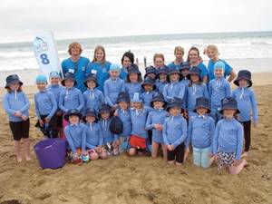
The initiative targeted training programs for junior lifeguards or "nippers," as they are called in Australia. Nippers have multiple risk factors for melanoma, including skin types I and II, summertime sun exposure, and a younger age. "Children are the most vulnerable," Dr. Anthony Dixon said at the annual meeting of the Florida Society of Dermatologic Surgeons.

"I knew I had to do something. Nippers are seen all over Australia. And they don’t wear a lot – they wear funny hats where the ears stick out," said Dr. Dixon, a dermatologic surgeon in private practice Geelong, Victoria. He felt compelled to act after seeing "more and more" of his patients develop skin cancer.
Dr. Dixon and his associates recommended four major changes to the nipper programs, which train children aged 6-12 years to become lifeguards. The recommended changes were:
P Do not hold nipper training between the hours of 11 a.m. and 4 p.m. "UVB is focused toward the middle of the day. We know that, the public does not," Dr. Dixon said. He estimated that nippers would receive less UV exposure during early-morning training.
P The children should wear rash vests, preferably with long sleeves.
P They should also wear broad-brimmed hats while awaiting training exercises
P Sunscreen should be available and reapplied frequently.
"We would all call that … common sense," said Dr. Dixon. "If only the lifesaving association did what they say in their manual." Their "Public Safety and Aquatic Rescue" manual addresses sun safety on page 15 of its 170 pages. "That is not too bad," he said. It advocates application of sunscreen before sun exposure, for example.
Dr. Dixon and his associates launched the campaign in January 2009 during the peak of summer in the southern hemisphere. Nipper training clubs agreed to the recommendations. However, some programs changed more readily than others, often for unexpected reasons.
For example, the nipper training club at Anglesea Beach did not immediately take to the recommendations, Dr. Dixon said. Program director Peter Foster ("Nipper Pete") briefed the children on the new program while they were standing in full sun. Sunscreen was repeatedly mentioned, but it was not applied until after the briefing. Also, the nippers did not don broad-brimmed hats because they had to wear hats with the sponsor’s name.
Dr. Dixon returned at a later date to observe any progress. "I saw kids at noon wearing only Speedos, [so they had] enormous sun exposure," Dr. Dixon said. "Nipper Pete saw me up on the sand dune with a camera and he panicked." He called the kids over and lectured them.
Sun exposure reduction is not as automatic as other protections, Dr. Dixon said. For example, "if that kid was too far out in the water, Nipper Pete would have gone out, pulled him in, and would have then given him the lecture about going out too deep."
Unbeknownst to Dr. Dixon, initial resistance to moving nipper training at Anglesea Beach to earlier in the morning had to do with a thriving business on the beach. Pressure from a sausage business that was making good money selling lunch to the nippers, their family members, and the trainers impeded the sun safety recommendation. Upon discovering this, Dr. Dixon intervened again. Eventually all parties agreed to conduct training earlier, and for the business to sell donuts instead.
Another nipper training program, in contrast, enacted the recommendations immediately. "The club at Jan Juc Beach decided to change everything with no briefing. They e-mailed the participants beforehand." All the nippers wore bucket hats while waiting for their training exercises, for example.
"So what others said was impossible was being done two beaches down," he said.
Part of the challenge of initiating behavioral changes is a misperception about the relative risk of sun exposure compared with other beach dangers, Dr. Dixon said. When asked about risk factors for death at the beach, "most people would mention drowning first," he said. However, there are 7 deaths from melanoma and 2 deaths from other skin cancers per 100,000 people in Australia each year, compared with 1.2 deaths per 100,000 people for accidental drowning. Put another way, for each person who dies from drowning, 46 Australians die from melanoma.
Other people might cite shark attacks as the greatest danger. In an average year, about four people worldwide die from a shark encounter. A shark attack "is an extremely rare way for humans to die, but when it happens, we all hear about it," Dr. Dixon said. "There are about 5,000 melanoma deaths in Australia for each shark death."
Dr. Dixon said that he did not have any relevant disclosures.
Visit http://www.coastalwatch.com for live Surfcams from Australia.
ORLANDO – Physicians faced unanticipated challenges when they launched a campaign to increase sun-protective behaviors for a high-risk group along the beaches of Australia.
The initiative targeted training programs for junior lifeguards or "nippers," as they are called in Australia. Nippers have multiple risk factors for melanoma, including skin types I and II, summertime sun exposure, and a younger age. "Children are the most vulnerable," Dr. Anthony Dixon said at the annual meeting of the Florida Society of Dermatologic Surgeons.
"I knew I had to do something. Nippers are seen all over Australia. And they don’t wear a lot – they wear funny hats where the ears stick out," said Dr. Dixon, a dermatologic surgeon in private practice Geelong, Victoria. He felt compelled to act after seeing "more and more" of his patients develop skin cancer.
Dr. Dixon and his associates recommended four major changes to the nipper programs, which train children aged 6-12 years to become lifeguards. The recommended changes were:
P Do not hold nipper training between the hours of 11 a.m. and 4 p.m. "UVB is focused toward the middle of the day. We know that, the public does not," Dr. Dixon said. He estimated that nippers would receive less UV exposure during early-morning training.
P The children should wear rash vests, preferably with long sleeves.
P They should also wear broad-brimmed hats while awaiting training exercises
P Sunscreen should be available and reapplied frequently.
"We would all call that … common sense," said Dr. Dixon. "If only the lifesaving association did what they say in their manual." Their "Public Safety and Aquatic Rescue" manual addresses sun safety on page 15 of its 170 pages. "That is not too bad," he said. It advocates application of sunscreen before sun exposure, for example.
Dr. Dixon and his associates launched the campaign in January 2009 during the peak of summer in the southern hemisphere. Nipper training clubs agreed to the recommendations. However, some programs changed more readily than others, often for unexpected reasons.
For example, the nipper training club at Anglesea Beach did not immediately take to the recommendations, Dr. Dixon said. Program director Peter Foster ("Nipper Pete") briefed the children on the new program while they were standing in full sun. Sunscreen was repeatedly mentioned, but it was not applied until after the briefing. Also, the nippers did not don broad-brimmed hats because they had to wear hats with the sponsor’s name.
Dr. Dixon returned at a later date to observe any progress. "I saw kids at noon wearing only Speedos, [so they had] enormous sun exposure," Dr. Dixon said. "Nipper Pete saw me up on the sand dune with a camera and he panicked." He called the kids over and lectured them.
Sun exposure reduction is not as automatic as other protections, Dr. Dixon said. For example, "if that kid was too far out in the water, Nipper Pete would have gone out, pulled him in, and would have then given him the lecture about going out too deep."
Unbeknownst to Dr. Dixon, initial resistance to moving nipper training at Anglesea Beach to earlier in the morning had to do with a thriving business on the beach. Pressure from a sausage business that was making good money selling lunch to the nippers, their family members, and the trainers impeded the sun safety recommendation. Upon discovering this, Dr. Dixon intervened again. Eventually all parties agreed to conduct training earlier, and for the business to sell donuts instead.
Another nipper training program, in contrast, enacted the recommendations immediately. "The club at Jan Juc Beach decided to change everything with no briefing. They e-mailed the participants beforehand." All the nippers wore bucket hats while waiting for their training exercises, for example.
"So what others said was impossible was being done two beaches down," he said.
Part of the challenge of initiating behavioral changes is a misperception about the relative risk of sun exposure compared with other beach dangers, Dr. Dixon said. When asked about risk factors for death at the beach, "most people would mention drowning first," he said. However, there are 7 deaths from melanoma and 2 deaths from other skin cancers per 100,000 people in Australia each year, compared with 1.2 deaths per 100,000 people for accidental drowning. Put another way, for each person who dies from drowning, 46 Australians die from melanoma.
Other people might cite shark attacks as the greatest danger. In an average year, about four people worldwide die from a shark encounter. A shark attack "is an extremely rare way for humans to die, but when it happens, we all hear about it," Dr. Dixon said. "There are about 5,000 melanoma deaths in Australia for each shark death."
Dr. Dixon said that he did not have any relevant disclosures.
Visit http://www.coastalwatch.com for live Surfcams from Australia.
FROM THE ANNUAL MEETING OF THE FLORIDA SOCIETY OF DERMATOLOGIC SURGEONS
Solutions to Challenges of Mohs Surgery for Lentigo Maligna
ORLANDO – Standard surgical margins often are inadequate for lentigo maligna; instead, perimeter excision techniques or Mohs surgery is preferable because these approaches consistently provide high cure rates, according to Dr. Basil S. Cherpelis.
Lentigo maligna is a subtype of melanoma in situ, characterized as an overgrowth of atypical melanocytes with the potential to become lentigo maligna melanoma. "I think of it as the problem child of the melanoma family," Dr. Cherpelis said at the annual meeting of the Florida Society of Dermatologic Surgeons.
"What margin should I use, and if I am going to do Mohs, how can I see margins on frozen sections?" Intraoperative margin control is recommended, but recent literature suggests that standard 5-mm margins are frequently inadequate (Clin. Plast. Surg. 2010;37:35-46; Int. J. Dermatol. 2010;49:482-91).
In general, traditional frozen sections permit only 1% of margins to be assessed. For that reason, Dr. Cherpelis said he prefers permanent processing or Mohs surgery for these patients. With permanent processing, a surgeon leaves a central island of tissue and removes peripheral strips. Excised samples sent to a lab for evaluation typically take several days for results.
"Permanent processing has excellent cure rates. But on the downside, a patient has to wait for pathology results, which can be inconvenient," said Dr. Cherpelis, a cutaneous oncologist at Moffitt Cancer Center at the University of South Florida in Tampa.
In contrast, Mohs micrographic excision and repair are performed on the same day. Because the entire margin is visualized, cure rates are "excellent," Dr. Cherpelis said.
As to why Mohs isn’t more widely used, one possible reason is that it can be difficult to distinguish melanocytes from keratinocytes on frozen section, he said.
Immunohistostains to the rescue: Melanoma antigen recognized by T cells (MART-1) and microphthalmia-associated transcription factor (MITF) staining each has a role.
"MART-1 is a valuable immunostain for melanoma in situ on photodamaged skin," said Dr. L. Frank Glass, a dermatopathologist at Moffitt Cancer Center. MART-1 on frozen sections can provide the same data as permanent processing. "But MART-1 is not a magic bullet," he added. "There can be false positives."
In addition, expertise and teamwork are required with immunohistostains, Dr. Glass said. "The right MART-1 staining takes a technical staff to do it right."
MITF works in frozen sections and produces results similar to permanent sections. Clinicians can use this nuclear immunostain to quantify melanocytes and assess other parameters that distinguish melanoma in situ from solar lentigines or skin with chronic sun damage, for example.
"MITF is particularly useful in combination with MART-1," Dr. Glass said.
Instead of dendritic processes that overlap and look like confluence, MITF reveals discreet melanocytic cells, Dr. Glass said. "You can feel more comfortable calling a case negative with MITF – if it shows no abnormal cytology – in the same patient where the MART-1 shows clumping."
"The bottom line is we are looking for 100% specificity for melanoma," Dr. Glass said. He and his colleagues also are developing cutoff values, based on melanocyte diameter and density, to distinguish melanoma in situ from solar lentigo or sun-damaged skin.
Dr. Cherpelis and Dr. Glass said they had no relevant financial disclosures.
EXPERT ANALYSIS FROM THE ANNUAL MEETING OF THE FLORIDA SOCIETY OF DERMATOLOGIC SURGEONS
Future Technologies Hold Promise for Hair Restoration
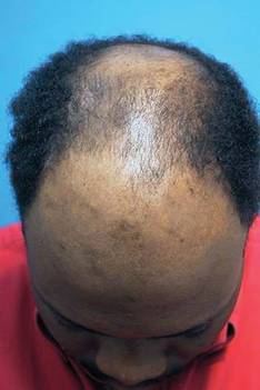
ORLANDO - Expect clinical options for hair restoration to grow in the future, said Dr. Ricardo Mejia.
Robotic hair transfer, multiple technologies to optimize new growth, and even hair cloning could help overcome current limitations in hair transplantation, Dr. Mejia said. Promising technologies could someday supplant donor strip and follicular unit extraction techniques. "We are getting to the age of robotics," Dr. Mejia said at the annual meeting of the Florida Society of Dermatologic Surgeons.
Historically, hair transplantation meant 4-mm plugs transferred at a rate of 10-200 grafts per session over a total of three to eight treatments. Because plugs were placed in a regular pattern, initial results appeared unnatural and very obvious. For some patients, a perception persists that this is still state-of-the-art for hair transplantation, Dr. Mejia said.
A natural, irregular hairline and greater hair density in fewer treatment sessions are now commonplace. "The average session these days of 2,500 grafts is not a big deal," Dr. Mejia said at the meeting.
"Restoring youthful hairlines can be done in single sessions. ... You can get a nice, age-appropriate appearance for an individual," said Dr. Mejia, a hair transplant surgeon in private practice in Jupiter, Fla. Even with recent advances, full growth of hair grafts still takes 6 months to a year, so realistic patient expectations are important.
A new device, NeoGraft Automated Hair Transplant System (NeoGraft), was cleared for marketing by the Food and Drug Administration in March 2009. After a rotating sharp punch scores the skin, a pneumatic suction device extracts the follicles. This technique minimizes injury to the lower half of hair follicles during follicular unit extraction, Dr. Mejia said. The device also implants grafts to a uniform depth.
Researchers are working on a variety of other means to protect grafts during the transfer process. For example, some are developing solutions to protect grafts that contain allopurinol, nitric oxide inhibitors, vitamins, and other components. Also, "we are starting to look at solutions used in organ transplantation." Small studies have shown increased hair survival and growth with these solutions used to optimize protection of organs during transfer, Dr. Mejia said.
Bathing follicular units in autologous platelet-rich plasma to promote healing angiogenesis is another approach. Growth factor components also could be beneficial, Dr. Mejia said. More studies are needed to determine the efficacy of injections of autologous platelet-rich plasma into both the donor area and the recipient areas in clinical practice.
Matching the size of the incision blade to the graft size can also help improve graft survival, Dr. Mejia said. Less trauma, less ischemia, and decreased overall bleeding are associated with finer blades. Although finer blades allow higher-density graft packing, he advised caution because some studies have shown more graft death with higher densities.
Investigators also are looking at technology to optimize new hair growth once the grafts are in place.
"Low-level lasers are getting a lot of attention," Dr. Mejia said. Wavelengths are in the range of 630-670 nm, power densities are between 5-50 mW/cm2, and fluences are 2-20 J/cm2.
The Food and Drug Administration cleared two devices that use low-level light therapy for hair growth: HairMax LaserComb (Lexington International) for men and the MPE-90 Hair Growth Stimulation System (Salon Lasers) for women.
"How good is the HairMax comb?" a meeting attendee asked. Dr. Mejia replied that reviews are mixed: "Hair restoration surgeons are on the fence – some believe in it, some don’t. Some patients are happy with it, some are not."
A lot of research also is underway to refine auto cloning and hair multiplication technologies, Dr. Mejia said.
Dermal papilla cells or fibroblasts are the starting point, because they stimulate formation of new hairs. Multiple companies are working on proprietary processes to spur these fibroblasts to produce enough follicles in culture to replace an entire scalp. This is where they hope "to make their windfall," he said. Research includes fibroblasts grown in subatmospheric oxygen tension, addition of wound-healing factors, and injections of a "hair-stimulating complex" to promote greater hair growth.
TrichoCyte is an example of a cell-based hair regeneration technology in phase II trials based on a proprietary dermal papilla cell process (Intercytex, Manchester, England). "The technique does work but [it is] not completely satisfactory at this point," Dr. Mejia said.
More than half of participants in one protocol for another proprietary cell treatment process showed significant hair growth 1 year later, according to a release announcing phase II study results for Aderans Research Institute.
Considerable work remains to be done before regenerative medical hair cloning becomes a clinically viable option, Dr. Mejia said. "How far out are we? I say 5-10 years."
Dr. Mejia said he had no relevant disclosures.
EXPERT ANALYSIS FROM THE ANNUAL MEETING OF THE FLORIDA SOCIETY OF DERMATOLOGIC SURGEONS
Pearls for Excision and Management of Cylindromas
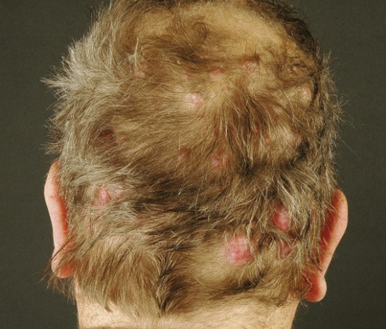
ORLANDO - Surgical excision is the preferred approach for treating a patient who presents with a cylindroma or with the more extensive familial cylindromatosis, Dr. Leonard Slazinski said.
There are some important distinctions between the two. Cylindromas are benign skin appendage tumors that commonly present as single, slow-growing lesions on the head and neck. Lesions often appear in conjunction with spiradenomas and trichoepitheliomas, they typically are 0.5-6.0 cm in size (although some can grow larger), and they affect females more than males. They arise sporadically with no known inheritance pattern.

The tumor can be excised with careful inspection to ensure removal of all tissue, Dr. Slazinski said. "In my experience, the cylindroma often does have a pseudocapsule, which allows for blunt dissection of the tumor under direct vision."
In contrast, familial cylindromatosis is an inherited autosomal dominant condition characterized by multiple lesions located on the head and neck. Treatment is trickier, as there is no effective field or systemic therapy. Surgery should be considered most effective, but many doctors "are overwhelmed by the number of these lesions," said Dr. Slazinski, a dermatologist in private practice in Sarasota, Fla.
Familial trichoepitheliomas, Brooke-Spiegler syndrome, and familial cylindromatosis share a genetic etiology – a mutation of the CYLD gene on chromosome 16. "Great advances have been made in etiology and cellular mechanisms of familial cylindromatosis and related conditions," Dr. Slazinski said. But "treatment has not advanced to the same degree."
Removal of these lesions is often performed for aesthetic and functional reasons. "These diseases can become quite severe if untreated," Dr. Slazinski said. The psychosocial impact can be great, and patients often become social recluses.
Even with traditional excision, recurrence "is quite high" at approximately 42%, Dr. Slazinski said. Mohs micrographic surgery is used more often for solitary lesions. Other options, depending on the individual presentation, include cryotherapy, curettage, electrosurgery and radiofrequency excision, and true scalp excision with split-thickness grafting, "which is advocated in severe cases."
Cylindroma patients often present with pseudoalopecia because the tumor does not grow hair. Scalp cylindromas present a unique challenge, with limited tissue and patients’ desire to spare their hair, Dr. Slazinski said. "Hair cosmesis is often the patient’s greatest concern. Paradoxically, as hair is conserved, [the area] can become a reservoir for future tumor formation."
Some clinicians advocate topical aspirin therapy for familial cylindromatosis, Dr. Slazinski said. There may be "less than stellar results, but my patient is seeing some benefit and wants to continue on compounded, topical salicylic acid."
Dr. Slazinski described a patient with an extensive number of lesions across her face, head, and neck. "I'm blessed to be able to take care of this patient, because no one else has really offered her any possibility of improvement. I’ve taken it on myself to at least try to palliate the situation. I am under no delusion that I can cure her in any way."
Dr. Slazinski said he had no relevant financial disclosures.
ORLANDO - Surgical excision is the preferred approach for treating a patient who presents with a cylindroma or with the more extensive familial cylindromatosis, Dr. Leonard Slazinski said.
There are some important distinctions between the two. Cylindromas are benign skin appendage tumors that commonly present as single, slow-growing lesions on the head and neck. Lesions often appear in conjunction with spiradenomas and trichoepitheliomas, they typically are 0.5-6.0 cm in size (although some can grow larger), and they affect females more than males. They arise sporadically with no known inheritance pattern.
The tumor can be excised with careful inspection to ensure removal of all tissue, Dr. Slazinski said. "In my experience, the cylindroma often does have a pseudocapsule, which allows for blunt dissection of the tumor under direct vision."
In contrast, familial cylindromatosis is an inherited autosomal dominant condition characterized by multiple lesions located on the head and neck. Treatment is trickier, as there is no effective field or systemic therapy. Surgery should be considered most effective, but many doctors "are overwhelmed by the number of these lesions," said Dr. Slazinski, a dermatologist in private practice in Sarasota, Fla.
Familial trichoepitheliomas, Brooke-Spiegler syndrome, and familial cylindromatosis share a genetic etiology – a mutation of the CYLD gene on chromosome 16. "Great advances have been made in etiology and cellular mechanisms of familial cylindromatosis and related conditions," Dr. Slazinski said. But "treatment has not advanced to the same degree."
Removal of these lesions is often performed for aesthetic and functional reasons. "These diseases can become quite severe if untreated," Dr. Slazinski said. The psychosocial impact can be great, and patients often become social recluses.
Even with traditional excision, recurrence "is quite high" at approximately 42%, Dr. Slazinski said. Mohs micrographic surgery is used more often for solitary lesions. Other options, depending on the individual presentation, include cryotherapy, curettage, electrosurgery and radiofrequency excision, and true scalp excision with split-thickness grafting, "which is advocated in severe cases."
Cylindroma patients often present with pseudoalopecia because the tumor does not grow hair. Scalp cylindromas present a unique challenge, with limited tissue and patients’ desire to spare their hair, Dr. Slazinski said. "Hair cosmesis is often the patient’s greatest concern. Paradoxically, as hair is conserved, [the area] can become a reservoir for future tumor formation."
Some clinicians advocate topical aspirin therapy for familial cylindromatosis, Dr. Slazinski said. There may be "less than stellar results, but my patient is seeing some benefit and wants to continue on compounded, topical salicylic acid."
Dr. Slazinski described a patient with an extensive number of lesions across her face, head, and neck. "I'm blessed to be able to take care of this patient, because no one else has really offered her any possibility of improvement. I’ve taken it on myself to at least try to palliate the situation. I am under no delusion that I can cure her in any way."
Dr. Slazinski said he had no relevant financial disclosures.
FROM THE ANNUAL MEETING OF THE FLORIDA SOCIETY OF DERMATOLOGIC SURGEONS
Pearls for Excision and Management of Cylindromas
ORLANDO – Surgical excision is the preferred approach for treating a patient who presents with a cylindroma or with the more extensive familial cylindromatosis, Dr. Leonard Slazinski said.
There are some important distinctions between the two. Cylindromas are benign skin appendage tumors that commonly present as single, slow-growing lesions on the head and neck. Lesions often appear in conjunction with spiradenomas and trichoepitheliomas, they typically are 0.5 to 6 cm in size (although some can grow larger), and they affect females more than males. They arise sporadically with no known inheritance pattern.
The tumor can be excised with careful inspection to ensure removal of all tissue, Dr. Slazinski said. "In my experience, the cylindroma often does have a pseudocapsule, which allows for blunt dissection of the tumor under direct vision."
In contrast, familial cylindromatosis is an inherited autosomal dominant condition characterized by multiple lesions located on the head and neck. Treatment is trickier, as there is no effective field or systemic therapy. Surgery should be considered most effective, but many doctors "are overwhelmed by the number of these lesions," said Dr. Slazinski, a dermatologist in private practice in Sarasota, Fla.
Familial trichoepitheliomas, Brooke-Spiegler syndrome, and familial cylindromatosis share a genetic etiology – a mutation of the CYLD gene on chromosome 16. "Great advances have been made in etiology and cellular mechanisms of familial cylindromatosis and related conditions," Dr. Slazinski said. But "treatment has not advanced to the same degree."
Removal of these lesions is often performed for aesthetic and functional reasons. "These diseases can become quite severe if untreated," Dr. Slazinski said. The psychosocial impact can be great, and patients often become social recluses.
Even with traditional excision, recurrence "is quite high" at approximately 42%, Dr. Slazinski said. Mohs micrographic surgery is used more often for solitary lesions. Other options, depending on the individual presentation, include cryotherapy, curettage, electrosurgery and radiofrequency excision, and true scalp excision with split-thickness grafting, "which is advocated in severe cases."
Cylindroma patients often present with pseudoalopecia because the tumor does not grow hair. Scalp cylindromas present a unique challenge, with limited tissue and patients’ desire to spare their hair, Dr. Slazinski said. "Hair cosmesis is often the patient’s greatest concern. Paradoxically, as hair is conserved, [the area] can become a reservoir for future tumor formation."
Some clinicians advocate topical aspirin therapy for familial cylindromatosis, Dr. Slazinski said. There may be "less than stellar results, but my patient is seeing some benefit and wants to continue on compounded, topical salicylic acid."
Dr. Slazinski described a patient with an extensive number of lesions across her face, head, and neck. "I’m blessed to be able to take care of this patient, because no one else has really offered her any possibility of improvement. I’ve taken it on myself to at least try to palliate the situation. I am under no delusion that I can cure her in any way."
Dr. Slazinski said he had no relevant financial disclosures.
FROM THE ANNUAL MEETING OF THE FLORIDA SOCIETY OF DERMATOLOGIC SURGEONS
Future Technologies Hold Promise for Hair Restoration
ORLANDO – Expect clinical options for hair restoration to grow in the future, said Dr. Ricardo Mejia.
Robotic hair transfer, multiple technologies to optimize new growth, and even hair cloning could help overcome current limitations in hair transplantation, Dr. Mejia said. Promising technologies could someday supplant donor strip and follicular unit extraction techniques. "We are getting to the age of robotics," Dr. Mejia said at the annual meeting of the Florida Society of Dermatologic Surgeons.
Historically, hair transplantation meant 4-mm plugs transferred at a rate of 10-200 grafts per session over a total of three to eight treatments. Because plugs were placed in a regular pattern, initial results appeared unnatural and very obvious. For some patients, a perception persists that this is still state-of-the-art for hair transplantation, Dr. Mejia said.
A natural, irregular hairline and greater hair density in fewer treatment sessions are now commonplace. "The average session these days of 2,500 grafts is not a big deal," Dr. Mejia said at the meeting.
"Restoring youthful hairlines can be done in single sessions. ... You can get a nice, age-appropriate appearance for an individual," said Dr. Mejia, a hair transplant surgeon in private practice in Jupiter, Fla. Even with recent advances, full growth of hair grafts still takes 6 months to a year, so realistic patient expectations are important.
A new device, NeoGraft Automated Hair Transplant System (NeoGraft), was cleared for marketing by the Food and Drug Administration in March 2009. After a rotating sharp punch scores the skin, a pneumatic suction device extracts the follicles. This technique minimizes injury to the lower half of hair follicles during follicular unit extraction, Dr. Mejia said. The device also implants grafts to a uniform depth.
Researchers are working on a variety of other means to protect grafts during the transfer process. For example, some are developing solutions to protect grafts that contain allopurinol, nitric oxide inhibitors, vitamins, and other components. Also, "we are starting to look at solutions used in organ transplantation." Small studies have shown increased hair survival and growth with these solutions used to optimize protection of organs during transfer, Dr. Mejia said.
Bathing follicular units in autologous platelet-rich plasma to promote healing angiogenesis is another approach. Growth factor components also could be beneficial, Dr. Mejia said. More studies are needed to determine the efficacy of injections of autologous platelet-rich plasma into both the donor area and the recipient areas in clinical practice.
Matching the size of the incision blade to the graft size can also help improve graft survival, Dr. Mejia said. Less trauma, less ischemia, and decreased overall bleeding are associated with finer blades. Although finer blades allow higher-density graft packing, he advised caution because some studies have shown more graft death with higher densities.
Investigators also are looking at technology to optimize new hair growth once the grafts are in place.
"Low-level lasers are getting a lot of attention," Dr. Mejia said. Wavelengths are in the range of 630-670 nm, power densities are between 5-50 mW/cm2, and fluences are 2-20 J/cm2.
The Food and Drug Administration cleared two devices that use low-level light therapy for hair growth: HairMax LaserComb (Lexington International) for men and the MPE-90 Hair Growth Stimulation System (Salon Lasers) for women.
"How good is the HairMax comb?" a meeting attendee asked. Dr. Mejia replied that reviews are mixed: "Hair restoration surgeons are on the fence – some believe in it, some don’t. Some patients are happy with it, some are not."
A lot of research also is underway to refine auto cloning and hair multiplication technologies, Dr. Mejia said.
Dermal papilla cells or fibroblasts are the starting point, because they stimulate formation of new hairs. Multiple companies are working on proprietary processes to spur these fibroblasts to produce enough follicles in culture to replace an entire scalp. This is where they hope "to make their windfall," he said. Research includes fibroblasts grown in subatmospheric oxygen tension, addition of wound-healing factors, and injections of a "hair-stimulating complex" to promote greater hair growth.
TrichoCyte is an example of a cell-based hair regeneration technology in phase II trials based on a proprietary dermal papilla cell process (Intercytex, Manchester, U.K.). "The technique does work but [it is] not completely satisfactory at this point," Dr. Mejia said.
More than half of participants in one protocol for another proprietary cell treatment process showed significant hair growth 1 year later, according to a release announcing phase II study results for Aderans Research Institute.
Considerable work remains to be done before regenerative medical hair cloning becomes a clinically viable option, Dr. Mejia said. "How far out are we? I say 5-10 years."
Dr. Mejia said he had no relevant disclosures.
Historically, hair transplantation
ORLANDO – Expect clinical options for hair restoration to grow in the future, said Dr. Ricardo Mejia.
Robotic hair transfer, multiple technologies to optimize new growth, and even hair cloning could help overcome current limitations in hair transplantation, Dr. Mejia said. Promising technologies could someday supplant donor strip and follicular unit extraction techniques. "We are getting to the age of robotics," Dr. Mejia said at the annual meeting of the Florida Society of Dermatologic Surgeons.
Historically, hair transplantation meant 4-mm plugs transferred at a rate of 10-200 grafts per session over a total of three to eight treatments. Because plugs were placed in a regular pattern, initial results appeared unnatural and very obvious. For some patients, a perception persists that this is still state-of-the-art for hair transplantation, Dr. Mejia said.
A natural, irregular hairline and greater hair density in fewer treatment sessions are now commonplace. "The average session these days of 2,500 grafts is not a big deal," Dr. Mejia said at the meeting.
"Restoring youthful hairlines can be done in single sessions. ... You can get a nice, age-appropriate appearance for an individual," said Dr. Mejia, a hair transplant surgeon in private practice in Jupiter, Fla. Even with recent advances, full growth of hair grafts still takes 6 months to a year, so realistic patient expectations are important.
A new device, NeoGraft Automated Hair Transplant System (NeoGraft), was cleared for marketing by the Food and Drug Administration in March 2009. After a rotating sharp punch scores the skin, a pneumatic suction device extracts the follicles. This technique minimizes injury to the lower half of hair follicles during follicular unit extraction, Dr. Mejia said. The device also implants grafts to a uniform depth.
Researchers are working on a variety of other means to protect grafts during the transfer process. For example, some are developing solutions to protect grafts that contain allopurinol, nitric oxide inhibitors, vitamins, and other components. Also, "we are starting to look at solutions used in organ transplantation." Small studies have shown increased hair survival and growth with these solutions used to optimize protection of organs during transfer, Dr. Mejia said.
Bathing follicular units in autologous platelet-rich plasma to promote healing angiogenesis is another approach. Growth factor components also could be beneficial, Dr. Mejia said. More studies are needed to determine the efficacy of injections of autologous platelet-rich plasma into both the donor area and the recipient areas in clinical practice.
Matching the size of the incision blade to the graft size can also help improve graft survival, Dr. Mejia said. Less trauma, less ischemia, and decreased overall bleeding are associated with finer blades. Although finer blades allow higher-density graft packing, he advised caution because some studies have shown more graft death with higher densities.
Investigators also are looking at technology to optimize new hair growth once the grafts are in place.
"Low-level lasers are getting a lot of attention," Dr. Mejia said. Wavelengths are in the range of 630-670 nm, power densities are between 5-50 mW/cm2, and fluences are 2-20 J/cm2.
The Food and Drug Administration cleared two devices that use low-level light therapy for hair growth: HairMax LaserComb (Lexington International) for men and the MPE-90 Hair Growth Stimulation System (Salon Lasers) for women.
"How good is the HairMax comb?" a meeting attendee asked. Dr. Mejia replied that reviews are mixed: "Hair restoration surgeons are on the fence – some believe in it, some don’t. Some patients are happy with it, some are not."
A lot of research also is underway to refine auto cloning and hair multiplication technologies, Dr. Mejia said.
Dermal papilla cells or fibroblasts are the starting point, because they stimulate formation of new hairs. Multiple companies are working on proprietary processes to spur these fibroblasts to produce enough follicles in culture to replace an entire scalp. This is where they hope "to make their windfall," he said. Research includes fibroblasts grown in subatmospheric oxygen tension, addition of wound-healing factors, and injections of a "hair-stimulating complex" to promote greater hair growth.
TrichoCyte is an example of a cell-based hair regeneration technology in phase II trials based on a proprietary dermal papilla cell process (Intercytex, Manchester, U.K.). "The technique does work but [it is] not completely satisfactory at this point," Dr. Mejia said.
More than half of participants in one protocol for another proprietary cell treatment process showed significant hair growth 1 year later, according to a release announcing phase II study results for Aderans Research Institute.
Considerable work remains to be done before regenerative medical hair cloning becomes a clinically viable option, Dr. Mejia said. "How far out are we? I say 5-10 years."
Dr. Mejia said he had no relevant disclosures.
Historically, hair transplantation
Historically, hair transplantation
EXPERT ANALYSIS FROM THE ANNUAL MEETING OF THE FLORIDA SOCIETY OF DERMATOLOGIC SURGEONS
Exposure to Maternal Drug Use Disorder Ups Risk for Children
BOCA RATON, Fla. – Exposure to a maternal drug use disorder, particularly during adolescence, significantly increases the risk for development of a drug use disorder in children, according to a follow-up study.
Risk was more than seven times greater for girls exposed to maternal drug use disorder, compared with others without such exposure (odds ratio, 7.04), according to secondary analysis of a study of 262 girls and their family members (Pediatrics 2000;106:792-7).
The initial study included structured psychiatric interviews of these girls, aged 6-17 years, as well as 290 of their siblings and 238 parents. The current study assessed risks for this cohort over 5 years. The average age of girls at 5-year follow-up was 18 years.
Timing of exposure made a significant difference, Dr. Amy M. Yule said. Risk of any offspring developing a drug use disorder was significantly higher if exposure to maternal drug use disorder occurred during adolescence (OR, 3.61), compared with preschool or childhood.
In a practical sense, these findings suggest physicians who treat children screen parents for drug use regularly, Dr. Yule said at the annual meeting of the American Academy of Addiction Psychiatry meeting. "I’m advocating people to ask parents about substance use. This can make a difference in the child’s risk of having a substance use disorder."
You can ask the questions during family history taking, but be sure not to overlook any relevant responses if this is part of a comprehensive initial consultation, said Dr. Yule, a clinical fellow in psychiatry at Harvard Medical School and Massachusetts General Hospital in Boston. Exposures were primarily to cannabis, she added. There was no significant correlation with maternal alcohol use and subsequent child risk.

"The message is also it’s not okay to smoke marijuana in front of your 14-year-old," study coauthor Dr. Timothy E. Wilens said. Dr. Wilens is director of Substance Abuse Services in Pediatric Psychopharmacology, department of psychiatry, at Massachusetts General. "Pediatricians are really good with the ‘No smoking in front of kids’ message."
"The main point is getting people to ask parents if they are using and engaging parents to seek treatment or do a trial of sobriety," Dr. Yule said.
Researchers controlled for familial risk, socioeconomic status, and presence of attention-deficit/hyperactivity disorder (the original study compared 140 children with ADHD to 162 others without). "We looked at exposure only, not at genetic influence," Dr. Yule said. "You cannot change your genetics, but that could also be a [contributing factor]."
Dr. Yule was surprised that their follow-up study did not reveal the same overall associations with substance use disorder exposure cited in the original study. The 2000 study found risk associated with alcohol use, drug use, and both.
The current study focused primarily on risks between mothers and daughters. "I assume the risk is the same for paternal use," Dr. Yule said, although the smaller number of fathers precluded any such official conclusion.
Ideally, pediatricians and other physicians would start screening for parental drug use disorders when the child is in 5th grade, Dr. Wilens said. Even though most children do not start using drugs until age 12 years or older, "we want them to start asking parents about substance use earlier."
Dr. Yule said she had no relevant disclosures. Dr. Wilens is a researcher and consultant for Abbott, Lilly, Merck, and Shire. He is also a researcher for McNeil; a consultant for Astra-Zeneca; and on the speakers bureau for Lilly, McNeil, Novartis, and Shire. He consults and receives research grants from the National Institutes of Health/National Institute for Drug Abuse.
BOCA RATON, Fla. – Exposure to a maternal drug use disorder, particularly during adolescence, significantly increases the risk for development of a drug use disorder in children, according to a follow-up study.
Risk was more than seven times greater for girls exposed to maternal drug use disorder, compared with others without such exposure (odds ratio, 7.04), according to secondary analysis of a study of 262 girls and their family members (Pediatrics 2000;106:792-7).
The initial study included structured psychiatric interviews of these girls, aged 6-17 years, as well as 290 of their siblings and 238 parents. The current study assessed risks for this cohort over 5 years. The average age of girls at 5-year follow-up was 18 years.
Timing of exposure made a significant difference, Dr. Amy M. Yule said. Risk of any offspring developing a drug use disorder was significantly higher if exposure to maternal drug use disorder occurred during adolescence (OR, 3.61), compared with preschool or childhood.
In a practical sense, these findings suggest physicians who treat children screen parents for drug use regularly, Dr. Yule said at the annual meeting of the American Academy of Addiction Psychiatry meeting. "I’m advocating people to ask parents about substance use. This can make a difference in the child’s risk of having a substance use disorder."
You can ask the questions during family history taking, but be sure not to overlook any relevant responses if this is part of a comprehensive initial consultation, said Dr. Yule, a clinical fellow in psychiatry at Harvard Medical School and Massachusetts General Hospital in Boston. Exposures were primarily to cannabis, she added. There was no significant correlation with maternal alcohol use and subsequent child risk.
"The message is also it’s not okay to smoke marijuana in front of your 14-year-old," study coauthor Dr. Timothy E. Wilens said. Dr. Wilens is director of Substance Abuse Services in Pediatric Psychopharmacology, department of psychiatry, at Massachusetts General. "Pediatricians are really good with the ‘No smoking in front of kids’ message."
"The main point is getting people to ask parents if they are using and engaging parents to seek treatment or do a trial of sobriety," Dr. Yule said.
Researchers controlled for familial risk, socioeconomic status, and presence of attention-deficit/hyperactivity disorder (the original study compared 140 children with ADHD to 162 others without). "We looked at exposure only, not at genetic influence," Dr. Yule said. "You cannot change your genetics, but that could also be a [contributing factor]."
Dr. Yule was surprised that their follow-up study did not reveal the same overall associations with substance use disorder exposure cited in the original study. The 2000 study found risk associated with alcohol use, drug use, and both.
The current study focused primarily on risks between mothers and daughters. "I assume the risk is the same for paternal use," Dr. Yule said, although the smaller number of fathers precluded any such official conclusion.
Ideally, pediatricians and other physicians would start screening for parental drug use disorders when the child is in 5th grade, Dr. Wilens said. Even though most children do not start using drugs until age 12 years or older, "we want them to start asking parents about substance use earlier."
Dr. Yule said she had no relevant disclosures. Dr. Wilens is a researcher and consultant for Abbott, Lilly, Merck, and Shire. He is also a researcher for McNeil; a consultant for Astra-Zeneca; and on the speakers bureau for Lilly, McNeil, Novartis, and Shire. He consults and receives research grants from the National Institutes of Health/National Institute for Drug Abuse.
FROM THE ANNUAL MEETING OF THE AMERICAN ACADEMY OF ADDICTION PSYCHIATRY
Exposure to Maternal Drug Use Disorder Ups Risk for Children
BOCA RATON, Fla. – Exposure to a maternal drug use disorder, particularly during adolescence, significantly increases the risk for development of a drug use disorder in children, according to a follow-up study.
Risk was more than seven times greater for girls exposed to maternal drug use disorder, compared with others without such exposure (odds ratio, 7.04), according to secondary analysis of a study of 262 girls and their family members (Pediatrics 2000;106:792-7).

The initial study included structured psychiatric interviews of these girls, aged 6-17 years, as well as 290 of their siblings and 238 parents. The current study assessed risks for this cohort over 5 years. The average age of girls at 5-year follow-up was 18 years.
Timing of exposure made a significant difference, Dr. Amy M. Yule said. Risk of any offspring developing a drug use disorder was significantly higher if exposure to maternal drug use disorder occurred during adolescence (OR, 3.61), compared with preschool or childhood.
In a practical sense, these findings suggest physicians who treat children screen parents for drug use regularly, Dr. Yule said at the annual meeting of the American Academy of Addiction Psychiatry meeting. "I’m advocating people to ask parents about substance use. This can make a difference in the child’s risk of having a substance use disorder."
You can ask the questions during family history taking, but be sure not to overlook any relevant responses if this is part of a comprehensive initial consultation, said Dr. Yule, a clinical fellow in psychiatry at Harvard Medical School and Massachusetts General Hospital in Boston. Exposures were primarily to cannabis, she added. There was no significant correlation with maternal alcohol use and subsequent child risk.
"The message is also it’s not okay to smoke marijuana in front of your 14-year-old," study coauthor Dr. Timothy E. Wilens said. Dr. Wilens is director of Substance Abuse Services in Pediatric Psychopharmacology, department of psychiatry, at Massachusetts General. "Pediatricians are really good with the ‘No smoking in front of kids’ message."
"The main point is getting people to ask parents if they are using and engaging parents to seek treatment or do a trial of sobriety," Dr. Yule said.
Researchers controlled for familial risk, socioeconomic status, and presence of attention-deficit/hyperactivity disorder (the original study compared 140 children with ADHD to 162 others without). "We looked at exposure only, not at genetic influence," Dr. Yule said. "You cannot change your genetics, but that could also be a [contributing factor]."
Dr. Yule was surprised that their follow-up study did not reveal the same overall associations with substance use disorder exposure cited in the original study. The 2000 study found risk associated with alcohol use, drug use, and both.
The current study focused primarily on risks between mothers and daughters. "I assume the risk is the same for paternal use," Dr. Yule said, although the smaller number of fathers precluded any such official conclusion.
Ideally, pediatricians and other physicians would start screening for parental drug use disorders when the child is in 5th grade, Dr. Wilens said. Even though most children do not start using drugs until age 12 years or older, "we want them to start asking parents about substance use earlier."
Dr. Yule said she had no relevant disclosures. Dr. Wilens is a researcher and consultant for Abbott, Lilly, Merck, and Shire. He is also a researcher for McNeil; a consultant for Astra-Zeneca; and on the speakers bureau for Lilly, McNeil, Novartis, and Shire. He consults and receives research grants from the National Institutes of Health/National Institute for Drug Abuse.
BOCA RATON, Fla. – Exposure to a maternal drug use disorder, particularly during adolescence, significantly increases the risk for development of a drug use disorder in children, according to a follow-up study.
Risk was more than seven times greater for girls exposed to maternal drug use disorder, compared with others without such exposure (odds ratio, 7.04), according to secondary analysis of a study of 262 girls and their family members (Pediatrics 2000;106:792-7).
The initial study included structured psychiatric interviews of these girls, aged 6-17 years, as well as 290 of their siblings and 238 parents. The current study assessed risks for this cohort over 5 years. The average age of girls at 5-year follow-up was 18 years.
Timing of exposure made a significant difference, Dr. Amy M. Yule said. Risk of any offspring developing a drug use disorder was significantly higher if exposure to maternal drug use disorder occurred during adolescence (OR, 3.61), compared with preschool or childhood.
In a practical sense, these findings suggest physicians who treat children screen parents for drug use regularly, Dr. Yule said at the annual meeting of the American Academy of Addiction Psychiatry meeting. "I’m advocating people to ask parents about substance use. This can make a difference in the child’s risk of having a substance use disorder."
You can ask the questions during family history taking, but be sure not to overlook any relevant responses if this is part of a comprehensive initial consultation, said Dr. Yule, a clinical fellow in psychiatry at Harvard Medical School and Massachusetts General Hospital in Boston. Exposures were primarily to cannabis, she added. There was no significant correlation with maternal alcohol use and subsequent child risk.
"The message is also it’s not okay to smoke marijuana in front of your 14-year-old," study coauthor Dr. Timothy E. Wilens said. Dr. Wilens is director of Substance Abuse Services in Pediatric Psychopharmacology, department of psychiatry, at Massachusetts General. "Pediatricians are really good with the ‘No smoking in front of kids’ message."
"The main point is getting people to ask parents if they are using and engaging parents to seek treatment or do a trial of sobriety," Dr. Yule said.
Researchers controlled for familial risk, socioeconomic status, and presence of attention-deficit/hyperactivity disorder (the original study compared 140 children with ADHD to 162 others without). "We looked at exposure only, not at genetic influence," Dr. Yule said. "You cannot change your genetics, but that could also be a [contributing factor]."
Dr. Yule was surprised that their follow-up study did not reveal the same overall associations with substance use disorder exposure cited in the original study. The 2000 study found risk associated with alcohol use, drug use, and both.
The current study focused primarily on risks between mothers and daughters. "I assume the risk is the same for paternal use," Dr. Yule said, although the smaller number of fathers precluded any such official conclusion.
Ideally, pediatricians and other physicians would start screening for parental drug use disorders when the child is in 5th grade, Dr. Wilens said. Even though most children do not start using drugs until age 12 years or older, "we want them to start asking parents about substance use earlier."
Dr. Yule said she had no relevant disclosures. Dr. Wilens is a researcher and consultant for Abbott, Lilly, Merck, and Shire. He is also a researcher for McNeil; a consultant for Astra-Zeneca; and on the speakers bureau for Lilly, McNeil, Novartis, and Shire. He consults and receives research grants from the National Institutes of Health/National Institute for Drug Abuse.
FROM THE ANNUAL MEETING OF THE AMERICAN ACADEMY OF ADDICTION PSYCHIATRY
Major Finding: Exposure to maternal drug use disorder significantly increases risk of development of same disorder for offspring (odds ratio, 7.04), compared with unexposed children.
Data Source: Secondary analysis of 262 girls and their families followed 5 years after initial study enrollment.
Disclosures: Dr. Amy M. Yule had no relevant disclosures. Dr. Timothy Wilens is a researcher, consultant, and speaker for NIDA and multiple pharmaceutical companies.
Program Improves Hip Fracture Outcomes : Targeted intervention that includes progressive resistance training lowered mortality, dependency.
Major Finding: Age-adjusted risk of death was significantly reduced in the intervention group, compared with usual care (odds ratio = 0.19).
Data Source: The Hip Fracture Intervention Trial (HIPFIT) compared outcomes for 62 hip fracture patients randomized to resistance training and up to 12 other interventions versus 62 randomized to usual care (6-12 weeks of physiotherapy, an orthopedic consult at 6 weeks, and any recommended therapies).
Disclosures: Dr. Singh said she had no relevant financial disclosures.
NEW ORLEANS — Compared with usual care after hip fracture, a comprehensive and targeted intervention that includes high-intensity progressive resistance training over 12 months lowers mortality, decreases nursing home admissions, improves activities of daily living dependency, and decreases the use of assistive devices, according to a randomized, controlled trial.
“It is possible to change the most important outcomes for these people,” Dr. Maria A. Fiatarone Singh said.
Functional dependency, however, did not significantly differ between groups.
Many facets of hip fractures have been studied, from pharmacologic prevention of osteoporosis to acute hospital interventions to fracture rehabilitation. “Although we've done a lot of studies, we still have not figured out how to prevent people from entering a nursing home or dying,” said Dr. Singh, professor of medicine and chair of exercise and sport science at the University of Sydney.
So Dr. Singh and her colleagues launched the Hip Fracture Intervention Trial (HIPFIT). They compared outcomes for 62 hip fracture patients randomized to resistance training and up to 12 other interventions vs. 62 patients randomized to usual care. Intervention was associated with an 84% reduction in the likelihood of nursing home admission (odds ratio, 0.16), compared with usual care, Dr. Singh said. In absolute numbers, 5 intervention patients (8%) and 12 control patients (19%) were admitted to a nursing home during the 12 months of follow-up.
“Hip fracture is associated with chronic pain, reduced mobility, disability, and increasing degree of dependence. After hip fracture, 10%-20% of formerly community-dwelling people require long-term nursing home care,” Dr. Singh said.
Four intervention patients and eight usual-care patients died. Age-adjusted risk of death was significantly reduced in the intervention group, compared with usual care (OR = 0.19). Cardiovascular disease, infection, and stroke were among the causes.
Dr. Singh and her associates hypothesized that long-term disability and nursing home utilization after hip fracture would be reduced by targeted, multifactorial intervention aimed at the primary risk factors. They chose modifiable risk factors to make application of their findings more practical, including sarcopenia/muscle weakness, poor balance or gait, malnutrition or weight loss, vitamin D insufficiency, and vision concerns.
All intervention group participants received hip protectors and supervised, high-intensity, progressive resistance training for 12 months. The protocol included seven exercises designed for both upper and lower body strength. A meeting attendee questioned how patients were able to exercise after hip fracture. The intervention began with an isometric measure of strength and actual strength training started about 6 months after fracture, Dr. Singh replied.
Balance training exercises were progressive as well. As tasks were mastered, participants graduated to a more difficult level. For example, if a person could balance holding on to something with two hands, next they progressed to one hand and then to one finger.
Interventions were added for individual participants as needed, up to a total of 13. Treatment of depression, nutritional supplementation, medication management, and vision assessment are examples. Some participants received home assessment and referral to community services. Others received interventions to address risk and/or fear of falling, low self-efficacy, and polypharmacy.
Evaluations were done at baseline and at 4 and 12 months after fracture, with regular review by geriatricians, general practitioners, and ophthalmologists.
A meeting attendee asked which interventions were most useful. “Our specific intent was not to break apart the 13 interventions,” Dr. Singh said. She said many were intertwined, for example, vision improvements allowed balance training to be more effective. The effects of strength and balance training were most robust because they were performed twice a week for 12 months.
Usual care included 6-12 weeks of physiotherapy, an orthopedic consult at 6 weeks, and any recommended therapies. “We sent letter to general practitioners if people [in the usual-care group] were depressed, had low vitamin D, or abnormal cognitive function. We did not prescribe for this group,” she said.
Even though overall functional dependency did not differ significantly, intervention was associated with significantly less decline in some functional dependency KATZ scores (total, continence, and transfer) at 12 months, compared with their prefracture baseline. This is relevant, Dr. Singh said, because previous research they did showed that overall function declines for most people after a hip fracture. Only 20% of participants in the Sarcopenia and Hip Fracture Study (SHIP) returned to baseline function at 12 months (J. Gerontol. A. Biol. Sci. Med. Sci. 2009;64:568-74). In the current study, after the researchers controlled for age, there was less of a decline in function for total KATZ score, transfer change, and continence change if patients were in intervention group vs. usual care, according to Dr. Singh.
“Did changes in KATZ activities of daily living total score and scales mediate the nursing home admissions we saw? It seemed to be the case,” Dr. Singh said. “The nursing home residents had greater decline in KATZ function and toileting [continence] scores vs. others.”
All results are based on an intent-to-treat analysis. The dropout rate was low, she said: nine HIPFIT patients and three usual-care patients did not complete follow-up.
At baseline, the community-dwelling participants were 69% female; mean age, 79 years; 83% at nutritional risk; 88% vitamin D insufficient; 90% living independently (vs. 10% in nursing homes); and 38% were cognitively impaired. A total 45% were depressed. The mean number of chronic diseases was 3.4. The usual-care group reported worse bodily pain, the only significant difference between groups. There were no adverse events, except for some musculoskeletal soreness after activity.
Acute Alcohol Withdrawal: DT Risk Higher After Age 65
BOCA RATON, Fla. – Older patients hospitalized with acute alcohol withdrawal experience significantly more delirium tremens and delirium than do inpatients younger than 65 years, according to a retrospective study.
Screen older patients for alcohol consumption and consider medications other than benzodiazepines to treat withdrawal in this population, advised Dr. Jeremy Doniger, a resident at Harvard Longwood Psychiatry in Boston.
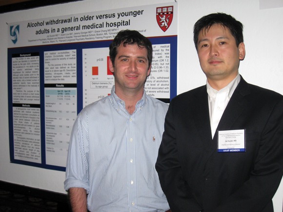
"Older people are drinking more than we realize. Be consistent about screening for it," Dr. Doniger said. In fact, with the aging of the population, the number of older adults with an alcohol use disorder is expected to double by 2020 (Brit. Med. J. 2003;327:664-7). A likely outcome is that more older patients will be admitted to hospitals for severe alcohol withdrawal in the future.
"Benzodiazepines can cause delirium, and older patients may be more susceptible [to these effects]," said Dr. Doniger. N-methyl-d-aspartate antagonists, for example, alone or in combination with benzodiazepines, are an effective alternative strategy, he added.
Reports in the literature are mixed as to whether older age predicts worse withdrawal, said Dr. Joji Suzuki, lead study investigator, and director of the Addiction Recovery Program at Brigham and Women’s Hospital and Harvard Medical School, Boston.
To find a more definitive answer, the investigators reviewed the charts of all medical inpatients at Brigham and Women’s Hospital for whom a psychiatry consultation was requested from 2008 to 2010. They identified 141 patients diagnosed and treated for alcohol withdrawal during hospital admission.
They compared 31 older patients to 110 younger ones. "We wanted to know if older patients, those 65 and older, have worse or the same outcomes as younger folks," said Dr. Suzuki. "We found something very striking: that DTs [delirium tremens] or delirium from any cause approaches 50% in older patients," Dr. Suzuki said. "Delirium is very serious, and can be a predictor for death."
Delirium from any cause affected 52% of the older group versus 30% of the younger patients, a statistically significant difference. In addition, 44% of older patients experienced DTs while hospitalized, compared with 20% of younger patients. This disparity also was significantly different.
"Part of it might be severity of comorbidities," Dr. Doniger said. The presence of more severe comorbidities was significantly associated with development of any delirium (odds ratio, 1.2), but not with DTs (OR, 1.13) or withdrawal seizures (OR, 1.02).
Prevalence of withdrawal seizures did not differ significantly between older patients (10%) and younger patients (6%). Nor did hospital length of stay differ significantly. Older patients stayed at Brigham and Women’s Hospital a mean 12.5 days compared with 8.6 days for the younger group.
The take-home message is that "younger patients going through withdrawal will fare about the same or a little worse than someone in a detox setting. But older patients are at greater risk for adverse outcomes, and the longer they stay in the hospital, the higher their risk for not doing well [because of nosocomial infections, falls, and other risk factors]," Dr. Suzuki said.
Inpatients who experienced DTs received significantly higher doses of benzodiazepines than did those who did not. In terms of lorazepam equivalents, the mean dose was 64.6 mg versus 30.5 mg in the non-DT group.
Dr. Doniger said differences in body composition and metabolism could contribute to greater benzodiazepine delirium in older patients because they typically have less water and more fat, which holds medications, and they have slower liver metabolism, he said.
The study was supported in part by the Department of Psychiatry at Harvard Medical School. Dr. Suzuki and Dr. Doniger had no relevant financial disclosures.
BOCA RATON, Fla. – Older patients hospitalized with acute alcohol withdrawal experience significantly more delirium tremens and delirium than do inpatients younger than 65 years, according to a retrospective study.
Screen older patients for alcohol consumption and consider medications other than benzodiazepines to treat withdrawal in this population, advised Dr. Jeremy Doniger, a resident at Harvard Longwood Psychiatry in Boston.
"Older people are drinking more than we realize. Be consistent about screening for it," Dr. Doniger said. In fact, with the aging of the population, the number of older adults with an alcohol use disorder is expected to double by 2020 (Brit. Med. J. 2003;327:664-7). A likely outcome is that more older patients will be admitted to hospitals for severe alcohol withdrawal in the future.
"Benzodiazepines can cause delirium, and older patients may be more susceptible [to these effects]," said Dr. Doniger. N-methyl-d-aspartate antagonists, for example, alone or in combination with benzodiazepines, are an effective alternative strategy, he added.
Reports in the literature are mixed as to whether older age predicts worse withdrawal, said Dr. Joji Suzuki, lead study investigator, and director of the Addiction Recovery Program at Brigham and Women’s Hospital and Harvard Medical School, Boston.
To find a more definitive answer, the investigators reviewed the charts of all medical inpatients at Brigham and Women’s Hospital for whom a psychiatry consultation was requested from 2008 to 2010. They identified 141 patients diagnosed and treated for alcohol withdrawal during hospital admission.
They compared 31 older patients to 110 younger ones. "We wanted to know if older patients, those 65 and older, have worse or the same outcomes as younger folks," said Dr. Suzuki. "We found something very striking: that DTs [delirium tremens] or delirium from any cause approaches 50% in older patients," Dr. Suzuki said. "Delirium is very serious, and can be a predictor for death."
Delirium from any cause affected 52% of the older group versus 30% of the younger patients, a statistically significant difference. In addition, 44% of older patients experienced DTs while hospitalized, compared with 20% of younger patients. This disparity also was significantly different.
"Part of it might be severity of comorbidities," Dr. Doniger said. The presence of more severe comorbidities was significantly associated with development of any delirium (odds ratio, 1.2), but not with DTs (OR, 1.13) or withdrawal seizures (OR, 1.02).
Prevalence of withdrawal seizures did not differ significantly between older patients (10%) and younger patients (6%). Nor did hospital length of stay differ significantly. Older patients stayed at Brigham and Women’s Hospital a mean 12.5 days compared with 8.6 days for the younger group.
The take-home message is that "younger patients going through withdrawal will fare about the same or a little worse than someone in a detox setting. But older patients are at greater risk for adverse outcomes, and the longer they stay in the hospital, the higher their risk for not doing well [because of nosocomial infections, falls, and other risk factors]," Dr. Suzuki said.
Inpatients who experienced DTs received significantly higher doses of benzodiazepines than did those who did not. In terms of lorazepam equivalents, the mean dose was 64.6 mg versus 30.5 mg in the non-DT group.
Dr. Doniger said differences in body composition and metabolism could contribute to greater benzodiazepine delirium in older patients because they typically have less water and more fat, which holds medications, and they have slower liver metabolism, he said.
The study was supported in part by the Department of Psychiatry at Harvard Medical School. Dr. Suzuki and Dr. Doniger had no relevant financial disclosures.
FROM THE ANNUAL MEETING OF THE AMERICAN ACADEMY OF ADDICTION PSYCHIATRY
Major Finding: A greater percentage of hospitalized patients 65 and older with alcohol withdrawal experience delirium, 52%, compared with 30% among younger patients.
Data Source: Review of 141 patient charts involving a psychiatry consultation from 2008 to 2010.
Disclosures: Dr. Joji Suzuki and Dr. Jeremy Doniger had no relevant disclosures.