User login
M. Alexander Otto began his reporting career early in 1999 covering the pharmaceutical industry for a national pharmacists' magazine and freelancing for the Washington Post and other newspapers. He then joined BNA, now part of Bloomberg News, covering health law and the protection of people and animals in medical research. Alex next worked for the McClatchy Company. Based on his work, Alex won a year-long Knight Science Journalism Fellowship to MIT in 2008-2009. He joined the company shortly thereafter. Alex has a newspaper journalism degree from Syracuse (N.Y.) University and a master's degree in medical science -- a physician assistant degree -- from George Washington University. Alex is based in Seattle.
VIDEO: Measuring, treating brain hypoxia looks promising for TBI
SAN DIEGO – It’s been possible for over 15 years for neurointensivists to measure the partial pressure of oxygen in the brain of patients following traumatic brain injury.
But the technology has not been widely adopted because there have been no high-quality data showing that it’s useful. As a result, in most hospitals, TBI treatment is guided mostly by intracranial pressure.
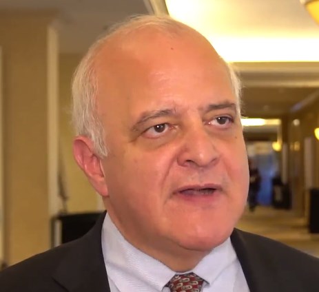
The evidence gap is being filled. In a recent phase 2 trial, there was a trend towards benefit when treatment was guided by both intracranial pressure and the brain oxygenation (Crit Care Med. 2017 Nov;45[11]:1907-14). The study was powered for nonfutility, not clinically meaningful change, but the National Institute of Neurological Disorders and Stroke has recently funded a 45-site, phase 3 trial that will definitively answer whether treatment protocols informed by both pressure and oxygen improve neurologic outcomes, said principal investigator Ramon Diaz-Arrastia, MD, PhD, a professor of neurology at the University of Pennsylvania, Philadelphia.
In an interview at the annual meeting of the American Neurological Association, he explained the work, and exactly how paying attention to brain oxygen levels changed treatment in the phase 2 study. It didn’t take anything unusual to maintain oxygen partial pressure above 20 mm Hg.
The video associated with this article is no longer available on this site. Please view all of our videos on the MDedge YouTube channel
SAN DIEGO – It’s been possible for over 15 years for neurointensivists to measure the partial pressure of oxygen in the brain of patients following traumatic brain injury.
But the technology has not been widely adopted because there have been no high-quality data showing that it’s useful. As a result, in most hospitals, TBI treatment is guided mostly by intracranial pressure.
The evidence gap is being filled. In a recent phase 2 trial, there was a trend towards benefit when treatment was guided by both intracranial pressure and the brain oxygenation (Crit Care Med. 2017 Nov;45[11]:1907-14). The study was powered for nonfutility, not clinically meaningful change, but the National Institute of Neurological Disorders and Stroke has recently funded a 45-site, phase 3 trial that will definitively answer whether treatment protocols informed by both pressure and oxygen improve neurologic outcomes, said principal investigator Ramon Diaz-Arrastia, MD, PhD, a professor of neurology at the University of Pennsylvania, Philadelphia.
In an interview at the annual meeting of the American Neurological Association, he explained the work, and exactly how paying attention to brain oxygen levels changed treatment in the phase 2 study. It didn’t take anything unusual to maintain oxygen partial pressure above 20 mm Hg.
The video associated with this article is no longer available on this site. Please view all of our videos on the MDedge YouTube channel
AT ANA 2017
VIDEO: Alzheimer’s blood test expected soon
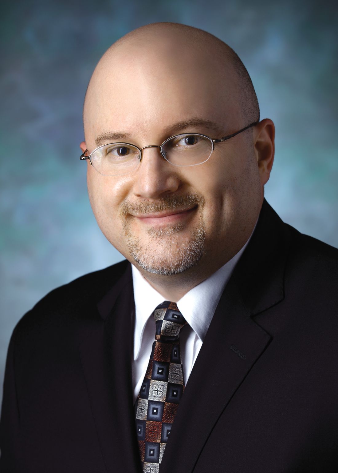
SAN DIEGO – A blood test that could be on the market in as little as 2 years has an accuracy of 89% for detecting amyloid plaques in the brain, according to a report at the annual meeting of the American Neurological Association.
It measures the ratio of amyloid-beta 42 to amyloid-beta 40; the numbers refer to how many amino acids are in the proteins. In healthy individuals, the ratio is “remarkably consistent, but when beta-42 starts to stick to plaques in the brain, it doesn’t get out into the blood, and the ratio drops; that’s what we are detecting.” It’s highly accurate in both “Alzheimer’s patients and people who are completely normal who have amyloid plaques in their brains,” said Randall Bateman, MD, a professor of neurology at Washington University, St. Louis.
The test is being developed by C2N Diagnostics; Dr. Bateman is a cofounder and scientific adviser. The company is working with the Food and Drug Administration and the Centers for Medicare and Medicaid Services to commercialize the test.
If it makes it to market – which seems likely – it could be a game changer, not only for Alzheimer’s research, but also for screening, preclinical detection, and early treatment. At present, CNS amyloidosis is detected largely by positron emission tomography and radioactive tracers.
In an interview at the meeting, Dr. Bateman explained the test, the research behind it, and what it could mean for neurologists and patients. In short, “when effective drugs are found, the blood beta-amyloid test [could] be used to screen millions of people in the general public to identify who is at risk for Alzheimer’s disease” so they can “start treatments even before memory loss and brain damage begin,” he said.
AT ANA 2017

VIDEO: Sildenafil improves cerebrovascular reactivity in chronic TBI
SAN DIEGO – The healthy brain is a master of autoregulation, continuously adjusting blood flow to meet metabolic demand.
But in traumatic brain injury, cerebrovascular reactivity (CVR) breaks down; blood vessels don’t dilate as they should to deliver nutrients and oxygen, leading to progressive neurologic decline.
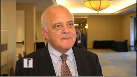
Sildenafil (Viagra) – a vasodilator in injured blood vessels – might help, according to ongoing research at the University of Pennsylvania, Philadelphia.
Researchers there gave sildenafil to inpatients with persistent symptoms at least 6 months after traumatic brain injury and measured CVR by a novel MRI technique an hour later. “Sildenafil was able to correct the deficit in CVR in many cases. We are hopeful this could be a useful therapy,” said principal investigator Ramon Diaz-Arrastia, MD, a professor of neurology at the university.
He explained the work in an interview at annual meeting of the American Neurological Association. The next step is to see if sildenafil helps CVR in acute traumatic brain injury, and in people who have had multiple, mild brain traumas, including professional athletes.
SAN DIEGO – The healthy brain is a master of autoregulation, continuously adjusting blood flow to meet metabolic demand.
But in traumatic brain injury, cerebrovascular reactivity (CVR) breaks down; blood vessels don’t dilate as they should to deliver nutrients and oxygen, leading to progressive neurologic decline.
Sildenafil (Viagra) – a vasodilator in injured blood vessels – might help, according to ongoing research at the University of Pennsylvania, Philadelphia.
Researchers there gave sildenafil to inpatients with persistent symptoms at least 6 months after traumatic brain injury and measured CVR by a novel MRI technique an hour later. “Sildenafil was able to correct the deficit in CVR in many cases. We are hopeful this could be a useful therapy,” said principal investigator Ramon Diaz-Arrastia, MD, a professor of neurology at the university.
He explained the work in an interview at annual meeting of the American Neurological Association. The next step is to see if sildenafil helps CVR in acute traumatic brain injury, and in people who have had multiple, mild brain traumas, including professional athletes.
AT ANA 2017
MRI brainstem volume loss predicts SUDEP
SAN DIEGO – Brainstem volume loss is extensive in sudden unexplained death in epilepsy, suggesting that loss of brainstem volume on MRI might predict who is at risk, according to investigators from the Center for SUDEP Research, a multicenter research collaborative.
The “MRI can detect potentially life threatening brainstem damage before SUDEP [sudden unexplained death in epilepsy] onset and ought to be further studied as a clinically useful biomarker to identify patients at risk for SUDEP,” said lead investigator Alica Goldman, MD, PhD, associate professor of neurology and neurophysiology at Baylor College of Medicine, Houston, and an investigator for the research collaborative.

It’s possible SUDEP is due to autonomic failure secondary to damage to areas of the brainstem that control autonomic functions such as breathing and heartbeat. Cardiorespiratory failure has been reported in cases of witnessed SUDEP, and the team previously reported structural mesencephalic and lower brainstem abnormalities in two SUDEP cases with temporal lobe epilepsy (Neuroimage Clin. 2014 Jul 9;5:208-16).
“It was a logical transition to have a larger study,” Dr. Goldman said at the annual meeting of the American Neurological Association.
The team compared findings from standardized 3T MRI exams in 18 patients with focal-onset epilepsy, 27 SUDEP cases that had focal-onset epilepsy and one or more MRIs within 10 years of death, and 11 controls without epilepsy.
They also looked at heart rate variability based on ECG readings, a proxy of autonomic control. Abnormal variability is a risk factor for arrhythmias and sudden cardiac death, and has been shown previously to correlate with epilepsy duration and frequency. It also seems worse at night, when SUDEP risk is highest.
In the living epilepsy patients, the team found structural volume loss in the dorsal mesencephalon and other brainstem areas, and the loss correlated with abnormal heart rate variability (P less than .001).
In the SUDEP cases, “we found that patients who died from SUDEP had widespread brainstem volume loss in their last MRI before death, and the extent of volume loss in the brainstem correlated with shorter survival time” from the final MRI (P = .03).
The SUDEP cases were a mean of 23 years old at their last MRI; the majority of the cases were men.
The National Institutes of Health and the Epilepsy Foundation supported the work. Dr. Goldman had no relevant disclosures.
SAN DIEGO – Brainstem volume loss is extensive in sudden unexplained death in epilepsy, suggesting that loss of brainstem volume on MRI might predict who is at risk, according to investigators from the Center for SUDEP Research, a multicenter research collaborative.
The “MRI can detect potentially life threatening brainstem damage before SUDEP [sudden unexplained death in epilepsy] onset and ought to be further studied as a clinically useful biomarker to identify patients at risk for SUDEP,” said lead investigator Alica Goldman, MD, PhD, associate professor of neurology and neurophysiology at Baylor College of Medicine, Houston, and an investigator for the research collaborative.
It’s possible SUDEP is due to autonomic failure secondary to damage to areas of the brainstem that control autonomic functions such as breathing and heartbeat. Cardiorespiratory failure has been reported in cases of witnessed SUDEP, and the team previously reported structural mesencephalic and lower brainstem abnormalities in two SUDEP cases with temporal lobe epilepsy (Neuroimage Clin. 2014 Jul 9;5:208-16).
“It was a logical transition to have a larger study,” Dr. Goldman said at the annual meeting of the American Neurological Association.
The team compared findings from standardized 3T MRI exams in 18 patients with focal-onset epilepsy, 27 SUDEP cases that had focal-onset epilepsy and one or more MRIs within 10 years of death, and 11 controls without epilepsy.
They also looked at heart rate variability based on ECG readings, a proxy of autonomic control. Abnormal variability is a risk factor for arrhythmias and sudden cardiac death, and has been shown previously to correlate with epilepsy duration and frequency. It also seems worse at night, when SUDEP risk is highest.
In the living epilepsy patients, the team found structural volume loss in the dorsal mesencephalon and other brainstem areas, and the loss correlated with abnormal heart rate variability (P less than .001).
In the SUDEP cases, “we found that patients who died from SUDEP had widespread brainstem volume loss in their last MRI before death, and the extent of volume loss in the brainstem correlated with shorter survival time” from the final MRI (P = .03).
The SUDEP cases were a mean of 23 years old at their last MRI; the majority of the cases were men.
The National Institutes of Health and the Epilepsy Foundation supported the work. Dr. Goldman had no relevant disclosures.
AT ANA 2017
Key clinical point:
Major finding: Patients who died from SUDEP had widespread brainstem volume loss in their last MRI before death (P = .03).
Data source: Imaging review of 27 SUDEP cases, 18 patients with focal epilepsy, and 11 controls.
Disclosures: The National Institutes of Health and the Epilepsy Foundation supported the work. The lead investigator had no relevant disclosures.
Eye movement, not CT or MRI, rules out posterior stroke
SAN DIEGO – When a patient with vertigo and dizziness presents to the emergency department, the first order of business is to figure out if they’re due to benign inner ear problems or a posterior fossa stroke.
Emergency department physicians usually use noncontrast CT (NCCT) to rule out stroke, while neurologists turn to MRI with diffusion-weighted imaging (MRI-DWI) to make the call.
Neither are good enough. The sensitivity of NCCT for acute posterior fossa strokes is just 30.8%. The sensitivity of MRI-DWI is better at 76.4%, but “false negatives [occur] in roughly one [of] four brainstem strokes in the first 48 hours,” according to a meta-analysis from Johns Hopkins University, Baltimore. The study, presented at the annual meeting of the American Neurological Association, involved more than 800 patients in the 14 strongest studies to look into the issue since 1990.

“Anterior circulation strokes are obvious, but posterior circulation strokes are subtle. We’ve surveyed ED physicians, and there’s a certain rate in the population who just don’t understand how bad CTs are for detecting acute stroke.” Meanwhile, “neurologists know” they can’t rely on CTs, “but they think MRIs are good enough. That turns out not to be true either,” he said.
Dr. Newman-Toker’s comments were sparked by a discussion about the meta-analysis, but he spoke from years of work trying to improve the situation. He was clear about what’s at stake: The early recognition of posterior fossa strokes, treatment with thrombolytics and surgical decompression (when warranted), and prevention of a second, larger stroke, can mean the difference between walking out of the hospital and dying or being wheelchair bound for life.
He and his colleagues have developed a way to detect posterior fossa strokes using eye movement abnormalities. They call it HINTS, which stands for “head impulse, nystagmus, and test of skew. “It turns out that the eye movements of inner ear disease look slightly different than the eye movements of brain disease; the subtle differences are enough to distinguish between the two.” When the technique is mastered, “our best estimate is that the sensitivity for posterior circulation stroke is around 99%,” Dr. Newman-Toker said. He is working to get the message out and train people; a video of the technique is online (Semin Neurol. 2015 Oct;35[5]:506-21).
As for the meta-analysis, “none of us were terribly surprised that CT wasn’t much good, and I knew that MRI wasn’t going to be perfect, but it was worse than I expected when we crunched all the numbers,” he said.
It’s no surprise that eye movement trumps imaging. “Physiology beats anatomy in the acute phase. It’s takes a little while for anatomy to change” on imaging, but eye movements change immediately “when patients become symptomatic with dizziness and vertigo,” he said.
Meanwhile, “if you don’t know how to evaluate peoples’ eye movements, I think MRIs are the next best thing,” he said.
There was no industry funding for the work. Johns Hopkins is working with a company called Natus to develop HINTS training. Dr. Newman-Toker might earn royalties, but so far “I haven’t made any money off it, and there’s no document saying I’m ever going to make any money from it,” he said.
SAN DIEGO – When a patient with vertigo and dizziness presents to the emergency department, the first order of business is to figure out if they’re due to benign inner ear problems or a posterior fossa stroke.
Emergency department physicians usually use noncontrast CT (NCCT) to rule out stroke, while neurologists turn to MRI with diffusion-weighted imaging (MRI-DWI) to make the call.
Neither are good enough. The sensitivity of NCCT for acute posterior fossa strokes is just 30.8%. The sensitivity of MRI-DWI is better at 76.4%, but “false negatives [occur] in roughly one [of] four brainstem strokes in the first 48 hours,” according to a meta-analysis from Johns Hopkins University, Baltimore. The study, presented at the annual meeting of the American Neurological Association, involved more than 800 patients in the 14 strongest studies to look into the issue since 1990.
“Anterior circulation strokes are obvious, but posterior circulation strokes are subtle. We’ve surveyed ED physicians, and there’s a certain rate in the population who just don’t understand how bad CTs are for detecting acute stroke.” Meanwhile, “neurologists know” they can’t rely on CTs, “but they think MRIs are good enough. That turns out not to be true either,” he said.
Dr. Newman-Toker’s comments were sparked by a discussion about the meta-analysis, but he spoke from years of work trying to improve the situation. He was clear about what’s at stake: The early recognition of posterior fossa strokes, treatment with thrombolytics and surgical decompression (when warranted), and prevention of a second, larger stroke, can mean the difference between walking out of the hospital and dying or being wheelchair bound for life.
He and his colleagues have developed a way to detect posterior fossa strokes using eye movement abnormalities. They call it HINTS, which stands for “head impulse, nystagmus, and test of skew. “It turns out that the eye movements of inner ear disease look slightly different than the eye movements of brain disease; the subtle differences are enough to distinguish between the two.” When the technique is mastered, “our best estimate is that the sensitivity for posterior circulation stroke is around 99%,” Dr. Newman-Toker said. He is working to get the message out and train people; a video of the technique is online (Semin Neurol. 2015 Oct;35[5]:506-21).
As for the meta-analysis, “none of us were terribly surprised that CT wasn’t much good, and I knew that MRI wasn’t going to be perfect, but it was worse than I expected when we crunched all the numbers,” he said.
It’s no surprise that eye movement trumps imaging. “Physiology beats anatomy in the acute phase. It’s takes a little while for anatomy to change” on imaging, but eye movements change immediately “when patients become symptomatic with dizziness and vertigo,” he said.
Meanwhile, “if you don’t know how to evaluate peoples’ eye movements, I think MRIs are the next best thing,” he said.
There was no industry funding for the work. Johns Hopkins is working with a company called Natus to develop HINTS training. Dr. Newman-Toker might earn royalties, but so far “I haven’t made any money off it, and there’s no document saying I’m ever going to make any money from it,” he said.
AT ANA 2017
Key clinical point:
Major finding: The sensitivity of CT for acute posterior strokes was just 30.8%. The sensitivity of MRI was better at 76.4%, but it still missed one in four.
Data source: Meta-analysis of more than 800 patients
Disclosures: There was no industry funding for the work. The senior investigator might profit from commercialization of HINTS training.
Slow-wave sleep linked to Parkinson’s disease cognition
SAN DIEGO – Slow-wave sleep improves cognition in Parkinson’s disease, according to University of Alabama at Birmingham investigators.
After sleep studies, they compared the cognitive performance of 16 patients who spent more than 14% of their sleep time in slow-wave (SW) sleep with the cognitive performance of 16 who spent less than 14% in SW sleep; 13 of the patients in the low SW group (81%) met the criteria for mild Parkinson’s disease (PD) cognitive impairment versus 7 of the patients in the higher SW sleep group (44%); the patients were well matched for age, sex, disease duration, and other variables.
The normative value for SW sleep is about 15%-20%, although people spend less time in SW sleep as they age. The study subjects were in their mid-60s on average, so the 14% cut point wasn’t too far off from what might be considered typical.
More SW sleep in PD was associated with better performance on measures of global function, attention/working memory, executive function, and language comprehension. Patients in the low SW group, for instance, performed 0.25 standard deviations below the normative mean on attention/working memory tests, while subjects in the high SW group performed 0.5 standard deviations above, meaning that they outperformed people in their age group who didn’t have PD. These differences were statistically significant.
It raises the question of what can be done to help PD patients sleep better. “I’m interested in exercise to improve sleep, and we have some primary data that show it’s helpful for sleep in general but also SW sleep,” said Dr. Amara. Sodium oxybate (Xyrem) might also help, but for now, it’s a controlled substance approved only for narcolepsy. “We might have to branch out and try something novel,” she said.

The next step is to “see if we can use SW sleep to predict who might have cognitive declines and find interventions to [prevent] it,” she said.
Patients in the study had been diagnosed with PD for a mean of about 7 years. People in the high SW group were on lower amounts of levodopa equivalents, however, the investigators controlled for that, as well as for the fact that women tend to spend more time in SW sleep than men. There were no differences between the groups in measures of movement problems and of subjective scores of sleep quality and daytime sleepiness.
People with high SW sleep fell asleep sooner than did those in the low SW groups, about 9 minutes versus 20 minutes. There was no correlation between visual-spatial function and SW sleep, but visual-spatial function did correlate with the amount of time spent in rapid eye movement sleep, suggesting that dreaming might be important for visual-spatial function, Dr. Amara said.
The work was funded by the National Institutes of Health. Dr. Amara had no relevant disclosures.
SAN DIEGO – Slow-wave sleep improves cognition in Parkinson’s disease, according to University of Alabama at Birmingham investigators.
After sleep studies, they compared the cognitive performance of 16 patients who spent more than 14% of their sleep time in slow-wave (SW) sleep with the cognitive performance of 16 who spent less than 14% in SW sleep; 13 of the patients in the low SW group (81%) met the criteria for mild Parkinson’s disease (PD) cognitive impairment versus 7 of the patients in the higher SW sleep group (44%); the patients were well matched for age, sex, disease duration, and other variables.
The normative value for SW sleep is about 15%-20%, although people spend less time in SW sleep as they age. The study subjects were in their mid-60s on average, so the 14% cut point wasn’t too far off from what might be considered typical.
More SW sleep in PD was associated with better performance on measures of global function, attention/working memory, executive function, and language comprehension. Patients in the low SW group, for instance, performed 0.25 standard deviations below the normative mean on attention/working memory tests, while subjects in the high SW group performed 0.5 standard deviations above, meaning that they outperformed people in their age group who didn’t have PD. These differences were statistically significant.
It raises the question of what can be done to help PD patients sleep better. “I’m interested in exercise to improve sleep, and we have some primary data that show it’s helpful for sleep in general but also SW sleep,” said Dr. Amara. Sodium oxybate (Xyrem) might also help, but for now, it’s a controlled substance approved only for narcolepsy. “We might have to branch out and try something novel,” she said.
The next step is to “see if we can use SW sleep to predict who might have cognitive declines and find interventions to [prevent] it,” she said.
Patients in the study had been diagnosed with PD for a mean of about 7 years. People in the high SW group were on lower amounts of levodopa equivalents, however, the investigators controlled for that, as well as for the fact that women tend to spend more time in SW sleep than men. There were no differences between the groups in measures of movement problems and of subjective scores of sleep quality and daytime sleepiness.
People with high SW sleep fell asleep sooner than did those in the low SW groups, about 9 minutes versus 20 minutes. There was no correlation between visual-spatial function and SW sleep, but visual-spatial function did correlate with the amount of time spent in rapid eye movement sleep, suggesting that dreaming might be important for visual-spatial function, Dr. Amara said.
The work was funded by the National Institutes of Health. Dr. Amara had no relevant disclosures.
AT ANA 2017
Key clinical point:
Major finding: Thirteen patients in the low SW group (81%) met criteria for mild Parkinson’s disease (PD) cognitive impairment versus seven (44%) in the higher SW sleep group.
Data source: Cognitive testing of 32 patients with PD stratified by amount of SW sleep
Disclosures: The work was funded by the National Institutes of Health. The presenter had no relevant disclosures.
Post-Ebola syndrome includes neurologic sequelae
SAN DIEGO – Add neurologic issues to the growing list of medical problems faced by survivors of Ebola virus.
Among 153 Liberian patients about a year out from their acute illness, “there were only a handful who didn’t have” some lingering neurologic problem. “The most commonly reported ongoing symptoms were headache and memory loss. A couple of people had seizures possibly related to Ebola.” Depression, anxiety, and posttraumatic stress disorder were common, said neurologist Jeanne Billioux, MD, a clinical fellow at the National Institute of Neurological Disorders and Stroke, Bethesda, Md.
Almost two-thirds of the patients had abnormal neurologic exams. The most common findings were tremors, pathological reflexes, mild dysmetria, and abnormalities of eye pursuits and saccades, plus nystagmus. The findings were statistically significant, compared with 81 close contacts, generally household members, who served as controls in the ongoing natural history study, which was presented at the annual meeting of the American Neurological Association.

What’s become clear in the wake of the recent outbreak in West Africa, by far the worst to date with over 28,000 cases and more than 11,000 deaths, is that there is a post-Ebola syndrome that includes ophthalmologic, cardiac, and rheumatologic problems. It now appears that “neurologic sequelae are a part of it,” as well, she said.
The natural history study – dubbed the Partnership for Research on Ebola Virus in Liberia (PREVAIL III) – is a collaboration between the National Institutes of Health and the Ministry of Health of Liberia, one of the hardest-hit countries; the neurology investigation is just one component of the study, which includes about 1,500 patients overall.
Serology testing confirmed that cases truly did have Ebola, and the controls did not. After the first evaluation a year or so after the acute illness, patients have been followed up every 6 months, with some out to about 3 years.
Although patients aren’t back to normal, the good news is that their symptoms and exams are improving. “We started with only 6 who had no symptoms; now we have 15. Headaches are getting better; memory is getting better. It’s wonderful,” Dr. Billioux said.
The patients were asked to recall their acute symptoms during their first study visit. Many reported headaches, weakness, altered mental status, and cranial nerve symptoms. About 2% described convulsions or strokelike symptoms, and about 25% described symptoms consistent with meningitis.
It’s unclear how the virus affected the CNS, which isn’t considered to be a target organ. Perhaps fluid loss from severe diarrhea led to cerebral hypoperfusion. The cytokine storm during the acute phase might also have played a role. The virus has, however, been isolated from cerebral spinal fluid and, although uncommon, there are the reports of meningitis symptoms, so perhaps it does have direct CNS effects. Much remains to be learned.
Both cases and controls were a mean of about 35 years old, and evenly split between the sexes; 108 patients (70.6%) spent more than 2 weeks in an Ebola treatment unit.
The work was funded by the National Institutes of Health. Dr. Billioux had no disclosures.
SAN DIEGO – Add neurologic issues to the growing list of medical problems faced by survivors of Ebola virus.
Among 153 Liberian patients about a year out from their acute illness, “there were only a handful who didn’t have” some lingering neurologic problem. “The most commonly reported ongoing symptoms were headache and memory loss. A couple of people had seizures possibly related to Ebola.” Depression, anxiety, and posttraumatic stress disorder were common, said neurologist Jeanne Billioux, MD, a clinical fellow at the National Institute of Neurological Disorders and Stroke, Bethesda, Md.
Almost two-thirds of the patients had abnormal neurologic exams. The most common findings were tremors, pathological reflexes, mild dysmetria, and abnormalities of eye pursuits and saccades, plus nystagmus. The findings were statistically significant, compared with 81 close contacts, generally household members, who served as controls in the ongoing natural history study, which was presented at the annual meeting of the American Neurological Association.
What’s become clear in the wake of the recent outbreak in West Africa, by far the worst to date with over 28,000 cases and more than 11,000 deaths, is that there is a post-Ebola syndrome that includes ophthalmologic, cardiac, and rheumatologic problems. It now appears that “neurologic sequelae are a part of it,” as well, she said.
The natural history study – dubbed the Partnership for Research on Ebola Virus in Liberia (PREVAIL III) – is a collaboration between the National Institutes of Health and the Ministry of Health of Liberia, one of the hardest-hit countries; the neurology investigation is just one component of the study, which includes about 1,500 patients overall.
Serology testing confirmed that cases truly did have Ebola, and the controls did not. After the first evaluation a year or so after the acute illness, patients have been followed up every 6 months, with some out to about 3 years.
Although patients aren’t back to normal, the good news is that their symptoms and exams are improving. “We started with only 6 who had no symptoms; now we have 15. Headaches are getting better; memory is getting better. It’s wonderful,” Dr. Billioux said.
The patients were asked to recall their acute symptoms during their first study visit. Many reported headaches, weakness, altered mental status, and cranial nerve symptoms. About 2% described convulsions or strokelike symptoms, and about 25% described symptoms consistent with meningitis.
It’s unclear how the virus affected the CNS, which isn’t considered to be a target organ. Perhaps fluid loss from severe diarrhea led to cerebral hypoperfusion. The cytokine storm during the acute phase might also have played a role. The virus has, however, been isolated from cerebral spinal fluid and, although uncommon, there are the reports of meningitis symptoms, so perhaps it does have direct CNS effects. Much remains to be learned.
Both cases and controls were a mean of about 35 years old, and evenly split between the sexes; 108 patients (70.6%) spent more than 2 weeks in an Ebola treatment unit.
The work was funded by the National Institutes of Health. Dr. Billioux had no disclosures.
AT ANA 2017
Key clinical point:
Major finding: A year after their acute infection, almost two-thirds of Ebola survivors had abnormal neurologic exams.
Data source: Natural history study involving 153 patients
Disclosures: The work was funded by the National Institutes of Health. The lead investigator had no disclosures.

Higher stroke risk found for TAVR versus SAVR
SAN DIEGO – There was an 86% greater risk of ischemic stroke after transcatheter aortic valve replacement, compared with surgical aortic valve replacement, and a more than sixfold increased risk of hemorrhagic stroke, in a review of more than 44,000 patients in the Nationwide Readmissions Database who were followed out to a year after having one procedure or the other.
“Our data suggest an elevated risk of both ischemic and hemorrhagic stroke after TAVR [transcatheter aortic valve replacement]. I see a lot of people that have strokes after” TAVR, so “I wasn’t all that surprised that there is an increased risk, but could I have guessed it would have been so high? No.” Perhaps “we are offering it to people we shouldn’t be offering it to,” said lead investigator Laura Stein, MD, a vascular neurology fellow at Mount Sinai Hospital, New York.

The 2013 Nationwide Readmissions Database used in the study captured more than 14 million readmissions in the United States across all payers and the uninsured. “We used this database because its captures all comers” and reflects “more real-world practice,” Dr. Stein said.
There were 6,015 TAVR and 38,624 SAVR cases in 2013, and the team found consistently elevated cumulative risks of ischemic and hemorrhagic stroke after TAVR, compared with SAVR, according to a presentation at the annual meeting of the American Neurological Association.
Compared with SAVR, the hazard ratio for ischemic stroke with TAVR was 1.86 (95% confidence interval, 1.12-3.08; P = .016) and, for hemorrhagic stroke, 6.17 (95% CI, 1.97-19.33; P = .0018). Dr. Stein declined to release absolute numbers of strokes in the two groups, pending publication.
“A lot of attention is being paid to this topic because there has been a push, a lot of it by the device makers, to prove that [TAVR] outcomes are just as good as with traditional surgery, and that we should be offering [TAVR] to more people with higher risk factor profiles who might not have been offered repair otherwise. Our job is to help patients make the most informed decisions. Having another source of data like [ours]” adds to the conversation about risks and benefits, she said.
The investigators adjusted for a large number of potential confounders to make sure the comparison was as fair as possible given the limits of database reviews. Among other variables, they controlled for baseline cardiovascular risk factors, carotid artery disease, heart failure, obesity, smoking, surgical complications, mortality, and illness severity scores, as well as hospital size, teaching hospital status, and urban versus rural location.
“What we can’t know is what medications these patients were on that might have increased their bleeding or ischemia risk. Also, we were relying on coding done by other people,” Dr. Stein said.
The next step is to look at the impact of stenting and other concomitant procedures. “We were surprised by the number of people that had multiple procedures at the same time.” The ultimate goal is to develop a risk score to help patients and doctors decide between the two procedures, she said.
Meanwhile, the team found no difference in stroke risk between coronary artery bypass grafting and percutaneous coronary interventions in the 2013 database.
Three was no industry funding for the work, and Dr. Stein did not have any relevant disclosures.
SAN DIEGO – There was an 86% greater risk of ischemic stroke after transcatheter aortic valve replacement, compared with surgical aortic valve replacement, and a more than sixfold increased risk of hemorrhagic stroke, in a review of more than 44,000 patients in the Nationwide Readmissions Database who were followed out to a year after having one procedure or the other.
“Our data suggest an elevated risk of both ischemic and hemorrhagic stroke after TAVR [transcatheter aortic valve replacement]. I see a lot of people that have strokes after” TAVR, so “I wasn’t all that surprised that there is an increased risk, but could I have guessed it would have been so high? No.” Perhaps “we are offering it to people we shouldn’t be offering it to,” said lead investigator Laura Stein, MD, a vascular neurology fellow at Mount Sinai Hospital, New York.
The 2013 Nationwide Readmissions Database used in the study captured more than 14 million readmissions in the United States across all payers and the uninsured. “We used this database because its captures all comers” and reflects “more real-world practice,” Dr. Stein said.
There were 6,015 TAVR and 38,624 SAVR cases in 2013, and the team found consistently elevated cumulative risks of ischemic and hemorrhagic stroke after TAVR, compared with SAVR, according to a presentation at the annual meeting of the American Neurological Association.
Compared with SAVR, the hazard ratio for ischemic stroke with TAVR was 1.86 (95% confidence interval, 1.12-3.08; P = .016) and, for hemorrhagic stroke, 6.17 (95% CI, 1.97-19.33; P = .0018). Dr. Stein declined to release absolute numbers of strokes in the two groups, pending publication.
“A lot of attention is being paid to this topic because there has been a push, a lot of it by the device makers, to prove that [TAVR] outcomes are just as good as with traditional surgery, and that we should be offering [TAVR] to more people with higher risk factor profiles who might not have been offered repair otherwise. Our job is to help patients make the most informed decisions. Having another source of data like [ours]” adds to the conversation about risks and benefits, she said.
The investigators adjusted for a large number of potential confounders to make sure the comparison was as fair as possible given the limits of database reviews. Among other variables, they controlled for baseline cardiovascular risk factors, carotid artery disease, heart failure, obesity, smoking, surgical complications, mortality, and illness severity scores, as well as hospital size, teaching hospital status, and urban versus rural location.
“What we can’t know is what medications these patients were on that might have increased their bleeding or ischemia risk. Also, we were relying on coding done by other people,” Dr. Stein said.
The next step is to look at the impact of stenting and other concomitant procedures. “We were surprised by the number of people that had multiple procedures at the same time.” The ultimate goal is to develop a risk score to help patients and doctors decide between the two procedures, she said.
Meanwhile, the team found no difference in stroke risk between coronary artery bypass grafting and percutaneous coronary interventions in the 2013 database.
Three was no industry funding for the work, and Dr. Stein did not have any relevant disclosures.
AT ANA 2017
Key clinical point:
Major finding: There was an 86% greater risk of ischemic stroke after transcatheter aortic valve replacement, compared with surgical aortic valve replacement, and a more than sixfold increased risk of hemorrhagic stroke.
Data source: Database review of more than 44,000 patients
Disclosures: Three was no industry funding for the work, and the lead investigator did not have any relevant disclosures.
BP accuracy is the ghost in the machine
SAN FRANCISCO – Amid all the talk about subgroup blood pressure targets and tiny differences in drug regimens at a recent hypertension meeting, there was an elephant in the room that attendees refused to ignore.
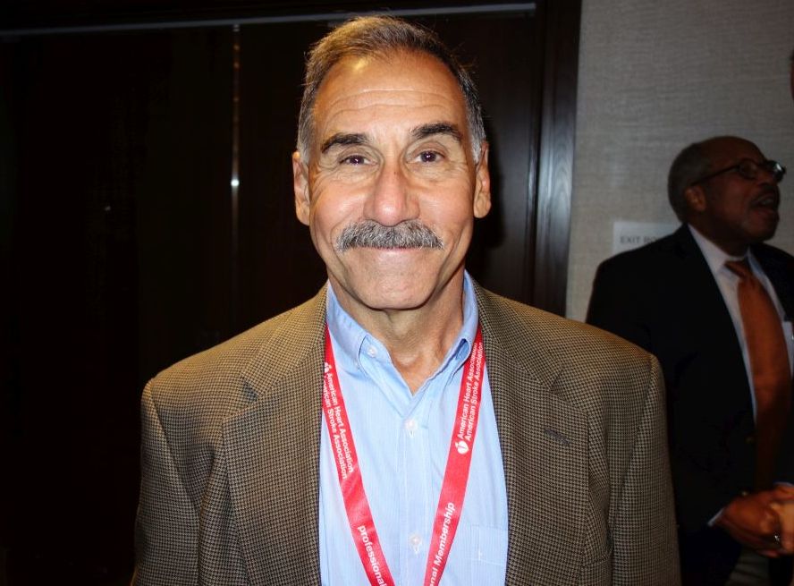
“We do it wrong,” said Dr. Steven Yarows, a primary care physician in Chelsea, Mich., who estimated he’s taken 44,000 blood pressures in his 36 years of practice.
Everyone in medicine is taught that people should rest a bit and not talk while their blood pressure is taken; that the last measurement matters more than the first; and that most Americans need a large-sized cuff. Current guidelines are based on patients sitting for 5-10 minutes alone in a quiet room while an automatic machine averages their last 3-5 blood pressures.
But when Dr. Yarows asked his 300 or so audience members – hypertension physicians who paid to come to the meeting – how many actually followed those rules, four hands went up. It’s not good enough; “if you are going to make a diagnosis that lasts a lifetime, you have to be accurate,” he said at the joint scientific sessions of the American Heart Association Council on Hypertension, AHA Council on Kidney Cardiovascular Disease, and American Society of Hypertension.
There’s resistance. No one has a room set aside for blood pressure; staff don’t want to deal with it; and at a time when primary care doctors are nickel and dimed for everything they do, insurers haven’t stepped up to pay to make accurate blood pressure a priority.
To do it right, you have to ask patients to come in 10 minutes early and have a room set up for them where they can sit alone with a large oscillometric cuff to average a few blood pressures at rest, Dr. Yarows said. They also need at least one 24-hour monitoring.
“Most of the time, the patient walks over from the waiting room, they get on the scale which automatically elevates the blood pressure tremendously, and then they sit down and talk about their family while their blood pressure is being taken.” Even in normotensive patients, that alone could raise systolic pressure 20 mm Hg or more, he said. It makes one-time blood pressure pretty much meaningless.
The biggest problem is that blood pressure is hugely variable, so it’s hard to know what matters. In one of Dr. Yarows’ normotensive patients, BP varied 44 mm Hg systolic and 37 mm Hg diastolic over 24 hours. In a hypertensive patient, systolic pressure varied 62 mm Hg and diastolic 48 mm Hg over 24 hours. Another patient was 114/85 mm Hg at noon, and 159/73 mm Hg an hour later. “That’s a huge spread,” he said.
Twenty-four hour monitoring is the only way to really know if patients are hypertensive and need treatment. “Any person you suspect of having hypertension, before you place them on medicine, you should have 24 hour blood pressure monitoring. This is the most effective way to determine if they do have high blood pressure,” and how much it needs to be lowered, he said.
Dr. Yarows had no disclosures.
SAN FRANCISCO – Amid all the talk about subgroup blood pressure targets and tiny differences in drug regimens at a recent hypertension meeting, there was an elephant in the room that attendees refused to ignore.
“We do it wrong,” said Dr. Steven Yarows, a primary care physician in Chelsea, Mich., who estimated he’s taken 44,000 blood pressures in his 36 years of practice.
Everyone in medicine is taught that people should rest a bit and not talk while their blood pressure is taken; that the last measurement matters more than the first; and that most Americans need a large-sized cuff. Current guidelines are based on patients sitting for 5-10 minutes alone in a quiet room while an automatic machine averages their last 3-5 blood pressures.
But when Dr. Yarows asked his 300 or so audience members – hypertension physicians who paid to come to the meeting – how many actually followed those rules, four hands went up. It’s not good enough; “if you are going to make a diagnosis that lasts a lifetime, you have to be accurate,” he said at the joint scientific sessions of the American Heart Association Council on Hypertension, AHA Council on Kidney Cardiovascular Disease, and American Society of Hypertension.
There’s resistance. No one has a room set aside for blood pressure; staff don’t want to deal with it; and at a time when primary care doctors are nickel and dimed for everything they do, insurers haven’t stepped up to pay to make accurate blood pressure a priority.
To do it right, you have to ask patients to come in 10 minutes early and have a room set up for them where they can sit alone with a large oscillometric cuff to average a few blood pressures at rest, Dr. Yarows said. They also need at least one 24-hour monitoring.
“Most of the time, the patient walks over from the waiting room, they get on the scale which automatically elevates the blood pressure tremendously, and then they sit down and talk about their family while their blood pressure is being taken.” Even in normotensive patients, that alone could raise systolic pressure 20 mm Hg or more, he said. It makes one-time blood pressure pretty much meaningless.
The biggest problem is that blood pressure is hugely variable, so it’s hard to know what matters. In one of Dr. Yarows’ normotensive patients, BP varied 44 mm Hg systolic and 37 mm Hg diastolic over 24 hours. In a hypertensive patient, systolic pressure varied 62 mm Hg and diastolic 48 mm Hg over 24 hours. Another patient was 114/85 mm Hg at noon, and 159/73 mm Hg an hour later. “That’s a huge spread,” he said.
Twenty-four hour monitoring is the only way to really know if patients are hypertensive and need treatment. “Any person you suspect of having hypertension, before you place them on medicine, you should have 24 hour blood pressure monitoring. This is the most effective way to determine if they do have high blood pressure,” and how much it needs to be lowered, he said.
Dr. Yarows had no disclosures.
EXPERT ANALYSIS FROM JOINT HYPERTENSION 2017
Rosacea: Expert recommends treating erythema, papules/pustules simultaneously
The results of a recently published trial helps answer one of the toughest questions in rosacea treatment: when patients present with both papules/pustules and erythema, which problem do you treat first?
In the past, Hilary Baldwin, MD, tended to target papules and pustules first, usually with ivermectin cream (Soolantra) and, when warranted, anti-inflammatory doses of doxycycline. Going after the erythema first and making the skin paler could make the cherry red inflammatory lesions stand out even more, she said.
In the study, one group was put on ivermectin for 12 weeks, with the vasoconstrictor brimonidine 0.33% (Mirvaso topical gel) added after 4 weeks to help with the erythema; and another group was treated with both ivermectin 1% cream and brimonidine daily for the entire 12 weeks. The third group received vehicles of both applied every day for 12 weeks (J Drugs Dermatol. 2017 Sep 1;16[9]:909-16).
“What they found was that both the combinations worked better than ivermectin alone,” and that treatment with both agents for the full 12 weeks worked best, with no increased risk of irritation or worsening of erythema than when brimonidine was brought in after 4 weeks of ivermectin, said Dr. Baldwin, a clinical associate professor of dermatology at Rutgers Robert Wood Johnson Medical School, New Brunswick, N.J., said in an interview. The vasoconstriction might have somehow helped with the papules and pustules, she noted.
The lesions might have looked more prominent “for the week or two it took for the ivermectin to kick in, but the patients didn’t care. They were happier campers by virtue of treating both aspects of their rosacea at the same time,” she added.
The new kid on the block for vasoconstriction – oxymetazoline (Rhofade cream) – appears to be gentler than brimonidine. “It takes a little bit longer to reach peak effect, and the peak doesn’t give you quite as much vasoconstriction as brimonidine, which for some people is a good thing,” she said. “Perhaps they’re a little bit too white with brimonidine. For other people who are bright red, oxymetazoline might not be enough. I think there’s a place for both drugs.”
Both vasoconstrictors might actually make erythema temporarily worse; it’s a known side effect. Dr. Baldwin has her patients try them for the first time when they’re at home and don’t have any important impending social engagements, just in case. “I like to give a tube of each one and say, ‘use one on one side of your face and the other on the other side and see which makes you happier.’ ”

Some patients can get away with “a really low dose and be completely cleared,” she commented. “I have some fully controlled on 10 mg twice weekly. As long as there’s no pregnancy risk, there’s no reason you can’t do this almost indefinitely.”
This publication and the Global Academy for Medical Education are both owned by Frontline Medical News. Dr. Baldwin is a speaker and advisor for Allergan, Galderma, and Valeant; and is an investigator for Dermira, Galderma, Novan, and Valeant.
The results of a recently published trial helps answer one of the toughest questions in rosacea treatment: when patients present with both papules/pustules and erythema, which problem do you treat first?
In the past, Hilary Baldwin, MD, tended to target papules and pustules first, usually with ivermectin cream (Soolantra) and, when warranted, anti-inflammatory doses of doxycycline. Going after the erythema first and making the skin paler could make the cherry red inflammatory lesions stand out even more, she said.
In the study, one group was put on ivermectin for 12 weeks, with the vasoconstrictor brimonidine 0.33% (Mirvaso topical gel) added after 4 weeks to help with the erythema; and another group was treated with both ivermectin 1% cream and brimonidine daily for the entire 12 weeks. The third group received vehicles of both applied every day for 12 weeks (J Drugs Dermatol. 2017 Sep 1;16[9]:909-16).
“What they found was that both the combinations worked better than ivermectin alone,” and that treatment with both agents for the full 12 weeks worked best, with no increased risk of irritation or worsening of erythema than when brimonidine was brought in after 4 weeks of ivermectin, said Dr. Baldwin, a clinical associate professor of dermatology at Rutgers Robert Wood Johnson Medical School, New Brunswick, N.J., said in an interview. The vasoconstriction might have somehow helped with the papules and pustules, she noted.
The lesions might have looked more prominent “for the week or two it took for the ivermectin to kick in, but the patients didn’t care. They were happier campers by virtue of treating both aspects of their rosacea at the same time,” she added.
The new kid on the block for vasoconstriction – oxymetazoline (Rhofade cream) – appears to be gentler than brimonidine. “It takes a little bit longer to reach peak effect, and the peak doesn’t give you quite as much vasoconstriction as brimonidine, which for some people is a good thing,” she said. “Perhaps they’re a little bit too white with brimonidine. For other people who are bright red, oxymetazoline might not be enough. I think there’s a place for both drugs.”
Both vasoconstrictors might actually make erythema temporarily worse; it’s a known side effect. Dr. Baldwin has her patients try them for the first time when they’re at home and don’t have any important impending social engagements, just in case. “I like to give a tube of each one and say, ‘use one on one side of your face and the other on the other side and see which makes you happier.’ ”
Some patients can get away with “a really low dose and be completely cleared,” she commented. “I have some fully controlled on 10 mg twice weekly. As long as there’s no pregnancy risk, there’s no reason you can’t do this almost indefinitely.”
This publication and the Global Academy for Medical Education are both owned by Frontline Medical News. Dr. Baldwin is a speaker and advisor for Allergan, Galderma, and Valeant; and is an investigator for Dermira, Galderma, Novan, and Valeant.
EXPERT ANALYSIS FROM THE COASTAL DERMATOLOGY SYMPOSIUM