User login
American Academy of Allergy, Asthma, & Immunology (AAAAI): Annual Meeting
AAAAI: Grass allergy tablets show favorable NNT
HOUSTON – The number needed to treat with Timothy grass sublingual immunotherapy tablets for allergic rhinitis to achieve a clinically meaningful response is 7.9, Dr. Stephen R. Durham reported at the annual meeting of the American Academy of Allergy, Asthma, and Immunology.
What the low number needed to treat (NNT) means in this instance is that, on average, 7.9 children or adults with Timothy grass–induced allergic rhinitis with or without conjunctivitis would need to be treated with Merck’s sublingual immunotherapy tablet (SLIT) rather than placebo daily for 3 years in order for 1 additional patient to obtain sustained benefit. Sustained benefit was defined as at least 50% well days for the entire grass pollen season during each of the 3 treatment years plus the subsequent 2 years of no treatment, explained Dr. Durham of Royal Brompton and Harefield Hospitals and Imperial College, London.
A ‘well day’ was considered as a day with no use of open-label rescue medication and in which the worst score recorded was ‘none’ or ‘mild’ for each of the four nasal and two ocular symptoms measured. This NNT analysis was based upon pooled data from six pivotal randomized, double-blind, phase III, placebo-controlled clinical trials totaling 3,094 patients, Dr. Durham added.
A separate analysis of the same pooled data using a different definition of favorable response – that is, a total combined daily symptom and daily medication score of 3 or less during the entire grass pollen season during the 3 treatment years, plus the following 2 no-treatment years – yielded an NNT of 9.4. The maximum total daily symptom score during any given year was 18, while the maximum daily medication score per year was 30-36, depending upon whether the participant was a child or adult, and whether the study was conducted in Europe or the United States.
The risk-benefit ratio of SLIT for allergic rhinitis in the pooled analysis was favorable as reflected in a number needed to harm of 303, with harm defined as a treatment-related systemic allergic reaction. When the NNH was recalculated using epinephrine usage as the harmful endpoint, the NNH was closely similar at 305.
Merck’s Timothy grass SLIT, marketed as Grastek, is FDA-approved for treatment of allergic rhinitis in adults and children as young as 5 years of age.
Dr. Durham reported receiving research grants and serving as a consultant to Merck, ALK, and Stallergenes. The NNT analysis was funded by Merck, and the phase III clinical trials on which the analysis was based were supported by ALK and Merck.
AT 2015 AAAAI ANNUAL MEETING
Key clinical point: The number of allergic rhinitis patients who need to be treated with Timothy grass immunotherapy tablets to obtain a sustained clinically important response is 7.9.
Major finding: For every 7.9 patients who took the sublingual immunotherapy tablets rather than placebo daily for 3 years, one additional patient experienced ‘well days’ for the entire grass pollen season during those 3 years, along with the next 2 years of no treatment.
Data source: This analysis was based upon six pivotal phase III, randomized, double-blind, placebo-controlled clinical trials totaling 3,094 adults and children with allergic rhinitis.
Disclosures: This NNT analysis was sponsored by Merck. The presenter reported receiving a research grant from the company.
Send kids home 2 hours after food challenge testing
HOUSTON – Food-allergic children undergoing a double-blind, placebo-controlled food challenge test can safely be discharged home after 2 hours provided they haven’t experienced a severe immediate reaction in the interim, according to a large retrospective Dutch study.
Late reactions are unpredictable and very seldom severe, Jacquelien Saleh-Langenberg reported at the annual meeting of the American Academy of Allergy, Asthma, and Immunology.
She presented a study of 1,142 children who underwent double-blind, placebo-controlled food challenge testing at a tertiary clinic at the University of Groningen in the Netherlands, where she is a combined medical student and Ph.D. candidate. The food-allergic children were challenged with cow’s milk, peanut, cashew, hazelnut, and egg.
A total of 400 children developed late reactions: 20.8% of children reported late reactions only on an active challenge day, 9.6% only on a placebo challenge day, and 4.6% reported reactions on both active and placebo challenge days.
Of particular interest was the finding that 89 subjects developed isolated reactions on an active challenge day and 92 did so on a placebo challenge day.
“Isolated late reactions occurred with comparable frequency after active and placebo challenge and are thus unlikely to be a real phenomenon,” Ms. Saleh-Langenberg concluded.
Late reactions were manifest as gastrointestinal symptoms in 45% of cases and cutaneous symptoms in about one-third, with respiratory symptoms accounting for most of the remainder. Ninety-eight percent of late reactions were rated as mild to moderate, having a score of 1-6 on a 12-point severity scale.
The investigators developed a predictive model for late reactions occurring on an active challenge day. It proved to have little practical value, though. The model, which included age, allergic rhinitis, severity of any immediate reaction, and hazelnut allergy, explained a mere 8% of the variance in the incidence of late reactions.
When late reactions occurred on an active challenge day, they did so a mean of 3.5 hours after testing. When they occurred on a placebo challenge day, they happened a mean of 4 hours after the challenge. The reactions took an average of 2 hours and 1 hour, respectively, to disappear.
Ms. Saleh-Langenberg reported no conflicts of interest with regard to this university-supported study.
AT 2015 AAAAI ANNUAL MEETING
Key clinical point: Food-allergic children who don’t experience a severe immediate reaction following double-blind, placebo-controlled food challenges can safely be discharged home 2 hours post-testing.
Major finding: No severe late reactions occurred in a large cohort of children who underwent double-blind, placebo-controlled food challenges with cow’s milk, cashew, peanut, egg, and hazelnut.
Data source: This retrospective study included 1,142 Dutch children who underwent double-blind, placebo-controlled food challenges at a university clinic.
Disclosures: The presenter reported no conflicts of interest with regard to this university-supported study.
Aspirin desensitization making headway in U.S.
HOUSTON – About 63% of allergists and fellows in training perform aspirin desensitization for aspirin-exacerbated respiratory disease, according to a national survey.
That figure is lower than it should be, given the wealth of published evidence that aspirin desensitization is a safe and effective component of the treatment of aspirin-exacerbated respiratory disease (AERD), Dr. Jeremy D. Waldram asserted in presenting the survey findings at the annual meeting of the American Academy of Allergy, Asthma, and Immunology.
Moreover, the figure likely overcalls the true rate, since participation in the survey was voluntary and fans of aspirin desensitization were probably more inclined to complete the 16-item questionnaire, added Dr. Waldram, a fellow in allergy and immunology at the Scripps Clinic in San Diego.
Was he surprised to find that aspirin desensitization isn’t more widely utilized?
“I think the number that surprised me more was that among the 37.5% of allergists who don’t do aspirin desensitization, almost 30% of them don’t even refer their patients to others who do the procedure. We don’t know why they don’t refer out; it wasn’t a question included in the survey. Perhaps they see patients who are of a less severe phenotype,” he said in an interview.
The 684 survey responses represented a 15% response rate. While 37.5% of respondents indicated they don’t perform aspirin desensitization, 73% of those who reported doing the procedure said they do an average of 1-5 cases annually.
Among allergists who don’t perform aspirin desensitization, safety concerns were the leading reason cited. Indeed, 70% of those who don’t do aspirin desensitization indicated safety risks were the main reason. More than one reason could be given, however, and 30% of allergists cited poor compensation for the procedure as a deterrent, nearly 60% said the logistics of monitoring care were too onerous, and one-third said they didn’t perform aspirin desensitization because they hadn’t been trained to do it.
Of allergists who reported doing aspirin desensitization, 52% perform the procedure in an outpatient setting unattached to a hospital. Another 21% do so in an outpatient clinic that’s physically attached to a hospital.
Within the past 5 years, 9% of respondents said that they’ve had a patient react severely to aspirin desensitization, requiring an unanticipated transfer to a higher level of care. That’s contrary to the experience among allergists at the Scripps Clinic, which is widely credited with pioneering the outpatient approach.
“We essentially do all our aspirin desensitizations for AERD in the outpatient setting. In 1,500 treated patients we’ve never had one that we had to transfer to a higher level of care. We don’t have any special setup. It’s a typical outpatient clinic. We usually don’t start IVs or do anything above and beyond,” Dr. Waldram said.
While 26% of respondents reported they generally recommend aspirin desensitization immediately upon identifying a patient history that supports the diagnosis of AERD, another 54% said they usually recommend the procedure to patients only after they’ve failed to improve on typical medical therapy.
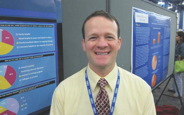
Twenty percent of physicians rated aspirin desensitization as “extremely helpful for the majority of patients,” another 49% said they find it most beneficial as an adjuvant to ongoing medical therapy.
Forty-four percent of allergists who perform aspirin desensitization reported that they learned to do the procedure during fellowship training. Fourteen percent said they learned to the procedure at an annual meeting, and 36% picked it up by reviewing the relevant literature.
Several allergists commented that had Dr. Waldram’s survey been conducted even a couple of years ago the rate of utilization of aspirin desensitization would have been far lower. They interpreted his reported 62.5% rate as a sign of progress. Dr. Waldram said he believes the key to further boosting utilization of aspirin desensitization lies in increasing exposure to the procedure during fellowship training. He noted that internal medicine-trained fellows who responded to the survey had a significantly higher aspirin desensitization utilization rate than those who came to their allergy fellowship with a background in pediatrics.
The hallmarks of AERD are difficult-to-treat nasal polyps, chronic eosinophilic sinusitis, and asthma in a patient with sensitivity to aspirin and other COX-1 inhibitors.
Dr. Waldram reported having no financial conflicts with regard to his study, which was conducted free of commercial support.
AT 2015 AAAAI ANNUAL MEETING
Key clinical point: Aspirin desensitization for patients with aspirin-exacerbated respiratory disease is catching on among U.S. allergists.
Major finding: Roughly 63% of allergists and allergy fellows who responded to a national survey indicated they perform aspirin desensitization for aspirin-exacerbated respiratory disease.
Data source: This was a 16-question survey of aspirin desensitization practices among U.S. allergists and allergy fellows. The national survey drew 684 responses.
Disclosures: The presenter reported having no financial conflicts with regard to his study, which was funded without commercial support.
Risk factors identified for gestational eczema
HOUSTON– New-onset eczema during pregnancy is a common phenomenon with several newly identified risk factors.
This disease entity deserves a proper name: gestational eczema, Dr. Wilfried J.J. Karmaus asserted at the annual meeting of the American Academy of Allergy, Asthma, and Immunology.
In contrast, the likelihood of new-onset asthma arising during pregnancy isn’t significantly more common than in an affected woman’s male partner during the same time frame.
“There was no large difference in wheezing between the women and men. Therefore, we cannot propose the term ‘gestational asthma,’” said Dr. Karmaus, professor of epidemiology at the University of Memphis. “Investigations into how to prevent eczema and asthma in pregnancy are really important, because eczema and asthma in pregnancy can increase the risk of these diseases in the offspring. This is a totally undeveloped field.”
He presented new findings from the Isle of Wight study, a prospective study in which a cohort of women has been followed from birth through pregnancy across three generations.
Eczema and asthma are common atopic diseases, and they are particularly common during pregnancy. Indeed, eczema is the most common skin disease seen in pregnancy, accounting for 35%-50% of all dermatoses in previous studies by other investigators. In those studies, only 20%-40% of women with eczema during pregnancy had a prepregnancy history of the disease.
In the Isle of Wight cohort, women were evaluated for asthma and eczema symptoms at ages 1, 2, 4, 10, and 18 years and again during pregnancy at gestational weeks 20 and 28. A total of 26 of 116 women developed eczema during pregnancy, with eight of them (31%) experiencing the skin disease for the first time in their lives. In contrast, only six of their male partners had eczema during the pregnancy time frame, and just one of them had new-onset eczema.
A history of maternal eczema in the preceding generation was associated with a 52% increased relative risk of having eczema by age 18 and a 3.1-fold increased likelihood of eczema during pregnancy. Also, methylation of the filaggrin gene at the cytosine-phosphate-guanine site cg13447818 when assessed at age 18 was associated with a significantly increased likelihood of eczema in a subject’s mother as well as increased risk of gestational eczema 1-7 years later, Dr. Karmaus continued.
Eighteen percent of women in the Isle of Wight cohort had asthma during pregnancy, as did a similar proportion of their male partners. Twenty-seven percent of women with asthma during pregnancy had no previous history of the respiratory disease, a rate which was again comparable in their male partners with asthma.
DNA methylation of the IL1RL1 gene at cg17738684 was significantly associated with asthma heritability across three generations in the Isle of Wight study. The IL1RL1 gene at cg17738684 is a candidate gene for asthma that encodes for interleukin-33. This finding raises the possibility that addressing this DNA methylation could prove fruitful as a transgenerational asthma prevention strategy.
The Isle of Wight birth cohort study is funded by the National Institutes of Health, Asthma UK, and the Isle of Wight Trust. Dr. Karmaus reported having no financial conflicts of interest.
AT 2015 AAAAI ANNUAL MEETING
Key clinical point: Thirty-one percent of cases of eczema and 27% of asthma in a group of pregnant women occurred for the first time in the woman’s life.
Major finding: A history of maternal eczema was associated with a 3.1-fold increased likelihood of eczema during the offspring’s pregnancy.
Data source: The Isle of Wight birth cohort study is a prospective study following three generations from birth through pregnancy.
Disclosures: The study is funded by the National Institutes of Health, Asthma UK, and the Isle of Wight Trust. The presenter reported having no financial conflicts of interest.
Bronchial thermoplasty gains momentum for severe asthma
HOUSTON – Bronchial thermoplasty has emerged as an important treatment option for patients with severe asthma at specialized centers, Dr. Mario Castro observed at the annual meeting of the American Academy of Allergy, Asthma, and Immunology.
The most recent international European Respiratory Society/American Thoracic Society practice guidelines on severe asthma recommend that bronchial thermoplasty for severe persistent asthma be utilized only in the setting of a clinical study or independent registry. The guidelines cited “very low confidence” in the available estimates of the novel treatment’s longer-term benefits and harms, as well as the lack of data regarding the phenotypes of asthma patients most likely to benefit (Eur. Respir. J. 2014 Feb;43:343-73).
Dr. Castro, a member of the task force that developed the ERS/ATS guidelines, said the group’s cautious stance was appropriate given the evidence available at the time of deliberations. However, at the AAAAI meeting, he highlighted more recent study results that address many of the task force’s concerns and that he said might lead to a more enthusiastic recommendation for bronchial thermoplasty in future guidelines.
One key piece of evidence unavailable to the task force comes from results reported in the 5-year prospective follow-up of 162 bronchial thermoplasty-treated patients in the international Asthma Intervention Research 2 (AIR2) trial.
“It’s quite striking that the exacerbation rate did not start to creep back up over time in this severe asthma population. We believe this study shows for the first time that this therapy may actually be a disease modifier, and that you can do this procedure in an identified population and the benefits of this one-time treatment are sustained over at least a 5-year time period,” said Dr. Castro, an AIR2 investigator and professor of pulmonary and critical care medicine and pediatrics at Washington University in St. Louis.
Compared with the baseline established during the year prior to bronchial thermoplasty, at 5 years post procedure, there was a 44% decrease in the percentage of AIR2 participants with severe exacerbations requiring oral corticosteroids, and a 48% reduction in the severe exacerbation event rate. Moreover, there was a 78% reduction in the percentage of patients with an emergency department visit for asthma and an 88% drop in the ED visit event rate (J. Allergy Clin. Immunol. 2013;132:1295-302).
With regard to safety, annual high-resolution CT scans showed no structural abnormalities from baseline to 5 years post-bronchial thermoplasty that could be attributed to the procedure. Prebronchodilator forced expiratory volume in 1 second (FEV1) values remained steady between years 1 and 5 post procedure despite an 18% decrease in the average daily dose of inhaled corticosteroids.
In a separate study, Dr. Castro and coinvestigators at Washington University identified a number of predictors of who will get the best responses to bronchial thermoplasty. This was a small study involving 42 patients with severe persistent asthma as reflected in their baseline mean inhaled corticosteroid dose of 2,185 mcg/day. Eighty percent of patients required bursts of oral corticosteroids during the year prior to the procedure. Their average baseline Asthma Quality of Life Questionnaire (AQLQ) score was 3.42. The baseline FEV1 postbronchodilator averaged 70%, with a range of 44%-121%.
Predictors of a clinically meaningful improvement in quality of life as defined by at least a 0.5-point improvement in AQLQ score 1 year post procedure included a shorter duration of asthma – 19 years, as compared with an average of 45 years in nonresponders – and a greater number of severe exacerbations during the year prior to bronchial thermoplasty.
Using another important yardstick of clinical improvement – at least a 240 mcg/day dose reduction in inhaled corticosteroids or a 2.5 mg/day decrease in oral corticosteroids at 1 year post procedure – significant predictors of benefit included older age (55 vs. 43 years), a lower baseline AQLQ score (2.4 vs. 4.0), and greater need for oral corticosteroids.
In addition, several quantitative metrics obtained through multidetector CT scans of the chest showed promise as predictors of a corticosteroid dose reduction. Responders showed less baseline air trapping, with an average of 6.1% of the lung having a density below –850 Hounsfield units, compared with 12.1% in nonresponders. Responders also had less baseline emphysema-like lung, with 3.2% of the lung having a density below –950 Hounsfield units at total lung capacity, compared with 5.8% in nonresponders, according to Dr. Castro.
This as-yet unpublished study was funded by the National Institutes of Health. AIR2 was sponsored by Boston Scientific. Dr. Castro reported research grants from the NIH, the American Lung Association, Boston Scientific, and other companies.
An estimated 5% of asthma patients are categorized as having severe disease. Bronchial thermoplasty has been approved by the FDA for the treatment of severe asthma since 2010. The outpatient procedure entails delivery of radiofrequency energy to the lungs in three sessions spaced several weeks apart.
EXPERT ANALYSIS FROM THE 2015 AAAAI ANNUAL MEETING
Mast cells predict hypersensitive reactions in rituximab desensitization
HOUSTON – For rituximab patients undergoing desensitization, mast cell degranulation is a more effective predictor of hypersensitive reactions (HSR) than are the traditionally employed skin tests, according to a study presented at the annual meeting of the American Academy of Allergy, Asthma, and Immunology.
“In this particular study, we were interested in investigating the roles of immunoglobulin E (IgE) in the mast cells in rituximab hypersensitivity,” said Dr. Johnson T. Wong of Massachusetts General Hospital, Boston, who presented the findings during a session dedicated to “Novel Insights in Drug Allergy.”
The study comprised 25 patients with persistent or severe rituximab sensitivity (RITS) who were treated at Massachusetts General between 2008 and 2013. Dr. Wong and his coinvestigators performed skin testing on 18 subjects, of which 7 (39%) reacted positively at least once during the course of testing.
HSR reactions occurred at similar rates in subjects who reacted positively and negatively to skin testing. Tryptase levels were taken in 18 subjects (72%), during 67% of the HSR desensitizations and 21% of the asymptomatic desensitizations.
Overall, elevated levels were noted in 21% of the HSR desensitizations.
The elevated tryptase levels in patients experiencing HSR indicates that mast cell degranulation is likely a major contributing factor in causing HSR, with cytokine release and tumor lysis also playing an important role, Dr. Wong said. Mast cell degranulations also points to an association with IgE mechanisms as a reliable indicator of HSR likelihood.
Dr. Wong and his coauthors concluded via tryptase level testing that asymptomatic mast cell degranulation happened very rarely, as only 1 out of 27 assessments (3%) indicated that result. One patient was excluded from the HSR desensitization cohort’s results because of probable mast cell activation syndrome, which caused abnormally elevated tryptase levels throughout the study period.
“In both positive and negative skin test patients, our desensitization protocol was able to reduce the reaction rate to less than 30%, so around 70% of patients had no reaction at all during desensitization,” said Dr. Wong, adding that nearly all desensitizations were completed successfully.
Dr. Wong had no disclosures.
AT THE 2015 AAAAI ANNUAL MEETING
Key clinical point: Look to mast cell degranulation, not skin tests, for accurate information regarding the likelihood of hypersensitive reactions in rituximab patients undergoing desensitization.
Major finding: Tryptase levels were elevated in 21% of patients who experienced hypersensitive reactions during desensitization.
Data source: Study of 25 patients with rituximab sensitivity at Massachusetts General Hospital.
Disclosures: Dr. Wong reported no relevant financial disclosures.
‘Mossy Oak sign’ suggests delayed anaphylaxis to red meat
HOUSTON– In central Virginia, where IgE-mediated delayed allergic reactions to red meat have become the most common cause of anaphylaxis in adults, physicians have taken to looking for what they call the ‘Mossy Oak sign.’
“If a patient shows up in a blaze-orange cap and hunter’s camouflage fatigues, an allergy fellow will tell me, ‘There’s a positive Mossy Oak sign in room 2,’ and I know that probably means the patient has delayed anaphylaxis to alpha-gal,” Dr. Scott P. Commins said at the annual meeting of the American Academy of Allergy, Asthma, and Immunology.
Alpha-gal is short for galactose-alpha-1,3,-galactose, an oligosaccharide present on thyroglobulin and other tissues in nonprimate mammals. It’s not normally present in humans, but when a lone star tick (Amblyomma americanum) that has fed on a nonprimate mammal bites a human, the alpha-gal is transferred, eliciting serum IgE antibodies.
Mossy Oak is a popular brand of hunter’s camouflage clothing. A positive Mossy Oak sign is useful in clinical practice because a patient who presents to a medical clinic dressed in hunting regalia is someone who spends a lot of time outdoors in the woods and fields where ticks lurk. He’s also typically someone who enjoys eating red meat. And whether it’s venison, beef, pork, lamb, goat, or bison, it contains alpha-gal. The result, in a patient who’s been primed via tick bite, can be a life-threatening anaphylactic or urticarial reaction arising 3-6 hours later, explained Dr. Commins, an allergist at the University of Virginia, Charlottesville.
He and his coinvestigators have played a central role in the still-unfolding story of this novel disease involving late-onset anaphylaxis to mammalian meat. Dr. Commins was the lead author of the paper that first described the syndrome (J. Allergy Clin. Immunol. 2009;123:426-33), as well as a subsequent paper that established the lone star tick as the culprit, making this syndrome the first known example of a response to an ectoparasite giving rise to a serious form of food allergy (J. Allergy Clin. Immunol. 2011;127:1286-93). More recently, the investigators have shown that alpha-gal-specific IgE does not cause or worsen asthma (Am. J. Respir. Crit. Care Med. 2012;185:723-30).
In a wide-ranging talk, Dr. Commins addressed the diagnosis and management of delayed anaphylaxis to red meat. He also touched upon some provocative emerging issues, including the possible risks posed by placing a porcine heart valve or bioprosthetic ligament in a patient with serum IgE antibodies to alpha-gal.
The investigators stumbled upon the phenomenon of tick-transmitted delayed anaphylaxis to red meat while they were trying to unravel the explanation for the markedly regional occurrence of IgE-mediated hypersensitivity reactions to the chimeric monoclonal antibody cetuximab (Erbitux) previously reported in the oncology literature (J. Clin. Oncol. 2007;25:3644-8). Dr. Commins and his colleagues realized that the same southeastern and south-central states where reactions to the initial infusion of cetuximab were concentrated were the states where the lone star tick abounds.
Incidentally, scientists at the Centers for Disease Control and Prevention follow the lone star tick closely because it is the primary vector for ehrlichiosis. CDC researchers say the tick’s range is steadily expanding and now includes 28 states, with New York’s Long Island a hot spot.
There is no public health requirement to report serum IgE-mediated delayed reactions to red meat, so the exact number of affected patients is unknown. But it’s clear that many thousands of individuals are affected, and estimates are being revised upward as the novel syndrome becomes more widely known. The disorder is common in Europe and Australia as well.
Classically, IgE-mediated anaphylactic reactions occur within 5-30 minutes after exposure to the offending agent. Thus, the 3- to 6-hour delay in symptom onset in patients with a reaction to the alpha-gal in red meat is remarkable; the explanation for the time lag remains unclear.
Dr. Commins and others have shown that individuals with serum IgE antibodies to alpha-gal also typically have serum IgE antibodies to cat, dog, beef, and pork, but not to egg, peanut, chicken, fish, or house dust mite.
Diagnosis of IgE-mediated urticarial or anaphylactic reactions to mammalian meat is made on the basis of the presence of serum IgE antibodies to alpha-gal. Dr. Commins recommended considering the diagnosis and ordering the blood test in the setting of new-onset anaphylaxis in a patient who enjoys hunting or other outdoor activities in a state where the lone star tick is found, particularly if the symptoms occur at night, hours after a big meat-heavy meal. A history of recent or persistent tick bites is an obvious clue.
“Also, it’s striking how many patients develop palmar erythema and itching during an episode. Not all report it, but when they do it’s usually a pretty good giveaway that they might have IgE to alpha-gal,” according to the allergist.
He added that it’s entirely reasonable to order a screening test for IgE to alpha-gal in patients in lone star tick–abiding states whose anaphylactic reactions seem to occur randomly without an apparent trigger.
Dr. Commins and his coinvestigators have assembled a database of roughly 500 of their patients with IgE to alpha-gal, about half of whom have a history of atopy. The investigators have found that an individual’s atopic status has no bearing on IgE antibody titer or the severity of the delayed reactions. Moreover, neither the alpha-gal IgE antibody level, the ratio of alpha-gal-specific IgE to total IgE, nor IgG antibodies correlate with reaction severity, he continued.
Based upon their study of 45 affected children, Dr. Commins and coworkers concluded that the clinical presentation and serum IgE pattern are the same as in adults (Pediatrics 2013 May [doi:10.1542/peds.2012-2585]). Since that publication, however, the investigators have realized there is a subgroup of affected teenagers who present with GI symptoms, he added.
Turning to disease management, Dr. Commins said he advocates an avoidance diet that eliminates mammalian meats, rich desserts, and super-premium ice cream.
“I also counsel patients to avoid broths, gravies, and anything that might be a mystery sauce,” he said. “You’d be surprised at how many people with alpha-gal order chicken at a Mexican restaurant thinking that they’re doing the right thing and end up reacting. I don’t know exactly why it happens, so I just say ‘avoid mystery sauces.’ Dairy and cheese are actually fairly well tolerated, although soft cheeses, like brie, can cause a reaction.”
Reactions are inconsistent, and symptoms can vary from episode to episode. Cofactors are a concern, with exercise and alcohol tending to make patients more sensitive to an alpha-gal exposure. The degree of risk posed by vaccines containing gelatin constitutes an emerging and unresolved issue.
“We’ve heard of several reactions to the shingles vaccine because of the gelatin, and the MMR vaccine is also on the radar,” Dr. Commins said.
Implantation of porcine bioprosthetic heart valves in patients with serum IgE antibodies to alpha-gal has been associated with reports of early valve failure; all bioprosthetic valves contain alpha-gal unless they’ve been decellularized. In addition, Dr. Commins is familiar with a case at another university in which three separate attempts to place a bioprosthetic ligament during repeated arthroscopic knee surgeries failed in a patient who had a “screamingly high” level of IgE to alpha-gal.
“I think this bioprosthesis issue is yet to be resolved,” he added.
One audience member, a Texas allergist, said she has a lot of trouble convincing her patients who are avid hunters to give up eating red meats. Dr. Commins said he faces the same issue.
“There is a recalcitrant group that just wants to eat a side of beef every day. I tell them if you’re not going to be on an avoidance diet, at least avoid the fattier cuts and don’t eat tremendous amounts. We believe that the antigen is possibly a glycolipid. Those cuts of meat that are high in fat are the ones patients tell us over and over again give them the worst reactions,” he said.
“The inconsistency of the allergic reactions keeps some patients from taking this disease seriously,” the allergist added. “What eventually happens for some patients is they end up having a really bad reaction. And then that convinces them.”
He monitors affected patients’ alpha-gal IgE levels over time. If and when the IgE becomes negative, he recommends a food challenge test. The patient comes in at 8 a.m., eats three pork sausage patties, and spends the day under observation at the clinic, walking the stairs periodically since exercise is a cofactor. If the challenge goes off without a hitch, the patient is free to go home at 4 or 5 p.m. In the past, that was the patient’s ticket to clearance to safely eat a big meat meal with alcohol, but Dr. Commins has pulled back of late from that recommendation.
“We believe that additional tick bites can make the allergy come back. So if someone passes a challenge in October and then the following spring gets more tick bites, you may have set them up to have a reaction because you’ve told them they can eat meat again. So the utility of a negative food challenge is unclear unless you’re pretty confident a patient is not going to have more tick bites,” he explained.
Dr. Commins reported receiving research grants from the National Institutes of Health to conduct his studies on delayed anaphylactic reactions to red meat. He serves on speakers bureaus for Genentech and Teva.
EXPERT ANALYSIS FROM THE 2015 AAAAI ANNUAL MEETING
Consider cephalosporin a safe alternative for patients with penicillin allergy
HOUSTON– Given the low incidence of adverse drug reactions to cephalosporin antibiotics among nearly a million California health plan patients, patients with a history of penicillin allergy can safely be given cephalosporins, according to Dr. Eric M. Macy.
The recommendation is based on the findings of a retrospective, population-based analysis of the records of 949,323 Kaiser Permanente Southern California health plan members, which was presented by Dr. Macy, of the Kaiser Permanente Medical Center in San Diego, at the annual meeting of the American Academy of Allergy, Asthma, and Immunology.
Dr. Macy and his colleagues examined the records of 622,456 health plan members who were given 901,908 regimens of oral cephalosporins and 326,867 members given 487,630 parenteral cephalosporin regimens between Jan. 1, 2010, and Dec. 31, 2012.
Clostridium difficile infection within 90 days, nephropathy within 30 days, and all-cause death within 1 day were the most common cephalosporin-associated adverse drug reactions (ADRs) reported by physicians, with rates of 0.91%, 0.15%, and 0.10%, respectively. Cephalosporin-associated anaphylaxis was documented by physicians a total of five times in oral patients and eight times in parenteral patients (P = .0761), while only three serious cutaneous adverse reactions (SCARs) were recorded. All of those SCARS were attributable to other antibiotics taken at the same time as the cephalosporins, according to Dr. Macy.
Patients who reported developing new cephalosporin allergies over the course of the study period were significantly more likely to be female than male: 0.56% vs. 0.43% (P < .0001). And patients with a history of penicillin allergy were more likely to report a new cephalosporin allergy within 30 days than patients with no drug allergy, another cephalosporin allergy, or a non–beta-lactam allergy.
The numbers are smaller than those reported recently in the American Journal of Medicine (2009;122: 778.e1–7), Dr. Macy noted, “because there it was new allergy reports in the year after any cephalosporin exposure, as opposed to our new report, which looks at reactions within 30 days of exposure.”
Dr. Macy disclosed that he has received research support from ALK and BioMarin.
AT THE 2015 AAAAI ANNUAL MEETING
Key clinical point: Given the low risk of adverse drug reactions with cephalosporins, patients with a history of penicillin allergy can safely take cephalosporins.
Major finding: The most frequent ADRs in patients taking either oral or parenteral cephalosporins were Clostridium difficile infection within 90 days (0.91%), nephropathy within 30 days (0.15%), and all-cause death within 1 day (0.10%).
Data source: Retrospective, population-based analysis of 949,323 Kaiser Permanente Southern California health plan members from 2010 to 2012.
Disclosures: Dr. Macy disclosed receiving research support from ALK and BioMarin.
Self-reported penicillin allergy may be undiagnosed chronic urticaria
HOUSTON – The higher prevalence of chronic urticaria in patients with self-reported penicillin allergy suggests that these patients may be confusing one condition with the other, according to a late-breaking study presented at the annual meeting of the American Academy of Allergy, Asthma, and Immunology.
A retrospective chart review of 1,419 patients with self-reported penicillin allergy revealed that 175 patients (12.3%) had a diagnosis of chronic urticaria, a significantly higher percentage than the typical prevalence range of 0.5%-5% that has been reported in the general population.
“Patients are potentially mistakenly attributing symptoms of chronic urticaria to a penicillin allergy,” explained study author Dr. Susanna G. Silverman of the University of Pennsylvania, Philadelphia.
The study included patients at the University of Pennsylvania’s allergy and immunology clinic who self-reported penicillin allergy from June 2007 to August 2014. Patients were identified as having penicillin allergy if penicillin, amoxicillin, amoxicillin-clavulanate, or piperacillin-tazobactam were present on the allergy list of their medical records.
Dr. Silverman then identified all patients from that group who also received a diagnosis of urticaria – a total of 343 patients – then narrowed the list to those who were diagnosed with chronic urticaria or the presence of urticaria for at least 6 weeks.
Of the 175 patients who had chronic urticaria, all were between the ages of 20 years and 92 years; 84% were female, and 53% were white.
“We think it’s important for physicians to think about this and to ask patients about symptoms of chronic urticaria when they report penicillin allergy,” Dr. Silverman noted, “to better determine what is truly penicillin allergy versus simply chronic urticaria symptoms.”
Dr. Silverman did not report any financial disclosures.
AT 2015 AAAAI ANNUAL MEETING
Key clinical point: Patients self-reporting penicillin allergy may have undiagnosed chronic urticaria.
Major finding: 12.3% of patients with self-reported penicillin allergy had chronic urticaria diagnoses, a significantly higher rate than the 5% rate in the general population.
Data source: Retrospective chart review of 1,419 patients at the University of Pennsylvania’s allergy and immunology clinic.
Disclosures: Dr. Silverman did not report any financial disclosures.
VIDEO: Novel three-shot immunotherapy regimen
HOUSTON– Dr. Amber M. Patterson of Nationwide Children’s Hospital and Research Institute in Columbus, Ohio, provides the lowdown on intralymphatic immunotherapy, in which a three-injection protocol of ultrasound-guided lymph node injections spaced 4 weeks apart is being investigated as a treatment for grass pollen-induced rhinoconjunctivitis in adolescents. It’s an attractive alternative to the conventional 3-year regimen of weekly, then monthly subcutaneous immunotherapy injections.
The video associated with this article is no longer available on this site. Please view all of our videos on the MDedge YouTube channel
AT 2015 AAAAI ANNUAL MEETING


