User login
Talk about déjà vu: Senators set to re-enact drug price hearing of 60 years ago
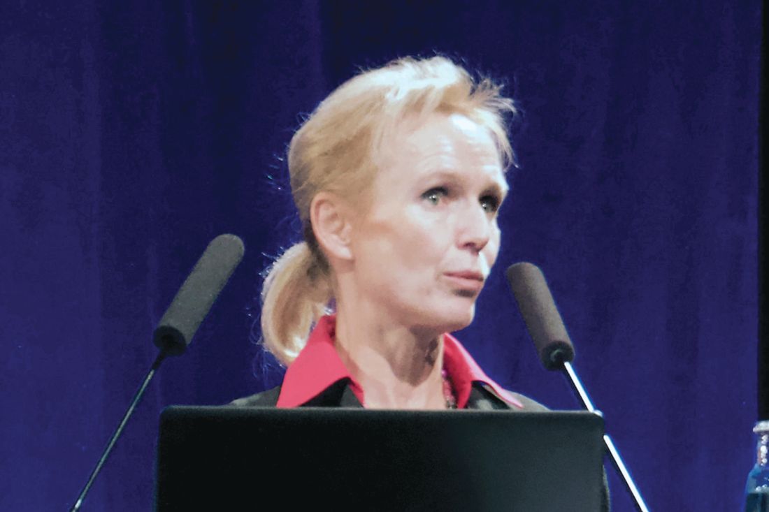
Kenneth Frazier, CEO of pharma giant Merck, is set to face senators Feb. 26 who say drug costs are “sky-high” and “out of control.”

But Frazier doesn’t need new talking points. Sixty years ago, a different panel of senators grilled a different Merck boss about the same problem.
To a striking degree, the subjects likely to surface during the hearing – high drug prices and profits, limited price transparency, aggressive marketing, alleged patent abuse and mediocre, “me-too” drugs – are identical to the issues senators investigated decades ago, historical transcripts show.
Frazier is scheduled to testify before the Senate Finance Committee, led by Sen. Chuck Grassley (R-Iowa), along with the CEOs of AbbVie, AstraZeneca, Bristol-Myers Squibb, Pfizer, and Sanofi and a top executive for Johnson & Johnson.
Hearings led by Sen. Estes Kefauver (D-Tenn.) in 1959 and 1960 were the first significant congressional inquiry into rising drug costs and drug-company profits. While that showdown led to new legal standards for drug safety and effectiveness, cost-control measures never made it to the final bill.
Health policy scholars say the similar hearings show just how much unfinished business remains and how well pharma companies have protected profits and limited regulation over the years.
“Every decade since the Kefauver hearings has seen at least one set of congressional hearings into the increasing prices of prescription drugs,” said Jeremy Greene, MD, PhD, a drug-industry historian at Johns Hopkins University, Baltimore.
“Drug prices, especially at the high end, are only ever higher” since the 1960s, said Scott Podolsky, MD, a health care historian at Harvard Medical School, Boston. “The issues of transparency and profitability certainly have been there from the very first day of Kefauver.”
A Tennessee Democrat known for investigating the Mafia, Sen. Kefauver launched a series of hearings on business in the late 1950s. His Senate antitrust subcommittee began taking testimony on high pharmaceutical prices in late 1959.
“While this country has the best drugs in the world, it would appear from the great number of letters which the subcommittee has received that many of our citizens are experiencing difficulty in being able to purchase them,” Sen. Kefauver said in opening remarks.
The sessions, which lasted off and on until Sept. 1960, were “among the most sensational” hearings of that Congress, a syndicated columnist wrote at the time. Appearing were the bosses of Merck, Pfizer, Schering, Bristol-Myers, Upjohn, Smith Kline, and American Home Products. Senators dug into the prices of antibiotics, corticosteroids and tranquilizers, the wonder drugs of the time.
John Connor, then Merck’s president, said he had “deep sympathy” for people unable to afford medicine. So did Schering’s boss, but he said it wasn’t the industry’s fault.
“Undoubtedly some people find it difficult to pay for needed medication. They will also have difficulty in meeting their rent and food bills,” said Francis Brown, Schering’s president at the time. “It is a matter of inadequate income rather than excessive prices.”
The executives urged Congress to create government programs to help people pay for health care, which it did a few years later. Today Medicare, Medicaid, and subsidies for private health coverage cost taxpayers more than $1 trillion annually.
But drugs are still unaffordable for many. A fourth of Americans in a recent survey by the Kaiser Family Foundation said somebody in their family skipped doses or left prescriptions unfilled because medicine costs too much. (Kaiser Health News is an editorially independent program of the foundation.)
Sixty years ago, as now, policymakers wondered why Americans pay so much more for medicine than people elsewhere.
“I still don’t understand why druggists in London buy this drug for $7.53 and our drugstores have to pay $17.90,” Sen. Kefauver told Connor, the Merck executive.
“We meet different market conditions in different countries,” was Connor’s response. Americans were getting “a reasonable bargain,” he said. “Merck’s Head Defends Drug Prices” was the front-page headline the next day in The New York Times.
Senators accused companies of marking up the cost of drugs by thousands of percent. Then as now, executives defended high profits as necessary to finance research and development, even though they often spent more on ads and marketing than R&D.
“They were advised that whenever the senators mentioned high prices, just mention research and how difficult it is, how expensive it is,” said Donald Light, a health policy professor at Rowan University in New Jersey. “Since 1959, that is the repeated and successful theme of Big Pharma.”
“The few successful products have to pay for the hundreds of research failures,” Alvin Brush, president of American Home Products, told Sen. Kefauver’s committee.
Like those of today, patients of 1960 were baffled about what medicine cost until they got the bill. Such obscurity led to overpayment.
“The consumer, in this field, cannot exercise his normal, economic prerogative of shopping or pricing before a purchase,” the head of a generic-drug manufacturer told the committee. “The normal laws of supply and demand have no application here.”
Drug companies engaged in a “pharmaceutical numbers racket” by promoting different strengths of established drugs as new medicine and charging more, Louis Lasagna, a pharmacology professor from Johns Hopkins University, told the committee.
“Now this is like saying that a dime is more potent than two nickels because you can use one coin instead of two,” he said.
Companies often worked not to develop breakthrough medicine but to take an existing product and “modify the original drug just enough to get a patentable alternative,” said Dr. Frederick Meyers, a pharmacology professor at what later became the University of California-San Francisco.
In contrast to the nonstop consumer TV commercials of today, ads for prescription medicines in 1960 were aimed at doctors and hospitals, appearing in medical journals. Even those were deemed aggressive. A committee staffer dumped out a huge pile of samples, ads, and flyers that a Minnesota doctor said he had received in only one month.
Maybe if the companies spent less on ads, armies of salesmen and “expensive stock options” for executives, Sen. Kefauver suggested, “you could lower the price of drugs, too.”
Senators proposed sweeping solutions.
Sen. Kefauver’s draft bill would have withheld patents for modified drugs unless the new molecule gave a therapeutic response “significantly greater” than the original. It would have promoted competition by allowing anybody to license and sell a patented drug, in return for royalties paid to the patent holder, after three years.
One expert urged the government to publish an official list of patented drugs and their generic equivalents along with prices so everybody could see what they cost.
None of this happened. The industry fought back. Any legislation seemed doomed until the tragedy of thalidomide, a pill taken for morning sickness that produced deformities in babies, prompted lawmakers to act.
But the Kefauver Harris Amendment of 1962, which set requirements for medical trials and laid ground for the modern drug-approval process, did little to control cost.
There was no need to limit drug prices, Austin Smith, president of the Pharmaceutical Manufacturers Association, told the Kefauver committee. Attempts to prove excessive pharma profits were “doomed to failure,” he said. Drug prices were rising more slowly than consumer prices generally, Smith said.
Smith’s counterpart today is Stephen Ubl, who runs what is now known as the Pharmaceutical Research and Manufacturers of America. He takes a similar line, one that is likely to be repeated on Feb. 26. “Drug prices are rapidly decelerating,” Ubl tweeted last month.
Kaiser Health News is a nonprofit national health policy news service. It is an editorially independent program of the Henry J. Kaiser Family Foundation that is not affiliated with Kaiser Permanente.
Kenneth Frazier, CEO of pharma giant Merck, is set to face senators Feb. 26 who say drug costs are “sky-high” and “out of control.”
But Frazier doesn’t need new talking points. Sixty years ago, a different panel of senators grilled a different Merck boss about the same problem.
To a striking degree, the subjects likely to surface during the hearing – high drug prices and profits, limited price transparency, aggressive marketing, alleged patent abuse and mediocre, “me-too” drugs – are identical to the issues senators investigated decades ago, historical transcripts show.
Frazier is scheduled to testify before the Senate Finance Committee, led by Sen. Chuck Grassley (R-Iowa), along with the CEOs of AbbVie, AstraZeneca, Bristol-Myers Squibb, Pfizer, and Sanofi and a top executive for Johnson & Johnson.
Hearings led by Sen. Estes Kefauver (D-Tenn.) in 1959 and 1960 were the first significant congressional inquiry into rising drug costs and drug-company profits. While that showdown led to new legal standards for drug safety and effectiveness, cost-control measures never made it to the final bill.
Health policy scholars say the similar hearings show just how much unfinished business remains and how well pharma companies have protected profits and limited regulation over the years.
“Every decade since the Kefauver hearings has seen at least one set of congressional hearings into the increasing prices of prescription drugs,” said Jeremy Greene, MD, PhD, a drug-industry historian at Johns Hopkins University, Baltimore.
“Drug prices, especially at the high end, are only ever higher” since the 1960s, said Scott Podolsky, MD, a health care historian at Harvard Medical School, Boston. “The issues of transparency and profitability certainly have been there from the very first day of Kefauver.”
A Tennessee Democrat known for investigating the Mafia, Sen. Kefauver launched a series of hearings on business in the late 1950s. His Senate antitrust subcommittee began taking testimony on high pharmaceutical prices in late 1959.
“While this country has the best drugs in the world, it would appear from the great number of letters which the subcommittee has received that many of our citizens are experiencing difficulty in being able to purchase them,” Sen. Kefauver said in opening remarks.
The sessions, which lasted off and on until Sept. 1960, were “among the most sensational” hearings of that Congress, a syndicated columnist wrote at the time. Appearing were the bosses of Merck, Pfizer, Schering, Bristol-Myers, Upjohn, Smith Kline, and American Home Products. Senators dug into the prices of antibiotics, corticosteroids and tranquilizers, the wonder drugs of the time.
John Connor, then Merck’s president, said he had “deep sympathy” for people unable to afford medicine. So did Schering’s boss, but he said it wasn’t the industry’s fault.
“Undoubtedly some people find it difficult to pay for needed medication. They will also have difficulty in meeting their rent and food bills,” said Francis Brown, Schering’s president at the time. “It is a matter of inadequate income rather than excessive prices.”
The executives urged Congress to create government programs to help people pay for health care, which it did a few years later. Today Medicare, Medicaid, and subsidies for private health coverage cost taxpayers more than $1 trillion annually.
But drugs are still unaffordable for many. A fourth of Americans in a recent survey by the Kaiser Family Foundation said somebody in their family skipped doses or left prescriptions unfilled because medicine costs too much. (Kaiser Health News is an editorially independent program of the foundation.)
Sixty years ago, as now, policymakers wondered why Americans pay so much more for medicine than people elsewhere.
“I still don’t understand why druggists in London buy this drug for $7.53 and our drugstores have to pay $17.90,” Sen. Kefauver told Connor, the Merck executive.
“We meet different market conditions in different countries,” was Connor’s response. Americans were getting “a reasonable bargain,” he said. “Merck’s Head Defends Drug Prices” was the front-page headline the next day in The New York Times.
Senators accused companies of marking up the cost of drugs by thousands of percent. Then as now, executives defended high profits as necessary to finance research and development, even though they often spent more on ads and marketing than R&D.
“They were advised that whenever the senators mentioned high prices, just mention research and how difficult it is, how expensive it is,” said Donald Light, a health policy professor at Rowan University in New Jersey. “Since 1959, that is the repeated and successful theme of Big Pharma.”
“The few successful products have to pay for the hundreds of research failures,” Alvin Brush, president of American Home Products, told Sen. Kefauver’s committee.
Like those of today, patients of 1960 were baffled about what medicine cost until they got the bill. Such obscurity led to overpayment.
“The consumer, in this field, cannot exercise his normal, economic prerogative of shopping or pricing before a purchase,” the head of a generic-drug manufacturer told the committee. “The normal laws of supply and demand have no application here.”
Drug companies engaged in a “pharmaceutical numbers racket” by promoting different strengths of established drugs as new medicine and charging more, Louis Lasagna, a pharmacology professor from Johns Hopkins University, told the committee.
“Now this is like saying that a dime is more potent than two nickels because you can use one coin instead of two,” he said.
Companies often worked not to develop breakthrough medicine but to take an existing product and “modify the original drug just enough to get a patentable alternative,” said Dr. Frederick Meyers, a pharmacology professor at what later became the University of California-San Francisco.
In contrast to the nonstop consumer TV commercials of today, ads for prescription medicines in 1960 were aimed at doctors and hospitals, appearing in medical journals. Even those were deemed aggressive. A committee staffer dumped out a huge pile of samples, ads, and flyers that a Minnesota doctor said he had received in only one month.
Maybe if the companies spent less on ads, armies of salesmen and “expensive stock options” for executives, Sen. Kefauver suggested, “you could lower the price of drugs, too.”
Senators proposed sweeping solutions.
Sen. Kefauver’s draft bill would have withheld patents for modified drugs unless the new molecule gave a therapeutic response “significantly greater” than the original. It would have promoted competition by allowing anybody to license and sell a patented drug, in return for royalties paid to the patent holder, after three years.
One expert urged the government to publish an official list of patented drugs and their generic equivalents along with prices so everybody could see what they cost.
None of this happened. The industry fought back. Any legislation seemed doomed until the tragedy of thalidomide, a pill taken for morning sickness that produced deformities in babies, prompted lawmakers to act.
But the Kefauver Harris Amendment of 1962, which set requirements for medical trials and laid ground for the modern drug-approval process, did little to control cost.
There was no need to limit drug prices, Austin Smith, president of the Pharmaceutical Manufacturers Association, told the Kefauver committee. Attempts to prove excessive pharma profits were “doomed to failure,” he said. Drug prices were rising more slowly than consumer prices generally, Smith said.
Smith’s counterpart today is Stephen Ubl, who runs what is now known as the Pharmaceutical Research and Manufacturers of America. He takes a similar line, one that is likely to be repeated on Feb. 26. “Drug prices are rapidly decelerating,” Ubl tweeted last month.
Kaiser Health News is a nonprofit national health policy news service. It is an editorially independent program of the Henry J. Kaiser Family Foundation that is not affiliated with Kaiser Permanente.
Mucinous ovarian tumor survival rates stress correct diagnosis
Women with invasive, well-differentiated mucinous ovarian cancer are more likely to die from their disease within 10 years of diagnosis than women with mucinous borderline ovarian tumors, according to a retrospective study of more than 2,700 cases.
The analysis also revealed different clinical characteristics, reported lead author Koji Matsuo, MD, PhD of the University of Southern California, Los Angeles, and his colleagues. For example, compared with borderline ovarian tumors (BOT), patients with ovarian cancer (OC) were usually older and had undergone hysterectomy.
“Our study endorses the importance of making the proper diagnosis for invasive cancer when the ovarian tumor is of mucinous histology,” the investigators wrote in Gynecologic Oncology.
Using Surveillance, Epidemiology, and End Results data from 1988-2000, the analysis included 581 cases of stage I, invasive, well-differentiated mucinous OC and 2,130 cases of stage I mucinous BOT. The investigators noted “histological misclassification of BOT as OC or OC as BOT is not uncommon,” because of similar histopathologic characteristics.
Multivariable analysis showed that, compared with cases of BOT, women with OC were more often from the eastern United States (22.0% vs. 13.6%), older (51.9 vs. 48.0 years), and had a history of lymphadenectomy (47.0% vs. 23.2%) or hysterectomy (64.4% vs. 35.8%). Women with OC were also more likely to have tumors smaller than 4 cm (12.9% vs. 8.9%) and stage T1c disease (15.7% vs. 7.3%). Rates of OC declined over time, from 34.7% during 1988-1991 to 22.0% during 1997-2000. Following propensity score matching, multivariable analysis showed that 10-year cause-specific survival rates for OC and BOT were 92.7% and 97.5%, respectively, giving OC a hazard ratio of 2.03 (P = .007). Overall survival showed a similar disparity, of 76.1% for OC, compared with 83.6% for BOT. The investigators concluded that “survival of these two diseases is completely different.”
Regarding histopathologic misclassification, the investigators proposed “a standardized specimen sectioning protocol and diagnostic criteria for mucinous ovarian tumors.”
The study was funded by Ensign Endowment for Gynecologic Cancer Research. The investigators reported financial relationships with KIYATEC, BioPath, M-Trap, and Chugai.
SOURCE: Matsuo K et al. Gynecol Oncol. 2019 Feb 20. doi: 10.1016/j.ygyno.2019.02.003.
FROM GYNECOLOGIC ONCOLOGY
Insulin-treated diabetes in pregnancy carries preterm risk
Women with insulin-treated diabetes are at significantly greater risk of preterm birth and of delivering babies who are large for gestational age (LGA), regardless of prepregnancy body weight, new findings suggest.

Researchers examined the role of maternal diabetes and weight on pregnancy outcomes in the population-based cohort study. The study comprised 649,043 live births in Finland between Jan. 1, 2004, and Dec. 31, 2014, including 4,000 in women with insulin-treated diabetes, 3,740 in women with type 2 diabetes, and 98,568 women with gestational diabetes.
Prepregnancy body mass index was normal for nearly 60% of mothers, while 4% were underweight, 21% were overweight, 8% were moderately obese, and 4% were severely obese.
Overall, the researchers found that women with insulin-treated diabetes had a 43-fold higher odds of having an LGA infant, compared with the reference group of women of normal BMI without diabetes (adjusted odds ratio [aOR], 43.80; 95% confidence interval, 40.88-46.93). And there was an 11-fold greater odds of having a preterm birth in this group (aOR, 11.17; 95% CI, 10.46-11.93).
The findings were published in JAMA Pediatrics.
“Smaller, but clearly statistically significant, increased LGA risks were found also for mothers with type 2 diabetes and gestational diabetes not treated with insulin, especially in combination with prepregnancy overweight or obesity that were stronger for type 2 diabetes than gestational diabetes,” wrote Linghua Kong, MSc, of the department of molecular medicine and surgery at Karolinska Institutet, and coauthors.
The aOR for LGA among women with type 2 diabetes was 9.57 (95% CI, 8.65-10.58), compared with the reference group. And for women with maternal gestational diabetes, the aOR for LGA was 3.80 (95% CI, 3.66-3.96).
Looking at the risk for preterm birth, the researchers found that the aOR among women with type 2 diabetes was 2.12 (95% CI, 1.90-2.36), while there was no association between gestational diabetes and preterm birth.
The researchers also reported that for women with gestational diabetes or no diabetes, the odds of preterm birth increased slightly as maternal prepregnancy BMI increased.
“Maternal glucose metabolism during pregnancy differs from that in the non-pregnant state; insulin resistance is increased, directing fat as the mother’s energy source to ensure adequate carbohydrate supply for the growing fetus,” the researchers wrote. “This increase in insulin resistance is mediated by a number of factors, such as increased levels of progesterone, estrogen, and human placental lactogen.”
The authors noted that their data did not include information on congenital anomalies, maternal complications such as preeclampsia, and grade of diabetes control during pregnancy. In addition, the data on maternal BMI was derived from a single time point.
“These findings may have implications for counseling and managing pregnancies to prevent adverse birth outcomes,” they wrote.
The study and some authors were supported by the THL National Institute for Health and Welfare, the Swedish Research Council, Stockholm County Council, the China Scholarship Council, and the Swedish Brain Foundation.
SOURCE: Kong L et al. JAMA Pediatr. 2019 Feb 25. doi: 10.1001/jamapediatrics.2018.5541.
Women with insulin-treated diabetes are at significantly greater risk of preterm birth and of delivering babies who are large for gestational age (LGA), regardless of prepregnancy body weight, new findings suggest.
Researchers examined the role of maternal diabetes and weight on pregnancy outcomes in the population-based cohort study. The study comprised 649,043 live births in Finland between Jan. 1, 2004, and Dec. 31, 2014, including 4,000 in women with insulin-treated diabetes, 3,740 in women with type 2 diabetes, and 98,568 women with gestational diabetes.
Prepregnancy body mass index was normal for nearly 60% of mothers, while 4% were underweight, 21% were overweight, 8% were moderately obese, and 4% were severely obese.
Overall, the researchers found that women with insulin-treated diabetes had a 43-fold higher odds of having an LGA infant, compared with the reference group of women of normal BMI without diabetes (adjusted odds ratio [aOR], 43.80; 95% confidence interval, 40.88-46.93). And there was an 11-fold greater odds of having a preterm birth in this group (aOR, 11.17; 95% CI, 10.46-11.93).
The findings were published in JAMA Pediatrics.
“Smaller, but clearly statistically significant, increased LGA risks were found also for mothers with type 2 diabetes and gestational diabetes not treated with insulin, especially in combination with prepregnancy overweight or obesity that were stronger for type 2 diabetes than gestational diabetes,” wrote Linghua Kong, MSc, of the department of molecular medicine and surgery at Karolinska Institutet, and coauthors.
The aOR for LGA among women with type 2 diabetes was 9.57 (95% CI, 8.65-10.58), compared with the reference group. And for women with maternal gestational diabetes, the aOR for LGA was 3.80 (95% CI, 3.66-3.96).
Looking at the risk for preterm birth, the researchers found that the aOR among women with type 2 diabetes was 2.12 (95% CI, 1.90-2.36), while there was no association between gestational diabetes and preterm birth.
The researchers also reported that for women with gestational diabetes or no diabetes, the odds of preterm birth increased slightly as maternal prepregnancy BMI increased.
“Maternal glucose metabolism during pregnancy differs from that in the non-pregnant state; insulin resistance is increased, directing fat as the mother’s energy source to ensure adequate carbohydrate supply for the growing fetus,” the researchers wrote. “This increase in insulin resistance is mediated by a number of factors, such as increased levels of progesterone, estrogen, and human placental lactogen.”
The authors noted that their data did not include information on congenital anomalies, maternal complications such as preeclampsia, and grade of diabetes control during pregnancy. In addition, the data on maternal BMI was derived from a single time point.
“These findings may have implications for counseling and managing pregnancies to prevent adverse birth outcomes,” they wrote.
The study and some authors were supported by the THL National Institute for Health and Welfare, the Swedish Research Council, Stockholm County Council, the China Scholarship Council, and the Swedish Brain Foundation.
SOURCE: Kong L et al. JAMA Pediatr. 2019 Feb 25. doi: 10.1001/jamapediatrics.2018.5541.
FROM JAMA PEDIATRICS
Key clinical point:
Major finding: Pregnant women with insulin-treated diabetes have a 43-fold higher odds of having a child who is large for gestational age and 11-fold high risk for preterm birth.
Study details: A population-based cohort study of 649,043 live births in Finland between 2004 and 2014.
Disclosures: The study and some authors were supported by the THL National Institute for Health and Welfare, the Swedish Research Council, Stockholm County Council, the China Scholarship Council, and the Swedish Brain Foundation.
Source: Kong L et al. JAMA Pediatr. 2019 Feb 25. doi: 10.1001/jamapediatrics.2018.5541.
TCA and punch excision are two options for icepick acne scars
WAIKOLOA, HAWAII – Dermatologists can certainly improve icepick acne scars, but they have to be careful not to make them worse, according to dermatologist Nazanin Saedi, MD, director of the Jefferson Laser Surgery and Cosmetic Dermatology Center at Thomas Jefferson University, Philadelphia.
.
After about three to five TCA treatments, most patients will have a better than 50% improvement, Dr. Saedi said, but the treatment isn’t for darker skin types – Fitzpatrick types V or VI – because of the risk of pigmentation changes. Dr. Saedi uses toothpicks to apply a small amount of 80% TCA to the base of the scar, and waits for the “frost” to appear. It’s important not to reapply the TCA. “A lot of people double dip and ... keep dipping into the scar,” which causes more damage.
For patients with darker skin types, or those who don’t want to go through a series of treatments, punch excision is an option, with nonablative laser treatment a week later when sutures are removed. “Some patients heal beautifully,” but some patients may have a spread scar or a small atrophic scar at the punch site, she noted. Options to treat atrophic scarring after treatment are laser treatments and fillers.
She offered her advice in an interview at the Hawaii Dermatology Seminar, provided by the Global Academy for Medical Education/Skin Disease Education Foundation. It’s important to set realistic expectations, Dr. Saedi said.
SDEF/Global Academy for Medical Education and this news organization are owned by the same parent company.
EXPERT ANALYSIS FROM SDEF HAWAII DERMATOLOGY SEMINAR

Subcutaneous FVIIa marzeptacog alfa reduced bleeding days
PRAGUE – Patients with hemophilia A or B and inhibitors may have reduced bleeding days when given subcutaneous factor VIIa (FVIIa) marzeptacog alfa, according to early results from a phase 2/3 trial.

The ongoing study is evaluating pharmacokinetics, pharmacodynamics, efficacy, and safety of marzeptacog alfa, which has four engineered amino acid substitutions within the FVIIa protein to increase catalytic activity, reported lead author Howard Levy, MBBCh, PhD, chief medical officer of Catalyst Biosciences in San Francisco, and his colleagues.
“[Marzeptacog alfa] is about nine times as potent as NovoSeven [RT],” Dr. Levy said, referring to the FVIIa product from Novo Nordisk. “This allows for subcutaneous dosing. One of the advantages of subcutaneous dosing is that it further prolongs the half-life.”
Dr. Levy presented the findings at the annual congress of the European Association for Haemophilia and Allied Disorders.
The study involved 12 patients with hemophilia A or B and inhibitors, all of whom began with an annualized bleeding rate (ABR) of more than 12 days per year; of these, 7 have completed dosing.
Following pharmacokinetic analysis, subcutaneous dosing began at an initial dose of 30 mcg/kg daily. As-needed dose escalations were allowed at regular intervals. Specifically, if spontaneous bleeding occurred, then at the next 50-day interval, an individual patient’s dose would be increased to the next level. Dose levels were 30 mcg/kg, 60 mcg/kg, 90 mcg/kg, and 120 mcg/kg.
Any patient completing a 50-day interval without bleeding was finished with the study and proceeded to a 30-day follow-up period, during which time resumption of bleeding was monitored. The primary endpoint was a reduction in the ABR at the final dose level. Secondary endpoints were safety, tolerability, and a lack of inhibitor formation.
As Dr. Levy discussed results, he expressed concern that ABR is an inadequate measure of efficacy. “There is a significant amount hidden by the raw statistic of an annualized bleed rate.” He elaborated further by describing two trial participants who each had an ABR of about 23 days per year but had very different proportions of days with bleeding in the 6-month lead-in period (22% vs. 9%), suggesting that this proportion may provide a clearer impression of efficacy.
Prior to treatment, the mean ABR among all patients was 19 days per year and the median proportion of days with bleeding was 11%. This latter value dropped from 11% to 1% with treatment. The former value, ABR, was reduced in a “clinically significant” fashion, although exact values were not provided (P = .009).
Two patients required dose escalation to 60 mcg/kg, and five out of seven patients had no bleeding (traumatic or spontaneous), at their final dose levels.
Subcutaneous half-life was 13.1 hours, compared with intravenous half-life of 3.9 hours. After more than 260 cumulative days of subcutaneous injections, no thromboses or antidrug antibodies were detected. Two subjects had a total of six injection site reactions, with mild to moderate redness and moderate swelling that resolved without sequelae.
One patient died on day 11 of the trial because of fatal hemorrhagic stroke, but this patient had uncontrolled hypertension and the death was not considered drug related.
More clinical data from this trial will be reported in July 2019 at the annual congress of the International Society on Thrombosis and Haemostasis in Melbourne.
The study was funded by Catalyst Biosciences. Dr. Levy and multiple coauthors are employees, shareholders, or consultants for Catalyst Biosciences.
SOURCE: Levy H et al. EAHAD 2019, Abstract OR11.
PRAGUE – Patients with hemophilia A or B and inhibitors may have reduced bleeding days when given subcutaneous factor VIIa (FVIIa) marzeptacog alfa, according to early results from a phase 2/3 trial.
The ongoing study is evaluating pharmacokinetics, pharmacodynamics, efficacy, and safety of marzeptacog alfa, which has four engineered amino acid substitutions within the FVIIa protein to increase catalytic activity, reported lead author Howard Levy, MBBCh, PhD, chief medical officer of Catalyst Biosciences in San Francisco, and his colleagues.
“[Marzeptacog alfa] is about nine times as potent as NovoSeven [RT],” Dr. Levy said, referring to the FVIIa product from Novo Nordisk. “This allows for subcutaneous dosing. One of the advantages of subcutaneous dosing is that it further prolongs the half-life.”
Dr. Levy presented the findings at the annual congress of the European Association for Haemophilia and Allied Disorders.
The study involved 12 patients with hemophilia A or B and inhibitors, all of whom began with an annualized bleeding rate (ABR) of more than 12 days per year; of these, 7 have completed dosing.
Following pharmacokinetic analysis, subcutaneous dosing began at an initial dose of 30 mcg/kg daily. As-needed dose escalations were allowed at regular intervals. Specifically, if spontaneous bleeding occurred, then at the next 50-day interval, an individual patient’s dose would be increased to the next level. Dose levels were 30 mcg/kg, 60 mcg/kg, 90 mcg/kg, and 120 mcg/kg.
Any patient completing a 50-day interval without bleeding was finished with the study and proceeded to a 30-day follow-up period, during which time resumption of bleeding was monitored. The primary endpoint was a reduction in the ABR at the final dose level. Secondary endpoints were safety, tolerability, and a lack of inhibitor formation.
As Dr. Levy discussed results, he expressed concern that ABR is an inadequate measure of efficacy. “There is a significant amount hidden by the raw statistic of an annualized bleed rate.” He elaborated further by describing two trial participants who each had an ABR of about 23 days per year but had very different proportions of days with bleeding in the 6-month lead-in period (22% vs. 9%), suggesting that this proportion may provide a clearer impression of efficacy.
Prior to treatment, the mean ABR among all patients was 19 days per year and the median proportion of days with bleeding was 11%. This latter value dropped from 11% to 1% with treatment. The former value, ABR, was reduced in a “clinically significant” fashion, although exact values were not provided (P = .009).
Two patients required dose escalation to 60 mcg/kg, and five out of seven patients had no bleeding (traumatic or spontaneous), at their final dose levels.
Subcutaneous half-life was 13.1 hours, compared with intravenous half-life of 3.9 hours. After more than 260 cumulative days of subcutaneous injections, no thromboses or antidrug antibodies were detected. Two subjects had a total of six injection site reactions, with mild to moderate redness and moderate swelling that resolved without sequelae.
One patient died on day 11 of the trial because of fatal hemorrhagic stroke, but this patient had uncontrolled hypertension and the death was not considered drug related.
More clinical data from this trial will be reported in July 2019 at the annual congress of the International Society on Thrombosis and Haemostasis in Melbourne.
The study was funded by Catalyst Biosciences. Dr. Levy and multiple coauthors are employees, shareholders, or consultants for Catalyst Biosciences.
SOURCE: Levy H et al. EAHAD 2019, Abstract OR11.
REPORTING FROM EAHAD 2019
Midostaurin maintenance may reduce relapse risk in FLT3-ITD+ AML
HOUSTON – Midostaurin maintenance therapy along with standard-of-care treatment after allogeneic stem cell transplant (alloSCT) in patients with acute myeloid leukemia (AML) appears to reduce the risk of relapse, according to findings from the randomized, phase 2 RADIUS trial.

Notably, the effect of midostaurin in this open-label, exploratory trial was most pronounced in patients with high levels of phosphorylated FLT3 (pFLT3) inhibition as assessed by plasma inhibitor activity assay, Richard T. Maziarz, MD, reported at the Transplantation & Cellular Therapy Meetings.
“The median [pFLT3 reduction] was less than 70% ... those patients who had the deepest level inhibition maintained the highest likelihood of staying free of disease,” Dr. Maziarz, a professor of medicine at Oregon Health & Science University, Portland, said at the meeting held by the American Society for Blood and Marrow Transplantation and the Center for International Blood and Marrow Transplant Research. At its meeting, the American Society for Blood and Marrow Transplantation announced a new name for the society: American Society for Transplantation and Cellular Therapy (ASTCT).
Midostaurin is a multitargeted tyrosine kinase inhibitor (TKI) that was shown in the pivotal RATIFY trial to significantly improve event-free and overall survival versus placebo when interspersed with induction and consolidation chemotherapy and also when used for maintenance in adults with newly diagnosed FLT3-mutated AML, Dr. Maziarz explained. He noted that patients in the RATIFY study who underwent alloSCT did not receive midostaurin maintenance (N Engl J Med. 2017; 377:454-64).
Although alloSCT provides the greatest likelihood of sustained remission in AML, relapse rates remain high at 30%-59%, he said, adding that, “in the setting of transplantation, FLT3 expression, or FLT3-ITD [internal tandem duplication] ... is a poor risk feature.”
Studies are increasingly suggesting that posttransplant maintenance therapy may improve this outcome. For example, the small, randomized, phase 2 SORMAIN study presented at the 2018 annual meeting of the American Society of Hematology showed a signal for benefit with posttransplant maintenance with the TKI sorafenib. Data regarding midostaurin in this setting are limited, Dr. Maziarz noted.
The RADIUS trial was a small study designed to look for a similar signal with midostaurin and thus was not adequately powered to detect a statistical difference between the arms, he explained.
RADIUS included 60 AML patients aged 18-70 years who underwent myeloablative alloSCT and were in their first complete remission. The primary endpoint was relapse-free survival (RFS) at 18 months after transplant. Results were presented at ASH 2018.
RFS was 89% in 16 of 30 patients who were randomized to receive 50 mg of midostaurin twice daily along with standard-of-care (SOC) treatment and completed 12 4-week cycles. This compared with an RFS rate of 76% in 14 of 30 patients who received SOC only and completed 12 cycles (hazard ratio, 0.46).
The predicted relative reduction in the risk of relapse with the addition of midostaurin was 54%, and at 24 months, both RFS and overall survival were 85% in the midostaurin group and 76% in the SOC-only group, Dr. Maziarz reported.
The median duration of exposure to midostaurin was 10.5 months and the median dose intensity was 93 mg/day, indicating that full-dose therapy was achievable in most patients who stayed on the study.
Treatment was generally well tolerated; there was a comparable number of early discontinuations in the midostaurin and SOC-only arms. The discontinuations were caused mainly by adverse events (typically gastrointestinal toxicities) in the midostaurin arm and by consent withdrawal in the SOC-only arm, he said, adding that there were no significant differences between the groups with respect to serious adverse events or acute or chronic graft-versus-host disease.
Following the presentation of the primary RADIUS results at ASH 2018, an exploratory analysis was conducted to assess midostaurin’s inhibitory effects on FLT3 in plasma.
FLT3 plasma inhibitor activity, assessed by coculturing plasma samples taken on the first day of the treatment cycles with the FLT3-positive AML to look for a reduction in pFLT3, was evaluable in 28 patients in each arm.
“What we see is when you start there are high levels of FLT3, but the pFLT3 drops significantly with exposure to the plasma,” he said, noting that the effect was most prominent during the first two cycles of therapy.
The patients with the highest levels of inhibition had the greatest likelihood of RFS, whereas RFS in those with suboptimal pFLT3 inhibition was similar to that seen in the SOC-only arm, Dr. Maziarz said. Two patients in the midostaurin group who relapsed did so after 12 months – when midostaurin had been discontinued, he noted.
“Our conclusion is that maintenance midostaurin may contribute to a reduction in relapse risk at 18 months post transplant ... and can be safely administered in the posttransplant setting,” Dr. Maziarz said. “pFLT3 inhibition to less than 70% of baseline, at least in this study, was associated with improved relapse-free survival and overall survival, and it was achieved in more than 50% of patients on the midostaurin.”
It is likely that a more definitive answer will be provided by the Blood and Marrow Transplant Clinical Trials Network Protocol 1506, a large, multinational, placebo-controlled trial now recruiting to look at this question of whether maintenance therapy in the posttransplant setting will improve outcomes.
However, it is important to note that no patient in the RADIUS trial received pretransplant midostaurin, as RADIUS was conducted at the same time as the RATIFY trial.
“Patients today who will go to transplant with FLT3-ITD, the vast majority will have been treated during induction ... and we may have a totally different biology going forward,” he said.
Dr. Maziarz reported financial relationships with Incyte, Novartis, Celgene/Juno, Kite/Gilead, Juno Therapeutics, Kite Therapeutics, and Athersys.
HOUSTON – Midostaurin maintenance therapy along with standard-of-care treatment after allogeneic stem cell transplant (alloSCT) in patients with acute myeloid leukemia (AML) appears to reduce the risk of relapse, according to findings from the randomized, phase 2 RADIUS trial.
Notably, the effect of midostaurin in this open-label, exploratory trial was most pronounced in patients with high levels of phosphorylated FLT3 (pFLT3) inhibition as assessed by plasma inhibitor activity assay, Richard T. Maziarz, MD, reported at the Transplantation & Cellular Therapy Meetings.
“The median [pFLT3 reduction] was less than 70% ... those patients who had the deepest level inhibition maintained the highest likelihood of staying free of disease,” Dr. Maziarz, a professor of medicine at Oregon Health & Science University, Portland, said at the meeting held by the American Society for Blood and Marrow Transplantation and the Center for International Blood and Marrow Transplant Research. At its meeting, the American Society for Blood and Marrow Transplantation announced a new name for the society: American Society for Transplantation and Cellular Therapy (ASTCT).
Midostaurin is a multitargeted tyrosine kinase inhibitor (TKI) that was shown in the pivotal RATIFY trial to significantly improve event-free and overall survival versus placebo when interspersed with induction and consolidation chemotherapy and also when used for maintenance in adults with newly diagnosed FLT3-mutated AML, Dr. Maziarz explained. He noted that patients in the RATIFY study who underwent alloSCT did not receive midostaurin maintenance (N Engl J Med. 2017; 377:454-64).
Although alloSCT provides the greatest likelihood of sustained remission in AML, relapse rates remain high at 30%-59%, he said, adding that, “in the setting of transplantation, FLT3 expression, or FLT3-ITD [internal tandem duplication] ... is a poor risk feature.”
Studies are increasingly suggesting that posttransplant maintenance therapy may improve this outcome. For example, the small, randomized, phase 2 SORMAIN study presented at the 2018 annual meeting of the American Society of Hematology showed a signal for benefit with posttransplant maintenance with the TKI sorafenib. Data regarding midostaurin in this setting are limited, Dr. Maziarz noted.
The RADIUS trial was a small study designed to look for a similar signal with midostaurin and thus was not adequately powered to detect a statistical difference between the arms, he explained.
RADIUS included 60 AML patients aged 18-70 years who underwent myeloablative alloSCT and were in their first complete remission. The primary endpoint was relapse-free survival (RFS) at 18 months after transplant. Results were presented at ASH 2018.
RFS was 89% in 16 of 30 patients who were randomized to receive 50 mg of midostaurin twice daily along with standard-of-care (SOC) treatment and completed 12 4-week cycles. This compared with an RFS rate of 76% in 14 of 30 patients who received SOC only and completed 12 cycles (hazard ratio, 0.46).
The predicted relative reduction in the risk of relapse with the addition of midostaurin was 54%, and at 24 months, both RFS and overall survival were 85% in the midostaurin group and 76% in the SOC-only group, Dr. Maziarz reported.
The median duration of exposure to midostaurin was 10.5 months and the median dose intensity was 93 mg/day, indicating that full-dose therapy was achievable in most patients who stayed on the study.
Treatment was generally well tolerated; there was a comparable number of early discontinuations in the midostaurin and SOC-only arms. The discontinuations were caused mainly by adverse events (typically gastrointestinal toxicities) in the midostaurin arm and by consent withdrawal in the SOC-only arm, he said, adding that there were no significant differences between the groups with respect to serious adverse events or acute or chronic graft-versus-host disease.
Following the presentation of the primary RADIUS results at ASH 2018, an exploratory analysis was conducted to assess midostaurin’s inhibitory effects on FLT3 in plasma.
FLT3 plasma inhibitor activity, assessed by coculturing plasma samples taken on the first day of the treatment cycles with the FLT3-positive AML to look for a reduction in pFLT3, was evaluable in 28 patients in each arm.
“What we see is when you start there are high levels of FLT3, but the pFLT3 drops significantly with exposure to the plasma,” he said, noting that the effect was most prominent during the first two cycles of therapy.
The patients with the highest levels of inhibition had the greatest likelihood of RFS, whereas RFS in those with suboptimal pFLT3 inhibition was similar to that seen in the SOC-only arm, Dr. Maziarz said. Two patients in the midostaurin group who relapsed did so after 12 months – when midostaurin had been discontinued, he noted.
“Our conclusion is that maintenance midostaurin may contribute to a reduction in relapse risk at 18 months post transplant ... and can be safely administered in the posttransplant setting,” Dr. Maziarz said. “pFLT3 inhibition to less than 70% of baseline, at least in this study, was associated with improved relapse-free survival and overall survival, and it was achieved in more than 50% of patients on the midostaurin.”
It is likely that a more definitive answer will be provided by the Blood and Marrow Transplant Clinical Trials Network Protocol 1506, a large, multinational, placebo-controlled trial now recruiting to look at this question of whether maintenance therapy in the posttransplant setting will improve outcomes.
However, it is important to note that no patient in the RADIUS trial received pretransplant midostaurin, as RADIUS was conducted at the same time as the RATIFY trial.
“Patients today who will go to transplant with FLT3-ITD, the vast majority will have been treated during induction ... and we may have a totally different biology going forward,” he said.
Dr. Maziarz reported financial relationships with Incyte, Novartis, Celgene/Juno, Kite/Gilead, Juno Therapeutics, Kite Therapeutics, and Athersys.
REPORTING FROM TCT 2019
Padua variant factor IX gene shines in AMT-061
PRAGUE – AMT-061, a gene therapy consisting of an adeno-associated virus serotype 5 (AAV5) vector and a Padua variant human factor IX (hFIX) gene, provides clinically meaningful FIX activity, based on early results from an ongoing, phase 2b study.
The findings provide traction for a phase 3 trial (HOPE-B; NCT03569891) which is currently enrolling, according to lead author Annette Von Drygalski, MD, PharmD, director of the Hemophilia and Thrombosis Treatment Center at the University of California, San Diego.
Dr. Von Drygalski said that the results also demonstrate the positive impact of switching from a wild-type hFIX gene in AMT-060 to a Padua hFIX variant in AMT-061, achieved by replacing arginine with leucine at R338L.
Speaking at the annual congress of the European Association for Haemophilia and Allied Disorders, Dr. Von Drygalski referred to AMT-061 as the “enhanced version” of AMT-060. Prior to human trials, nonhuman primate studies comparing AMT-060 with AMT-061 demonstrated a 550% increase in factor IX activity, similar to previous reports of 600%-800% boosts from the Padua variant.
The current findings revealed early outcomes for three men with severe hemophilia B who were given AMT-061. The patients were between 43 years and 50 years of age and had been taking clotting factors prophylactically. Among them, annualized bleed rates ranged from one to five events per year. Two of the three men were HIV-positive, one had IgM antibodies against AAV5, and all three had neutralizing antibodies against AAV5.
“Of note,” Dr. Von Drygalski said, “two of the participants had previously been excluded from participation in gene therapy due to preexisting antibodies against AAV.”
Each man was given a single dose of 2 x 10¹³ gene copies/kg of AMT-061 intravenously. The primary endpoint was efficacy at 6 weeks after administration. Ongoing secondary endpoints include patient-reported bleeding and FIX concentrate use, joint health, patient-reported quality of life, and laboratory parameters. Patients will be followed for 1 year.
Within 2 weeks, all three men achieved meaningful levels of FIX. Two of three patients demonstrated an upward trend of FIX activity that peaked at about 50% of normal. The third patient’s levels plateaued sooner, with the most recent reading at week 14 showing 25%. Mean factor IX activity level was 38% of normal 12 weeks after gene therapy.
“These clotting factor activity levels translated into complete control [of bleeding] with no requirement for factor IX replacement after infusion,” Dr. Von Drygalski said. For comparison, AMT-060, the wild-type version, only achieved levels of 3%-13%, which are less clinically meaningful.
Along with demonstrating efficacy, AMT-061 was well tolerated. One patient had two adverse events that resolved without treatment: very mild C-reactive protein elevation and transient headache. One patient had a very mild and transient AST elevation (week 2, 43 U/L; week 4, 48 U/L) that resolved without treatment. No patients had ALT elevations and immunosuppression was not needed.
These findings provide a strong foundation for the phase 3 HOPE-B trial, which will include about 50 patients with FIX activity no greater than 2%. Dr. Von Drygalski noted that patients with AAV5-neutralizing antibodies will not be excluded.
The investigators reported financial relationships with Bayer, UniQure, Pfizer, Novo Nordisk, Shire, and others.
SOURCE: Von Drygalski A et al. EAHAD 2019, Abstract OR10.
REPORTING FROM EAHAD 2019
Medicine grapples with COI reporting
Conflict of interest (COI) reporting has moved center stage again in recent months, with some medical journals, professional societies, cancer centers, and academic medical institutions reviewing policies and practices in the wake of a highly publicized disclosure failure last fall at Memorial Sloan Kettering Cancer Center (MSK).

And in some settings, oncologists and other physician researchers are being encouraged to check what the federal Open Payments database says about their payments from industry.
The spotlight is on the field of cancer research and treatment, where MSK’s chief medical officer, José Baselga, MD, PhD, resigned in September 2018 after the New York Times and ProPublica reported that he’d failed to disclose millions of dollars of industry payments and ownership interests in the majority of journal articles he wrote or cowrote over a 4-year period.
COI disclosure issues have a broad reach, however, and the policy reviews, debates, and hashing out of responsibilities that are now taking place likely will have implications for all of medicine.
Among the questions: Who enforces disclosure rules and how should cases of incomplete or inconsistent disclosure be handled? How can COI declarations be made easier for researchers? Should disclosure be based on self-reported relevancy, or more comprehensive in nature?
Such questions are being debated nationally. On Feb. 12, 2019, leaders from academia, journals, and medical societies came together in Washington, D.C., at the offices of the Association of American Medical Colleges (AAMC) for a closed-door meeting focused on COI disclosures. MSK, the Journal of the American Medical Association (JAMA), the American Society of Clinical Oncology (ASCO), and the Council of Medical Specialty Societies led the charge as cosponsors of the meeting.
“We’ve been dealing with disclosure issues in a siloed way in exchanges [between journal editors and authors, for instance, or between speakers and CME providers]. And academic institutions have their own robust disclosure mechanisms that they use internally,” said Heather Pierce, JD, MPH, senior director of science policy and regulatory counsel for the AAMC. “There’s a growing understanding that these conversations need to be happening across these different sectors.”
Pleas for accuracy
At academic medical institutions, conflicts of interest are identified and then managed; it’s common for researchers’ COI management plans to include requirements for disclosure in all presentations and publications.
Journals and professional medical societies require authors and speakers to submit disclosure forms of varying lengths and with differing questions about relationships with industry, often based on the notion of relevancy to the subject at hand. Disclosure forms are reviewed, but editors and other reviewers rely largely – if not entirely – on the honor system.
Dr. Baselga’s disclosure lapses and his subsequent resignation have rattled leaders in each of these settings. Researchers at MSK were instructed to review their COI disclosures and submit corrections when necessary, and in December 2018 the hospital was reportedly evaluating its process for reviewing conflicts of interest, according to reports in the New York Times and ProPublica. (MSK did not respond to requests for comment about actions taken.)
The Dana Farber Cancer Institute in Boston similarly has “been reminding faculty and other researchers” of their disclosure responsibilities and is conducting a review of “all our policies in this area,” a spokeswoman said. And at Fred Hutchinson Cancer Research Center in Seattle, a spokesman said they have established an internal task force to review individual and institutional COI policies to ensure that COIs are “appropriately managed while also enabling research collaborations that bring scientific advances to our patients.”
Other centers contacted for this article, such as the Cleveland Clinic Cancer Center and the Mayo Clinic Cancer Center, said that they have no new reviews ongoing and no plans to change policies at this time.
The heightened attention to disclosure has, in turn, shone a spotlight on increasingly complex physician-industry relationships and on the Open Payments website run by the Centers for Medicare and Medicaid Services. Open Payments is a disclosure program and database that tracks payments made to physicians and teaching hospitals by drug and device companies.
Journalists, including those who reported on Dr. Baselga’s disclosures, have searched the public database for industry payment data. So have other researchers who have studied financial disclosure statements; a study reported in JAMA Oncology last year, for instance, concluded through the use of Open Payments data that about one-third of authors of cancer drug trial reports did not completely disclose payments from trial sponsors (JAMA Oncol. 2018;4[10]:1426-8
In a column published in December 2018 in AAMC News, AAMC President and CEO Darrell G. Kirch, MD, wrote that failures to disclose can raise questions about the integrity of research, whether or not there is an actual conflict. He advised institutions to “encourage faculty to review the information posted about them on the Open Payments website” of the CMS to “ensure it is accurate and consistent with disclosures related to all their professional responsibilities.”
ASCO issues similar advice, encouraging authors and CME speakers and participants of other ASCO activities to double-check their disclosures against other sources, including “publicly reported interactions with companies that may have been inadvertently omitted.”
In the world of journals, the New England Journal of Medicine (NEJM) began asking authors at the end of 2018 to “certify that they have reconciled their disclosures” with the Open Payments database, said Jennifer Zeis, spokeswoman for journal.
Time may tell how well such requests work. When the Institute of Medicine (now called the National Academy of Medicine) called on Congress in 2009 to create a national program requiring pharmaceutical, device, and biotechnology companies to publicly report their payments, it envisioned universities, journals, and others using the program to verify disclosures made to them. But the resulting Open Payments database has limitations – for instance, it doesn’t include payments from companies without FDA-approved products, it is not necessarily up to date, and its payment categories do not necessarily match categories of disclosure.
“Some entries in the Open Payments database need further explanation,” said Ms. Zeis of NEJM. “Some authors, for example, have said that the database does not fully and accurately explain that the funds were disbursed not to them personally, but to their academic institutions.” While the database provides transparency, it also “needs context that’s not currently provided,” she said.
Mistakes in the database can also be “very hard to challenge,” said Clifford A. Hudis, MD, CEO of the American Society of Clinical Oncology, which produces the Journal of Clinical Oncology (JCO).

All in all, he said, “there’s really no timely, comprehensive, and fully reliable source of information with which to verify an individual’s disclosures.”
Policies and practices are also under review at the American Association for Cancer Research (AACR) and the American Society of Hematology (ASH).
The AACR has appointed a panel of experts, including physicians, basic scientists, a patient advocate, and others to conduct “a comprehensive review of [its] disclosure policies and to explore whether any current policies need to be revised,” said Rachel Salis-Silverman, director of public relations. It also will convene a session on COI disclosures at its 2019 annual meeting at the end of March, she said.
ASH, which publishes the journal Blood, is exploring possible changes in its “internal processes with regards to ASH publications,” said Matt Gertzog, deputy executive director of the group. COI disclosure is “more of a journey than a destination,” he said. “We are continuously reflecting on and refining our processes.”
Moving away from ‘relevancy’
Physicians and others who have relationships with industry have long complained about a patchwork of disclosure requirements, and significant efforts have been made in the last decade to standardize forms and practices. However, the current system is still “a little bit of a Tower of Babel,” said Dr. Hudis of ASCO. “Every day, physicians have to complete from scratch similar, but not identical, disclosure forms that ask similar, but not identical, questions.”
A disclosable compensation amount “might be first dollar, or it might be over $10,000. [Time periods] might cover 1 year, or 3 years. ... Stock ownership might be dollar value, or a percentage of shares,” he explained. “If you want a system that would make it hard to be compliant and easy to mess up, that’s what we have.”
To standardize the COI disclosure process for all Society-related publications and activities – including CME, JCO, and practice guidelines – ASCO moved about 5 years ago to a system of general disclosure, asking physicians and others to disclose all financial interests and industry relationships rather than what they deem relevant.
The thinking was that general disclosure “would be easier for disclosers, and nobody would ever be accused of hiding anything,” Dr. Hudis said.
“We’d [also] recognized,” he explained, “that there was a risk to the relevancy approach in that it put the judgment for the potential conflict in the hands of the potentially conflicted, while others might have a different point of view about what is or isn’t relevant.”
Those concerned about general disclosure worry that it may “obscure [for the reader or listener] what’s really important or the most meaningful,” he said.
Some physicians have expressed in interviews for this article, moreover, the concern that too many disclosures – too long a list of financial relationships – will be viewed negatively. This is something ASCO aims to guard against as it strives to achieve full transparency, Dr. Hudis said. “If one were to suggest that engagement itself is automatically a negative, then you’re starting to put negative pressure on compliance with disclosure.”
And full disclosure matters, he said. “We have to err in the direction of believing that disclosure is good,” he said, “even if we can’t prove it has clear and measurable impact. That is why our goal is to make full disclosure easier. What potential conflicts are acceptable, or not, is an important but entirely separate matter.”
Howard Bauchner, MD, editor in chief of JAMA and the JAMA Network, frames the pros and cons of general and relevant disclosure similarly, and emphasizes that the relationships of authors with industry – particularly with private equity start-up companies – has changed dramatically over the past decade. Editors have “talked about complete versus [more narrowly] relevant disclosures at length,” he said, and have been moving overall “toward more complete disclosure where the reader can make a decision on their own.”
Other journals also are taking this approach. In 2009, in an effort to reduce variability in reporting processes and formats, the International Committee of Medical Journal Editors (ICMJE) developed a uniform electronic disclosure form that asks about financial relationships and interactions with any entity that could be considered “broadly relevant” to the submitted work. The group updated the form in December 2018.
As an example, the form reads, an article about testing an epidermal growth factor receptor (EGFR) antagonist in lung cancer requires the reporting of “all associations with entities pursuing diagnostic or therapeutic strategies in cancer in general, not just in the area of EGFR or lung cancer.” JAMA, NEJM, and The Lancet are among those journals that embrace the ICMJE’s policies and use its form.
To simplify its own disclosure process, JAMA and the network’s journals ended the practice in January 2019 of requiring both the ICMJE form and JAMA’s own separate disclosure form. The journals now use a single electronic form that includes questions from the ICMJE form. And to promote more consistent and complete reporting, the electronic form contains prompts that ask authors each time they answer “no” to one of four specific questions about potential COI whether they are certain of their answers and whether their answers are consistent with other disclosures they recently made.
While relevancy statements “create struggles for authors,” about two-thirds of the disclosure inaccuracies reported by readers and verified by JAMA’s editors (most often through editor-author discussions) involve a complete lack of disclosure rather than questions of relevancy, Dr. Bauchner noted. (JAMA and the network’s journals receive about 30,000 disclosure forms each year.)
The AAMC, in the meantime, has developed a central web-based repository for disclosures called Convey. Physicians and others can maintain secure records of financial interests in the repository, and these records can then be disclosed directly to any journal or organization that uses the system. The tool – born from discussions that followed the 2009 IOM report on COI – is “intended to facilitate more complete, more accurate, and more consistent” disclosures,” said Ms. Pierce of the AAMC. It is now live and in its early stages of use; NEJM assisted in its development and has been one of its pilot testers.
Enforcement questions
Some experts believe that institutions should maintain public databases of disclosures and/or that disclosure requirements should be better enforced in-house.
“There often are no clear guidelines in institutions about how to respond to people who are negligent in how they’re managing their disclosures,” said Jeffrey R. Botkin, MD, MPH, professor of pediatrics and associate vice president for research at the University of Utah, Salt Lake City, who has served on a variety of ethics committees and is an elected member of the Hastings Center. Dr. Botkin proposed in a Viewpoint published last October in JAMA that failure to disclose significant COIs should be considered research misconduct (JAMA 2018;320[22]:2307-8).

At the University of Utah, “we’re getting better at saying, ‘show us that you’ve disclosed,’ ” he said. “In some cases we’ll do spot checks of journal articles to make sure [researchers have] followed through with their disclosures.”
John Abramson, MD, a lecturer in the department of health care policy at Harvard Medical School, Boston, contends that incomplete declarations of COI have been shown to correlate with reporting of manufacturer-friendly research results. Journals should have “zero tolerance” standards for incomplete or inaccurate COI declarations and should, among other things, “inform academic institutions of breaches of integrity.”
At JAMA, which in 2017 published a theme issue on COI and COI declarations, editors have been discussing whether they will contact an author’s institution “if there’s a pattern involved [with disclosure problems] or if there’s a lack of declaration of multiple COIs,” Dr. Bauchner said.
AGA COI and interactions with industry policies
AGA has a set of robust policies to manage potential conflicts of interest and interactions with industry for its leadership, boards of editors, task forces, educational activities and staff. These policies can be found on AGA’s website at www.gastro.org/aga-leadership/committees and http://partner.gastro.org/.
Conflict of interest (COI) reporting has moved center stage again in recent months, with some medical journals, professional societies, cancer centers, and academic medical institutions reviewing policies and practices in the wake of a highly publicized disclosure failure last fall at Memorial Sloan Kettering Cancer Center (MSK).
And in some settings, oncologists and other physician researchers are being encouraged to check what the federal Open Payments database says about their payments from industry.
The spotlight is on the field of cancer research and treatment, where MSK’s chief medical officer, José Baselga, MD, PhD, resigned in September 2018 after the New York Times and ProPublica reported that he’d failed to disclose millions of dollars of industry payments and ownership interests in the majority of journal articles he wrote or cowrote over a 4-year period.
COI disclosure issues have a broad reach, however, and the policy reviews, debates, and hashing out of responsibilities that are now taking place likely will have implications for all of medicine.
Among the questions: Who enforces disclosure rules and how should cases of incomplete or inconsistent disclosure be handled? How can COI declarations be made easier for researchers? Should disclosure be based on self-reported relevancy, or more comprehensive in nature?
Such questions are being debated nationally. On Feb. 12, 2019, leaders from academia, journals, and medical societies came together in Washington, D.C., at the offices of the Association of American Medical Colleges (AAMC) for a closed-door meeting focused on COI disclosures. MSK, the Journal of the American Medical Association (JAMA), the American Society of Clinical Oncology (ASCO), and the Council of Medical Specialty Societies led the charge as cosponsors of the meeting.
“We’ve been dealing with disclosure issues in a siloed way in exchanges [between journal editors and authors, for instance, or between speakers and CME providers]. And academic institutions have their own robust disclosure mechanisms that they use internally,” said Heather Pierce, JD, MPH, senior director of science policy and regulatory counsel for the AAMC. “There’s a growing understanding that these conversations need to be happening across these different sectors.”
Pleas for accuracy
At academic medical institutions, conflicts of interest are identified and then managed; it’s common for researchers’ COI management plans to include requirements for disclosure in all presentations and publications.
Journals and professional medical societies require authors and speakers to submit disclosure forms of varying lengths and with differing questions about relationships with industry, often based on the notion of relevancy to the subject at hand. Disclosure forms are reviewed, but editors and other reviewers rely largely – if not entirely – on the honor system.
Dr. Baselga’s disclosure lapses and his subsequent resignation have rattled leaders in each of these settings. Researchers at MSK were instructed to review their COI disclosures and submit corrections when necessary, and in December 2018 the hospital was reportedly evaluating its process for reviewing conflicts of interest, according to reports in the New York Times and ProPublica. (MSK did not respond to requests for comment about actions taken.)
The Dana Farber Cancer Institute in Boston similarly has “been reminding faculty and other researchers” of their disclosure responsibilities and is conducting a review of “all our policies in this area,” a spokeswoman said. And at Fred Hutchinson Cancer Research Center in Seattle, a spokesman said they have established an internal task force to review individual and institutional COI policies to ensure that COIs are “appropriately managed while also enabling research collaborations that bring scientific advances to our patients.”
Other centers contacted for this article, such as the Cleveland Clinic Cancer Center and the Mayo Clinic Cancer Center, said that they have no new reviews ongoing and no plans to change policies at this time.
The heightened attention to disclosure has, in turn, shone a spotlight on increasingly complex physician-industry relationships and on the Open Payments website run by the Centers for Medicare and Medicaid Services. Open Payments is a disclosure program and database that tracks payments made to physicians and teaching hospitals by drug and device companies.
Journalists, including those who reported on Dr. Baselga’s disclosures, have searched the public database for industry payment data. So have other researchers who have studied financial disclosure statements; a study reported in JAMA Oncology last year, for instance, concluded through the use of Open Payments data that about one-third of authors of cancer drug trial reports did not completely disclose payments from trial sponsors (JAMA Oncol. 2018;4[10]:1426-8
In a column published in December 2018 in AAMC News, AAMC President and CEO Darrell G. Kirch, MD, wrote that failures to disclose can raise questions about the integrity of research, whether or not there is an actual conflict. He advised institutions to “encourage faculty to review the information posted about them on the Open Payments website” of the CMS to “ensure it is accurate and consistent with disclosures related to all their professional responsibilities.”
ASCO issues similar advice, encouraging authors and CME speakers and participants of other ASCO activities to double-check their disclosures against other sources, including “publicly reported interactions with companies that may have been inadvertently omitted.”
In the world of journals, the New England Journal of Medicine (NEJM) began asking authors at the end of 2018 to “certify that they have reconciled their disclosures” with the Open Payments database, said Jennifer Zeis, spokeswoman for journal.
Time may tell how well such requests work. When the Institute of Medicine (now called the National Academy of Medicine) called on Congress in 2009 to create a national program requiring pharmaceutical, device, and biotechnology companies to publicly report their payments, it envisioned universities, journals, and others using the program to verify disclosures made to them. But the resulting Open Payments database has limitations – for instance, it doesn’t include payments from companies without FDA-approved products, it is not necessarily up to date, and its payment categories do not necessarily match categories of disclosure.
“Some entries in the Open Payments database need further explanation,” said Ms. Zeis of NEJM. “Some authors, for example, have said that the database does not fully and accurately explain that the funds were disbursed not to them personally, but to their academic institutions.” While the database provides transparency, it also “needs context that’s not currently provided,” she said.
Mistakes in the database can also be “very hard to challenge,” said Clifford A. Hudis, MD, CEO of the American Society of Clinical Oncology, which produces the Journal of Clinical Oncology (JCO).
All in all, he said, “there’s really no timely, comprehensive, and fully reliable source of information with which to verify an individual’s disclosures.”
Policies and practices are also under review at the American Association for Cancer Research (AACR) and the American Society of Hematology (ASH).
The AACR has appointed a panel of experts, including physicians, basic scientists, a patient advocate, and others to conduct “a comprehensive review of [its] disclosure policies and to explore whether any current policies need to be revised,” said Rachel Salis-Silverman, director of public relations. It also will convene a session on COI disclosures at its 2019 annual meeting at the end of March, she said.
ASH, which publishes the journal Blood, is exploring possible changes in its “internal processes with regards to ASH publications,” said Matt Gertzog, deputy executive director of the group. COI disclosure is “more of a journey than a destination,” he said. “We are continuously reflecting on and refining our processes.”
Moving away from ‘relevancy’
Physicians and others who have relationships with industry have long complained about a patchwork of disclosure requirements, and significant efforts have been made in the last decade to standardize forms and practices. However, the current system is still “a little bit of a Tower of Babel,” said Dr. Hudis of ASCO. “Every day, physicians have to complete from scratch similar, but not identical, disclosure forms that ask similar, but not identical, questions.”
A disclosable compensation amount “might be first dollar, or it might be over $10,000. [Time periods] might cover 1 year, or 3 years. ... Stock ownership might be dollar value, or a percentage of shares,” he explained. “If you want a system that would make it hard to be compliant and easy to mess up, that’s what we have.”
To standardize the COI disclosure process for all Society-related publications and activities – including CME, JCO, and practice guidelines – ASCO moved about 5 years ago to a system of general disclosure, asking physicians and others to disclose all financial interests and industry relationships rather than what they deem relevant.
The thinking was that general disclosure “would be easier for disclosers, and nobody would ever be accused of hiding anything,” Dr. Hudis said.
“We’d [also] recognized,” he explained, “that there was a risk to the relevancy approach in that it put the judgment for the potential conflict in the hands of the potentially conflicted, while others might have a different point of view about what is or isn’t relevant.”
Those concerned about general disclosure worry that it may “obscure [for the reader or listener] what’s really important or the most meaningful,” he said.
Some physicians have expressed in interviews for this article, moreover, the concern that too many disclosures – too long a list of financial relationships – will be viewed negatively. This is something ASCO aims to guard against as it strives to achieve full transparency, Dr. Hudis said. “If one were to suggest that engagement itself is automatically a negative, then you’re starting to put negative pressure on compliance with disclosure.”
And full disclosure matters, he said. “We have to err in the direction of believing that disclosure is good,” he said, “even if we can’t prove it has clear and measurable impact. That is why our goal is to make full disclosure easier. What potential conflicts are acceptable, or not, is an important but entirely separate matter.”
Howard Bauchner, MD, editor in chief of JAMA and the JAMA Network, frames the pros and cons of general and relevant disclosure similarly, and emphasizes that the relationships of authors with industry – particularly with private equity start-up companies – has changed dramatically over the past decade. Editors have “talked about complete versus [more narrowly] relevant disclosures at length,” he said, and have been moving overall “toward more complete disclosure where the reader can make a decision on their own.”
Other journals also are taking this approach. In 2009, in an effort to reduce variability in reporting processes and formats, the International Committee of Medical Journal Editors (ICMJE) developed a uniform electronic disclosure form that asks about financial relationships and interactions with any entity that could be considered “broadly relevant” to the submitted work. The group updated the form in December 2018.
As an example, the form reads, an article about testing an epidermal growth factor receptor (EGFR) antagonist in lung cancer requires the reporting of “all associations with entities pursuing diagnostic or therapeutic strategies in cancer in general, not just in the area of EGFR or lung cancer.” JAMA, NEJM, and The Lancet are among those journals that embrace the ICMJE’s policies and use its form.
To simplify its own disclosure process, JAMA and the network’s journals ended the practice in January 2019 of requiring both the ICMJE form and JAMA’s own separate disclosure form. The journals now use a single electronic form that includes questions from the ICMJE form. And to promote more consistent and complete reporting, the electronic form contains prompts that ask authors each time they answer “no” to one of four specific questions about potential COI whether they are certain of their answers and whether their answers are consistent with other disclosures they recently made.
While relevancy statements “create struggles for authors,” about two-thirds of the disclosure inaccuracies reported by readers and verified by JAMA’s editors (most often through editor-author discussions) involve a complete lack of disclosure rather than questions of relevancy, Dr. Bauchner noted. (JAMA and the network’s journals receive about 30,000 disclosure forms each year.)
The AAMC, in the meantime, has developed a central web-based repository for disclosures called Convey. Physicians and others can maintain secure records of financial interests in the repository, and these records can then be disclosed directly to any journal or organization that uses the system. The tool – born from discussions that followed the 2009 IOM report on COI – is “intended to facilitate more complete, more accurate, and more consistent” disclosures,” said Ms. Pierce of the AAMC. It is now live and in its early stages of use; NEJM assisted in its development and has been one of its pilot testers.
Enforcement questions
Some experts believe that institutions should maintain public databases of disclosures and/or that disclosure requirements should be better enforced in-house.
“There often are no clear guidelines in institutions about how to respond to people who are negligent in how they’re managing their disclosures,” said Jeffrey R. Botkin, MD, MPH, professor of pediatrics and associate vice president for research at the University of Utah, Salt Lake City, who has served on a variety of ethics committees and is an elected member of the Hastings Center. Dr. Botkin proposed in a Viewpoint published last October in JAMA that failure to disclose significant COIs should be considered research misconduct (JAMA 2018;320[22]:2307-8).
At the University of Utah, “we’re getting better at saying, ‘show us that you’ve disclosed,’ ” he said. “In some cases we’ll do spot checks of journal articles to make sure [researchers have] followed through with their disclosures.”
John Abramson, MD, a lecturer in the department of health care policy at Harvard Medical School, Boston, contends that incomplete declarations of COI have been shown to correlate with reporting of manufacturer-friendly research results. Journals should have “zero tolerance” standards for incomplete or inaccurate COI declarations and should, among other things, “inform academic institutions of breaches of integrity.”
At JAMA, which in 2017 published a theme issue on COI and COI declarations, editors have been discussing whether they will contact an author’s institution “if there’s a pattern involved [with disclosure problems] or if there’s a lack of declaration of multiple COIs,” Dr. Bauchner said.
AGA COI and interactions with industry policies
AGA has a set of robust policies to manage potential conflicts of interest and interactions with industry for its leadership, boards of editors, task forces, educational activities and staff. These policies can be found on AGA’s website at www.gastro.org/aga-leadership/committees and http://partner.gastro.org/.
Cancer researchers awarded for breakthroughs
The Royal Swedish Academy of Sciences has awarded the 2019 Sjöberg Prize to Dennis J. Slamon, MD, PhD, and Brian J. Druker, MD.

The pair received the prize, which is worth $1 million, “for their groundbreaking contributions to the clinical development of targeted therapy directed against genetic aberrations in cancer.”
Dr. Slamon, a professor at the University of California, Los Angeles, conducted research that led to the development of trastuzumab as a treatment for HER2-positive breast cancer.
Dr. Druker, a professor at Oregon Health and Science University in Portland, found that imatinib could treat chronic myeloid leukemia by inhibiting BCR-ABL.

Dr. Druker and Dr. Slamon both plan to use the prize money to identify new targets for cancer therapies.
In other news, Steven A. Rosenberg, MD, PhD, of the National Cancer Institute in Bethesda, Md., won the 2019 Szent-Györgyi Prize for Progress in Cancer Research.
Dr. Rosenberg won the $25,000 prize for his “pioneering role in the development of adoptive immunotherapy to treat cancer.”

Dr. Rosenberg is credited with conducting trials of interleukin-2 that led to its U.S. approval, identifying tumor-infiltrating lymphocytes in metastatic melanoma, showing that adoptive cell transfer can prompt tumor regression in advanced melanoma and breast cancer, and being the first person to use chimeric antigen receptor (CAR) T cells to treat aggressive lymphomas.
With his current work, Dr. Rosenberg is exploring the use of adoptive cell transfer in epithelial cancers.
Dr. Rosenberg also received the 2019 Dr. Nathan Davis Award for Outstanding Government Service. He is one of eight individuals who won the award this year.
The award is named after the founding father of the American Medical Association and is given to “elected and career officials in federal, state, or municipal service whose outstanding contributions have promoted the art and science of medicine and the betterment of public health.”
Movers in Medicine highlights career moves and personal achievements by hematologists and oncologists. Did you switch jobs, take on a new role, climb a mountain? Tell us all about it at [email protected], and you could be featured in Movers in Medicine.
The Royal Swedish Academy of Sciences has awarded the 2019 Sjöberg Prize to Dennis J. Slamon, MD, PhD, and Brian J. Druker, MD.
The pair received the prize, which is worth $1 million, “for their groundbreaking contributions to the clinical development of targeted therapy directed against genetic aberrations in cancer.”
Dr. Slamon, a professor at the University of California, Los Angeles, conducted research that led to the development of trastuzumab as a treatment for HER2-positive breast cancer.
Dr. Druker, a professor at Oregon Health and Science University in Portland, found that imatinib could treat chronic myeloid leukemia by inhibiting BCR-ABL.
Dr. Druker and Dr. Slamon both plan to use the prize money to identify new targets for cancer therapies.
In other news, Steven A. Rosenberg, MD, PhD, of the National Cancer Institute in Bethesda, Md., won the 2019 Szent-Györgyi Prize for Progress in Cancer Research.
Dr. Rosenberg won the $25,000 prize for his “pioneering role in the development of adoptive immunotherapy to treat cancer.”
Dr. Rosenberg is credited with conducting trials of interleukin-2 that led to its U.S. approval, identifying tumor-infiltrating lymphocytes in metastatic melanoma, showing that adoptive cell transfer can prompt tumor regression in advanced melanoma and breast cancer, and being the first person to use chimeric antigen receptor (CAR) T cells to treat aggressive lymphomas.
With his current work, Dr. Rosenberg is exploring the use of adoptive cell transfer in epithelial cancers.
Dr. Rosenberg also received the 2019 Dr. Nathan Davis Award for Outstanding Government Service. He is one of eight individuals who won the award this year.
The award is named after the founding father of the American Medical Association and is given to “elected and career officials in federal, state, or municipal service whose outstanding contributions have promoted the art and science of medicine and the betterment of public health.”
Movers in Medicine highlights career moves and personal achievements by hematologists and oncologists. Did you switch jobs, take on a new role, climb a mountain? Tell us all about it at [email protected], and you could be featured in Movers in Medicine.
Medicine grapples with COI reporting
Conflict of interest (COI) reporting has moved center stage again in recent months, with some medical journals, professional societies, cancer centers, and academic medical institutions reviewing policies and practices in the wake of a highly publicized disclosure failure last fall at Memorial Sloan Kettering Cancer Center (MSK).
And in some settings, oncologists and other physician researchers are being encouraged to check what the federal Open Payments database says about their payments from industry.
The spotlight is on the field of cancer research and treatment, where MSK’s chief medical officer, José Baselga, MD, PhD, resigned in September 2018 after the New York Times and ProPublica reported that he’d failed to disclose millions of dollars of industry payments and ownership interests in the majority of journal articles he wrote or cowrote over a 4-year period.

COI disclosure issues have a broad reach, however, and the policy reviews, debates, and hashing out of responsibilities that are now taking place likely will have implications for all of medicine.
Among the questions: Who enforces disclosure rules and how should cases of incomplete or inconsistent disclosure be handled? How can COI declarations be made easier for researchers? Should disclosure be based on self-reported relevancy, or more comprehensive in nature?
Such questions are being debated nationally. On Feb. 12, 2019, leaders from academia, journals, and medical societies came together in Washington, D.C., at the offices of the Association of American Medical Colleges (AAMC) for a closed-door meeting focused on COI disclosures. MSK, the Journal of the American Medical Association (JAMA), the American Society of Clinical Oncology (ASCO), and the Council of Medical Specialty Societies led the charge as cosponsors of the meeting.
“We’ve been dealing with disclosure issues in a siloed way in exchanges [between journal editors and authors, for instance, or between speakers and CME providers]. And academic institutions have their own robust disclosure mechanisms that they use internally,” said Heather Pierce, JD, MPH, senior director of science policy and regulatory counsel for the AAMC. “There’s a growing understanding that these conversations need to be happening across these different sectors.”
Pleas for accuracy
At academic medical institutions, conflicts of interest are identified and then managed; it’s common for researchers’ COI management plans to include requirements for disclosure in all presentations and publications.
Journals and professional medical societies require authors and speakers to submit disclosure forms of varying lengths and with differing questions about relationships with industry, often based on the notion of relevancy to the subject at hand. Disclosure forms are reviewed, but editors and other reviewers rely largely – if not entirely – on the honor system.
Dr. Baselga’s disclosure lapses and his subsequent resignation have rattled leaders in each of these settings. Researchers at MSK were instructed to review their COI disclosures and submit corrections when necessary, and in December 2018 the hospital was reportedly evaluating its process for reviewing conflicts of interest, according to reports in the New York Times and ProPublica. (MSK did not respond to requests for comment about actions taken.)
The Dana Farber Cancer Institute in Boston similarly has “been reminding faculty and other researchers” of their disclosure responsibilities and is conducting a review of “all our policies in this area,” a spokeswoman said. And at Fred Hutchinson Cancer Research Center in Seattle, a spokesman said they have established an internal task force to review individual and institutional COI policies to ensure that COIs are “appropriately managed while also enabling research collaborations that bring scientific advances to our patients.”
Other centers contacted for this article, such as the Cleveland Clinic Cancer Center and the Mayo Clinic Cancer Center, said that they have no new reviews ongoing and no plans to change policies at this time.
The heightened attention to disclosure has, in turn, shone a spotlight on increasingly complex physician-industry relationships and on the Open Payments website run by the Centers for Medicare and Medicaid Services. Open Payments is a disclosure program and database that tracks payments made to physicians and teaching hospitals by drug and device companies.
Journalists, including those who reported on Dr. Baselga’s disclosures, have searched the public database for industry payment data. So have other researchers who have studied financial disclosure statements; a study reported in JAMA Oncology last year, for instance, concluded through the use of Open Payments data that about one-third of authors of cancer drug trial reports did not completely disclose payments from trial sponsors (JAMA Oncol. 2018;4[10]:1426-8
In a column published in December 2018 in AAMC News, AAMC President and CEO Darrell G. Kirch, MD, wrote that failures to disclose can raise questions about the integrity of research, whether or not there is an actual conflict. He advised institutions to “encourage faculty to review the information posted about them on the Open Payments website” of the CMS to “ensure it is accurate and consistent with disclosures related to all their professional responsibilities.”
ASCO issues similar advice, encouraging authors and CME speakers and participants of other ASCO activities to double-check their disclosures against other sources, including “publicly reported interactions with companies that may have been inadvertently omitted.”
In the world of journals, the New England Journal of Medicine (NEJM) began asking authors at the end of 2018 to “certify that they have reconciled their disclosures” with the Open Payments database, said Jennifer Zeis, a spokeswoman forthe journal.
Time may tell how well such requests work. When the Institute of Medicine (now called the National Academy of Medicine) called on Congress in 2009 to create a national program requiring pharmaceutical, device, and biotechnology companies to publicly report their payments, it envisioned universities, journals, and others using the program to verify disclosures made to them. But the resulting Open Payments database has limitations – for instance, it doesn’t include payments from companies without FDA-approved products, it is not necessarily up to date, and its payment categories do not necessarily match categories of disclosure.
“Some entries in the Open Payments database need further explanation,” said Ms. Zeis of NEJM. “Some authors, for example, have said that the database does not fully and accurately explain that the funds were disbursed not to them personally, but to their academic institutions.” While the database provides transparency, it also “needs context that’s not currently provided,” she said.
Mistakes in the database can also be “very hard to challenge,” said Clifford A. Hudis, MD, CEO of the American Society of Clinical Oncology, which produces the Journal of Clinical Oncology (JCO).
All in all, he said, “there’s really no timely, comprehensive, and fully reliable source of information with which to verify an individual’s disclosures.”
Policies and practices are also under review at the American Association for Cancer Research (AACR) and the American Society of Hematology (ASH).
The AACR has appointed a panel of experts, including physicians, basic scientists, a patient advocate, and others to conduct “a comprehensive review of [its] disclosure policies and to explore whether any current policies need to be revised,” said Rachel Salis-Silverman, director of public relations. It also will convene a session on COI disclosures at its 2019 annual meeting at the end of March, she said.
ASH, which publishes the journal Blood, is exploring possible changes in its “internal processes with regards to ASH publications,” said Matt Gertzog, deputy executive director of the group. COI disclosure is “more of a journey than a destination,” he said. “We are continuously reflecting on and refining our processes.”
Moving away from ‘relevancy’
Physicians and others who have relationships with industry have long complained about a patchwork of disclosure requirements, and significant efforts have been made in the last decade to standardize forms and practices. However, the current system is still “a little bit of a Tower of Babel,” said Dr. Hudis of ASCO. “Every day, physicians have to complete from scratch similar, but not identical, disclosure forms that ask similar, but not identical, questions.”
A disclosable compensation amount “might be first dollar, or it might be over $10,000. [Time periods] might cover 1 year, or 3 years. ... Stock ownership might be dollar value, or a percentage of shares,” he explained. “If you want a system that would make it hard to be compliant and easy to mess up, that’s what we have.”
To standardize the COI disclosure process for all Society-related publications and activities – including CME, JCO, and practice guidelines – ASCO moved about 5 years ago to a system of general disclosure, asking physicians and others to disclose all financial interests and industry relationships rather than what they deem relevant.
The thinking was that general disclosure “would be easier for disclosers, and nobody would ever be accused of hiding anything,” Dr. Hudis said.
“We’d [also] recognized,” he explained, “that there was a risk to the relevancy approach in that it put the judgment for the potential conflict in the hands of the potentially conflicted, while others might have a different point of view about what is or isn’t relevant.”
Those concerned about general disclosure worry that it may “obscure [for the reader or listener] what’s really important or the most meaningful,” he said.
Some physicians have expressed in interviews for this article, moreover, the concern that too many disclosures – too long a list of financial relationships – will be viewed negatively. This is something ASCO aims to guard against as it strives to achieve full transparency, Dr. Hudis said. “If one were to suggest that engagement itself is automatically a negative, then you’re starting to put negative pressure on compliance with disclosure.”
And full disclosure matters, he said. “We have to err in the direction of believing that disclosure is good,” he said, “even if we can’t prove it has clear and measurable impact. That is why our goal is to make full disclosure easier. What potential conflicts are acceptable, or not, is an important but entirely separate matter.”
Howard Bauchner, MD, editor in chief of JAMA and the JAMA Network, frames the pros and cons of general and relevant disclosure similarly, and emphasizes that the relationships of authors with industry – particularly with private equity start-up companies – has changed dramatically over the past decade. Editors have “talked about complete versus [more narrowly] relevant disclosures at length,” he said, and have been moving overall “toward more complete disclosure where the reader can make a decision on their own.”
Other journals also are taking this approach. In 2009, in an effort to reduce variability in reporting processes and formats, the International Committee of Medical Journal Editors (ICMJE) developed a uniform electronic disclosure form that asks about financial relationships and interactions with any entity that could be considered “broadly relevant” to the submitted work. The group updated the form in December 2018.
As an example, the form reads, an article about testing an epidermal growth factor receptor (EGFR) antagonist in lung cancer requires the reporting of “all associations with entities pursuing diagnostic or therapeutic strategies in cancer in general, not just in the area of EGFR or lung cancer.” JAMA, NEJM, and The Lancet are among those journals that embrace the ICMJE’s policies and use its form.
To simplify its own disclosure process, JAMA and the network’s journals ended the practice in January 2019 of requiring both the ICMJE form and JAMA’s own separate disclosure form. The journals now use a single electronic form that includes questions from the ICMJE form. And to promote more consistent and complete reporting, the electronic form contains prompts that ask authors each time they answer “no” to one of four specific questions about potential COI whether they are certain of their answers and whether their answers are consistent with other disclosures they recently made.
While relevancy statements “create struggles for authors,” about two-thirds of the disclosure inaccuracies reported by readers and verified by JAMA’s editors (most often through editor-author discussions) involve a complete lack of disclosure rather than questions of relevancy, Dr. Bauchner noted. (JAMA and the network’s journals receive about 30,000 disclosure forms each year.)
The AAMC, in the meantime, has developed a central web-based repository for disclosures called Convey. Physicians and others can maintain secure records of financial interests in the repository, and these records can then be disclosed directly to any journal or organization that uses the system. The tool – born from discussions that followed the 2009 IOM report on COI – is “intended to facilitate more complete, more accurate, and more consistent” disclosures,” said Ms. Pierce of the AAMC. It is now live and in its early stages of use; NEJM assisted in its development and has been one of its pilot testers.
Enforcement questions
Some experts believe that institutions should maintain public databases of disclosures and/or that disclosure requirements should be better enforced in-house.
“There often are no clear guidelines in institutions about how to respond to people who are negligent in how they’re managing their disclosures,” said Jeffrey R. Botkin, MD, MPH, professor of pediatrics and associate vice president for research at the University of Utah, Salt Lake City, who has served on a variety of ethics committees and is an elected member of the Hastings Center. Dr. Botkin proposed in a Viewpoint published last October in JAMA that failure to disclose significant COIs should be considered research misconduct (JAMA 2018;320[22]:2307-8).
At the University of Utah, “we’re getting better at saying, ‘show us that you’ve disclosed,’ ” he said. “In some cases we’ll do spot checks of journal articles to make sure [researchers have] followed through with their disclosures.”
John Abramson, MD, a lecturer in the department of health care policy at Harvard Medical School, Boston, contends that incomplete declarations of COI have been shown to correlate with reporting of manufacturer-friendly research results. Journals should have “zero tolerance” standards for incomplete or inaccurate COI declarations and should, among other things, “inform academic institutions of breaches of integrity.”
At JAMA, which in 2017 published a theme issue on COI and COI declarations, editors have been discussing whether they will contact an author’s institution “if there’s a pattern involved [with disclosure problems] or if there’s a lack of declaration of multiple COIs,” Dr. Bauchner said.
Conflict of interest (COI) reporting has moved center stage again in recent months, with some medical journals, professional societies, cancer centers, and academic medical institutions reviewing policies and practices in the wake of a highly publicized disclosure failure last fall at Memorial Sloan Kettering Cancer Center (MSK).
And in some settings, oncologists and other physician researchers are being encouraged to check what the federal Open Payments database says about their payments from industry.
The spotlight is on the field of cancer research and treatment, where MSK’s chief medical officer, José Baselga, MD, PhD, resigned in September 2018 after the New York Times and ProPublica reported that he’d failed to disclose millions of dollars of industry payments and ownership interests in the majority of journal articles he wrote or cowrote over a 4-year period.
COI disclosure issues have a broad reach, however, and the policy reviews, debates, and hashing out of responsibilities that are now taking place likely will have implications for all of medicine.
Among the questions: Who enforces disclosure rules and how should cases of incomplete or inconsistent disclosure be handled? How can COI declarations be made easier for researchers? Should disclosure be based on self-reported relevancy, or more comprehensive in nature?
Such questions are being debated nationally. On Feb. 12, 2019, leaders from academia, journals, and medical societies came together in Washington, D.C., at the offices of the Association of American Medical Colleges (AAMC) for a closed-door meeting focused on COI disclosures. MSK, the Journal of the American Medical Association (JAMA), the American Society of Clinical Oncology (ASCO), and the Council of Medical Specialty Societies led the charge as cosponsors of the meeting.
“We’ve been dealing with disclosure issues in a siloed way in exchanges [between journal editors and authors, for instance, or between speakers and CME providers]. And academic institutions have their own robust disclosure mechanisms that they use internally,” said Heather Pierce, JD, MPH, senior director of science policy and regulatory counsel for the AAMC. “There’s a growing understanding that these conversations need to be happening across these different sectors.”
Pleas for accuracy
At academic medical institutions, conflicts of interest are identified and then managed; it’s common for researchers’ COI management plans to include requirements for disclosure in all presentations and publications.
Journals and professional medical societies require authors and speakers to submit disclosure forms of varying lengths and with differing questions about relationships with industry, often based on the notion of relevancy to the subject at hand. Disclosure forms are reviewed, but editors and other reviewers rely largely – if not entirely – on the honor system.
Dr. Baselga’s disclosure lapses and his subsequent resignation have rattled leaders in each of these settings. Researchers at MSK were instructed to review their COI disclosures and submit corrections when necessary, and in December 2018 the hospital was reportedly evaluating its process for reviewing conflicts of interest, according to reports in the New York Times and ProPublica. (MSK did not respond to requests for comment about actions taken.)
The Dana Farber Cancer Institute in Boston similarly has “been reminding faculty and other researchers” of their disclosure responsibilities and is conducting a review of “all our policies in this area,” a spokeswoman said. And at Fred Hutchinson Cancer Research Center in Seattle, a spokesman said they have established an internal task force to review individual and institutional COI policies to ensure that COIs are “appropriately managed while also enabling research collaborations that bring scientific advances to our patients.”
Other centers contacted for this article, such as the Cleveland Clinic Cancer Center and the Mayo Clinic Cancer Center, said that they have no new reviews ongoing and no plans to change policies at this time.
The heightened attention to disclosure has, in turn, shone a spotlight on increasingly complex physician-industry relationships and on the Open Payments website run by the Centers for Medicare and Medicaid Services. Open Payments is a disclosure program and database that tracks payments made to physicians and teaching hospitals by drug and device companies.
Journalists, including those who reported on Dr. Baselga’s disclosures, have searched the public database for industry payment data. So have other researchers who have studied financial disclosure statements; a study reported in JAMA Oncology last year, for instance, concluded through the use of Open Payments data that about one-third of authors of cancer drug trial reports did not completely disclose payments from trial sponsors (JAMA Oncol. 2018;4[10]:1426-8
In a column published in December 2018 in AAMC News, AAMC President and CEO Darrell G. Kirch, MD, wrote that failures to disclose can raise questions about the integrity of research, whether or not there is an actual conflict. He advised institutions to “encourage faculty to review the information posted about them on the Open Payments website” of the CMS to “ensure it is accurate and consistent with disclosures related to all their professional responsibilities.”
ASCO issues similar advice, encouraging authors and CME speakers and participants of other ASCO activities to double-check their disclosures against other sources, including “publicly reported interactions with companies that may have been inadvertently omitted.”
In the world of journals, the New England Journal of Medicine (NEJM) began asking authors at the end of 2018 to “certify that they have reconciled their disclosures” with the Open Payments database, said Jennifer Zeis, a spokeswoman forthe journal.
Time may tell how well such requests work. When the Institute of Medicine (now called the National Academy of Medicine) called on Congress in 2009 to create a national program requiring pharmaceutical, device, and biotechnology companies to publicly report their payments, it envisioned universities, journals, and others using the program to verify disclosures made to them. But the resulting Open Payments database has limitations – for instance, it doesn’t include payments from companies without FDA-approved products, it is not necessarily up to date, and its payment categories do not necessarily match categories of disclosure.
“Some entries in the Open Payments database need further explanation,” said Ms. Zeis of NEJM. “Some authors, for example, have said that the database does not fully and accurately explain that the funds were disbursed not to them personally, but to their academic institutions.” While the database provides transparency, it also “needs context that’s not currently provided,” she said.
Mistakes in the database can also be “very hard to challenge,” said Clifford A. Hudis, MD, CEO of the American Society of Clinical Oncology, which produces the Journal of Clinical Oncology (JCO).
All in all, he said, “there’s really no timely, comprehensive, and fully reliable source of information with which to verify an individual’s disclosures.”
Policies and practices are also under review at the American Association for Cancer Research (AACR) and the American Society of Hematology (ASH).
The AACR has appointed a panel of experts, including physicians, basic scientists, a patient advocate, and others to conduct “a comprehensive review of [its] disclosure policies and to explore whether any current policies need to be revised,” said Rachel Salis-Silverman, director of public relations. It also will convene a session on COI disclosures at its 2019 annual meeting at the end of March, she said.
ASH, which publishes the journal Blood, is exploring possible changes in its “internal processes with regards to ASH publications,” said Matt Gertzog, deputy executive director of the group. COI disclosure is “more of a journey than a destination,” he said. “We are continuously reflecting on and refining our processes.”
Moving away from ‘relevancy’
Physicians and others who have relationships with industry have long complained about a patchwork of disclosure requirements, and significant efforts have been made in the last decade to standardize forms and practices. However, the current system is still “a little bit of a Tower of Babel,” said Dr. Hudis of ASCO. “Every day, physicians have to complete from scratch similar, but not identical, disclosure forms that ask similar, but not identical, questions.”
A disclosable compensation amount “might be first dollar, or it might be over $10,000. [Time periods] might cover 1 year, or 3 years. ... Stock ownership might be dollar value, or a percentage of shares,” he explained. “If you want a system that would make it hard to be compliant and easy to mess up, that’s what we have.”
To standardize the COI disclosure process for all Society-related publications and activities – including CME, JCO, and practice guidelines – ASCO moved about 5 years ago to a system of general disclosure, asking physicians and others to disclose all financial interests and industry relationships rather than what they deem relevant.
The thinking was that general disclosure “would be easier for disclosers, and nobody would ever be accused of hiding anything,” Dr. Hudis said.
“We’d [also] recognized,” he explained, “that there was a risk to the relevancy approach in that it put the judgment for the potential conflict in the hands of the potentially conflicted, while others might have a different point of view about what is or isn’t relevant.”
Those concerned about general disclosure worry that it may “obscure [for the reader or listener] what’s really important or the most meaningful,” he said.
Some physicians have expressed in interviews for this article, moreover, the concern that too many disclosures – too long a list of financial relationships – will be viewed negatively. This is something ASCO aims to guard against as it strives to achieve full transparency, Dr. Hudis said. “If one were to suggest that engagement itself is automatically a negative, then you’re starting to put negative pressure on compliance with disclosure.”
And full disclosure matters, he said. “We have to err in the direction of believing that disclosure is good,” he said, “even if we can’t prove it has clear and measurable impact. That is why our goal is to make full disclosure easier. What potential conflicts are acceptable, or not, is an important but entirely separate matter.”
Howard Bauchner, MD, editor in chief of JAMA and the JAMA Network, frames the pros and cons of general and relevant disclosure similarly, and emphasizes that the relationships of authors with industry – particularly with private equity start-up companies – has changed dramatically over the past decade. Editors have “talked about complete versus [more narrowly] relevant disclosures at length,” he said, and have been moving overall “toward more complete disclosure where the reader can make a decision on their own.”
Other journals also are taking this approach. In 2009, in an effort to reduce variability in reporting processes and formats, the International Committee of Medical Journal Editors (ICMJE) developed a uniform electronic disclosure form that asks about financial relationships and interactions with any entity that could be considered “broadly relevant” to the submitted work. The group updated the form in December 2018.
As an example, the form reads, an article about testing an epidermal growth factor receptor (EGFR) antagonist in lung cancer requires the reporting of “all associations with entities pursuing diagnostic or therapeutic strategies in cancer in general, not just in the area of EGFR or lung cancer.” JAMA, NEJM, and The Lancet are among those journals that embrace the ICMJE’s policies and use its form.
To simplify its own disclosure process, JAMA and the network’s journals ended the practice in January 2019 of requiring both the ICMJE form and JAMA’s own separate disclosure form. The journals now use a single electronic form that includes questions from the ICMJE form. And to promote more consistent and complete reporting, the electronic form contains prompts that ask authors each time they answer “no” to one of four specific questions about potential COI whether they are certain of their answers and whether their answers are consistent with other disclosures they recently made.
While relevancy statements “create struggles for authors,” about two-thirds of the disclosure inaccuracies reported by readers and verified by JAMA’s editors (most often through editor-author discussions) involve a complete lack of disclosure rather than questions of relevancy, Dr. Bauchner noted. (JAMA and the network’s journals receive about 30,000 disclosure forms each year.)
The AAMC, in the meantime, has developed a central web-based repository for disclosures called Convey. Physicians and others can maintain secure records of financial interests in the repository, and these records can then be disclosed directly to any journal or organization that uses the system. The tool – born from discussions that followed the 2009 IOM report on COI – is “intended to facilitate more complete, more accurate, and more consistent” disclosures,” said Ms. Pierce of the AAMC. It is now live and in its early stages of use; NEJM assisted in its development and has been one of its pilot testers.
Enforcement questions
Some experts believe that institutions should maintain public databases of disclosures and/or that disclosure requirements should be better enforced in-house.
“There often are no clear guidelines in institutions about how to respond to people who are negligent in how they’re managing their disclosures,” said Jeffrey R. Botkin, MD, MPH, professor of pediatrics and associate vice president for research at the University of Utah, Salt Lake City, who has served on a variety of ethics committees and is an elected member of the Hastings Center. Dr. Botkin proposed in a Viewpoint published last October in JAMA that failure to disclose significant COIs should be considered research misconduct (JAMA 2018;320[22]:2307-8).
At the University of Utah, “we’re getting better at saying, ‘show us that you’ve disclosed,’ ” he said. “In some cases we’ll do spot checks of journal articles to make sure [researchers have] followed through with their disclosures.”
John Abramson, MD, a lecturer in the department of health care policy at Harvard Medical School, Boston, contends that incomplete declarations of COI have been shown to correlate with reporting of manufacturer-friendly research results. Journals should have “zero tolerance” standards for incomplete or inaccurate COI declarations and should, among other things, “inform academic institutions of breaches of integrity.”
At JAMA, which in 2017 published a theme issue on COI and COI declarations, editors have been discussing whether they will contact an author’s institution “if there’s a pattern involved [with disclosure problems] or if there’s a lack of declaration of multiple COIs,” Dr. Bauchner said.
