User login
Drug shows promise for treating AML, MDS

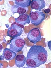
Preclinical results support clinical testing of an experimental agent in acute myeloid leukemia (AML) and myelodysplastic syndromes (MDS), according to researchers.
The agent, ALRN-6924, was shown to combat AML and MDS by restoring activity of the tumor-suppressing protein p53.
ALRN-6924 exhibited antileukemic activity in AML cells and mouse models of the disease, as well as in a patient with MDS and excess leukemic blasts who received the drug on a compassionate-use basis.
These results, published in Science Translational Medicine, have led to a phase 1 trial of ALRN-6924 in patients with AML or MDS.
ALRN-6924 was developed by Aileron Therapeutics Inc., to target p53 by inhibiting 2 naturally occurring proteins, MDMX and MDM2. Overexpression of these proteins inactivates p53, allowing cancer cells to multiply unchecked.
In the current study, researchers set out to confirm ALRN-6924’s mechanism of action and determine the efficacy of the drug in AML/MDS. This work was supported, in part, by grants from Aileron Therapeutics Inc., and the National Institutes of Health.
The researchers did find that ALRN-6924 targets both MDMX and MDM2, blocking their interaction with p53 in AML cells.
The team said ALRN-6924 inhibited proliferation by inducing cell-cycle arrest and apoptosis in AML cell lines and AML patient cells, including leukemic stem cell-enriched populations.
“This is important because AML is driven by stem cells, and, if you don’t target stem cells, the disease will come back very quickly,” said study author Ulrich Steidl, MD, PhD, of Albert Einstein College of Medicine in Bronx, New York.
The researchers also found that ALRN-6924 greatly increased survival in a mouse model of AML. The median survival was 34 days in vehicle-treated control mice, 83 days in mice that received ALRN-6924 at 20 mg/kg twice a week, and 151 days in mice that received ALRN-6924 at 20 mg/kg three times a week.
“This is a very striking response,” Dr Steidl said. “Most experimental drugs for leukemia achieve an increase in survival of only a few days in these preclinical models. Even more importantly, ALRN-6924 effectively cured about 40% of the treated mice, meaning they were disease-free more than 1 year after treatment, essentially a lifetime for a mouse.”
Finally, the researchers assessed the effects of ALRN-6924 in a patient who had high-risk MDS with excess leukemic blasts.
The team found the p53 protein “was rapidly induced” in CD34+ leukemic blasts but not in healthy lymphocytes. And ALRN-6924 reduced the number of malignant cells circulating in the blood.
“This test was not designed to assess the efficacy of the drug in humans,” Dr Steidl noted. “That has to be done in a proper clinical trial. Our goal was to determine whether it can hit the desired target in human cells in a clinical setting, which it did in this individual.”
ALRN-6924 is a stapled alpha-helical peptide, a class of drugs whose helical structure is stabilized using hydrocarbon “staples.” The stapling prevents the peptides from being degraded by enzymes before reaching their intended target. ALRN-6924 is the first stapled peptide therapeutic to be tested in patients.
In the phase 1 trial (NCT02909972), researchers are testing ALRN-6924 in patients with relapsed/refractory AML or advanced MDS.
Group identifies novel genes involved in MM development
Researchers have identified novel genes involved in the development of multiple myeloma (MM), according to a paper published in Leukemia.
The team’s analyses revealed regions of coding and non-coding DNA that appear to drive MM development.
The researchers analyzed whole-exome sequencing data from 804 MM patients and whole-genome sequencing data from 765 MM patients.
This revealed 16 novel genes that were disrupted in coding regions of DNA and 15 novel genes disrupted in non-coding regions.
There were 5 genes disrupted by structural variants in coding regions—CD96, PRDM1, FBXW7, MAP3K14, and CCND2.
There were also 11 genes disrupted by single nucleotide variants (SNVs) and indels in coding regions—BAX, C8orf86, FAM154B, FTL, HIST1H4H, LEMD2, PABPC1, RPN1, RPS3A, SGPP1, and TBC1D29.
Among the novel genes disrupted by mutations in non-coding regions was NBPF1, a promoter disrupted by SNVs. The researchers noted that NBPF1 is directly regulated by NF-κB, and the NF-κB pathway is recurrently affected in MM.
The team also identified 7 cis-regulatory elements (CREs) disrupted by SNVs—CALCB, COBLL1, HOXB3, ST6GAL1, PAX5, ATP13A2, and TPRG1.
The researchers said the SNVs in PAX5 and HOXB3 reduced gene expression, suggesting PAX5 and HOXB3 function as tumor suppressors in MM. On the other hand, the SNVs in ST6GAL1 increased gene expression, which may contribute to the aberrant immunoglobulin-G glycosylation seen in MM.
Finally, there were 7 CREs disrupted by copy number variations—MYC, PLD4, KDM3B, SP110, RAB36, PACS2, and TEX22.
The researchers noted that, with the exception of MYC, these genes reside close to regions of common structural variation, so their relevance in MM is not clear.
The team also said it’s well known that MYC is upregulated in MM through gene amplification or translocation, but this research shows that MYC can be dysregulated by alternative mechanisms.
“We need smarter, kinder treatments for myeloma that are more tailored to each person’s cancer,” said study author Richard Houlston, MD, PhD, of The Institute of Cancer Research in London, UK.
“Exhaustive genetic research like this is helping us to make that possible. Our findings should now open up new avenues for discovering treatments that target the genes driving myeloma.”
Generic antiemetic now available in US
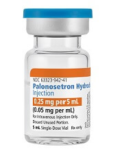
Palonosetron Hydrochloride Injection, a generic alternative to Aloxi®, is now available in the US.
Fresenius Kabi’s Palonosetron Hydrochloride Injection is a 5-HT3 serotonin receptor that is approved for the prevention of nausea and vomiting in certain adults.
Palonosetron Hydrochloride Injection is available in a single-dose vial (0.25 mg per 5 mL).
In the US, Palonosetron Hydrochloride Injection is approved for the prevention of acute and delayed nausea and vomiting associated with initial and repeat courses of moderately emetogenic cancer chemotherapy.
The drug is also approved for the prevention of acute nausea and vomiting associated with initial and repeat courses of highly emetogenic cancer chemotherapy.
And Palonosetron Hydrochloride Injection is approved for the prevention of post-operative nausea and vomiting for up to 24 hours after surgery. Efficacy beyond 24 hours has not been demonstrated.
The full prescribing information for Palonosetron Hydrochloride Injection can be found on the Fresenius Kabi website.
Lighter skin on left cheek
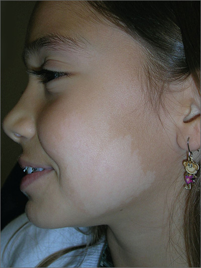
The FP made the diagnosis of nevus depigmentosus.
Nevus depigmentosus is usually present at birth or develops in early childhood. There is a decreased number of melanosomes within a normal number of melanocytes. The hypopigmented area typically has a serrated or jagged edge and has been compared to the appearance of a continent.
Nevus depigmentosus presents no danger or increased malignant potential. Excision, which is not an advisable option, is the only available treatment. Patients who want to camouflage the appearance of the hypopigmentation can use cover-up makeup.
Photos and text for Photo Rounds Friday courtesy of Richard P. Usatine, MD. This case was adapted from: Smith M, Usatine R. Benign nevi. In: Usatine R, Smith M, Mayeaux EJ, et al. Color Atlas of Family Medicine. 2nd ed. New York, NY: McGraw-Hill; 2013:945-952.
To learn more about the Color Atlas of Family Medicine, see: www.amazon.com/Color-Family-Medicine-Richard-Usatine/dp/0071769641/.
You can now get the second edition of the Color Atlas of Family Medicine as an app by clicking on this link: usatinemedia.com.
Rare, serious alemtuzumab adverse events emerge
according to three new case reports and series in Neurology.
The complications include acute coronary syndrome in one patient, hemophagocytic lymphohistiocytosis (HLH) in two patients, and acute acalculous cholecystitis in eight patients.
Postmarketing surveillance aims to identify rare and late adverse events that may not be discovered during clinical trials.
The cardiac arrest incident occurred in a 24-year old woman with spinal-onset relapsing-remitting multiple sclerosis (RRMS) during alemtuzumab infusion. She had experienced two clinical relapses and a significant increase in cerebral and spinal MRI lesion load, despite previous treatment with natalizumab (Tysabri). The only striking thing about her medical history was allergic asthma.
On the third day of treatment, she developed severe asymptomatic sinus bradycardia (HR, 38 beats per minute), which resolved with treatment, and clinicians continued the regimen. The following morning, she experienced acute coronary syndrome, which later resolved.
The authors recommend that clinicians treating RRMS patients with alemtuzumab consider a baseline preinfusion ECG and monitoring heart rate at least hourly while infusion is being carried out.
The HLH cases occurred in a man and a woman with RRMS, both in their 20s. The woman was switched from natalizumab to alemtuzumab because of a high anti-JC virus antibody index. Both cases presented in the time frame typical of secondary autoimmunity. The female patient had an acalculous gallbladder with thickened walls and pericholecystic fluid and responded to intravenous antibiotics, but 3 weeks later she developed thrombocytopenia, coagulopathy, and anemia with abnormal liver enzymes. Treatment failed and she succumbed to the condition.
The man received alemtuzumab as first-line therapy for RRMS during the Keratinocyte Growth Factor to Prevent Autoimmunity After Alemtuzumab Treatment of Multiple Sclerosis (CAM-THY) clinical trial. He received boluses of either placebo or palifermin (Kepivance) before and after each alemtuzumab dose in order to reduce secondary autoimmunity. At 30 months after his initial cycle of alemtuzumab, he was admitted to the hospital with HLH and also developed acquired factor VIII hemophilia. He responded positively to 4 months of corticosteroid treatment combined with two doses of rituximab (Rituxan) and ultimately returned to work. However, he experienced a relapse of acquired hemophilia during later doses of corticosteroids and rituximab.
A search of the Food and Drug Administration Adverse Event Reporting System (FAERS) revealed eight cases of acute acalculous cholecystitis (AAC) – four assessed as possibly caused by alemtuzumab, and four assessed as probable – that occurred in patients with RRMS. Seven of the cases involved presentation during treatment or soon afterward, which suggests that acute cytokine release syndrome (ACRS) may be the cause. The cases did not share all of the manifestations of typical ACRS, but the researchers noted that coadministration of methylprednisolone in the RRMS dosing schedule may have masked some of the features.
Unlike typical AAC, these cases occurred primarily in females (six of eight) and were not associated with concurrent critical illnesses. Other risk factors were not consistently present. Historically, AAC is associated with treatment of older male patients in the ICU. Three of the patients underwent surgical treatment, and five received conservative treatment of antibiotics or an unspecified treatment. Seven patients recovered. The outcome in the eighth patient was not reported.
Despite these good outcomes, the authors of the report note that longer-term outcomes in this AAC population are not yet understood.
The findings led to the addition of AAC to the Warnings and Precautions section of the alemtuzumab label in October 2017.
None of the reports received funding. Some of the authors had financial ties to the pharmaceutical industry.
SOURCES: Croteau D et al. Neurology. 2018 Mar 30. doi: 10.1212/WNL.0000000000005422. Saarela M et al. Neurology. 2018 Mar 30. doi: 10.1212/WNL.0000000000005420. Ferraro D et al. Neurology. 2018 Mar 30. doi: 10.1212/WNL.0000000000005417.
Clinical trials find most adverse events for a new drug, but postmarketing surveillance is essential to find rare events, even though they may underestimate the frequency of these events because of underreporting.
Other reports suggest the emergence of uncommon infectious complications associated with alemtuzumab, especially Listeria meningitis. Listeria infection can occur within days of alemtuzumab treatment, and the use of preventive sulfamethoxazole-trimethoprim treatment is now advocated in the United Kingdom.
Overall, these adverse events remind clinicians that they must balance the benefits of alemtuzumab, which is highly effective, against its potential risks.
Paolo A. Muraro, MD, PhD is a professor of neurology, neuroimmunology, and immunotherapy at Imperial College London. Neil J. Scolding MD, PhD, is director of the Bristol Institute of Clinical Neurosciences at the University of Bristol (England). Robert J. Fox, MD, is a neurologist at the Mellen Center for Multiple Sclerosis at the Cleveland Clinic in Ohio. All of the authors reported financial relationships with companies marketing MS drugs. Their comments are derived from an editorial accompanying the adverse event reports (Neurology. 2018 Mar 30. doi: 10.1212/WNL.0000000000005409).
according to three new case reports and series in Neurology.
The complications include acute coronary syndrome in one patient, hemophagocytic lymphohistiocytosis (HLH) in two patients, and acute acalculous cholecystitis in eight patients.
Postmarketing surveillance aims to identify rare and late adverse events that may not be discovered during clinical trials.
The cardiac arrest incident occurred in a 24-year old woman with spinal-onset relapsing-remitting multiple sclerosis (RRMS) during alemtuzumab infusion. She had experienced two clinical relapses and a significant increase in cerebral and spinal MRI lesion load, despite previous treatment with natalizumab (Tysabri). The only striking thing about her medical history was allergic asthma.
On the third day of treatment, she developed severe asymptomatic sinus bradycardia (HR, 38 beats per minute), which resolved with treatment, and clinicians continued the regimen. The following morning, she experienced acute coronary syndrome, which later resolved.
The authors recommend that clinicians treating RRMS patients with alemtuzumab consider a baseline preinfusion ECG and monitoring heart rate at least hourly while infusion is being carried out.
The HLH cases occurred in a man and a woman with RRMS, both in their 20s. The woman was switched from natalizumab to alemtuzumab because of a high anti-JC virus antibody index. Both cases presented in the time frame typical of secondary autoimmunity. The female patient had an acalculous gallbladder with thickened walls and pericholecystic fluid and responded to intravenous antibiotics, but 3 weeks later she developed thrombocytopenia, coagulopathy, and anemia with abnormal liver enzymes. Treatment failed and she succumbed to the condition.
The man received alemtuzumab as first-line therapy for RRMS during the Keratinocyte Growth Factor to Prevent Autoimmunity After Alemtuzumab Treatment of Multiple Sclerosis (CAM-THY) clinical trial. He received boluses of either placebo or palifermin (Kepivance) before and after each alemtuzumab dose in order to reduce secondary autoimmunity. At 30 months after his initial cycle of alemtuzumab, he was admitted to the hospital with HLH and also developed acquired factor VIII hemophilia. He responded positively to 4 months of corticosteroid treatment combined with two doses of rituximab (Rituxan) and ultimately returned to work. However, he experienced a relapse of acquired hemophilia during later doses of corticosteroids and rituximab.
A search of the Food and Drug Administration Adverse Event Reporting System (FAERS) revealed eight cases of acute acalculous cholecystitis (AAC) – four assessed as possibly caused by alemtuzumab, and four assessed as probable – that occurred in patients with RRMS. Seven of the cases involved presentation during treatment or soon afterward, which suggests that acute cytokine release syndrome (ACRS) may be the cause. The cases did not share all of the manifestations of typical ACRS, but the researchers noted that coadministration of methylprednisolone in the RRMS dosing schedule may have masked some of the features.
Unlike typical AAC, these cases occurred primarily in females (six of eight) and were not associated with concurrent critical illnesses. Other risk factors were not consistently present. Historically, AAC is associated with treatment of older male patients in the ICU. Three of the patients underwent surgical treatment, and five received conservative treatment of antibiotics or an unspecified treatment. Seven patients recovered. The outcome in the eighth patient was not reported.
Despite these good outcomes, the authors of the report note that longer-term outcomes in this AAC population are not yet understood.
The findings led to the addition of AAC to the Warnings and Precautions section of the alemtuzumab label in October 2017.
None of the reports received funding. Some of the authors had financial ties to the pharmaceutical industry.
SOURCES: Croteau D et al. Neurology. 2018 Mar 30. doi: 10.1212/WNL.0000000000005422. Saarela M et al. Neurology. 2018 Mar 30. doi: 10.1212/WNL.0000000000005420. Ferraro D et al. Neurology. 2018 Mar 30. doi: 10.1212/WNL.0000000000005417.
FROM NEUROLOGY
Key clinical point: Rare events associated with alemtuzumab are emerging that were not uncovered during clinical trials.
Major finding: Eight cases of acute acalculous cholecystitis formed the basis for its addition to the Warnings and Precautions section of the alemtuzumab label in October 2017.
Study details: Case reports of acute coronary syndrome in one patient, hemophagocytic lymphohistiocytosis in two patients, and acute acalculous cholecystitis in eight patients.
Disclosures: None of the reports received funding. Some of the authors had financial ties to the pharmaceutical industry.
Sources: Croteau D et al. Neurology. 2018 Mar 30. doi: 10.1212/WNL.0000000000005422; Saarela M et al. Neurology. 2018 Mar 30. doi: 10.1212/WNL.0000000000005420; Ferraro D et al. Neurology. 2018 Mar 30. doi: 10.1212/WNL.0000000000005417.
This month in the journal CHEST®
Giants In Chest Medicine
Professor Emeritus Elizabeth F. Juniper, MCSP, MSc
By Dr. P. M. O’Byrne
Original Research
A Population-Based Cohort Study on the Drug-Specific Effect of Statins on Sepsis Outcome. 
A Multicenter Randomized Trial of a Checklist for Endotracheal Intubation of Critically Ill Adults.
By Dr. D. R. Janz, et al.
Determinants of Unintentional Leaks During CPAP Treatment in OSA.
By Dr. M. Lebret, et al.
Evidence-Based Medicine
Screening for Lung Cancer: CHEST Guideline and Expert Panel Report.
By Dr. P. J. Mazzone, et al.
Treating Cough Due to Non-CF and CF Bronchiectasis With Nonpharmacological Airway Clearance: CHEST Expert Panel Report.
By Dr. A. T. Hill, et al.
Giants In Chest Medicine
Professor Emeritus Elizabeth F. Juniper, MCSP, MSc
By Dr. P. M. O’Byrne
Original Research
A Population-Based Cohort Study on the Drug-Specific Effect of Statins on Sepsis Outcome.
A Multicenter Randomized Trial of a Checklist for Endotracheal Intubation of Critically Ill Adults.
By Dr. D. R. Janz, et al.
Determinants of Unintentional Leaks During CPAP Treatment in OSA.
By Dr. M. Lebret, et al.
Evidence-Based Medicine
Screening for Lung Cancer: CHEST Guideline and Expert Panel Report.
By Dr. P. J. Mazzone, et al.
Treating Cough Due to Non-CF and CF Bronchiectasis With Nonpharmacological Airway Clearance: CHEST Expert Panel Report.
By Dr. A. T. Hill, et al.
SAVE LIVES: Clean your hands
The World Health Organization (WHO) has announced its annual SAVE LIVES: Clean Your Hands 2018 campaign (Saito, et al. J Hosp Infect. 2018;98[4]:321), designating May 5, 2018, as world hand hygiene day.
Health-care-associated infections are a major patient safety problem. Unfortunately, their spread is common in hospitals and ICUs around the globe. The vehicle for these infections, including multidrug-resistant organisms, is frequently the contaminated hands of health-care workers. Health-care-acquired infections, as any other infection, can lead to sepsis and death. Infections acquired in the ICU are especially deadly, with mortalities that can be as high as 80%. Proper hand hygiene, despite being simple and inexpensive, is the single most important means of reducing the prevalence of hospital-acquired infections and the spread of antimicrobial resistance.
We have known about the significance of hand washing since the early 19th century. More recent data show that hand washing can reduce the overall prevalence of hospital-acquired infections and the cross-transmission of multidrug-resistant organisms. It is estimated that we can prevent 15% to 30% of these infections with adequate hand washing alone.
Despite the clear benefit and the understanding of the importance of hand washing, compliance with this simple intervention is only about 50%. Health-care workers tend to overestimate these rates, self-reporting a compliance of 75%. Even the latter number represents a lot of missed opportunities, and we must do something about it.
A multifaceted approach that combines education with written material, reminders, and continued feedback on performance can have an important effect on hand washing compliance and rates of hospital-acquired infections.
Sepsis is the single most important cause of death in hospitals in the United States. The campaign (http://www.who.int/infection-prevention/campaigns/clean-hands/en/), sponsored by the World Health Organization, should serve as a reminder to all health-care workers about the importance of adequate hand washing and as an opportunity to improve our compliance moving forward.
Despite the progress made, there is still a lot of room for improvement. We can have an impact on the number of deaths from sepsis by preventing them to occur in the first place. Wash your hands and do it well, it does not cost us anything.
Remember: It is in our hands – prevent sepsis and save lives!
Shruti Gadre, MD
Steering Committee Member, Critical Care NetWork
Angel Coz, MD, FCCP
Chair, Critical Care NetWork
AMA Insights
As many who read CHEST® Physician may know, we have a nucleus of dedicated volunteers who give unselfishly of their time and talent to represent our members in the area of “regulatory advocacy” and “policy advocacy” in the areas of pulmonary, critical care, and sleep medicine. It is our goal to recognize and support this valuable group of individuals who represent us in the space of coding and reimbursement, RUC activities, relationships with organizations like the ACP and the AMA, as well as our sister societies, such as ATS, SCCM, NAMDRC, CCNA, APSR, ALAT, and ERS, among others.
One of our goals, in addition to recognizing this group, is to identify and mentor the next generation of representatives. A great example of this mentorship is reflected in our involvement with the AMA. Dr. Bob McCaffree has represented CHEST for 22 years and is now mentoring Dr. Raj Desai who will be assuming this role of AMA Delegate this year. Special thanks to Dr. McCaffree for his unselfish service in this capacity and for his mentorship of Dr. Desai. I hope that you enjoy this and future CHEST® Physician articles summarizing and reflecting on the activities pertinent to CHEST at the AMA.
John Studdard, MD, FCCP
CHEST President
Collaborating with societies: CHEST and AMA
While the American Medical Association (AMA) is the oldest and largest national medical association, many physicians, both members and nonmembers, have limited understanding of the policies, processes, and strategic foci of the AMA. It is our goal to inform our membership about the workings of the AMA and how those interact with the goals of CHEST and our members. We hope to do this by publishing periodic articles in CHEST® Physician. One of the authors (DRM) has been the CHEST delegate to the AMA for more than 20 years, and the other (NRD) is CHEST’s new delegate.

- Create thriving physician practices.
- Create the medical school of the future.
- Improve health outcomes.
We will expand on these in future articles.
The AMA is both an individual member organization and a federation of geographic, ie, county and state, societies and specialty societies, as well as the uniformed services and the VA. It is this federation that comprises the House of Delegates (HOD or House), which is the principle policy-making body of the AMA. The number of delegates from each member organization (now numbering more than 170 organizations) depends on the number of individual AMA members among that organization’s members. Due to recent bylaws changes, CHEST now has two delegates. The HOD meets twice per year to establish policy on health, medical, professional, and governance matters, as well as the principles within which the AMA’s business activities are conducted.

Most member societies meet in caucuses or Section Councils prior to the voting in the House to discuss the pending business. The Specialty and Service Society (SSS) is the largest caucus in the AMA’s House of Delegates. The SSS meets twice annually in conjunction with the Interim and Annual Meetings of the HOD. There are two categories of groups in the SSS: those societies that have seats in the HOD and those seeking admission to the house.
SSS groups in the HOD include:
- 119 national medical specialties
- 2 professional interest medical associations
- 5 military service groups
An association must first be represented in the SSS for 3 years and meet the required number of AMA members before it is eligible to seek admission to the HOD.
The American College of Chest Physicians (CHEST) is an active member of the SSS but also joins with other societies of similar interests in the Section Council on Chest and Allergic Diseases. This caucus includes the ATS, SCCM, ASSM, and several allergy societies. Through the HOD, the SSS, and the Section Council, CHEST can partner with the AMA and other societies, such as ATS, to support each other’s resolutions or important regulatory issues.
In summary, the AMA plays an important role in many areas of interest to our members. And, it can be a useful forum for connecting with societies with similar interests in directing advocacy and setting policy. We plan to continue this update in future issues of CHEST® Physician.
References
1. https://www.ama-assn.org/content/ama-house-delegates Accessed: January 28, 2018
2. https://www.ama-assn.org/practice-management/ama-steps-forward-practice-improvement-strategies Accessed: January 28, 2018
As many who read CHEST® Physician may know, we have a nucleus of dedicated volunteers who give unselfishly of their time and talent to represent our members in the area of “regulatory advocacy” and “policy advocacy” in the areas of pulmonary, critical care, and sleep medicine. It is our goal to recognize and support this valuable group of individuals who represent us in the space of coding and reimbursement, RUC activities, relationships with organizations like the ACP and the AMA, as well as our sister societies, such as ATS, SCCM, NAMDRC, CCNA, APSR, ALAT, and ERS, among others.
One of our goals, in addition to recognizing this group, is to identify and mentor the next generation of representatives. A great example of this mentorship is reflected in our involvement with the AMA. Dr. Bob McCaffree has represented CHEST for 22 years and is now mentoring Dr. Raj Desai who will be assuming this role of AMA Delegate this year. Special thanks to Dr. McCaffree for his unselfish service in this capacity and for his mentorship of Dr. Desai. I hope that you enjoy this and future CHEST® Physician articles summarizing and reflecting on the activities pertinent to CHEST at the AMA.
John Studdard, MD, FCCP
CHEST President
Collaborating with societies: CHEST and AMA
While the American Medical Association (AMA) is the oldest and largest national medical association, many physicians, both members and nonmembers, have limited understanding of the policies, processes, and strategic foci of the AMA. It is our goal to inform our membership about the workings of the AMA and how those interact with the goals of CHEST and our members. We hope to do this by publishing periodic articles in CHEST® Physician. One of the authors (DRM) has been the CHEST delegate to the AMA for more than 20 years, and the other (NRD) is CHEST’s new delegate.
- Create thriving physician practices.
- Create the medical school of the future.
- Improve health outcomes.
We will expand on these in future articles.
The AMA is both an individual member organization and a federation of geographic, ie, county and state, societies and specialty societies, as well as the uniformed services and the VA. It is this federation that comprises the House of Delegates (HOD or House), which is the principle policy-making body of the AMA. The number of delegates from each member organization (now numbering more than 170 organizations) depends on the number of individual AMA members among that organization’s members. Due to recent bylaws changes, CHEST now has two delegates. The HOD meets twice per year to establish policy on health, medical, professional, and governance matters, as well as the principles within which the AMA’s business activities are conducted.
Most member societies meet in caucuses or Section Councils prior to the voting in the House to discuss the pending business. The Specialty and Service Society (SSS) is the largest caucus in the AMA’s House of Delegates. The SSS meets twice annually in conjunction with the Interim and Annual Meetings of the HOD. There are two categories of groups in the SSS: those societies that have seats in the HOD and those seeking admission to the house.
SSS groups in the HOD include:
- 119 national medical specialties
- 2 professional interest medical associations
- 5 military service groups
An association must first be represented in the SSS for 3 years and meet the required number of AMA members before it is eligible to seek admission to the HOD.
The American College of Chest Physicians (CHEST) is an active member of the SSS but also joins with other societies of similar interests in the Section Council on Chest and Allergic Diseases. This caucus includes the ATS, SCCM, ASSM, and several allergy societies. Through the HOD, the SSS, and the Section Council, CHEST can partner with the AMA and other societies, such as ATS, to support each other’s resolutions or important regulatory issues.
In summary, the AMA plays an important role in many areas of interest to our members. And, it can be a useful forum for connecting with societies with similar interests in directing advocacy and setting policy. We plan to continue this update in future issues of CHEST® Physician.
References
1. https://www.ama-assn.org/content/ama-house-delegates Accessed: January 28, 2018
2. https://www.ama-assn.org/practice-management/ama-steps-forward-practice-improvement-strategies Accessed: January 28, 2018
Zika virus: Sexual contact risk may be limited to short window
Shedding of infectious Zika virus in the semen of symptomatic infected men appears to be uncommon and limited to the first few weeks after onset of illness, according to results of a recent prospective study.
Out of all semen samples with detectable Zika virus RNA, the only ones with infectious virus were those that had been obtained within 30 days of illness onset, study authors reported in the New England Journal of Medicine.
Sexual transmission of Zika virus, first documented in 2011, has now been reported in at least 13 countries, Dr. Mead and his colleagues wrote.
Usually, the cases have involved transmission from a symptomatic man to a woman, they added.
Previously, some investigators had proposed that sexual transmission of Zika virus could pose a greater risk of fetal infection than could mosquito-borne transmission, Dr. Mead and colleagues noted in their report. “If so, the interruption of sexual transmission could play a critical role in preventing the serious complications that have been associated with fetal infection,” they wrote.
To investigate further, Dr. Mead and his colleagues conducted a prospective study of men with symptomatic Zika virus infection. They collected 1,327 semen samples from 184 men and 1,038 urine samples from 183 men, according to the report.
They obtained specimens twice monthly for 6 months. Samples were tested for Zika RNA using real-time reverse transcriptase polymerase chain reaction assay and for infectious Zika virus using Vero cell culture and plaque assay.
Investigators detected Zika virus RNA in the semen of 60 men (33%), including semen samples from 22 of the 36 men (61%) tested within 30 days after illness onset, investigators said in the report.
While Zika virus RNA shedding decreased considerably in the 3 months after illness onset, it did continue for 281 days in one man, they noted.
Men who were older and those who ejaculated less frequently were more likely to have prolonged RNA shedding in semen, results of multivariable analysis showed.
Infectious Zika virus was isolated from just 3 out of the 78 semen samples with detectable Zika virus RNA that were tested by culture, investigators said. Notably, all 3 of the cases were among the 19 of those samples obtained within 30 days of illness onset, they reported.
Detection of Zika virus RNA in urine was rare, occurring in only 7 men (4%), possibly because of the timing of the first specimen collection, according to investigators. They said previous studies suggest a rapid decline in Zika virus shedding in urine during the first few weeks after onset of illness.
Important questions remain regarding sexual transmission of Zika virus, such as whether maternal infection through sex poses similar risks to the fetus as compared with maternal infection via mosquito bite, Dr. Mead and his coauthors said in the report.
“A better understanding of these issues is needed to guide the development of effective prevention strategies,” they wrote.
The study was supported by the Centers for Disease Control and Prevention. Dr. Mead and his coauthors reported they had no disclosures related to the study.
SOURCE: Mead PS et al. N Engl J Med. 2018;378(15):1377-85.
This study illustrates the apparent shortcomings of current virus-detection standards in terms of their relevance to public health, according to Heinz Feldmann, MD.
Approximately 4% of Zika virus RNA-positive semen samples were infectious, according to the report, and of those infectious samples, all were obtained within 30 days of the onset of illness. “This finding suggests that there is a short period during which Zika virus–infected men might transmit this virus through sexual contact,” Dr. Feldmann wrote in an editorial.
Current practice in some areas is to test semen samples sequentially until two or more consecutive negative results are obtained; however, that approach is controversial, according to Dr. Feldmann, because the person could be shedding the virus intermittently because of the potential for virus latency and reactivation.
“This also raises the question of whether modern molecular approaches are properly positioned to detect virus latency rather than persistence,” he said in his editorial. The goal, he added, should be to determine infectivity, which is probably best assessed by means of viral isolation – which is believed to be less sensitive than molecular detection.
“Thus, the diagnostic situation is far more complicated than it seems,” he noted.
However, he added, these diagnostic scenarios may be less applicable for public health entities, which have “quickly” disseminated recommendations for safer sex to prevent Zika virus spread and the potentially devastating consequences of fetal infection.
“These recommendations leverage the best data available and have been implemented, but ought to be updated as new data emerge,” Dr. Feldmann wrote.
Dr. Feldmann is with the National Institute of Allergy and Infectious Diseases, National Institutes of Health, and Rocky Mountain Laboratories, Hamilton, Mont. These comments are derived from his editorial N Engl J Med 2018;378:1377-85 . Dr. Feldmann reported that he had nothing to disclose related to the editorial.
Shedding of infectious Zika virus in the semen of symptomatic infected men appears to be uncommon and limited to the first few weeks after onset of illness, according to results of a recent prospective study.
Out of all semen samples with detectable Zika virus RNA, the only ones with infectious virus were those that had been obtained within 30 days of illness onset, study authors reported in the New England Journal of Medicine.
Sexual transmission of Zika virus, first documented in 2011, has now been reported in at least 13 countries, Dr. Mead and his colleagues wrote.
Usually, the cases have involved transmission from a symptomatic man to a woman, they added.
Previously, some investigators had proposed that sexual transmission of Zika virus could pose a greater risk of fetal infection than could mosquito-borne transmission, Dr. Mead and colleagues noted in their report. “If so, the interruption of sexual transmission could play a critical role in preventing the serious complications that have been associated with fetal infection,” they wrote.
To investigate further, Dr. Mead and his colleagues conducted a prospective study of men with symptomatic Zika virus infection. They collected 1,327 semen samples from 184 men and 1,038 urine samples from 183 men, according to the report.
They obtained specimens twice monthly for 6 months. Samples were tested for Zika RNA using real-time reverse transcriptase polymerase chain reaction assay and for infectious Zika virus using Vero cell culture and plaque assay.
Investigators detected Zika virus RNA in the semen of 60 men (33%), including semen samples from 22 of the 36 men (61%) tested within 30 days after illness onset, investigators said in the report.
While Zika virus RNA shedding decreased considerably in the 3 months after illness onset, it did continue for 281 days in one man, they noted.
Men who were older and those who ejaculated less frequently were more likely to have prolonged RNA shedding in semen, results of multivariable analysis showed.
Infectious Zika virus was isolated from just 3 out of the 78 semen samples with detectable Zika virus RNA that were tested by culture, investigators said. Notably, all 3 of the cases were among the 19 of those samples obtained within 30 days of illness onset, they reported.
Detection of Zika virus RNA in urine was rare, occurring in only 7 men (4%), possibly because of the timing of the first specimen collection, according to investigators. They said previous studies suggest a rapid decline in Zika virus shedding in urine during the first few weeks after onset of illness.
Important questions remain regarding sexual transmission of Zika virus, such as whether maternal infection through sex poses similar risks to the fetus as compared with maternal infection via mosquito bite, Dr. Mead and his coauthors said in the report.
“A better understanding of these issues is needed to guide the development of effective prevention strategies,” they wrote.
The study was supported by the Centers for Disease Control and Prevention. Dr. Mead and his coauthors reported they had no disclosures related to the study.
SOURCE: Mead PS et al. N Engl J Med. 2018;378(15):1377-85.
FROM THE NEW ENGLAND JOURNAL OF MEDICINE
Key clinical point: While Zika virus RNA is common and may persist for months in the semen of symptomatic infected men, shedding of infectious virus appears to be much less common and limited to the first few weeks after onset of illness.
Major finding: Out of 78 semen samples with detectable Zika virus RNA, 3 had infectious virus, and all 3 were obtained within 30 days of illness onset.
Study details: A prospective study of 1,327 semen samples from 184 men with symptomatic Zika virus infection.
Disclosures: The study was supported by the Centers for Disease Control and Prevention. Study authors reported they had nothing to disclose relative to the study.
Source: Mead PS et al. N Engl J Med. 2018 Apr 12;378(15):1377-85.

New genetic subtypes could facilitate precision medicine in DLBCL
Four genetic subtypes of diffuse large B-cell lymphoma (DLBCL) showed multiple distinct mutations, gene expression signatures, and treatment responses, researchers reported.
The findings “may provide a conceptual edifice on which to develop precision therapies for these aggressive cancers,” Roland Schmitz, PhD, and his associates wrote in the New England Journal of Medicine.
Other DLBCL studies have focused on individual mutations, but therapeutic response probably hinges on “constellations of genetic aberrations,” wrote Dr. Schmitz of the National Cancer Institute and his associates.
Therefore, they used exome and transcriptome sequencing, deep amplicon resequencing of 372 genes, and DNA copy-number analysis to analyze 572 fresh-frozen DLBCL biopsy specimens, nearly all of which were treatment-naïve.
This multiplatform approach yielded four genetic subtypes: MCD, so named for its co-occurring MYD88L265P and CD79B mutations; BN2, which has BCL6 fusions and NOTCH2 mutations; N1, which has NOTCH1 mutations; and EZB, which has EZH2 mutations and BCL2 translocations. Most MCD and N1 specimens were activated B-cell–like (ABC) tumors, EZB specimens were primarily germinal-center B-cell–like (GCB) tumors, and BN2 specimens included ABC, GCB, and unclassified cases.
A closer look at 119 previously untreated patients linked genetic subtypes with significant differences in progression-free survival (P less than .0001) and overall survival (P = .0002) following R-CHOP or CHOP-like chemotherapy.
The BN2 and EZB subtypes “[had] much more favorable outcomes than the MCD and N1 subtypes,” Dr. Schmitz and his associates said. “Analysis of genetic pathways suggested that MCD and BN2 DLBCLs rely on ‘chronic active’ B-cell receptor signaling that is amenable to therapeutic inhibition.”
Genetically subtyping DLBCL could help guide patients into appropriate clinical trials, the investigators wrote. For example, patients with the N1 subtype might be candidates for immune checkpoint inhibitor therapy, given N1’s prominent T-cell gene expression and poor response to R-CHOP.
Funders included the National Institutes of Health, the National Cancer Institute, the Dr. Mildred Scheel Stiftung fur Krebsforschung (Deutsche Krebshilfe), the Washington University in St. Louis, and the Kay Kendall Leukaemia Fund. Dr. Schmitz disclosed research funding from Dr. Mildred Scheel Stiftung fur Krebsforschung (Deutsche Krebshilfe).
SOURCE: Schmitz et al. New Eng J Med. 2018 Apr 11. doi: 10.1056/NEJMoa1801445.
FROM NEJM
Key clinical point: Multiplatform analyses identified four new genetic subtypes of DLBCL.
Major finding: The subtypes were distinguishable based on multiple genetic aberrations, phenotypes, and treatment responses.
Study details: Study of 574 DLBCL samples using exome and transcriptome sequencing, array-based DNA copy-number analysis, and targeted amplicon resequencing of 372 genes.
Disclosures: Funders included the National Institutes of Health, the National Cancer Institute, the Dr. Mildred Scheel Stiftung fur Krebsforschung (Deutsche Krebshilfe), the Washington University in St. Louis, and the Kay Kendall Leukaemia Fund. Dr. Schmitz disclosed research funding from Dr. Mildred Scheel Stiftung fur Krebsforschung (Deutsche Krebshilfe).
Source: Schmitz et al. New Eng J Med. 2018 Apr 11. doi: 10.1056/NEJMoa1801445.