User login
2 in 1: Rosacea-like demodicosis, papulopustular rosacea may be phenotypes of same disease
according to the authors of a retrospective study.
Currently, there is a lack of consensus among physicians and health care organizations on how Demodex mites affect the development of rosacea. Many experts separate rosacea into two camps: PPR not caused by Demodex and rosacea-like demodicosis caused by Demodex.
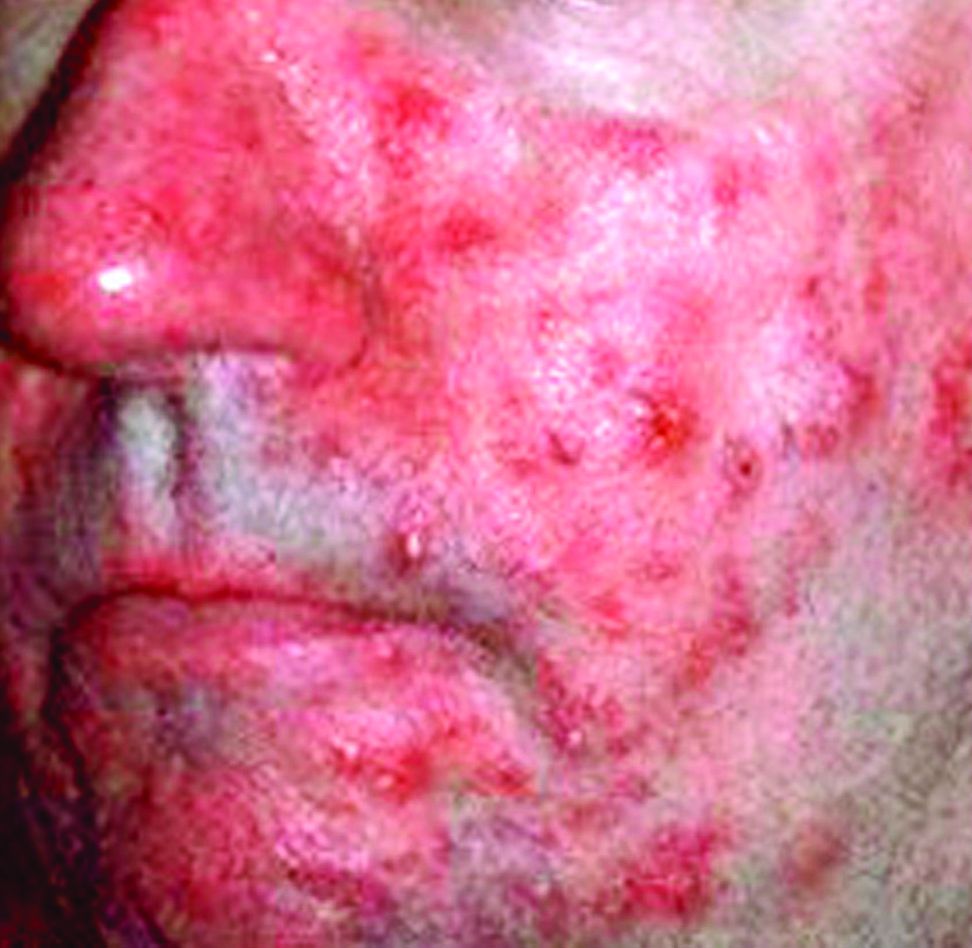
During each evaluation session, each patient underwent two consecutive standardized skin surface biopsies (SSSBs), a small 1 square centimeter sample of the horny skin layer and the follicular content, on each cheek. The first sample, SSSB1, was a superficial sample, and SSSB2, the second sample, was a deep sample. The sum of the two samples, SSSB1+2, also was noted.
During the same session, patients had Demodex densities (Dds) measured. To avoid any confounding factors that could affect facial skin symptoms, Dr. Forton and Dr. de Maertelaer evaluated a subgroup of 132 patients who had not been treated in the previous 3 months and had no other facial dermatoses, such as acne vulgaris and seborrheic dermatitis.
The study revealed that, among the 242 patients in the primary analysis group, those with persistent erythema had higher Dds than did those without and the differences were statistically significant when comparing SSSB2 (208 D/cm2 vs. 130 D/cm2; P = 0.031) and SSB1+2 (298 D/cm2 vs. 191 D/cm2; P = 0.025), respectively.
This pattern of greater Dds density among patients with persistent erythema, compared with patients without, also held true among the subgroup of 132 patients who had not received any recent dermatological treatments, but the difference was not statistically significant. Nonetheless, patients with follicular scales did have greater SSSBs than did those without follicular scales.
As part of the study, Dr. Forton and Dr. de Maertelaer analyzed the findings of a case report of a 19-year-old woman who had a facial papulopustular eruption that had been present for 1 year. She had two SSSBs taken on each cheek: the right had follicular scales and papulopustules, and the left was clinically normal. This revealed that she had much higher Dds on the affected cheek than on the clinically normal one (108 and 216 D/cm2 vs. 12 and 20 D/cm2). She was subsequently diagnosed with rosacea-like demodicosis.
After treating the areas with acaricidal ointment, the symptoms improved. But 27 months after stopping maintenance treatments, the facial eruptions reappeared, and the papulopustules were larger than during her original consultation, leading to the diagnosis of PPR. This time, the Dds was high on both cheeks but responded to acaricidal treatment, which indicates that her eruptions were caused Demodex mites.
“All our observations, therefore, highlight the nosological confusion that persists between PPR and rosacea-like demodicosis and the need to update the consensus concerning the definition and classification of rosacea. Moreover, they suggest that PPR and rosacea-like demodicosis may be phenotypes of the same disease,” wrote Dr. Forton and Dr. de Maertelaer. “This concept is supported by our case report, with many features indicating that the second presentation was an evolution of the first.”
They cautioned that their research is not definitive but should provide strong evidence that PPR and rosacea-like demodicosis are the same disease.
“While our observations do not prove a causative role of Demodex in rosacea, they nevertheless support the idea that PPR and rosacea-like demodicosis should no longer be considered as two separate entities but, rather, as two phenotypes of the same disease,” they wrote. “As such, the definition of rosacea subtype II (PPR) should be reconsidered and simplified to include all patients with central face papulopustules – with or without persistent erythema – and thus also patients with ‘rosacea-like demodicosis,’ which is a term that should therefore disappear.”
This study received no external funding. Dr. Forton works for Galderma as a consultant. Dr. de Maertelaer had no conflicts of interest to declare.
SOURCE: Forton F et al. J Eur Acad Dermatol Venereol. 2018 Feb 25. doi: 10.1111/jdv.14885.
according to the authors of a retrospective study.
Currently, there is a lack of consensus among physicians and health care organizations on how Demodex mites affect the development of rosacea. Many experts separate rosacea into two camps: PPR not caused by Demodex and rosacea-like demodicosis caused by Demodex.
During each evaluation session, each patient underwent two consecutive standardized skin surface biopsies (SSSBs), a small 1 square centimeter sample of the horny skin layer and the follicular content, on each cheek. The first sample, SSSB1, was a superficial sample, and SSSB2, the second sample, was a deep sample. The sum of the two samples, SSSB1+2, also was noted.
During the same session, patients had Demodex densities (Dds) measured. To avoid any confounding factors that could affect facial skin symptoms, Dr. Forton and Dr. de Maertelaer evaluated a subgroup of 132 patients who had not been treated in the previous 3 months and had no other facial dermatoses, such as acne vulgaris and seborrheic dermatitis.
The study revealed that, among the 242 patients in the primary analysis group, those with persistent erythema had higher Dds than did those without and the differences were statistically significant when comparing SSSB2 (208 D/cm2 vs. 130 D/cm2; P = 0.031) and SSB1+2 (298 D/cm2 vs. 191 D/cm2; P = 0.025), respectively.
This pattern of greater Dds density among patients with persistent erythema, compared with patients without, also held true among the subgroup of 132 patients who had not received any recent dermatological treatments, but the difference was not statistically significant. Nonetheless, patients with follicular scales did have greater SSSBs than did those without follicular scales.
As part of the study, Dr. Forton and Dr. de Maertelaer analyzed the findings of a case report of a 19-year-old woman who had a facial papulopustular eruption that had been present for 1 year. She had two SSSBs taken on each cheek: the right had follicular scales and papulopustules, and the left was clinically normal. This revealed that she had much higher Dds on the affected cheek than on the clinically normal one (108 and 216 D/cm2 vs. 12 and 20 D/cm2). She was subsequently diagnosed with rosacea-like demodicosis.
After treating the areas with acaricidal ointment, the symptoms improved. But 27 months after stopping maintenance treatments, the facial eruptions reappeared, and the papulopustules were larger than during her original consultation, leading to the diagnosis of PPR. This time, the Dds was high on both cheeks but responded to acaricidal treatment, which indicates that her eruptions were caused Demodex mites.
“All our observations, therefore, highlight the nosological confusion that persists between PPR and rosacea-like demodicosis and the need to update the consensus concerning the definition and classification of rosacea. Moreover, they suggest that PPR and rosacea-like demodicosis may be phenotypes of the same disease,” wrote Dr. Forton and Dr. de Maertelaer. “This concept is supported by our case report, with many features indicating that the second presentation was an evolution of the first.”
They cautioned that their research is not definitive but should provide strong evidence that PPR and rosacea-like demodicosis are the same disease.
“While our observations do not prove a causative role of Demodex in rosacea, they nevertheless support the idea that PPR and rosacea-like demodicosis should no longer be considered as two separate entities but, rather, as two phenotypes of the same disease,” they wrote. “As such, the definition of rosacea subtype II (PPR) should be reconsidered and simplified to include all patients with central face papulopustules – with or without persistent erythema – and thus also patients with ‘rosacea-like demodicosis,’ which is a term that should therefore disappear.”
This study received no external funding. Dr. Forton works for Galderma as a consultant. Dr. de Maertelaer had no conflicts of interest to declare.
SOURCE: Forton F et al. J Eur Acad Dermatol Venereol. 2018 Feb 25. doi: 10.1111/jdv.14885.
according to the authors of a retrospective study.
Currently, there is a lack of consensus among physicians and health care organizations on how Demodex mites affect the development of rosacea. Many experts separate rosacea into two camps: PPR not caused by Demodex and rosacea-like demodicosis caused by Demodex.
During each evaluation session, each patient underwent two consecutive standardized skin surface biopsies (SSSBs), a small 1 square centimeter sample of the horny skin layer and the follicular content, on each cheek. The first sample, SSSB1, was a superficial sample, and SSSB2, the second sample, was a deep sample. The sum of the two samples, SSSB1+2, also was noted.
During the same session, patients had Demodex densities (Dds) measured. To avoid any confounding factors that could affect facial skin symptoms, Dr. Forton and Dr. de Maertelaer evaluated a subgroup of 132 patients who had not been treated in the previous 3 months and had no other facial dermatoses, such as acne vulgaris and seborrheic dermatitis.
The study revealed that, among the 242 patients in the primary analysis group, those with persistent erythema had higher Dds than did those without and the differences were statistically significant when comparing SSSB2 (208 D/cm2 vs. 130 D/cm2; P = 0.031) and SSB1+2 (298 D/cm2 vs. 191 D/cm2; P = 0.025), respectively.
This pattern of greater Dds density among patients with persistent erythema, compared with patients without, also held true among the subgroup of 132 patients who had not received any recent dermatological treatments, but the difference was not statistically significant. Nonetheless, patients with follicular scales did have greater SSSBs than did those without follicular scales.
As part of the study, Dr. Forton and Dr. de Maertelaer analyzed the findings of a case report of a 19-year-old woman who had a facial papulopustular eruption that had been present for 1 year. She had two SSSBs taken on each cheek: the right had follicular scales and papulopustules, and the left was clinically normal. This revealed that she had much higher Dds on the affected cheek than on the clinically normal one (108 and 216 D/cm2 vs. 12 and 20 D/cm2). She was subsequently diagnosed with rosacea-like demodicosis.
After treating the areas with acaricidal ointment, the symptoms improved. But 27 months after stopping maintenance treatments, the facial eruptions reappeared, and the papulopustules were larger than during her original consultation, leading to the diagnosis of PPR. This time, the Dds was high on both cheeks but responded to acaricidal treatment, which indicates that her eruptions were caused Demodex mites.
“All our observations, therefore, highlight the nosological confusion that persists between PPR and rosacea-like demodicosis and the need to update the consensus concerning the definition and classification of rosacea. Moreover, they suggest that PPR and rosacea-like demodicosis may be phenotypes of the same disease,” wrote Dr. Forton and Dr. de Maertelaer. “This concept is supported by our case report, with many features indicating that the second presentation was an evolution of the first.”
They cautioned that their research is not definitive but should provide strong evidence that PPR and rosacea-like demodicosis are the same disease.
“While our observations do not prove a causative role of Demodex in rosacea, they nevertheless support the idea that PPR and rosacea-like demodicosis should no longer be considered as two separate entities but, rather, as two phenotypes of the same disease,” they wrote. “As such, the definition of rosacea subtype II (PPR) should be reconsidered and simplified to include all patients with central face papulopustules – with or without persistent erythema – and thus also patients with ‘rosacea-like demodicosis,’ which is a term that should therefore disappear.”
This study received no external funding. Dr. Forton works for Galderma as a consultant. Dr. de Maertelaer had no conflicts of interest to declare.
SOURCE: Forton F et al. J Eur Acad Dermatol Venereol. 2018 Feb 25. doi: 10.1111/jdv.14885.
FROM THE JOURNAL OF THE EUROPEAN ACADEMY OF DERMATOLOGY AND VENEREOLOGY
Key clinical point: Demodex densities were greater in patients with persistent erythema than in those without.
Major finding: Deep tissue samples showed higher mite densities in patients with persistent erythema than in those without (SSSB2: 208 D/cm2 and 130 D/cm2; P = 0.031).
Study details: A retrospective, observational, case-control study of 242 patients with central face papulopustules.
Disclosures: This study received no external funding. Dr. Forton works for Galderma as a consultant. Dr. de Maertelaer has no conflicts of interest to declare.
Source: Forton F et al. J Eur Acad Dermatol Venereol. 2018 Feb 25. doi: 10.1111/jdv.14885.
Hormonal contraception boosts breast cancer risk
KAUAI, HAWAII – Current or recent use of hormonal contraception is associated with an increased risk of breast cancer, although in response to the Danish study findings.
“The data looks clear. It’s not a big risk, but it’s clearly a higher risk,” Lawrence F. Eichenfield, MD, said at the Hawaii Dermatology Seminar provided by the Global Academy for Medical Education/Skin Disease Education Foundation.

Investigators at the University of Copenhagen conducted a prospective cohort study among all 1.8 million Danish women aged 15-49 years who had not had cancer, venous thromboembolism, or treatment for infertility. Comprehensive nationwide registries were used to follow the women for an average of 10.9 years, which represents 19.6 million person-years of follow-up.
During that time, 11,517 cases of invasive breast cancer occurred in the study population. The relative risk was 1.2-fold greater in current or recent users of hormonal contraception, compared with never users of oral estrogen/progestin contraceptives or progestin-containing intrauterine devices. The risk rose with duration of use: from 1.09-fold with less than 1 year of exposure to 1.38-fold with more than 10 years of hormonal contraception.
From a population standpoint, there were 13 additional breast cancers diagnosed per 100,000 person-years of hormonal contraception use. That worked out to one extra breast cancer for every 7,690 women using hormonal contraception for 1 year (N Engl J Med. 2017 Dec 7;377[23]:2228-39).
Dr. Eichenfield pointed out that among Danish patients who used an oral contraceptive for less than 5 years, the excess risk of breast cancer went away within a year of stopping the medication. If patients used hormonal contraception for more than 5 years, the excess breast cancer risk lasted for 5 years after stopping.
He noted that the antiandrogen spironolactone, widely used off-label to treat adult female acne, has been shown to be free of any increased breast cancer risk.
He reported serving as a consultant to and/or recipient of research grants from more than a dozen pharmaceutical companies.
SDEF/Global Academy for Medical Education and this news organization are owned by the same parent company..
KAUAI, HAWAII – Current or recent use of hormonal contraception is associated with an increased risk of breast cancer, although in response to the Danish study findings.
“The data looks clear. It’s not a big risk, but it’s clearly a higher risk,” Lawrence F. Eichenfield, MD, said at the Hawaii Dermatology Seminar provided by the Global Academy for Medical Education/Skin Disease Education Foundation.
Investigators at the University of Copenhagen conducted a prospective cohort study among all 1.8 million Danish women aged 15-49 years who had not had cancer, venous thromboembolism, or treatment for infertility. Comprehensive nationwide registries were used to follow the women for an average of 10.9 years, which represents 19.6 million person-years of follow-up.
During that time, 11,517 cases of invasive breast cancer occurred in the study population. The relative risk was 1.2-fold greater in current or recent users of hormonal contraception, compared with never users of oral estrogen/progestin contraceptives or progestin-containing intrauterine devices. The risk rose with duration of use: from 1.09-fold with less than 1 year of exposure to 1.38-fold with more than 10 years of hormonal contraception.
From a population standpoint, there were 13 additional breast cancers diagnosed per 100,000 person-years of hormonal contraception use. That worked out to one extra breast cancer for every 7,690 women using hormonal contraception for 1 year (N Engl J Med. 2017 Dec 7;377[23]:2228-39).
Dr. Eichenfield pointed out that among Danish patients who used an oral contraceptive for less than 5 years, the excess risk of breast cancer went away within a year of stopping the medication. If patients used hormonal contraception for more than 5 years, the excess breast cancer risk lasted for 5 years after stopping.
He noted that the antiandrogen spironolactone, widely used off-label to treat adult female acne, has been shown to be free of any increased breast cancer risk.
He reported serving as a consultant to and/or recipient of research grants from more than a dozen pharmaceutical companies.
SDEF/Global Academy for Medical Education and this news organization are owned by the same parent company..
KAUAI, HAWAII – Current or recent use of hormonal contraception is associated with an increased risk of breast cancer, although in response to the Danish study findings.
“The data looks clear. It’s not a big risk, but it’s clearly a higher risk,” Lawrence F. Eichenfield, MD, said at the Hawaii Dermatology Seminar provided by the Global Academy for Medical Education/Skin Disease Education Foundation.
Investigators at the University of Copenhagen conducted a prospective cohort study among all 1.8 million Danish women aged 15-49 years who had not had cancer, venous thromboembolism, or treatment for infertility. Comprehensive nationwide registries were used to follow the women for an average of 10.9 years, which represents 19.6 million person-years of follow-up.
During that time, 11,517 cases of invasive breast cancer occurred in the study population. The relative risk was 1.2-fold greater in current or recent users of hormonal contraception, compared with never users of oral estrogen/progestin contraceptives or progestin-containing intrauterine devices. The risk rose with duration of use: from 1.09-fold with less than 1 year of exposure to 1.38-fold with more than 10 years of hormonal contraception.
From a population standpoint, there were 13 additional breast cancers diagnosed per 100,000 person-years of hormonal contraception use. That worked out to one extra breast cancer for every 7,690 women using hormonal contraception for 1 year (N Engl J Med. 2017 Dec 7;377[23]:2228-39).
Dr. Eichenfield pointed out that among Danish patients who used an oral contraceptive for less than 5 years, the excess risk of breast cancer went away within a year of stopping the medication. If patients used hormonal contraception for more than 5 years, the excess breast cancer risk lasted for 5 years after stopping.
He noted that the antiandrogen spironolactone, widely used off-label to treat adult female acne, has been shown to be free of any increased breast cancer risk.
He reported serving as a consultant to and/or recipient of research grants from more than a dozen pharmaceutical companies.
SDEF/Global Academy for Medical Education and this news organization are owned by the same parent company..
EXPERT ANALYSIS FROM SDEF HAWAII DERMATOLOGY SEMINAR
Few physicians report adverse events related to body contouring devices
DALLAS – Most physicians do not report adverse events related to noninvasive body contouring devices to the Food and Drug Administration, a database review showed.
“The FDA mandates that manufacturers and device operators disclose medical device reports to monitor suspected injuries and device malfunctions to the Manufacturer and User Facility Device Experience [MAUDE] database,” study author Adam J. Wulkan, MD, said at the annual conference of the American Society for Laser Medicine and Surgery. “Given the rapid growth in the noninvasive fat reduction market, it is essential physicians be aware of associated adverse events.”

“Given that this is the largest database for adverse events, I was surprised how few reports were done by physicians,” Dr. Wulkan said in an interview.
He reported results from 83 of the 98 MDRs. A total of 26 MDRs reported on cryolipolysis and included newly diagnosed or exacerbation of prior umbilical hernias (10), neuropathies (5), paradoxical fat hyperplasias (4), blisters (3), rashes (1), flu-like symptoms (1), gastroenteritis (1), and prolapsed bladder/uterus/rectum (1). Of the 11 MDRs for radio-frequency devices, 10 were burns/blisters and 1 fire.
Dr. Wulkan went on to note that 17 MDRs were reported for 1060-nm laser lipolysis, including burns/blisters (5), nodules (4), pain (3), cellulitis/abscesses (2), excessive swelling (1), neuropathy (1), and vomiting (1). There were four cases of burns on MDRs from focused/pulsed ultrasound procedures. One was on a non–FDA approved body site and another after combination radio-frequency/infrared treatment for cellulite reduction.
There were eight MDRs reported with 1440-nm laser cellulite reduction, including burn (5), unacceptable cosmesis (2), and faulty power supply (1). In addition, there were 12 MDRs reported on high-frequency ultrasound, including burns/blisters (9), subcutaneous nodules (2), and excoriation (1). Finally, vacuum-assisted submission was associated with five MDRs, including festooning/unacceptable cosmesis (2), seroma/hematoma (1), laceration (1), and nodules (1).
Dr. Wulkan acknowledged certain limitations of the study, including the fact that not all MDRs are reported to MAUDE, and that the reliability of patient reporting can be questionable. “We have to figure out if there is a natural causation between the adverse event and the treatment itself,” he said at the meeting. “The number of MDRs for any given device must be correlated to the number of procedures performed before we jump to conclusions about the safety of any given device.”
He reported having no financial disclosures. One study coauthor holds positions on advisory boards of, is a consultant for, and has intellectual property and/or stock options with various industry companies, such as Soliton and Allergan.
SOURCE: Wulkan et. al., ASLMS 2018.
DALLAS – Most physicians do not report adverse events related to noninvasive body contouring devices to the Food and Drug Administration, a database review showed.
“The FDA mandates that manufacturers and device operators disclose medical device reports to monitor suspected injuries and device malfunctions to the Manufacturer and User Facility Device Experience [MAUDE] database,” study author Adam J. Wulkan, MD, said at the annual conference of the American Society for Laser Medicine and Surgery. “Given the rapid growth in the noninvasive fat reduction market, it is essential physicians be aware of associated adverse events.”
“Given that this is the largest database for adverse events, I was surprised how few reports were done by physicians,” Dr. Wulkan said in an interview.
He reported results from 83 of the 98 MDRs. A total of 26 MDRs reported on cryolipolysis and included newly diagnosed or exacerbation of prior umbilical hernias (10), neuropathies (5), paradoxical fat hyperplasias (4), blisters (3), rashes (1), flu-like symptoms (1), gastroenteritis (1), and prolapsed bladder/uterus/rectum (1). Of the 11 MDRs for radio-frequency devices, 10 were burns/blisters and 1 fire.
Dr. Wulkan went on to note that 17 MDRs were reported for 1060-nm laser lipolysis, including burns/blisters (5), nodules (4), pain (3), cellulitis/abscesses (2), excessive swelling (1), neuropathy (1), and vomiting (1). There were four cases of burns on MDRs from focused/pulsed ultrasound procedures. One was on a non–FDA approved body site and another after combination radio-frequency/infrared treatment for cellulite reduction.
There were eight MDRs reported with 1440-nm laser cellulite reduction, including burn (5), unacceptable cosmesis (2), and faulty power supply (1). In addition, there were 12 MDRs reported on high-frequency ultrasound, including burns/blisters (9), subcutaneous nodules (2), and excoriation (1). Finally, vacuum-assisted submission was associated with five MDRs, including festooning/unacceptable cosmesis (2), seroma/hematoma (1), laceration (1), and nodules (1).
Dr. Wulkan acknowledged certain limitations of the study, including the fact that not all MDRs are reported to MAUDE, and that the reliability of patient reporting can be questionable. “We have to figure out if there is a natural causation between the adverse event and the treatment itself,” he said at the meeting. “The number of MDRs for any given device must be correlated to the number of procedures performed before we jump to conclusions about the safety of any given device.”
He reported having no financial disclosures. One study coauthor holds positions on advisory boards of, is a consultant for, and has intellectual property and/or stock options with various industry companies, such as Soliton and Allergan.
SOURCE: Wulkan et. al., ASLMS 2018.
DALLAS – Most physicians do not report adverse events related to noninvasive body contouring devices to the Food and Drug Administration, a database review showed.
“The FDA mandates that manufacturers and device operators disclose medical device reports to monitor suspected injuries and device malfunctions to the Manufacturer and User Facility Device Experience [MAUDE] database,” study author Adam J. Wulkan, MD, said at the annual conference of the American Society for Laser Medicine and Surgery. “Given the rapid growth in the noninvasive fat reduction market, it is essential physicians be aware of associated adverse events.”
“Given that this is the largest database for adverse events, I was surprised how few reports were done by physicians,” Dr. Wulkan said in an interview.
He reported results from 83 of the 98 MDRs. A total of 26 MDRs reported on cryolipolysis and included newly diagnosed or exacerbation of prior umbilical hernias (10), neuropathies (5), paradoxical fat hyperplasias (4), blisters (3), rashes (1), flu-like symptoms (1), gastroenteritis (1), and prolapsed bladder/uterus/rectum (1). Of the 11 MDRs for radio-frequency devices, 10 were burns/blisters and 1 fire.
Dr. Wulkan went on to note that 17 MDRs were reported for 1060-nm laser lipolysis, including burns/blisters (5), nodules (4), pain (3), cellulitis/abscesses (2), excessive swelling (1), neuropathy (1), and vomiting (1). There were four cases of burns on MDRs from focused/pulsed ultrasound procedures. One was on a non–FDA approved body site and another after combination radio-frequency/infrared treatment for cellulite reduction.
There were eight MDRs reported with 1440-nm laser cellulite reduction, including burn (5), unacceptable cosmesis (2), and faulty power supply (1). In addition, there were 12 MDRs reported on high-frequency ultrasound, including burns/blisters (9), subcutaneous nodules (2), and excoriation (1). Finally, vacuum-assisted submission was associated with five MDRs, including festooning/unacceptable cosmesis (2), seroma/hematoma (1), laceration (1), and nodules (1).
Dr. Wulkan acknowledged certain limitations of the study, including the fact that not all MDRs are reported to MAUDE, and that the reliability of patient reporting can be questionable. “We have to figure out if there is a natural causation between the adverse event and the treatment itself,” he said at the meeting. “The number of MDRs for any given device must be correlated to the number of procedures performed before we jump to conclusions about the safety of any given device.”
He reported having no financial disclosures. One study coauthor holds positions on advisory boards of, is a consultant for, and has intellectual property and/or stock options with various industry companies, such as Soliton and Allergan.
SOURCE: Wulkan et. al., ASLMS 2018.
REPORTING FROM ASLMS 2018
Key clinical point: Most physicians do not report adverse events related to noninvasive body contouring devices to the FDA .
Major finding: Of 83 medical device reports, 26 were related to cryolipolysis.
Study details: An analysis of 83 complications of noninvasive fat reduction and cellulite reduction devices reported to the FDA.
Disclosures: Dr. Wulkan reported having no financial disclosures. One study coauthor holds positions on advisory boards of, is a consultant for, and has intellectual property and/or stock options with various industry companies, such as Soliton and Allergan.
Source: Wulkan et al. ASLMS 2018.
Laser treatment of port wine stains in infancy found safe, effective
DALLAS – Laser treatment of port wine stains in infancy is both safe and effective, with no incidence of scarring or pigmentary changes, according to results from a single-center analysis.
“Early intervention allows for treatment without general anesthesia, with faster and more complete clearance than what has been reported for treatments begun at older ages,” Hana Jeon, MD, said at the annual conference of the American Society for Laser Medicine and Surgery.
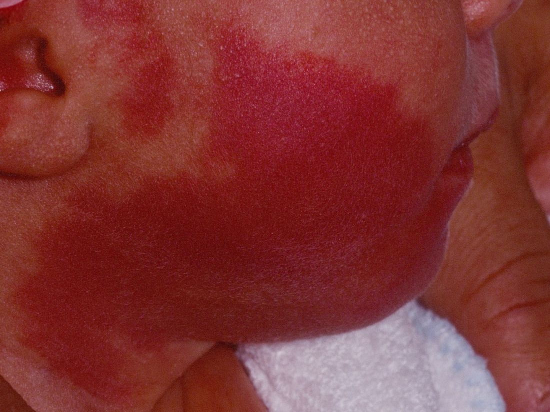
A recent Food and Drug Administration Drug Safety Communication warned that “repeated or lengthy used of general anesthetic and sedation drugs during surgeries or procedures in children younger than 3 years or in pregnant women during their third trimester may affect the development of children’s brains.” Dr. Jeon, a dermatologist in private practice in New York, noted that the FDA warning “places a greater importance on the already controversial topic of when to initiate port wine stain [PWS] treatments in pediatric patients, which requires repeated treatments and are often performed with general anesthesia. Without treatment, these lesions tend to get larger and thicker with time. Starting the treatment during infancy has the potential to limit the use of general anesthesia and to facilitate clearing.”
In what she said is the largest retrospective study of its kind to date, Dr. Jeon and her associates evaluated the success and safety of treating PWSs with a pulsed dye laser at the age of 1 year or younger in the office setting without general anesthesia. The patients received their first PWS treatment at their center during 2000-2017. They reviewed the charts of 197 patients to extract relevant data, including demographic information, age at the time of procedure, and treatment dates. The data cutoff was at 1 year following the initial treatment. Four physicians independently reviewed before and after photos and used the visual analogue scale to grade them.
The pulsed dye laser with dynamic cooling spray was used to minimize patient discomfort. No topical, local, or general anesthesia was used. Patients were immobilized by ancillary staff with parents present in most cases. Ocular shields were placed to allow treatment of periocular lesions.
Of the 197 patients, 63% were female, 90.1% had Fitzpatrick skin types I-III, 8.1% had type IV skin, and the rest had type V-VI skin. Most of the lesions were facial (75.6%), and 41.1% had periocular involvement. The average lesion size was 61 cm2.
The treatment settings for the pulsed dye laser were a 10-12 mm spot size delivered at a fluence of 6.5-9 J/cm2 in a pulse duration of 0.45-1.5 milliseconds. The average age at the time of first treatment was 3.4 months (range, 5-355 days), and the average number of treatments was 9.8 (range, 2-23). Most of the patients (116) were aged 0-3 months at the time of first treatment, followed by 51 aged 3-6 months, 19 aged 6-9 months, and 11 aged 9-12 months.
According to the averaged scores on the visual analogue scale assigned by physicians, 27.4% showed a 100% clearance, 39.1% showed 76%-99% improvement, 15.1% showed 51%-75% improvement, 10.7% showed 26%-50% improvement, and 7.7% showed 0%-25% improvement. No scarring or pigmentary changes were observed.
An analysis of dermatomal distribution revealed that the presence of a V1 lesion was a statistically significant predictor of a higher clearance rate, while the presence of a V3 lesion was a statistically significant predictor of a low clearance rate. “Regardless of where the lesion was, all patients did well,” she said.
Given the small proportion of patients who received their first treatment between the ages of 6 and 12 months of age, Dr. Jeon said that “further studies would be needed to elucidate whether earlier intervention helps to achieve better results.”*
Advantages of early treatment, she said, include the ability to “treat lesions before they get larger and thicker, which also decreases the risk of spontaneous bleeding. Thinner skin allows for better penetration of the laser beam, and we can also avoid using general anesthesia. Psychologically, if we’re able to clear these lesions, it can result in improved quality of life and self-esteem; not just for the patient but for the family as well.”
Dr. Jeon acknowledged certain limitations of the study, including its retrospective design, variable follow-up times, and non-standardization of photographs.
She reported having no financial disclosures.
SOURCE: Jeon H et al. ASLMS 2018.
Correction, 4/18/18: An earlier version of this article misstated the age range of the study participants.
DALLAS – Laser treatment of port wine stains in infancy is both safe and effective, with no incidence of scarring or pigmentary changes, according to results from a single-center analysis.
“Early intervention allows for treatment without general anesthesia, with faster and more complete clearance than what has been reported for treatments begun at older ages,” Hana Jeon, MD, said at the annual conference of the American Society for Laser Medicine and Surgery.
A recent Food and Drug Administration Drug Safety Communication warned that “repeated or lengthy used of general anesthetic and sedation drugs during surgeries or procedures in children younger than 3 years or in pregnant women during their third trimester may affect the development of children’s brains.” Dr. Jeon, a dermatologist in private practice in New York, noted that the FDA warning “places a greater importance on the already controversial topic of when to initiate port wine stain [PWS] treatments in pediatric patients, which requires repeated treatments and are often performed with general anesthesia. Without treatment, these lesions tend to get larger and thicker with time. Starting the treatment during infancy has the potential to limit the use of general anesthesia and to facilitate clearing.”
In what she said is the largest retrospective study of its kind to date, Dr. Jeon and her associates evaluated the success and safety of treating PWSs with a pulsed dye laser at the age of 1 year or younger in the office setting without general anesthesia. The patients received their first PWS treatment at their center during 2000-2017. They reviewed the charts of 197 patients to extract relevant data, including demographic information, age at the time of procedure, and treatment dates. The data cutoff was at 1 year following the initial treatment. Four physicians independently reviewed before and after photos and used the visual analogue scale to grade them.
The pulsed dye laser with dynamic cooling spray was used to minimize patient discomfort. No topical, local, or general anesthesia was used. Patients were immobilized by ancillary staff with parents present in most cases. Ocular shields were placed to allow treatment of periocular lesions.
Of the 197 patients, 63% were female, 90.1% had Fitzpatrick skin types I-III, 8.1% had type IV skin, and the rest had type V-VI skin. Most of the lesions were facial (75.6%), and 41.1% had periocular involvement. The average lesion size was 61 cm2.
The treatment settings for the pulsed dye laser were a 10-12 mm spot size delivered at a fluence of 6.5-9 J/cm2 in a pulse duration of 0.45-1.5 milliseconds. The average age at the time of first treatment was 3.4 months (range, 5-355 days), and the average number of treatments was 9.8 (range, 2-23). Most of the patients (116) were aged 0-3 months at the time of first treatment, followed by 51 aged 3-6 months, 19 aged 6-9 months, and 11 aged 9-12 months.
According to the averaged scores on the visual analogue scale assigned by physicians, 27.4% showed a 100% clearance, 39.1% showed 76%-99% improvement, 15.1% showed 51%-75% improvement, 10.7% showed 26%-50% improvement, and 7.7% showed 0%-25% improvement. No scarring or pigmentary changes were observed.
An analysis of dermatomal distribution revealed that the presence of a V1 lesion was a statistically significant predictor of a higher clearance rate, while the presence of a V3 lesion was a statistically significant predictor of a low clearance rate. “Regardless of where the lesion was, all patients did well,” she said.
Given the small proportion of patients who received their first treatment between the ages of 6 and 12 months of age, Dr. Jeon said that “further studies would be needed to elucidate whether earlier intervention helps to achieve better results.”*
Advantages of early treatment, she said, include the ability to “treat lesions before they get larger and thicker, which also decreases the risk of spontaneous bleeding. Thinner skin allows for better penetration of the laser beam, and we can also avoid using general anesthesia. Psychologically, if we’re able to clear these lesions, it can result in improved quality of life and self-esteem; not just for the patient but for the family as well.”
Dr. Jeon acknowledged certain limitations of the study, including its retrospective design, variable follow-up times, and non-standardization of photographs.
She reported having no financial disclosures.
SOURCE: Jeon H et al. ASLMS 2018.
Correction, 4/18/18: An earlier version of this article misstated the age range of the study participants.
DALLAS – Laser treatment of port wine stains in infancy is both safe and effective, with no incidence of scarring or pigmentary changes, according to results from a single-center analysis.
“Early intervention allows for treatment without general anesthesia, with faster and more complete clearance than what has been reported for treatments begun at older ages,” Hana Jeon, MD, said at the annual conference of the American Society for Laser Medicine and Surgery.
A recent Food and Drug Administration Drug Safety Communication warned that “repeated or lengthy used of general anesthetic and sedation drugs during surgeries or procedures in children younger than 3 years or in pregnant women during their third trimester may affect the development of children’s brains.” Dr. Jeon, a dermatologist in private practice in New York, noted that the FDA warning “places a greater importance on the already controversial topic of when to initiate port wine stain [PWS] treatments in pediatric patients, which requires repeated treatments and are often performed with general anesthesia. Without treatment, these lesions tend to get larger and thicker with time. Starting the treatment during infancy has the potential to limit the use of general anesthesia and to facilitate clearing.”
In what she said is the largest retrospective study of its kind to date, Dr. Jeon and her associates evaluated the success and safety of treating PWSs with a pulsed dye laser at the age of 1 year or younger in the office setting without general anesthesia. The patients received their first PWS treatment at their center during 2000-2017. They reviewed the charts of 197 patients to extract relevant data, including demographic information, age at the time of procedure, and treatment dates. The data cutoff was at 1 year following the initial treatment. Four physicians independently reviewed before and after photos and used the visual analogue scale to grade them.
The pulsed dye laser with dynamic cooling spray was used to minimize patient discomfort. No topical, local, or general anesthesia was used. Patients were immobilized by ancillary staff with parents present in most cases. Ocular shields were placed to allow treatment of periocular lesions.
Of the 197 patients, 63% were female, 90.1% had Fitzpatrick skin types I-III, 8.1% had type IV skin, and the rest had type V-VI skin. Most of the lesions were facial (75.6%), and 41.1% had periocular involvement. The average lesion size was 61 cm2.
The treatment settings for the pulsed dye laser were a 10-12 mm spot size delivered at a fluence of 6.5-9 J/cm2 in a pulse duration of 0.45-1.5 milliseconds. The average age at the time of first treatment was 3.4 months (range, 5-355 days), and the average number of treatments was 9.8 (range, 2-23). Most of the patients (116) were aged 0-3 months at the time of first treatment, followed by 51 aged 3-6 months, 19 aged 6-9 months, and 11 aged 9-12 months.
According to the averaged scores on the visual analogue scale assigned by physicians, 27.4% showed a 100% clearance, 39.1% showed 76%-99% improvement, 15.1% showed 51%-75% improvement, 10.7% showed 26%-50% improvement, and 7.7% showed 0%-25% improvement. No scarring or pigmentary changes were observed.
An analysis of dermatomal distribution revealed that the presence of a V1 lesion was a statistically significant predictor of a higher clearance rate, while the presence of a V3 lesion was a statistically significant predictor of a low clearance rate. “Regardless of where the lesion was, all patients did well,” she said.
Given the small proportion of patients who received their first treatment between the ages of 6 and 12 months of age, Dr. Jeon said that “further studies would be needed to elucidate whether earlier intervention helps to achieve better results.”*
Advantages of early treatment, she said, include the ability to “treat lesions before they get larger and thicker, which also decreases the risk of spontaneous bleeding. Thinner skin allows for better penetration of the laser beam, and we can also avoid using general anesthesia. Psychologically, if we’re able to clear these lesions, it can result in improved quality of life and self-esteem; not just for the patient but for the family as well.”
Dr. Jeon acknowledged certain limitations of the study, including its retrospective design, variable follow-up times, and non-standardization of photographs.
She reported having no financial disclosures.
SOURCE: Jeon H et al. ASLMS 2018.
Correction, 4/18/18: An earlier version of this article misstated the age range of the study participants.
REPORTING FROM ASLMS 2018
Key clinical point:
Major finding: On the visual analogue scale, 27.4% of infants achieved complete clearance of their port wine lesion after treatment.
Study details: An analysis of 197 infants aged 1 year or younger with port wine stains who were treated with a pulsed dye laser during 2000-2017.
Disclosures: Dr. Jeon reported having no financial disclosures.
Source: Jeon H et al. ASLMS 2018.
Enhanced recovery led to fewer complications for major oncologic procedures
CHICAGO – Use of an enhanced recovery protocol for oncology patients has been shown to improve outcomes in colorectal surgery but has been largely unproven in other types of major oncology operations. That prompted researchers at the University of Texas MD Anderson Cancer Center in Houston to investigate They found that the enhanced recovery protocol led to a reduction in complication rates and a decrease in hospital stay with no increase in readmissions, according to an analysis of more than 3,000 oncologic operations presented at the Society of Surgical Oncology Annual Cancer Symposium here.

“Patients treated with enhanced recovery did better,” Rebecca Marcus, MD, said in reporting the results. “There were decreased rates of perioperative transfusions, decreased rates of surgical site infections, decreased rates of complications, including severe complications such as wound dehiscence, pneumonia, renal failure, and unintended returns to the operating room.” She noted that the shorter hospital stays – 4 days for patients on the enhanced recovery protocol versus 5 days for those on the traditional postoperative protocol – did not result in increased readmissions.
The study reviewed 3,256 operations performed during 2011-2016 in the MD Anderson institutional American College of Surgeons National Surgical Quality Improvement Program database. The operations were colorectal (20.4%), gynecologic (19.5%), hepatobiliary (8.9%), thoracic (41.9%) and urologic (9.3%). Most employed the traditional postoperative protocol (53.4%). Colorectal and thoracic/vascular surgery were early adopters of the enhanced recovery protocol at MD Anderson.
Dr. Marcus noted that the overall complication rates were 21.9% for those treated with enhanced recovery–protocol versus 33.9% for those treated with traditional postoperative protocol (P less than .0001). The group treated with enhanced recovery protocol also had lower rates of severe complications: 8.7% vs. 11.7% (P =.0048). The study also noted a trend toward reduced National Surgical Quality Improvement Program 30-day mortality with the enhanced recover protocol (0.4% vs. 0.86%; P = .097). Readmission rates were similar between the two groups: 8.3% for enhanced recovery protocol versus 8.9% for traditional postoperative protocol.
The researchers performed a subanalysis of high-magnitude cases that had a relative value unit of 30 or more and involved operations of greater complexity, which constituted 38% of the study population. “In this group, we still saw the benefit of having treatment with an enhanced recovery protocol with decreased rates of preoperative transfusions, complications, and shorter length of stay without any recent readmission,” Dr. Marcus said. Complication rates in the high-magnitude group were 19% for the enhanced recovery protocol versus 26% for the traditional postoperative protocol in colorectal cases, 21% vs. 40% in gynecology, and 19% vs. 28% in thoracic/vascular.
“The beneficial impact of enhanced recovery appears to be maintained across all specialties and to be independent of case magnitude,” Dr. Marcus said.
She said future research of enhanced recovery in surgical oncology should focus on more long-term outcomes, “such as oncologic benefits of these protocols, especially given the known detrimental effect of the delayed return to adjuvant therapy for this patient population.”
Dr. Marcus and her coauthors reported having no financial disclosures.
SOURCE: Marcus RK et al. SSO 2018, Abstract 21.
CHICAGO – Use of an enhanced recovery protocol for oncology patients has been shown to improve outcomes in colorectal surgery but has been largely unproven in other types of major oncology operations. That prompted researchers at the University of Texas MD Anderson Cancer Center in Houston to investigate They found that the enhanced recovery protocol led to a reduction in complication rates and a decrease in hospital stay with no increase in readmissions, according to an analysis of more than 3,000 oncologic operations presented at the Society of Surgical Oncology Annual Cancer Symposium here.
“Patients treated with enhanced recovery did better,” Rebecca Marcus, MD, said in reporting the results. “There were decreased rates of perioperative transfusions, decreased rates of surgical site infections, decreased rates of complications, including severe complications such as wound dehiscence, pneumonia, renal failure, and unintended returns to the operating room.” She noted that the shorter hospital stays – 4 days for patients on the enhanced recovery protocol versus 5 days for those on the traditional postoperative protocol – did not result in increased readmissions.
The study reviewed 3,256 operations performed during 2011-2016 in the MD Anderson institutional American College of Surgeons National Surgical Quality Improvement Program database. The operations were colorectal (20.4%), gynecologic (19.5%), hepatobiliary (8.9%), thoracic (41.9%) and urologic (9.3%). Most employed the traditional postoperative protocol (53.4%). Colorectal and thoracic/vascular surgery were early adopters of the enhanced recovery protocol at MD Anderson.
Dr. Marcus noted that the overall complication rates were 21.9% for those treated with enhanced recovery–protocol versus 33.9% for those treated with traditional postoperative protocol (P less than .0001). The group treated with enhanced recovery protocol also had lower rates of severe complications: 8.7% vs. 11.7% (P =.0048). The study also noted a trend toward reduced National Surgical Quality Improvement Program 30-day mortality with the enhanced recover protocol (0.4% vs. 0.86%; P = .097). Readmission rates were similar between the two groups: 8.3% for enhanced recovery protocol versus 8.9% for traditional postoperative protocol.
The researchers performed a subanalysis of high-magnitude cases that had a relative value unit of 30 or more and involved operations of greater complexity, which constituted 38% of the study population. “In this group, we still saw the benefit of having treatment with an enhanced recovery protocol with decreased rates of preoperative transfusions, complications, and shorter length of stay without any recent readmission,” Dr. Marcus said. Complication rates in the high-magnitude group were 19% for the enhanced recovery protocol versus 26% for the traditional postoperative protocol in colorectal cases, 21% vs. 40% in gynecology, and 19% vs. 28% in thoracic/vascular.
“The beneficial impact of enhanced recovery appears to be maintained across all specialties and to be independent of case magnitude,” Dr. Marcus said.
She said future research of enhanced recovery in surgical oncology should focus on more long-term outcomes, “such as oncologic benefits of these protocols, especially given the known detrimental effect of the delayed return to adjuvant therapy for this patient population.”
Dr. Marcus and her coauthors reported having no financial disclosures.
SOURCE: Marcus RK et al. SSO 2018, Abstract 21.
CHICAGO – Use of an enhanced recovery protocol for oncology patients has been shown to improve outcomes in colorectal surgery but has been largely unproven in other types of major oncology operations. That prompted researchers at the University of Texas MD Anderson Cancer Center in Houston to investigate They found that the enhanced recovery protocol led to a reduction in complication rates and a decrease in hospital stay with no increase in readmissions, according to an analysis of more than 3,000 oncologic operations presented at the Society of Surgical Oncology Annual Cancer Symposium here.
“Patients treated with enhanced recovery did better,” Rebecca Marcus, MD, said in reporting the results. “There were decreased rates of perioperative transfusions, decreased rates of surgical site infections, decreased rates of complications, including severe complications such as wound dehiscence, pneumonia, renal failure, and unintended returns to the operating room.” She noted that the shorter hospital stays – 4 days for patients on the enhanced recovery protocol versus 5 days for those on the traditional postoperative protocol – did not result in increased readmissions.
The study reviewed 3,256 operations performed during 2011-2016 in the MD Anderson institutional American College of Surgeons National Surgical Quality Improvement Program database. The operations were colorectal (20.4%), gynecologic (19.5%), hepatobiliary (8.9%), thoracic (41.9%) and urologic (9.3%). Most employed the traditional postoperative protocol (53.4%). Colorectal and thoracic/vascular surgery were early adopters of the enhanced recovery protocol at MD Anderson.
Dr. Marcus noted that the overall complication rates were 21.9% for those treated with enhanced recovery–protocol versus 33.9% for those treated with traditional postoperative protocol (P less than .0001). The group treated with enhanced recovery protocol also had lower rates of severe complications: 8.7% vs. 11.7% (P =.0048). The study also noted a trend toward reduced National Surgical Quality Improvement Program 30-day mortality with the enhanced recover protocol (0.4% vs. 0.86%; P = .097). Readmission rates were similar between the two groups: 8.3% for enhanced recovery protocol versus 8.9% for traditional postoperative protocol.
The researchers performed a subanalysis of high-magnitude cases that had a relative value unit of 30 or more and involved operations of greater complexity, which constituted 38% of the study population. “In this group, we still saw the benefit of having treatment with an enhanced recovery protocol with decreased rates of preoperative transfusions, complications, and shorter length of stay without any recent readmission,” Dr. Marcus said. Complication rates in the high-magnitude group were 19% for the enhanced recovery protocol versus 26% for the traditional postoperative protocol in colorectal cases, 21% vs. 40% in gynecology, and 19% vs. 28% in thoracic/vascular.
“The beneficial impact of enhanced recovery appears to be maintained across all specialties and to be independent of case magnitude,” Dr. Marcus said.
She said future research of enhanced recovery in surgical oncology should focus on more long-term outcomes, “such as oncologic benefits of these protocols, especially given the known detrimental effect of the delayed return to adjuvant therapy for this patient population.”
Dr. Marcus and her coauthors reported having no financial disclosures.
SOURCE: Marcus RK et al. SSO 2018, Abstract 21.
REPORTING FROM SSO 2018
Key clinical point: Enhanced recovery protocol implementation is feasible in major oncologic surgery.
Major finding: Complication rates were 21.9% for enhanced recovery protocol versus 33.9% for traditional postoperative protocol.
Study details: Analysis of 3,256 oncology operations in an institutional ACS NSQIP database performed from 2011 to 2016.
Disclosures: Dr. Marcus and her coauthors reported having no financial disclosures.
Source: Marcus RK et al. SSO 2018, Abstract 21
High engraftment with new umbilical transplant technique
SALT LAKE CITY – Recipients of hematopoietic cell transplant with umbilical cord blood CD34+ cells expanded with an aryl hydrocarbon receptor (AHR) antagonist had a significantly higher rate of engraftment and comparable survival to a historical cohort of umbilical cord blood recipients.
The robust expansion of donor umbilical cord blood seen with the new technique opens the door for better use of umbilical cord blood inventory with superior human leukocyte antigen (HLA) matching, John Wagner, MD, said at a top abstracts session of the combined annual meetings of the Center for International Blood & Marrow Transplant Research and the American Society for Blood and Marrow Transplantation.
The new technique still shared the benefits of low rates of graft-versus-host disease (GVHD) and high survival that have been seen in previous umbilical cord blood transplants, with no significant difference in overall survival, relapse, or acute or chronic GVHD.
Compared to historical controls (n = 151), patients receiving the AHR antagonist–expanded umbilical cord blood (UCB) cells with myeloablative conditioning (n = 9) saw complete and more rapid engraftment (100% vs. 89% engraftment at a median 14 days vs. 23 days; P less than .01), reported Dr. Wagner of the University of Minnesota, Minneapolis.
These and other results came from two arms of a phase 2 trial of MGTA-456 (the working name of the AHR-expanded UCB cells). Twenty patients were to receive MGTA-456 derived from partially matched umbilical cord blood units after either myeloablative or nonmyeloablative conditioning; one patient in each arm had low expansion of UCB, so a total of 18 patients received MGTA-456. Each intervention arm was compared with a historical control arm that had received conventional UCB units.
In the myeloablative arm, patient demographics and disease characteristics were similar to the control cohort except that the MGTA-456 patients were significantly heavier (93.8 kg vs. 66.7 kg; P less than .04).
Platelet recovery also rebounded faster with MGTA-456 plus myeloablative conditioning than it did with historical controls: 89% of patients had platelet recovery by a median 46 days, compared with 71% with platelet recovery by a median 64 days in the historical cohort (P less than .01).
Patients achieved rapid complete chimerism if they received myeloablative conditioning, and they had rapid rebound of CD4 counts to at least 200 by 2-3 months posttransplant, Dr. Wagner reported.
The nonmyeloablative arm had a historical control cohort of 132 patients. Characteristics were similar between the two groups except that the MGTA-456 patients were older and more likely to have high-risk disease.
Again, all patients had rapid neutrophil recovery and saw 100% engraftment with MGTA-456. Median time to engraftment was 7 days with MGTA-456 and 15 days for the historical controls (P less than .01). Platelet recovery took longer for the MGTA-456 (median of 47 vs. 107 days), but the difference was not statistically significant.
Complete chimerism was achieved rapidly with the nonmyeloablative regimen as well, and CD4 recovery was brisk, as had been seen with myeloablative conditioning before MGTA-456 transplantation.
Compared with historical controls, “MGTA-456 retains the benefits of low chronic-graft-versus host disease and high survival despite higher disease risk and age” in the study group, Dr. Wagner said. There were no significant differences between the intervention and historical control arms of the nonmyeloablative study in acute or chronic GVHD, relapse, or overall survival.
The use of MGTA-456 occurs against the backdrop of a history of high survival rates with UCB transplantation – about 70% at 5 years, Dr. Wagner said. However, when conventional culture and expansion methods for UCB were used, the median time to engraftment had been reported to be 25 days with a 79% engraftment rate. This contrasts with the mean 13 days to engraftment for peripheral blood transplants and 18 days for bone marrow transplants. All of these transplant sources, regardless of whether the transplant was matched or mismatched, have engraftment rates of 92%-96%, said Dr. Wagner (Lancet Oncol. 2010; 11[7]:653-60).
When an AHR antagonist is used for UCB expansion, hematopoietic stem cell renewal is upped because cell differentiation is blocked, which means expansion is all driven toward hematopoietic stem cell self-renewal, Dr. Wagner said. Of the 36 available samples, MGTA-456 achieved a median 327-fold expansion of CD34+ cells, which enabled investigators to deliver a median CD34+ dose of 17.5 X 10⁶ cells/kg.
The downstream effect of the robust expansion rates is that more cord blood will be available for transplantation, and HLA matches will improve, Dr. Wagner said. Using current expansion techniques, fewer than 5% of cord blood units have a total nucleated cell count sufficient for an adult 80 kg recipient, he said, adding that use of MGTA-456 could make more than 80% of cord blood units available for adults.
According to the UCB transplant history at the University of Minnesota – where Dr. Wagner directs the pediatric blood and marrow transplantation program – of the patients who received 4/6 HLA-matched cord blood, 63% would move to a 5/6 match, and 8% would move to a full HLA match with the MGTA-456 technique. Of patients who received 5/6-matched transplants, almost one in four (23%) would move to a full 6/6 match.
Dr. Wagner and his colleagues had previously shown that adding an AHR antagonist resulted in enhanced T-cell recovery and rapid and sustained engraftment (Science. 2010;329:1345-8).
The researchers then proceeded to a phase 1-2, first-in-human trial of MGTA-456 that used a myeloablative conditioning regimen that met its primary safety endpoint of a lack of infusional toxicity or primary/secondary graft failure (Cell Stem Cell. 2016;18:144-55).
For reasons of safety, this earlier study used a double-transplant platform in which one infusion was uncultured umbilical cord blood and the other was MGTA-456. This study showed rapid neutrophil recovery when MGTA-456 was infused, with median 10.5 days to recovery, compared with a median 26.5 days for historical controls (P less than .001).
Additionally, the study showed a 19-day decrease in duration of the initial hospitalization, and all patients who received MGTA-456 had successful engraftment, Dr. Wagner said. On the strength of these results, the current trials of MGTA-456 alone – with both nonmyeloablative and myeloablative conditioning – were approved.
Multicenter clinical trials of MGTA-456 transplantation are now planned for both malignant and nonmalignant diseases. Enrollment is currently open for a phase 2 clinical trial of MGTA for inherited metabolic disorders (NCT03406962).
The study was funded by Novartis and Magenta Therapeutics. Dr. Wagner reported no other relevant disclosures.
SOURCE: Wagner J et al. 2018 BMT Tandem Meetings, Abstract 4.
SALT LAKE CITY – Recipients of hematopoietic cell transplant with umbilical cord blood CD34+ cells expanded with an aryl hydrocarbon receptor (AHR) antagonist had a significantly higher rate of engraftment and comparable survival to a historical cohort of umbilical cord blood recipients.
The robust expansion of donor umbilical cord blood seen with the new technique opens the door for better use of umbilical cord blood inventory with superior human leukocyte antigen (HLA) matching, John Wagner, MD, said at a top abstracts session of the combined annual meetings of the Center for International Blood & Marrow Transplant Research and the American Society for Blood and Marrow Transplantation.
The new technique still shared the benefits of low rates of graft-versus-host disease (GVHD) and high survival that have been seen in previous umbilical cord blood transplants, with no significant difference in overall survival, relapse, or acute or chronic GVHD.
Compared to historical controls (n = 151), patients receiving the AHR antagonist–expanded umbilical cord blood (UCB) cells with myeloablative conditioning (n = 9) saw complete and more rapid engraftment (100% vs. 89% engraftment at a median 14 days vs. 23 days; P less than .01), reported Dr. Wagner of the University of Minnesota, Minneapolis.
These and other results came from two arms of a phase 2 trial of MGTA-456 (the working name of the AHR-expanded UCB cells). Twenty patients were to receive MGTA-456 derived from partially matched umbilical cord blood units after either myeloablative or nonmyeloablative conditioning; one patient in each arm had low expansion of UCB, so a total of 18 patients received MGTA-456. Each intervention arm was compared with a historical control arm that had received conventional UCB units.
In the myeloablative arm, patient demographics and disease characteristics were similar to the control cohort except that the MGTA-456 patients were significantly heavier (93.8 kg vs. 66.7 kg; P less than .04).
Platelet recovery also rebounded faster with MGTA-456 plus myeloablative conditioning than it did with historical controls: 89% of patients had platelet recovery by a median 46 days, compared with 71% with platelet recovery by a median 64 days in the historical cohort (P less than .01).
Patients achieved rapid complete chimerism if they received myeloablative conditioning, and they had rapid rebound of CD4 counts to at least 200 by 2-3 months posttransplant, Dr. Wagner reported.
The nonmyeloablative arm had a historical control cohort of 132 patients. Characteristics were similar between the two groups except that the MGTA-456 patients were older and more likely to have high-risk disease.
Again, all patients had rapid neutrophil recovery and saw 100% engraftment with MGTA-456. Median time to engraftment was 7 days with MGTA-456 and 15 days for the historical controls (P less than .01). Platelet recovery took longer for the MGTA-456 (median of 47 vs. 107 days), but the difference was not statistically significant.
Complete chimerism was achieved rapidly with the nonmyeloablative regimen as well, and CD4 recovery was brisk, as had been seen with myeloablative conditioning before MGTA-456 transplantation.
Compared with historical controls, “MGTA-456 retains the benefits of low chronic-graft-versus host disease and high survival despite higher disease risk and age” in the study group, Dr. Wagner said. There were no significant differences between the intervention and historical control arms of the nonmyeloablative study in acute or chronic GVHD, relapse, or overall survival.
The use of MGTA-456 occurs against the backdrop of a history of high survival rates with UCB transplantation – about 70% at 5 years, Dr. Wagner said. However, when conventional culture and expansion methods for UCB were used, the median time to engraftment had been reported to be 25 days with a 79% engraftment rate. This contrasts with the mean 13 days to engraftment for peripheral blood transplants and 18 days for bone marrow transplants. All of these transplant sources, regardless of whether the transplant was matched or mismatched, have engraftment rates of 92%-96%, said Dr. Wagner (Lancet Oncol. 2010; 11[7]:653-60).
When an AHR antagonist is used for UCB expansion, hematopoietic stem cell renewal is upped because cell differentiation is blocked, which means expansion is all driven toward hematopoietic stem cell self-renewal, Dr. Wagner said. Of the 36 available samples, MGTA-456 achieved a median 327-fold expansion of CD34+ cells, which enabled investigators to deliver a median CD34+ dose of 17.5 X 10⁶ cells/kg.
The downstream effect of the robust expansion rates is that more cord blood will be available for transplantation, and HLA matches will improve, Dr. Wagner said. Using current expansion techniques, fewer than 5% of cord blood units have a total nucleated cell count sufficient for an adult 80 kg recipient, he said, adding that use of MGTA-456 could make more than 80% of cord blood units available for adults.
According to the UCB transplant history at the University of Minnesota – where Dr. Wagner directs the pediatric blood and marrow transplantation program – of the patients who received 4/6 HLA-matched cord blood, 63% would move to a 5/6 match, and 8% would move to a full HLA match with the MGTA-456 technique. Of patients who received 5/6-matched transplants, almost one in four (23%) would move to a full 6/6 match.
Dr. Wagner and his colleagues had previously shown that adding an AHR antagonist resulted in enhanced T-cell recovery and rapid and sustained engraftment (Science. 2010;329:1345-8).
The researchers then proceeded to a phase 1-2, first-in-human trial of MGTA-456 that used a myeloablative conditioning regimen that met its primary safety endpoint of a lack of infusional toxicity or primary/secondary graft failure (Cell Stem Cell. 2016;18:144-55).
For reasons of safety, this earlier study used a double-transplant platform in which one infusion was uncultured umbilical cord blood and the other was MGTA-456. This study showed rapid neutrophil recovery when MGTA-456 was infused, with median 10.5 days to recovery, compared with a median 26.5 days for historical controls (P less than .001).
Additionally, the study showed a 19-day decrease in duration of the initial hospitalization, and all patients who received MGTA-456 had successful engraftment, Dr. Wagner said. On the strength of these results, the current trials of MGTA-456 alone – with both nonmyeloablative and myeloablative conditioning – were approved.
Multicenter clinical trials of MGTA-456 transplantation are now planned for both malignant and nonmalignant diseases. Enrollment is currently open for a phase 2 clinical trial of MGTA for inherited metabolic disorders (NCT03406962).
The study was funded by Novartis and Magenta Therapeutics. Dr. Wagner reported no other relevant disclosures.
SOURCE: Wagner J et al. 2018 BMT Tandem Meetings, Abstract 4.
SALT LAKE CITY – Recipients of hematopoietic cell transplant with umbilical cord blood CD34+ cells expanded with an aryl hydrocarbon receptor (AHR) antagonist had a significantly higher rate of engraftment and comparable survival to a historical cohort of umbilical cord blood recipients.
The robust expansion of donor umbilical cord blood seen with the new technique opens the door for better use of umbilical cord blood inventory with superior human leukocyte antigen (HLA) matching, John Wagner, MD, said at a top abstracts session of the combined annual meetings of the Center for International Blood & Marrow Transplant Research and the American Society for Blood and Marrow Transplantation.
The new technique still shared the benefits of low rates of graft-versus-host disease (GVHD) and high survival that have been seen in previous umbilical cord blood transplants, with no significant difference in overall survival, relapse, or acute or chronic GVHD.
Compared to historical controls (n = 151), patients receiving the AHR antagonist–expanded umbilical cord blood (UCB) cells with myeloablative conditioning (n = 9) saw complete and more rapid engraftment (100% vs. 89% engraftment at a median 14 days vs. 23 days; P less than .01), reported Dr. Wagner of the University of Minnesota, Minneapolis.
These and other results came from two arms of a phase 2 trial of MGTA-456 (the working name of the AHR-expanded UCB cells). Twenty patients were to receive MGTA-456 derived from partially matched umbilical cord blood units after either myeloablative or nonmyeloablative conditioning; one patient in each arm had low expansion of UCB, so a total of 18 patients received MGTA-456. Each intervention arm was compared with a historical control arm that had received conventional UCB units.
In the myeloablative arm, patient demographics and disease characteristics were similar to the control cohort except that the MGTA-456 patients were significantly heavier (93.8 kg vs. 66.7 kg; P less than .04).
Platelet recovery also rebounded faster with MGTA-456 plus myeloablative conditioning than it did with historical controls: 89% of patients had platelet recovery by a median 46 days, compared with 71% with platelet recovery by a median 64 days in the historical cohort (P less than .01).
Patients achieved rapid complete chimerism if they received myeloablative conditioning, and they had rapid rebound of CD4 counts to at least 200 by 2-3 months posttransplant, Dr. Wagner reported.
The nonmyeloablative arm had a historical control cohort of 132 patients. Characteristics were similar between the two groups except that the MGTA-456 patients were older and more likely to have high-risk disease.
Again, all patients had rapid neutrophil recovery and saw 100% engraftment with MGTA-456. Median time to engraftment was 7 days with MGTA-456 and 15 days for the historical controls (P less than .01). Platelet recovery took longer for the MGTA-456 (median of 47 vs. 107 days), but the difference was not statistically significant.
Complete chimerism was achieved rapidly with the nonmyeloablative regimen as well, and CD4 recovery was brisk, as had been seen with myeloablative conditioning before MGTA-456 transplantation.
Compared with historical controls, “MGTA-456 retains the benefits of low chronic-graft-versus host disease and high survival despite higher disease risk and age” in the study group, Dr. Wagner said. There were no significant differences between the intervention and historical control arms of the nonmyeloablative study in acute or chronic GVHD, relapse, or overall survival.
The use of MGTA-456 occurs against the backdrop of a history of high survival rates with UCB transplantation – about 70% at 5 years, Dr. Wagner said. However, when conventional culture and expansion methods for UCB were used, the median time to engraftment had been reported to be 25 days with a 79% engraftment rate. This contrasts with the mean 13 days to engraftment for peripheral blood transplants and 18 days for bone marrow transplants. All of these transplant sources, regardless of whether the transplant was matched or mismatched, have engraftment rates of 92%-96%, said Dr. Wagner (Lancet Oncol. 2010; 11[7]:653-60).
When an AHR antagonist is used for UCB expansion, hematopoietic stem cell renewal is upped because cell differentiation is blocked, which means expansion is all driven toward hematopoietic stem cell self-renewal, Dr. Wagner said. Of the 36 available samples, MGTA-456 achieved a median 327-fold expansion of CD34+ cells, which enabled investigators to deliver a median CD34+ dose of 17.5 X 10⁶ cells/kg.
The downstream effect of the robust expansion rates is that more cord blood will be available for transplantation, and HLA matches will improve, Dr. Wagner said. Using current expansion techniques, fewer than 5% of cord blood units have a total nucleated cell count sufficient for an adult 80 kg recipient, he said, adding that use of MGTA-456 could make more than 80% of cord blood units available for adults.
According to the UCB transplant history at the University of Minnesota – where Dr. Wagner directs the pediatric blood and marrow transplantation program – of the patients who received 4/6 HLA-matched cord blood, 63% would move to a 5/6 match, and 8% would move to a full HLA match with the MGTA-456 technique. Of patients who received 5/6-matched transplants, almost one in four (23%) would move to a full 6/6 match.
Dr. Wagner and his colleagues had previously shown that adding an AHR antagonist resulted in enhanced T-cell recovery and rapid and sustained engraftment (Science. 2010;329:1345-8).
The researchers then proceeded to a phase 1-2, first-in-human trial of MGTA-456 that used a myeloablative conditioning regimen that met its primary safety endpoint of a lack of infusional toxicity or primary/secondary graft failure (Cell Stem Cell. 2016;18:144-55).
For reasons of safety, this earlier study used a double-transplant platform in which one infusion was uncultured umbilical cord blood and the other was MGTA-456. This study showed rapid neutrophil recovery when MGTA-456 was infused, with median 10.5 days to recovery, compared with a median 26.5 days for historical controls (P less than .001).
Additionally, the study showed a 19-day decrease in duration of the initial hospitalization, and all patients who received MGTA-456 had successful engraftment, Dr. Wagner said. On the strength of these results, the current trials of MGTA-456 alone – with both nonmyeloablative and myeloablative conditioning – were approved.
Multicenter clinical trials of MGTA-456 transplantation are now planned for both malignant and nonmalignant diseases. Enrollment is currently open for a phase 2 clinical trial of MGTA for inherited metabolic disorders (NCT03406962).
The study was funded by Novartis and Magenta Therapeutics. Dr. Wagner reported no other relevant disclosures.
SOURCE: Wagner J et al. 2018 BMT Tandem Meetings, Abstract 4.
REPORTING FROM THE 2018 BMT TANDEM MEETINGS
Key clinical point:
Major finding: The engraftment rate was 100% with both myeloablative and nonmyeloablative conditioning.
Study details: A phase 2 trial of 20 patients receiving MGTA-456, compared with a historical cohort of umbilical cord blood hematopoietic cell transplant recipients.
Disclosures: The study was sponsored by Novartis and Magenta Therapeutics. Dr. Wagner reported no other conflicts of interest.
Source: Wagner J et al. 2018 BMT Tandem Meetings, Abstract 4.
Drug appears to aid chemo in AML

Adding an experimental compound to chemotherapy is a “promising” treatment approach for certain patients with acute myeloid leukemia (AML), according to researchers.
They tested the compound, CPI-613, in combination with high-dose cytarabine and mitoxantrone in a phase 1 trial of patients with relapsed or refractory AML.
The combination produced similar response rates in the overall patient population (50%), patients age 60 and older (47%), and those with poor-risk cytogenetics (46%).
The most common grade 3/4 adverse events (AEs) were hematologic toxicities, and there was 1 fatal AE—hypotension.
Mortality rates in this trial were similar to those observed in historical controls treated with high-dose cytarabine, mitoxantrone, and asparaginase.
“These data are very encouraging, especially for patients 60 years of age or older who have historically done very poorly with this disease,” said Timothy Pardee, MD, PhD, a professor at Wake Forest Baptist Health in Winston-Salem, North Carolina, and chief medical officer of Rafael Pharmaceuticals, Inc., the company developing CPI-613.
Dr Pardee and his colleagues reported these results in Clinical Cancer Research.
The researchers noted that CPI-613 is designed to target mitochondrial metabolism in cancer cells, and preclinical research showed that CPI-613 sensitized AML cells to chemotherapy.
To investigate this further, the team tested CPI-613 in combination with high-dose cytarabine and mitoxantrone in the phase 1 trial. The study included 66 patients with relapsed or refractory AML, as well as a patient with advanced-phase chronic myeloid leukemia (CML) who was mistakenly enrolled.
The patients’ median age was 60 (range, 21-79), and 54% were age 60 and older. Their median percentage of marrow blasts was 43%. Forty percent of patients had poor-risk cytogenetics, and 49% had intermediate-risk cytogenetics.
Most patients (72%) had no prior salvage therapy, 13% had 1 prior line of salvage, 10% had 2 prior lines, and 4% had more than 2. Thirty-one percent of patients had refractory disease.
Seven percent of patients had previously received high-dose cytarabine and mitoxantrone, and 25% had previous salvage including high-dose or intermediate-dose cytarabine.
Treatment
Patients received CPI-613, given over 2 hours, on days 1 to 5 of cycle 1. Doses ranged from 500 mg/m2 to 2750 mg/m2.
Starting on day 3, patients received 5 doses of cytarabine at 3 gm/m2 (for patients younger than 60) or 1.5 gm/m2 (for older patients) in 500 mL normal saline, over 3 hours, every 12 hours.
Patients also received 3 daily doses of mitoxantrone at 6 mg/m2 in 50 mL normal saline, given over 15 minutes, after the first, third, and fifth doses of cytarabine.
Patients were initially assigned to receive 1 cycle of treatment. Those with at least 5% blasts after the first cycle could receive a second course—either a full course or a 3-day course. And patients who responded to the first course could receive up to 2 cycles of the 3-day course.
Safety
There were 2 dose-limiting toxicities when CPI-613 was given at the 2750 mg/m2 dose. One of these toxicities was grade 3 diarrhea that didn’t respond to anti-diarrheals, and the other was grade 3 nausea that didn’t respond to antiemetics.
Because of these events, 2500 mg/m2 was deemed the maximum-tolerated dose. However, the recommended phase 2 dose is 2000 mg/m2.
The most common AEs—occurring in at least 50% of all patients who received CPI-613 (n=67)—included hemoglobin decrease (67%), hyperglycemia (67%), neutropenia (67%), thrombocytopenia (67%), hypomagnesemia (66%), leukopenia (66%), lymphopenia (66%), hypoalbuminemia (65%), hypokalemia (60%), hypocalcemia (57%), and diarrhea (55%).
All cases of neutropenia, thrombocytopenia, leukopenia, and lymphopenia were grade 3/4. Other common grade 3/4 AEs (occurring in at least 20% of patients) included hemoglobin decrease (62%), febrile neutropenia (28%), hypophosphatemia (24%), and hypokalemia (23%).
The only grade 5 AE was hypotension.
The mortality rate was 12% (n=8) at 30 days and 19% (n=13) at 60 days. The researchers said this was similar to the historical experience with high-dose cytarabine, mitoxantrone, and asparaginase. Mortality rates with this regimen were 13% at 30 days and 22% at 60 days.
Efficacy
Sixty-two patients were evaluable for response. Of the 5 patients who were not evaluable, 1 didn’t complete the first cycle of treatment, 1 was the CML patient, and 3 died before assessment.
The overall response rate was 50% (31/62). This included 26 patients with a complete response (CR) and 5 patients who had a CR with incomplete count recovery (CRi).
The rate of CR/CRi was 47% (15/32) in patients older than 60 years of age, 46% (11/24) in patients who had poor-risk cytogenetics, and 53% (8/15) when CPI-613 was given at the recommended phase 2 dose—2000 mg/m2.
The median overall survival (OS) was 6.7 months for all evaluable patients and 13.2 months for patients who achieved a CR/CRi.
The median OS was 6.9 months for patients age 60 and older, which was not significantly different from the median OS in younger patients (P=0.9642).
This study was sponsored by Wake Forest University Health Sciences and the National Cancer Institute.
Adding an experimental compound to chemotherapy is a “promising” treatment approach for certain patients with acute myeloid leukemia (AML), according to researchers.
They tested the compound, CPI-613, in combination with high-dose cytarabine and mitoxantrone in a phase 1 trial of patients with relapsed or refractory AML.
The combination produced similar response rates in the overall patient population (50%), patients age 60 and older (47%), and those with poor-risk cytogenetics (46%).
The most common grade 3/4 adverse events (AEs) were hematologic toxicities, and there was 1 fatal AE—hypotension.
Mortality rates in this trial were similar to those observed in historical controls treated with high-dose cytarabine, mitoxantrone, and asparaginase.
“These data are very encouraging, especially for patients 60 years of age or older who have historically done very poorly with this disease,” said Timothy Pardee, MD, PhD, a professor at Wake Forest Baptist Health in Winston-Salem, North Carolina, and chief medical officer of Rafael Pharmaceuticals, Inc., the company developing CPI-613.
Dr Pardee and his colleagues reported these results in Clinical Cancer Research.
The researchers noted that CPI-613 is designed to target mitochondrial metabolism in cancer cells, and preclinical research showed that CPI-613 sensitized AML cells to chemotherapy.
To investigate this further, the team tested CPI-613 in combination with high-dose cytarabine and mitoxantrone in the phase 1 trial. The study included 66 patients with relapsed or refractory AML, as well as a patient with advanced-phase chronic myeloid leukemia (CML) who was mistakenly enrolled.
The patients’ median age was 60 (range, 21-79), and 54% were age 60 and older. Their median percentage of marrow blasts was 43%. Forty percent of patients had poor-risk cytogenetics, and 49% had intermediate-risk cytogenetics.
Most patients (72%) had no prior salvage therapy, 13% had 1 prior line of salvage, 10% had 2 prior lines, and 4% had more than 2. Thirty-one percent of patients had refractory disease.
Seven percent of patients had previously received high-dose cytarabine and mitoxantrone, and 25% had previous salvage including high-dose or intermediate-dose cytarabine.
Treatment
Patients received CPI-613, given over 2 hours, on days 1 to 5 of cycle 1. Doses ranged from 500 mg/m2 to 2750 mg/m2.
Starting on day 3, patients received 5 doses of cytarabine at 3 gm/m2 (for patients younger than 60) or 1.5 gm/m2 (for older patients) in 500 mL normal saline, over 3 hours, every 12 hours.
Patients also received 3 daily doses of mitoxantrone at 6 mg/m2 in 50 mL normal saline, given over 15 minutes, after the first, third, and fifth doses of cytarabine.
Patients were initially assigned to receive 1 cycle of treatment. Those with at least 5% blasts after the first cycle could receive a second course—either a full course or a 3-day course. And patients who responded to the first course could receive up to 2 cycles of the 3-day course.
Safety
There were 2 dose-limiting toxicities when CPI-613 was given at the 2750 mg/m2 dose. One of these toxicities was grade 3 diarrhea that didn’t respond to anti-diarrheals, and the other was grade 3 nausea that didn’t respond to antiemetics.
Because of these events, 2500 mg/m2 was deemed the maximum-tolerated dose. However, the recommended phase 2 dose is 2000 mg/m2.
The most common AEs—occurring in at least 50% of all patients who received CPI-613 (n=67)—included hemoglobin decrease (67%), hyperglycemia (67%), neutropenia (67%), thrombocytopenia (67%), hypomagnesemia (66%), leukopenia (66%), lymphopenia (66%), hypoalbuminemia (65%), hypokalemia (60%), hypocalcemia (57%), and diarrhea (55%).
All cases of neutropenia, thrombocytopenia, leukopenia, and lymphopenia were grade 3/4. Other common grade 3/4 AEs (occurring in at least 20% of patients) included hemoglobin decrease (62%), febrile neutropenia (28%), hypophosphatemia (24%), and hypokalemia (23%).
The only grade 5 AE was hypotension.
The mortality rate was 12% (n=8) at 30 days and 19% (n=13) at 60 days. The researchers said this was similar to the historical experience with high-dose cytarabine, mitoxantrone, and asparaginase. Mortality rates with this regimen were 13% at 30 days and 22% at 60 days.
Efficacy
Sixty-two patients were evaluable for response. Of the 5 patients who were not evaluable, 1 didn’t complete the first cycle of treatment, 1 was the CML patient, and 3 died before assessment.
The overall response rate was 50% (31/62). This included 26 patients with a complete response (CR) and 5 patients who had a CR with incomplete count recovery (CRi).
The rate of CR/CRi was 47% (15/32) in patients older than 60 years of age, 46% (11/24) in patients who had poor-risk cytogenetics, and 53% (8/15) when CPI-613 was given at the recommended phase 2 dose—2000 mg/m2.
The median overall survival (OS) was 6.7 months for all evaluable patients and 13.2 months for patients who achieved a CR/CRi.
The median OS was 6.9 months for patients age 60 and older, which was not significantly different from the median OS in younger patients (P=0.9642).
This study was sponsored by Wake Forest University Health Sciences and the National Cancer Institute.
Adding an experimental compound to chemotherapy is a “promising” treatment approach for certain patients with acute myeloid leukemia (AML), according to researchers.
They tested the compound, CPI-613, in combination with high-dose cytarabine and mitoxantrone in a phase 1 trial of patients with relapsed or refractory AML.
The combination produced similar response rates in the overall patient population (50%), patients age 60 and older (47%), and those with poor-risk cytogenetics (46%).
The most common grade 3/4 adverse events (AEs) were hematologic toxicities, and there was 1 fatal AE—hypotension.
Mortality rates in this trial were similar to those observed in historical controls treated with high-dose cytarabine, mitoxantrone, and asparaginase.
“These data are very encouraging, especially for patients 60 years of age or older who have historically done very poorly with this disease,” said Timothy Pardee, MD, PhD, a professor at Wake Forest Baptist Health in Winston-Salem, North Carolina, and chief medical officer of Rafael Pharmaceuticals, Inc., the company developing CPI-613.
Dr Pardee and his colleagues reported these results in Clinical Cancer Research.
The researchers noted that CPI-613 is designed to target mitochondrial metabolism in cancer cells, and preclinical research showed that CPI-613 sensitized AML cells to chemotherapy.
To investigate this further, the team tested CPI-613 in combination with high-dose cytarabine and mitoxantrone in the phase 1 trial. The study included 66 patients with relapsed or refractory AML, as well as a patient with advanced-phase chronic myeloid leukemia (CML) who was mistakenly enrolled.
The patients’ median age was 60 (range, 21-79), and 54% were age 60 and older. Their median percentage of marrow blasts was 43%. Forty percent of patients had poor-risk cytogenetics, and 49% had intermediate-risk cytogenetics.
Most patients (72%) had no prior salvage therapy, 13% had 1 prior line of salvage, 10% had 2 prior lines, and 4% had more than 2. Thirty-one percent of patients had refractory disease.
Seven percent of patients had previously received high-dose cytarabine and mitoxantrone, and 25% had previous salvage including high-dose or intermediate-dose cytarabine.
Treatment
Patients received CPI-613, given over 2 hours, on days 1 to 5 of cycle 1. Doses ranged from 500 mg/m2 to 2750 mg/m2.
Starting on day 3, patients received 5 doses of cytarabine at 3 gm/m2 (for patients younger than 60) or 1.5 gm/m2 (for older patients) in 500 mL normal saline, over 3 hours, every 12 hours.
Patients also received 3 daily doses of mitoxantrone at 6 mg/m2 in 50 mL normal saline, given over 15 minutes, after the first, third, and fifth doses of cytarabine.
Patients were initially assigned to receive 1 cycle of treatment. Those with at least 5% blasts after the first cycle could receive a second course—either a full course or a 3-day course. And patients who responded to the first course could receive up to 2 cycles of the 3-day course.
Safety
There were 2 dose-limiting toxicities when CPI-613 was given at the 2750 mg/m2 dose. One of these toxicities was grade 3 diarrhea that didn’t respond to anti-diarrheals, and the other was grade 3 nausea that didn’t respond to antiemetics.
Because of these events, 2500 mg/m2 was deemed the maximum-tolerated dose. However, the recommended phase 2 dose is 2000 mg/m2.
The most common AEs—occurring in at least 50% of all patients who received CPI-613 (n=67)—included hemoglobin decrease (67%), hyperglycemia (67%), neutropenia (67%), thrombocytopenia (67%), hypomagnesemia (66%), leukopenia (66%), lymphopenia (66%), hypoalbuminemia (65%), hypokalemia (60%), hypocalcemia (57%), and diarrhea (55%).
All cases of neutropenia, thrombocytopenia, leukopenia, and lymphopenia were grade 3/4. Other common grade 3/4 AEs (occurring in at least 20% of patients) included hemoglobin decrease (62%), febrile neutropenia (28%), hypophosphatemia (24%), and hypokalemia (23%).
The only grade 5 AE was hypotension.
The mortality rate was 12% (n=8) at 30 days and 19% (n=13) at 60 days. The researchers said this was similar to the historical experience with high-dose cytarabine, mitoxantrone, and asparaginase. Mortality rates with this regimen were 13% at 30 days and 22% at 60 days.
Efficacy
Sixty-two patients were evaluable for response. Of the 5 patients who were not evaluable, 1 didn’t complete the first cycle of treatment, 1 was the CML patient, and 3 died before assessment.
The overall response rate was 50% (31/62). This included 26 patients with a complete response (CR) and 5 patients who had a CR with incomplete count recovery (CRi).
The rate of CR/CRi was 47% (15/32) in patients older than 60 years of age, 46% (11/24) in patients who had poor-risk cytogenetics, and 53% (8/15) when CPI-613 was given at the recommended phase 2 dose—2000 mg/m2.
The median overall survival (OS) was 6.7 months for all evaluable patients and 13.2 months for patients who achieved a CR/CRi.
The median OS was 6.9 months for patients age 60 and older, which was not significantly different from the median OS in younger patients (P=0.9642).
This study was sponsored by Wake Forest University Health Sciences and the National Cancer Institute.
Dermatology practice gaps: improving medication management
KAUAI, HAWAII – Dermatologists don’t ordinarily peruse the ophthalmology literature. So they may be unaware that the American Academy of Ophthalmology has issued Erik J. Stratman, MD, noted at the Hawaii Dermatology Seminar provided by Global Academy for Medical Education/Skin Disease Education Foundation.
Most dermatologists routinely dose hydroxychloroquine at 400 mg/day, regardless of body weight. The former AAO recommendation, which dates back to 2011, called for dosing at up to 6.5 mg/kg of ideal body weight or 400 mg/day, whichever is lower. However, the AAO recommendation has changed in light of a large, retrospective case-control study that suggested this practice may be overdosing thin patients – thereby exposing them to increased risk of retinal toxicity and other drug-related adverse events – while at the same time possibly underdosing some obese patients, said Dr. Stratman, chairman of the department of dermatology at the Marshfield (Wisc.) Clinic.
This was one of two dermatology practice gaps he highlighted involving suboptimal medication management, the other being most dermatologists’ failure to protect their patients’ gut when prescribing prednisone.
“I think the push over the last 5 years has been ‘protect the bones, protect the bones, protect the bones.’ We’ve done better and better about protecting the bones and getting that into our conversations with patients on prednisone. But we’re not thinking so much about the gut,” the dermatologist said.
Hydroxychloroquine dosing
The former AAO recommendation was revised in response to a retrospective case-control study of retinal toxicity rates in 2,361 patients on the drug continuously for longer than 5 years. The study demonstrated that the risk of retinopathy jumped 5.7-fold with daily consumption of hydroxychloroquine at more than 5.0 mg/kg (JAMA Ophthalmol. 2014 Dec;132[12]:1453-60).
The current AAO recommendation (Ophthalmology. 2016 Jun;123[6]:1386-94) is to dose hydroxychloroquine at a daily maximum of 5.0 mg/kg of real weight, which correlated better with retinopathy risk in the case-control study than did ideal body weight. Hydroxychloroquine doesn’t accumulate well in fat.
Until now, most dermatologists have not routinely measured patients’ body weight in the office or calculated their body mass index. But Dr. Stratman advised against reliance upon a patient’s self-reported body weight, which may diverge substantially from reality. “Get yourself a good office scale – they’re not that expensive – and use it when prescribing drugs with a tight therapeutic window,” he urged.
Another key to minimizing retinopathy risk in patients on hydroxychloroquine is to pay careful attention to how long they’ve been on the drug. As the years go by in patients being treated for cutaneous lupus or other dermatologic disorders where decades-long therapy is often a mainstay, it’s important to check with patients and make sure they’re getting annual ophthalmologic screening for irreversible retinal toxicity by both threshold visual fields and spectral domain optical coherence tomography. In the large, practice-changing retrospective study, patients on hydroxychloroquine at 4.0-5.0 mg/kg daily had a prevalence of retinopathy of less than 2% during the first 10 years of therapy, but the rate shot up to nearly 20% after 20 years of use, Dr. Stratman observed.
He highlighted as helpful an updated review of the use of hydroxychloroquine in dermatology recently published by Anthony P. Fernandez, MD, PhD, of the department of dermatology at the Cleveland Clinic (J Am Acad Dermatol. 2017 Jun;76[6]:1176-82).
Dr. Fernandez recommends following the AAO guidance to dose the drug at 5.0 mg/kg or less of actual body weight in thin or normal-weight patients; however, he departed from the ophthalmologists with regard to treatment of obese patients. Because dosing based on actual weight could potentially lead to relative overdosing in obese patients, in that growing population he recommends calculating the dose based upon 5.0 mg/kg of actual body weight, as well as the dose based on 6.5 mg/kg of ideal body weight, then prescribing the lower of the two, up to a maximum of 400 mg/day.
“The current recommendation is really about not overdosing thin patients. Basically, dosing is not so difficult for obese people because if you weigh more than 175 pounds, you’re going to get 400 mg/day,” Dr. Stratman explained.
That 400 mg/day ceiling is not cast in stone, he continued. The guideline recommends that, if a patient is a nonresponder to several months of hydroxychloroquine at 400 mg/day, it’s worthwhile to order a drug blood level. If it’s not above the efficacy threshold of more than 750 ng/mL, it’s appropriate to titrate up.
Protecting against prednisone-induced gastritis
“We underprotect the gut,” Dr. Stratman asserted.
He referred to a recent comprehensive dermatologic review of the prevention and management of glucocorticoid-related side effects, especially the part on peptic ulcer disease (J Am Acad Dermatol. 2017 Jan;76[1]:11-6). This is an issue that heretofore hadn’t been much emphasized in the dermatology literature.
“I read this and thought, ‘Gosh, I’m not really having a conversation with my patients about a review of systems for gut protection as I should. And I certainly haven’t been thinking about prescribing PPIs [proton pump inhibitors] for my patients,’” he recalled.
Dr. Stratman polled his Hawaii Dermatology Seminar audience as to who had ever prescribed a PPI. Most indicated with their electronic clickers that they had never done so.
“This is what a practice gap is,” he commented. “You read the literature and you say, ‘Oh, I guess that makes sense. Maybe I should be doing that more often, or making sure it gets done.’”
“I don’t want to come across as saying, ‘For everybody we put on prednisone we should be giving vitamin D, calcium, and a PPI.’ That’s not the message. The message is, assess your patient – or make sure your patient is being assessed – for risk of peptic ulcer disease. And if you don’t feel comfortable prescribing a PPI, please get the patient connected with their primary care provider, who should,” Dr. Stratman said.
The authors of the dermatology review made a case for screening for GI risk factors in every patient who is going to receive an oral glucocorticoid. The ones who absolutely should be prescribed a PPI unless contraindicated include patients who are taking daily aspirin or NSAIDs for an essential reason, such as cardiovascular protection or significant arthritic pain. The authors suggest consideration of a PPI in patients with other, less potent risk factors for peptic ulcer disease, including a history of ulcer disease, gastroesophageal reflux disease, Barrett’s esophagus, heavy smoking, heavy alcohol consumption, age greater than 65, and concomitant use of other medications with an associated risk of peptic ulcer disease – such as bisphosphonates, “which you may have just put them on to protect their bones,” Dr. Stratman noted.
Of course, PPIs come with side effects of their own, including increased fracture risk, Clostridium difficile infections, and rebound acid secretion.
Dr. Stratman reported having no financial conflicts regarding his presentation.
SDEF/Global Academy for Medical Education and this news organization are owned by the same parent company.
KAUAI, HAWAII – Dermatologists don’t ordinarily peruse the ophthalmology literature. So they may be unaware that the American Academy of Ophthalmology has issued Erik J. Stratman, MD, noted at the Hawaii Dermatology Seminar provided by Global Academy for Medical Education/Skin Disease Education Foundation.
Most dermatologists routinely dose hydroxychloroquine at 400 mg/day, regardless of body weight. The former AAO recommendation, which dates back to 2011, called for dosing at up to 6.5 mg/kg of ideal body weight or 400 mg/day, whichever is lower. However, the AAO recommendation has changed in light of a large, retrospective case-control study that suggested this practice may be overdosing thin patients – thereby exposing them to increased risk of retinal toxicity and other drug-related adverse events – while at the same time possibly underdosing some obese patients, said Dr. Stratman, chairman of the department of dermatology at the Marshfield (Wisc.) Clinic.
This was one of two dermatology practice gaps he highlighted involving suboptimal medication management, the other being most dermatologists’ failure to protect their patients’ gut when prescribing prednisone.
“I think the push over the last 5 years has been ‘protect the bones, protect the bones, protect the bones.’ We’ve done better and better about protecting the bones and getting that into our conversations with patients on prednisone. But we’re not thinking so much about the gut,” the dermatologist said.
Hydroxychloroquine dosing
The former AAO recommendation was revised in response to a retrospective case-control study of retinal toxicity rates in 2,361 patients on the drug continuously for longer than 5 years. The study demonstrated that the risk of retinopathy jumped 5.7-fold with daily consumption of hydroxychloroquine at more than 5.0 mg/kg (JAMA Ophthalmol. 2014 Dec;132[12]:1453-60).
The current AAO recommendation (Ophthalmology. 2016 Jun;123[6]:1386-94) is to dose hydroxychloroquine at a daily maximum of 5.0 mg/kg of real weight, which correlated better with retinopathy risk in the case-control study than did ideal body weight. Hydroxychloroquine doesn’t accumulate well in fat.
Until now, most dermatologists have not routinely measured patients’ body weight in the office or calculated their body mass index. But Dr. Stratman advised against reliance upon a patient’s self-reported body weight, which may diverge substantially from reality. “Get yourself a good office scale – they’re not that expensive – and use it when prescribing drugs with a tight therapeutic window,” he urged.
Another key to minimizing retinopathy risk in patients on hydroxychloroquine is to pay careful attention to how long they’ve been on the drug. As the years go by in patients being treated for cutaneous lupus or other dermatologic disorders where decades-long therapy is often a mainstay, it’s important to check with patients and make sure they’re getting annual ophthalmologic screening for irreversible retinal toxicity by both threshold visual fields and spectral domain optical coherence tomography. In the large, practice-changing retrospective study, patients on hydroxychloroquine at 4.0-5.0 mg/kg daily had a prevalence of retinopathy of less than 2% during the first 10 years of therapy, but the rate shot up to nearly 20% after 20 years of use, Dr. Stratman observed.
He highlighted as helpful an updated review of the use of hydroxychloroquine in dermatology recently published by Anthony P. Fernandez, MD, PhD, of the department of dermatology at the Cleveland Clinic (J Am Acad Dermatol. 2017 Jun;76[6]:1176-82).
Dr. Fernandez recommends following the AAO guidance to dose the drug at 5.0 mg/kg or less of actual body weight in thin or normal-weight patients; however, he departed from the ophthalmologists with regard to treatment of obese patients. Because dosing based on actual weight could potentially lead to relative overdosing in obese patients, in that growing population he recommends calculating the dose based upon 5.0 mg/kg of actual body weight, as well as the dose based on 6.5 mg/kg of ideal body weight, then prescribing the lower of the two, up to a maximum of 400 mg/day.
“The current recommendation is really about not overdosing thin patients. Basically, dosing is not so difficult for obese people because if you weigh more than 175 pounds, you’re going to get 400 mg/day,” Dr. Stratman explained.
That 400 mg/day ceiling is not cast in stone, he continued. The guideline recommends that, if a patient is a nonresponder to several months of hydroxychloroquine at 400 mg/day, it’s worthwhile to order a drug blood level. If it’s not above the efficacy threshold of more than 750 ng/mL, it’s appropriate to titrate up.
Protecting against prednisone-induced gastritis
“We underprotect the gut,” Dr. Stratman asserted.
He referred to a recent comprehensive dermatologic review of the prevention and management of glucocorticoid-related side effects, especially the part on peptic ulcer disease (J Am Acad Dermatol. 2017 Jan;76[1]:11-6). This is an issue that heretofore hadn’t been much emphasized in the dermatology literature.
“I read this and thought, ‘Gosh, I’m not really having a conversation with my patients about a review of systems for gut protection as I should. And I certainly haven’t been thinking about prescribing PPIs [proton pump inhibitors] for my patients,’” he recalled.
Dr. Stratman polled his Hawaii Dermatology Seminar audience as to who had ever prescribed a PPI. Most indicated with their electronic clickers that they had never done so.
“This is what a practice gap is,” he commented. “You read the literature and you say, ‘Oh, I guess that makes sense. Maybe I should be doing that more often, or making sure it gets done.’”
“I don’t want to come across as saying, ‘For everybody we put on prednisone we should be giving vitamin D, calcium, and a PPI.’ That’s not the message. The message is, assess your patient – or make sure your patient is being assessed – for risk of peptic ulcer disease. And if you don’t feel comfortable prescribing a PPI, please get the patient connected with their primary care provider, who should,” Dr. Stratman said.
The authors of the dermatology review made a case for screening for GI risk factors in every patient who is going to receive an oral glucocorticoid. The ones who absolutely should be prescribed a PPI unless contraindicated include patients who are taking daily aspirin or NSAIDs for an essential reason, such as cardiovascular protection or significant arthritic pain. The authors suggest consideration of a PPI in patients with other, less potent risk factors for peptic ulcer disease, including a history of ulcer disease, gastroesophageal reflux disease, Barrett’s esophagus, heavy smoking, heavy alcohol consumption, age greater than 65, and concomitant use of other medications with an associated risk of peptic ulcer disease – such as bisphosphonates, “which you may have just put them on to protect their bones,” Dr. Stratman noted.
Of course, PPIs come with side effects of their own, including increased fracture risk, Clostridium difficile infections, and rebound acid secretion.
Dr. Stratman reported having no financial conflicts regarding his presentation.
SDEF/Global Academy for Medical Education and this news organization are owned by the same parent company.
KAUAI, HAWAII – Dermatologists don’t ordinarily peruse the ophthalmology literature. So they may be unaware that the American Academy of Ophthalmology has issued Erik J. Stratman, MD, noted at the Hawaii Dermatology Seminar provided by Global Academy for Medical Education/Skin Disease Education Foundation.
Most dermatologists routinely dose hydroxychloroquine at 400 mg/day, regardless of body weight. The former AAO recommendation, which dates back to 2011, called for dosing at up to 6.5 mg/kg of ideal body weight or 400 mg/day, whichever is lower. However, the AAO recommendation has changed in light of a large, retrospective case-control study that suggested this practice may be overdosing thin patients – thereby exposing them to increased risk of retinal toxicity and other drug-related adverse events – while at the same time possibly underdosing some obese patients, said Dr. Stratman, chairman of the department of dermatology at the Marshfield (Wisc.) Clinic.
This was one of two dermatology practice gaps he highlighted involving suboptimal medication management, the other being most dermatologists’ failure to protect their patients’ gut when prescribing prednisone.
“I think the push over the last 5 years has been ‘protect the bones, protect the bones, protect the bones.’ We’ve done better and better about protecting the bones and getting that into our conversations with patients on prednisone. But we’re not thinking so much about the gut,” the dermatologist said.
Hydroxychloroquine dosing
The former AAO recommendation was revised in response to a retrospective case-control study of retinal toxicity rates in 2,361 patients on the drug continuously for longer than 5 years. The study demonstrated that the risk of retinopathy jumped 5.7-fold with daily consumption of hydroxychloroquine at more than 5.0 mg/kg (JAMA Ophthalmol. 2014 Dec;132[12]:1453-60).
The current AAO recommendation (Ophthalmology. 2016 Jun;123[6]:1386-94) is to dose hydroxychloroquine at a daily maximum of 5.0 mg/kg of real weight, which correlated better with retinopathy risk in the case-control study than did ideal body weight. Hydroxychloroquine doesn’t accumulate well in fat.
Until now, most dermatologists have not routinely measured patients’ body weight in the office or calculated their body mass index. But Dr. Stratman advised against reliance upon a patient’s self-reported body weight, which may diverge substantially from reality. “Get yourself a good office scale – they’re not that expensive – and use it when prescribing drugs with a tight therapeutic window,” he urged.
Another key to minimizing retinopathy risk in patients on hydroxychloroquine is to pay careful attention to how long they’ve been on the drug. As the years go by in patients being treated for cutaneous lupus or other dermatologic disorders where decades-long therapy is often a mainstay, it’s important to check with patients and make sure they’re getting annual ophthalmologic screening for irreversible retinal toxicity by both threshold visual fields and spectral domain optical coherence tomography. In the large, practice-changing retrospective study, patients on hydroxychloroquine at 4.0-5.0 mg/kg daily had a prevalence of retinopathy of less than 2% during the first 10 years of therapy, but the rate shot up to nearly 20% after 20 years of use, Dr. Stratman observed.
He highlighted as helpful an updated review of the use of hydroxychloroquine in dermatology recently published by Anthony P. Fernandez, MD, PhD, of the department of dermatology at the Cleveland Clinic (J Am Acad Dermatol. 2017 Jun;76[6]:1176-82).
Dr. Fernandez recommends following the AAO guidance to dose the drug at 5.0 mg/kg or less of actual body weight in thin or normal-weight patients; however, he departed from the ophthalmologists with regard to treatment of obese patients. Because dosing based on actual weight could potentially lead to relative overdosing in obese patients, in that growing population he recommends calculating the dose based upon 5.0 mg/kg of actual body weight, as well as the dose based on 6.5 mg/kg of ideal body weight, then prescribing the lower of the two, up to a maximum of 400 mg/day.
“The current recommendation is really about not overdosing thin patients. Basically, dosing is not so difficult for obese people because if you weigh more than 175 pounds, you’re going to get 400 mg/day,” Dr. Stratman explained.
That 400 mg/day ceiling is not cast in stone, he continued. The guideline recommends that, if a patient is a nonresponder to several months of hydroxychloroquine at 400 mg/day, it’s worthwhile to order a drug blood level. If it’s not above the efficacy threshold of more than 750 ng/mL, it’s appropriate to titrate up.
Protecting against prednisone-induced gastritis
“We underprotect the gut,” Dr. Stratman asserted.
He referred to a recent comprehensive dermatologic review of the prevention and management of glucocorticoid-related side effects, especially the part on peptic ulcer disease (J Am Acad Dermatol. 2017 Jan;76[1]:11-6). This is an issue that heretofore hadn’t been much emphasized in the dermatology literature.
“I read this and thought, ‘Gosh, I’m not really having a conversation with my patients about a review of systems for gut protection as I should. And I certainly haven’t been thinking about prescribing PPIs [proton pump inhibitors] for my patients,’” he recalled.
Dr. Stratman polled his Hawaii Dermatology Seminar audience as to who had ever prescribed a PPI. Most indicated with their electronic clickers that they had never done so.
“This is what a practice gap is,” he commented. “You read the literature and you say, ‘Oh, I guess that makes sense. Maybe I should be doing that more often, or making sure it gets done.’”
“I don’t want to come across as saying, ‘For everybody we put on prednisone we should be giving vitamin D, calcium, and a PPI.’ That’s not the message. The message is, assess your patient – or make sure your patient is being assessed – for risk of peptic ulcer disease. And if you don’t feel comfortable prescribing a PPI, please get the patient connected with their primary care provider, who should,” Dr. Stratman said.
The authors of the dermatology review made a case for screening for GI risk factors in every patient who is going to receive an oral glucocorticoid. The ones who absolutely should be prescribed a PPI unless contraindicated include patients who are taking daily aspirin or NSAIDs for an essential reason, such as cardiovascular protection or significant arthritic pain. The authors suggest consideration of a PPI in patients with other, less potent risk factors for peptic ulcer disease, including a history of ulcer disease, gastroesophageal reflux disease, Barrett’s esophagus, heavy smoking, heavy alcohol consumption, age greater than 65, and concomitant use of other medications with an associated risk of peptic ulcer disease – such as bisphosphonates, “which you may have just put them on to protect their bones,” Dr. Stratman noted.
Of course, PPIs come with side effects of their own, including increased fracture risk, Clostridium difficile infections, and rebound acid secretion.
Dr. Stratman reported having no financial conflicts regarding his presentation.
SDEF/Global Academy for Medical Education and this news organization are owned by the same parent company.
EXPERT ANALYSIS FROM SDEF HAWAII DERMATOLOGY SEMINAR
Indoor tanning tax revenues continue to fall
since 2011, despite increased overall spending on services by consumers, according to investigators from the University of Pennsylvania.
Since the Affordable Care Act was passed in 2010 with a 10% excise tax on indoor tanning services, consumer spending, as reflected by collections of that tax, has dropped 13% – from $86.3 million in 2011 to $75.1 in 2016. The amount collected actually increased in 2012 and 2013, when revenue was $91.7 million, making the drop since that peak an even larger 18.1%. Overall personal spending on services, in the meantime, rose steadily from $7.1 billion in 2011 to $8.7 billion in 2016, or 22.6%, Kishore L. Jayakumar and Jules B. Lipoff, MD, said in JAMA Dermatology.
These findings, when combined with earlier surveys demonstrating “that the prevalence of indoor tanning had been increasing as late as 2009 [imply] a trend reversal coinciding with the tax’s implementation,” but they do not definitively show causation and should be considered “secondary to the public health objective of deterring indoor tanning,” the investigators wrote.
Mr. Jayakumar reported receiving “enrolled agent” status from the Internal Revenue Service. They did not report any other conflicts.
SOURCE: Jayakumar KL et al. JAMA Dermatol. 2018 Apr 11. doi: 10.1001/jamadermatol.2018.0161.
since 2011, despite increased overall spending on services by consumers, according to investigators from the University of Pennsylvania.
Since the Affordable Care Act was passed in 2010 with a 10% excise tax on indoor tanning services, consumer spending, as reflected by collections of that tax, has dropped 13% – from $86.3 million in 2011 to $75.1 in 2016. The amount collected actually increased in 2012 and 2013, when revenue was $91.7 million, making the drop since that peak an even larger 18.1%. Overall personal spending on services, in the meantime, rose steadily from $7.1 billion in 2011 to $8.7 billion in 2016, or 22.6%, Kishore L. Jayakumar and Jules B. Lipoff, MD, said in JAMA Dermatology.
These findings, when combined with earlier surveys demonstrating “that the prevalence of indoor tanning had been increasing as late as 2009 [imply] a trend reversal coinciding with the tax’s implementation,” but they do not definitively show causation and should be considered “secondary to the public health objective of deterring indoor tanning,” the investigators wrote.
Mr. Jayakumar reported receiving “enrolled agent” status from the Internal Revenue Service. They did not report any other conflicts.
SOURCE: Jayakumar KL et al. JAMA Dermatol. 2018 Apr 11. doi: 10.1001/jamadermatol.2018.0161.
since 2011, despite increased overall spending on services by consumers, according to investigators from the University of Pennsylvania.
Since the Affordable Care Act was passed in 2010 with a 10% excise tax on indoor tanning services, consumer spending, as reflected by collections of that tax, has dropped 13% – from $86.3 million in 2011 to $75.1 in 2016. The amount collected actually increased in 2012 and 2013, when revenue was $91.7 million, making the drop since that peak an even larger 18.1%. Overall personal spending on services, in the meantime, rose steadily from $7.1 billion in 2011 to $8.7 billion in 2016, or 22.6%, Kishore L. Jayakumar and Jules B. Lipoff, MD, said in JAMA Dermatology.
These findings, when combined with earlier surveys demonstrating “that the prevalence of indoor tanning had been increasing as late as 2009 [imply] a trend reversal coinciding with the tax’s implementation,” but they do not definitively show causation and should be considered “secondary to the public health objective of deterring indoor tanning,” the investigators wrote.
Mr. Jayakumar reported receiving “enrolled agent” status from the Internal Revenue Service. They did not report any other conflicts.
SOURCE: Jayakumar KL et al. JAMA Dermatol. 2018 Apr 11. doi: 10.1001/jamadermatol.2018.0161.
FROM JAMA DERMATOLOGY
Baseline Symptoms May Predict Psychosis in Parkinson’s Disease
REM sleep behavior disorder (RBD), excessive daytime sleepiness (EDS), and a high burden of autonomic symptoms are associated with increased risk of future psychotic symptoms in patients with Parkinson’s disease, according to research published online ahead of print April 4 in Neurology. These symptoms also correlate with low density of the cholinergic nucleus 4 (Ch4) of the basal forebrain.
Data From the Parkinson’s Progression Markers Initiative
Parkinson’s disease psychosis indicates advanced disease and is associated with dementia and increased mortality. Research indicates that degeneration of the nucleus basalis of Meynert is characteristic of Parkinson’s disease and Parkinson’s disease dementia, but this brain region is difficult to measure with MRI because of its size. It is possible, however, to measure Ch4, which incorporates the nucleus basalis of Meynert.
Matthew J. Barrett, MD, Assistant Professor of Neurology at the University of Virginia in Charlottesville, and colleagues studied a cohort of patients with de novo Parkinson’s disease to identify baseline clinical risk factors for future psychotic symptoms and to assess the relationship between baseline Ch4 density and future psychotic symptoms. All participants were untreated at baseline and had enrolled in the Parkinson’s Progression Markers Initiative (PPMI), a prospective, longitudinal, observational study.

Physicians assessed participants with the Movement Disorders Society-Unified Parkinson Disease Rating Scale (MDS-UPDRS) at each visit. Item 1.2 assesses hallucinations and psychosis during the previous week. The investigators considered any score above 0 on this item to indicate the presence of psychotic symptoms.
Investigators evaluated patients’ autonomic symptoms with the Scales for Outcomes in Parkinson’s Disease-Autonomic (SCOPA-AUT) and administered the RBD screening questionnaire and the Epworth Sleepiness Scale. They determined the density of each participant’s basal forebrain using brain MRI sequences that they obtained from the PPMI database.
Ch4 Density Could Predict Psychotic Symptoms
The investigators included 423 participants in their analysis. The population’s mean age at enrollment was approximately 61.5, and about 35% of the population was female. Mean disease duration was 0.57 years. The population’s median number of clinical visits was 11, and the median last visit occurred at 54 months after baseline.
In all, 138 participants (32.6%) reported psychotic symptoms at least once, and 84 (19.9%) reported them more than once. Dr. Barrett and colleagues identified 17 patients who, having once reported psychotic symptoms, reported these symptoms at each subsequent visit. Among patients who reported psychotic symptoms, approximately 79% reported them at only one visit.
The investigators categorized patients into three groups according to the number of psychotic events. The groups included participants with no psychotic events, those with one psychotic event, and those with two or more psychotic events.
After performing multivariate logistic regression and adjusting the results for age and sex, Dr. Barrett and colleagues found that greater autonomic symptoms (odds ratio [OR], 1.07 for a one-unit change in SCOPA-AUT), the presence of RBD (OR, 1.9), and EDS (OR, 2.5) at baseline were associated with increased risk of reporting psychotic symptoms on two or more occasions, compared to no or one psychotic event.
A logistic regression model adjusted for age and sex indicated that greater autonomic symptoms (OR, 1.08 for a one-unit change in SCOPA-AUT) and EDS (OR, 1.8) at baseline were associated with increased risk of psychotic symptoms on one or more occasions. A Cox regression model adjusted for EDS (hazard ratio [HR], 1.5) indicated that greater autonomic symptoms (HR, 1.03 for a one-unit change in SCOPA-AUT) and presence of RBD (HR, 1.47) were associated with increased risk of having a first psychotic event.
At the last assessment, patients with Parkinson’s disease and two or more psychotic events were more likely to have the postural instability and gait difficulty motor phenotype and lower scores on the Montreal Cognitive Assessment, Letter Number Sequencing test, and Symbol Digit Modalities Test.
The investigators had data about cholinergic nuclei densities at baseline for 228 participants with Parkinson’s disease and 101 controls. Ch4 density in Parkinson’s disease was associated with lower risk of reporting psychotic symptoms on two or more occasions (OR, 0.96 for an increase in density of one unit). Mean Ch4 densities were 1.7% greater for the control group, compared with the Parkinson’s disease group. Participants with Parkinson’s disease and two or more psychotic events had lower baseline Ch4 density, compared with healthy controls, but participants with Parkinson’s disease with no or one psychotic event did not.
In linear regression models adjusted for age and sex, autonomic symptoms were associated with baseline Ch4 density, but EDS and RBD were not. Having comorbid EDS and RBD was associated with lower Ch4 density.
Results Do Not Establish Causation
“Prior studies reported that EDS was associated with hallucinations in Parkinson’s disease, but we are not aware of other studies that found EDS to be a predictor of future psychotic symptoms,” said Dr. Barrett and colleagues.
“In univariate comparisons, participants with Parkinson’s disease with three or more psychotic events had worse visuospatial function, compared with those with two psychotic events. Considering the greater impairment in visuospatial and visuoperceptive function reported in Parkinson’s disease with visual hallucinations, this association should be evaluated in the future in the PPMI Parkinson’s disease cohort as additional follow-up visits occur.”
The current study’s finding of an association between autonomic dysfunction, RBD, and EDS and baseline Ch4 density in Parkinson’s disease is consistent with a recent analysis that found that the same symptoms were associated with a diffuse malignant subtype of Parkinson’s disease, said the authors. “Our finding that these three nonmotor symptoms are linked to future psychotic symptoms validates the prognostic value of these symptoms in predicting worse outcomes early in disease.”
Evidence does not support a causal relationship between lower Ch4 density and autonomic dysfunction, RBD, and EDS. These four characteristics may be associated with widespread subcortical pathology. “The relationship between this triad of clinical symptoms and lower Ch4 density supports the potential utility of this neuroimaging biomarker to identify a diffuse malignant subtype of Parkinson’s disease and to predict more rapid disease progression,” said Dr. Barrett and colleagues.
—Erik Greb
Suggested Reading
Barrett MJ, Blair JC, Sperling SA, et al. Baseline symptoms and basal forebrain volume predict future psychosis in early Parkinson disease. Neurology. 2018 Apr 4 [Epub ahead of print].
Bohnen NI, Teipel SJ. Cholinergic forebrain density loss in Parkinson disease: More than just cognitive changes. Neurology. 2018 Apr 4 [Epub ahead of print].
REM sleep behavior disorder (RBD), excessive daytime sleepiness (EDS), and a high burden of autonomic symptoms are associated with increased risk of future psychotic symptoms in patients with Parkinson’s disease, according to research published online ahead of print April 4 in Neurology. These symptoms also correlate with low density of the cholinergic nucleus 4 (Ch4) of the basal forebrain.
Data From the Parkinson’s Progression Markers Initiative
Parkinson’s disease psychosis indicates advanced disease and is associated with dementia and increased mortality. Research indicates that degeneration of the nucleus basalis of Meynert is characteristic of Parkinson’s disease and Parkinson’s disease dementia, but this brain region is difficult to measure with MRI because of its size. It is possible, however, to measure Ch4, which incorporates the nucleus basalis of Meynert.
Matthew J. Barrett, MD, Assistant Professor of Neurology at the University of Virginia in Charlottesville, and colleagues studied a cohort of patients with de novo Parkinson’s disease to identify baseline clinical risk factors for future psychotic symptoms and to assess the relationship between baseline Ch4 density and future psychotic symptoms. All participants were untreated at baseline and had enrolled in the Parkinson’s Progression Markers Initiative (PPMI), a prospective, longitudinal, observational study.
Physicians assessed participants with the Movement Disorders Society-Unified Parkinson Disease Rating Scale (MDS-UPDRS) at each visit. Item 1.2 assesses hallucinations and psychosis during the previous week. The investigators considered any score above 0 on this item to indicate the presence of psychotic symptoms.
Investigators evaluated patients’ autonomic symptoms with the Scales for Outcomes in Parkinson’s Disease-Autonomic (SCOPA-AUT) and administered the RBD screening questionnaire and the Epworth Sleepiness Scale. They determined the density of each participant’s basal forebrain using brain MRI sequences that they obtained from the PPMI database.
Ch4 Density Could Predict Psychotic Symptoms
The investigators included 423 participants in their analysis. The population’s mean age at enrollment was approximately 61.5, and about 35% of the population was female. Mean disease duration was 0.57 years. The population’s median number of clinical visits was 11, and the median last visit occurred at 54 months after baseline.
In all, 138 participants (32.6%) reported psychotic symptoms at least once, and 84 (19.9%) reported them more than once. Dr. Barrett and colleagues identified 17 patients who, having once reported psychotic symptoms, reported these symptoms at each subsequent visit. Among patients who reported psychotic symptoms, approximately 79% reported them at only one visit.
The investigators categorized patients into three groups according to the number of psychotic events. The groups included participants with no psychotic events, those with one psychotic event, and those with two or more psychotic events.
After performing multivariate logistic regression and adjusting the results for age and sex, Dr. Barrett and colleagues found that greater autonomic symptoms (odds ratio [OR], 1.07 for a one-unit change in SCOPA-AUT), the presence of RBD (OR, 1.9), and EDS (OR, 2.5) at baseline were associated with increased risk of reporting psychotic symptoms on two or more occasions, compared to no or one psychotic event.
A logistic regression model adjusted for age and sex indicated that greater autonomic symptoms (OR, 1.08 for a one-unit change in SCOPA-AUT) and EDS (OR, 1.8) at baseline were associated with increased risk of psychotic symptoms on one or more occasions. A Cox regression model adjusted for EDS (hazard ratio [HR], 1.5) indicated that greater autonomic symptoms (HR, 1.03 for a one-unit change in SCOPA-AUT) and presence of RBD (HR, 1.47) were associated with increased risk of having a first psychotic event.
At the last assessment, patients with Parkinson’s disease and two or more psychotic events were more likely to have the postural instability and gait difficulty motor phenotype and lower scores on the Montreal Cognitive Assessment, Letter Number Sequencing test, and Symbol Digit Modalities Test.
The investigators had data about cholinergic nuclei densities at baseline for 228 participants with Parkinson’s disease and 101 controls. Ch4 density in Parkinson’s disease was associated with lower risk of reporting psychotic symptoms on two or more occasions (OR, 0.96 for an increase in density of one unit). Mean Ch4 densities were 1.7% greater for the control group, compared with the Parkinson’s disease group. Participants with Parkinson’s disease and two or more psychotic events had lower baseline Ch4 density, compared with healthy controls, but participants with Parkinson’s disease with no or one psychotic event did not.
In linear regression models adjusted for age and sex, autonomic symptoms were associated with baseline Ch4 density, but EDS and RBD were not. Having comorbid EDS and RBD was associated with lower Ch4 density.
Results Do Not Establish Causation
“Prior studies reported that EDS was associated with hallucinations in Parkinson’s disease, but we are not aware of other studies that found EDS to be a predictor of future psychotic symptoms,” said Dr. Barrett and colleagues.
“In univariate comparisons, participants with Parkinson’s disease with three or more psychotic events had worse visuospatial function, compared with those with two psychotic events. Considering the greater impairment in visuospatial and visuoperceptive function reported in Parkinson’s disease with visual hallucinations, this association should be evaluated in the future in the PPMI Parkinson’s disease cohort as additional follow-up visits occur.”
The current study’s finding of an association between autonomic dysfunction, RBD, and EDS and baseline Ch4 density in Parkinson’s disease is consistent with a recent analysis that found that the same symptoms were associated with a diffuse malignant subtype of Parkinson’s disease, said the authors. “Our finding that these three nonmotor symptoms are linked to future psychotic symptoms validates the prognostic value of these symptoms in predicting worse outcomes early in disease.”
Evidence does not support a causal relationship between lower Ch4 density and autonomic dysfunction, RBD, and EDS. These four characteristics may be associated with widespread subcortical pathology. “The relationship between this triad of clinical symptoms and lower Ch4 density supports the potential utility of this neuroimaging biomarker to identify a diffuse malignant subtype of Parkinson’s disease and to predict more rapid disease progression,” said Dr. Barrett and colleagues.
—Erik Greb
Suggested Reading
Barrett MJ, Blair JC, Sperling SA, et al. Baseline symptoms and basal forebrain volume predict future psychosis in early Parkinson disease. Neurology. 2018 Apr 4 [Epub ahead of print].
Bohnen NI, Teipel SJ. Cholinergic forebrain density loss in Parkinson disease: More than just cognitive changes. Neurology. 2018 Apr 4 [Epub ahead of print].
REM sleep behavior disorder (RBD), excessive daytime sleepiness (EDS), and a high burden of autonomic symptoms are associated with increased risk of future psychotic symptoms in patients with Parkinson’s disease, according to research published online ahead of print April 4 in Neurology. These symptoms also correlate with low density of the cholinergic nucleus 4 (Ch4) of the basal forebrain.
Data From the Parkinson’s Progression Markers Initiative
Parkinson’s disease psychosis indicates advanced disease and is associated with dementia and increased mortality. Research indicates that degeneration of the nucleus basalis of Meynert is characteristic of Parkinson’s disease and Parkinson’s disease dementia, but this brain region is difficult to measure with MRI because of its size. It is possible, however, to measure Ch4, which incorporates the nucleus basalis of Meynert.
Matthew J. Barrett, MD, Assistant Professor of Neurology at the University of Virginia in Charlottesville, and colleagues studied a cohort of patients with de novo Parkinson’s disease to identify baseline clinical risk factors for future psychotic symptoms and to assess the relationship between baseline Ch4 density and future psychotic symptoms. All participants were untreated at baseline and had enrolled in the Parkinson’s Progression Markers Initiative (PPMI), a prospective, longitudinal, observational study.
Physicians assessed participants with the Movement Disorders Society-Unified Parkinson Disease Rating Scale (MDS-UPDRS) at each visit. Item 1.2 assesses hallucinations and psychosis during the previous week. The investigators considered any score above 0 on this item to indicate the presence of psychotic symptoms.
Investigators evaluated patients’ autonomic symptoms with the Scales for Outcomes in Parkinson’s Disease-Autonomic (SCOPA-AUT) and administered the RBD screening questionnaire and the Epworth Sleepiness Scale. They determined the density of each participant’s basal forebrain using brain MRI sequences that they obtained from the PPMI database.
Ch4 Density Could Predict Psychotic Symptoms
The investigators included 423 participants in their analysis. The population’s mean age at enrollment was approximately 61.5, and about 35% of the population was female. Mean disease duration was 0.57 years. The population’s median number of clinical visits was 11, and the median last visit occurred at 54 months after baseline.
In all, 138 participants (32.6%) reported psychotic symptoms at least once, and 84 (19.9%) reported them more than once. Dr. Barrett and colleagues identified 17 patients who, having once reported psychotic symptoms, reported these symptoms at each subsequent visit. Among patients who reported psychotic symptoms, approximately 79% reported them at only one visit.
The investigators categorized patients into three groups according to the number of psychotic events. The groups included participants with no psychotic events, those with one psychotic event, and those with two or more psychotic events.
After performing multivariate logistic regression and adjusting the results for age and sex, Dr. Barrett and colleagues found that greater autonomic symptoms (odds ratio [OR], 1.07 for a one-unit change in SCOPA-AUT), the presence of RBD (OR, 1.9), and EDS (OR, 2.5) at baseline were associated with increased risk of reporting psychotic symptoms on two or more occasions, compared to no or one psychotic event.
A logistic regression model adjusted for age and sex indicated that greater autonomic symptoms (OR, 1.08 for a one-unit change in SCOPA-AUT) and EDS (OR, 1.8) at baseline were associated with increased risk of psychotic symptoms on one or more occasions. A Cox regression model adjusted for EDS (hazard ratio [HR], 1.5) indicated that greater autonomic symptoms (HR, 1.03 for a one-unit change in SCOPA-AUT) and presence of RBD (HR, 1.47) were associated with increased risk of having a first psychotic event.
At the last assessment, patients with Parkinson’s disease and two or more psychotic events were more likely to have the postural instability and gait difficulty motor phenotype and lower scores on the Montreal Cognitive Assessment, Letter Number Sequencing test, and Symbol Digit Modalities Test.
The investigators had data about cholinergic nuclei densities at baseline for 228 participants with Parkinson’s disease and 101 controls. Ch4 density in Parkinson’s disease was associated with lower risk of reporting psychotic symptoms on two or more occasions (OR, 0.96 for an increase in density of one unit). Mean Ch4 densities were 1.7% greater for the control group, compared with the Parkinson’s disease group. Participants with Parkinson’s disease and two or more psychotic events had lower baseline Ch4 density, compared with healthy controls, but participants with Parkinson’s disease with no or one psychotic event did not.
In linear regression models adjusted for age and sex, autonomic symptoms were associated with baseline Ch4 density, but EDS and RBD were not. Having comorbid EDS and RBD was associated with lower Ch4 density.
Results Do Not Establish Causation
“Prior studies reported that EDS was associated with hallucinations in Parkinson’s disease, but we are not aware of other studies that found EDS to be a predictor of future psychotic symptoms,” said Dr. Barrett and colleagues.
“In univariate comparisons, participants with Parkinson’s disease with three or more psychotic events had worse visuospatial function, compared with those with two psychotic events. Considering the greater impairment in visuospatial and visuoperceptive function reported in Parkinson’s disease with visual hallucinations, this association should be evaluated in the future in the PPMI Parkinson’s disease cohort as additional follow-up visits occur.”
The current study’s finding of an association between autonomic dysfunction, RBD, and EDS and baseline Ch4 density in Parkinson’s disease is consistent with a recent analysis that found that the same symptoms were associated with a diffuse malignant subtype of Parkinson’s disease, said the authors. “Our finding that these three nonmotor symptoms are linked to future psychotic symptoms validates the prognostic value of these symptoms in predicting worse outcomes early in disease.”
Evidence does not support a causal relationship between lower Ch4 density and autonomic dysfunction, RBD, and EDS. These four characteristics may be associated with widespread subcortical pathology. “The relationship between this triad of clinical symptoms and lower Ch4 density supports the potential utility of this neuroimaging biomarker to identify a diffuse malignant subtype of Parkinson’s disease and to predict more rapid disease progression,” said Dr. Barrett and colleagues.
—Erik Greb
Suggested Reading
Barrett MJ, Blair JC, Sperling SA, et al. Baseline symptoms and basal forebrain volume predict future psychosis in early Parkinson disease. Neurology. 2018 Apr 4 [Epub ahead of print].
Bohnen NI, Teipel SJ. Cholinergic forebrain density loss in Parkinson disease: More than just cognitive changes. Neurology. 2018 Apr 4 [Epub ahead of print].