User login
Presenting the 2018 SHM Awards of Excellence winners
SHM’s Award of Excellence in Outstanding Service in Hospital Medicine
Dr. Flora Kisuule, MD, SFHM, is an assistant professor at Johns Hopkins School of Medicine and the vice chair for clinical operations for the department of medicine at Johns Hopkins Bayview Medical Center. While at Johns Hopkins University, both in Baltimore, she codeveloped a hospitalist fellowship program that she now directs, as well as a fellowship program specifically for nurse practitioners and physician assistants. Under her leadership as the associate director of the division of hospital medicine, she helped to bring Johns Hopkins Bayview hospitalists’ quality and mortality indicators into the top 5% nationwide and reduced hospital-acquired conditions at Hopkins Bayview to the best of the four regional Hopkins Health System hospitals.
Nationally, she has served on several SHM committees, facilitated at multiple SHM leadership courses, served as vice president of SHM’s Baltimore Chapter, and consulted on hospitalist programs around the country. Internationally, Dr. Kisuule has developed and mentored hospitalist programs in Saudi Arabia, the United Arab Emirates, and Central America. She is currently developing a training program in hospital medicine in Panama and is a Senior Fellow in Hospital Medicine.
SHM’s Award of Excellence in Teamwork in Quality Improvement
Since 2011, the Johns Hopkins Health System hospitals have conducted quality improvement projects to increase the value of care for their patients. Their amazing work catalyzed a grass-roots initiative involving faculty and residents at both Johns Hopkins Hospital and Johns Hopkins Bayview Medical Center.
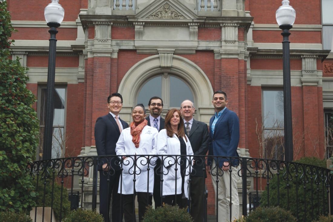
With a unification of the EHR across the health system, RedondaMiller, MD, MBA, president of Johns Hopkins Hospital, and Renee Demski, MSW, MBA, vice president of quality for Johns Hopkins Health System, created the Johns Hopkins Health System High-Value Care Committee to operationalize initiatives across all six hospitals and Johns Hopkins community physicians. The committee is under the leadership of Trushar Dungarani, DO, FHM, from Howard County General Hospital and Lenny Feldman, MD, SFHM, Amit Pahwa, MD, and Pamela Johnson, MD, from Johns Hopkins Hospital, and comprises provider representatives from each institution and other important contributors from various specialties, including Mike Borowitz, MD, PhD; Dr. Ken Lee, DrPH; Emily Pherson, PharmD; Amy Knight, MD; Tim Niessen, MD, MPH; Keisha Perrin, and Clare Rock.
Initiatives directed by the committee have included reducing inappropriate testing for Clostridium difficile, folate testing for anemia, and duplicative imaging exams, among others. In the 18 months since inception, the committee reduced charges to patients and payers by nearly $4 million and hospital costs by more than $200,000.
Members of this committee joined forces with like-minded institutions to create the High Value Practice Academic Alliance in 2016. Faculty leaders from more than 80 academic centers are now collaborating to increase health care value on a national scale through quality improvement projects, education, and dissemination.
SHM’s Award of Excellence in Teaching
Jennifer O’Toole, MD, MEd, is a med-peds hospitalist at the Cincinnati Children’s Hospital Medical Center and University of Cincinnati Medical Center. She serves as the director of the combined internal medicine and pediatrics residency program and is the director of education for the Children’s Hospital division of hospital medicine. In these roles, Dr. O’Toole has established an acute care track for her med-peds residents interested in careers in hospital medicine and a career development boot camp for early-career faculty members and fellows in the division of hospital medicine.
Perhaps Dr. O’Toole’s most instrumental role as an educator has been her involvement in influential educational programs nationally, including the I-PASS Handoff program. Implementation of I-PASS decreased medical errors by 23% and preventable adverse events by 30% in more than 10,000 patient admissions across nine North American hospitals. She has authored more than 30 peer-reviewed publications related to medical education and has presented more than 50 workshops at national meetings. After serving as a member of the planning committee for the Pediatric Hospital Medicine meeting, she is cochair for the 2018 meeting.
SHM’s Award of Clinical Excellence for Physicians
Rick Hilger, MD, SFHM, has been a hospitalist with HealthPartners Regions Hospital in St. Paul, Minnesota for 16 years. He is a national expert in the areas of readmission prevention and care delivery for high-utilizer patients. His work in this area was named Best Clinical Innovation at Hospital Medicine 2012. His collaborative approach to solving complex problems is demonstrated by the amount of time he has volunteered to help other organizations improve their care of high utilizers. Over 90 organizations and hospitals have reached out for assistance in starting their own committees, and Dr. Hilger has shared his time and care plan templates with each one. He was one of the first hospitalists asked to participate on a National Quality Forum committee and has improved hospital reimbursement by over 4 million dollars by developing an internal physician advisor program.
Dr. Hilger also has served on the SHM Annual Conference Committee and the Public Policy Committee, where he has worked with other committee members to advocate for policy changes related to observation status and readmission penalties.
SHM’s Award for Excellence in Humanitarian Services
Michelle Morse, MD, MPH, is a hospitalist and the assistant program director for the internal medicine residency at Brigham and Women’s Hospital in Boston. She is also the founding codirector of EqualHealth, an organization that aims to inspire and support the development of Haiti’s next generation of health care leaders. In 2015, Dr. Morse helped to found the Social Medicine Consortium, a global coalition of more than 450 people representing over 50 universities and organizations in 12 countries, which seeks to address the miseducation of health professionals.
In the aftermath of the 2010 earthquake in Haiti, Dr. Morse was compelled to collaborate with colleagues and friends to help build capacity of health care providers committed to social medicine, medical and nursing education, and social justice. She helped to open and operate a new 300-bed teaching hospital in rural Haiti and founded the first three residency training programs at the hospital. Since that time, the hospital has expanded to serve an area of 3 million people with an annual budget of $12 million.
SHM’s Award of Excellence in Research
Teryl Nuckols, MD, FHM, is a hospitalist and the director of general internal medicine at Cedars-Sinai Medical Center in Los Angeles. She also serves as associate professor of medicine at the University of California, Los Angeles, and a health services researcher at the RAND Corporation in Santa Monica.
Currently, she is principal investigator and coprincipal investigator on two projects funded by the Agency for Healthcare Research and Quality that are evaluating effects of the Medicare Hospital Readmissions Reduction Program. Dr. Nuckols was previously the principal investigator on two R01 research grants from the Agency for Healthcare Research and Quality and the recipient of a K08 Career Development Award. She has evaluated clinical practice guidelines on behalf of SHM as well as policy makers in California and Australia, and her work has led to more than 30 peer-reviewed publications in NEJM, Journal of Hospital Medicine, and JAMA Internal Medicine among others.
SHM’s Award of Clinical Excellence for NPs/PAs
Meredith K. Wold, PA-C, is the supervisor of advanced practice clinicians at HealthPartners Medical Group, which includes 20 nurse practitioners and physician assistants, at Regions Hospital in St. Paul, Minn. She is also the cofounder and co-curriculum director for the HealthPartners Hospital Medicine Physician Assistant Fellowship Program. Under her leadership, the fellowship program offers a clear curriculum that exposes key clinical scenarios and specialties crucial to hospital medicine, preparing new PAs to contribute immediately to team-based care upon completion of the fellowship.
To prevent burnout in a 7-on/7-off schedule, Ms. Wold thought creatively and developed a pooling and “draft” system to give some flexibility and breaks in the block schedule – almost like a fantasy football draft but with patient care shifts for noncontinuity services.
Excellence in Management in Hospital Medicine
Maria Lourdes Novelero, MA, MPA, is the associate chair for administration of the department of medicine at the University of California, San Francisco, and has also served as the administrator of the UCSF division of hospital medicine and its associated medical service from 2005 to 2016. In this role, she managed the department’s expansion from a 20-physician division to an 80-physician division. She also led the department’s pioneering efforts in quality, safety, and value.

Ms. Novelero created a structure to build and manage 10 different hospitalist-run services, including cancer, cardiology, neurosurgery, and liver transplant comanagement services; a procedure service; a palliative care service; and a large non–housestaff medicine service. She co-led a multidisciplinary effort to transform the discharge process, and established and cochaired the department of hospital medicine’s high value care committee, which catalyzed value improvement activities throughout UCSF. This committee’s work led to tangible value improvements, such as a 14% reduction in direct costs and a first-ever positive net margin for the medical service.
Ms. Novelero has more than 25 years of experience in management positions in the United States and Japan.
Certificate of Leadership in Hospital Medicine
Benji K. Mathews, MD, SFHM, CLHM, graduated from the Institute of Technology at the University of Minnesota and the University of Minnesota Medical School. He completed his internal medicine residency and chief residency through the University of Minnesota, Minneapolis. He joined HealthPartners as an academic hospitalist and holds the titles of section head of hospital medicine at Regions Hospital and director of point of care ultrasound (POCUS) for Hospital Medicine at HealthPartners. He is the president for the Minnesota Chapter of the Society of Hospital Medicine (SHM).
Dr. Mathews has a passion for medical education, care delivery, and quality. He is an assistant professor of medicine at the University of Minnesota. As such, he is core faculty for the internal medicine residency program and chair of the Clinical Competency Committee. Dr. Mathews is an active member of SHM as a member of SHM's annual meeting planning committee, organizing and setting the agenda for SHM's record breaking meetings for the last 4 years. Dr. Mathews also serves on SHM's Quality Improvement and Patient Safety Committee and is the chair of the Diagnostic Error Subcommittee. He has completed the Certificate of Leadership in Hospital Medicine (CLHM) with his area of focus on ultrasound in hospital medicine. His work in diagnostic error and POCUS has been well recognized with multiple workshops and presentations given locally and nationally. He is a Fellow in Diagnostic Safety through the Society to Improve Diagnosis in Medicine.
Dr. Mathews also has a passion for global health, rooted in his commitment to reducing health care disparities both locally and globally. He has worked with medical missions, NGOs, and orphanages in Nepal, India, Bolivia, Honduras, and Costa Rica. This led him to complete the global health course at the University of Minnesota.
SHM’s Junior Investigator Award
Christine D. Jones, MD, MS, is an assistant professor of medicine at the University of Colorado's Anschutz medical campus, Aurora, where she is the director of care transitions and director of scholarship for the division of hospital medicine.
Dr. Jones’ research is focused on improving care coordination between clinicians in different settings to improve outcomes for patients discharged with home health care. Her research is supported by a K08 career development award from the Agency for Healthcare Research and Quality. Her overall goal is to improve the quality of care transitions for hospitalized patients discharged to post-acute care settings, including home health care. Dr. Jones has presented at every SHM annual conference since she joined in 2013.
Chapter Excellence Awards
The Society of Hospital Medicine is proud to recognize outstanding SHM chapters at HM18 for the fifth annual Chapter Excellence Awards. Each year, chapters strive to demonstrate growth, sustenance, and innovation within their chapter activities, which are then applauded for their successes at SHM’s annual conference.
Platinum Chapters: Gulf States, Iowa, Maryland, Michigan, Minnesota, New Mexico, North Carolina Triangle, St. Louis.
Gold Chapters: Hampton Roads, Kentucky, North Jersey, Pacific Northwest.
Silver Chapters: Boston/Eastern Massachusetts, Houston, Los Angeles, Maine, NYC/Westchester, Rocky Mountain, San Francisco Bay Area, South Central PA, Southwest Florida, Wiregrass.
Outstanding Chapter of the Year
New Mexico. SHM’s New Mexico Chapter has been nominated to receive the Outstanding Chapter of the Year Award for 2017. Consistently, leadership and members go above and beyond the basic requirements to maintain status by creating and cultivating multiple innovative and dynamic opportunities for attendees to educate and network. The chapter participated in SHM’s pilot program to offer CME for chapters, bringing a CME-accredited educational presentation on the care of diverse patients. Their annual meeting this year featured a poster competition for residents, students, and early-career hospitalists; a didactic on antimicrobial stewardship; and a business meeting with elections, member benefits, and highlights of the chapter’s year, and chapter awards. The chapter initiated breakfast meetings with didactics on DACA and on burnout and wellness, and collaborated with the University of New Mexico, Albuquerque on a grand rounds series on physician wellness that was available via telecast to numerous hospitals and providers off site. The chapter hosted a book club, a networking session at HM17, and an informational meeting on SHM membership and the Fellows Program. They have supported the future of hospital medicine by having a table at the UNM School of Medicine’s student activities fair, a Resident of the Year Award, and a position on our council for a resident representative. They have collaborated with the sole internal medicine residency program in New Mexico so that residents in the hospitalist training track have membership in SHM, and they have expanded our juried poster competition to include students. The chapter’s level of originality is not only a benefit to the chapter, but SHM’s chapter program as a whole.
Rising Star Chapter
Kentucky. SHM’s Kentucky Chapter has been nominated to receive the Rising Star Chapter Award for 2017 for their innovation and growth. The chapter’s leadership was involved with the establishment of the Heartland Hospital Medicine Conference and hosted a hospital medicine career panel and bustling Research, Innovations, and Clinical Vignettes abstract and poster competition. Nearly 50 abstracts were submitted, and the top 40 were invited to present posters at the competition. These posters included a mix of attendings, advanced practice providers, residents, and students interested in participating in the local hospital medicine community. The chapter sponsored the first prize winner’s travel to attend Hospital Medicine 2018 and gave awards to best resident and best student posters. The Kentucky Chapter directly recruited 27 hospitalists to join SHM membership in 2017. The Kentucky Chapter is an active, enthusiastic chapter that is rapidly growing and thriving.
Student Hospitalist Scholarship Recipients
The Society of Hospital Medicine’s Physician in Training Committee launched a Student Hospitalist Scholar Grant program in 2015 for medical students to conduct mentored scholarly projects related to quality improvement and patient safety.
The program was expanded in 2017 and now includes both a summer and longitudinal program for students.
The committee is happy to announce the 4th year of scholar grant recipients to six students based on their ability and interest in hospital medicine, general qualifications, prior educational training, and promise for scholarly activity.
Summer Program
Ilana Scandariato
Cornell University, New York
Project: Understanding the experience of the long-term hospitalized patient with provider fragmentation: A qualitative study
Mentor: Ernie Esquivel, MD, FHM
Maximilian Hemmrich
University of Chicago
Project: Derivation and validation of a COPD readmission risk prediction tool
Mentor: Valerie Press, MD, MPH, SFHM
Sandeep Bala
University of Central Florida, Orlando
Project: The impact of plain language open medical notes on patient activation
Mentor: Marisha Burden, MD, SFHM
Longitudinal Program
Erin Rainosek
University of Texas, San Antonio
Project Title: Design thinking to improve patient experience
Mentor: Luci Leykum, MD, SFHM
Matthew Fallon
Creighton University, Omaha, Neb.
Project: Reducing the hospital readmission rate of congestive heart failure (CHF)
Mentor: Venkata Andukuri, MD, MPH, FHM
Philip Huang
University of Iowa, Iowa City
Project: Expanded patient regionalization to improve care efficiency
Mentor: Ethan Kuperman, MD, FHM
Resident Travel Grant Recipients
The Society of Hospital Medicine’s Physician in Training Committee launched a Resident Travel Grant Program in 2017 for residents to receive funding to attend SHM’s Annual Conference and be recognized for their scholarly work.
The Committee is pleased to announce the first year of Resident Travel Grant Recipients to 10 Residents based on their ability and interest in hospital medicine, research originality, and teaching value, and general qualifications.
Ashley M. Jenkins, MD
Cincinnati Children’s Hospital Medical Center
Poster 41 - Aren’t adults just big kids?: Standardizing care of adults in pediatric hospitals
Brian A. MacDonald Jr., MD, PhD
State University of New York at Buffalo
Poster 135 – Phosphatidylethanol level as a predictor of acute alcohol withdrawal
Christopher S. Bartlett, MD, MPH
University of New Mexico Health Sciences Center, Albuquerque
Poster 47 - Lessons learned from a resident-created experiential quality improvement and patient safety curriculum for medical and nursing students at the University of New Mexico
Christopher T. Su, MD, MPH
Montefiore Medical Center/Albert Einstein College of Medicine, New York
Poster 173 - Concurrent NSAID and warfarin use is associated with increased blood transfusions in hospitalized patients
Madeleine Ivrit Matthiesen, MD
Harvard Medical School/Massachusetts General Hospital Medicine–Pediatrics, Boston
Poster 69 - Resident perceptions of feedback and teaching
Neil Keshvani, MD
University of Texas Southwestern Medical Center, Dallas
Poster 237 - Improving respiratory rate measurement accuracy in the hospital: A quality improvement initiative
Peter N. Barish, MD
University of California, San Francisco
Poster 344 - Costs Related to potential overuse of respiratory viral panel PCRs in general medicine patients
Rachna Rawal, MD
Saint Louis University
Poster 259 - Empowering medicine residents to think before ordering daily labs: A quality improvement study
Yihan Chen, MD
University of California, Los Angeles, Medical Center
Poster 12 - Hospitalist-directed transfers improve emergency room length of stay
Zachary G. Jacobs, MD
University of California, San Francisco
Poster 233 - The prevalence of treating asymptomatic elevated blood pressure with intravenous antihypertensives on the general medicine wards: A potential target for a quality improvement inter-vention
Poster 96 - Factors Impacting time to antibiotic administration in patients with sepsis: A single-center study of electronic health record data
SHM’s Award of Excellence in Outstanding Service in Hospital Medicine
Dr. Flora Kisuule, MD, SFHM, is an assistant professor at Johns Hopkins School of Medicine and the vice chair for clinical operations for the department of medicine at Johns Hopkins Bayview Medical Center. While at Johns Hopkins University, both in Baltimore, she codeveloped a hospitalist fellowship program that she now directs, as well as a fellowship program specifically for nurse practitioners and physician assistants. Under her leadership as the associate director of the division of hospital medicine, she helped to bring Johns Hopkins Bayview hospitalists’ quality and mortality indicators into the top 5% nationwide and reduced hospital-acquired conditions at Hopkins Bayview to the best of the four regional Hopkins Health System hospitals.
Nationally, she has served on several SHM committees, facilitated at multiple SHM leadership courses, served as vice president of SHM’s Baltimore Chapter, and consulted on hospitalist programs around the country. Internationally, Dr. Kisuule has developed and mentored hospitalist programs in Saudi Arabia, the United Arab Emirates, and Central America. She is currently developing a training program in hospital medicine in Panama and is a Senior Fellow in Hospital Medicine.
SHM’s Award of Excellence in Teamwork in Quality Improvement
Since 2011, the Johns Hopkins Health System hospitals have conducted quality improvement projects to increase the value of care for their patients. Their amazing work catalyzed a grass-roots initiative involving faculty and residents at both Johns Hopkins Hospital and Johns Hopkins Bayview Medical Center.
With a unification of the EHR across the health system, RedondaMiller, MD, MBA, president of Johns Hopkins Hospital, and Renee Demski, MSW, MBA, vice president of quality for Johns Hopkins Health System, created the Johns Hopkins Health System High-Value Care Committee to operationalize initiatives across all six hospitals and Johns Hopkins community physicians. The committee is under the leadership of Trushar Dungarani, DO, FHM, from Howard County General Hospital and Lenny Feldman, MD, SFHM, Amit Pahwa, MD, and Pamela Johnson, MD, from Johns Hopkins Hospital, and comprises provider representatives from each institution and other important contributors from various specialties, including Mike Borowitz, MD, PhD; Dr. Ken Lee, DrPH; Emily Pherson, PharmD; Amy Knight, MD; Tim Niessen, MD, MPH; Keisha Perrin, and Clare Rock.
Initiatives directed by the committee have included reducing inappropriate testing for Clostridium difficile, folate testing for anemia, and duplicative imaging exams, among others. In the 18 months since inception, the committee reduced charges to patients and payers by nearly $4 million and hospital costs by more than $200,000.
Members of this committee joined forces with like-minded institutions to create the High Value Practice Academic Alliance in 2016. Faculty leaders from more than 80 academic centers are now collaborating to increase health care value on a national scale through quality improvement projects, education, and dissemination.
SHM’s Award of Excellence in Teaching
Jennifer O’Toole, MD, MEd, is a med-peds hospitalist at the Cincinnati Children’s Hospital Medical Center and University of Cincinnati Medical Center. She serves as the director of the combined internal medicine and pediatrics residency program and is the director of education for the Children’s Hospital division of hospital medicine. In these roles, Dr. O’Toole has established an acute care track for her med-peds residents interested in careers in hospital medicine and a career development boot camp for early-career faculty members and fellows in the division of hospital medicine.
Perhaps Dr. O’Toole’s most instrumental role as an educator has been her involvement in influential educational programs nationally, including the I-PASS Handoff program. Implementation of I-PASS decreased medical errors by 23% and preventable adverse events by 30% in more than 10,000 patient admissions across nine North American hospitals. She has authored more than 30 peer-reviewed publications related to medical education and has presented more than 50 workshops at national meetings. After serving as a member of the planning committee for the Pediatric Hospital Medicine meeting, she is cochair for the 2018 meeting.
SHM’s Award of Clinical Excellence for Physicians
Rick Hilger, MD, SFHM, has been a hospitalist with HealthPartners Regions Hospital in St. Paul, Minnesota for 16 years. He is a national expert in the areas of readmission prevention and care delivery for high-utilizer patients. His work in this area was named Best Clinical Innovation at Hospital Medicine 2012. His collaborative approach to solving complex problems is demonstrated by the amount of time he has volunteered to help other organizations improve their care of high utilizers. Over 90 organizations and hospitals have reached out for assistance in starting their own committees, and Dr. Hilger has shared his time and care plan templates with each one. He was one of the first hospitalists asked to participate on a National Quality Forum committee and has improved hospital reimbursement by over 4 million dollars by developing an internal physician advisor program.
Dr. Hilger also has served on the SHM Annual Conference Committee and the Public Policy Committee, where he has worked with other committee members to advocate for policy changes related to observation status and readmission penalties.
SHM’s Award for Excellence in Humanitarian Services
Michelle Morse, MD, MPH, is a hospitalist and the assistant program director for the internal medicine residency at Brigham and Women’s Hospital in Boston. She is also the founding codirector of EqualHealth, an organization that aims to inspire and support the development of Haiti’s next generation of health care leaders. In 2015, Dr. Morse helped to found the Social Medicine Consortium, a global coalition of more than 450 people representing over 50 universities and organizations in 12 countries, which seeks to address the miseducation of health professionals.
In the aftermath of the 2010 earthquake in Haiti, Dr. Morse was compelled to collaborate with colleagues and friends to help build capacity of health care providers committed to social medicine, medical and nursing education, and social justice. She helped to open and operate a new 300-bed teaching hospital in rural Haiti and founded the first three residency training programs at the hospital. Since that time, the hospital has expanded to serve an area of 3 million people with an annual budget of $12 million.
SHM’s Award of Excellence in Research
Teryl Nuckols, MD, FHM, is a hospitalist and the director of general internal medicine at Cedars-Sinai Medical Center in Los Angeles. She also serves as associate professor of medicine at the University of California, Los Angeles, and a health services researcher at the RAND Corporation in Santa Monica.
Currently, she is principal investigator and coprincipal investigator on two projects funded by the Agency for Healthcare Research and Quality that are evaluating effects of the Medicare Hospital Readmissions Reduction Program. Dr. Nuckols was previously the principal investigator on two R01 research grants from the Agency for Healthcare Research and Quality and the recipient of a K08 Career Development Award. She has evaluated clinical practice guidelines on behalf of SHM as well as policy makers in California and Australia, and her work has led to more than 30 peer-reviewed publications in NEJM, Journal of Hospital Medicine, and JAMA Internal Medicine among others.
SHM’s Award of Clinical Excellence for NPs/PAs
Meredith K. Wold, PA-C, is the supervisor of advanced practice clinicians at HealthPartners Medical Group, which includes 20 nurse practitioners and physician assistants, at Regions Hospital in St. Paul, Minn. She is also the cofounder and co-curriculum director for the HealthPartners Hospital Medicine Physician Assistant Fellowship Program. Under her leadership, the fellowship program offers a clear curriculum that exposes key clinical scenarios and specialties crucial to hospital medicine, preparing new PAs to contribute immediately to team-based care upon completion of the fellowship.
To prevent burnout in a 7-on/7-off schedule, Ms. Wold thought creatively and developed a pooling and “draft” system to give some flexibility and breaks in the block schedule – almost like a fantasy football draft but with patient care shifts for noncontinuity services.
Excellence in Management in Hospital Medicine
Maria Lourdes Novelero, MA, MPA, is the associate chair for administration of the department of medicine at the University of California, San Francisco, and has also served as the administrator of the UCSF division of hospital medicine and its associated medical service from 2005 to 2016. In this role, she managed the department’s expansion from a 20-physician division to an 80-physician division. She also led the department’s pioneering efforts in quality, safety, and value.
Ms. Novelero created a structure to build and manage 10 different hospitalist-run services, including cancer, cardiology, neurosurgery, and liver transplant comanagement services; a procedure service; a palliative care service; and a large non–housestaff medicine service. She co-led a multidisciplinary effort to transform the discharge process, and established and cochaired the department of hospital medicine’s high value care committee, which catalyzed value improvement activities throughout UCSF. This committee’s work led to tangible value improvements, such as a 14% reduction in direct costs and a first-ever positive net margin for the medical service.
Ms. Novelero has more than 25 years of experience in management positions in the United States and Japan.
Certificate of Leadership in Hospital Medicine
Benji K. Mathews, MD, SFHM, CLHM, graduated from the Institute of Technology at the University of Minnesota and the University of Minnesota Medical School. He completed his internal medicine residency and chief residency through the University of Minnesota, Minneapolis. He joined HealthPartners as an academic hospitalist and holds the titles of section head of hospital medicine at Regions Hospital and director of point of care ultrasound (POCUS) for Hospital Medicine at HealthPartners. He is the president for the Minnesota Chapter of the Society of Hospital Medicine (SHM).
Dr. Mathews has a passion for medical education, care delivery, and quality. He is an assistant professor of medicine at the University of Minnesota. As such, he is core faculty for the internal medicine residency program and chair of the Clinical Competency Committee. Dr. Mathews is an active member of SHM as a member of SHM's annual meeting planning committee, organizing and setting the agenda for SHM's record breaking meetings for the last 4 years. Dr. Mathews also serves on SHM's Quality Improvement and Patient Safety Committee and is the chair of the Diagnostic Error Subcommittee. He has completed the Certificate of Leadership in Hospital Medicine (CLHM) with his area of focus on ultrasound in hospital medicine. His work in diagnostic error and POCUS has been well recognized with multiple workshops and presentations given locally and nationally. He is a Fellow in Diagnostic Safety through the Society to Improve Diagnosis in Medicine.
Dr. Mathews also has a passion for global health, rooted in his commitment to reducing health care disparities both locally and globally. He has worked with medical missions, NGOs, and orphanages in Nepal, India, Bolivia, Honduras, and Costa Rica. This led him to complete the global health course at the University of Minnesota.
SHM’s Junior Investigator Award
Christine D. Jones, MD, MS, is an assistant professor of medicine at the University of Colorado's Anschutz medical campus, Aurora, where she is the director of care transitions and director of scholarship for the division of hospital medicine.
Dr. Jones’ research is focused on improving care coordination between clinicians in different settings to improve outcomes for patients discharged with home health care. Her research is supported by a K08 career development award from the Agency for Healthcare Research and Quality. Her overall goal is to improve the quality of care transitions for hospitalized patients discharged to post-acute care settings, including home health care. Dr. Jones has presented at every SHM annual conference since she joined in 2013.
Chapter Excellence Awards
The Society of Hospital Medicine is proud to recognize outstanding SHM chapters at HM18 for the fifth annual Chapter Excellence Awards. Each year, chapters strive to demonstrate growth, sustenance, and innovation within their chapter activities, which are then applauded for their successes at SHM’s annual conference.
Platinum Chapters: Gulf States, Iowa, Maryland, Michigan, Minnesota, New Mexico, North Carolina Triangle, St. Louis.
Gold Chapters: Hampton Roads, Kentucky, North Jersey, Pacific Northwest.
Silver Chapters: Boston/Eastern Massachusetts, Houston, Los Angeles, Maine, NYC/Westchester, Rocky Mountain, San Francisco Bay Area, South Central PA, Southwest Florida, Wiregrass.
Outstanding Chapter of the Year
New Mexico. SHM’s New Mexico Chapter has been nominated to receive the Outstanding Chapter of the Year Award for 2017. Consistently, leadership and members go above and beyond the basic requirements to maintain status by creating and cultivating multiple innovative and dynamic opportunities for attendees to educate and network. The chapter participated in SHM’s pilot program to offer CME for chapters, bringing a CME-accredited educational presentation on the care of diverse patients. Their annual meeting this year featured a poster competition for residents, students, and early-career hospitalists; a didactic on antimicrobial stewardship; and a business meeting with elections, member benefits, and highlights of the chapter’s year, and chapter awards. The chapter initiated breakfast meetings with didactics on DACA and on burnout and wellness, and collaborated with the University of New Mexico, Albuquerque on a grand rounds series on physician wellness that was available via telecast to numerous hospitals and providers off site. The chapter hosted a book club, a networking session at HM17, and an informational meeting on SHM membership and the Fellows Program. They have supported the future of hospital medicine by having a table at the UNM School of Medicine’s student activities fair, a Resident of the Year Award, and a position on our council for a resident representative. They have collaborated with the sole internal medicine residency program in New Mexico so that residents in the hospitalist training track have membership in SHM, and they have expanded our juried poster competition to include students. The chapter’s level of originality is not only a benefit to the chapter, but SHM’s chapter program as a whole.
Rising Star Chapter
Kentucky. SHM’s Kentucky Chapter has been nominated to receive the Rising Star Chapter Award for 2017 for their innovation and growth. The chapter’s leadership was involved with the establishment of the Heartland Hospital Medicine Conference and hosted a hospital medicine career panel and bustling Research, Innovations, and Clinical Vignettes abstract and poster competition. Nearly 50 abstracts were submitted, and the top 40 were invited to present posters at the competition. These posters included a mix of attendings, advanced practice providers, residents, and students interested in participating in the local hospital medicine community. The chapter sponsored the first prize winner’s travel to attend Hospital Medicine 2018 and gave awards to best resident and best student posters. The Kentucky Chapter directly recruited 27 hospitalists to join SHM membership in 2017. The Kentucky Chapter is an active, enthusiastic chapter that is rapidly growing and thriving.
Student Hospitalist Scholarship Recipients
The Society of Hospital Medicine’s Physician in Training Committee launched a Student Hospitalist Scholar Grant program in 2015 for medical students to conduct mentored scholarly projects related to quality improvement and patient safety.
The program was expanded in 2017 and now includes both a summer and longitudinal program for students.
The committee is happy to announce the 4th year of scholar grant recipients to six students based on their ability and interest in hospital medicine, general qualifications, prior educational training, and promise for scholarly activity.
Summer Program
Ilana Scandariato
Cornell University, New York
Project: Understanding the experience of the long-term hospitalized patient with provider fragmentation: A qualitative study
Mentor: Ernie Esquivel, MD, FHM
Maximilian Hemmrich
University of Chicago
Project: Derivation and validation of a COPD readmission risk prediction tool
Mentor: Valerie Press, MD, MPH, SFHM
Sandeep Bala
University of Central Florida, Orlando
Project: The impact of plain language open medical notes on patient activation
Mentor: Marisha Burden, MD, SFHM
Longitudinal Program
Erin Rainosek
University of Texas, San Antonio
Project Title: Design thinking to improve patient experience
Mentor: Luci Leykum, MD, SFHM
Matthew Fallon
Creighton University, Omaha, Neb.
Project: Reducing the hospital readmission rate of congestive heart failure (CHF)
Mentor: Venkata Andukuri, MD, MPH, FHM
Philip Huang
University of Iowa, Iowa City
Project: Expanded patient regionalization to improve care efficiency
Mentor: Ethan Kuperman, MD, FHM
Resident Travel Grant Recipients
The Society of Hospital Medicine’s Physician in Training Committee launched a Resident Travel Grant Program in 2017 for residents to receive funding to attend SHM’s Annual Conference and be recognized for their scholarly work.
The Committee is pleased to announce the first year of Resident Travel Grant Recipients to 10 Residents based on their ability and interest in hospital medicine, research originality, and teaching value, and general qualifications.
Ashley M. Jenkins, MD
Cincinnati Children’s Hospital Medical Center
Poster 41 - Aren’t adults just big kids?: Standardizing care of adults in pediatric hospitals
Brian A. MacDonald Jr., MD, PhD
State University of New York at Buffalo
Poster 135 – Phosphatidylethanol level as a predictor of acute alcohol withdrawal
Christopher S. Bartlett, MD, MPH
University of New Mexico Health Sciences Center, Albuquerque
Poster 47 - Lessons learned from a resident-created experiential quality improvement and patient safety curriculum for medical and nursing students at the University of New Mexico
Christopher T. Su, MD, MPH
Montefiore Medical Center/Albert Einstein College of Medicine, New York
Poster 173 - Concurrent NSAID and warfarin use is associated with increased blood transfusions in hospitalized patients
Madeleine Ivrit Matthiesen, MD
Harvard Medical School/Massachusetts General Hospital Medicine–Pediatrics, Boston
Poster 69 - Resident perceptions of feedback and teaching
Neil Keshvani, MD
University of Texas Southwestern Medical Center, Dallas
Poster 237 - Improving respiratory rate measurement accuracy in the hospital: A quality improvement initiative
Peter N. Barish, MD
University of California, San Francisco
Poster 344 - Costs Related to potential overuse of respiratory viral panel PCRs in general medicine patients
Rachna Rawal, MD
Saint Louis University
Poster 259 - Empowering medicine residents to think before ordering daily labs: A quality improvement study
Yihan Chen, MD
University of California, Los Angeles, Medical Center
Poster 12 - Hospitalist-directed transfers improve emergency room length of stay
Zachary G. Jacobs, MD
University of California, San Francisco
Poster 233 - The prevalence of treating asymptomatic elevated blood pressure with intravenous antihypertensives on the general medicine wards: A potential target for a quality improvement inter-vention
Poster 96 - Factors Impacting time to antibiotic administration in patients with sepsis: A single-center study of electronic health record data
New for HM19: Call for content expanded to include speaker, topic proposals
The Call for Content for HM19 – open to both Society of Hospital Medicine members and nonmembers – is being expanded from solely workshop submissions to include speaker and topic proposals.
“The open call for workshops has been in place for a number of years,” said Dustin T. Smith, MD, FHM, an associate professor of medicine at Emory University in Atlanta. Dr. Smith is an HM18 assistant course director and will be the primary course director for HM19. “In the past, we used a targeted survey method to get speaker/topic suggestions for the didactic sessions. The change was made, in part, based on member and annual meeting feedback along with strong SHM leadership support and guidance.”
This change, for both workshops and sessions, will allow for more speaker diversity, as well as open the door for new presenters. “We really feel this ability to propose content and speakers will resonate with our members and nonmembers alike, giving them a voice in annual conference planning,” he continued. “Our overall goal is to create new and exciting meeting formats each year that spark interest and promote overall attendee learning.”
Dr. Smith hosted an informational presentation on the process as part of the MEDTalks session on Monday.
When asked about themes for next year’s conference, Dr. Smith stated, “I anticipate that some of the areas of focus will center around health policy, advocacy for patients, and the latest in hospital medicine.” The SHM Annual Meeting Committee is responsible for HM19’s didactic content, the foundation of which will be rooted in hot topics and subject areas in the field. Planning also will take into account momentum and feedback from HM18.
“We have a few innovative and exciting educational tracks at HM18, including The Great Debate and Seasoning Your Career. If they are successful, we hope to offer similar ones next year,” Dr. Smith explained. “Some already popular tracks such as Rapid Fire clinical topics and Clinical Updates will likely remain meeting staples, although their content will certainly change.
“I am a working member of a talented group of incredibly knowledgeable hospitalists who form the Annual Conference Committee for both HM18 and HM19. With amazing SHM leadership and staff liaison guidance and support, we will be planning content for the bulk of the educational portion of next year’s conference,” he noted.
Dr. Smith, an Atlanta native, is an academic hospitalist and assistant chief of medicine for education in the Medical Specialty Care Services Line at the Atlanta Veterans Affairs (VA) Medical Center. He graduated from Emory University in Atlanta and completed his residency at the University of California, San Francisco, with distinction.
“I was immersed in large hospital medicine programs full of terrific practicing hospitalists at both institutions,” Dr. Smith recalled. “It became apparent that, when I completed my training, I wanted to pursue an academic hospital medicine career in a large, urban setting. Being from Atlanta, I wanted to reconnect with Emory, so I accepted a position at the Atlanta VA Medical Center as a hospitalist.” (The Atlanta VA is an Emory academic affiliate.)
The recipient of numerous teaching awards, Dr. Smith is an associate program director for the J. Willis Hurst Internal Medicine Residency Program at Emory, is the chair of the Emory division of hospital medicine Education Council, and has been codirector of the annual Southern Hospital Medicine Conference since 2012. The Emory department of medicine has named him “Distinguished Physician” for his significant clinical contributions.
“I am lucky enough to have been surrounded by great mentors in hospital medicine, both during training and as early- and now mid-career faculty. Mentoring and professional development are really key elements to building a successful and sustainable career in hospital medicine,” he said. “Additionally, I am incredibly humbled and thankful to have the opportunity to care for veterans in my various clinical roles at the Atlanta VA.”
When asked about the field, Dr. Smith said, “In my opinion, hospital medicine is and has been vital for the successful operation of health care and the management of hospitalized patients for more than a decade now. Hospitalists not only provide top-notch care for inpatients but also play so many other important roles in medicine in areas such as quality improvement, practice management, comanagement, research, and education.”
Understanding palliative care: An important part of practicing hospital medicine
according to Brett Hendel-Paterson, MD, FHM, a hospitalist at Region’s Hospital in St. Paul, Minn. and a presenter for this session.
Dr. Hendel-Paterson, Jeffrey L. Greenwald, MD, SFHM, of Massachusetts General Hospital, Boston, and Jeffrey Frank, MD, MBA, of Vituity, will each present on the topic of administering palliative care as a hospitalist and why it is important for hospitalists to better understand this area of medicine.
A common misunderstanding about palliative care is that it is end-of-life care only, a misconception within both the medical and patient community. Most people believe that palliative care is associated with the “angel of death,” as Dr. Greenwald stated. Palliative care does encompass end-of-life care but is also associated with life-limiting illness. Both areas of palliative can be improved with better patient communication and symptom management.
As frontline providers at times of critical illness, and throughout illness, hospitalists are ideally positioned to provide palliative care services, Dr. Greenwald stated during an interview.
With hospitalists in such a prominent role in providing palliative care, Dr. Hendel-Paterson offered a detailed explanation about why the information from this session is important for hospitalists.
“The majority of Americans who die in this country die in hospitals. We see and we know that patients sometimes get more aggressive care leading to greater suffering in their final days,” he said. “As hospitalists, we are expected to be the primary physicians in the hospital caring for patients with a variety of health conditions. We are expected to have a basic expertise and be able to independently manage health conditions. For example, we are expected to be able to diagnose and treat pneumonia without consulting infectious disease or pulmonology specialists for basic care. In the same way, we must be able to communicate well with our patients and families and help lead them through discussions of prognosis and advance care planning. Primary palliative care refers to the skill set that includes communications about serious illness and basic symptom management.”
Dr. Greenwald expanded on Dr. Hendel-Paterson’s point concerning the growing need for hospitalists who are competent in palliative care.
“As the population ages, this issue is going to become more and more important for our field, because there isn’t a sufficient pipeline, current state – or predicted future state – of palliative care providers in hospitals to meet the need. So there’s a gap in the need, and that need is increasing.”
According to Dr. Hendel-Paterson, he and his copresenters “hope that, after this session, participants will better understand primary palliative care, take ownership of end-of-life care of their patients, and will be motivated to increase skills in areas where they are lacking.”
Building on this idea of increasing one’s skills as a hospitalist, he emphasized the importance of understanding palliative care.
“The ability to practice high-quality primary palliative care is essential to being a competent hospitalist.”
Primary Palliative Care – What Every Hospitalist Should Know
Wednesday, 10:00-10:40 a.m.
Crystal Ballroom J1
Using data to drive quality improvement projects
Measuring and analyzing data is essential for any quality improvement project. Data can validate whether an anecdotal assumption indicates the need for a quality improvement initiative and can help showcase the success of any intervention.
At today’s session, “Using Data to Inform Quality Improvement,” attendees will learn how to develop balanced metrics for quality improvement projects, understand distinctions between quality improvement and traditional research projects, and identify how different measures of the same data can tell vastly different stories.
The session will be divided into three 20-minute sections. Ethan Kuperman, MD, MSc, FHM, clinical assistant professor, University of Iowa Health Care, Iowa City, will begin by discussing how to develop balanced metrics by exploring a venous thromboembolism prophylaxis quality improvement project.
Next, Aparna Kamath, MD, MS, SFHM, assistant professor and academic hospitalist, Duke University Health System, Durham, N.C., will demonstrate the distinction between quality improvement and research by exploring medication reconciliation. “I will also emphasize the need for feasibility and rigor in data gathering and analysis in addition to accuracy when doing a quality improvement project,” she said. “Our workshop will make these tasks less daunting for hospitalists doing quality improvement projects.”
Finally, Justin Glasgow, MD, PhD, inpatient medicine faculty and Value Institute senior clinical scholar, Christiana Care Health System, Newark, Del., will explore how improper data definitions led to differing interpretations while developing and deploying an early warning system. “Attendees will learn how to design a quality improvement project to ensure that they measure interventions in a manner that ensures they have had true success, while not creating any unintended consequences,” Dr. Glasgow said.
“The session is important as it will help attendees develop successful projects that their colleagues will respect and that their bosses will admire,” added Dr. Glasgow. “They will gain confidence to turn their experience into distributable scholarship.”
“Hospitalists at all stages of their careers can benefit from this presentation,” Dr. Kuperman said. “Whether you’re preparing for your first quality improvement project or getting ready for a systemwide redesign, collecting the wrong data or misusing collected data can ruin your results or lead you to the wrong conclusions.”
Each of the three presenters are well versed on the topic and have successfully analyzed data for numerous quality improvement projects and presented these data both in publications and in presentations, including for the Society of Hospital Medicine.
“My best qualification for this talk is the number of times I’ve messed up,” Dr. Kuperman said. “Early in my career, I had several projects in which I spent valuable resources creating an initiative, rolling out the implementation, and then wasn’t able to capture reliable outcomes – ruining the chances for scholarship or renewal.”
Through this presentation, Dr. Kuperman hopes that he can reach other hospitalists and soon-to-be hospitalists and teach them some hard-earned lessons so that they can be more successful from the start.
None of the three presenters have any financial disclosures.
Using Data to Inform Quality Improvement
Wednesday, 8:40-9:40 a.m.
Crystal Ballroom G1/A&B
A call for innovation: Dr. Robert Wachter to discuss evolution of HM at closing plenary
As has become tradition, Robert Wachter, MD, MHM, of the University of California, San Francisco, will close the conference today and will focus on the broader changes that must happen as the role of the hospitalist continues to evolve.
“I am going to talk about the changes in the world of hospital care and the importance of the field to innovate,” Dr. Wachter said. “To me, there are gravitational forces in the health care world that are making … patients who are in hospitals sicker than they were before. More and more patients are going to be cared for in outpatient settings and at home. We are going start to ... see things like sensors and telemedicine to enable more care outside of the hospital.”
Dr. Wachter said hospital medicine must evolve and mature to continue to prove that hospitalists are indispensable staff members within the hospital.
“That was why the field became the fastest-growing profession in medical history. We can’t sit on our laurels. We have to continue to innovate,” he said. “Even as the system changes around us, I am confident that we will innovate. My talk will be a pep talk and will include reflections on how the world of health care is changing and what those changes will mean to hospitalists.”
Dr. Wachter noted that the trend of steering patients who are less sick to the outpatient setting, as well as other economic factors, would change the nature of hospitalist practice.
“It will be more acuity, more intensity, more complex relationships with your own hospital and often with partner hospitals,” he said. “More of the work will be digitally enabled than it would have been 5 or 10 years ago.”
Integration of data and technology innovation will be a key to better serving this sicker population, Dr. Wachter predicted. We need “to take much fuller advantage than we have so far of the fact that we are all dealing with digital records, and the decision support, the data analytics, the artificial intelligence that we get from our computer systems is pretty puny,” he said. “That is partly why physicians don’t love their computers so much. They spend huge amounts of time entering data into computers and don’t get much useful information out of it.”
Dr. Wachter also warned that too much data could have negative effects on the delivery of care.
“One of the challenges we face is continuing to stay alert, not turn our brains off, and become increasingly dependent on the computer to give us information,” he said. “How do we avoid the challenges we’ve already seen from things like alert and alarm fatigue as the computer becomes more robust as an information source? There is always the risk our computers are going to overwhelm us with too much information, and we are going to fall asleep at the switch. Or when the computer says something that really is not right for a patient, we will not be thinking clearly enough to catch it.”
Despite the looming challenges and industry consolidations that are expected, Dr. Wachter doesn’t believe there will be any shortage of demand for hospitalists.
“I think, in most circumstances, [hospitalists are a protected] profession given the complexity, the high variations, and the dependence that it still has on seeing the patient, talking to the patient, and having discussions with multiple consultants,” he said. “It’s a pretty hard thing to replace with technology. Overall, the job situation is pretty bright.”
Learn to employ cultural humility
Many health care providers are guilty of having implicit or unconscious biases against patients, which can negatively affect the care they give. “Once providers come to this realization, they can make a conscious effort to neutralize these biases from manifesting throughout a practice,” said Aziz Ansari, DO, SFHM, associate chief medical officer, Clinical Optimization and Revenue Integrity, and associate professor of medicine, Loyola University Medical Center, Maywood, Ill., who will present today’s session “Winning Hearts and Minds at the Bedside: Battling Unconscious Bias Through Cultural Humility.”
By practicing cultural humility, hospitalists can gain trust from patients and their families, whom they do not know in their everyday practice. This encourages providers to be humble, ascertain what is important to the patient, and learn from every patient encounter.
The session will begin with a case study involving bias, followed by a self-reflection exercise. To determine whether you may be biased against some patient groups, Dr. Ansari recommends taking the online implicit association test at Implicit.harvard.edu.
As a palliative care specialist, Dr. Ansari has repeatedly faced situations in which a lack of cultural humility caused patients or their family member to foster mistrust toward a provider. Consequently, patients and family members may choose aggressive measures that providers might consider futile.
Dr. Ansari also will define what implicit or unconscious biases entail in greater detail. The discussion will then circle back to the original case and reveal how providers can improve their mindset when facing difficult situations by employing a tool called “The 5 Rs of Cultural Humility,” which include reflection, respect, regard, relevance, and resiliency.
Dr. Ansari spearheaded the development of the 5 Rs tool when he chaired the Cultural Humility Workgroup of SHM’s Practice Management Committee. “The goal is to use the tool to attain cultural humility and transform a potentially negative encounter into a gratifying one,” he said.
At a minimum, attendees should take time during the session to reflect on their own thoughts and biases. “This introspection can bring to light practices that providers may have been unaware of and, ultimately, can change their behaviors so every patient and their family feels that they are being seen and heard,” Dr. Ansari said. “In today’s current climate it is more important than ever for providers to self-reflect on their attitudes and perceptions because an increasing number of groups in our diverse society are feeling more marginalized.”
Winning Hearts and Minds at the Bedside: Battling Unconscious Bias Through Cultural Humility
Wednesday, 9:10-9:50 a.m.
Crystal Ballroom G2/C-F
Session to cover expanding HM scope of practice to pre- and posthospitalization care
As the field of hospital medicine continues to grow, it is experiencing more requests for assistance with pre- and posthospital care. These increasing demands will be the focus of today’s session “Addressing the Expanding Scope of Practice in Hospital Medicine.”
“As hospital medicine continues to advance, we are being asked to help hospitals and health care systems with challenges that extend beyond the hospital,” presenter Nick Fitterman, MD, SFHM, vice chair of hospital medicine of Northwell Health in Long Island, N.Y., explained in an interview. “Sometimes, this is a natural extension of hospital medicine; other times, it may reflect gaps in primary care or the health care system in general. Hospital medicine can be supportive, but our course must be deliberate and not extend beyond our scope of practice.
“The health care system is facing a variety of challenges, which, in turn, make an impact on hospital medicine,” stated Dr. Fitterman. “Factors that result in increased demands on the field include lack of access to posthospitalization care, high-acuity patients in subacute rehabilitation, rising health care costs, and value-based payments that include ‘bundles’ of care stretching beyond hospitalization. Finally, the nation has a population that’s aging,” he continued, “thanks to the ‘silver tsunami’ of baby boomers.”
The session will include presentations by leaders in the fields of post-acute clinics and hospitals at home. Lauren Doctoroff, MD, FHM, of Beth Israel Deaconess Medical Center in Boston will discuss post-acute care clinics; David Levine, MD, of Brigham and Women’s Hospital and Harvard Medical School, both in Boston, will cover some hospital-at-home developments; and Efren Manjarrez, MD, SFHM, of the University of Miami Health System will speak about preoperative care. Dr. Fitterman will provide examples of the expanded scope of practice issues that can arise in a large health care system.
“There are a finite number of beds in any given hospital and extending our reach into pre- or posthospitalization care is an avenue to expand our book of business, cultivate new skills, and engage patients and colleagues in new settings,” Dr. Fitterman explained. “Done with a clear vision and plan and within our scope of practice, this can be quite rewarding. Executed only to meet a demand but without proper resources, this can pose a new challenge and become frustrating.”
“HM18 participants who attend the session will receive some insights into what works and what doesn’t work regarding extrahospital care for hospital medicine,” continued Dr. Fitterman. “And we hope it will help hospital medicine practitioners and groups as they determine where to focus their efforts.”
When asked about an overall take-home message for the session, Dr. Fitterman stated, “We hospital medicine professionals must be wary of accepting every challenge posed to us to solve. This session should provide those in attendance with tools that will assist in their decision making.”
Addressing the Expanding Scope of Practice in Hospital Medicine
Wednesday, 7:30-8:30 a.m.
Grand Ballroom 7B
Welcome to Day 3 at HM18
What an amazing 3 days! As we spoke on the first day, delivering higher value of care has been a highlight of this annual conference. With thought-provoking sessions from Kate Goodrich, MD, MHS, on payment reform; Eric Howell, MD, on “Value is NOT a 4-Letter Word”; and Lenny Feldman, MD, SFHM, on “Things We Do for No Reason,” hospital medicine’s focus on value is clear. I hope you also had the chance to attend the sessions on clinical topics of medicine, education, and practice management.
The final day of the conference is no exception when it comes to topics with impact. Starting off at 7:30 a.m., there is a diverse group of sessions on clinical medicine for adults and pediatrics, including “Peter Pan Grows Up: How to Care for Your Patients with Chronic Pediatric Conditions,” topics in health policy, how to advance in academic medicine with a focus on writing a winning abstract, and applying what you have learned at HM18 back home.
This day also offers sessions on advancement with topics for mid-career hospitalists and those aspiring to leadership roles. Promotion in academic medicine also will be addressed.
You can further strengthen your presentation skills in sessions that will teach you how to create valuable teaching points on the fly and deliver memorable lectures and talks. Other sessions will enhance your skills in emotional intelligence.
As always, we are extremely fortunate to wrap up the day with Bob Wachter, MD, MHM, who is sure to deliver a thought-provoking and entertaining presentation entitled “Hospitalists as Drivers of Innovation and System Change: Are We Doing Enough?”
As you wrap up your final day, I wish you a safe journey back home. Please continue your engagement with the Society through chapter meetings in your area, signing up for committees and interest groups, and dropping us a line – we look forward to continuing to work for you in the coming year.
Dr. Afsar is the incoming president of the Society of Hospital Medicine and the chief ambulatory officer & CMO for ACOs at UC Irvine Health in Orange, Calif.
Gene variants linked to survival after HSCT

New research has revealed a link between rare gene variants and survival after hematopoietic stem cell transplant (HSCT).
Researchers performed exome sequencing in nearly 2500 HSCT recipients and their matched, unrelated donors.
The sequencing revealed several gene variants—in both donors and recipients—that were significantly associated with overall survival (OS), transplant-related mortality (TRM), and disease-related mortality (DRM) after HSCT.
Qianqian Zhu, PhD, of Roswell Park Comprehensive Cancer Center in Buffalo, New York, and her colleagues described these findings in Blood.
The team performed exome sequencing—using the Illumina HumanExome BeadChip—in patients who participated in the DISCOVeRY-BMT study.
This included 2473 HSCT recipients who had acute myeloid leukemia, acute lymphoblastic leukemia, or myelodysplastic syndromes. It also included 2221 donors who were a 10/10 human leukocyte antigen match for each recipient.
The researchers looked at genetic variants in donors and recipients and assessed the variants’ associations with OS, TRM, and DRM.
Variants in recipients
Analyses revealed an increased risk of TRM when there was a mismatch between donors and recipients for a variant in TEX38—rs200092801. The increased risk was even more pronounced when either the recipient or the donor was female.
Among the recipients mismatched with their donors at rs200092801, every female recipient and every recipient with a female donor died from TRM. In comparison, 44% of the male recipients with male donors died from TRM.
The researchers said the rs200092801 variant may prompt the production of a mutant peptide that can be presented by MHC-I molecules to immune cells to trigger downstream immune response and TRM.
Dr Zhu and her colleagues also identified variants that appeared to have a positive impact on TRM and OS.
Recipients who had any of 6 variants in the gene OR51D1 had a decreased risk of TRM and improved OS.
The variants (rs138224979, rs148606808, rs141786655, rs61745314, rs200394876, and rs149135276) were not associated with DRM, so the researchers concluded that the improvement in OS was driven by protection against TRM.
Donor variants linked to OS
Donors had variants in 4 genes—ALPP, EMID1, SLC44A5, and LRP1—that were associated with OS but not TRM or DRM.
The 3 variants identified in ALPP (rs144454460, rs140078460, and rs142493383) were associated with improved OS.
And the 2 variants in SLC44A5 (rs143004355 and rs149696907) were associated with worse OS.
There were 2 variants in EMID1. One was associated with improved OS (rs34772704), and the other was associated with decreased OS (rs139996840).
And there were 27 variants in LRP1. Some had a positive association with OS, and others had a negative association.
Donor variants linked to TRM and DRM
Six variants in the HHAT gene were associated with TRM. Five of the variants appeared to have a protective effect against TRM (rs145455128, rs146916002, rs61744143, rs149597734, and rs145943928). For the other variant (rs141591165), the apparent effect was inconsistent between patient cohorts.
There were 3 variants in LYZL4 associated with DRM. Two were associated with an increased risk of DRM (rs147770623 and rs76947105), and 1 appeared to have a protective effect (rs181886204).
Six variants in NT5E appeared to have a protective effect against DRM (rs200250022, rs200369370, rs41271617, rs200648774, rs144719925, and rs145505137).
The researchers said the variants in NT5E probably reduce the enzyme activity of the gene. This supports preclinical findings showing that targeted blockade of NT5E can slow tumor growth.
“We have just started to uncover the biological relevance of these new and unexpected genes to a patient’s survival after [HSCT],” Dr Zhu said.
“Our findings shed light on new areas that were not considered before, but we need to further replicate and test our findings. We’re hoping that additional studies of this type will continue to discover novel genes leading to improved outcomes for patients.”