User login
Personal omics profiling here to stay
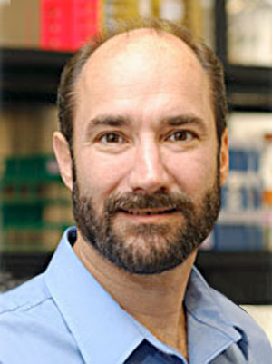
LOS ANGELES – Michael Snyder, PhD, wears his research on his sleeve, so to speak. On any given day, he wears up to eight devices on his body that measure everything from radiation exposure to fasting glucose and sleep activity.
The way he sees it,wearables to monitor physiomes, plus personal omics profiling technology, give a high-resolution view of how health changes over periods of wellness and disease.
“We know intuitively that your health state is influenced by many things, including your genome and all of the things you’re exposed to, from pathogens to food, stress, and exercise,” Dr. Snyder said at the World Congress on Insulin Resistance, Diabetes & Cardiovascular Disease. “They all impact your health state, but in a future not far away, people will be born with their genome sequenced, and we will understand in a probabilistic fashion how, with a set of variants in your genome, if you are exposed to certain environmental conditions, you will have certain health outcomes. As an example, if you’re at risk for Parkinson’s disease, you probably shouldn’t be a pesticide worker, because that greatly increases the chances by which you get Parkinson’s.”
About 8 years ago, he and his associates launched an ongoing longitudinal personal omics profiling project of 105 individuals, 55% of whom are prediabetic. After undergoing genome sequencing, each person undergoes measurement of 14 different omics every 3 months, including their RNA, proteins, lipidomics, cytokines, and microbiome (Cell Host Microbe. 2014 Sep 10;16[3]:276-89).
When a perturbation comes along, like a viral infection or positive results from a colonoscopy, the researchers gather additional samples. “We are trying to understand how the different omics relate to one another,” said Dr. Snyder, who in 2012 used his own genome sequence to predict and help diagnose his own type 2 diabetes, a story that received international media attention. “If you know the inputs into a system, you should be able to calculate the outputs, no matter how complex the system is. You should be able to make meaningful associations. In this case, the inputs are your genome, your epigenome and your microbiome, and the foods you eat. The outputs would be the metabolome, and things like that. We try to understand how responses to, say, viral infections or other perturbations are similar to one another, like congestion and fever, but also why some people get more ill than others, or have varying disease-specific symptoms.”
To date, Dr. Snyder and his associates have collected about 1,800 time points and roughly 10,000 samples. The first 1,000 of those time points have been analyzed. Of the first 70 people who underwent genome sequencing, 12 had pathogenic mutations that are clinically actionable, including mutations in BRCA1, which is associated with breast and ovarian cancer; APC and MUTYH, which are associated with colon cancer; SHBD, which is associated with a high frequency of neuroendocrine tumors; and RBM20, which is associated with dilated cardiomyopathy. One such person “underwent stress testing, and it turns out he does have a heart defect,” noted Dr. Snyder, who is also the author of Genomics and Personalized Medicine: What Everyone Needs to Know (New York: Oxford University Press, 2016). “So some of this information is extremely valuable. This is why we argue that genome sequencing, much like family history, will one day move into the clinic for those who want to use it.”
So far, the longitudinal personal omics analysis has revealed other important diagnoses that likely would have flown under the radar of conventional Western medicine, including a heart defect in one person that was detected by a simple wearable device, a case of early lymphoma, and a case of MGUS (monoclonal gammopathy of unknown significance), which is a precancerous condition.
“We have also had many metabolic cases; a lot of folks were prediabetic and others were diabetic,” Dr. Snyder said. “It’s hard to predict exactly what you’re going to see for any one person. But when you collect a lot of this data, you do find things that are important for their health.”
Of the current study participants, 23 underwent a dietary perturbation. Of these, 13 were insulin resistant and 10 were healthy controls, matched for body mass index. The subjects consumed an extra 1,000 calories per day for 30 days, maintained their peak weight for 7 days, and embarked on a weight loss program for 60 days. “We looked at the effects of weight gain and weight loss in incredible molecular detail,” Dr. Snyder explained, noting that the work will appear in a forthcoming edition of Cell Systems. “At baseline, we can tell the insulin-resistant from the -sensitive folks. After people gain and lose weight, we can identify all the compounds and biochemical pathways that change by using integrative c-means clustering to identify pattern recognition across RNA sequencing, proteome, metabolome, microbiome, and cytokines. From blood, we can actually see these compounds changing. We also can tease out differences between resistance and sensitivity in their reaction to weight gain and weight loss. For example, in the microbiome, the insulin-resistant folks were more resistant to changes in weight gain and weight loss, while insulin-sensitive folks go up and down pretty well.”
The researchers also observed that the omics profile of each person who participated in the dietary perturbation study was different at baseline. “So everybody is special, and not just to our mothers,” Dr. Snyder said. “That is to say, if I profile you, you will not look like anyone sitting next to you. If you go through a perturbation, you will still look more like you than the person sitting next to you.” Going forward, he predicted that personal omics profiling “will build a personal dashboard that relays into your smartphone and can measure your health. Just like your car dashboard, we hope that you’ll have indicators of health, which will be your command center of the future.”
He disclosed that he is a scientific adviser to Personalis, Genapsys, SensOmics, and Qbio.
EXPERT ANALYSIS FROM WCIRDC 2017
Children of moms with RA have higher risk of RA, too
The prevalence of rheumatoid arthritis, as well as thyroid disease and epilepsy, was significantly higher in children whose mothers had RA, according to data from a nationwide cohort study in Denmark.
RA runs in families, and many women with RA are concerned about the possible impact of their disease on a developing fetus, wrote Line R. Jølving of Odense (Denmark) University Hospital and the University of Southern Denmark in Odense and her colleagues in Arthritis Care & Research. “However, we do not have sufficient knowledge on the association between maternal RA and long-term chronic childhood outcomes, and still we do not know which specific chronic diseases to be especially aware of in children of women with RA,” they said.

Overall, the risk of RA was almost three times as high for the children of mothers with RA (hazard ratio, 2.89) as it was for the children of women without RA. In addition, the risk of thyroid disease was more than twice as high (HR, 2.19), and the risk of epilepsy was more than 50% higher (HR, 1.61). Maternal RA during pregnancy had no significant impact on children’s anxiety and personality disorders or on the presence of chronic lung disease.
Mothers with RA were generally older than were those without the condition. The children of mothers with RA were significantly more likely to be born via cesarean section, preterm, and small for gestational age, the researchers said.
The results were limited by the potential inclusion of misclassified diagnoses of child outcomes and by the lack of data on medication use. In addition, the study was not designed to determine the effect of biologic mechanisms or genes, the researchers said. Despite these limitations, “the findings in this study are relevant for pediatricians, rheumatologists, and general practitioners in order to have special awareness of early symptoms of RA, thyroid disease, and epilepsy in offspring of mothers with RA,” they said.
The researchers had no financial conflicts to disclose. The study was funded by several sources, including the Region of Southern Denmark, the University of Southern Denmark in Odense, the Center for Clinical Epidemiology, Odense University Hospital, and the Colitis-Crohn Association in Denmark.
SOURCE: Jølving L et al. Arthritis Care Res. 2017 Dec 11. doi: 10.1002/acr.23461.
The prevalence of rheumatoid arthritis, as well as thyroid disease and epilepsy, was significantly higher in children whose mothers had RA, according to data from a nationwide cohort study in Denmark.
RA runs in families, and many women with RA are concerned about the possible impact of their disease on a developing fetus, wrote Line R. Jølving of Odense (Denmark) University Hospital and the University of Southern Denmark in Odense and her colleagues in Arthritis Care & Research. “However, we do not have sufficient knowledge on the association between maternal RA and long-term chronic childhood outcomes, and still we do not know which specific chronic diseases to be especially aware of in children of women with RA,” they said.
Overall, the risk of RA was almost three times as high for the children of mothers with RA (hazard ratio, 2.89) as it was for the children of women without RA. In addition, the risk of thyroid disease was more than twice as high (HR, 2.19), and the risk of epilepsy was more than 50% higher (HR, 1.61). Maternal RA during pregnancy had no significant impact on children’s anxiety and personality disorders or on the presence of chronic lung disease.
Mothers with RA were generally older than were those without the condition. The children of mothers with RA were significantly more likely to be born via cesarean section, preterm, and small for gestational age, the researchers said.
The results were limited by the potential inclusion of misclassified diagnoses of child outcomes and by the lack of data on medication use. In addition, the study was not designed to determine the effect of biologic mechanisms or genes, the researchers said. Despite these limitations, “the findings in this study are relevant for pediatricians, rheumatologists, and general practitioners in order to have special awareness of early symptoms of RA, thyroid disease, and epilepsy in offspring of mothers with RA,” they said.
The researchers had no financial conflicts to disclose. The study was funded by several sources, including the Region of Southern Denmark, the University of Southern Denmark in Odense, the Center for Clinical Epidemiology, Odense University Hospital, and the Colitis-Crohn Association in Denmark.
SOURCE: Jølving L et al. Arthritis Care Res. 2017 Dec 11. doi: 10.1002/acr.23461.
FROM ARTHRITIS CARE & RESEARCH
Key clinical point: Children whose mothers had RA were significantly more likely to have RA, epilepsy, and thyroid problems than children born to mothers without RA.
Major finding: Children of mothers with RA were almost three times as likely to have RA (hazard ratio, 2.89).
Data source: A nationwide cohort study of live births in Denmark that included 2,106 children born to women with RA and 1,378,539 children born to women without RA.
Disclosures: The researchers had no financial conflicts to disclose. The study was funded by several sources, including the Region of Southern Denmark, the University of Southern Denmark in Odense, the Center for Clinical Epidemiology, Odense University Hospital, and the Colitis-Crohn Association in Denmark.
Source: Jølving L et al. Arthritis Care Res. 2017 Dec 11. doi: 10.1002/acr.23461.
Cardiosphere-derived cells may reverse Duchenne heart scarring
ANAHEIM, CALIF. – One-time infusion of cardiosphere-derived cells into the three major coronary arteries seemed to prevent and perhaps even reverse cardiac scarring, as well as improve arm function, in the open-label phase 1-2 HOPE-Duchenne trial of 25 boys with advanced Duchenne muscular dystrophy.
“Decreased scarring is really a big deal because this is counter to the natural history of Duchenne, where all the scars progress, senior investigator Ronald G. Victor, MD, said at the American Heart Association scientific sessions. “It was gratifying to see that the changes in scar coincided with improved regional systolic wall thickening, particularly in the inferior wall of the left ventricle, which is the very area disproportionately affected early and thus severely in Duchenne.”
Glucocorticoids are the only thing that help to date, prolonging ambulation by about 3 years, but with Cushingoid features and other well-known side effects.
Cardiosphere-derived cells (CDCs) – cardiac progenitor cells – have shown some promise for heart failure. They don’t seem to engraft and grow, but rather to release extracellular vesicles packed with proteins, RNA, and other bioactive molecules. “They act as a role model to get endogenous cells to do the right thing. That’s what we think,” Dr. Victor said.
The HOPE-Duchenne trial [Halt Cardiomyopathy Progression in Duchenne] randomized 12 boys with Duchenne to usual care and 13 others to usual care plus a single infusion of 75 million CDCs divided equally among the left anterior descending, circumflex, and right coronary arteries. The cells were derived from donated heart muscle. The specific CDC preparation tested was “CAP-1002” from Capricor Therapeutics, the study’s sponsor.
The boys were aged 12-22 years, with a mean age of 17.8 years. They had left ventricle scarring in at least four MRI segments; their mean left ventricle ejection fraction was just below 50%; 68% were wheel-chair bound, and all were on stable steroid regimens.
At 12 months, cardiac scarring had increased about 5% in the control group, but decreased by about 7% in the treatment arm, although with no change ejection fraction improvement (P = .03).
Skeletal muscle was assessed by mid-level and distal performance of upper limb scoring, a measure of arm function developed specifically for Duchenne. “For patients who have lost ambulation, the ability to use a joystick to drive a scooter, the ability to feed themselves and use a computer and cellphone are absolutely key to quality of life,” he said.
At 12 months, performance of upper limb scores were largely unchanged in the control group, but improved in about half of the treated boys. “A couple were quite dramatic,” Dr. Victor said. The differences were statistically significant (P = .007) when limited to subjects who started with scores below 55 points, with 58 points meaning normal function.
On the safety side, nothing unusual happened in the control arm, except a femur fracture.
One boy in the treatment arm went into ventricular fibrillation during the diagnostic angiogram. Five had periprocedural atrial fibrillation. All 13 had periprocedural troponin elevations, versus two boys in the control group over the entire course of the study. Dr. Victor didn’t report any deaths, but safety concerns are probably why Capricor is shifting to intravenous infusion for the next trial, HOPE-2.
Dr. Victor noted that troponin leaks “wax and wane” over time in Duchenne, so the transient increases in the treatment arm “were superimposed on a baseline of abnormal cardiac enzymes.”
The Food and Drug Administration has green-lighted the company’s HOPE-2 trial to start enrollment in early 2018, with a larger number of patients and intravenous CDC delivery at 3 month intervals.
Several investigators were Capricor employees. Others were consultants or reported ownership interests. Dr. Victor was the principal investigator at Cedars-Sinai, and was on the trial’s steering committee. The University of Florida, Gainesville, and Cincinnati Children’s Hospital Medical Center, Ohio, were the other two study sites.
SOURCE: Ronald Victor, MD; 2017 AHA Scientific Sessions abstract number S1177
This is a tough disease, and therapies are desperately needed.
The HOPE-Duchenne data look pretty good, and everything seems to be moving in the same direction, but 13 people treated for even 12 months doesn’t really tell us much about safety.

The pipeline for this disease is filling up, but a lot of therapies are aimed at younger patients with the hopes of keeping them functional for as long as possible. I’m glad to see a treatment aimed at more advanced disease.
In general, when the choice is made to enter a clinical trial, it may mean you can’t enter another clinical trial, and these people are in a desperate situation. We need to sort out what should be tested, not just what can be tested.
Robert M. Califf, MD, is professor of cardiology at Duke University, Durham, N.C. He was the study discussant, and wasn’t involved in the work.
This is a tough disease, and therapies are desperately needed.
The HOPE-Duchenne data look pretty good, and everything seems to be moving in the same direction, but 13 people treated for even 12 months doesn’t really tell us much about safety.
The pipeline for this disease is filling up, but a lot of therapies are aimed at younger patients with the hopes of keeping them functional for as long as possible. I’m glad to see a treatment aimed at more advanced disease.
In general, when the choice is made to enter a clinical trial, it may mean you can’t enter another clinical trial, and these people are in a desperate situation. We need to sort out what should be tested, not just what can be tested.
Robert M. Califf, MD, is professor of cardiology at Duke University, Durham, N.C. He was the study discussant, and wasn’t involved in the work.
ANAHEIM, CALIF. – One-time infusion of cardiosphere-derived cells into the three major coronary arteries seemed to prevent and perhaps even reverse cardiac scarring, as well as improve arm function, in the open-label phase 1-2 HOPE-Duchenne trial of 25 boys with advanced Duchenne muscular dystrophy.
“Decreased scarring is really a big deal because this is counter to the natural history of Duchenne, where all the scars progress, senior investigator Ronald G. Victor, MD, said at the American Heart Association scientific sessions. “It was gratifying to see that the changes in scar coincided with improved regional systolic wall thickening, particularly in the inferior wall of the left ventricle, which is the very area disproportionately affected early and thus severely in Duchenne.”
Glucocorticoids are the only thing that help to date, prolonging ambulation by about 3 years, but with Cushingoid features and other well-known side effects.
Cardiosphere-derived cells (CDCs) – cardiac progenitor cells – have shown some promise for heart failure. They don’t seem to engraft and grow, but rather to release extracellular vesicles packed with proteins, RNA, and other bioactive molecules. “They act as a role model to get endogenous cells to do the right thing. That’s what we think,” Dr. Victor said.
The HOPE-Duchenne trial [Halt Cardiomyopathy Progression in Duchenne] randomized 12 boys with Duchenne to usual care and 13 others to usual care plus a single infusion of 75 million CDCs divided equally among the left anterior descending, circumflex, and right coronary arteries. The cells were derived from donated heart muscle. The specific CDC preparation tested was “CAP-1002” from Capricor Therapeutics, the study’s sponsor.
The boys were aged 12-22 years, with a mean age of 17.8 years. They had left ventricle scarring in at least four MRI segments; their mean left ventricle ejection fraction was just below 50%; 68% were wheel-chair bound, and all were on stable steroid regimens.
At 12 months, cardiac scarring had increased about 5% in the control group, but decreased by about 7% in the treatment arm, although with no change ejection fraction improvement (P = .03).
Skeletal muscle was assessed by mid-level and distal performance of upper limb scoring, a measure of arm function developed specifically for Duchenne. “For patients who have lost ambulation, the ability to use a joystick to drive a scooter, the ability to feed themselves and use a computer and cellphone are absolutely key to quality of life,” he said.
At 12 months, performance of upper limb scores were largely unchanged in the control group, but improved in about half of the treated boys. “A couple were quite dramatic,” Dr. Victor said. The differences were statistically significant (P = .007) when limited to subjects who started with scores below 55 points, with 58 points meaning normal function.
On the safety side, nothing unusual happened in the control arm, except a femur fracture.
One boy in the treatment arm went into ventricular fibrillation during the diagnostic angiogram. Five had periprocedural atrial fibrillation. All 13 had periprocedural troponin elevations, versus two boys in the control group over the entire course of the study. Dr. Victor didn’t report any deaths, but safety concerns are probably why Capricor is shifting to intravenous infusion for the next trial, HOPE-2.
Dr. Victor noted that troponin leaks “wax and wane” over time in Duchenne, so the transient increases in the treatment arm “were superimposed on a baseline of abnormal cardiac enzymes.”
The Food and Drug Administration has green-lighted the company’s HOPE-2 trial to start enrollment in early 2018, with a larger number of patients and intravenous CDC delivery at 3 month intervals.
Several investigators were Capricor employees. Others were consultants or reported ownership interests. Dr. Victor was the principal investigator at Cedars-Sinai, and was on the trial’s steering committee. The University of Florida, Gainesville, and Cincinnati Children’s Hospital Medical Center, Ohio, were the other two study sites.
SOURCE: Ronald Victor, MD; 2017 AHA Scientific Sessions abstract number S1177
REPORTING FROM THE AHA SCIENTIFIC SESSIONS
Key clinical point: One-time infusion of cardiosphere-derived cells (CDCs) into the three major coronary arteries seemed to prevent and perhaps even reverse cardiac scarring, as well as improve arm function, in boys with advanced Duchenne muscular dystrophy.
Major finding: At 12 months, cardiac scarring increased about 5% in the control group, but decreased about 7% in the treatment arm, although with no change in ejection fraction (P = .03).
Study details: HOPE-Duchenne, an open-label, phase 1-2 trial involving 25 boys
Disclosures: The study was funded by Capricor Therapeutics, makers of the CDC preparation tested. Several investigators were Capricor employees. Others were consultants or reported ownership interests. The senior investigator and presenter was on the trial’s steering committee.
Source: Victor R, et al. 2017 AHA Scientific Sessions abstract number S1177.
Brain structural changes on MRI predict sudden death in epilepsy
WASHINGTON – It has long been hypothesized that sudden unexpected death in epilepsy (SUDEP) is the result of damage to areas of the brain that control breathing and heart rate, but two studies presented at the American Epilepsy Society annual meeting identified specific areas where structural changes correlate with SUDEP, suggesting the potential for screening.
In one of two studies that evaluated MRI images in patients who subsequently died of SUDEP and compared them to patients with epilepsy or normal healthy individuals, brainstem volume loss was not only greater in those with SUDEP but there was a correlation between greater volume loss and a shorter period of survival, reported Susanne G. Mueller, MD, a radiologist affiliated with the Center for Imaging of Neurodegenerative Diseases at the San Francisco Veterans Affairs Medical Center.
“This is the first evidence that brainstem damage, one of the mechanisms that has been shown to cause SUDEP in animals, could also play a role in people with epilepsy,” Dr. Mueller reported. Although more work is needed, volume loss observed on MRI “could be used as a potential biomarker to assess the individual SUDEP risk in epilepsy patients.”
In one of two populations studied, investigators analyzed MRI scans from 27 SUDEP patients taken prior to death. Focusing on brainstem areas involved in autonomic function, deformation morphometry generated profile maps that isolated areas of volume loss. The changes suggested that focal epilepsy produced structural changes in the mesencephalic region of the lower brainstem.
Damage to nuclei involved in control of heart rate variability and other autonomic functions would be consistent with increased risk of SUDEP.
“We can just report what we see on imaging. These data do not tell us the cause of SUDEP, but they do show correlations that are consistent with current theories,” Dr. Mueller explained.
In a second study that compared 18 patients with focal epilepsy to 11 controls, greater volume loss in patients with epilepsy correlated negatively with heart rate variability, a relationship not seen in the controls. The volume loss in periventricular gray and medulla oblongata nuclei was most closely associated with heart rate variability, which is considered a surrogate for altered autonomic function, Dr. Mueller reported.
Similar conclusions were reached in a different study conducted at a separate institution. In this study, MRI scans from 237 patients with epilepsy and 110 healthy controls were evaluated. Four of the epilepsy patients subsequently died of SUDEP.

“These are all areas that can be involved in autonomic function potentially involved in SUDEP,” Mr. George said. Like Dr. Mueller, Mr. George cautioned that clinically viable algorithms that would allow MRI to assess SUDEP risk may be years away, but these studies provide preliminary evidence that structural changes on MRI could eventually serve as a SUDEP biomarker.
There was limited overlap between the areas of structural change potentially associated with SUDEP in the studies presented by Dr. Mueller and Mr. George, but Dr. Mueller said that SUDEP might not stem from a single epilepsy-induced brain abnormality. She reported that more MRI scans to trace structural changes in SUDEP patients are likely to identify more areas of interest. Acquiring a large number of MRI scans is a challenge, but Dr. Mueller envisions a registry where routine scans could be submitted. This would permit this research to be conducted on a larger scale.
“There is a similar initiative to collect MRI brain images of patients with Alzheimer’s disease,” said Dr. Mueller, noting that this provides a precedent for the type of research needed in epilepsy. If a similar program could be undertaken in epilepsy, Dr. Mueller believes it might substantially accelerate the effort to understand and recognize risk of SUDEP.
Mr. George’s study was funded by FACES (Finding a CURE for Epilepsy/Seizures). Dr. Mueller’s study was funded by grants from UCSF, the Epilepsy Foundation, and the National Institutes of Health.
Dr. Mueller and Mr. George reported no potential conflicts of interest related to this topic.
SOURCE: Mueller S et al., AES 2017 abstract 3.205 and George A et al., AES 2017 abstract 3.214
WASHINGTON – It has long been hypothesized that sudden unexpected death in epilepsy (SUDEP) is the result of damage to areas of the brain that control breathing and heart rate, but two studies presented at the American Epilepsy Society annual meeting identified specific areas where structural changes correlate with SUDEP, suggesting the potential for screening.
In one of two studies that evaluated MRI images in patients who subsequently died of SUDEP and compared them to patients with epilepsy or normal healthy individuals, brainstem volume loss was not only greater in those with SUDEP but there was a correlation between greater volume loss and a shorter period of survival, reported Susanne G. Mueller, MD, a radiologist affiliated with the Center for Imaging of Neurodegenerative Diseases at the San Francisco Veterans Affairs Medical Center.
“This is the first evidence that brainstem damage, one of the mechanisms that has been shown to cause SUDEP in animals, could also play a role in people with epilepsy,” Dr. Mueller reported. Although more work is needed, volume loss observed on MRI “could be used as a potential biomarker to assess the individual SUDEP risk in epilepsy patients.”
In one of two populations studied, investigators analyzed MRI scans from 27 SUDEP patients taken prior to death. Focusing on brainstem areas involved in autonomic function, deformation morphometry generated profile maps that isolated areas of volume loss. The changes suggested that focal epilepsy produced structural changes in the mesencephalic region of the lower brainstem.
Damage to nuclei involved in control of heart rate variability and other autonomic functions would be consistent with increased risk of SUDEP.
“We can just report what we see on imaging. These data do not tell us the cause of SUDEP, but they do show correlations that are consistent with current theories,” Dr. Mueller explained.
In a second study that compared 18 patients with focal epilepsy to 11 controls, greater volume loss in patients with epilepsy correlated negatively with heart rate variability, a relationship not seen in the controls. The volume loss in periventricular gray and medulla oblongata nuclei was most closely associated with heart rate variability, which is considered a surrogate for altered autonomic function, Dr. Mueller reported.
Similar conclusions were reached in a different study conducted at a separate institution. In this study, MRI scans from 237 patients with epilepsy and 110 healthy controls were evaluated. Four of the epilepsy patients subsequently died of SUDEP.
“These are all areas that can be involved in autonomic function potentially involved in SUDEP,” Mr. George said. Like Dr. Mueller, Mr. George cautioned that clinically viable algorithms that would allow MRI to assess SUDEP risk may be years away, but these studies provide preliminary evidence that structural changes on MRI could eventually serve as a SUDEP biomarker.
There was limited overlap between the areas of structural change potentially associated with SUDEP in the studies presented by Dr. Mueller and Mr. George, but Dr. Mueller said that SUDEP might not stem from a single epilepsy-induced brain abnormality. She reported that more MRI scans to trace structural changes in SUDEP patients are likely to identify more areas of interest. Acquiring a large number of MRI scans is a challenge, but Dr. Mueller envisions a registry where routine scans could be submitted. This would permit this research to be conducted on a larger scale.
“There is a similar initiative to collect MRI brain images of patients with Alzheimer’s disease,” said Dr. Mueller, noting that this provides a precedent for the type of research needed in epilepsy. If a similar program could be undertaken in epilepsy, Dr. Mueller believes it might substantially accelerate the effort to understand and recognize risk of SUDEP.
Mr. George’s study was funded by FACES (Finding a CURE for Epilepsy/Seizures). Dr. Mueller’s study was funded by grants from UCSF, the Epilepsy Foundation, and the National Institutes of Health.
Dr. Mueller and Mr. George reported no potential conflicts of interest related to this topic.
SOURCE: Mueller S et al., AES 2017 abstract 3.205 and George A et al., AES 2017 abstract 3.214
REPORTING FROM AES 2017
Key clinical point: MRI-detected damage to brain areas mediating autonomic function is implicated in sudden unexpected death in epilepsy (SUDEP).
Major finding: Two separate studies drew the conclusion that brain structural difference seen on MRI may become an effective screening tool for identifying epilepsy patients at high risk for SUDEP.
Data source: Prospective studies.
Disclosures: Dr. Mueller’s study was funded by grants from UCSF, the Epilepsy Foundation, and the National Institutes of Health. Mr. George’s study was funded by FACES (Finding a CURE for Epilepsy/Seizures). Dr. Mueller and Mr. George reported no potential conflicts of interest related to this topic.
Source: Mueller S et al., AES 2017 abstract 3.205 and George A et al., AES 2017 abstract 3.214
A three-track concept proposed for regulating fecal microbiota transplantation
FROM SCIENCE
Researchers are proposing a three-track regulatory framework to help ensure that using fecal microbiota transplantations (FMTs) to treat recurring Clostridium difficile infection (CDI) can be done in a more safe and effective manner.
The need for better regulation is arising because, although FMT is considered by many to be the standard of care, samples are inconsistently screened for infectious pathogens.
“The use of prescreened stool obtained from a stool bank and shipped to a physician is increasing, but the stool banks are not regulated,” Diane Hoffmann, the Jacob A. France Professor of Health Care Law at the University of Maryland, Baltimore, and her colleagues wrote in Science.
The authors noted that the Food and Drug Administration has struggled with regulating FMT; so far, it has tried to use the drug approval pathway as a tool to oversee FMT.
However, the authors note that the transplanted material “is not a ‘typical’ drug, and thus may not be appropriate for the drug regulatory pathway.” To that end, Ms. Hoffmann and her colleagues offered three paths that the FDA could implement to regulate FMT.
The first track would cover instances in which a physician obtains the stool from a donor the patient or physician already know. In this case, “FMT would be regulated as the ‘practice of medicine’ subject primarily to state regulation,” the authors proposed. “Physicians performing FMT for CDI would be able to do so based on their scope of practice using clinical judgment and the relevant standard of care.” In this track, the investigational new drug (IND) pathway would be applied when FMT is used for any indication other than CDI unless the use meets clinical innovation requirements.
The second track covers stool obtained from stool banks. In this scenario, it would be “regulated in the same manner as human cell-tissue establishments with some additional oversight,” Ms. Hoffmann and her colleagues wrote. “This would include registering annually with the FDA and complying with rules for donor screening and testing and ‘good manufacturing processes.’ ” Physicians using stool from these banks would be required to report back any adverse events stemming from stool supplied by the banks, and the banks would submit that data to the FDA. Unless operating under an IND, stool banks would only be able to provide samples to treat CDI.
The third track deals with “modified stool-based products” (stool processed in such a way that the original relevant characteristics of the transferred community of microorganisms have been altered). “These products would be regulated as biological products or drugs with some alteration of IND requirements (i.e., elimination of some phase I IND requirements and modification of characterization specifications),” the authors wrote.
“Our proposal improves on the FDA’s current and proposed regulatory scheme as it allows stool banks to continue to provide stool but only under an approved regulatory framework,” Ms. Hoffmann and her colleagues wrote.
They believe that implementation of this three-track structure would not be difficult: “FDA would need to change its position and determine that microbiota derived from stool is a tissue, not a drug or biological product. The framework could be implemented through new guidance or by issuing formal regulations and no statutory changes would be required. The scheme would serve as a useful starting point for regulation of other types of [microbiota transplants].”
Seizures captured on a smartphone found diagnostic for epilepsy
WASHINGTON – Smartphone videos brought to the clinic by patients are valid tools for the diagnosis of epilepsy, according to a prospective blinded and multicenter study of 41 consecutive videos presented at the American Epilepsy Society annual meeting.
“These findings have global implications, because they suggest that smartphone videos are a cost effective tool that can accelerate the time to diagnosis even in places where video-EEG monitoring is not readily available,” reported William Tatum, DO, professor of neurology, Mayo Clinic, Jacksonville, Fla.

In addition to submitting a smartphone video, all patients in this study underwent a history and physical (H&P) and were evaluated with video-EEG monitoring. The smartphone videos underwent review by 10 epilepsy experts and 8 general neurology residents blinded to the video EEG findings. The latter group was selected to test the value of smartphone video in clinicians with general knowledge but no special expertise.
The final diagnosis was made on the basis of all the clinical information, including the video-EEG, which Dr. Tatum characterized as the gold standard for the diagnosis of epilepsy. Based on the video-EEG, 11 of the 41 patients (26.8%) had seizures, 26 (63.4%) had psychogenic nonepileptic seizures (PNES), 3 (7.4%) had physiologic nonepileptic events (PhysNEE), and 1 (2.4%) had both PhysNEE and PNES.
On the basis of the blinded smartphone video alone, the median correct diagnosis was 71.4% for experts and 66.7% for residents. Although this difference was not significant, Dr. Tatum reported that there was substantially less inter-rater variability among experts.
“Overall, smartphone video review correctly differentiated epilepsy from PNES in 68% of the videos evaluated by experts and 58% assessed by residents,” Dr. Tatum reported.
For experts, the smartphone video assessment yielded a specificity of 43% and sensitivity of 83% for epilepsy. For PNES, these figures were 80% and 54%, respectively. Among residents, the sensitivities and specificities for epilepsy (32% and 83%) and PNES (82% and 53%) were similar. Dr. Tatum noted that H&P predicted the definitive diagnosis in 75.6% of cases.
The rate of correct diagnoses with blinded smartphone video analysis in this study was respectable, but Dr. Tatum suggested that smartphone video should be an adjunctive tool that is reviewed in the context of H&P, which would be expected to further boost accuracy. Although he acknowledged that smartphone videos plus H&P will not completely supplant the need for video-EEG monitoring to reach a definitive diagnosis in all cases, he believes that it is accurate in many, and it accelerates the time to diagnosis.
“The median duration of the smartphone review was about a minute and a half. The median duration of H&P was 60 minutes, but the median time to a result with video-EEG was 2.54 days,” said Dr. Tatum, noting that this difference was highly significant (P lees than .001). If a diagnosis can be reached without video-EEG, it would also be expected to greatly reduce costs.
On a yes-no basis, 78% of those who evaluated the smartphone videos judged them to be adequate for a diagnosis. An analysis of those considered to be poor quality by expert and resident viewers was presented as a separate report. The most common reasons that smartphone videos were considered to have inadequate quality, according to this analysis, were inadequate duration, insufficient lighting, and poor audio. In other words, essentially all of the problems stemmed from inadequate technique, not technical limitations, reported Erin E. Coonan, an undergraduate research intern working with Dr. Tatum.
“We think that disseminating information to the general public about how to take an adequate quality smartphone video could increase the quality of these videos when they are brought to the clinic,” Ms. Coonan reported. She and Dr. Tatum believe that patient-submitted smartphone videos will be increasingly common tool in clinical medicine, making information about proper technique valuable.
Most U.S. adults now carry smartphones, and these are becoming increasingly common even in resource-poor areas of the world, according to Dr. Tatum. He said that clinical medicine should embrace this technology.
“The cost of a smartphone video is essentially zero, but our data suggest that they can be a useful adjunctive diagnostic tool,” Dr. Tatum said.
The presenters reported no potential conflicts of interest related to these studies.
SOURCE: Tatum W et al., AES 2017 abstract 3.161 and Coonan E et al., AES 2017 abstract 3.070
WASHINGTON – Smartphone videos brought to the clinic by patients are valid tools for the diagnosis of epilepsy, according to a prospective blinded and multicenter study of 41 consecutive videos presented at the American Epilepsy Society annual meeting.
“These findings have global implications, because they suggest that smartphone videos are a cost effective tool that can accelerate the time to diagnosis even in places where video-EEG monitoring is not readily available,” reported William Tatum, DO, professor of neurology, Mayo Clinic, Jacksonville, Fla.
In addition to submitting a smartphone video, all patients in this study underwent a history and physical (H&P) and were evaluated with video-EEG monitoring. The smartphone videos underwent review by 10 epilepsy experts and 8 general neurology residents blinded to the video EEG findings. The latter group was selected to test the value of smartphone video in clinicians with general knowledge but no special expertise.
The final diagnosis was made on the basis of all the clinical information, including the video-EEG, which Dr. Tatum characterized as the gold standard for the diagnosis of epilepsy. Based on the video-EEG, 11 of the 41 patients (26.8%) had seizures, 26 (63.4%) had psychogenic nonepileptic seizures (PNES), 3 (7.4%) had physiologic nonepileptic events (PhysNEE), and 1 (2.4%) had both PhysNEE and PNES.
On the basis of the blinded smartphone video alone, the median correct diagnosis was 71.4% for experts and 66.7% for residents. Although this difference was not significant, Dr. Tatum reported that there was substantially less inter-rater variability among experts.
“Overall, smartphone video review correctly differentiated epilepsy from PNES in 68% of the videos evaluated by experts and 58% assessed by residents,” Dr. Tatum reported.
For experts, the smartphone video assessment yielded a specificity of 43% and sensitivity of 83% for epilepsy. For PNES, these figures were 80% and 54%, respectively. Among residents, the sensitivities and specificities for epilepsy (32% and 83%) and PNES (82% and 53%) were similar. Dr. Tatum noted that H&P predicted the definitive diagnosis in 75.6% of cases.
The rate of correct diagnoses with blinded smartphone video analysis in this study was respectable, but Dr. Tatum suggested that smartphone video should be an adjunctive tool that is reviewed in the context of H&P, which would be expected to further boost accuracy. Although he acknowledged that smartphone videos plus H&P will not completely supplant the need for video-EEG monitoring to reach a definitive diagnosis in all cases, he believes that it is accurate in many, and it accelerates the time to diagnosis.
“The median duration of the smartphone review was about a minute and a half. The median duration of H&P was 60 minutes, but the median time to a result with video-EEG was 2.54 days,” said Dr. Tatum, noting that this difference was highly significant (P lees than .001). If a diagnosis can be reached without video-EEG, it would also be expected to greatly reduce costs.
On a yes-no basis, 78% of those who evaluated the smartphone videos judged them to be adequate for a diagnosis. An analysis of those considered to be poor quality by expert and resident viewers was presented as a separate report. The most common reasons that smartphone videos were considered to have inadequate quality, according to this analysis, were inadequate duration, insufficient lighting, and poor audio. In other words, essentially all of the problems stemmed from inadequate technique, not technical limitations, reported Erin E. Coonan, an undergraduate research intern working with Dr. Tatum.
“We think that disseminating information to the general public about how to take an adequate quality smartphone video could increase the quality of these videos when they are brought to the clinic,” Ms. Coonan reported. She and Dr. Tatum believe that patient-submitted smartphone videos will be increasingly common tool in clinical medicine, making information about proper technique valuable.
Most U.S. adults now carry smartphones, and these are becoming increasingly common even in resource-poor areas of the world, according to Dr. Tatum. He said that clinical medicine should embrace this technology.
“The cost of a smartphone video is essentially zero, but our data suggest that they can be a useful adjunctive diagnostic tool,” Dr. Tatum said.
The presenters reported no potential conflicts of interest related to these studies.
SOURCE: Tatum W et al., AES 2017 abstract 3.161 and Coonan E et al., AES 2017 abstract 3.070
Key clinical point: Videos taken with a smartphone can contribute to the accurate diagnosis of epilepsy, according to results of a blinded study.
Major finding: Experts correctly differentiated epileptic seizures from psychogenic nonepileptic seizures with smartphone video in 68% of cases.
Data source: A multicenter, prospective blinded trial of 41 consecutive videos.
Disclosures: The presenters reported no potential conflicts of interest related to this topic.
Source: Tatum W et al., AES 2017 abstract 3.161 and Coonan E et al., AES 2017 abstract 3.070
FDA okays dosing software for hemophilia A
The Food and Drug Administration has granted 510(k) clearance to myPKFiT for ADVATE (Antihemophilic Factor, Recombinant), pharmokinetic dosing software used for tailoring prophylaxis regimens for hemophilia A patients.
The software can be used for hemophilia A patients aged 16 years or older who weigh at least 45 kilograms (about 99 pounds) and are treated with ADVATE.
The free, web-based software estimates a patient’s pharmokinetic curve using age, body weight, and local laboratory FVIII one-stage clotting activity measurements and allows physicians to estimate a full pharmokinetic curve with just two measurable blood samples, according to Shire, the software’s manufacturer. That’s much lower than the 9-11 blood samples recommendation from the International Society on Thrombosis and Haemostasis.![]()
The software is expected to be available in the United States by the end of the first quarter of 2018. A version of the software is already marketed in Europe.
FDA: Gadolinium retention prompts new GBCA class warning, safety measures
Gadolinium-based contrast agents (GBCAs) used for MRI will now carry a warning regarding their potential retention in the bodies and brains of treated patients, according to the Food and Drug Administration.
The FDA is requiring the new class warning, along with other safety measures, based on evidence showing that trace amounts of gadolinium can be retained in the body for months to years after treatment.

Specifically, the agency will require that patients receiving GBCAs first receive a Medication Guide and that GBCA manufacturers conduct human and animal studies to further assess GBCA safety. At this time, the only known adverse health effect of gadolinium retention is nephrogenic systemic fibrosis, which affects a small subgroup of patients with pre-existing kidney failure. No causal association has been established between gadolinium retention and reported adverse events in those with normal kidney function.
The FDA recommended that health care professionals consider the retention characteristics of GBCAs for patients who may be at higher risk for retention, including those requiring multiple lifetime doses, pregnant women, children, and patients with inflammatory conditions, but stressed that, although repeated GBCA imaging studies should be minimized when possible, they should not be avoided or deferred when they are necessary. In the safety alert, the FDA noted that administration of the GBCAs Dotarem (gadoterate meglumine), Gadavist (gadobutrol), and ProHance (gadoteridol) produce the lowest gadolinium levels in the body, and the three agents leave similar gadolinium levels in the body.
The agency encourages reports of adverse events or side effects related to the use of GBCAs to its MedWatch Safety information and Adverse Event Reporting Program. Reports can be submitted online at www.fda.gov/MedWatch/report or by calling 1-800-332-1088 to request a preaddressed form that can be mailed or faxed to 1-800-FDA-0178.
Gadolinium-based contrast agents (GBCAs) used for MRI will now carry a warning regarding their potential retention in the bodies and brains of treated patients, according to the Food and Drug Administration.
The FDA is requiring the new class warning, along with other safety measures, based on evidence showing that trace amounts of gadolinium can be retained in the body for months to years after treatment.
Specifically, the agency will require that patients receiving GBCAs first receive a Medication Guide and that GBCA manufacturers conduct human and animal studies to further assess GBCA safety. At this time, the only known adverse health effect of gadolinium retention is nephrogenic systemic fibrosis, which affects a small subgroup of patients with pre-existing kidney failure. No causal association has been established between gadolinium retention and reported adverse events in those with normal kidney function.
The FDA recommended that health care professionals consider the retention characteristics of GBCAs for patients who may be at higher risk for retention, including those requiring multiple lifetime doses, pregnant women, children, and patients with inflammatory conditions, but stressed that, although repeated GBCA imaging studies should be minimized when possible, they should not be avoided or deferred when they are necessary. In the safety alert, the FDA noted that administration of the GBCAs Dotarem (gadoterate meglumine), Gadavist (gadobutrol), and ProHance (gadoteridol) produce the lowest gadolinium levels in the body, and the three agents leave similar gadolinium levels in the body.
The agency encourages reports of adverse events or side effects related to the use of GBCAs to its MedWatch Safety information and Adverse Event Reporting Program. Reports can be submitted online at www.fda.gov/MedWatch/report or by calling 1-800-332-1088 to request a preaddressed form that can be mailed or faxed to 1-800-FDA-0178.
Gene mutations tied to viral skin infection in pediatric AD patients
Filaggrin (FLG) polymorphism appears to be linked with Molluscum contagiosum virus skin infection in children with atopic dermatitis (AD), reported Sara Manti, MD, of the University of Messina (Italy), and her associates.
“To the best of our knowledge, this is the first study” to describe this association, they said.
In an Italian study of 100 children with AD and 97 healthy children who served as controls, both clinical and laboratory data showed that FLG gene mutations were linked to early AD onset, a more severe clinical course of disease, and a significantly higher risk of Molluscum contagiosum virus (MCV)–associated skin infection.

The data “suggest that FLG gene may serve as a potential biomarker to screen the atopic population, identifying patients at high risk of AD, to adopt preventive measures that can restore the barrier function of the skin and reduce patients’ susceptibility to recurrent skin infection,” they wrote. “Our findings also indicate that the identified SNPs [single-nucleotide polymorphisms] might have clinically relevant implications with respect to a major MCV [M. contagiosum virus] colonization of skin, also permitting the identification of a new AD phenotype in children. However, to date, because of the existence of controversial data, we believe that further and needed insights into AD pathophysiology may also be gained by studying people with the FLG gene who do not have atopic predisposition and/or AD. Thus, a large population-based study is necessary to further validate these early interpretations and to gain detailed information about the role of FLG variants,” Dr. Manti and her coinvestigators concluded.
Read more in Manti S et al. Ann Allergy Asthma Immunol. 2017;119:446-51.
Filaggrin (FLG) polymorphism appears to be linked with Molluscum contagiosum virus skin infection in children with atopic dermatitis (AD), reported Sara Manti, MD, of the University of Messina (Italy), and her associates.
“To the best of our knowledge, this is the first study” to describe this association, they said.
In an Italian study of 100 children with AD and 97 healthy children who served as controls, both clinical and laboratory data showed that FLG gene mutations were linked to early AD onset, a more severe clinical course of disease, and a significantly higher risk of Molluscum contagiosum virus (MCV)–associated skin infection.
The data “suggest that FLG gene may serve as a potential biomarker to screen the atopic population, identifying patients at high risk of AD, to adopt preventive measures that can restore the barrier function of the skin and reduce patients’ susceptibility to recurrent skin infection,” they wrote. “Our findings also indicate that the identified SNPs [single-nucleotide polymorphisms] might have clinically relevant implications with respect to a major MCV [M. contagiosum virus] colonization of skin, also permitting the identification of a new AD phenotype in children. However, to date, because of the existence of controversial data, we believe that further and needed insights into AD pathophysiology may also be gained by studying people with the FLG gene who do not have atopic predisposition and/or AD. Thus, a large population-based study is necessary to further validate these early interpretations and to gain detailed information about the role of FLG variants,” Dr. Manti and her coinvestigators concluded.
Read more in Manti S et al. Ann Allergy Asthma Immunol. 2017;119:446-51.
FROM THE ANNALS OF ALLERGY, ASTHMA & IMMUNOLOGY
FDA okays dosing software for hemophilia A
The Food and Drug Administration has granted 510(k) clearance to myPKFiT for ADVATE (Antihemophilic Factor, Recombinant), pharmokinetic dosing software used for tailoring prophylaxis regimens for hemophilia A patients.
The software can be used for hemophilia A patients aged 16 years or older who weigh at least 45 kilograms (about 99 pounds) and are treated with ADVATE.

The software is expected to be available in the United States by the end of the first quarter of 2018. A version of the software is already marketed in Europe.
The Food and Drug Administration has granted 510(k) clearance to myPKFiT for ADVATE (Antihemophilic Factor, Recombinant), pharmokinetic dosing software used for tailoring prophylaxis regimens for hemophilia A patients.
The software can be used for hemophilia A patients aged 16 years or older who weigh at least 45 kilograms (about 99 pounds) and are treated with ADVATE.
The software is expected to be available in the United States by the end of the first quarter of 2018. A version of the software is already marketed in Europe.