A treatment for narcolepsy reduced sleep problems in individuals with Parkinson’s disease who had excessive daytime sleepiness in a double-blind, randomized, placebo-controlled, phase 2a trial.
The trial results provide “class I evidence for the efficacy of sodium oxybate in treating sleep-wake disturbances in Parkinson’s disease. This finding is based on the extensive, electrophysiologically proven treatment effect that, to our knowledge, is unmatched by any other intervention reported so far,” Fabian Büchele, MD, of the department of neurology at University Hospital Zürich and his colleagues wrote in their report (JAMA Neurol. 2017 Nov 6. doi: 10.1001/jamaneurol.2017.3171).
Silvia Jansen/iStockphoto
Dr. Büchele and his coauthors observed that there was a reciprocal association between nighttime sleep and excessive daytime sleepiness.
“Sodium oxybate–related improvements of sleep and EDS [excessive daytime sleepiness] correlated significantly, whereas sodium oxybate–induced sleep disturbances predicted insufficient treatment response and AEs [adverse events],” they wrote.
The study of 12 patients used a crossover design to examine how well the sodium oxybate could improve sleep latency and scores on the Epworth Sleepiness Scale. The investigators randomized the patients to receive the central nervous system depressor sodium oxybate followed by placebo, or to placebo first and then the active drug, with a 2- to 4-week washout period in between crossovers. Sodium oxybate or placebo were taken at bedtime, and 2.5-4 hours later, with the dose individually titrated between 3 g and 9 g per night, for 6 weeks.
The treatment was associated with significant improvements, including a significant 2.9-minute increase in mean sleep latency on the intention-to-treat analysis (P = .002) and a 3.5-minute increase in the per-protocol analysis (P less than .001), as well as a 4.2-point reduction in Epworth Sleepiness Scale scores (P = .001).
Patients treated with sodium oxybate reported enhanced subjective sleep quality, shown as a 2-point mean reduction in Parkinson’s Disease Sleep Scale–2 scores and a 72.7-minute mean increase in slow-wave stage N3 sleep (and reduction in superficial stage N1 sleep).
In eight patients, there was at least a 50% clinically significant increase in mean sleep latency, and six patients had a normalization of Epworth Sleepiness Scale scores.
The sodium oxybate was fairly well tolerated, with no dropouts related to adverse events. All patients experienced adverse events, but three-quarters of these were described as not interfering with daily activities, and one-quarter as having a mild to moderate impact on daily activities. Most resolved after dose adjustment, but one-third of patients were still affected by the end of the study.
One patient dropped out during the washout phase and another was excluded from the per-protocol analysis; both of these patients developed sleep apnea during the course of the study. One patient also developed parasomnia during the study.
While the authors said that their results on efficacy and safety matched those found in trials of sodium oxybate for narcolepsy, their study was limited by small numbers, and larger follow-up trials of longer duration are needed to confirm the findings.
The study was funded by UCB Pharma, which provided the drug and placebo, and by the Clinical Research Priority Program Sleep and Health of the University of Zürich. Five authors declared speaker honoraria and grants from a range of organizations and pharmaceutical companies, including one author who had received these from UCB Pharma. No other conflicts were declared.
Key clinical point: Sodium oxybate may reduce sleep-wake disturbances of Parkinson’s disease.
Major finding: Treatment with sodium oxybate led to clinically significant improvements in sleep latency and in excessive daytime sleepiness in patients with Parkinson’s disease.
Data source: Randomized, placebo-controlled, crossover phase 2a trial in 12 patients with Parkinson’s disease.
Disclosures: The study was funded by UCB Pharma, which provided the drug and placebo, and by the Clinical Research Priority Program Sleep and Health of the University of Zürich. Five authors declared speaker honoraria and grants from a range of organizations and pharmaceutical companies, including one author who had received these from UCB Pharma. No other conflicts were declared.
Robotic thoracic surgery is associated with shorter hospital stays, shorter duration of chest tube usage, and lower postoperative pain intensity than open thoracotomy, new data suggests.
In a paper published in Surgical Endoscopy, researchers reported the results of a retrospective study comparing the outcomes from 38 individuals who underwent robotic thoracic surgery using the da Vinci System with those of 38 patients who underwent open thoracic surgery.
They saw significantly shorter hospital stays associated with robotic surgery, compared with open surgery (6.9 days vs. 8.0 days, respectively; P = .02), as well as a shorter duration of chest tube use (2.9 plus or minus 2.0 days vs. 4.9 plus or minus 2.2 days; P less than .001).
While robotic surgery did not result in significant reductions in postoperative pain intensity when patients coughed, pain intensity for patients at rest on days 4 and 5 after surgery was significantly lower among those who had undergone robotic surgery than among those who had undergone open surgery (Surg Endosc. 2017. doi: 10.1007/s00464-017-5464-6). On day 4, those who had undergone robotic surgery reported a mean pain score of 0.5 on a scale of 1-10, while those who underwent open surgery had a mean pain score of 1.1 (P = .04). On day 5, the mean pain scores were 0.7 in the robotic-surgery group and 1.6 in the open-surgery group (P = .003).
However, there was no difference in postoperative opioid use between the two groups. The robotic-surgery group showed a trend toward more postoperative nausea, but this did not reach statistical significance.
Most of the surgeries in the study were performed to remove malignant or benign tumors. Among both groups, 68% of patients received epidural analgesia, and 32% received systemic opioid-based postoperative analgesia.
“Current evidence shows that more than 70% of stage I lung cancers are still being performed by open technique,” wrote Christopher Darr, from the West German Lung Center at University Hospital Essen (Germany), and his coauthors. “However, the safety and the feasibility of robotic anatomic lung resection have been shown in several case series. Robotic thoracic surgery is gaining popularity as benefits can be suggested, such as improved ergonomics, three-dimensional optics, and simplifying operative procedure.”
One author declared financial support for meetings and presentations from Intuitive Surgical, the company that developed the da Vinci System. No other conflicts of interest were declared.
Key clinical point: Robotic thoracic surgery is associated with shorter hospital stays and shorter duration of chest tube use, compared with open surgery.
Major finding: Patients who underwent robotic thoracic surgery had a mean hospital stay of 6.9 days, compared with 8 days for those who underwent open thoracic surgery.
Data source: Retrospective study of 38 patients who underwent robotic thoracic surgery and 38 who underwent open thoracic surgery.
Disclosures: One investigator declared financial support from Intuitive Surgical, the company that developed the da Vinci System, for meetings and presentations. No other conflicts of interest were declared.
In 2015, Debasree Banerjee, MD, MS, received the CHEST Foundation Research Grant in Pulmonary Arterial Hypertension. She was also a 2016 NetWorks Challenge Travel Grantee as a member of the Women’s Health NetWork, allowing her to attend the 2016 CHEST Annual Meeting and network with peers and leaders in chest medicine. Read our follow-up interview with Dr. Banerjee on her research progress and how the grants she’s received have impacted her and the work she is doing.
What is the project you have been working on?
I have been researching the role of the specific sodium channel in the heart, how it affects the conductance in patients with pulmonary arterial hypertension, and how it might affect RV function. We know in some sources that about 25% of patients with PAH can die of sudden cardiac death, and sudden cardiac death is more common in patients with left-sided heart disease.
Instead of dying of sudden death or end stage heart failure, we wanted a way to see, just based on a physical exam, if there’s evidence of heart pump function not working well. With the funding, I’ve been able to more than double the sample size of the original pilot data and add in two more large objectives to complement my original aim.
What has receiving the grant meant to you?
One of the reasons I was able to stay at Brown was because of winning this grant from the CHEST Foundation. It was able to cement my interest in fully pursuing a physician scientist career, which is huge, because it is not what I had planned on doing. Because of this grant, I had an 80% protected research position in my first year. Winning the grant gave me a feeling of affirmation and validation, and that certainly motivates me to continue on this path.
Going into fellowship, if you had asked me what I had envisioned myself doing, I would have said I’d be a medical educator. I think I was surprised by my research year in fellowship when I was working on this project, because the grant created so much excitement. I felt like I could actually do this, and obtaining the grant uped the ante of investment and kept me excited. Plus, the grant allowed me to do everything, see the whole process, the full arc, and I’m not even done.
What barriers have you encountered with your research?
Dr. Debasree Banerjee
Not having all the control, like unplanned hospitalizations or advanced sickness in the patients. There are also things cost-wise that are needed for the research that I wouldn’t have had access to without the grant. I didn’t do much research in medical school and residency, since I was more focused on teaching, so I hadn’t been prepared for the administrative legwork. But, it’s something I’m learning.
Being able to follow up with the CHEST Foundation and attend the CHEST annual meeting are exciting ways to overcome any slumps or doubts, because you see the interest and encouragement for the work you’re doing. Receiving the travel grant and coming to the annual meeting as a new faculty member, it was the most high-yield conference I’ve ever attended. Every day, there is something new and interactive for development.
What advice would you give to someone who hasn’t received a grant before but is considering applying?
If they can get a good mentor, that’s invaluable. It takes perseverance, persistence, and passion, and if you believe your work is having an impact, it’s absolutely worth doing. Even if you apply and don’t get it the first time, try, try again. I have so much more faith in CHEST because of the positivity I see from the investment in my own mentor, who was a past foundation grant recipient and encouraged me to apply. CHEST gives ample opportunity to network and help to be steered in the right way. As a grant recipient and being folded into the CHEST community, you start to think, “I want this feeling again. Someone thinks this is important work.”
More than 90% of the image-based movements of a new robotic camera steering device were accurate in a study of 66 procedures sponsored by the device maker. The findings were published online in Surgical Endoscopy.
“A robotic laparoscopic positioner can perform the task of the surgical assistant and enables the surgeon to control camera movements personally,” wrote Paul J. M. Wijsman, MD, of Meander Medical Center, Amersfoort, the Netherlands, and colleagues (Surg. Endosc 2017. doi: 10.1007/s00464-017-5957-3).
To assess the accuracy of an image-guided robotic control system (AutoLap, MST, Israel), the researchers conducted a multicenter study of patients scheduled for abdominal surgeries including hernia repair and gallbladder removal. The primary outcomes were the number of successful movements and adverse events. The average age of the patients was 49 years, and approximately 75% were women.
“A movement is deemed successful if the laparoscope reached the desired position, which was verbally verified with the surgeon after each movement,” the researchers wrote. An average of 99 joystick movements and 12.8 “follow-me” movements were made during a procedure. The nine surgeons who participated in the study reported an average satisfaction of 4 on a scale of 1-5. Overall, no adverse events related to the procedures were reported.
The operational times using the robotic device were consistent with previous studies, the researchers said. The average time to set up the system was 4 minutes.
The findings were limited by several factors, including the limitations of the system and possible bias of the participants; factors affecting image quality included fogging and blurring of the lens, the researchers said. However, the results suggest that a robotic system such as AutoLap may have economic value by reducing the number of surgical team members needed for a procedure, and more research is needed to determine both economic and ergonomic benefits, they noted.
The study was sponsored by MST – Medical Surgery Technologies. Dr. Wijsman is a clinical field engineer for the company.
Key clinical point: The Autolap system was a safe and effective way to manage a robotic camera during a range of abdominal surgical procedures.
Major finding: An image-based steering device for a robotic camera was accurate more than 90% of the time when used to guide surgeons.
Data source: The data come from a review of 66 abdominal surgeries in adults.
Disclosures: The study was sponsored by MST – Medical Surgery Technologies – maker of the device. Dr. Wijsman is a clinical field engineer for the company.
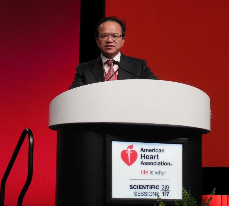
ANAHEIM, CALIF. – Treatment with dual-antiplatelet therapy following coronary artery bypass grafting with a saphenous vein maintained vein-graft patency better than aspirin alone in a randomized, multicenter trial with 500 patients.
After 1 year of dual-antiplatelet therapy (DAPT) with ticagrelor (Brilinta) and aspirin, 89% of saphenous-vein grafts remained patent, compared with a 77% patency rate in saphenous-vein grafts in patients treated with aspirin alone, a statistically significant difference for the study’s primary endpoint, Qiang Zhao, MD, said at the American Hart Association scientific sessions. The data, collected at six Chinese centers, also showed a nominal decrease in the combined rate of cardiovascular death, MI, and stroke: 2% with DAPT and 5% with aspirin alone. It further showed an increase in major or bypass-related bleeds: 2% with DAPT and none with aspirin alone, reported Dr. Zhao, professor and director of cardiac surgery at Ruijin Hospital in Shanghai, China.
Mitchel L. Zoler/Frontline Medical News
Dr. Qiang Zhao
But with a study of 500 patients that was only powered to address vein-graft patency the trial was underpowered to prove that the reductions in cardiovascular death, MI, and stroke outweighed the increase in major bleeds.
“If this result were repeated in a larger study it would be important,” John H. Alexander, MD, professor of medicine at Duke University in Durham, N.C., commented in a video interview.
The Compare the Efficacy of Different Antiplatelet Therapy Strategy After Coronary Artery Bypass Graft Surgery (DACAB) trial randomized patients who underwent coronary artery bypass grafting (CABG). They averaged about 64 years of age, and received an average of nearly four grafts each including an average of nearly three saphenous vein grafts. The study assigned patients to one of three treatment arms starting within 24 hours after surgery: 168 received ticagrelor 90 mg twice daily plus aspirin 100 mg once daily, 166 got ticagrelor alone, and 166 received aspirin alone. Treatment continued for 1 year.
Mitchel L. Zoler/Frontline Medical News
Dr. Timothy J. Gardner
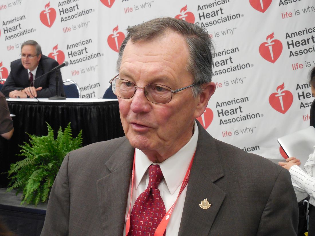
Although arterial grafts are much preferred for CABG, “saphenous vein grafts are still plenty used,” commented Timothy J. Gardner, MD, a cardiac surgeon and medical director of the Center for Heart & Vascular Health of Christiana Care in Newark, Del. That’s especially true when patients require multivessel bypass, in which case placement of saphenous veins grafts are a virtual given in current U.S. practice, Dr. Gardner said in an interview.
“Some surgeons and physicians currently prescribe DAPT to CABG patients, but there is not much evidence of its benefit. The DACAB trial is useful, but you need to show that it does not just improve patency but that patients also have better outcomes. The excess of major bleeds is a big deal. It gives one pause about adopting DAPT as standard treatment,” Dr. Gardner said.
DACAB received no commercial funding. Dr. Zhao has been a speaker on behalf of and has received research funding from AstraZeneca, the company that markets ticagrelor (Brilinta). He has also been a speaker for Johnson & Johnson and Medtronic and has received research funding from Bayer, Novartis, and Sanofi. Dr. Gardner had no disclosures.
Results from the DACAB trial showed that using aspirin and ticagrelor improved vein-graft patency, compared with using aspirin alone. It was a compelling result, but for the intermediate, imaging-based outcome of graft patency at 1 year after surgery. This finding is conclusive evidence that dual-antiplatelet therapy has some benefit.
But the findings from this trial, modestly sized with 500 patients, failed to prove that the clinical benefit from dual-antiplatelet therapy was worth the adverse effect of an increase in the rate of major and bypass-related bleeding. The study was underpowered to prove that dual-antiplatelet therapy had a clear beneficial impact on clinical outcomes such as cardiovascular death, MI, and stroke, although this combined rate went in the right direction with dual therapy, compared with aspirin alone. We need to see proof of a benefit for these clinical outcomes to justify using a treatment that causes an increase in major bleeds.
The DACAB results alone are not enough to justify a change in practice. It would be an important finding if the results could be replicated in a larger study. And if dual-antiplatelet therapy was proven to have a net clinical benefit for patients, we would still want to target it to patients with a higher ischemic risk and, in general, avoid using it in patients with a high bleeding risk.
The video associated with this article is no longer available on this site. Please view all of our videos on the MDedge YouTube channel
John H. Alexander, MD, is a cardiologist and professor of medicine at Duke University in Durham, N.C. He has been a consultant to and has received research funding from several companies, including AstraZeneca, the company that markets ticagrelor (Brilinta). He made these comments as designated discussant for the DACAB study and in a video interview .
Body
Results from the DACAB trial showed that using aspirin and ticagrelor improved vein-graft patency, compared with using aspirin alone. It was a compelling result, but for the intermediate, imaging-based outcome of graft patency at 1 year after surgery. This finding is conclusive evidence that dual-antiplatelet therapy has some benefit.
But the findings from this trial, modestly sized with 500 patients, failed to prove that the clinical benefit from dual-antiplatelet therapy was worth the adverse effect of an increase in the rate of major and bypass-related bleeding. The study was underpowered to prove that dual-antiplatelet therapy had a clear beneficial impact on clinical outcomes such as cardiovascular death, MI, and stroke, although this combined rate went in the right direction with dual therapy, compared with aspirin alone. We need to see proof of a benefit for these clinical outcomes to justify using a treatment that causes an increase in major bleeds.
The DACAB results alone are not enough to justify a change in practice. It would be an important finding if the results could be replicated in a larger study. And if dual-antiplatelet therapy was proven to have a net clinical benefit for patients, we would still want to target it to patients with a higher ischemic risk and, in general, avoid using it in patients with a high bleeding risk.
The video associated with this article is no longer available on this site. Please view all of our videos on the MDedge YouTube channel
John H. Alexander, MD, is a cardiologist and professor of medicine at Duke University in Durham, N.C. He has been a consultant to and has received research funding from several companies, including AstraZeneca, the company that markets ticagrelor (Brilinta). He made these comments as designated discussant for the DACAB study and in a video interview .
Title
DAPT must also show clinical benefits
DAPT must also show clinical benefits
ANAHEIM, CALIF. – Treatment with dual-antiplatelet therapy following coronary artery bypass grafting with a saphenous vein maintained vein-graft patency better than aspirin alone in a randomized, multicenter trial with 500 patients.
After 1 year of dual-antiplatelet therapy (DAPT) with ticagrelor (Brilinta) and aspirin, 89% of saphenous-vein grafts remained patent, compared with a 77% patency rate in saphenous-vein grafts in patients treated with aspirin alone, a statistically significant difference for the study’s primary endpoint, Qiang Zhao, MD, said at the American Hart Association scientific sessions. The data, collected at six Chinese centers, also showed a nominal decrease in the combined rate of cardiovascular death, MI, and stroke: 2% with DAPT and 5% with aspirin alone. It further showed an increase in major or bypass-related bleeds: 2% with DAPT and none with aspirin alone, reported Dr. Zhao, professor and director of cardiac surgery at Ruijin Hospital in Shanghai, China.
Mitchel L. Zoler/Frontline Medical News
Dr. Qiang Zhao
But with a study of 500 patients that was only powered to address vein-graft patency the trial was underpowered to prove that the reductions in cardiovascular death, MI, and stroke outweighed the increase in major bleeds.
“If this result were repeated in a larger study it would be important,” John H. Alexander, MD, professor of medicine at Duke University in Durham, N.C., commented in a video interview.
The Compare the Efficacy of Different Antiplatelet Therapy Strategy After Coronary Artery Bypass Graft Surgery (DACAB) trial randomized patients who underwent coronary artery bypass grafting (CABG). They averaged about 64 years of age, and received an average of nearly four grafts each including an average of nearly three saphenous vein grafts. The study assigned patients to one of three treatment arms starting within 24 hours after surgery: 168 received ticagrelor 90 mg twice daily plus aspirin 100 mg once daily, 166 got ticagrelor alone, and 166 received aspirin alone. Treatment continued for 1 year.
Mitchel L. Zoler/Frontline Medical News
Dr. Timothy J. Gardner
Although arterial grafts are much preferred for CABG, “saphenous vein grafts are still plenty used,” commented Timothy J. Gardner, MD, a cardiac surgeon and medical director of the Center for Heart & Vascular Health of Christiana Care in Newark, Del. That’s especially true when patients require multivessel bypass, in which case placement of saphenous veins grafts are a virtual given in current U.S. practice, Dr. Gardner said in an interview.
“Some surgeons and physicians currently prescribe DAPT to CABG patients, but there is not much evidence of its benefit. The DACAB trial is useful, but you need to show that it does not just improve patency but that patients also have better outcomes. The excess of major bleeds is a big deal. It gives one pause about adopting DAPT as standard treatment,” Dr. Gardner said.
DACAB received no commercial funding. Dr. Zhao has been a speaker on behalf of and has received research funding from AstraZeneca, the company that markets ticagrelor (Brilinta). He has also been a speaker for Johnson & Johnson and Medtronic and has received research funding from Bayer, Novartis, and Sanofi. Dr. Gardner had no disclosures.
Key clinical point: Using dual-antiplatelet therapy to treat patients receiving a saphenous vein coronary bypass graft led to better graft patency after 1 year, compared with bypass patients treated with aspirin alone.
Major finding: The 1-year saphenous-vein graft patency rate was 89% with DAPT treatment and 77% with aspirin alone. Data source: DACAB, a multicenter, randomized trial with 500 Chinese patients.
Disclosures: DACAB received no commercial funding. Dr. Zhao has been a speaker on behalf of and has received research funding from AstraZeneca, the company that markets ticagrelor (Brilinta). He has also been a speaker for Johnson & Johnson and Medtronic and has received research funding from Bayer, Novartis, and Sanofi.
In keeping with the commitment of the American College of Chest Physicians (CHEST) to be the home of the clinician educator, and supporting CHEST’s strategic vision of advancing best patient outcomes through innovative chest medicine education, a new designation intended to provide national-level recognition of excellence in continuing medical education has been established—the innovation award-winning Distinguished CHEST Educator.
Distinguished CHEST Educators are within the top 5% of CHEST’s faculty and are recognized for their achievements in making significant and long-term contributions to the design and delivery of CHEST education. With more than 108 ways to educate, these faculty members have exceeded expectations by serving as CHEST committee chairs, vice-chairs, faculty, and peer reviewers for programs such as the CHEST Annual Meeting.
“The greatest achievement I can imagine is seen in the people we train—as that lives on. Real values in medicine live only by being handed down to others. Over the past decade, CHEST has afforded me the privilege to represent the organization on a national platform, and, in doing so, I have been able to refine my own skills and those of my peers, as well as adding both quality and detail to my understanding of how young physicians learn,” says Nader Kamangar, MD, FCCP, of UCLA, CHEST member since 2000, and Distinguished CHEST Educator.
This designation will be granted to select clinical educators each year. The inaugural class of Distinguished CHEST Educators was honored at the end of October at CHEST 2017 in Toronto, as will be the tradition for the classes that follow.
Distinguished CHEST Educator
Congratulations to the inaugural class of Distinguished CHEST Educators.
Sandra Adams, MD, MS, FCCP
Doreen Addrizzo-Harris, MD, FCCP
A. Christine Argento, MD, FCCP
Robert Arntfield, MD, FCCP
Anthony Asciutto, RRT
Olivier Axler, MD, PhD, FCCP
Meyer Balter, MD, FCCP
Gisela Banauch, MD, MS, FCCP
Robert Baughman, MD, FCCP
David Bell, MD, FCCP
Michel Boivin, MD, FCCP
Gabriel Bosslet, MD, FCCP
Jean Bourbeau, MD, MS, FCCP
David Bowton, MD, FCCP
Kevin Brown, MD, FCCP
Jack Buckley, MD, MPH, FCCP
Kristin Burkart, MD, MS, FCCP
Brian Carlin, MD, FCCP
Christopher Carroll, MD, FCCP
Roberto Casal, MD
Richard Castriotta, MD, FCCP
Kevin Chan, MD, FCCP
Alexander Chen, MD
Michael Christian, MD, FCCP
Nancy Collop, MD, FCCP
Clayton Cowl, MD, MS, FCCP
Angel Coz Yataco, MD, FCCP
Gerard Criner, MD, FCCP
Carolyn D’Ambrosio, MD, FCCP
Mauricio Danckers, MD, FCCP
Aneesa Das, MD, FCCP
John Davies, RRT, MA, FCCP
Frank Detterbeck, MD, FCCP
Emily Diederich, MD, FCCP
Kevin Doerschug, MD, MS, FCCP
Meagan Dubosky, RRT-ACCS
Kevin Dushay, MD, FCCP
Eric Edell, MD, FCCP
William Enfinger
Michael Ezzie, MD, FCCP
David Feller-Kopman, MD, FCCP
Kevin Felner, MD, FCCP
Neil Freedman, MD, FCCP
Thomas Fuhrman, MD, MS, FCCP
John Gaillard, MD, FCCP
Colin Gillespie, MD
Maritza Groth, MD, FCCP
Mark Hall, MD
Jesse Hall, MD, FCCP
Nicola Hanania, MD, MBBS, FCCP
D. Kyle Hogarth, MD, FCCP
Steven Hollenberg, MD, FCCP
Robert Hyzy, MD, FCCP
Richard Irwin, MD, Master FCCP
Nader Kamangar, MD, MS, FCCP
Carl Kaplan, MD, FCCP
Brian Kaufman, MD, FCCP
William Kelly, MD, FCCP
Seth Koenig, MD, FCCP
Anastassios Koumbourlis, MD, MPH, FCCP
Lindsey Kreisher, RRT
Karol Kremens, MD, FCCP
Sunita Kumar, MD, MBBS, FCCP
Viera Lakticova, MD
Carla Lamb, MD, FCCP
Hans Lee, MD, FCCP
Peter Lenz, MD, MEd, FCCP
Stephanie Levine, MD, FCCP
Deborah Levine, MD, MS, FCCP
Kenneth Lyn-Kew, MD
Joao Alberto de Andrade, MD, FCCP
Neil MacIntyre, MD, FCCP
Donald Mahler, MD, FCCP
Fabien Maldonado, MD, FCCP
Atul Malhotra, MD, FCCP
Haney Mallemat, MD
Darcy Marciniuk, MD, FCCP
Diego Maselli Caceres, MD, FCCP
Paul Mayo, MD, FCCP
Peter Mazzone, MD, MPH, FCCP
John McIlwaine, DO, MBA, FCCP
Mark Metersky, MD, FCCP
Scott Millington, MD
Taro Minami, MD, FCCP
Lisa Moores, MD, FCCP
Amy Morris, MD
John Mullon, MD, FCCP
Septimiu Murgu, MD, FCCP
Mangala Narasimhan, DO, FCCP
Michael Niederman, MD, FCCP
Alexander Niven, MD, FCCP
Anne O’Donnell, MD, FCCP
Erik Osborn, MD
David Ost, MD, MPH, FCCP
Ronald Oudiz, MD, FCCP
Daniel Ouellette, MD, MS, FCCP
Nicholas Pastis, MD, FCCP
Paru Patrawalla, MD, FCCP
Jay Peters, MD, FCCP
Barbara Phillips, MD, MSPH, FCCP
Margaret Pisani, MD, MS, FCCP
Janos Porszasz, MD, PhD
Whitney Prince, MD, FCCP
Suhail Raoof, MBBS, FCCP
Marcos Restrepo, MD, MSc, FCCP
Otis Rickman, DO, FCCP
Roy Ridgeway
Mary Ried, RN, CCRN
Antoni Rosell, MD
Mark Rosen, MD, Master FCCP
Bernard Roth, MD, FCCP
Anthony Saleh, MD, FCCP
Juan Sanchez, MD, FCCP
Pralay Sarkar, MBBS, FCCP
Lewis Satterwhite, MD, FCCP
Paul Scanlon, MD, FCCP
Gregory Schmidt, MD, FCCP
David Schulman, MD, MPH, FCCP
Brady Scott, RRT, MS
Bernardo Selim, MD, FCCP
Curtis Sessler, MD, FCCP
Rakesh Shah, MD, FCCP
Ray Wes Shepherd, MD, FCCP
John Sherner, MD, FCCP
Ariel Shiloh, MD
Samira Shojaee, MD, FCCP
Gerard Silvestri, MD, MS, FCCP
Steven Simpson, MD, FCCP
James Stoller, MD, MS, FCCP
Mary Strek, MD, FCCP
William Stringer, MD, FCCP
Eleanor Summerhill, MD, FCCP
Lynn Tanoue, MD, FCCP
Victor Test, MD, FCCP
Arthur Tokarczyk, MD, FCCP
Anil Vachani, MD, FCCP
Momen Wahidi, MD, MBA, FCCP
Keith Wille, MD, FCCP
Lisa Wolfe, MD, FCCP
Richard Wunderink, MD, FCCP
Lonny Yarmus, DO, FCCP
Kazuhiro Yasufuku, MD, PhD, FCCP
Gulrukh Zaidi, MD
In keeping with the commitment of the American College of Chest Physicians (CHEST) to be the home of the clinician educator, and supporting CHEST’s strategic vision of advancing best patient outcomes through innovative chest medicine education, a new designation intended to provide national-level recognition of excellence in continuing medical education has been established—the innovation award-winning Distinguished CHEST Educator.
Distinguished CHEST Educators are within the top 5% of CHEST’s faculty and are recognized for their achievements in making significant and long-term contributions to the design and delivery of CHEST education. With more than 108 ways to educate, these faculty members have exceeded expectations by serving as CHEST committee chairs, vice-chairs, faculty, and peer reviewers for programs such as the CHEST Annual Meeting.
“The greatest achievement I can imagine is seen in the people we train—as that lives on. Real values in medicine live only by being handed down to others. Over the past decade, CHEST has afforded me the privilege to represent the organization on a national platform, and, in doing so, I have been able to refine my own skills and those of my peers, as well as adding both quality and detail to my understanding of how young physicians learn,” says Nader Kamangar, MD, FCCP, of UCLA, CHEST member since 2000, and Distinguished CHEST Educator.
This designation will be granted to select clinical educators each year. The inaugural class of Distinguished CHEST Educators was honored at the end of October at CHEST 2017 in Toronto, as will be the tradition for the classes that follow.
Distinguished CHEST Educator
Congratulations to the inaugural class of Distinguished CHEST Educators.
Stephen J. Welch was officially appointed Executive Vice President and CEO in April after serving as the interim for both positions since May 2016. Here’s a little “inside look” at what Steve is all about.
What is one major accomplishment you hope to achieve as Executive Vice President & Chief Executive Officer?
My goal as EVP/CEO is fairly simple and straightforward: to ensure the organization remains relevant and viable as a leader in providing clinically focused, innovative educational programs and content. I don’t really have one accomplishment that I’m focused on, but I do want to ensure that we achieve our annual organizational goals that support CHEST’s strategic plan. That may sound a little vague, but it’s true. We have so many outstanding programs and initiatives that I’d be doing a disservice to identify a single goal.
How does your previous experience with CHEST help you successfully lead the organization?
With CHEST being a not-for-profit organization, which relies on volunteer leadership and faculty, I think the relationships I’ve built over the past 23 years within the organization and the chest medicine community are invaluable. I personally know so many of our leadership because I’ve been part of the organization at the executive level working with them for those 23 years. They know me and how I approach opportunities, address issues, and handle challenges, which has helped build an immediate level of mutual respect, trust, and confidence between the staff and leadership. In addition, there was no disruption from having someone come in from the outside and have to get up to speed. It made the transition pretty seamless for the staff, as well.
During my time at CHEST, I’ve seen how the organization operates, from the journal, to the annual meeting and board reviews, to the simulation and hands-on skills training, to operational activities like the management of our finances and new global headquarters and training center. I’ve also had the opportunity to meet with many of our international members and sister societies. Those experiences have allowed me to work closely with many of our faculty, authors, and educators to understand their educational and professional needs, so we can ensure that we meet them.
CHEST is only as good as the education we provide, and it’s our subject matter experts who drive that content engine. In my previous role leading the Publishing Division and working on our journal CHEST® and programs like SEEK, I’ve had the honor and pleasure of working with some of the greatest minds in pulmonary, critical care, and sleep medicine. It’s humbling.
What will be some of the underlying themes as you work to outline the strategic plan for the next 5 years?
We are in the final stages of planning for 2018 and beyond, and although our proposed roadmap isn’t significantly different than what we have been doing, there’s some greater emphasis on a few key areas. For example, we’re looking at innovations in educational delivery. We’ve got some very forward thinking faculty educators and staff who are collaborating to develop innovations like gamification of educational and simulation programs, and augmented reality. Globalization and growth are also a key part of our strategic plan, and we are committed to the broad delivery of our educational programs and content both here and around the world. Finally, we have invested in a data analytics project that is maturing, and we’ll be leveraging that information to provide more personalized education plans – not just for the physician but for the entire health-care team. It’s important for us to stay relevant and viable.
Stephen J. Welch
Why has CHEST shifted to an interdisciplinary, team-focused approach?
I look at it as simply an evolution that reflects how health care is changing. It’s a team sport now, and our advanced practice providers (APPs) play a huge role in patient management and care. To be as effective and efficient as possible, and ensure the best patient outcomes, the whole team needs to be on the same page, and we believe that providing education for the interdisciplinary care team will help ensure that the best patient care is delivered.
There’s also a need for this education, and we want to fill it. Our APPs tell us that there is no formal pulmonary training or post-masters fellowship in pulmonary medicine for them. They are often left on their own to fill any gaps in knowledge and skills. That’s where our CHEST programs, such as our CHEST Annual Meeting, come in. We have an Interprofessional NetWork made up of APPs and physicians, and they were integral in working with the CHEST 2017 Program Committee to ensure plenty of relevant content was offered. Moving forward, we will continue to offer and build interdisciplinary programs designed for the entire team, as well as programs that address clinical issues across disciplines.
What are some of the critical skills CHEST physicians need to keep the population healthy during the ever-evolving field?
Educationally, we recognize that conferences like the annual CHEST meeting must provide more than just talking heads. We’ve invested heavily in high-fidelity medical simulation through small group, hands-on skill training in critical care techniques, airway management, EBUS, critical care ultrasound, bronchoscopy, and other chest medicine content. It’s like the old adage about fishing: instead of telling people how to fish, we teach them to fish.
Any final thoughts?
I always encourage our members to get involved with CHEST and experience the camaraderie and connectivity of the CHEST family. Ask any of our leadership, and you will surely hear their story of that special person who first introduced them to the College. Reach out and tell a colleague about CHEST. We are focused on clinically relevant education that our members can take back and put into action immediately. At the end of the day, it’s about providing state-of-the-art education via high fidelity medical simulation, hands-on skills training, clinically focused courses, case-based programming, and more—all intended to be immediately implemented to improve patient care and patient outcomes. That’s what the CHEST organization is all about.
Burden of Adult Community-Acquired, Health-care-Associated, Hospital-Acquired, and Ventilator-Associated Pneumonia: New York City, 2010 to 2014. By R. E. Corrado, et al.
Hyperbaric Oxygen Therapy Is Associated With Lower Short- and Long-Term Mortality in Patients With Carbon Monoxide Poisoning. By C-C Huang, et al.
Evidence-Based Medicine
Pharmacologic and Nonpharmacologic Treatment for Acute Cough Associated With the Common Cold: CHEST Expert Panel Report. By M. A. Malesker, et al, on behalf of the CHEST Expert Cough Panel.
Cough in Ambulatory Immunocompromised Adults: CHEST Expert Panel Report. By M. J. Rosen, et al, on behalf of the CHEST Expert Cough Panel.
Original Research
Burden of Adult Community-Acquired, Health-care-Associated, Hospital-Acquired, and Ventilator-Associated Pneumonia: New York City, 2010 to 2014. By R. E. Corrado, et al.
Hyperbaric Oxygen Therapy Is Associated With Lower Short- and Long-Term Mortality in Patients With Carbon Monoxide Poisoning. By C-C Huang, et al.
Evidence-Based Medicine
Pharmacologic and Nonpharmacologic Treatment for Acute Cough Associated With the Common Cold: CHEST Expert Panel Report. By M. A. Malesker, et al, on behalf of the CHEST Expert Cough Panel.
Cough in Ambulatory Immunocompromised Adults: CHEST Expert Panel Report. By M. J. Rosen, et al, on behalf of the CHEST Expert Cough Panel.
Great attention has been paid to the SERVE-HF trial (“Treatment of Sleep-disordered Breathing with Predominant Central Sleep Apnea by Adaptive Servo Ventilation in Patients with Heart Failure”), which showed increased all-cause mortality and cardiovascular mortality in the Adaptive Servo-ventilation (ASV) group compared with the control group of conventional heart failure management alone. The results of this trial led to the recommendation by multiple ASV manufacturers and medical societies to withdraw clinical use of ASV from patients with heart failure and a reduced ejection fraction (HFrEF) less than 45%.
Sleep-disordered breathing is common in patients with HFrEF with prevalence rates of 50% to 75%. Central sleep apnea (CSA) is associated with increased mortality in heart failure (HF) and is found in 25% to 40% of this subpopulation. It is estimated that the severity of CSA increases in parallel with the severity of the HF. For several years, treatment of CSA with positive pressure ventilation was believed to favor outcomes in HFrEF with a protective effect.
In the Canadian Positive Airway Pressure for Patients with CSA and HF (CANPAP) trial, subjects were randomized to treatment with CPAP or no CPAP. This trial was terminated early; it did not show an advantage of CPAP in morbidity or mortality. A post-hoc analysis suggested that mortality might be reduced if the frequency of respiratory events per hour or apnea hypopnea index (AHI) is reduced to 15/hour or less while using CPAP.
Dr. Jairo H. Barrantes
Hoping to improve the outcomes of HF, SERVE-HF was the first randomized, large scale, multinational trial directed to treat CSA in patients with HFrEF < 45% and concomitant clinically significant sleep apnea with AHI > 15/hour of central predominance (CSA index >10/hour). Treatment arms compared the addition the ASV, one of the most effective noninvasive positive pressure ventilation technologies for central apneas that offers automated inspiratory pressure support in addition to expiratory positive pressure vs conventional medical treatment alone in the control group.
The study published in the New England Journal of Medicine in September 2015 was designed in an intention-to-treat basis with the primary end point of time to first event, a composite of death from any cause, lifesaving cardiovascular intervention (heart transplant, implantation of LVAD, resuscitation after sudden cardiac arrest, or defibrillation for ventricular arrhythmia), or unplanned hospitalization for heart failure. The study did not show significant differences in the primary end point between the ASV and control group (54.1% and 50.8%, respectively; hazard ratio, 1.13; 95% confidence interval, 0.97 to 1.31; P=.10).1
The most interesting and unexpected outcome was an increase in the all cause mortality and cardiovascular mortality in the ASV group (hazard ratio for death from any cause, 1.28; 95% CI, 1.06 to 1.55; P=.01; and hazard ratio from cardiovascular death, 1.34; 95% CI, 1.09 to 1.65; P=.006).1 These findings led to the above recommendations from manufacturers, as well as a position statement from the American Academy of Sleep Medicine. These findings cannot be extrapolated to the obstructive sleep apnea population with concomitant HFrEF or to patients with HF with preserved ejection fraction, where positive pressure ventilation has offered an advantage1 likely by a different physiologic mechanism not fully uncovered at this time, believed to be an overall effect of afterload reduction.
Selection and self-selection bias in this study was addressed in a new analysis by the same SERVE-HF investigator group published August 2017, where a time-dependent model of on-treatment analysis (done to tease out if the original results were related to the treatment assignment or to poor adherence) was conducted to understand potential causes of the initial findings in the original study. It showed patients randomized to ASV who crossed over to the control group were at higher risk of cardiovascular death than control subjects; also the control patients with crossover to ASV had a signal of lower risk of cardiovascular death risk compared with patients assigned to ASV.2 Reduced adherence to ASV treatment during SERVE-HF was a concern, since it resulted in a reduced exposure to the treatment. The on-treatment analysis showed again an increase of cardiovascular death in HFrEF patients with predominant CSA treated with ASV in addition to conventional heart failure treatment compared with the control group.2 There was no increase in cardiovascular death risk associated with ASV use intervals (dose effect). This effect is not related to the amount of hours used per night.
The effect of the recommended withdrawal of treatment in HFrEF patients with EF<45% and moderate to severe central predominant sleep apnea is being addressed in smaller studies. A single center retrospective analysis observed the effects after ASV discontinuation in this population. Thirteen out of 126 patients treated with ASV who met SERVE-HF criteria were followed for at least a year; 93% of the subjects who met criteria had ASV removed; immediate recurrence of the central apnea was observed in most (except two patients), while adverse events were not identified (defined as need for emergency hospitalization). Day and nighttime symptoms were reported by 61% of the group, and they were started on alternative treatments.3 At 1 year after ASV removal, 88% of patients were still alive, overall cardiac function did not change in 1 year (P=0.17), and seven patients required adjustment of medications for heart failure. Symptomatic patients were treated with oxygen supplementation for nocturnal symptoms or CPAP if they had daytime sleepiness. None was treated with bi-level PAP, acetazolamide, or phrenic nerve stimulation. Four patients insisted on continuation of ASV despite understanding physician concerns. 3 This study helps to demonstrate that ASV discontinuation is feasible but requires close follow-up. However, larger, long-term prospective reviews are required to draw statistically meaningful conclusions about the consequences and safety of ASV removal; these studies will be difficult to conduct under the current indications for ASV in the interest group.
At this time, investigators have shifted to further understand the causes of the increase in cardiovascular mortality, overall mortality, and the understanding of the pathophysiologic processes associated with ASV use in HFrEF. It is not known whether the effect in mortality is related to the specific ASV device/algorithm used to suppress CSA or is related to the ASV principle itself. Upcoming studies will assist in clarifying these details. Currently, there is an ongoing trial looking at the effect of ASV on survival and hospital admissions in heart failure (ADVENT–HF) using a different ASV device; this study will hopefully elucidate the impact of class effect vs device effect. It may also provide better insight of the etiology of mortality and the impact of improved ASV compliance, first addressed by the on-treatment analysis of the SERVE-HF.4
Although the reasons for increased mortality related to ASV are unclear, proposed hypotheses include: central apnea is an adaptive mechanism to HFrEF and the reversal of central apneas might adversely affect the underlying disease process,1 low adherence to ASV may impact outcomes, and specific devices may induce hyper-/hypoventilation generated by the algorithm designs of the specific ASV device and this may result in electrolyte abnormalities that generate arrhythmias.
The ADVENT-HF trial, although similar in design, has significant differences from SERVE-HF: different ASV devices may have a different impact on cardiac output and ventilation, recruited patients included those with less daytime sleepiness, and the potential to assess the effect of ASV in patients with OSA and low daytime sleepiness in patients with reduced EF.5,6 This ongoing study may help us to further understand why there is increased mortality and what effect ASV has on the treatment of sleep apnea in patients with HFrEF.
References
1. Cowie MR, et al. N Engl J Med. 2015;373(12):1095-1105.
2. Woehrle H, et al. Eur Respir J. 2017; 50:1601.
3. Brill AK, et al. Sleep Med. 2017;37:201-207.
4. Bradley TD, et al. Can Respir. 2015;22(6):313.
5. Lyons OD, et al. Eur J Heart Fail. 2017;19(4):579-587.
6. Haruki N, et al. Can J Cardiol. 2016;32(12):1402-1410.
Dr. Barrantes is an assistant professor, Department of Pulmonary, Critical Care, and Sleep Medicine, Baylor College of Medicine, Houston, Texas.
Alex M. Azar II, a former pharmaceutical executive and member of the George W. Bush administration, has been selected by President Donald Trump to lead the Department of Health & Human Services.
Mr. Azar served as president of Eli Lilly in the United States for 5 years from 2012 to 2017, after joining the company in 2007. Prior to that, he served President Bush at HHS from 2001 to 2007, serving first as general counsel and later as deputy secretary under Secretary Michael O. Leavitt.
Wikimedia Commons/WWsgConnect/CC-SA 4.0
Alex M. Azar II
President Trump announced the appointment via Twitter on Nov. 13. “Happy to announce, I am nominating Alex Azar to be the next HHS Secretary. He will be a star for better healthcare and lower drug prices!”
“The challenges plaguing the American health care system are serious. For too long, hardworking, middle-class families have been forced to bear the brunt of Obamacare’s failures in the form of higher premiums and fewer choices,” Senate Finance Committee Chairman Orrin Hatch (R-Utah) said in a statement. “The leader of HHS will be at the tip of the spear, working to not only right the wrongs of this deeply flawed law but also ensure the long-term sustainability of both Medicare and Medicaid.”
The Senate Finance Committee must first approve the nomination before it is considered by the full chamber.
“We commend President Trump for nominating Alex Azar for secretary of Health & Human Services,” House Energy & Commerce Committee Chairman Greg Walden (R-Ore.) and Health Subcommittee Chairman Michael Burgess, MD, (R-Texas) said in a joint statement. “He is a veteran of HHS, bringing with him a wealth of institutional knowledge that will be instrumental in delivering patient-centered health care and combating the opioid crisis. We look forward to working with Mr. Azar on these critical issues and many others in the future.”
The Campaign for Sustainable Rx Pricing, a coalition of physicians and other stakeholders across the health care industry, was more measured in its reaction to the news.
“We sincerely hope that Secretary-nominee Azar will follow through on the President’s commitment to achieve lower drug prices for all Americans,” according to a statement from CSRxP. “We look forward to working with him, once confirmed, to end anticompetitive practices that artificially inflate drug prices, restore a functioning prescription drug market, and rein in the exorbitant price hikes that harm patients, job creators, and taxpayers alike.”
The nomination process could be bumpy, as Mr. Azar has made statements in the news in the past that were in support the dismantling of the Affordable Care Act. But keeping him from the post will be difficult, as he would only need a simple majority vote in the Senate to gain approval. With Republicans holding 52 seats, it would only require three dissenting GOP senators, assuming the Democrats vote against the appointment. If two crossed the aisle, Vice President Mike Pence would cast the deciding vote.
Finance Committee Democrats boycotted the committee vote on Mr. Azar’s predecessor, Secretary Tom Price, MD, forcing committee Chairman Hatch to suspend rules in order to move the appointment to the full chamber for consideration.