User login
Target self-medication of mood and anxiety symptoms
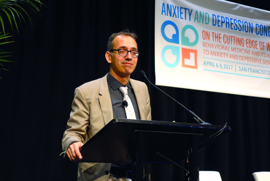
SAN FRANCISCO – Drinking to alleviate mood or anxiety symptoms is responsible for 12%-16% of cases of new-onset alcohol use disorder in affected individuals, Jitender Sareen, MD, said at the annual conference of the Anxiety and Depression Association of America.
Similarly, the use of prescription or nonprescription drugs to self-medicate mood or anxiety symptoms accounts for 20% of new-onset drug use disorders in this population, added Dr. Sareen, professor and head of the department of psychiatry at the University of Manitoba in Winnipeg.
He was a coauthor of two landmark longitudinal epidemiologic studies that support the concept of self-medication as a direct causal mechanism that explains a phenomenon often observed in clinical practice: namely, the high rate of comorbid mood or anxiety disorders accompanied by an alcohol or substance use disorder.
“The clinical implication is that questions about self-medication with alcohol or drugs should be included in the assessment of patients with anxiety and mood symptoms, because self-medication is a marker of higher likelihood of psychopathology. And psychologic therapies like cognitive-behavioral therapy and dialectical behavior therapy could prevent onset of substance use disorders by teaching patients emotion regulation skills to manage their mood and anxiety symptoms without self-medication,” Dr. Sareen said.
The first longitudinal study of the role of self-medication in the development of comorbid anxiety and substance use disorders included 34,653 nationally representative adults who completed both the initial face-to-face National Epidemiologic Survey on Alcohol and Related Conditions in 2001-2002 and a follow-up survey conducted 3 years later.
During the 3-year follow-up period, 9.7% of subjects developed a new-onset anxiety disorder, 5.9% of participants newly met DSM-IV diagnostic criteria for alcohol use disorder, and 2% developed a new-onset drug use disorder.
Among subjects who met the criteria for an anxiety disorder at baseline and at that time also reported self-medication with alcohol, 12.6% developed an incident alcohol use disorder during follow-up. Among those who self-medicated with drugs, 10.4% developed a drug use disorder.
In contrast, only 4.7% of subjects with a baseline anxiety disorder who did not self-medicate with alcohol at baseline developed an incident alcohol use disorder. And an incident drug use disorder occurred in 1.7% of patients with a baseline anxiety disorder who did not self-medicate with drugs.
Among patients with a baseline alcohol or other substance use disorder, self-medication with alcohol was associated with an adjusted 2.13-fold increased likelihood of developing social phobia during 3 years of follow-up, while self-medication with other drugs was independently associated with a 3.27-fold increased likelihood of subsequently developing social phobia.
In a multivariate logistic regression analysis, Dr. Sareen and his coinvestigators determined that self-medication with alcohol by patients with an anxiety disorder at baseline was associated with a 2.63-fold increased risk of incident alcohol use disorder during follow-up. Self-medication with drugs in patients with a baseline anxiety disorder was associated with a 4.99-fold risk of a new-onset substance use disorder during the 3 years of follow-up (Arch Gen Psychiatry. 2011;68[8]:800-7).
In a subsequent analysis of the same prospectively studied population, Dr. Sareen and his colleagues focused specifically on drinking to self-medicate mood symptoms. They found that self-medication with alcohol was associated with an adjusted 3.1-fold increased likelihood of new-onset alcohol dependence during the 3-year follow-up, as well as with a 3.45-fold increased risk of persistence of alcohol dependence. Roughly 12% of all cases of incident alcohol dependence arising during follow-up of patients with baseline mood symptoms were attributed to self-medication with alcohol. The increased risk of new-onset alcohol dependence was observed not only in subjects who met DSM-IV criteria for an affective disorder, but in those with subthreshold mood symptoms as well (JAMA Psychiatry. 2013 Jul;70[7]:718-26).
Again, this points to drinking as a behavior employed to self-medicate mood symptoms as a potential target for preventive interventions aimed at reducing the occurrence of alcohol dependence. As yet, however, no formal studies have been done to confirm the effectiveness of this strategy, the psychiatrist continued.
Dr. Sareen was not involved in the third iteration of the National Epidemiologic Survey on Alcohol and Related Conditions, in which a different group of 36,309 nationally representative adults was interviewed during 2011-2013 to assess the impact of the DSM-5 criteria for alcohol use disorder. Using DSM-5, 13.9% of the population met criteria for an alcohol use disorder during the past 12 months, and the lifetime prevalence of alcohol use disorder was 29.1%. Fewer than one in five subjects with a lifetime DSM-5 alcohol use disorder had ever been treated.
In the first national survey, which used DSM-IV criteria, the 12-month and lifetime prevalences of alcohol abuse and/or dependence were 8.5% and 30.3%, respectively.
DSM-5 alcohol use disorder was highly comorbid. Both lifetime and 12-month alcohol use disorder were associated with significantly increased likelihood of other substance use disorders, major depression, bipolar I disorder, borderline personality disorder, and antisocial personality disorder.
These data indicate “an urgent need to educate the public and policy makers about alcohol use disorder and its treatment alternatives, to destigmatize the disorder, and to encourage those who cannot reduce their alcohol consumption on their own, despite substantial harm to themselves and others, to seek treatment,” the investigators wrote (JAMA Psychiatry. 2015 Aug;72[8]:757-66).
The surveys were supported by the National Institute on Alcohol Abuse and Alcoholism. Dr. Sareen reported having no financial conflicts of interest.
EXPERT ANALYSIS FROM THE ANXIETY AND DEPRESSION CONFERENCE 2017
Sarilumab showed sustained effect on RA progression at 3 years
BIRMINGHAM, ENGLAND – A “durable clinical response and stabilization of structural damage” was observed at 3 years of follow-up in the Long Term Evaluation of Sarilumab in Rheumatoid Arthritis Patients (SARIL-RA-EXTEND) study.
Désirée van der Heijde, MD, PhD, who reported the findings of the EXTEND study at the British Society for Rheumatology annual conference, noted that the benefit was seen regardless of the initial treatment that had been given at baseline.

The aim of the EXTEND trial was to examine the continuity of response to sarilumab seen in the MOBILITY trial, said Dr. van der Heijde, professor of rheumatology at Leiden (The Netherlands) University Medical Center.
Sarilumab is a fully human (IgG1) monoclonal antibody that binds to interleukin (IL)-6 receptors, both soluble and membrane-bound, and thus inhibits IL-6-mediated signaling through the soluble IL-6R alpha and membrane-bound IL-6R alpha receptors. It is undergoing regulatory approval in the United States, European Union, and Japan, but was recently approved in Canada for treatment of adult patients with moderate to severe rheumatoid arthritis (RA) who have had an inadequate response to one or more biologic or nonbiologic disease-modifying antirheumatic drugs (DMARDs) (Drugs. 2017;77:705-12).
In the MOBILITY trial, 1,197 patients who were being treated with methotrexate but who had an inadequate response were randomized to receive placebo (n = 398) or sarilumab given subcutaneously every 2 weeks at one of two doses: 150 mg (n = 400) or 200 mg (n = 399).
EXTEND allowed patients completing this trial who still had active disease to continue or start (if they had been given placebo) treatment with sarilumab at a dose of 200 mg given every 2 weeks, with dose reduction to 150 mg every 2 weeks if needed, in addition to methotrexate. A total of 901 patients participated in the extension study.
“In the MOBILITY trial, all three groups were very balanced, and if you then take the patients who entered the EXTEND trial, they are very similar to the patients who also were randomized into MOBILITY,” Dr. van der Heijde said.
This was a fairly typical RA population, she observed: About 80% were female, the mean age was 50 years, and the mean duration of RA was 9 years. About 20%-25% had prior treatment with a DMARD, more than 80% were rheumatoid factor or anti-CCP antibody positive. There were similar mean C-reactive protein (CRP) levels between the groups, and the 28-joint disease activity score (DAS28) with CRP was around 6, and Clinical Disease Activity Index (CDAI) score around 40.
Radiographs that were taken at baseline, at the end of year 2, and at the end of year 3 were reread by two independent readers and scored together in one session. Data had to be extrapolated for 29 patients who did not have a radiographs taken at year 3 but who had been seen during the third year of treatment.
The significant radiographic inhibition seen at the end of the MOBILITY trial in both the 150-mg and 200-mg active treatment groups was sustained in the EXTEND study.
“There is a small progression between year 2 and 3, and this progression is quite similar in all the three treatment arms,” Dr. van der Heijde reported, nothing that all patients were taking 200 mg of sarilumab at this point.
The mean change in the modified total Sharp score (mTSS) from year 2 to 3 was 0.35 in the patients who had originally been randomized to the placebo arm, 0.64 in patients originally randomized to 150 mg sarilumab every 2 weeks, and 0.44 in those originally taking 200 mg sarilumab every 2 weeks.
From baseline to year 3, the mean change in mTSS were a respective 3.3, 1.9, and 0.8.
“If you present the data in a different way, like the percentage of patients showing no progression, you see the differentiation between the patients who started on placebo versus those who were started on sarilumab 150 mg or 200 mg,” said Dr. van der Heijde.
At year 2, 67%, 59%, and 48% of patients treated with sarilumab 200 mg, sarilumab 150 mg, or placebo, respectively had no progression in mTSS (signified by a change from baseline of 0.5 points or more).
At year 3, corresponding rates were a respective 75%, 55%, and 49%.
DAS-28-CRP response at year 3 was similar across the initial treatment groups, Dr. van der Heijde observed. Reductions seen in the MOBILITY trial were clearly continued, she said. The percentage of patients achieving DAS-28-CRP of less than 2.6 was 22%, 34%, and 36% of placebo, sarilumab 150-mg, and sarilumab 200-mg treated patients at the end of the MOBILITY study. At the end of year 2, the corresponding numbers were 60%, 62%, and 62%, and by year 3, not much had changed in the percentage of patients achieving DAS-28-CRP of less than 2.6: 58% for placebo, 62% for sarilumab 150 mg, and 68% for sarilumab 200 mg.
Similar results were seen for patients achieving a CDAI of 2.8 or lower at years 2 and 3 in the extension study.
Treatment-emergent adverse events (TEAEs) occurred in 89.7% of patients over 3 years. One in five (20%) patients experienced serious adverse events, with 23% of patients discontinuing treatment because of TEAEs. There were 9 (0.8%) deaths during the trial.
TEAEs that occurred at rates of 5% or higher in any treatment group were neutropenia in 19.4%, increased alanine aminotransferase in 13.0%, and upper respiratory tract infections in 12.7%.
There were some changes in laboratory values, Dr. van der Heijde said, but “most of the changes were very small” and in line with the effects expected with IL-6 inhibition.
The study was sponsored by Sanofi Genzyme and Regeneron Pharmaceuticals. Dr. van der Heijde and coauthors disclosed receiving consulting fees, research grants, or both from multiple pharmaceutical companies, including the sponsors of the study. Dr. van der Heijde is director of Imaging Rheumatology.
BIRMINGHAM, ENGLAND – A “durable clinical response and stabilization of structural damage” was observed at 3 years of follow-up in the Long Term Evaluation of Sarilumab in Rheumatoid Arthritis Patients (SARIL-RA-EXTEND) study.
Désirée van der Heijde, MD, PhD, who reported the findings of the EXTEND study at the British Society for Rheumatology annual conference, noted that the benefit was seen regardless of the initial treatment that had been given at baseline.
The aim of the EXTEND trial was to examine the continuity of response to sarilumab seen in the MOBILITY trial, said Dr. van der Heijde, professor of rheumatology at Leiden (The Netherlands) University Medical Center.
Sarilumab is a fully human (IgG1) monoclonal antibody that binds to interleukin (IL)-6 receptors, both soluble and membrane-bound, and thus inhibits IL-6-mediated signaling through the soluble IL-6R alpha and membrane-bound IL-6R alpha receptors. It is undergoing regulatory approval in the United States, European Union, and Japan, but was recently approved in Canada for treatment of adult patients with moderate to severe rheumatoid arthritis (RA) who have had an inadequate response to one or more biologic or nonbiologic disease-modifying antirheumatic drugs (DMARDs) (Drugs. 2017;77:705-12).
In the MOBILITY trial, 1,197 patients who were being treated with methotrexate but who had an inadequate response were randomized to receive placebo (n = 398) or sarilumab given subcutaneously every 2 weeks at one of two doses: 150 mg (n = 400) or 200 mg (n = 399).
EXTEND allowed patients completing this trial who still had active disease to continue or start (if they had been given placebo) treatment with sarilumab at a dose of 200 mg given every 2 weeks, with dose reduction to 150 mg every 2 weeks if needed, in addition to methotrexate. A total of 901 patients participated in the extension study.
“In the MOBILITY trial, all three groups were very balanced, and if you then take the patients who entered the EXTEND trial, they are very similar to the patients who also were randomized into MOBILITY,” Dr. van der Heijde said.
This was a fairly typical RA population, she observed: About 80% were female, the mean age was 50 years, and the mean duration of RA was 9 years. About 20%-25% had prior treatment with a DMARD, more than 80% were rheumatoid factor or anti-CCP antibody positive. There were similar mean C-reactive protein (CRP) levels between the groups, and the 28-joint disease activity score (DAS28) with CRP was around 6, and Clinical Disease Activity Index (CDAI) score around 40.
Radiographs that were taken at baseline, at the end of year 2, and at the end of year 3 were reread by two independent readers and scored together in one session. Data had to be extrapolated for 29 patients who did not have a radiographs taken at year 3 but who had been seen during the third year of treatment.
The significant radiographic inhibition seen at the end of the MOBILITY trial in both the 150-mg and 200-mg active treatment groups was sustained in the EXTEND study.
“There is a small progression between year 2 and 3, and this progression is quite similar in all the three treatment arms,” Dr. van der Heijde reported, nothing that all patients were taking 200 mg of sarilumab at this point.
The mean change in the modified total Sharp score (mTSS) from year 2 to 3 was 0.35 in the patients who had originally been randomized to the placebo arm, 0.64 in patients originally randomized to 150 mg sarilumab every 2 weeks, and 0.44 in those originally taking 200 mg sarilumab every 2 weeks.
From baseline to year 3, the mean change in mTSS were a respective 3.3, 1.9, and 0.8.
“If you present the data in a different way, like the percentage of patients showing no progression, you see the differentiation between the patients who started on placebo versus those who were started on sarilumab 150 mg or 200 mg,” said Dr. van der Heijde.
At year 2, 67%, 59%, and 48% of patients treated with sarilumab 200 mg, sarilumab 150 mg, or placebo, respectively had no progression in mTSS (signified by a change from baseline of 0.5 points or more).
At year 3, corresponding rates were a respective 75%, 55%, and 49%.
DAS-28-CRP response at year 3 was similar across the initial treatment groups, Dr. van der Heijde observed. Reductions seen in the MOBILITY trial were clearly continued, she said. The percentage of patients achieving DAS-28-CRP of less than 2.6 was 22%, 34%, and 36% of placebo, sarilumab 150-mg, and sarilumab 200-mg treated patients at the end of the MOBILITY study. At the end of year 2, the corresponding numbers were 60%, 62%, and 62%, and by year 3, not much had changed in the percentage of patients achieving DAS-28-CRP of less than 2.6: 58% for placebo, 62% for sarilumab 150 mg, and 68% for sarilumab 200 mg.
Similar results were seen for patients achieving a CDAI of 2.8 or lower at years 2 and 3 in the extension study.
Treatment-emergent adverse events (TEAEs) occurred in 89.7% of patients over 3 years. One in five (20%) patients experienced serious adverse events, with 23% of patients discontinuing treatment because of TEAEs. There were 9 (0.8%) deaths during the trial.
TEAEs that occurred at rates of 5% or higher in any treatment group were neutropenia in 19.4%, increased alanine aminotransferase in 13.0%, and upper respiratory tract infections in 12.7%.
There were some changes in laboratory values, Dr. van der Heijde said, but “most of the changes were very small” and in line with the effects expected with IL-6 inhibition.
The study was sponsored by Sanofi Genzyme and Regeneron Pharmaceuticals. Dr. van der Heijde and coauthors disclosed receiving consulting fees, research grants, or both from multiple pharmaceutical companies, including the sponsors of the study. Dr. van der Heijde is director of Imaging Rheumatology.
AT RHEUMATOLOGY 2017
Key clinical point:
Major finding: The mean change in the modified total Sharp score (mTSS) from year 2 to 3 was 0.35 in the patients who had originally been randomized to the placebo arm, 0.64 in patients originally randomized to 150 mg sarilumab every 2 weeks, and 0.44 in those originally taking 200 mg sarilumab every 2 weeks.
Data source: SARIL-RA-EXTEND: a multicenter, uncontrolled extension study involving 1,197 patients who had participated in the phase III SARIL-RA-MOBILITY trial.
Disclosures: The study was sponsored by Sanofi Genzyme and Regeneron Pharmaceuticals. Dr. van der Heijde and her coauthors disclosed receiving consulting fees, research grants, or both from multiple pharmaceutical companies, including the sponsors of the study. Dr. van der Heijde is director of Imaging Rheumatology.

Here’s what’s trending at SHM
SHM gives QI a new look
SHM is proud to announce that its Center for Hospital Innovation & Improvement has a fresh look and name: SHM’s Center for Quality Improvement. While the name may have changed, SHM’s Center for QI will remain your partner in quality and patient safety.
“SHM’s Center for QI provides a comprehensive set of resources and programs to support hospitalists and other hospital clinicians as they work to improve quality and safety in their hospitals,” says Eric E. Howell, MD, MHM, senior physician advisor for SHM’s Center for QI.
SHM’s Center for QI’s mentored implementation programs are deployed in hundreds of hospitals and have been recognized with the John M. Eisenberg Award. More recently, its opioid-safety program (RADEO) was recognized by the CMS for its efforts to enhance patient safety.
Visit http://www.hospitalmedicine.org/QI to learn more about SHM’s Center for QI and about opportunities for partnerships, solutions, and tools to address your QI needs.
PHM 2017 is coming! Book your ticket to Nashville today
Pediatric Hospital Medicine (PHM) 2017 is the largest, leading educational event for health care professionals who specialize in the care of hospitalized children. This year’s meeting will be held July 20-23 at the Omni Nashville in Tennessee.
Attendees will have the opportunity to network with colleagues from across the nation, learn from renowned faculty from throughout the discipline, and acquire skills, tools, and resources to directly benefit their patients and practice.
PHM 2017 has been designed to provide participants with tools to improve clinical skills and practice, address management issues, lead change and innovation within their institutions, and network with thought leaders to collaborate and learn about new innovations.
View the full meeting schedule, educational objectives, and more at www.peds2017.org.![]()
Benchmark your HMG appropriately with the State of Hospital Medicine Report
The State of Hospital Medicine Report continues to be the best source of detail regarding the configuration and operation of hospital medicine groups. The biennial report provides current data on hospitalist compensation and production, in addition to cutting-edge knowledge covering practice demographics, staffing levels, turnover, staff growth, compensation methods, and financial support for solid, evidence-based management decisions.
“We’ve used data from the report to hold more informed discussions with the group that provides our note-coding services and to determine how to benchmark our nocturnists’ workloads and pay,” said Andrew White, MD, SFHM, director of the Hospital Medicine Service at the University of Washington in Seattle. “The results are broken into region and academic practice type, which gives me the confidence that I’m looking at results from groups like mine, rather than comparing to the country-wide average.”
The report is designed for hospital medicine leaders (both physician leaders and nonphysician practice administrators and executives), as well as frontline hospitalists, nurse practitioners, physician assistants, pediatricians, and internal and family medicine physicians.
In addition to the print version, the 2016 State of Hospital Medicine Report is also available in an enhanced, fully searchable digital version. To order your copy in either print or digital, visit www.hospitalmedicine.org/survey.
Learn how to drive change as a leader in hospital medicine
A successful hospitalist program requires strong leadership from the floor to the C-suite. SHM’s Leadership Academy prepares clinical and academic leaders with vital skills that, traditionally, are not taught in medical school or typical residency programs. This year’s meeting will be held October 23-26 at the JW Marriott Camelback Inn in Scottsdale, Ariz.
New for Leadership Academy 2017, Strategic Essentials (formerly Leadership Foundations), Influential Management, and Mastering Teamwork will be available to all attendees, regardless of previous attendance. SHM provides recommendations for interested registrants so they can determine which course fits them best in their leadership journey.
Take the Strategic Essentials course to evaluate your personal leadership strengths and weaknesses, understand key hospital drivers, and more.
If you are looking to learn skills needed to drive culture change through specific leadership behaviors as well as financial storytelling, then Leadership: Influential Management would be a great course for you.
The third course, Leadership: Mastering Teamwork, will help attendees learn to critically assess program growth opportunities, lead and motivate teams, and design effective communication strategies. Learn more at www.shmleadershipacademy.org.![]()
Stay ahead of the MACRA curve with SHM
The Medicare Access and CHIP Reauthorization Act (MACRA) put into motion the new Quality Payment Program, which replaces past pay-for-performance programs, such as the Physician Quality Reporting System and physician value-based modifier. The new program has many complicated requirements, and hospitalists will be impacted.
The first year of the program has flexible participation, yet hospitalists need to do at least one thing (report one quality measure, attest to one improvement activity) in the program in order to avoid a 4% penalty to Medicare payments. 2017 is the first reporting year, so now is the time for providers to familiarize themselves with the requirements.
To support hospitalists who are looking for hospital medicine–specific ways to participate and avoid penalties, SHM hosted a webinar that is now available at www.macraforhm.org under “Resources.” SHM’s policy staff broke down the program requirements and went into detail on ways in which hospitalists can and should participate in the new program. Updates and other resources are also available at www.macraforhm.org.
Looking to be a speaker at Hospital Medicine 2018?
The Society of Hospital Medicine reminds you to submit your workshop proposal for the 2018 Annual Meeting to be held April 8-11, 2018, at the Orlando World Center Marriott. Workshops should involve topics in one of ten categories: clinical, career development, research, academic, patient experience/communication, perioperative, information technology, practice management, quality and patient safety, and evidence‐based medicine/high‐value care. Each workshop should last 90 minutes.
Proposals that are the most likely to be accepted will be innovative as well as highly interactive, utilizing small groups and limiting didactic/lecture content. Workshops previously presented at national or regional meetings will be considered. Four faculty members from each workshop that is accepted will receive 50% off their annual meeting registration, although workshops may include a maximum of six additional facilitators.
The submission deadline is Friday, May 12, 2017 at 8:00 a.m. EST. Visit www.hospitalmedicine2018.org for more information.
Brett Radler is SHM’s communications specialist.
Scientists create online database to aid CML research

A newly launched online database provides researchers with access to data on gene expression in chronic myeloid leukemia (CML).
The LEUKomics database includes datasets relating to clinical parameters in CML, normal and leukemic stem cells, CML disease stages, treatments for the disease, and mouse models of CML.
The database is free for researchers to use and share.
The scientists who developed the database hope it will increase our understanding of CML and lead to new treatments for the disease.
“LEUKomics is a very valuable resource and could help us to reveal new underlying mechanisms that drive CML,” said Jeff Evans, Director of the Institute of Cancer Sciences at the University of Glasgow in Scotland.
“It has the potential to transform CML research on a global level, as the findings can be downloaded and shared with other researchers across the world. We also hope it inspires new research ideas and ultimately fuels a global search into finding cures for CML.”
The LEUKomics database includes datasets with information on gene expression related to:
- Clinical parameters in CML, such as disease aggressiveness and response to treatment
- Stem and progenitor cells from CML patients and healthy individuals
- Chronic, accelerated, and blast phases of CML
- CML treatment (currently only tyrosine kinase inhibitors)
- Stem and progenitor cells from mouse models of CML.
The LEUKomics database was launched by scientists at the University of Glasgow and the University of Melbourne.
The website has been built as part of the stem cell database Stemformatics, with funding from the Scottish Cancer Foundation and Bloodwise.
“Thanks to research, most patients with CML will now live a normal life by taking a single pill,” said Alasdair Rankin, Director of Research at Bloodwise in London, England.
“But treatment is life-long, and not everyone can tolerate the side effects from their treatment or may not respond and see their CML return. There remains a need to develop a permanent cure for all people with this blood cancer. LEUKomics is a highly innovative way to speed up this search for a cure and should be a valuable asset for the global blood cancer research community. We look forward to seeing its impact in the months to come.”
The LEUKomics database can be accessed at: www.stemformatics.org/leukomics. ![]()
England did not benefit from CDF, analysis suggests

The National Health Service’s (NHS) Cancer Drugs Fund (CDF) has not benefitted the people of England and may have been detrimental for English cancer patients, according to researchers.
The group analyzed 29 drugs that were approved for use through the CDF and found the fund did not “deliver meaningful value for patients or society” and may have resulted in patients suffering unnecessarily from adverse effects.
The CDF is money the English government sets aside to pay for cancer drugs that haven’t been approved by the National Institute for Health and Care Excellence (NICE), the health technology assessment body in the UK, and aren’t available within the NHS, the publicly funded national healthcare system for England.
The original CDF was closed in March 2016, but a new CDF was opened at the end of July 2016.
In a study published in Annals of Oncology, researchers looked at 29 drugs that had been approved for use through the CDF for 47 specific indications and were available in January 2015.
The team said only 18 of the indications (38%) were based on clinical trials that reported a statistically significant benefit in terms of patients’ overall survival. The median overall survival benefit was 3.2 months, ranging from 1.4 months to 15.7 months.
The researchers also considered other factors, such as quality of life and adverse effects, to measure the drugs’ value to patients. And the team found that most of the drugs failed to show any evidence of meaningful clinical benefit.
In fact, the researchers said the benefit to patients in real-world situations was probably less than the benefit observed in the clinical trials, since trial participants are carefully selected, have fewer comorbidities, and tend to be younger than patients not included in trials.
“From 2010 when it started to 2016 when it closed, the Cancer Drugs Fund cost the UK taxpayer a total of £1.27 billion, the equivalent of 1 year’s total spending on all cancer drugs in the NHS,” said study author Ajay Aggarwal, of the London School of Hygiene & Tropical Medicine in England.
“The majority of cancer medicines funded through the CDF were found wanting with respect to what patients, clinicians, and NICE would count as clinically meaningful benefit. In addition, no data on the outcome of patients who used drugs accessed through the fund were collected.”
The researchers said basic information on patients who accessed the fund—including date of treatment cessation, side effects, deaths after 30 days of treatment, and date of death or relapse—was supposed to have been collected by April 2012.
However, even after it became mandatory to collect these data in 2014, 93% of outcome data were incomplete for 2014-2015.
“We also lost a major opportunity to understand how these medicines work in the real world,” said study author Richard Sullivan, MD, PhD, of King’s College London in England.
The original CDF was closed in March 2016 because it had become financially unsustainable, but a new CDF was opened at the end of July 2016.
The new CDF provides managed access to new cancer drugs for a limited period in circumstances where the clinical and cost-effectiveness of the drug is deemed uncertain by NICE. The new CDF works in close collaboration with NICE, to which all newly licensed drugs will be referred for appraisal first.
“This addresses some of the problems with the old CDF; namely, the fund would no longer support the provision of drugs that have been appraised but not recommended by NICE,” Dr Sullivan said.
“It still provides funding for new drugs awaiting NICE appraisal that have potential benefit. However, the issue here is one of fairness: why should cancer medicines be treated in this way and not all medicines and, indeed, all technologies?” ![]()
EMA recommends orphan status for drug for DLBCL

The European Medicines Agency (EMA) has recommended that PQR309 receive orphan drug designation for the treatment of patients with diffuse large B-cell lymphoma (DLBCL).
PQR309, is an oral, brain-penetrant, dual inhibitor of the PI3K/mTOR pathway being developed by PIQUR Therapeutics AG.
PQR309 is currently being investigated in phase 1 and 2 studies in relapsed or refractory lymphoma (NCT02249429), relapsed or refractory primary central nervous system lymphoma (NCT02669511), and other malignancies.
Preclinical research of PQR309 in lymphomas was presented at the AACR Annual Meeting 2017 (abstract 2652).
Researchers tested the drug in 40 cell lines—27 DLBCL, 10 mantle cell lymphoma, and 3 splenic marginal zone lymphoma.
The researchers reported that PQR309 had “potent antiproliferative activity” in most of the cell lines.
The median IC50 was 166 nM in DLBCL cell lines, 235 nM in mantle cell lymphoma cell lines, and 214 nM in splenic marginal zone lymphoma cell lines.
In addition, PQR309 demonstrated similar activity in activated B-cell like DLBCL and germinal center B-cell like DLBCL.
About orphan designation
Orphan designation provides regulatory and financial incentives for companies to develop and market therapies that treat life-threatening or chronically debilitating conditions affecting no more than 5 in 10,000 people in the European Union, and where no satisfactory treatment is available.
Orphan designation provides a 10-year period of marketing exclusivity if the drug receives regulatory approval. The designation also provides incentives for companies seeking protocol assistance from the EMA during the product development phase and direct access to the centralized authorization procedure.
The EMA adopts an opinion on the granting of orphan drug designation, and that opinion is submitted to the European Commission for a final decision. The European Commission typically makes a decision within 30 days. ![]()
How choline increases the risk of thrombotic events

Past research indicated that choline, a nutrient found in animal products, is associated with a heightened risk of thrombosis.
Now, researchers have found evidence to suggest that consuming excess choline increases levels of the gut microbial metabolite trimethylamine N-oxide (TMAO), which increases platelet responsiveness, and this raises the risk of thrombotic events.
However, taking low-dose aspirin may reduce that risk.
“This is the first study in humans to directly demonstrate that dietary choline substantially elevates TMAO production by gut bacteria, impacting platelet function,” said Stanley Hazen, MD, PhD, of the Cleveland Clinic in Ohio.
“It provides direct evidence of a mechanistic link between TMAO levels and risk for blood clotting events like heart attack and stroke, the major culprit for the development of cardiovascular events.”
Dr Hazen and his colleagues reported these findings in Circulation.
For this study, the team tested the effects of oral choline supplements in healthy vegans/vegetarians (n=8) and omnivores (n=10). The subjects received choline bitartrate at 500 mg twice daily (about 450 mg total choline per day) for 2 months.
Both omnivores and vegans/vegetarians had at least a 10-fold increase in plasma levels of TMAO at 1 month and 2 months after starting choline supplementation, as well as an increase in platelet responsiveness.
In fact, the researchers observed a dose-dependent association between plasma TMAO levels and platelet function.
The team then set out to determine whether platelet hyper-responsiveness associated with choline supplementation and elevated TMAO would occur in the presence of aspirin.
For this, the researchers evaluated the 10 omnivores after a choline-free washout period of at least 1 month.
The subjects had their platelet function tested at baseline and after taking low-dose aspirin daily (81 mg each evening) for at least a month.
The subjects were then followed for another 2 months while continuing to take aspirin and a daily supplement of choline.
The researchers found that elevated TMAO levels and enhanced susceptibility for platelet activation still occurred, but the TMAO levels were attenuated by aspirin.
These findings suggest that a low dose of aspirin may partially counter the pro-thrombotic effects of a high TMAO plasma level associated with a diet rich in choline, and a high TMAO level can partially overcome the beneficial antiplatelet effects of taking low-dose aspirin.
“Further research is necessary to confirm these findings, but these studies suggest patients without known cardiovascular disease but with elevated TMAO levels may benefit from aspirin and diet modification in preventing blood clotting, which can lead to heart attack and stroke,” Dr Hazen said.
“They also suggest that a high TMAO level in a patient with known cardiovascular disease should be considered for more aggressive antiplatelet therapy.” ![]()
Decision pathway for periprocedural management of anticoagulation in patients with nonvalvular atrial fibrillation
Clinical question: This work group synthesized available data to address whether and when anticoagulant therapy should be interrupted, whether and how anticoagulant bridging with a parenteral agent should be performed, and when and how anticoagulant therapy should be restarted for those who require temporary interruption.
Background: Atrial fibrillation is the most common sustained arrhythmia worldwide. Antithrombotic therapy, with a strong preference to oral anticoagulant (vitamin K antagonists [VKA] or Direct oral anticoagulant [DOAC]) over antiplatelet, is recommended for patients with high thrombotic risk. Temporary interruption is frequently necessary to mitigate bleed risk with surgical or invasive procedures. Although several factors go into the decision to interrupt anticoagulation, practice varies widely.
Study design: Data review and commentary.
Setting: Veterans’ Affairs Hospitals.
For the assessment of patient-related bleed risk, consider the HAS-BLED (Hypertension, Abnormal Renal and Liver Function, Stroke–Bleeding, Labile INRs, Elderly, Drugs or Alcohol) score: bleeding in the preceding 3 months, bleeding with a similar procedure or prior bridging, abnormalities of platelet function, concomitant use of antiplatelet therapy, and/or supratherapeutic international normalized ratio.
Vitamin K Antagonists:
- Do not interrupt for no clinically important or low bleed risk AND absence of patient-related bleed risk factor(s).
- Interrupt for procedures with intermediate or high bleed risk OR procedures with uncertain bleed risk and the presence of patient-related bleed risk factor(s).
- Consider interruption for procedure with no clinically important or low bleed risk AND the presence of patient-related bleed risk factor(s) OR procedures with uncertain bleed risk AND the absence of patient-related bleed risk factor(s).
Direct Oral Anticoagulants:
Can interrupt therapy for all bleed risks; duration based on creatinine clearance.
A procedure performed at the trough level may allow reinitiation the evening of or the day after the procedure with 1 or fewer dose(s) missed.
Bottom line: VKAs should be held based on surgical and patient bleed risk factors. Guidelines provide tools to calculate and consider. DOACs can always be held, preferably at trough times to minimize interruptions and for durations based on creatinine clearance.
Citation: Doherty JU, Gluckman TJ, Hucker WJ, et al. “2017 ACC Expert consensus decision pathway for periprocedural management of anticoagulation in patients with nonvalvular atrial fibrillation.” J Am Coll Cardiol. 2017 Feb 21;69(7):871-98.
Dr. White is an instructor in the Division of Hospital Medicine, Loyola University Chicago.
HM17 satellite symposia schedule, information
Patient Care Transitions in COPD: Improving Collaboration Between Inpatient and Outpatient Providers to Reduce Readmissions
Monday, May 1
5–7 p.m., Mandalay Bay J
Dinner at 5 p.m.
Overview: The faculty panel will be composed of leading experts representing hospital medicine and pulmonary specialties. For detailed faculty information please visit www.practitionersedge.com/shmcopd.
Integrity Continuing Education Inc. is accredited by the Accreditation Council for Continuing Medical Education to provide continuing medical education for physicians. Integrity Continuing Education designates this live activity for a maximum of 1.5 AMA PRA Category 1 Credits™. Register: www.practitionersedge.com/shmcopd.
Alcoholic Hepatitis: Advances in Therapeutic Management
Monday, May 1
5:30–7:30 p.m., Jasmine CDGH
Dinner at 5:30 p.m.
Overview: Because alcohol potentiates the fibrosis- and cancer-inducing actions of the hepatitis virus, people with heavy alcohol intake are particularly vulnerable to hepatitis infection and are most in need of treatment. In this activity, we will discuss advances in acute and long-term management of alcoholic hepatitis and liver function. Specifically, we will aim to review guideline-based screening and diagnostic considerations for alcoholic hepatitis; assess standards of care for management of acute alcoholic hepatitis, including lifestyle-, pharmacologic-, and device-related methods; and evaluate investigational methods of treatment with respect to efficacy, safety, and long-term management.
Presenters: Ram Mohan Subramanian, MD, associate professor of medicine and surgery, medical director of liver transplant, Emory University, Atlanta; Julie Thompson, MD, MPH, assistant professor of medicine, division of gastroenterology, hepatology, and nutrition, University of Minnesota, Minneapolis.
This activity has been approved for AMA PRA Category 1 Credit(s)™. Full accreditation information available: www.akhcme.com/SHM. This activity is supported by an educational grant from Vital Therapies, Inc. Register: https://akhinc.formstack.com/forms/shm.
A Physician’s Keys to Locking Out Lawsuits and Reducing Taxes
Tuesday, May 2
Noon–1 p.m., Oceanside G
Overview: This course educates on the dangers of lawsuits in the healthcare community. Focused primarily toward physicians and how they should be structured to protect themselves.
Presenter: Art McOmber business owner, retired FBI Agent.
Sponsored by Legally Mine.
A Master Class in Understanding & Applying Updated Treatment Guidelines and Scientific Advances to Reduce Mortality and Hospitalizations in Chronic Heart Failure Patients
Tuesday, May 2
7:30–9:30 p.m., Breakers ABGH
Dinner at 7 p.m.
Presenters: Alpesh Amin, MD, MBA, MACP, SFHM, University of California Irvine, California; Mark H. Drazner, MD, MSc, University of Texas Southwestern Medical Center, Dallas.
This CME activity is jointly provided by Medical Learning Institute and PVI, PeerView Institute for Medical Education. This activity is accredited by the ACCME to provide continuing medical education for physicians.
The Medical Learning Institute designates this live activity for a maximum of 2.0 AMA PRA Category 1 Credits™. This activity is supported by educational grants from Novartis Pharmaceuticals Corporation and ZS Pharma, a member of the AstraZeneca Group. Register: www.peerviewpress.com/CHF2017.
Assessing VTE Risk in Medically Ill Patients: The Critical Role of the Hospitalist
Tuesday, May 2
7:30–9:30 p.m., Jasmine CDGH
Dinner at 7 p.m.
Presenters: Ebrahim Barkoudah, MD, MPH, instructor in medicine, associate director, global clinical scholars research training program, office of global education, Harvard Medical School, associate director, hospital medicine unit, department of medicine, Brigham and Women’s Hospital (BWH), Boston; Elaine M. Hylek, MD, MPH, professor of medicine, Boston University School of Medicine, director, thrombosis and anticoagulation service, Boston Medical Center; Aaron P. Kithcart, MD, PhD, vascular medicine fellow, BWH; John Fanikos, RPh, MBA, assistant professor of clinical pharmacy practice at Northeastern University, Massachusetts College of Pharmacy, director of pharmacy business and financial services, BWH; Arman Qamar, MD, vascular medicine and cardiology fellow, BWH.
This activity is held in conjunction with the North American Thrombosis Forum and with HM17.
Medscape LLC is accredited by the American Nurses Credentialing Center (ANCC), the Accreditation Council for Pharmacy Education (ACPE), and the Accreditation Council for Continuing Medical Education (ACCME), to provide continuing education for the health care team. Medscape LLC designates this live activity for a maximum of 2.0 AMA PRA Category 1 Credit(s)™. Physicians should claim only the credit commensurate with the extent of their participation in the activity. This activity is supported by an independent educational grant from Portola Pharmaceuticals. Register: www.medscape.org/townhall/vte-risk.
Understanding and Managing Hyponatremia: Key Information for Today’s Hospitalist
Wednesday, May 3
7:30 - 9:30 p.m., Jasmine CDGH
Dinner at 7:30 p.m.
Presenters: Jeffrey S. Shapiro, MD, regional medical director and hospitalist, Southern California Hospitalist Network, Anaheim; Alpesh N. Amin, MD, MBA, MACP, SFHM, FACC, Thomas and Mary Cesario chair, Department of Medicine, professor of medicine, business, public health, nursing science, and biomedical engineering, executive director, hospitalist program, University of California, Irvine; Mohammed S. Ahmed, DO, CSH, FASN, associate professor, Department of Internal Medicine, Midwestern University, Chicago College of Osteopathic Medicine
Sponsored by Otsuka Pharmaceuticals.
Multimodal breast cancer treatment linked with greater risk of lymphedema
Chemotherapy, radiation, and higher body mass index are strongly associated with increased risk of lymphedema in breast cancer patients who have undergone sentinel node biopsy or axillary lymph node dissection, according to analysis of data from the Rochester Epidemiology Project.
Judy C. Boughey, MD, professor of surgery and vice chair of research at the Mayo Clinic, Rochester, Minn., and her associates performed a chart review of 1,794 patients diagnosed with a first breast cancer in Olmsted County, Minn., between 1990 and 2010. Patients’ median age at diagnosis was 60 years. About half (48%) were diagnosed at stage I, while 17% were diagnosed at Stage 0, 29% at Stage II, and 6% at Stage III.
At a median of 10 years of follow-up, 209 patients had been diagnosed with breast cancer–related lymphedema, with most diagnoses occurring within 5 years of surgery. No cases of lymphedema were found in patients who did not have axillary surgery.
There was no significant difference in the rate of lymphedema based on type of breast cancer surgery (mastectomy vs. lumpectomy with breast-conserving surgery); however, lymphedema occurred significantly more frequently in patients who received ALND as compared to those who received SLN (15.9% vs. 5.3%). Lymphedema rates did not differ based on type of axillary surgery (3.5% for ALND and 4.1% for SLN) in a subset of 453 patients who did not receive radiation or chemotherapy.
Almost a third (31.3%) of patients who had nodal radiation, with or without breast or chest wall radiation, developed lymphedema by 5 years, as compared with 5.9% of patients who did not receive radiation (P less than .001). Similarly, patients who received chemotherapy were significantly more likely to develop lymphedema. At 5 years, lymphedema was present in 27.2% of patients who received anthracylcline/cyclophosphamide (AC) with a taxane agent, 29.7% of those who received a taxane agent without AC , and 6.0% of those who did not get chemotherapy (P less than .001).
Five-year incidence of lymphedema also increased significantly with body mass index, occurring in 17.1% of patients with a BMI greater than 35 kg/m2, 13% of those with a BMI between 30-34.99, and 14.4% of those with a BMI between 25-29.99, as compared with 8% of those with a BMI below 25.
On univariate analysis, increased stage was associated with risk of lymphedema, Dr. Boughey said. However, on multivariate analysis, stage was no longer associated with risk of lymphedema and “the dominate factors were BMI, type of axillary surgery, use of radiation therapy and particularly nodal radiation, and the use of chemotherapy.”
The highest rate of lymphedema was seen in a patients with the most advanced disease who had ALND with nodal radiation and AC with a taxane agent, she said.
“We think this is primarily due to the modalities of treatment rather that the pure phenomenon of stage,” Dr. Boughey added. “This study can help identify patients at a higher risk of lymphedema so that we can individualize the surveillance of these patients to allow them to have earlier identification and earlier treatment of lymphedema.”
[email protected]
On Twitter @denisefulton
FROM ASBS 2017
Key clinical point:
Major finding: More than 30% of patients receiving nodal radiation developed lymphedema vs. 5.9% of those who did not. Similarly, lymphedema developed in just under 30% of patients who received a taxane vs. 6.9% of those who did not.
Data source: Analysis of all 1,794 cases of first breast cancers diagnosed in Olmsted County, Minn., 1990-2010.
Disclosures: The Rochester Epidemiology Project receives federal funding from agencies of the Health & Human Services Department. Dr. Boughey reported no relevant financial conflicts of interest.