User login
Tips for Caring for Someone With Autism

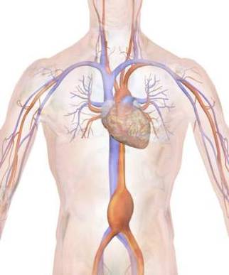
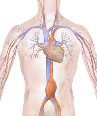
Abdominal compartment syndrome – a common adverse event after rAAA repair
Abdominal compartment syndrome (ACS) was common after ruptured abdominal aortic aneurysm (AAA) repair, with a similar incidence for both open surgical and endovascular repair (EVAR), according to a report published in the European Journal of Vascular and Endovascular Surgery.
Samuel Ersryd, a doctoral student, and his colleagues at Uppsala (Sweden) University performed their study to determine the contemporary incidence, treatment, and outcomes of ACS after AAA repair.
The analysis included 6,634 patients in the Swedish vascular registry who were treated for abdominal aortic aneurysm repair at 31 institutions from May 2008 to December 2013. The mean patient age was 72.8 years, and 16.6% were women. There were 5,271 intact AAA (iAAA) repairs and 1,341 ruptured AAA (rAAA repairs). A total of 41.9% of iAAA repairs were open, as were 72.0% of the rAAA repairs (Eur J Vasc Endovasc Surg. 2016;52:158-65).The study found an incidence of ACS in the rAAA group of 6.8% after open surgery and 6.9% after EVAR.
The morbidity and mortality rates for iAAA and rAAA with ACS were “devastating” in both groups, according to the authors.
Mortality at 90 days for patients with ACS after rAAA was 58.7%, twice that of patients without ACS. In patients with iAAA repair with ACS, the 90-day mortality was 19.2%, six times higher than for those without ACS.
Prophylactic open abdomen treatment was performed in 10.7% of open-surgery patients.
The researchers found no differences in mortality among patients in either group that developed ACS, whether they were treated with decompression laparotomy or not.
Age, sex, and perioperative comorbidities were not associated with ACS, Mr. Ersryd and his associates said. Within the rAAA group, however, ACS was associated with the lowest measured preoperative blood pressure and with preoperative unconsciousness. In addition, ACS was more common in both the iAAA and rAAA groups after perioperative bleeding greater than 5 L, in the iAAA group after reimplantation of a renal artery, and in the rAAA group after the use of balloon occlusion after EVAR. In those patients operated on for iAAA, the risk of developing ACS was 8.1% in patients who had perioperative bleeding greater than 5 L, compared with only 0.8% if bleeding was less than 5 L (P less than .001).
“With such poor results among patients who developed ACS, prevention is the obvious key to success. Massive transfusion protocols and permissive hypotension in patients with ongoing bleeding are important, as well as being restrictive with crystalloids,” the authors said. In addition, they recommended a proactive strategy treating intra-abdominal hypertension with medical therapy, effective pain relief, and neuromuscular blockade as important preventive measures.
“ACS is associated with a devastating effect on outcome after surgery for both ruptured and intact AAA. There was no difference in outcome among those who developed ACS, depending on whether the primary treatment had been performed with an open or endovascular technique,” the researchers concluded.
The authors reported they had no conflicts of interest and the study was funded by the Swedish Research Council and Uppsala University.
On Twitter @VascularTweets
The authors of this paper provide compelling data demonstrating the seriousness of abdominal compartment syndrome developing following abdominal aortic surgery. Recognition of this complication is therefore mandatory and techniques to relieve it should be instituted immediately. In my practice, I have successfully used the Wittmann patch, but recently I switched to the VAC (vacuum-assisted closure) device. Following an endovascular approach for a ruptured AAA, patients may require concomitant exploration for retrograde bleeding sources, such as an inferior mesenteric artery or large lumbars that will continue to bleed unless ligated.
Dr. Russell Samson is the medical editor of Vascular Specialist.
Abdominal compartment syndrome (ACS) was common after ruptured abdominal aortic aneurysm (AAA) repair, with a similar incidence for both open surgical and endovascular repair (EVAR), according to a report published in the European Journal of Vascular and Endovascular Surgery.
Samuel Ersryd, a doctoral student, and his colleagues at Uppsala (Sweden) University performed their study to determine the contemporary incidence, treatment, and outcomes of ACS after AAA repair.
The analysis included 6,634 patients in the Swedish vascular registry who were treated for abdominal aortic aneurysm repair at 31 institutions from May 2008 to December 2013. The mean patient age was 72.8 years, and 16.6% were women. There were 5,271 intact AAA (iAAA) repairs and 1,341 ruptured AAA (rAAA repairs). A total of 41.9% of iAAA repairs were open, as were 72.0% of the rAAA repairs (Eur J Vasc Endovasc Surg. 2016;52:158-65).The study found an incidence of ACS in the rAAA group of 6.8% after open surgery and 6.9% after EVAR.
The morbidity and mortality rates for iAAA and rAAA with ACS were “devastating” in both groups, according to the authors.
Mortality at 90 days for patients with ACS after rAAA was 58.7%, twice that of patients without ACS. In patients with iAAA repair with ACS, the 90-day mortality was 19.2%, six times higher than for those without ACS.
Prophylactic open abdomen treatment was performed in 10.7% of open-surgery patients.
The researchers found no differences in mortality among patients in either group that developed ACS, whether they were treated with decompression laparotomy or not.
Age, sex, and perioperative comorbidities were not associated with ACS, Mr. Ersryd and his associates said. Within the rAAA group, however, ACS was associated with the lowest measured preoperative blood pressure and with preoperative unconsciousness. In addition, ACS was more common in both the iAAA and rAAA groups after perioperative bleeding greater than 5 L, in the iAAA group after reimplantation of a renal artery, and in the rAAA group after the use of balloon occlusion after EVAR. In those patients operated on for iAAA, the risk of developing ACS was 8.1% in patients who had perioperative bleeding greater than 5 L, compared with only 0.8% if bleeding was less than 5 L (P less than .001).
“With such poor results among patients who developed ACS, prevention is the obvious key to success. Massive transfusion protocols and permissive hypotension in patients with ongoing bleeding are important, as well as being restrictive with crystalloids,” the authors said. In addition, they recommended a proactive strategy treating intra-abdominal hypertension with medical therapy, effective pain relief, and neuromuscular blockade as important preventive measures.
“ACS is associated with a devastating effect on outcome after surgery for both ruptured and intact AAA. There was no difference in outcome among those who developed ACS, depending on whether the primary treatment had been performed with an open or endovascular technique,” the researchers concluded.
The authors reported they had no conflicts of interest and the study was funded by the Swedish Research Council and Uppsala University.
On Twitter @VascularTweets
FROM THE EUROPEAN JOURNAL OF VASCULAR AND ENDOVASCULAR SURGERY
Key clinical point: Abdominal compartment syndrome is a dangerous and frequent complication after treatment of ruptured abdominal aortic aneurysms.
Major finding: After ruptured AAA repair, ACS developed in 6.8% of patients with open repair and 6.9% of patients with EVAR.
Data source: Researchers performed a population-based study using the Swedish vascular registry and the Swedish national population registry.
Disclosures: The authors reported they had no conflicts of interest and the study was funded by the Swedish Research Council and Uppsala University.
Burning Mouth Syndrome Due to Varicella Zoster Virus
Burning mouth syndrome(BMS)—persistent burning pain for which no dental or medical cause is found—may be associated with herpes simplex virus (HSV) type 1 or varicella zoster virus (VZV), say clinicians at University of Colorado in Aurora. They report on 2 female patients with BMS who had elevated levels of serum anti-VZV IgM antibodies.
One patient had experienced BMS for 8 months; the other for 2 years. Routine blood counts, liver, kidney, thyroid, and other tests were normal for both patients. No VZV, HSV-1, or HSV-2 DNA were detected in saliva samples, but serum anti-VZV IgG antibody was present and anti-ZV IgM antibody was elevated at 1.88 in 1 patient and 1.02 in the other (normal < 0.90).
Related: Experimental Malaria Vaccine Shows Promise for Longer Protection
Both patients had received zoster vaccine within a year of the onset of BMS, but the clinicians said it “seems unlikely” that immunization contributed to their condition, because none of more than 19,000 adults who had received zoster vaccine in the Shingles Prevention Study developed BMS.
Oral valacyclovir, 1 g 3 times daily for 2 months reduced the pain to minimal for the first patient, although severe pain recurred when the dose was lowered or the drug was discontinued. The clinicians say she has continued to improve on the daily dose. The second patient also was started on oral valacyclovir 1 g 3 times daily for 3 months. Her anti-VZV IgM antibody remained elevated after 3 months. The same dose, continued for another 3 months, reduced the pain by 60%. The dose was lowered to 1 g daily. After 8 months, she is pain free for 3 to 4 days a week; otherwise, the pain is mild.
Related: A New Kind of Flu Drug
The clinicians suggest not only evaluating patients with BMS for VZV or HSV-1, but also being aware that prolonged antiviral treatment may be required.
Source:
Nagel MA, Gilden D. BMJ Case Rep. 2016; pii: bcr2016215953.
doi: 10.1136/bcr-2016-215953.
New and Noteworthy Information—August 2016
People with epilepsy are at increased risk of autism spectrum disorder, especially if epilepsy appears in childhood, according to a study published July 12 in Neurology. Researchers used the Swedish Patient Register to identify 85,201 individuals with epilepsy, their siblings, and their offspring. Each person with epilepsy was compared with five controls matched for age, sex, calendar period, and county. Patients' siblings and offspring were compared with siblings and offspring of controls. During follow-up, 1.6% of people with epilepsy and 0.2% of controls were diagnosed with autism spectrum disorder. People with epilepsy were at increased risk of future autism spectrum disorder, with the highest risk seen in individuals diagnosed with epilepsy in childhood. Siblings and offspring of patients were at increased risk of autism spectrum disorder, compared with controls.
Among patients with chorea associated with Huntington's disease, deutetrabenazine, compared with placebo, results in improved motor signs at 12 weeks, according to a study published July 5 in JAMA. For this study, 90 adults (mean age, 53.7; 44.4% women) with Huntington's disease and a baseline total maximal chorea score of eight or higher were enrolled from August 2013 to August 2014. Participants were randomized to receive deutetrabenazine or placebo. In the deutetrabenazine group, the mean total maximal chorea scores improved from 12.1 to 7.7, whereas in the placebo group, scores improved from 13.2 to 11.3. In the deutetrabenazine group, the mean 36-Item Short Form physical functioning subscale scores decreased from 47.5 to 47.4, whereas in the placebo group, scores decreased from 43.2 to 39.9.
The estimated suicide rate among people with epilepsy in a large US population exceeds that in the general population, according to a study published online ahead of print June 30 in Epilepsy & Behavior. Among people age 10 and older, researchers identified 972 suicide cases with epilepsy and 81,529 suicide cases without epilepsy in 17 states from 2003 through 2011. Investigators estimated their suicide rates, evaluated suicide risk among people with epilepsy, and investigated suicide risk factors specific to epilepsy by comparing those with and without epilepsy. The estimated annual suicide mortality rate among people with epilepsy was 22% higher than that in the general population. Overall, compared with people without epilepsy, those with epilepsy were more likely to have died from suicide and were twice as likely to poison themselves.
Low-dose methylene blue increases functional MRI (fMRI) activity during sustained attention and short-term memory tasks and enhances memory retrieval, according to a study published online ahead of print June 28 in Radiology. Twenty-six people ages 22 to 62 were enrolled. Researchers performed fMRI imaging with a psychomotor vigilance task and delayed match-to-sample tasks before and one hour after administration of low-dose methylene blue or placebo. Cerebrovascular reactivity effects were measured with the carbon dioxide challenge. Multiple comparison correction also was applied. Administration of methylene blue increased response in the bilateral insular cortex during the psychomotor vigilance task and fMRI response during the short-term memory task involving the prefrontal, parietal, and occipital cortex. Methylene blue also was associated with a 7% increase in correct responses during memory retrieval.
Traumatic brain injury (TBI) with loss of consciousness is associated with an increased risk for clinical and neuropathologic findings of Parkinson's disease, but not Alzheimer's disease, according to a study published online ahead of print July 11 in JAMA Neurology. Researchers retrospectively analyzed data from three prospective cohort studies that included annual or biennial cognitive and clinical testing to identify incident cases of dementia and Alzheimer's disease. Of 7,130 participants, 865 reported a history of TBI with loss of consciousness. In 45,190 person-years of follow-up, 1,537 incident cases of dementia and 117 of Parkinson's disease were identified. No association was found between TBI with loss of consciousness and incident dementia or Alzheimer's disease. TBI with loss of consciousness was associated with incident Parkinson's disease, progression of parkinsonian signs, Lewy bodies, and microinfarcts.
Genetically elevated BMI is associated with risk of multiple sclerosis (MS), which suggests a causal role for obesity in MS etiology, according to an article published June 28 in PLOS Medicine. Researchers used summary statistics from the Genetic Investigation of Anthropometric Traits (GIANT) consortium and the International MS Genetics Consortium (IMSGC). The effect of each single-nucleotide polymorphism (SNP) on MS was weighted by its effect on BMI. Seventy SNPs had genome-wide significance for BMI in GIANT and were investigated for their association with MS risk in the IMSGC. It was found that increased BMI influences MS susceptibility, where a one-standard-deviation increase in genetically determined BMI increased odds of MS by 41%. Sensitivity analyses, including MR-Egger regression, and the weighted median approach provided no evidence of pleiotropic effects.
The FDA has accepted the Biologics License Application for Ocrevus (ocrelizumab) for the treatment of relapsing multiple sclerosis and primary progressive multiple sclerosis. The agency granted the application Priority Review Designation with a targeted action date of December 28, 2016. The Ocrevus Marketing Authorization Application has also been validated by the European Medicines Agency. The Ocrevus marketing applications are based on results from three phase III studies that met primary and key secondary end points. Data from OPERA I and OPERA II in people with relapsing multiple sclerosis showed superior efficacy of Ocrevus in reducing annualized relapse rates and disability progression sustained for at least three and for at least six months, respectively, compared with interferon beta-1a. Genentech, which will manufacture the drug, is headquartered in South San Francisco, California.
Thirty-eight independent genomic regions are associated with migraine, according to a study published online ahead of print June 20 in Nature Genetics. The identified loci showed enrichment for genes expressed in vascular and smooth muscle tissues, which is consistent with a predominant theory of migraine that highlights vascular etiologies. To identify new genomic loci associated with susceptibility to migraine, researchers carried out a genetic study of migraine on 59,674 subjects with migraine and 316,078 controls who participated in 22 genome-wide association studies. Investigators identified 44 independent single-nucleotide polymorphisms significantly associated with migraine risk that mapped to 38 distinct genomic loci, including 28 loci not previously reported and a locus that is the first to be identified on chromosome X. The findings may promote the development of personalized treatments for migraine.
Long-term risks of recurrent stroke and poststroke dementia remain high after stroke and are substantially influenced by prestroke risk factors, emphasizing the need for optimizing primary prevention, according to a study published online ahead of print July 14 in Stroke. Researchers monitored 1,237 patients with first-ever stroke and 4,928 stroke-free participants, matched by age, sex, examination round, and stroke date, for the occurrence of stroke or dementia. Beyond one year after stroke, patients retained a threefold increased risk of recurrent stroke and an almost twofold increased risk of dementia, compared with people without stroke. In all, 39% of recurrent strokes and 10% of poststroke dementia cases were attributable to prestroke cardiovascular risk factors. These percentages were similar for first-ever stroke and dementia in the matched stroke-free population.
Molecular evidence indicates that nilotinib significantly increases brain dopamine and reduces toxic proteins linked to disease progression in patients with Parkinson's disease or dementia with Lewy bodies, according to a phase I study published July 11 in the Journal of Parkinson's Disease. Twelve participants were randomized to 150 mg/day or 300 mg/day of oral nilotinib for 24 weeks. The CSF levels of homovanillic acid were significantly increased between baseline and 24 weeks of treatment. The researchers found that nilotinib is safe and well tolerated for people with advanced Parkinson's disease. In addition, nilotinib is detectable in the CSF and engages the target ABL1. Improvements in motor and cognitive function suggest that nilotinib may have clinical benefits. Nilotinib should be evaluated in larger randomized trials, said the researchers.
The FDA has approved ExAblate Neuro, the first focused ultrasound device to treat essential tremor in patients who have not responded to medication. ExAblate Neuro uses MRI to deliver focused ultrasound to destroy brain tissue in a small area thought to be responsible for causing tremors. Of 76 patients with essential tremor, 56 randomly received the ExAblate Neuro treatment in one study. Patients in the control group were able to cross over into the treatment group three months later. Patients treated with ExAblate Neuro showed a nearly 50% improvement in tremor and motor function three months after treatment, compared with their baseline scores. To determine whether ExAblate Neuro treatment is appropriate, patients should first have MRI and CT scans. InSightec, the device's manufacturer, is headquartered in Tirat Carmel, Israel.
The driving ability of patients with mild cognitive impairment (MCI) and Alzheimer's disease is related to their degree of cognitive impairment, according to a systematic review published May 11 in the Journal of Alzheimer's Disease. Researchers investigated the predictive utility of cognitive tests and domains, and the areas and degree of driving impairment in patients with MCI and Alzheimer's disease. Effect sizes were derived and analyzed in a random effects model. Executive function, attention, visuospatial function, and global cognition significantly predicted driving performance. Trail Making Test Part B and Maze test were the best single predictors of driving performance. Patients with very mild Alzheimer's disease and mild Alzheimer's disease were more likely to fail an on-road test than healthy control drivers, with failure rates of 13.6%, 33.3%, and 1.6%, respectively.
Functional brain scans may help predict recovery and guide treatment after stroke, according to a study published online ahead of print July 11 in the Proceedings of the National Academy of Sciences. Researchers investigated whether functional connectivity MRI (fcMRI) could provide useful information for assessing stroke damage. They used fcMRI to assess communication between brain areas in 132 patients with stroke and 31 people without stroke. The technology allowed the authors to identify large disruptions in brain communication that occurred as a result of stroke. Each participant also underwent a battery of neuropsychologic tests. Network-specific patterns of dysfunction predicted specific behavioral deficits, and loss of interhemispheric communication across a set of regions was associated with impairment across multiple behavioral domains.
—Kimberly Williams

Links found between NAFLD, type 2 diabetes, and NASH in children
Nearly one in three children with nonalcoholic fatty liver disease have abnormal glucose metabolism, and this co-morbidity is also associated with a greater risk of nonalcoholic steatohepatitis, according to a cross-sectional study published online Aug. 1 in JAMA Pediatrics.
The study used data from 675 children with biopsy-confirmed nonalcoholic fatty liver disease (NAFLD) who were enrolled in the NASH Clinical Research Network. The mean age of the children was 12.6 years, and they had a mean BMI of 32.5. Most of the children in the study were boys and Hispanic.
Overall, 23.4% of study participants had prediabetes and 6.5% met the clinical criteria for type 2 diabetes. However, girls with NAFLD had a 60% greater risk of prediabetes and a fivefold greater risk of type 2 diabetes than boys, even after controlling for BMI and waist circumference (JAMA Pediatr. 2016 Aug 1. doi: 10.1001/jamapediatrics.2016.1971), reported Dr. Kimberly P. Newton and her coauthors.
The researchers also noted a significant association between nonalcoholic steatohepatitis (NASH) and glucose metabolism. Individuals with type 2 diabetes were three times more likely to also have NASH, while those with prediabetes had a 90% higher incidence of NASH, compared with individuals with normal glucose metabolism. They also found that those with NASH had significantly higher mean fasting glucose and insulin concentrations than children without NASH.
Dr. Newton, of the University of California, San Diego, and her coauthors wrote that while abnormal glucose metabolism is known to be common in adults with NAFLD, and that type diabetes is a risk factor for progression to NASH and liver-related mortality, the association in children with NAFLD is less well understood.
“Among our cohort, the prevalence of children with type 2 diabetes was much higher than would be expected based on contributions from obesity alone,” they wrote. “Although systemic insulin resistance is believed be important in the pathogenesis of both pediatric NAFLD and type 2 diabetes, to our knowledge, there are no longitudinal studies that evaluate the cause-effect relationship between these two associated conditions.”
The authors drew particular attention to the threefold higher odds of NASH in children with NAFLD and type 2 diabetes, pointing out that while the prognostic implications of NASH in childhood are not fully known, the NASH phenotype is associated with a “substantially” greater risk of cirrhosis. This risk is likely to be compounded by the presence of type 2 diabetes.
“Our study advances the literature by showing that as early as childhood, prediabetes and type 2 diabetes emerge as clear risk factors for NASH with potential downstream implications for future morbidity and mortality.”
Commenting on the greater incidence of prediabetes and type 2 diabetes among girls with NAFLD, the authors said this had been observed in other studies and that sex differences represented a major unmet research need.
The Nonalcoholic Steatohepatitis Clinical Research Network is supported by the National Institute of Diabetes and Digestive and Kidney Diseases. Additional support was received from the National Center for Advancing Translational Sciences. The researchers reported no conflicts of interest.
FROM JAMA PEDIATRICS
Key clinical point: Nearly one in three children with nonalcoholic fatty liver disease have abnormal glucose metabolism, and this combination also is associated with a greater risk of nonalcoholic steatohepatitis.
Major finding: Among children with biopsy-diagnosed non-alcoholic fatty liver disease, 23.4% had prediabetes and 6.5% met the clinical criteria for type 2 diabetes.
Data source: Cross-sectional study of 675 children with biopsy-confirmed nonalcoholic fatty liver disease enrolled in the NASH Clinical Research Network.
Disclosures: The Nonalcoholic Steatohepatitis Clinical Research Network is supported by the National Institute of Diabetes and Digestive and Kidney Diseases. Additional support was received from the National Center for Advancing Translational Sciences. The researchers reported no conflicts of interest.
Two incretin-based drugs linked to increased bile duct disease but not pancreatitis
At least two incretin-based drugs – glucagon-like peptide 1 agonists and dipeptidyl peptidase 4 inhibitors – do not appear to increase the risk of acute pancreatitis in individuals with diabetes but are associated with an increased risk of bile duct and gallbladder disease.
Two studies examining the impact on the pancreas of incretin-based drugs, including dipeptidyl peptidase 4 (DPP-4) inhibitors and glucagon-like peptide 1 (GLP-1) agonists, have been published online August 1 in JAMA Internal Medicine.
Incretin-based drugs have been associated with increased risk of elevated pancreatic enzyme levels, while GLP-1 has been shown to increase the proliferation and activity of cholangiocytes, which have raised concerns of an impact on the bile duct, gallbladder, and pancreas.
The first study was an international, population-based cohort study using the health records of more than 1.5 million individuals with type 2 diabetes, who began treatment with antidiabetic drugs between January 2007 and June 2013.
Analysis of these data showed there was no difference in the risk of hospitalization for acute pancreatitis between those taking incretin-based drugs and those on two or more other oral antidiabetic medications (JAMA Intern Med. 2016 Aug 1. doi: 10.1001/jamainternmed.2016.1522).
The study also found no significant increase in the risk of acute pancreatitis either with DPP-4 inhibitors or GLP-1 agonists, nor was there any increase with a longer duration of use or in patients with a history of acute or chronic pancreatitis.
Most previous observational studies of incretin-based drugs and pancreatitis had reported null findings, but four studies did find a positive association. Laurent Azoulay, PhD, from the Lady Davis Institute at Montreal’s Jewish General Hospital, and his coauthors suggested this heterogeneity was likely the result of methodologic shortcomings such as the use of inappropriate comparator groups and confoundings.
“Although it remains possible that these drugs may be associated with acute pancreatitis, the upper limit of our 95% [confidence interval] suggests that this risk is likely to be small,” the authors wrote. “Thus, the findings of this study should provide some reassurance to patients treated with incretin-based drugs.”
Meanwhile, a second population-based cohort study in 71,368 patients starting an antidiabetic drug found the use of GLP-1 analogues was associated with a significant 79% increase in the risk of bile duct and gallbladder disease, compared with the use of at least two other oral antidiabetic medications.
When stratified by duration of use, individuals taking GLP-1 analogues for less than 180 days showed a twofold increase in the risk of bile duct and gallbladder disease (adjusted hazard ratio, 2.01; 95% CI, 1.23-3.29) but those taking the drugs for longer than 180 days did not show an increased risk.
The use of GLP-1 analogues was also associated with a two-fold increase in the risk of undergoing a cholecystectomy.
However, the study found no increased risk of bile duct or gallbladder disease with DPP-4 inhibitors (JAMA Intern Med. 2016 Aug 1. doi: 10.1001/jamainternmed.2016.1531).
Jean-Luc Faillie, MD, PhD, of the University of Montpellier (France) and his associates suggested that rapid weight loss associated with GLP-1 analogues may explain the association with bile duct and gallbladder disease, which would also account for the observation that the association did not occur in patients taking the drugs for a longer period of time.
“Weight loss leads to supersaturation of cholesterol in the bile, a known risk factor for gallstones,” the authors wrote.
DPP-4 inhibitors have different effects on the GLP-1 pharmacologic factors and a weaker incretin action, which the authors suggested may explain the lack of association with bile duct and gallbladder disease, as well as their lower incidence of gastrointestinal adverse events.
“Although further studies are needed to confirm our findings and the mechanisms involved, physicians prescribing GLP-1 analogues should be aware of this association and carefully monitor patients for biliary tract complications.”
The first study was enabled by data-sharing agreements with the Canadian Network for Observational Drug Effect Studies, which is funded by the Canadian Institutes of Health Research. Two authors declared consulting fees, grant support, or financial compensation from the pharmaceutical industry, but there were no other conflicts of interest declared.
The second study was funded by the Canadian Institutes of Health Research. No conflicts of interest were declared.
FROM JAMA INTERNAL MEDICINE
Key clinical point: Glucagon-like peptide 1 agonists do not appear to increase the risk of acute pancreatitis in individuals with diabetes but are associated with an increased risk of bile duct and gallbladder disease.
Major finding: GLP-1 agonists are associated with a 79% increase in the risk of bile duct and gallbladder disease, compared with other oral antidiabetic medications, but do not increase the risk of acute pancreatitis.
Data source: Two population-based cohort studies; one involving more than 1.5 million individuals with type 2 diabetes across three countries, and the other involving 71,368 patients with type 2 diabetes.
Disclosures: The first study was enabled by data-sharing agreements with the Canadian Network for Observational Drug Effect Studies, which is funded by the Canadian Institutes of Health Research. Two authors declared consulting fees, grant support, or financial compensation from the pharmaceutical industry, but there were no other conflicts of interest declared. The second study was funded by the Canadian Institutes of Health Research. No conflicts of interest were declared.
Acute liver failure
To the Editor: In the article by Drs. Singh et al in your June issue, I was surprised that the role of hepatitis delta wasn’t mentioned as a potential cause of acute liver failure. My understanding is that this peculiar virus can only infect those with hepatitis B surface antigenemia, but when it does, it results in far more serious liver injury, including acute liver failure in some.
To the Editor: In the article by Drs. Singh et al in your June issue, I was surprised that the role of hepatitis delta wasn’t mentioned as a potential cause of acute liver failure. My understanding is that this peculiar virus can only infect those with hepatitis B surface antigenemia, but when it does, it results in far more serious liver injury, including acute liver failure in some.
To the Editor: In the article by Drs. Singh et al in your June issue, I was surprised that the role of hepatitis delta wasn’t mentioned as a potential cause of acute liver failure. My understanding is that this peculiar virus can only infect those with hepatitis B surface antigenemia, but when it does, it results in far more serious liver injury, including acute liver failure in some.
In reply: Acute liver failure
In Reply: We thank Dr. Homler for bringing hepatitis D as a potential cause of acute liver failure to our attention.
Hepatitis D virus, first described in the 1970s, requires the hepatitis B surface antigen (HBsAg) capsid to enter the hepatocyte and, thus, can only cause liver injury when the patient is also infected simultaneously with hepatitis B virus.1 Hepatitis D virus can cause either coinfection (presence of immunoglobulin M anti-HB core antibody with or without HBsAg) or superinfection (presence of HBsAg without immunoglobulin M anti-HB core antibody) with hepatitis B virus. In India, coinfection has been reported to be the cause of acute liver failure in about 4% of all patients, and superinfection in 4.5%.2
While simultaneous treatment for hepatitis D and B viruses with pegylated interferon and any of the agents used for treatment of hepatitis B has been successful in treating chronic hepatitis, it has not been proven useful in patients with acute liver failure, and liver transplant remains the only treatment option.3
- Rizzetto M. The adventure of delta. Liver Int 2016; 36(suppl 1):135–140.
- Irshad M, Acharya SK. Hepatitis D virus (HDV) infection in severe forms of liver diseases in North India. Eur J Gastroenterol Hepatol 1996; 8:995–998.
- Noureddin M, Gish R. Hepatitis delta: epidemiology, diagnosis and management 36 years after discovery. Curr Gastroenterol Rep 2014; 16:365.
In Reply: We thank Dr. Homler for bringing hepatitis D as a potential cause of acute liver failure to our attention.
Hepatitis D virus, first described in the 1970s, requires the hepatitis B surface antigen (HBsAg) capsid to enter the hepatocyte and, thus, can only cause liver injury when the patient is also infected simultaneously with hepatitis B virus.1 Hepatitis D virus can cause either coinfection (presence of immunoglobulin M anti-HB core antibody with or without HBsAg) or superinfection (presence of HBsAg without immunoglobulin M anti-HB core antibody) with hepatitis B virus. In India, coinfection has been reported to be the cause of acute liver failure in about 4% of all patients, and superinfection in 4.5%.2
While simultaneous treatment for hepatitis D and B viruses with pegylated interferon and any of the agents used for treatment of hepatitis B has been successful in treating chronic hepatitis, it has not been proven useful in patients with acute liver failure, and liver transplant remains the only treatment option.3
- Rizzetto M. The adventure of delta. Liver Int 2016; 36(suppl 1):135–140.
- Irshad M, Acharya SK. Hepatitis D virus (HDV) infection in severe forms of liver diseases in North India. Eur J Gastroenterol Hepatol 1996; 8:995–998.
- Noureddin M, Gish R. Hepatitis delta: epidemiology, diagnosis and management 36 years after discovery. Curr Gastroenterol Rep 2014; 16:365.
Sustained Remissions After Discontinuation of Ibrutinib in Relapsed/Refractory CLL: A Basis for Reducing Drug Toxicity and Treatment Costs?
In contrast to traditional chemotherapy, patients responding to biological or targeted therapies often are treated indefinitely until progression or toxicity. This therapeutic model, however, increases treatment costs, may induce greater toxicity and theoretically could select for earlier emergence of drug resistance. Moreover, little data are available regarding the outcomes of patients who discontinue targeted therapies after achieving remission. In this regard, we report 2 patients with relapsed/refractory chronic lymphocytic leukemia (CLL) who chose to stop therapy unrelated to toxicity or disease status after the induction of remission by the BTK inhibitor ibrutinib.
Patient A started ibrutinib for progressive CLL at an absolute lymphocyte count (ALC) of 137,000 mm3 and recurrent hemolytic anemia. After 5 months, the hemolysis had resolved (Hgb 15.6 g/dL), while the ALC had declined to 9,200 mm3. Treatment was then interrupted due to patient preference. One month after drug discontinuation, the ALC was in the normal range at 1,400 mm3 and remained within or near the normal range for a total of 12 months. Two months later, the ALC was again markedly elevated at 68,000 mm3 and anemia recurred. The patient then agreed to restart ibrutinib. After 4 months of re-treatment, he has had prompt resolution of the anemia and achieved a partial remission thus far.
Patient B was started on ibrutinib for a rising ALC (26,000 mm3) and severe hemolytic anemia. After 9 months of treatment, the hemoglobin was 13 g/dL and the ALC was in the normal range at 3,300 mm3. Due to unrelated medical problems, ibrutinib therapy was stopped. Currently, 6 months since drug discontinuation, the ALC remains in the normal range, and no other signs of CLL are present.
These clinical observations suggest that interruption of ibrutinib may be feasible in at least some CLL patients who achieve remission. Even if flow cytometry were performed at monthly intervals to detect early recurrence and ensure prompt re-institution of therapy, the cost savings would still be considerable. Of course, clinical trials will be necessary to confirm equivalent long-term efficacy and overall survival for intermittent versus continuous ibrutinib therapy in CLL.
The Impact of an Integrated Veterans Health Administration RN Disease Management Program: The Oncology Aspect
In an effort to decrease the timeline from suspicion and/or cancer diagnosis to treatment, the Oncology Disease Management RNs incorporated a streamlined and integrated clinical approach. A Disease Case Management (DM) team model is a useful approach to enhance the veteran experience and expedite health care delivery. In 2007, the DM program was begun. The team consisted of 3 registered nurses (RN) who worked under the Quality Management Department rather than the oncology clinic in order to have a more wide ranged approach in both treatment and interventions.
Methods: All patients who had either a suspicion for, or an actual diagnosis of cancer were followed by the DM RNs. We assessed the timeline in days for veterans to get scheduled for recommended tests and consults before and after the DM team involvement. We reviewed timeline data for patients diagnosed during fiscal years 2009 to 2015. The percentage change was calculated for Oncology Consults and Intra-facility Consults to DM team. Times from diagnosis to treatment intervention pre and post DM implementation were reviewed for Primary liver, colon and lung cancers.
Results: Rate of change: Oncology consults for Disease Management team intervention: 62% increase. Reduction in time from diagnosis to treatment intervention: Colon cancer and Lung Cancer 50% and 46% respectively. Reduction in the time required for getting primary Liver cancer patients into diagnostic tests and tumor board presentations for treatment intervention – days shortened from 92 to 31 days from baseline data 2010/11 to 2015. This reflected a 66% decrease in time to treatment /intervention.
Conclusion: The data reflect that in a VA healthcare setting, the Disease Management team model is feasible for facilitating an integrated and coordinated approach in reducing timelines for treatment. The data support that the early introduction of the disease management team model shortened the number of days from diagnosis to treatment by facilitating referrals, clinical testing, and/or treatment initiation in Veteran cancer patients resulting in timely care intervention.