User login
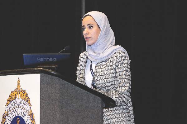
The role ob.gyns. can play in preventing Zika virus
In February of 2016, the World Health Organization declared Zika a Public Health Emergency of International Concern and the Centers for Disease and Prevention elevated its Zika Emergency Operation Center activation to Level 1, its highest level. The Zika epidemic is unique in many ways, particularly in that women and reproductive health care are at the center of the epidemic.
Most people who are infected with Zika are asymptomatic; those who do have symptoms generally have a mild, self-limited infection. However, a pregnant woman can pass the infection to her fetus, which can lead to severe outcomes. As ob.gyns., we are called upon to guide pregnant women who have been exposed or infected regarding testing, diagnosis, options counseling, surveillance, and management. Additionally, we can help pregnant women prevent Zika infection. Discuss avoiding travel for her and her partner(s) to areas with Zika. Encourage strategies for mosquito bite prevention if traveling to endemic areas and at home. Recommend abstaining from sex or using a condom for those who have partners who have recently traveled to areas with Zika transmission.

With that said, most of our patients are not pregnant. Many are capable of pregnancy and potentially at risk for unintended pregnancy since almost half of pregnancies in the United States are unintended (N Engl J Med. 2016 Mar 3;374[9]:843-52). Contraception for women who are not desiring pregnancy may help prevent unintended pregnancy, but also may help prevent the serious effects that can be seen with mother-to-fetus transmission of Zika. We can play a critical role in this important prevention message for nonpregnant women.
We do not know how long the Zika virus will continue to spread nor do we know whether we will have local transmission in the continental U.S. But we do know that travel is common and sexual transmission is possible. As such, all women need to be educated about the Zika virus. All women should be aware of the risks it poses for their reproductive health and be given an opportunity to discuss what that might mean to each of them. Whether a woman is planning a pregnancy, trying to avoid one, or is unsure of what she wants, having a conversation with her ob.gyn. may help her feel informed and supported in making the best decision for her right now. This may be particularly true of her plans for pregnancy timing and the use of contraception and condoms.
Beyond travel screening and mosquito bite prevention strategies, there are several key messages that we are uniquely poised to relay about Zika prevention. First, women who have been exposed to Zika should delay trying to conceive for 8 weeks. The recommendation for male partners to delay conception ranges from 8 weeks to 6 months depending upon local transmission and presence of symptoms. For those who are not trying to become pregnant or who want to delay pregnancy because of Zika concerns or exposures, we should offer the full range of contraceptive options and work with each patient to optimize her contraceptive selection, based on her individual preferences, goals, and needs. Finally, women should be counseled to avoid transmission from sex by choosing to not have sex or by using a condom with each act of sex with a partner who may have been exposed to Zika.
Our understanding of Zika infection is rapidly evolving. It will be particularly important for us to stay current on this issue. Providers can access up-to-date information about Zika, provider updates, patient information, posters and other information about the emergency response from the CDC’s website at www.cdc.gov/zika/index.html.
There are several tools available for guidance in these conversations. On July 1, 2016, the Office of Population Affairs released a toolkit for Providing Family Planning Care for Non-Pregnant Women and Men of Reproductive Age in the Context of Zika. It is available online at: www.hhs.gov/opa/news#toolkit.
The CDC also offers comprehensive guidance on providing contraception. Providers can access the Medical Eligibility Criteria for Contraceptive Use, the Selected Practice Recommendations for Contraceptive Use, and Providing Quality Family Planning Services, all of which are available on the CDC website.
Dr. Kottke is an associate professor in the department of gynecology and obstetrics at Emory University in Atlanta, Georgia. She is the director of the Jane Fonda Center at Emory, which is involved in research and program development focused on adolescent sexual and reproductive health. She also serves as the medical consultant for the state of Georgia’s Family Planning Program. Dr. Kottke reported that she is a Nexplanon (Merck) trainer, is a consultant to CSL Behring, and serves on the advisory board for Evofem.
In February of 2016, the World Health Organization declared Zika a Public Health Emergency of International Concern and the Centers for Disease and Prevention elevated its Zika Emergency Operation Center activation to Level 1, its highest level. The Zika epidemic is unique in many ways, particularly in that women and reproductive health care are at the center of the epidemic.
Most people who are infected with Zika are asymptomatic; those who do have symptoms generally have a mild, self-limited infection. However, a pregnant woman can pass the infection to her fetus, which can lead to severe outcomes. As ob.gyns., we are called upon to guide pregnant women who have been exposed or infected regarding testing, diagnosis, options counseling, surveillance, and management. Additionally, we can help pregnant women prevent Zika infection. Discuss avoiding travel for her and her partner(s) to areas with Zika. Encourage strategies for mosquito bite prevention if traveling to endemic areas and at home. Recommend abstaining from sex or using a condom for those who have partners who have recently traveled to areas with Zika transmission.
With that said, most of our patients are not pregnant. Many are capable of pregnancy and potentially at risk for unintended pregnancy since almost half of pregnancies in the United States are unintended (N Engl J Med. 2016 Mar 3;374[9]:843-52). Contraception for women who are not desiring pregnancy may help prevent unintended pregnancy, but also may help prevent the serious effects that can be seen with mother-to-fetus transmission of Zika. We can play a critical role in this important prevention message for nonpregnant women.
We do not know how long the Zika virus will continue to spread nor do we know whether we will have local transmission in the continental U.S. But we do know that travel is common and sexual transmission is possible. As such, all women need to be educated about the Zika virus. All women should be aware of the risks it poses for their reproductive health and be given an opportunity to discuss what that might mean to each of them. Whether a woman is planning a pregnancy, trying to avoid one, or is unsure of what she wants, having a conversation with her ob.gyn. may help her feel informed and supported in making the best decision for her right now. This may be particularly true of her plans for pregnancy timing and the use of contraception and condoms.
Beyond travel screening and mosquito bite prevention strategies, there are several key messages that we are uniquely poised to relay about Zika prevention. First, women who have been exposed to Zika should delay trying to conceive for 8 weeks. The recommendation for male partners to delay conception ranges from 8 weeks to 6 months depending upon local transmission and presence of symptoms. For those who are not trying to become pregnant or who want to delay pregnancy because of Zika concerns or exposures, we should offer the full range of contraceptive options and work with each patient to optimize her contraceptive selection, based on her individual preferences, goals, and needs. Finally, women should be counseled to avoid transmission from sex by choosing to not have sex or by using a condom with each act of sex with a partner who may have been exposed to Zika.
Our understanding of Zika infection is rapidly evolving. It will be particularly important for us to stay current on this issue. Providers can access up-to-date information about Zika, provider updates, patient information, posters and other information about the emergency response from the CDC’s website at www.cdc.gov/zika/index.html.
There are several tools available for guidance in these conversations. On July 1, 2016, the Office of Population Affairs released a toolkit for Providing Family Planning Care for Non-Pregnant Women and Men of Reproductive Age in the Context of Zika. It is available online at: www.hhs.gov/opa/news#toolkit.
The CDC also offers comprehensive guidance on providing contraception. Providers can access the Medical Eligibility Criteria for Contraceptive Use, the Selected Practice Recommendations for Contraceptive Use, and Providing Quality Family Planning Services, all of which are available on the CDC website.
Dr. Kottke is an associate professor in the department of gynecology and obstetrics at Emory University in Atlanta, Georgia. She is the director of the Jane Fonda Center at Emory, which is involved in research and program development focused on adolescent sexual and reproductive health. She also serves as the medical consultant for the state of Georgia’s Family Planning Program. Dr. Kottke reported that she is a Nexplanon (Merck) trainer, is a consultant to CSL Behring, and serves on the advisory board for Evofem.

Turning down treatment
How do you treat a patient who doesn’t want to be treated?
It depends. If the problem is acne or a wart, then it’s all right to let it go.
Harriet, however, has HIV. And a facial basal cell carcinoma.
Now what?
Perhaps it still depends.

After showing me a red spot on her right cheek, Harriet put me on notice right away. “I don’t believe in unnecessary treatments,” she said.
I asked her what she meant.
“I’ve been HIV positive since the mid-1980s,” she said. “Last year, my doctor wanted to pump me full of those poisons. So I changed doctors.”
“What does your new doctor think?” I asked.
“My T-cell counts aren’t very good. He also thinks I should take those medicines, but I don’t see him much.
“I went to another dermatologist about a different problem. He got very angry at me, because I had called in advance to ask not to be seen by a resident. They told me that would be OK, but the resident showed up anyway. When the doctor also came in and I tried to explain, he threw me out of the clinic.”
By now, I’d gotten the picture: Harriet has her own ideas about things and is not about to take advice with which she doesn’t agree. What makes Harriet different is not that she ignores medical advice – people do that all the time – but that she comes right out and says so to the doctor’s face. Others wait till they get home.
So what do you do when a patient shows that he or she is ready to look you in the eye and turn you down flat?
One response is to leave the room in a huff. After all, who’s the expert here, and who’s trying to help whom?
That reaction is understandable, but if the doctor walks out, who is being helped? The patient, or the doctor?
There is, of course, another approach, which is to suppress professional ego considerations and ask:
1. What are the actual medical stakes? What is the worst that could happen if the patient refuses treatment?
2. What realistic options, if any, are there to change the patient’s mind?
3. Why is the patient behaving that way, anyway?
In Harriet’s case, what are the medical stakes? I am no HIV specialist, but how many patients who seroconverted in the 1980s are still around to consider their options? (Other such patients have told me their doctors really don’t understand how patients like them survived.) If Harriet is sexually inactive and does not try to donate blood, how sure are we that she isn’t better off doing nothing?
Besides, what other options are there to change her mind?
Before turning to my third question, I have to plan what to do if – when – the biopsy confirms my clinical diagnosis of basal cell. I will inform Harriet, and recommend Mohs surgery. Suppose she refuses and wants a lesser procedure, or no surgery at all. What next?
This happens. If the patient is elderly, and the lesion is not near a strategic organ, such as the eye, it may be acceptable just to watch the lesion. Some basal cells grow fast, others barely grow at all. If Harriet decides to do nothing, I could explain my preference – surgery – and her risks – lesion growth and bigger surgery later – and insist on seeing her every 2 months.
Which brings us to our third question: Why would Harriet act this way?
Two possibilities occur to me. The first is fear. Scared people often act aggressively. Calmed down, they relent.
The second is that Harriet is what the English call bloody minded; in other words, deliberately uncooperative.
Battling with the bloody minded is not helpful for anybody.
Negotiating with difficult people is much less gratifying than giving advice and being respectfully thanked for our efforts and expertise.
Sometimes, though, the best we can do is to swallow our professional pride, try to defuse what fear we can, and show that we will push only as hard as clinical risk truly justifies. This is not always easy to do but is often worth the effort. It may certainly be the best available alternative.
Harriet’s biopsy showed a basal cell. She readily agreed to surgery.
You never know.
Dr. Rockoff practices dermatology in Brookline, Mass. He is on the clinical faculty at Tufts University School of Medicine, Boston, and has taught senior medical students and other trainees for 30 years. Dr. Rockoff has contributed to the Under My Skin column in Dermatology News since January 2002. Write to him at [email protected].
How do you treat a patient who doesn’t want to be treated?
It depends. If the problem is acne or a wart, then it’s all right to let it go.
Harriet, however, has HIV. And a facial basal cell carcinoma.
Now what?
Perhaps it still depends.
After showing me a red spot on her right cheek, Harriet put me on notice right away. “I don’t believe in unnecessary treatments,” she said.
I asked her what she meant.
“I’ve been HIV positive since the mid-1980s,” she said. “Last year, my doctor wanted to pump me full of those poisons. So I changed doctors.”
“What does your new doctor think?” I asked.
“My T-cell counts aren’t very good. He also thinks I should take those medicines, but I don’t see him much.
“I went to another dermatologist about a different problem. He got very angry at me, because I had called in advance to ask not to be seen by a resident. They told me that would be OK, but the resident showed up anyway. When the doctor also came in and I tried to explain, he threw me out of the clinic.”
By now, I’d gotten the picture: Harriet has her own ideas about things and is not about to take advice with which she doesn’t agree. What makes Harriet different is not that she ignores medical advice – people do that all the time – but that she comes right out and says so to the doctor’s face. Others wait till they get home.
So what do you do when a patient shows that he or she is ready to look you in the eye and turn you down flat?
One response is to leave the room in a huff. After all, who’s the expert here, and who’s trying to help whom?
That reaction is understandable, but if the doctor walks out, who is being helped? The patient, or the doctor?
There is, of course, another approach, which is to suppress professional ego considerations and ask:
1. What are the actual medical stakes? What is the worst that could happen if the patient refuses treatment?
2. What realistic options, if any, are there to change the patient’s mind?
3. Why is the patient behaving that way, anyway?
In Harriet’s case, what are the medical stakes? I am no HIV specialist, but how many patients who seroconverted in the 1980s are still around to consider their options? (Other such patients have told me their doctors really don’t understand how patients like them survived.) If Harriet is sexually inactive and does not try to donate blood, how sure are we that she isn’t better off doing nothing?
Besides, what other options are there to change her mind?
Before turning to my third question, I have to plan what to do if – when – the biopsy confirms my clinical diagnosis of basal cell. I will inform Harriet, and recommend Mohs surgery. Suppose she refuses and wants a lesser procedure, or no surgery at all. What next?
This happens. If the patient is elderly, and the lesion is not near a strategic organ, such as the eye, it may be acceptable just to watch the lesion. Some basal cells grow fast, others barely grow at all. If Harriet decides to do nothing, I could explain my preference – surgery – and her risks – lesion growth and bigger surgery later – and insist on seeing her every 2 months.
Which brings us to our third question: Why would Harriet act this way?
Two possibilities occur to me. The first is fear. Scared people often act aggressively. Calmed down, they relent.
The second is that Harriet is what the English call bloody minded; in other words, deliberately uncooperative.
Battling with the bloody minded is not helpful for anybody.
Negotiating with difficult people is much less gratifying than giving advice and being respectfully thanked for our efforts and expertise.
Sometimes, though, the best we can do is to swallow our professional pride, try to defuse what fear we can, and show that we will push only as hard as clinical risk truly justifies. This is not always easy to do but is often worth the effort. It may certainly be the best available alternative.
Harriet’s biopsy showed a basal cell. She readily agreed to surgery.
You never know.
Dr. Rockoff practices dermatology in Brookline, Mass. He is on the clinical faculty at Tufts University School of Medicine, Boston, and has taught senior medical students and other trainees for 30 years. Dr. Rockoff has contributed to the Under My Skin column in Dermatology News since January 2002. Write to him at [email protected].

Anatomic repair of ccTGA did not yield superior survival
BALTIMORE – Anatomic repair did not outperform physiologic repair in patients with congenitally corrected transposition of the great arteries (ccTGA), according to a study presented by Maryam Al-Omair, M.D., of the University of Toronto at the annual meeting of the American Association for Thoracic Surgery.
Dr. Al-Omair and her colleagues hypothesized that patients undergoing anatomic repair for ccTGA would have superior systemic ventricular function and survival. However, their results showed that anatomic repair of ccTGA did not yield superior survival, compared with physiologic repair, and the long-term impact on systemic ventricular function was not certain.
Because of early evidence showing better outcomes of anatomic over physiologic repair for ccTGA, the surgical trend over time greatly favored the use of anatomic repair: At her team’s institution, anatomic repair went from 2.3% in the 1982-1989 period to 92.3% in the 2010-2015 period, Dr. Al-Omair said.
Their study assessed 200 patients (165 with biventricular ccTGA and 35 Fontan patients) who were managed from 1982 to 2015 at the Hospital for Sick Children, Toronto. The patient treatment groups were anatomic repair (38 patients), physiologic repair (89), single-ventricle (Fontan) repair (35), and palliated (no intracardiac repair) patients (38). The median follow-up was 3.4 years for anatomic repair, 13.5 years for physiologic repair, 7.5 years for single-ventricle repair, and 11.8 years with no repair (11.8 years), reflecting their change in practice.
The investigators followed the primary outcome of transplant-free survival and secondary outcomes of late systemic ventricular function and systemic atrioventricular valve function.

They found no significant difference in transplant-free survival at 20 years in the three repair groups assessed from 1892 to 2105: anatomic repair (58%), physiologic repair (71%), and single-ventricle (Fontan) repair (78%). Looking at the latter period of 2000-2015 for 10-year transplant-free survival, they found similar results: anatomic repair (77%), physiologic repair (85%), and single-ventricle (Fontan) repair (100%).
They also found that transplant-free survival in patients who required no intracadiac repair and had no associated lesions such as ventral septal defect or ventral septal defect with pulmonary stenosis was nearly 95% at 25 years.
A multivariate analysis showed no independent predictors of mortality among the three treatments, patient age at index operation, or period of treatment, as well as the need for a permanent pacemaker, or moderately to severely reduced ventricular function or moderate to severe valve regurgitation after the index operation, according to Dr. Al-Omair.
For the secondary outcome of late systemic ventricular function, a multivariate analysis showed that two of the variables were independent predictors: Index operation at or after 2000 was shown to be protective (hazard ratio, 0.152), while a negative association was seen with moderately to severely reduced ventricular function after the index operation (HR, 12.4).
For the secondary outcome of late systemic valve function, a multivariate analysis showed that three of the variables were independent predictors: Fontan operation (HR, 0.124) and index operation at or after 2000 (HR, 0.258) were shown to be protective, while a negative association was seen with moderately to severely reduced valve regurgitation after the index operation (HR, 9.00).
The researchers concluded that midterm Fontan survival was relatively favorable, pushing borderline repair may not be necessary, and “prophylactic banding” and the double-switch procedure should be looked on with caution for lower-risk patients.
“Our study also showed that survival was best in those having no associated lesions requiring operation, indicating that performing an anatomic repair for those not having associated lesions could be counterproductive,” Dr. Al-Omair concluded.
The webcast of the annual meeting presentation is available at www.aats.org.
Dr. Al-Omair reported that she and her colleagues had no relevant financial disclosures.
The choice of anatomic vs. physiologic repair of congenitally corrected transposition of the great arteries is a controversial area, with many well-known surgeons and centers advocating for anatomic repair (a much tougher and more challenging operation) as opposed to physiologic repair. The Toronto group is to be applauded for this honest conclusion, which goes a bit against the currently fashionable “more is better” approach.
Robert Jaquiss, M.D., of Duke University, Durham, N.C., is the congenital heart disease associate medical editor for Thoracic Surgery News.
BALTIMORE – Anatomic repair did not outperform physiologic repair in patients with congenitally corrected transposition of the great arteries (ccTGA), according to a study presented by Maryam Al-Omair, M.D., of the University of Toronto at the annual meeting of the American Association for Thoracic Surgery.
Dr. Al-Omair and her colleagues hypothesized that patients undergoing anatomic repair for ccTGA would have superior systemic ventricular function and survival. However, their results showed that anatomic repair of ccTGA did not yield superior survival, compared with physiologic repair, and the long-term impact on systemic ventricular function was not certain.
Because of early evidence showing better outcomes of anatomic over physiologic repair for ccTGA, the surgical trend over time greatly favored the use of anatomic repair: At her team’s institution, anatomic repair went from 2.3% in the 1982-1989 period to 92.3% in the 2010-2015 period, Dr. Al-Omair said.
Their study assessed 200 patients (165 with biventricular ccTGA and 35 Fontan patients) who were managed from 1982 to 2015 at the Hospital for Sick Children, Toronto. The patient treatment groups were anatomic repair (38 patients), physiologic repair (89), single-ventricle (Fontan) repair (35), and palliated (no intracardiac repair) patients (38). The median follow-up was 3.4 years for anatomic repair, 13.5 years for physiologic repair, 7.5 years for single-ventricle repair, and 11.8 years with no repair (11.8 years), reflecting their change in practice.
The investigators followed the primary outcome of transplant-free survival and secondary outcomes of late systemic ventricular function and systemic atrioventricular valve function.
They found no significant difference in transplant-free survival at 20 years in the three repair groups assessed from 1892 to 2105: anatomic repair (58%), physiologic repair (71%), and single-ventricle (Fontan) repair (78%). Looking at the latter period of 2000-2015 for 10-year transplant-free survival, they found similar results: anatomic repair (77%), physiologic repair (85%), and single-ventricle (Fontan) repair (100%).
They also found that transplant-free survival in patients who required no intracadiac repair and had no associated lesions such as ventral septal defect or ventral septal defect with pulmonary stenosis was nearly 95% at 25 years.
A multivariate analysis showed no independent predictors of mortality among the three treatments, patient age at index operation, or period of treatment, as well as the need for a permanent pacemaker, or moderately to severely reduced ventricular function or moderate to severe valve regurgitation after the index operation, according to Dr. Al-Omair.
For the secondary outcome of late systemic ventricular function, a multivariate analysis showed that two of the variables were independent predictors: Index operation at or after 2000 was shown to be protective (hazard ratio, 0.152), while a negative association was seen with moderately to severely reduced ventricular function after the index operation (HR, 12.4).
For the secondary outcome of late systemic valve function, a multivariate analysis showed that three of the variables were independent predictors: Fontan operation (HR, 0.124) and index operation at or after 2000 (HR, 0.258) were shown to be protective, while a negative association was seen with moderately to severely reduced valve regurgitation after the index operation (HR, 9.00).
The researchers concluded that midterm Fontan survival was relatively favorable, pushing borderline repair may not be necessary, and “prophylactic banding” and the double-switch procedure should be looked on with caution for lower-risk patients.
“Our study also showed that survival was best in those having no associated lesions requiring operation, indicating that performing an anatomic repair for those not having associated lesions could be counterproductive,” Dr. Al-Omair concluded.
The webcast of the annual meeting presentation is available at www.aats.org.
Dr. Al-Omair reported that she and her colleagues had no relevant financial disclosures.
AT THE AATS ANNUAL MEETING
Key clinical point: Performing an anatomic repair for ccTGA in patients without associated lesions could be counterproductive.
Major finding: There was no significant difference in transplant-free survival at 20 years among anatomic repair (58%), physiologic repair (71%), and single-ventricle repair (78%).
Data source: A single-institution study assessing 200 patients with ccGTA/Fontan who were managed from 1982 to 2015.
Disclosures: Dr. Al-Omair reported that she and her colleagues had no relevant financial disclosures.
FDA grants priority review to nivolumab for head and neck cancer
The Food and Drug Administration has granted a priority review for expanded use of nivolumab for patients with previously treated recurrent or metastatic squamous cell carcinoma of the head and neck (SCCHN), based on results of Checkmate-141.
CheckMate-141 was stopped early in January 2016 after the study met its primary endpoint of improved overall survival in SCCHN patients receiving nivolumab after platinum-based therapy, compared to investigator’s choice of therapy (methotrexate, docetaxel, or cetuximab).

The FDA is expected to act on the review by Nov. 11, 2016, according to a written statement from Bristol-Myers Squib, makers of nivolumab.
The PD-1 immune checkpoint inhibitor, marketed as Opdivo, was also granted breakthrough therapy designation by the FDA for the treatment of unresectable locally advanced or metastatic urothelial carcinoma that has progressed during or following a platinum-based regimen, according to another written statement from Bristol-Myers Squibb. This is the sixth breakthrough therapy designation for nivolumab.
The breakthrough therapy designation in bladder cancer is based on the results of the phase II CA209-275 trial and other supportive data.
On Twitter @jessnicolecraig
The Food and Drug Administration has granted a priority review for expanded use of nivolumab for patients with previously treated recurrent or metastatic squamous cell carcinoma of the head and neck (SCCHN), based on results of Checkmate-141.
CheckMate-141 was stopped early in January 2016 after the study met its primary endpoint of improved overall survival in SCCHN patients receiving nivolumab after platinum-based therapy, compared to investigator’s choice of therapy (methotrexate, docetaxel, or cetuximab).
The FDA is expected to act on the review by Nov. 11, 2016, according to a written statement from Bristol-Myers Squib, makers of nivolumab.
The PD-1 immune checkpoint inhibitor, marketed as Opdivo, was also granted breakthrough therapy designation by the FDA for the treatment of unresectable locally advanced or metastatic urothelial carcinoma that has progressed during or following a platinum-based regimen, according to another written statement from Bristol-Myers Squibb. This is the sixth breakthrough therapy designation for nivolumab.
The breakthrough therapy designation in bladder cancer is based on the results of the phase II CA209-275 trial and other supportive data.
On Twitter @jessnicolecraig

Study Examines Long-term Trends in Type 2 Diabetes Medication Use
NEW ORLEANS – Treatment options for patients with type 2 diabetes mellitus have increased markedly post metformin therapy, results from a long-term study suggests. However, the proportion of patients who have maintained a hemoglobin A1c level of less than 7% has remained steady since 2008.
“It would seem that further research and guidance for personalized treatment pathways is needed to help patients achieve optimal diabetes control,” lead study author Victoria Higgins said at the annual scientific sessions of the American Diabetes Association.
Ms. Higgins, franchise director at Adelphi Real World, Cheshire, United Kingdom, presented data from the Adelphi Real World Diabetes Disease Specific Program, a cross-sectional, observational study of patients with type 2 diabetes in France, Germany, Italy, Spain, the United Kingdom, and the United States. Patients were older than 18 years of age with a confirmed diagnosis of type 2 diabetes and were prescribed at least one antidiabetic drug and/or insulin. Data were collected from the second quarter of 2000 to the second quarter of 2015, gleaned from face-to-face interviews with 3,555 diabetes specialists and 5,109 primary care physicians (PCPs) and completion of physician-reported forms from consultations with patients with type 2 diabetes. Ms. Higgins reported data from 70,657 patients. Of these, 38,489 consulted with a PCP, while 32,168 consulted with a diabetes specialist.
The researchers found that between 2000 and 2015, the number of PCPs who indicated that they would introduce insulin at an HbA1c level less than 8% fell from 24% to 7%, while among diabetes specialists, it fell from 34% to 7%. In addition, a similar proportion of respondents said they would introduce insulin at an HbA1c of 9% or higher in 2015 (42% of PCPs and 39% of diabetes specialists), than in 2004 (36% of PCPs and 24% of diabetes specialists). The introduction of new therapies – such as DPP-4, and more recently GLP-1 and SGLT2 agents – affected treatment patterns over the time period studied. “The main treatment is noninsulin only, but among specialists, a higher prevalence of patients are on noninsulin plus insulin, as well as insulin only,” Ms. Higgins said. “Also interesting to see is there are still some type 2 diabetics who are still on diet and exercise only.” (In 2015, the proportion on a diet and exercise only–regimen was 10% of patients who consulted with primary care physicians and 6% of patients who consulted with diabetes specialists.)
Between 2000 and 2015, the mean number of drugs per patient rose from 1.4 to 1.7 among those who consulted with PCPs, while the mean number of drugs per patient rose from 1.6 to 2.1 among those who consulted with diabetes specialists. A metformin-only regimen is used more often by PCPs than by diabetes specialists, moving toward a higher polypharmacy among the specialists.
Ms. Higgins and her colleagues also found that while there were improvements in HbA1c levels between 2000 and 2008, there has not been any substantial improvement in HbA1c since that time. In 2008, 40% of patients who consulted with PCPs achieved an HbA1c level of less than 7%, compared with 39% of those who consulted with diabetes care specialists. In 2015, those percentages were 50% and 36%, respectively. Ms. Higgins reported having no financial disclosures.
AT THE ADA SCIENTIFIC SESSIONS
Study examines long-term trends in type 2 diabetes medication use
NEW ORLEANS – Treatment options for patients with type 2 diabetes mellitus have increased markedly post metformin therapy, results from a long-term study suggests. However, the proportion of patients who have maintained a hemoglobin A1c level of less than 7% has remained steady since 2008.
“It would seem that further research and guidance for personalized treatment pathways is needed to help patients achieve optimal diabetes control,” lead study author Victoria Higgins said at the annual scientific sessions of the American Diabetes Association.
Ms. Higgins, franchise director at Adelphi Real World, Cheshire, United Kingdom, presented data from the Adelphi Real World Diabetes Disease Specific Program, a cross-sectional, observational study of patients with type 2 diabetes in France, Germany, Italy, Spain, the United Kingdom, and the United States. Patients were older than 18 years of age with a confirmed diagnosis of type 2 diabetes and were prescribed at least one antidiabetic drug and/or insulin. Data were collected from the second quarter of 2000 to the second quarter of 2015, gleaned from face-to-face interviews with 3,555 diabetes specialists and 5,109 primary care physicians (PCPs) and completion of physician-reported forms from consultations with patients with type 2 diabetes. Ms. Higgins reported data from 70,657 patients. Of these, 38,489 consulted with a PCP, while 32,168 consulted with a diabetes specialist.
The researchers found that between 2000 and 2015, the number of PCPs who indicated that they would introduce insulin at an HbA1c level less than 8% fell from 24% to 7%, while among diabetes specialists, it fell from 34% to 7%. In addition, a similar proportion of respondents said they would introduce insulin at an HbA1c of 9% or higher in 2015 (42% of PCPs and 39% of diabetes specialists), than in 2004 (36% of PCPs and 24% of diabetes specialists). The introduction of new therapies – such as DPP-4, and more recently GLP-1 and SGLT2 agents – affected treatment patterns over the time period studied. “The main treatment is noninsulin only, but among specialists, a higher prevalence of patients are on noninsulin plus insulin, as well as insulin only,” Ms. Higgins said. “Also interesting to see is there are still some type 2 diabetics who are still on diet and exercise only.” (In 2015, the proportion on a diet and exercise only–regimen was 10% of patients who consulted with primary care physicians and 6% of patients who consulted with diabetes specialists.)
Between 2000 and 2015, the mean number of drugs per patient rose from 1.4 to 1.7 among those who consulted with PCPs, while the mean number of drugs per patient rose from 1.6 to 2.1 among those who consulted with diabetes specialists. A metformin-only regimen is used more often by PCPs than by diabetes specialists, moving toward a higher polypharmacy among the specialists.
Ms. Higgins and her colleagues also found that while there were improvements in HbA1c levels between 2000 and 2008, there has not been any substantial improvement in HbA1c since that time. In 2008, 48% of patients who consulted with PCPs achieved an HbA1c level of less than 7%, compared with 39% of those who consulted with diabetes care specialists. In 2015, those percentages were 50% and 36%, respectively.* Ms. Higgins reported having no financial disclosures.
*CORRECTION 11/7/16: An earlier version of this article misstated the percentage of patients who consulted with PCPs and achieved an HbA1c level of less than 7%.
AT THE ADA SCIENTIFIC SESSIONS
Key clinical point: Treatment options for patients with type 2 diabetes have increased markedly since 2000.
Major finding: Between 2000 and 2015, the mean number of drugs per patient rose from 1.4 to 1.7 among those who consulted with primary care physicians, while the mean number of drugs per patient rose from 1.6 to 2.1 among those who consulted with diabetes specialists.
Data source: A cross-sectional study of 38,489 type 2 diabetes patients who consulted with a PCP and 32,168 who consulted with a diabetes specialist between 2000 and 2015.
Disclosures: Ms. Higgins reported having no financial disclosures.
Laser and electrocautery plume
A recent publication by Chuang et al. carefully characterizes the content of smoke (or plume) created from laser hair removal.1 At the University of California, Los Angeles, discarded terminal hairs from the trunk and extremities were collected from two adult volunteers. The hair samples were sealed in glass gas chromatography chambers and treated with either an 810-nm diode laser (Lightsheer, Lumenis) or 755-nm alexandrite laser (Gentlelase, Candela). During laser hair removal (LHR) treatment, two 6-L negative-pressure canisters were used to capture 30 seconds of laser plume, and a portable condensation particle counter was used to measure ultrafine particulates (less than 1 mcm). Ultrafine particle concentrations were measured within the treatment room, within the waiting room, and outside the building. The laser plume was then analyzed by gas chromatography–mass spectrometry (GC-MS) at the Boston University department of chemistry.
Analysis with GC-MS identified 377 chemical compounds. Sixty-two of the compounds exhibited strong absorption peaks, of which 13 are known or suspected carcinogens (including benzene, ethylbenzene, benzeneacetonitrile, acetonitrile, quinoline, isoquinoline, sterene, diethyl phthalate, 2-methylpyridine, naphthalene carbonitrile, and propene) and more than 20 are known environmental toxins causing acute toxic effects on exposure (including carbon monoxide, p-xylene, phenol, toluene, benzaldehyde, benzenedicarboxylic acid [phthalic acid], and long-chain and cyclic hydrocarbons).

During LHR, the portable condensation particle counters documented an eightfold increase, compared with the ambient room baseline level of ultrafine particle concentrations (ambient room baseline, 15,300 particles per cubic centimeter [ppc]; LHR with smoke evacuator, 129,376 ppc), even when a smoke evacuator was in close proximity (5.0 cm) to the procedure site. When the smoke evacuator was turned off for 30 seconds, there was a more than 26-fold increase in particulate count, compared with ambient baseline levels (ambient baseline, 15,300 ppc; LHR without smoke evacuator for 30 seconds, 435,888 ppc).
It has long been known that smoke created from electrocautery also may impose a risk on the health care worker. In 2011, Lewin et al. published a comprehensive review of the risk that surgical smoke and laser imposes on the dermatologist.2 At this time, most of the laser data was for the plume created by ablative CO2 lasers. In their review, it was stated that surgical smoke is composed of 95% water and 5% particulate matter (made up of chemicals, blood and tissue particles, viruses, and bacteria). The size of the particulate matter is dictated by the device used, with electrosurgical units creating particles of roughly 0.07 mcm and lasers liberating particles of 0.31 mcm. The size of liberated particles is important as those smaller than 100 mcm in diameter remain airborne, and particles less than 2 mcm are deposited in the bronchioles and alveoli.
Electrocautery plume is composed mostly of hydrocarbons, phenols, nitriles, and fatty acids, but most notably carbon monoxide, acrylonitrile, hydrogen cyanide, and benzene, which may have carcinogenic potential. On the mucosa of the canine tongue, the mutagenic effect of the smoke from 1 g of cauterized tissue with laser and electrocautery was equivalent to those from three or six cigarettes, respectively. Pulmonary changes also may occur. Blood vessel hypertrophy, alveolar congestion, and emphysematous changes were seen in rats after plume created from both electrocautery and Nd:Yag ablation of porcine skin. In that study, pulmonary changes were less severe in rats exposed to surgical smoke collected with single- and double-filtered smoke evacuators than unfiltered smoke.
Infection also poses a risk, with bovine papillomavirus and HPV detected in CO2 laser plume as early as 1988. In 1995, Gloster and Roenigk at the Mayo Clinic conducted a comparative study using questionnaires sent to members of the American Society for Laser Surgeons and the American Society of Dermatologic Surgery.3 The comparison groups were CO2 laser surgeons and two large groups of patients in the community with a diagnosis of warts. Analysis revealed that CO2 laser surgeons had a statistically significant greater risk of acquiring nasopharyngeal warts but were less likely to acquire plantar, genital, and perianal warts than the Mayo Clinic patient group did, demonstrating that laser plume is a likely means by which HPV can be transmitted to the upper airway, suggesting that those using lasers to treat HPV lesions are at greater risk. Staphylococcus, Corynebacterium, and Neisseria also have been detected during laser resurfacing.

Traditional surgical masks are able to capture particles greater than 5 mcm but offer no protection against particulate matter produced by electrosurgical and laser devices liberating byproducts less than 1 mcm. Laser masks or high-filtration masks provide greater protection than do standard surgical masks and are able to filter particles to 1.1 mcm; however, it has been shown that approximately 77% of particulate matter in surgical smoke is 1.1 mcm and smaller. Smoke evacuators consist of a suction unit (vacuum pump), filter, hose, and inlet nozzle. The smoke evacuator should have a capture velocity of approximately 30-45 m/min at the inlet nozzle. As the effectiveness of the smoke evacuator decreases with farther distance away from the procedure site, the current study of laser plume from LHR recommends that the smoke evacuator be placed within 5 cm of plume generation.4 In a study of warts treated with CO2 laser or electrocoagulation, smoke evacuators are 98.6% effective when placed 1 cm from the treatment site, with efficacy decreasing to 50% when moved to 2 cm from the treatment site.
As our specialty likely conducts the highest proportion of laser and electrosurgical procedures of any specialty, current recommendations for electrocautery, laser resurfacing, and LHR should include adequate air filtration and the use of high-filtration masks and smoke evacuators by the health care practitioner.
References
1. JAMA Dermatol. 2016 Jul 6. doi: 10.1001/jamadermatol.2016.2097. [Epub ahead of print]
2. J Am Acad Dermatol. 2011 Sep;65(3):636-41.
3. J Am Acad Dermatol. 1995 Mar;32(3):436-41.
4. J Am Acad Dermatol. 1989 Jul;21(1):41-9.
Dr. Wesley and Dr. Talakoub are co-contributors to this column. Dr. Talakoub is in private practice in McLean, Va. Dr. Wesley practices dermatology in Beverly Hills, Calif. This month’s column is by Dr. Wesley. Write to them at [email protected].
A recent publication by Chuang et al. carefully characterizes the content of smoke (or plume) created from laser hair removal.1 At the University of California, Los Angeles, discarded terminal hairs from the trunk and extremities were collected from two adult volunteers. The hair samples were sealed in glass gas chromatography chambers and treated with either an 810-nm diode laser (Lightsheer, Lumenis) or 755-nm alexandrite laser (Gentlelase, Candela). During laser hair removal (LHR) treatment, two 6-L negative-pressure canisters were used to capture 30 seconds of laser plume, and a portable condensation particle counter was used to measure ultrafine particulates (less than 1 mcm). Ultrafine particle concentrations were measured within the treatment room, within the waiting room, and outside the building. The laser plume was then analyzed by gas chromatography–mass spectrometry (GC-MS) at the Boston University department of chemistry.
Analysis with GC-MS identified 377 chemical compounds. Sixty-two of the compounds exhibited strong absorption peaks, of which 13 are known or suspected carcinogens (including benzene, ethylbenzene, benzeneacetonitrile, acetonitrile, quinoline, isoquinoline, sterene, diethyl phthalate, 2-methylpyridine, naphthalene carbonitrile, and propene) and more than 20 are known environmental toxins causing acute toxic effects on exposure (including carbon monoxide, p-xylene, phenol, toluene, benzaldehyde, benzenedicarboxylic acid [phthalic acid], and long-chain and cyclic hydrocarbons).
During LHR, the portable condensation particle counters documented an eightfold increase, compared with the ambient room baseline level of ultrafine particle concentrations (ambient room baseline, 15,300 particles per cubic centimeter [ppc]; LHR with smoke evacuator, 129,376 ppc), even when a smoke evacuator was in close proximity (5.0 cm) to the procedure site. When the smoke evacuator was turned off for 30 seconds, there was a more than 26-fold increase in particulate count, compared with ambient baseline levels (ambient baseline, 15,300 ppc; LHR without smoke evacuator for 30 seconds, 435,888 ppc).
It has long been known that smoke created from electrocautery also may impose a risk on the health care worker. In 2011, Lewin et al. published a comprehensive review of the risk that surgical smoke and laser imposes on the dermatologist.2 At this time, most of the laser data was for the plume created by ablative CO2 lasers. In their review, it was stated that surgical smoke is composed of 95% water and 5% particulate matter (made up of chemicals, blood and tissue particles, viruses, and bacteria). The size of the particulate matter is dictated by the device used, with electrosurgical units creating particles of roughly 0.07 mcm and lasers liberating particles of 0.31 mcm. The size of liberated particles is important as those smaller than 100 mcm in diameter remain airborne, and particles less than 2 mcm are deposited in the bronchioles and alveoli.
Electrocautery plume is composed mostly of hydrocarbons, phenols, nitriles, and fatty acids, but most notably carbon monoxide, acrylonitrile, hydrogen cyanide, and benzene, which may have carcinogenic potential. On the mucosa of the canine tongue, the mutagenic effect of the smoke from 1 g of cauterized tissue with laser and electrocautery was equivalent to those from three or six cigarettes, respectively. Pulmonary changes also may occur. Blood vessel hypertrophy, alveolar congestion, and emphysematous changes were seen in rats after plume created from both electrocautery and Nd:Yag ablation of porcine skin. In that study, pulmonary changes were less severe in rats exposed to surgical smoke collected with single- and double-filtered smoke evacuators than unfiltered smoke.
Infection also poses a risk, with bovine papillomavirus and HPV detected in CO2 laser plume as early as 1988. In 1995, Gloster and Roenigk at the Mayo Clinic conducted a comparative study using questionnaires sent to members of the American Society for Laser Surgeons and the American Society of Dermatologic Surgery.3 The comparison groups were CO2 laser surgeons and two large groups of patients in the community with a diagnosis of warts. Analysis revealed that CO2 laser surgeons had a statistically significant greater risk of acquiring nasopharyngeal warts but were less likely to acquire plantar, genital, and perianal warts than the Mayo Clinic patient group did, demonstrating that laser plume is a likely means by which HPV can be transmitted to the upper airway, suggesting that those using lasers to treat HPV lesions are at greater risk. Staphylococcus, Corynebacterium, and Neisseria also have been detected during laser resurfacing.
Traditional surgical masks are able to capture particles greater than 5 mcm but offer no protection against particulate matter produced by electrosurgical and laser devices liberating byproducts less than 1 mcm. Laser masks or high-filtration masks provide greater protection than do standard surgical masks and are able to filter particles to 1.1 mcm; however, it has been shown that approximately 77% of particulate matter in surgical smoke is 1.1 mcm and smaller. Smoke evacuators consist of a suction unit (vacuum pump), filter, hose, and inlet nozzle. The smoke evacuator should have a capture velocity of approximately 30-45 m/min at the inlet nozzle. As the effectiveness of the smoke evacuator decreases with farther distance away from the procedure site, the current study of laser plume from LHR recommends that the smoke evacuator be placed within 5 cm of plume generation.4 In a study of warts treated with CO2 laser or electrocoagulation, smoke evacuators are 98.6% effective when placed 1 cm from the treatment site, with efficacy decreasing to 50% when moved to 2 cm from the treatment site.
As our specialty likely conducts the highest proportion of laser and electrosurgical procedures of any specialty, current recommendations for electrocautery, laser resurfacing, and LHR should include adequate air filtration and the use of high-filtration masks and smoke evacuators by the health care practitioner.
References
1. JAMA Dermatol. 2016 Jul 6. doi: 10.1001/jamadermatol.2016.2097. [Epub ahead of print]
2. J Am Acad Dermatol. 2011 Sep;65(3):636-41.
3. J Am Acad Dermatol. 1995 Mar;32(3):436-41.
4. J Am Acad Dermatol. 1989 Jul;21(1):41-9.
Dr. Wesley and Dr. Talakoub are co-contributors to this column. Dr. Talakoub is in private practice in McLean, Va. Dr. Wesley practices dermatology in Beverly Hills, Calif. This month’s column is by Dr. Wesley. Write to them at [email protected].

Cabozantinib improves overall survival in renal cell carcinoma
CHICAGO – The oral multitargeted tyrosine kinase inhibitor cabozantinib confers an overall survival benefit to patients with previously treated renal cell carcinoma, compared with everolimus, according to the final survival analysis of the phase III METEOR trial.
An analysis of the 658 patients enrolled in the study revealed that “treatment with cabozantinib significantly improved overall survival, compared to everolimus. The median overall survival was 21.4 months in the cabozantinib group, compared to 16.5 months in the everolimus group,” Toni Choueiri, M.D., of the Dana-Farber Cancer Institute in Boston reported at the annual meeting of the American Society of Clinical Oncology.
”The hazard ratio of 0.66 does represent a 34% reduction in the rate of deaths,” he said.

An overall survival benefit associated with cabozantinib was consistently observed in all prespecified groups including the groups stratified by Memorial Sloan Kettering Cancer Center (MSKCC) risk criteria, the number and duration of prior vascular endothelial growth factor receptor tyrosine kinase inhibitor therapies, and tumor MET status.
An analysis of the same 658 patients enrolled in the METEOR trial, presented at the Genitourinary Cancers Symposium in early 2016, indicated cabozantinib improved progression-free survival, compared with everolimus (7.4 months vs. 3.9 months, HR, 0.52; P less than .001).
Cabozantinib is the only agent to demonstrate a significant benefit in overall survival, progression-free survival, and overall response rate in a phase III trial in previously treated patients with advanced renal cell carcinoma, according to Dr. Choueiri. “Cabozantinib is an important new treatment option for these patients,” he said.
This study was funded by Exelixis. Dr. Choueiri reported serving in advisory roles and receiving research funding and honoraria from multiple companies.
On Twitter @jessicolecraig
CHICAGO – The oral multitargeted tyrosine kinase inhibitor cabozantinib confers an overall survival benefit to patients with previously treated renal cell carcinoma, compared with everolimus, according to the final survival analysis of the phase III METEOR trial.
An analysis of the 658 patients enrolled in the study revealed that “treatment with cabozantinib significantly improved overall survival, compared to everolimus. The median overall survival was 21.4 months in the cabozantinib group, compared to 16.5 months in the everolimus group,” Toni Choueiri, M.D., of the Dana-Farber Cancer Institute in Boston reported at the annual meeting of the American Society of Clinical Oncology.
”The hazard ratio of 0.66 does represent a 34% reduction in the rate of deaths,” he said.
An overall survival benefit associated with cabozantinib was consistently observed in all prespecified groups including the groups stratified by Memorial Sloan Kettering Cancer Center (MSKCC) risk criteria, the number and duration of prior vascular endothelial growth factor receptor tyrosine kinase inhibitor therapies, and tumor MET status.
An analysis of the same 658 patients enrolled in the METEOR trial, presented at the Genitourinary Cancers Symposium in early 2016, indicated cabozantinib improved progression-free survival, compared with everolimus (7.4 months vs. 3.9 months, HR, 0.52; P less than .001).
Cabozantinib is the only agent to demonstrate a significant benefit in overall survival, progression-free survival, and overall response rate in a phase III trial in previously treated patients with advanced renal cell carcinoma, according to Dr. Choueiri. “Cabozantinib is an important new treatment option for these patients,” he said.
This study was funded by Exelixis. Dr. Choueiri reported serving in advisory roles and receiving research funding and honoraria from multiple companies.
On Twitter @jessicolecraig
FROM 2016 ASCO ANNUAL MEETING
Key clinical point: Cabozantinib significantly improved overall survival, compared with everolimus in patients with renal cell carcinoma.
Major finding: For cabozantinib the median overall survival was 21.4 months, compared with 16.5 months in the everolimus group (HR, 0.66; 95% CI, 0.53-0.83; P = .0003).
Data source: A survival analysis of 658 patients with previously treated advanced renal cell carcinoma enrolled in the randomized phase III METEOR trial.
Disclosures: This study was funded by Exelixis. Dr. Choueiri reported serving in advisory roles and receiving research funding and honoraria from multiple companies.

Anxiety predicts cognitive decline and dementia
Anxiety is strongly associated with a higher risk of cognitive impairment, according to a meta-analysis by B. Gulpers and coauthors from Maastricht University Medical Center in the Netherlands.
Investigators performed a literature search up to January 2015 to identify studies on the relationship between anxiety and cognition. Pooled relative risks were calculated to examine anxiety as a possible factor for cognitive impairment, cognitive decline, and dementia in community studies, and for conversion to dementia in patients referred to memory clinics.
Anxiety predicted cognitive impairment (RR = 1.77; 95% confidence interval, 1.38-2.26; z = 4.50; P less than .001) and dementia (RR = 1.57; 95% CI, 1.02-2.42; z = 2.05; P = .040) in the community, especially with increased mean age, the authors reported. Anxiety did not, however, predict conversion to dementia (RR = 1.21; 95% CI, 0.90-1.63; z = 1.28; P = .200).
“Future studies should include mediating mechanism when studying anxiety as a predictor for cognitive decline and/or dementia,” the authors concluded.
Read the full article in the American Journal of Geriatric Psychiatry.
FROM THE AMERICAN JOURNAL OF GERIATRIC PSYCHIATRY
Brodalumab Approval Recommended, Despite Possible Suicide Signal
A Food and Drug Administration advisory committee has unanimously supported approval of the monoclonal antibody brodalumab for the treatment of moderate to severe plaque psoriasis, with the majority recommending risk management options beyond labeling to address concerns about the six completed suicides among patients treated with brodalumab in clinical trials.
Though no members of the FDA’s Dermatologic and Ophthalmic Drugs Advisory Committee disputed the drug’s efficacy for plaque psoriasis, the possibility of a signal for suicidal ideation and behavior (SIB) among the brodalumab group prompted the committee’s chair, Michael Bigby, M.D., to remark, “The big problem is that you have six completed suicides. … I would say it’s a fairly big number for a randomized controlled trial. Patients and clinicians need to be aware of this as a fact.”

Dr. Bigby, professor of dermatology at Harvard Medical School, Boston, headed the 18-member panel, which included dermatologists, psychiatrists, and cardiologists. Fourteen members voted in favor of approval with risk management measures beyond labeling; four members voted for approval with labeling alone.
Brodalumab targets interleukin-17 receptors, inhibiting IL-17 uptake and thus blocking the pathway responsible for the cutaneous and systemic signs and symptoms of psoriasis. The pivotal phase III brodalumab studies, AMAGINE-1, -2, and -3, enrolled a total of 4,373 patients with moderate to severe plaque psoriasis. AMAGINE-1 enrolled 661 patients in a 1:1:1 ratio to a randomized, double-blind placebo-controlled study that included two doses of brodalumab: 210 mg and 140 mg. The placebo-controlled phase of the study ran for 12 weeks, followed by a withdrawal and retreatment phase, and after 52 weeks, by an open label long-term extension phase.
AMAGINE-2 (1,831 patients) and AMAGINE-3 (1,881 patients) were identically designed studies that pitted both doses of brodalumab against an active comparator, ustekinumab (Stelara), as well as placebo, in a 2:2:1:1 ratio. This double-blind phase of the study also lasted 12 weeks. During weeks 12-52, patients on the brodalumab arms continued, with an exploration of wider-dose intervals for the 140-mg dose. Patients also continued with ustekinumab, with a rescue option to transition to brodalumab 210 mg. The placebo group could continue on brodalumab 210 mg as well. These two studies also had an open label long-term extension phase.
At the 12-week mark in AMAGINE-1, 41.9% of patients achieved the coprimary endpoint of 100% reduction in the Psoriasis Area and Severity Index (PASI 100) score, compared with 0.5% of those on placebo (P less than .001). AMAGINE-2 and -3 saw 44.4% and 36.7% of the brodalumab 210-mg arms achieving PASI 100 at the 12-week mark, respectively. These figures were significantly higher than the 21.7% and 18.5% reaching PASI 100 on ustekinumab (P less than .001), as well as significantly higher than the less than 1% of patients in the placebo arm who reached that endpoint.
At the end of 52 weeks, 51% of those on brodalumab 210 mg had completely clear skin, compared with 28.1% of the patients receiving ustekinumab (P less than .001).
The safety analysis for brodalumab showed that the most common adverse events were nasopharyngitis, upper respiratory tract infection, headache, and arthralgia; of these, the last two were considered adverse drug reactions. At 52 weeks, those on brodalumab had a slightly greater risk of having nonserious fungal infections than did those on ustekinumab. Overall, however, there was no difference between the brodalumab and ustekinumab arms at week 52 for treatment-emergent adverse events or for serious adverse events.
A cluster of SIB events occurred in 2013 and 2014 during the clinical trials, and the sponsor consulted the FDA. Additional monitoring for suicidal ideation and depression via the Columbia Suicide Severity Rating Scale (C-SSRS) and the Patient Health Questionnaire (PHQ) were implemented at a point where 84% of trial patients were already in the uncontrolled open label extension stage of the trial; in addition, a retrospective data review searched for suicidal ideation and attempts before the implementation of these two tools.
A total of six brodalumab patients completed suicide in all clinical trials, including those for psoriatic arthritis and RA. Suicides included two during the 12- to 52-week follow-up and four during the long-term follow-up phase. Another 29 patients attempted suicide, had intentional self-injury, or had suicidal ideation during these phases of the study, for a total of 35 SIB events. (SIB includes completed suicide, suicide attempt, suicide behavior, and suicide ideation.)*
The significance of the number of completed suicides was difficult to ascertain, given the increased baseline prevalence of depression and related illnesses among patients with psoriasis. Compared with the general population, individuals with psoriasis have hazard ratios of 1.39, 1.31, and 1.44 for depression, anxiety, and suicidality, according to a cohort study cited by Robert Levin, M.D. , director of the division of pharmacovigilance I at the FDA’s Center for Drug Evaluation and Research (CDER).
In addition, monitoring for suicidal ideation and attempts was stepped up during the course of the clinical trials. Brodalumab’s sponsor, Valeant Pharmaceuticals, noted that the rates of suicidal ideation and attempts increased after implementation of the C-SSRS, while other neuropsychiatric symptoms remained stable or declined during the course of the studies. “This makes us think this is ascertainment bias,” said Valeant consultant Lauren Marangell, M.D., a psychiatrist and president of Brain Health Consultants.
Dr. Marangell also reported that, after stratification for preexisting psychiatric comorbidities and risk factors, “there is no question that the rate of SIB is higher in both arms with patients with baseline risk factors.” The FDA’s data analysis showed an 18-fold increase in the rate of SIB for brodalumab arm participants with a prior history of suicidality.
During its presentation, Valeant also pointed out that patients with histories of drug and alcohol abuse, depression, and suicidality were not specifically excluded from the clinical trials. This differentiates the brodalumab program from other biologic studies, “making the studies more characteristic of the real world,” said R.K. Pillai, Ph.D., Valeant vice president.
Dr. Levin’s analysis, one of several independent analyses conducted by different FDA divisions, found that the data were inconclusive. “We currently can’t conclude whether or not these are drug-related risks,” he said. Considering the critical severity of suicide as an outcome, “we must consider the application and regulatory actions carefully.”
Jean Kim, M.D., medical officer in CDER’s division of psychiatry products (DPP), noted that the number of suicides seen in the brodalumab trials “is higher than typically seen in DPP’s large psychiatric drug trials, which involve populations with higher psychiatric morbidity than patients with psoriasis.”
Patients with psoriasis are also known to have a baseline elevated risk of cardiovascular disease, and no committee member felt the brodalumab data showed any additional signal for major adverse cardiovascular events (MACEs) beyond what would be suspected in this population. “I don’t right now see a safety signal for MACE,” said panelist Michael Blaha, M.D., a cardiologist and director of clinical research at Johns Hopkins Ciccarone Center for Prevention of Heart Disease, Baltimore. He noted, however, that research is ongoing to elucidate the relationship between cytokine levels and MACE. “This question about whether cytokines raise or reduce rates for [cardiovascular] events is an open one.”
One of the dermatologists on the committee, Lynn Drake, M.D., said that her decision making was influenced by the disease burden psoriasis inflicts on the patients she sees in her daily practice. “These patients are ill. People tend to think it’s just skin disease. It is not just skin disease. … I’d hate to see our patients deprived of a drug that might help alleviate their symptoms,” said Dr. Drake.
Though committee members voiced some disagreement about whether a patient registry should be established, and if it should be voluntary, they were in agreement that patients and prescribers both be aware of the potential risks of brodalumab, without making any risk management strategy so burdensome that appropriate patients would be deterred from considering the medication. Psoriasis, Dr. Drake said, “is a devastating disease. We need this in our armamentarium.”
The proposed dosing for brodalumab is 210 mg injected subcutaneously weekly for 3 weeks, followed by 210 mg every 2 weeks thereafter.
The FDA usually follows the recommendations of its advisory committees. The panelists had no relevant financial disclosures.
A Food and Drug Administration advisory committee has unanimously supported approval of the monoclonal antibody brodalumab for the treatment of moderate to severe plaque psoriasis, with the majority recommending risk management options beyond labeling to address concerns about the six completed suicides among patients treated with brodalumab in clinical trials.
Though no members of the FDA’s Dermatologic and Ophthalmic Drugs Advisory Committee disputed the drug’s efficacy for plaque psoriasis, the possibility of a signal for suicidal ideation and behavior (SIB) among the brodalumab group prompted the committee’s chair, Michael Bigby, M.D., to remark, “The big problem is that you have six completed suicides. … I would say it’s a fairly big number for a randomized controlled trial. Patients and clinicians need to be aware of this as a fact.”
Dr. Bigby, professor of dermatology at Harvard Medical School, Boston, headed the 18-member panel, which included dermatologists, psychiatrists, and cardiologists. Fourteen members voted in favor of approval with risk management measures beyond labeling; four members voted for approval with labeling alone.
Brodalumab targets interleukin-17 receptors, inhibiting IL-17 uptake and thus blocking the pathway responsible for the cutaneous and systemic signs and symptoms of psoriasis. The pivotal phase III brodalumab studies, AMAGINE-1, -2, and -3, enrolled a total of 4,373 patients with moderate to severe plaque psoriasis. AMAGINE-1 enrolled 661 patients in a 1:1:1 ratio to a randomized, double-blind placebo-controlled study that included two doses of brodalumab: 210 mg and 140 mg. The placebo-controlled phase of the study ran for 12 weeks, followed by a withdrawal and retreatment phase, and after 52 weeks, by an open label long-term extension phase.
AMAGINE-2 (1,831 patients) and AMAGINE-3 (1,881 patients) were identically designed studies that pitted both doses of brodalumab against an active comparator, ustekinumab (Stelara), as well as placebo, in a 2:2:1:1 ratio. This double-blind phase of the study also lasted 12 weeks. During weeks 12-52, patients on the brodalumab arms continued, with an exploration of wider-dose intervals for the 140-mg dose. Patients also continued with ustekinumab, with a rescue option to transition to brodalumab 210 mg. The placebo group could continue on brodalumab 210 mg as well. These two studies also had an open label long-term extension phase.
At the 12-week mark in AMAGINE-1, 41.9% of patients achieved the coprimary endpoint of 100% reduction in the Psoriasis Area and Severity Index (PASI 100) score, compared with 0.5% of those on placebo (P less than .001). AMAGINE-2 and -3 saw 44.4% and 36.7% of the brodalumab 210-mg arms achieving PASI 100 at the 12-week mark, respectively. These figures were significantly higher than the 21.7% and 18.5% reaching PASI 100 on ustekinumab (P less than .001), as well as significantly higher than the less than 1% of patients in the placebo arm who reached that endpoint.
At the end of 52 weeks, 51% of those on brodalumab 210 mg had completely clear skin, compared with 28.1% of the patients receiving ustekinumab (P less than .001).
The safety analysis for brodalumab showed that the most common adverse events were nasopharyngitis, upper respiratory tract infection, headache, and arthralgia; of these, the last two were considered adverse drug reactions. At 52 weeks, those on brodalumab had a slightly greater risk of having nonserious fungal infections than did those on ustekinumab. Overall, however, there was no difference between the brodalumab and ustekinumab arms at week 52 for treatment-emergent adverse events or for serious adverse events.
A cluster of SIB events occurred in 2013 and 2014 during the clinical trials, and the sponsor consulted the FDA. Additional monitoring for suicidal ideation and depression via the Columbia Suicide Severity Rating Scale (C-SSRS) and the Patient Health Questionnaire (PHQ) were implemented at a point where 84% of trial patients were already in the uncontrolled open label extension stage of the trial; in addition, a retrospective data review searched for suicidal ideation and attempts before the implementation of these two tools.
A total of six brodalumab patients completed suicide in all clinical trials, including those for psoriatic arthritis and RA. Suicides included two during the 12- to 52-week follow-up and four during the long-term follow-up phase. Another 29 patients attempted suicide, had intentional self-injury, or had suicidal ideation during these phases of the study, for a total of 35 SIB events. (SIB includes completed suicide, suicide attempt, suicide behavior, and suicide ideation.)*
The significance of the number of completed suicides was difficult to ascertain, given the increased baseline prevalence of depression and related illnesses among patients with psoriasis. Compared with the general population, individuals with psoriasis have hazard ratios of 1.39, 1.31, and 1.44 for depression, anxiety, and suicidality, according to a cohort study cited by Robert Levin, M.D. , director of the division of pharmacovigilance I at the FDA’s Center for Drug Evaluation and Research (CDER).
In addition, monitoring for suicidal ideation and attempts was stepped up during the course of the clinical trials. Brodalumab’s sponsor, Valeant Pharmaceuticals, noted that the rates of suicidal ideation and attempts increased after implementation of the C-SSRS, while other neuropsychiatric symptoms remained stable or declined during the course of the studies. “This makes us think this is ascertainment bias,” said Valeant consultant Lauren Marangell, M.D., a psychiatrist and president of Brain Health Consultants.
Dr. Marangell also reported that, after stratification for preexisting psychiatric comorbidities and risk factors, “there is no question that the rate of SIB is higher in both arms with patients with baseline risk factors.” The FDA’s data analysis showed an 18-fold increase in the rate of SIB for brodalumab arm participants with a prior history of suicidality.
During its presentation, Valeant also pointed out that patients with histories of drug and alcohol abuse, depression, and suicidality were not specifically excluded from the clinical trials. This differentiates the brodalumab program from other biologic studies, “making the studies more characteristic of the real world,” said R.K. Pillai, Ph.D., Valeant vice president.
Dr. Levin’s analysis, one of several independent analyses conducted by different FDA divisions, found that the data were inconclusive. “We currently can’t conclude whether or not these are drug-related risks,” he said. Considering the critical severity of suicide as an outcome, “we must consider the application and regulatory actions carefully.”
Jean Kim, M.D., medical officer in CDER’s division of psychiatry products (DPP), noted that the number of suicides seen in the brodalumab trials “is higher than typically seen in DPP’s large psychiatric drug trials, which involve populations with higher psychiatric morbidity than patients with psoriasis.”
Patients with psoriasis are also known to have a baseline elevated risk of cardiovascular disease, and no committee member felt the brodalumab data showed any additional signal for major adverse cardiovascular events (MACEs) beyond what would be suspected in this population. “I don’t right now see a safety signal for MACE,” said panelist Michael Blaha, M.D., a cardiologist and director of clinical research at Johns Hopkins Ciccarone Center for Prevention of Heart Disease, Baltimore. He noted, however, that research is ongoing to elucidate the relationship between cytokine levels and MACE. “This question about whether cytokines raise or reduce rates for [cardiovascular] events is an open one.”
One of the dermatologists on the committee, Lynn Drake, M.D., said that her decision making was influenced by the disease burden psoriasis inflicts on the patients she sees in her daily practice. “These patients are ill. People tend to think it’s just skin disease. It is not just skin disease. … I’d hate to see our patients deprived of a drug that might help alleviate their symptoms,” said Dr. Drake.
Though committee members voiced some disagreement about whether a patient registry should be established, and if it should be voluntary, they were in agreement that patients and prescribers both be aware of the potential risks of brodalumab, without making any risk management strategy so burdensome that appropriate patients would be deterred from considering the medication. Psoriasis, Dr. Drake said, “is a devastating disease. We need this in our armamentarium.”
The proposed dosing for brodalumab is 210 mg injected subcutaneously weekly for 3 weeks, followed by 210 mg every 2 weeks thereafter.
The FDA usually follows the recommendations of its advisory committees. The panelists had no relevant financial disclosures.
FROM AN FDA ADVISORY COMMITTEE HEARING