User login
Benefits, challenges emerge in evolution of rheumatology-dermatology clinics
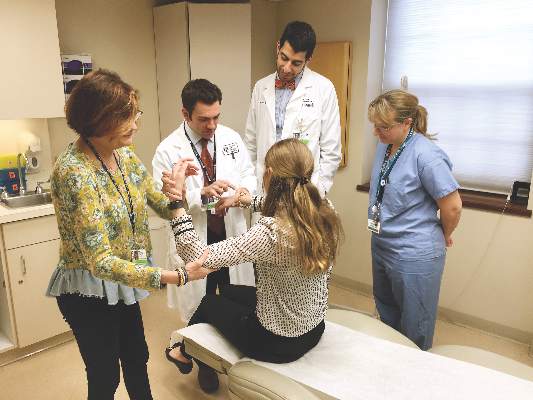
MIAMI – Combined rheumatology-dermatology clinics to help people with psoriasis, psoriatic arthritis, and other relevant overlapping conditions continue to evolve, with evidence suggesting advantages and challenges for both patients and physicians.
Physicians like the improved communication and greater collaboration but still have reservations about billing and scheduling, according to preliminary results of a survey conducted by the Psoriasis and Psoriatic Arthritis Clinic Multicenter Advancement Network (PPACMAN). For patients, the advantages go beyond convenience, according to a 1-year study of outcomes at a Rhode Island Hospital–Brown University combined clinic in Providence.
“The goal of the combined clinics survey is to gauge strengths, barriers, and challenges to creating these models, to learn from one another to improve current clinical care, and to support propagation of these models,” said Joseph F. Merola, MD, at the annual meeting of the Group for Research and Assessment of Psoriasis and Psoriatic Arthritis.
Multiple clinic models
The clinic setups differ. In those settings where rheumatologists and dermatologists see patients on the same day, approximately three-quarters provide care together in the same room. The remaining clinics see patients through serial visits. Another 30% of respondents said dermatologists and rheumatologists generally see patients in a combined clinic on different days.
“Rheumatologists said it is a satisfying and rewarding endeavor, they form closer ties with colleagues, and it allows early, improved communication,” said Dr. Merola, a rheumatologist and dermatologist who is co-director of the Center for Skin and Related Musculoskeletal Diseases, a combined clinic at Brigham and Women’s Hospital in Boston.
Some common benefits, each cited by more than 80% of respondents, include a prompt and accurate diagnosis of psoriatic arthritis, the ability of physicians to learn from each other, and multiple training opportunities for residents and fellows. In fact, 72% said their combined clinic has rheumatology fellows, 82% have dermatology residents, and 27% incorporate internal medicine trainees.
Other than rheumatologists and dermatologists, 31% said their clinic has dedicated nursing, 15% have a cardiologist, and 15% have a psychiatrist.
A majority of physicians (92%) said they bill through their own department. At a subsequent roundtable discussion, Dr. Merola noted that patients pay separate copays at the combined clinic. “We’ve had some complaints from patients. They don’t like having two copayments.”
“You have to tell patients in advance,” suggested Soumya M. Reddy, MD, a rheumatologist at NYU Langone Medical Center in New York and co-director of its Psoriasis and Psoriatic Arthritis Center.
Common conditions and challenges
Not surprisingly, psoriasis and psoriatic arthritis are the most common conditions treated in these clinics, followed by lupus and dermatomyositis. Most clinics see patients either once weekly or once monthly.
A major concern expressed by 75% surrounds scheduling. More than half, 58%, worry about demonstrating value to their institution. Only a minority, 17%, responded that achieving consensus on patient management is a challenge.
In general, rheumatology evaluations take more time than dermatology assessments, presenting a challenge for scheduling and maximizing physician time. “You don’t really want the dermatologist sitting there for 20 minutes not really doing a lot,” said Alison Ehrlich, MD, professor and chair of dermatology at George Washington University in Washington.
The solution at Brigham and Women’s Hospital is to staff the clinic with two dermatologists and one rheumatologist. The two dermatologists see about 20 patients each in a half-day; the rheumatologist probably treats about 6 to 8 of those 40 patients, Dr. Merola said. At George Washington, Dr. Ehrlich sees patients along with the rheumatologist, leaves the room to treat other patients, later consults with her colleague, and they go back in together as necessary.
“The patients are incredibly appreciative” of the combined clinic, Dr. Ehrlich said. “They really love it.”
The survey is ongoing. The preliminary responses above are based on 17 responses, a 52% response rate. Of the respondents, 10 are dermatologists, 6 are rheumatologists, and 1 is dual trained. Since the meeting, Dr. Merola indicated that another two dermatologists and three rheumatologists have responded to the survey.
The Brown University experience
Charis Gn, MD, and colleagues studied outcomes for 167 patients treated at a combined clinic at Rhode Island Hospital. The ultimate goal of the clinic is “to identify patients with psoriatic arthritis early on,” Dr. Gn said at a poster presentation at the GRAPPA annual meeting. “We wanted to see what kind of outcomes [we get] for patients who see rheumatology and dermatology.”
About one-third of patients left the clinic with changes in diagnosis. About 1 in 5 patients with psoriasis were diagnosed with psoriatic arthritis as well. For the psoriasis patients newly diagnosed with psoriatic arthritis, 80% had an escalation in treatment, said Dr. Gn, an internal medicine resident.
Of the 41 patients with psoriasis, 17 (41%) had a treatment escalation. The same was true for 15 (79%) of the 19 patients with psoriatic arthritis post-evaluation.
Regarding the combined clinic, “it’s shown to be very beneficial for patients with psoriatic arthritis,” Dr. Gn said.
The record review from July 2014 to February 2016 shows the clinic also serves patients with cutaneous lupus, dermatomyositis, pyoderma gangrenosum, vasculitis, and rheumatoid arthritis, among others.
The presenters had no relevant disclosures.
AT 2016 GRAPPA ANNUAL MEETING

Belonging and grieving: Lessons from Orlando
We belong in our family, in our group, and in our community. Belonging is a basic human need, and we work throughout our lives to seek or create safe places where we can belong. For the LGBTQ+ community, the Pulse nightclub was one of those rare places where they could feel like they truly belonged. It was a place where they did not need to pretend to be someone else or to keep their guard up against insults or criticism. For survivors of the shooting, the families, and the LGBTQ+ community, that sense of belonging is now fractured.
Pulse reportedly represented every LGBTQ+’s queer bar, where a safe environment is sought – an environment in which to grow and express identity. For the LGBTQ+ community, the June 12 massacre threatened all safe havens – where few currently exist – especially in the night scene, where the increased risk of violence is a constant factor in deciding where to party. There are few gay bars.

What must psychiatrists understand about the impact of the event, besides the regular work of grieving? Some specifics pertinent to this attack must be understood: the preponderance of Latinos1, the youthfulness of most victims, and the impact and reverberations throughout the LGBTQ+ community. This column highlights the unique aspects of this tragedy, and the complexity of the grieving that will take place in these families and communities.
Grief, illness, and resilience: What do we know?
Grieving after sudden and violent deaths, compared with natural deaths, is more likely to be associated with mental health disorders and a slower recovery (Psychiatry. 2012 Spring;75[1]:76-97). Traumatic loss can cause incapacitating grief and a search for meaning that can continue for years, especially in those who identify closely with the victims (J Trauma Stress. 2016 Feb;29[1]41-8).
The LGBTQ+ community identifies strongly with the victims of the Orlando shooting, and so is more likely to have a prolonged and difficult grieving process. Feelings of “it could have been me” or “this might happen to me or my loved ones,” can result in symptoms of anxiety and survivor guilt. The LGBTQ+ community also is grieving the loss of a safe place and that associated sense of belonging. Loss of safety and feelings of reduced control over one’s life result in a more difficult grieving process, irrespective of the cause of death (Death Stud. 2006;30[5]:403-28). It is as if the loss provides further evidence of life’s injustices and unpredictability, provoking feelings of helplessness and fear.
Being able to make sense or make meaning after a disaster is a key component of developing resilience (J Clin Psychol. 2016 Feb 22. doi: 10.1002/jclp.22270). To make meaning out of a loss, people inevitably will examine their own lives for meaning and purpose. Meaning-making also can result in post-traumatic growth, such as an increased appreciation for life, stronger connections with family and friends, or greater awareness of one’s strengths.
The meaning-making process, often facilitated by psychotherapy, plays a critical role in recovery. Meaning-making takes different forms. For some people, the record-breaking turnout at NYC Pride, in which the owner of Pulse and others rode on a float and received great support, was a significant event. Meanwhile, another person who might be scared might want to see his family, regardless of whether or not the family is fully accepting.
Grief as a social process
Coping with grief is the working out of “the meaning of the loss,” both personally and in community. Grieving is a social process, and eulogies, grief accounts in popular literature, and elegies help construct the identity of the loved one and the meaning of the event. Grieving involves reviewing the relationship to the lost loved one and establishing a sense of continuity between life before and life after the loss (Death Stud. 2014 Jul-Dec; 38[6-10]:485-98).
One challenge for the LGBTQ+ community is how to direct the meaning of the loss. What is the meaning of this loss and how will the dead be remembered? The outpouring of support across the world is heartwarming and reflects how the LGBTQ+ community has become more accepted in the world at large.
The LGBTQ+ community experiences much less stigma than 50 years ago. In 1973, after the firebombing of a club in New Orleans, some relatives reportedly refused to claim the bodies of their gay sons. However, there remains much variation in how communities/societies view LGBTQ+ individuals and relationships.
Cultural considerations of being LGBTQ+
Nonbinary gender identity is recognized in several cultures around the world. The word “Mahu” in Polynesian culture and the concept of being Two-Spirit in some Native American cultures describe those who do not identify as their assigned sex at birth, play out gender roles opposite their assigned sex, or are attracted to same-sex partners, according to biologist Joan Roughgarden, PhD, (“Evolution’s Rainbow: Diversity, Gender and Sexuality in Nature and People” Los Angeles: University of California Press, 2004). In Dr. Roughgarden’s 2010 book, “The Genial Gene” (Los Angeles: University of California Press), she promotes social-selection theory as an alternative to the Darwinian selfish gene theory. She lists 26 phenomena, not explained by current sexual-selection theory, that are better explained by social selection, a population-based explanation of partner selection. Those ideas are beginning to reach a wider audience in mainstream culture, although for many communities and religions, those ideas are seen as “wrong” or “blasphemous.”
For all minority groups, the importance of belonging to a safe, accepting community ranks highly. The pressure for all minority group members to conform to mainstream society is strong and persistent. A good parallel is found when thinking about race and the pressure of dark-skinned individuals to conform to white society. Skin bleaching remains a common practice; some people turn dark skin whiter in order to conform to a societal ideal. Similarly, in order to fit in and avoid discrimination or persecution, many LGBTQ+ individuals work hard to keep their gender conforming face forward. Pulse offered an opportunity for LGBTQ+ individuals and couples to feel like they could be themselves, express themselves, and forget about the societal pressures of conformity. Belonging to a safe community is, therefore, vital to maintaining a sense of self and self-worth.
The acknowledgment of LGBTQ+ status varies by culture. Ninety percent of the Pulse shooting victims reportedly were gay Latino men; a group that expresses its gender identity differently from gay white men. Verbal disclosure benefits gay white men’s well-being but doesn’t affect gay Latino men’s well-being, either positively or negatively. For gay white men, the more they verbally disclose their gay identity to others, the more they feel they are showing their true, feelings of authenticity, research shows). Additionally, LGBTQ+ members may be out in English but not in their Spanish-speaking church, according to Marianne Duddy-Burke, executive director of Dignity/USA, the largest national lay movement of LGBTQ Catholics, their families, and friends. Sadly, many of the Latino families of the Pulse shooting victims are Spanish-speaking only and reportedly had difficulty finding someone to explain what had happened.
Family considerations of being LGBTQ+.
Some families may know privately that they have an LGBTQ member but do not publicly acknowledge the fact. Family members may say things like “being married is maybe not for you, and that’s OK,” or “People should be allowed to be whoever they are.” This communication, while masked, is supportive and illustrates “an understanding” in the family. It may be some time before a family can say “we support our child, regardless” or “I know you are gay, and that’s OK.”
Gender identity can, therefore, be communicated nonverbally and behaviorally. For example, gay Puerto Rican and gay Dominican men in New York City discussed how they disclosed their gender identity by bringing their same-sex partner to family functions without the direct verbalization of a romantic relationship. This strategy, by which gay identity is implied and unspoken, is known as “tacit subjectivity,” or “tacitness.” The authors of this study postulated that for gay Latino men, their relationships in the family and Latino community are more important than the acknowledgment of individual difference and the expression of authenticity.
This perspective is consistent with the importance of community and family identity, a common aspect of non-Western cultures. At the extreme, some communities are very hostile to LGBTQ+ individuals, including in the United States. After sending “thoughts and prayers” to Orlando, the GOP House chair blocked legislation aimed at making sure that federal contractors cannot discriminate based on gender identity or sexual orientation.
Meanwhile, in Pakistani Muslim families, an LGBTQ+ member threatens the marriage prospects of the girls and may place the family in danger of being killed. Yet, 50 top Pakistani clerics recently issued a religious decree, or fatwa, declaring that transgender people have full marriage, inheritance, and funeral rights under Islamic law. The fatwa stated that a female-born transgender person having “visible signs of being a male” may marry a woman or a male-born transgender with “visible signs of being a female,” and vice versa. However, it ruled that a transgender person carrying “visible signs of both genders” – or intersex – may not marry anyone.
Currently in Pakistan, gay marriage is punishable by life imprisonment. The new fatwa also declared that any act intended to “humiliate, insult, or tease” the community was “haraam” (forbidden). The fatwa also states that transgender people should not be deprived of family inheritances, nor the right to be buried in Muslim ceremonies and that parents who deprived their transgender sons or daughters of inheritances were “inviting the wrath of God.”
Complexity of grieving after Pulse
The unique cultural and family aspects of the Pulse shooting makes the process of grieving complex. The cultural aspects of LGBTQ+ identity, both within that community and within Latino culture, bring new variables to the grieving process. Many of the previously held truths about grieving may turn out to be unimportant in this tragedy, and new factors may emerge.
Previously held findings on complex grief include:
• The importance of personal risk factors, such as female gender and preexisting psychiatric difficulties, in increasing the probability for mental distress.
• Interpersonal risk factors, such as kinship and social support, especially the loss of a child.
• Low perceived social support associated with depression after disaster-related bereavement.
• Social isolation related to difficulties in adjustment after the sudden and violent loss of a child.
• Finding meaning in the loss is related to lower mental distress and grief after violent losses.
• Social support has shown nonconclusive results in the general bereavement literature. Still, some studies suggest that social support may exert a protective effect on mental health adversities after sudden and violent losses.
After the Pulse shooting, these variables need to be reanalyzed. Specific personal risk factors are likely to emerge, such as a victim’s comfort with LGBTQ+ identity and their feelings of connection with the victims. Interpersonal factors will relate to the acceptance of their families and the presence of other safe places. The social status of LGBTQ+ community vis-à-vis the mainstream culture also will have an impact. Meaning-finding is a universal need that may be more difficult to reach for the LGBTQ+ community.
An assumption that likely will be upheld is that sudden, unexpected, and violent losses, compared with losses from natural deaths, are followed by a more difficult grieving process. This has been confirmed in several empirical studies that show a heightened risk for prolonged grief disorder (PGD), major depressive disorder, and posttraumatic stress disorder (PTSD) after violent losses.
In a study measuring PGD in 1,723 college students who had experienced either sudden and violent or natural losses, Joseph Currier, PhD, and his colleagues found that the violence of the loss, not the suddenness, predicted the increased PGD risk (Death Stud. 2006;30:403-28).
Also, it was more difficult to make sense of violent losses, and those students spent more time talking about the loss. In line with studies of PGD, the violence of the loss – not the suddenness – has been shown to account for the increased PTSD risk in the bereaved (J Anxiety Disord. 2003; 17[2]131-47).
DSM-5 diagnosis of persistent complex bereavement disorder
How to make a diagnosis? There is controversy surrounding the accuracy of DSM-5 proposed criteria for persistent complex bereavement disorder (PCBD). In a study of family members of U.S. military service members who died of any cause since Sept. 11, 2001 (n = 1,732), the DSM-5 PCBD criteria accurately excluded nonclinical, normative grief, but also excluded nearly half of clinical cases (Am J Psychiatry. 2016 May 24. [doi: 10.1176/appi.ajp.2016.15111442]).
PCBD previously has been referred to in the literature as complicated grief and prolonged grief disorder. However, those three labels refer to the same syndrome of clinically impairing grief, which affects approximately 7%-15% of bereaved individuals. This syndrome is diagnosed when persistent and severe grief symptoms continue beyond 6-12 months after the death of a loved one and are associated with functional impairment.
Typical symptoms include difficulty accepting the death or a strong sense of disbelief about the death, intense yearning and longing for the deceased, anger and bitterness, distressing and intrusive thoughts related to the death, and excessive avoidance of reminders of the painful loss. In recognition of their lack of validation, PCBD criteria were included in section 3 of DSM-5 Conditions for Further Study.
In summary, the Pulse shooting highlights the importance of assessing grief from a variety of new perspectives. First, assess the patient’s own personal sense of identity and how much she identifies with the victims. Second, assess the cultural and family aspects pertinent to the patient’s expression of gender identity. Third, assess the degree of acceptance the person feels and her ability to access a safe community. Assess the degree of persecution she feels in society at large. This assessment includes identifying where the patient experiences a true sense of belonging. Lastly, the path forward is always a meaning-making endeavor.
Summary of proposed PCBD in DSM-5
• Criterion A requires that the individual has experienced the death of a loved one.
• Criterion B requires the presence of 1 of 4 symptoms related to yearning, longing, and sorrow.
• Criterion C requires 6 of 12 symptoms demonstrating reactive distress to the death or social/identity disruption.
• Criterion D requires clinically significant distress or functional impairment.
• Criterion E requires that distress or impairment is outside of sociocultural norms.
Symptoms present for at least 12 months and that are not better accounted for by major depressive disorder, generalized anxiety disorder, or posttraumatic stress disorder.
Source: Dr. Heru
Thank you to Shiona Heru, JD, for her comments.
References
1. Gender-inclusive version is to use either Latinx (the x symbol represents the absence of o/a) or Latin@ (using the @ symbol mixes the o and @).
2. For more on “The Singular Experience of the Queer Latin Nightclub”: http://www.theatlantic.com/entertainment/archive/2016/06/orlando-shooting-pulse-latin-queer-gay-nightclub-ramon-rivera-servera-intrerview/487442/
Dr. Heru is with the department of psychiatry at the University of Colorado Denver, Aurora. She is editor of “Working With Families in Medical Settings: A Multidisciplinary Guide for Psychiatrists and Other Health Professionals (New York: Routledge, 2013). She has no conflicts of interest to disclose. .
We belong in our family, in our group, and in our community. Belonging is a basic human need, and we work throughout our lives to seek or create safe places where we can belong. For the LGBTQ+ community, the Pulse nightclub was one of those rare places where they could feel like they truly belonged. It was a place where they did not need to pretend to be someone else or to keep their guard up against insults or criticism. For survivors of the shooting, the families, and the LGBTQ+ community, that sense of belonging is now fractured.
Pulse reportedly represented every LGBTQ+’s queer bar, where a safe environment is sought – an environment in which to grow and express identity. For the LGBTQ+ community, the June 12 massacre threatened all safe havens – where few currently exist – especially in the night scene, where the increased risk of violence is a constant factor in deciding where to party. There are few gay bars.
What must psychiatrists understand about the impact of the event, besides the regular work of grieving? Some specifics pertinent to this attack must be understood: the preponderance of Latinos1, the youthfulness of most victims, and the impact and reverberations throughout the LGBTQ+ community. This column highlights the unique aspects of this tragedy, and the complexity of the grieving that will take place in these families and communities.
Grief, illness, and resilience: What do we know?
Grieving after sudden and violent deaths, compared with natural deaths, is more likely to be associated with mental health disorders and a slower recovery (Psychiatry. 2012 Spring;75[1]:76-97). Traumatic loss can cause incapacitating grief and a search for meaning that can continue for years, especially in those who identify closely with the victims (J Trauma Stress. 2016 Feb;29[1]41-8).
The LGBTQ+ community identifies strongly with the victims of the Orlando shooting, and so is more likely to have a prolonged and difficult grieving process. Feelings of “it could have been me” or “this might happen to me or my loved ones,” can result in symptoms of anxiety and survivor guilt. The LGBTQ+ community also is grieving the loss of a safe place and that associated sense of belonging. Loss of safety and feelings of reduced control over one’s life result in a more difficult grieving process, irrespective of the cause of death (Death Stud. 2006;30[5]:403-28). It is as if the loss provides further evidence of life’s injustices and unpredictability, provoking feelings of helplessness and fear.
Being able to make sense or make meaning after a disaster is a key component of developing resilience (J Clin Psychol. 2016 Feb 22. doi: 10.1002/jclp.22270). To make meaning out of a loss, people inevitably will examine their own lives for meaning and purpose. Meaning-making also can result in post-traumatic growth, such as an increased appreciation for life, stronger connections with family and friends, or greater awareness of one’s strengths.
The meaning-making process, often facilitated by psychotherapy, plays a critical role in recovery. Meaning-making takes different forms. For some people, the record-breaking turnout at NYC Pride, in which the owner of Pulse and others rode on a float and received great support, was a significant event. Meanwhile, another person who might be scared might want to see his family, regardless of whether or not the family is fully accepting.
Grief as a social process
Coping with grief is the working out of “the meaning of the loss,” both personally and in community. Grieving is a social process, and eulogies, grief accounts in popular literature, and elegies help construct the identity of the loved one and the meaning of the event. Grieving involves reviewing the relationship to the lost loved one and establishing a sense of continuity between life before and life after the loss (Death Stud. 2014 Jul-Dec; 38[6-10]:485-98).
One challenge for the LGBTQ+ community is how to direct the meaning of the loss. What is the meaning of this loss and how will the dead be remembered? The outpouring of support across the world is heartwarming and reflects how the LGBTQ+ community has become more accepted in the world at large.
The LGBTQ+ community experiences much less stigma than 50 years ago. In 1973, after the firebombing of a club in New Orleans, some relatives reportedly refused to claim the bodies of their gay sons. However, there remains much variation in how communities/societies view LGBTQ+ individuals and relationships.
Cultural considerations of being LGBTQ+
Nonbinary gender identity is recognized in several cultures around the world. The word “Mahu” in Polynesian culture and the concept of being Two-Spirit in some Native American cultures describe those who do not identify as their assigned sex at birth, play out gender roles opposite their assigned sex, or are attracted to same-sex partners, according to biologist Joan Roughgarden, PhD, (“Evolution’s Rainbow: Diversity, Gender and Sexuality in Nature and People” Los Angeles: University of California Press, 2004). In Dr. Roughgarden’s 2010 book, “The Genial Gene” (Los Angeles: University of California Press), she promotes social-selection theory as an alternative to the Darwinian selfish gene theory. She lists 26 phenomena, not explained by current sexual-selection theory, that are better explained by social selection, a population-based explanation of partner selection. Those ideas are beginning to reach a wider audience in mainstream culture, although for many communities and religions, those ideas are seen as “wrong” or “blasphemous.”
For all minority groups, the importance of belonging to a safe, accepting community ranks highly. The pressure for all minority group members to conform to mainstream society is strong and persistent. A good parallel is found when thinking about race and the pressure of dark-skinned individuals to conform to white society. Skin bleaching remains a common practice; some people turn dark skin whiter in order to conform to a societal ideal. Similarly, in order to fit in and avoid discrimination or persecution, many LGBTQ+ individuals work hard to keep their gender conforming face forward. Pulse offered an opportunity for LGBTQ+ individuals and couples to feel like they could be themselves, express themselves, and forget about the societal pressures of conformity. Belonging to a safe community is, therefore, vital to maintaining a sense of self and self-worth.
The acknowledgment of LGBTQ+ status varies by culture. Ninety percent of the Pulse shooting victims reportedly were gay Latino men; a group that expresses its gender identity differently from gay white men. Verbal disclosure benefits gay white men’s well-being but doesn’t affect gay Latino men’s well-being, either positively or negatively. For gay white men, the more they verbally disclose their gay identity to others, the more they feel they are showing their true, feelings of authenticity, research shows). Additionally, LGBTQ+ members may be out in English but not in their Spanish-speaking church, according to Marianne Duddy-Burke, executive director of Dignity/USA, the largest national lay movement of LGBTQ Catholics, their families, and friends. Sadly, many of the Latino families of the Pulse shooting victims are Spanish-speaking only and reportedly had difficulty finding someone to explain what had happened.
Family considerations of being LGBTQ+.
Some families may know privately that they have an LGBTQ member but do not publicly acknowledge the fact. Family members may say things like “being married is maybe not for you, and that’s OK,” or “People should be allowed to be whoever they are.” This communication, while masked, is supportive and illustrates “an understanding” in the family. It may be some time before a family can say “we support our child, regardless” or “I know you are gay, and that’s OK.”
Gender identity can, therefore, be communicated nonverbally and behaviorally. For example, gay Puerto Rican and gay Dominican men in New York City discussed how they disclosed their gender identity by bringing their same-sex partner to family functions without the direct verbalization of a romantic relationship. This strategy, by which gay identity is implied and unspoken, is known as “tacit subjectivity,” or “tacitness.” The authors of this study postulated that for gay Latino men, their relationships in the family and Latino community are more important than the acknowledgment of individual difference and the expression of authenticity.
This perspective is consistent with the importance of community and family identity, a common aspect of non-Western cultures. At the extreme, some communities are very hostile to LGBTQ+ individuals, including in the United States. After sending “thoughts and prayers” to Orlando, the GOP House chair blocked legislation aimed at making sure that federal contractors cannot discriminate based on gender identity or sexual orientation.
Meanwhile, in Pakistani Muslim families, an LGBTQ+ member threatens the marriage prospects of the girls and may place the family in danger of being killed. Yet, 50 top Pakistani clerics recently issued a religious decree, or fatwa, declaring that transgender people have full marriage, inheritance, and funeral rights under Islamic law. The fatwa stated that a female-born transgender person having “visible signs of being a male” may marry a woman or a male-born transgender with “visible signs of being a female,” and vice versa. However, it ruled that a transgender person carrying “visible signs of both genders” – or intersex – may not marry anyone.
Currently in Pakistan, gay marriage is punishable by life imprisonment. The new fatwa also declared that any act intended to “humiliate, insult, or tease” the community was “haraam” (forbidden). The fatwa also states that transgender people should not be deprived of family inheritances, nor the right to be buried in Muslim ceremonies and that parents who deprived their transgender sons or daughters of inheritances were “inviting the wrath of God.”
Complexity of grieving after Pulse
The unique cultural and family aspects of the Pulse shooting makes the process of grieving complex. The cultural aspects of LGBTQ+ identity, both within that community and within Latino culture, bring new variables to the grieving process. Many of the previously held truths about grieving may turn out to be unimportant in this tragedy, and new factors may emerge.
Previously held findings on complex grief include:
• The importance of personal risk factors, such as female gender and preexisting psychiatric difficulties, in increasing the probability for mental distress.
• Interpersonal risk factors, such as kinship and social support, especially the loss of a child.
• Low perceived social support associated with depression after disaster-related bereavement.
• Social isolation related to difficulties in adjustment after the sudden and violent loss of a child.
• Finding meaning in the loss is related to lower mental distress and grief after violent losses.
• Social support has shown nonconclusive results in the general bereavement literature. Still, some studies suggest that social support may exert a protective effect on mental health adversities after sudden and violent losses.
After the Pulse shooting, these variables need to be reanalyzed. Specific personal risk factors are likely to emerge, such as a victim’s comfort with LGBTQ+ identity and their feelings of connection with the victims. Interpersonal factors will relate to the acceptance of their families and the presence of other safe places. The social status of LGBTQ+ community vis-à-vis the mainstream culture also will have an impact. Meaning-finding is a universal need that may be more difficult to reach for the LGBTQ+ community.
An assumption that likely will be upheld is that sudden, unexpected, and violent losses, compared with losses from natural deaths, are followed by a more difficult grieving process. This has been confirmed in several empirical studies that show a heightened risk for prolonged grief disorder (PGD), major depressive disorder, and posttraumatic stress disorder (PTSD) after violent losses.
In a study measuring PGD in 1,723 college students who had experienced either sudden and violent or natural losses, Joseph Currier, PhD, and his colleagues found that the violence of the loss, not the suddenness, predicted the increased PGD risk (Death Stud. 2006;30:403-28).
Also, it was more difficult to make sense of violent losses, and those students spent more time talking about the loss. In line with studies of PGD, the violence of the loss – not the suddenness – has been shown to account for the increased PTSD risk in the bereaved (J Anxiety Disord. 2003; 17[2]131-47).
DSM-5 diagnosis of persistent complex bereavement disorder
How to make a diagnosis? There is controversy surrounding the accuracy of DSM-5 proposed criteria for persistent complex bereavement disorder (PCBD). In a study of family members of U.S. military service members who died of any cause since Sept. 11, 2001 (n = 1,732), the DSM-5 PCBD criteria accurately excluded nonclinical, normative grief, but also excluded nearly half of clinical cases (Am J Psychiatry. 2016 May 24. [doi: 10.1176/appi.ajp.2016.15111442]).
PCBD previously has been referred to in the literature as complicated grief and prolonged grief disorder. However, those three labels refer to the same syndrome of clinically impairing grief, which affects approximately 7%-15% of bereaved individuals. This syndrome is diagnosed when persistent and severe grief symptoms continue beyond 6-12 months after the death of a loved one and are associated with functional impairment.
Typical symptoms include difficulty accepting the death or a strong sense of disbelief about the death, intense yearning and longing for the deceased, anger and bitterness, distressing and intrusive thoughts related to the death, and excessive avoidance of reminders of the painful loss. In recognition of their lack of validation, PCBD criteria were included in section 3 of DSM-5 Conditions for Further Study.
In summary, the Pulse shooting highlights the importance of assessing grief from a variety of new perspectives. First, assess the patient’s own personal sense of identity and how much she identifies with the victims. Second, assess the cultural and family aspects pertinent to the patient’s expression of gender identity. Third, assess the degree of acceptance the person feels and her ability to access a safe community. Assess the degree of persecution she feels in society at large. This assessment includes identifying where the patient experiences a true sense of belonging. Lastly, the path forward is always a meaning-making endeavor.
Summary of proposed PCBD in DSM-5
• Criterion A requires that the individual has experienced the death of a loved one.
• Criterion B requires the presence of 1 of 4 symptoms related to yearning, longing, and sorrow.
• Criterion C requires 6 of 12 symptoms demonstrating reactive distress to the death or social/identity disruption.
• Criterion D requires clinically significant distress or functional impairment.
• Criterion E requires that distress or impairment is outside of sociocultural norms.
Symptoms present for at least 12 months and that are not better accounted for by major depressive disorder, generalized anxiety disorder, or posttraumatic stress disorder.
Source: Dr. Heru
Thank you to Shiona Heru, JD, for her comments.
References
1. Gender-inclusive version is to use either Latinx (the x symbol represents the absence of o/a) or Latin@ (using the @ symbol mixes the o and @).
2. For more on “The Singular Experience of the Queer Latin Nightclub”: http://www.theatlantic.com/entertainment/archive/2016/06/orlando-shooting-pulse-latin-queer-gay-nightclub-ramon-rivera-servera-intrerview/487442/
Dr. Heru is with the department of psychiatry at the University of Colorado Denver, Aurora. She is editor of “Working With Families in Medical Settings: A Multidisciplinary Guide for Psychiatrists and Other Health Professionals (New York: Routledge, 2013). She has no conflicts of interest to disclose. .
Pitt Hopkins Scientific Symposium to Take Place in November
The Pitt Hopkins Research Foundation Scientific Symposium and Family Conference will take place November 3-5 in Dallas, Texas.
The Pitt Hopkins Research Foundation Scientific Symposium and Family Conference will take place November 3-5 in Dallas, Texas.
The Pitt Hopkins Research Foundation Scientific Symposium and Family Conference will take place November 3-5 in Dallas, Texas.
HCU Network Australia Develops Q & A Video With Homocystinuria Expert
Clinician Andrew Morris of the UK is interviewed about the importance of timely diagnosis of homocystinuria in a brief video published recently by the HCU Network Australia.
Clinician Andrew Morris of the UK is interviewed about the importance of timely diagnosis of homocystinuria in a brief video published recently by the HCU Network Australia.
Clinician Andrew Morris of the UK is interviewed about the importance of timely diagnosis of homocystinuria in a brief video published recently by the HCU Network Australia.

Foundation Seeks Proposals to Improve Care of Children With Chronic and Complex Diseases
The Lucile Packard Foundation for Children’s Health seeks applications for grants to develop single-topic issue briefs that describe goals and processes to improve aspects of the systems of care for children with chronic and complex health conditions in the US. Proposals are due September 15.
The Lucile Packard Foundation for Children’s Health seeks applications for grants to develop single-topic issue briefs that describe goals and processes to improve aspects of the systems of care for children with chronic and complex health conditions in the US. Proposals are due September 15.
The Lucile Packard Foundation for Children’s Health seeks applications for grants to develop single-topic issue briefs that describe goals and processes to improve aspects of the systems of care for children with chronic and complex health conditions in the US. Proposals are due September 15.

ACP Develops Toolkit for Transition From Pediatric to Adult Care
The American College of Physicians Initiative has developed a toolkit to facilitate more effective transition of young adults from pediatric to adult care. The kit includes a major focus on providing a framework for the adult care provider.
The American College of Physicians Initiative has developed a toolkit to facilitate more effective transition of young adults from pediatric to adult care. The kit includes a major focus on providing a framework for the adult care provider.
The American College of Physicians Initiative has developed a toolkit to facilitate more effective transition of young adults from pediatric to adult care. The kit includes a major focus on providing a framework for the adult care provider.

Fresh Press: ACS Surgery News digital July issue is available on the website
The July issue of ACS Surgery News in digital format is available online. Use the mobile app to download or view as a pdf.
This month’s issue features coverage of the report, “A National Trauma Care System: Integrating Civilian and Military Trauma Systems to Achieve Zero Preventable Deaths After Injury,” released by the National Academies of Sciences, Engineering, and Medicine. The report, supported in part by the American College of Surgeons, gives a detailed list of policy recommendations that would create a unified trauma response system incorporating lessons learned from combat mission with the aim of eliminating unnecessary deaths in civilian trauma situations (p. 1).

Looking for some summer reading? Dr. Carol Scott-Conner, moderator of the ACS Communities Surgeon Writers discussion group, reviews three books by surgeons that should be of special interest to the readers of ACS Surgery News. All three works concern personal experience and reflections on the profession (p. 4).
Don’t miss the story (p. 7) on peer review hearings and how best to approach this situation. These hearings should be informative and educational, but can also play out in unexpected ways. Be prepared!
The July issue of ACS Surgery News in digital format is available online. Use the mobile app to download or view as a pdf.
This month’s issue features coverage of the report, “A National Trauma Care System: Integrating Civilian and Military Trauma Systems to Achieve Zero Preventable Deaths After Injury,” released by the National Academies of Sciences, Engineering, and Medicine. The report, supported in part by the American College of Surgeons, gives a detailed list of policy recommendations that would create a unified trauma response system incorporating lessons learned from combat mission with the aim of eliminating unnecessary deaths in civilian trauma situations (p. 1).
Looking for some summer reading? Dr. Carol Scott-Conner, moderator of the ACS Communities Surgeon Writers discussion group, reviews three books by surgeons that should be of special interest to the readers of ACS Surgery News. All three works concern personal experience and reflections on the profession (p. 4).
Don’t miss the story (p. 7) on peer review hearings and how best to approach this situation. These hearings should be informative and educational, but can also play out in unexpected ways. Be prepared!
Lessons learned from merging EHR systems
WASHINGTON – As practices merge, how hard is it to merge EHRs?
Even in what might seem to be the best circumstances, it can be a huge challenge, according to Jacqueline Fincher, MD, of McDuffie Medical Associates, Thompson, Ga.
One appealing aspect of the merger of Dr. Fincher’s practice with another was that each practice used an EHR from the same vendor and each was operating with the same updates.

“This is the perfect setup,” she said at the annual meeting of the American College of Physicians. “We don’t have to go to Epic. We can stay on the same EHR. In fact, this group had gone on the same EHR back in 2006 within a month of the time that we went on; we were on the exact same version and the same everything. We were totally even. The thought among the corporate and IT staff of the new entity was that this is going to be seamless. We’re just going to renumber the accounts and everything will be just fine.”
A call to the EHR vendor, whom she did not name, revealed that the process would be anything but seamless.
“Our IT staff contacted our common EHR vendor and said we want to merge this practice with our bigger practice and the EHR company said, ‘Wow, we’ve never done that before.’ What? In this day of consolidation and integration, they had never done it before? Nor did they have a business model to do so, much less a digital plan to do so. That was pretty shocking,” she said.
Dr. Fincher noted that the EHR vendor recommended a third party vendor to handle creating an interface between the two EHRs. “Most EHR companies do not, I say, do not have a dedicated service to migrate data. It’s almost always going out to a third-party conversion service that doesn’t know you, doesn’t know your work flow, and makes everything even more difficult.”
Two and a half months – and 10 interfaces – later, the launch of the combined EHR was a disaster, Dr. Fincher said.
Even though both practices were using the exact same version of the EHR, each had very different work flows, defaults, and other nuances that meant data didn’t transfer smoothly – cheaply.
Among the surprise expenses: about $55,000 for additional hardware, network cabling and interfaces; $35,000 for additional servers; and at least $100,000 for personnel expenses related to the data migration.
Given her experience, Dr. Fincher advised her peers “to start at least 6 months in advance to map and convert the data.”
And it is vital to get input and participation from all office stakeholders – both clinical and administrative staff – regarding how the data is migrated, she said. Be sure to completely understand all work flows from both practices so that you know how the data is going to migrate.
“Understanding work flow, that is absolutely critical. Every single office has a different work flow for every type of encounter by any method. How these work flows are the same or different between your office and the new practice or your EHR and the new EHR that you’re going to, they are different. You have to understand the differences,” she added.
“We didn’t perceive that there was that much difference but when everything crashed, we discovered there were because they had different work flows, they had different defaults in place, those types of things.”
Other key questions: Which data needs to be migrated? How long should the old system remain in place? Should the data be migrated manually or digitally? How much time will the EHR merger take?
“You want to establish that structured planning time,” Dr. Fincher said. “You’ve got to carve out scheduled time with the group in order to do this. Establish the tasks that need to be accomplished, so just making a to-do list every week and who’s going to be accountable to accomplish those parts of the list” is important.
WASHINGTON – As practices merge, how hard is it to merge EHRs?
Even in what might seem to be the best circumstances, it can be a huge challenge, according to Jacqueline Fincher, MD, of McDuffie Medical Associates, Thompson, Ga.
One appealing aspect of the merger of Dr. Fincher’s practice with another was that each practice used an EHR from the same vendor and each was operating with the same updates.
“This is the perfect setup,” she said at the annual meeting of the American College of Physicians. “We don’t have to go to Epic. We can stay on the same EHR. In fact, this group had gone on the same EHR back in 2006 within a month of the time that we went on; we were on the exact same version and the same everything. We were totally even. The thought among the corporate and IT staff of the new entity was that this is going to be seamless. We’re just going to renumber the accounts and everything will be just fine.”
A call to the EHR vendor, whom she did not name, revealed that the process would be anything but seamless.
“Our IT staff contacted our common EHR vendor and said we want to merge this practice with our bigger practice and the EHR company said, ‘Wow, we’ve never done that before.’ What? In this day of consolidation and integration, they had never done it before? Nor did they have a business model to do so, much less a digital plan to do so. That was pretty shocking,” she said.
Dr. Fincher noted that the EHR vendor recommended a third party vendor to handle creating an interface between the two EHRs. “Most EHR companies do not, I say, do not have a dedicated service to migrate data. It’s almost always going out to a third-party conversion service that doesn’t know you, doesn’t know your work flow, and makes everything even more difficult.”
Two and a half months – and 10 interfaces – later, the launch of the combined EHR was a disaster, Dr. Fincher said.
Even though both practices were using the exact same version of the EHR, each had very different work flows, defaults, and other nuances that meant data didn’t transfer smoothly – cheaply.
Among the surprise expenses: about $55,000 for additional hardware, network cabling and interfaces; $35,000 for additional servers; and at least $100,000 for personnel expenses related to the data migration.
Given her experience, Dr. Fincher advised her peers “to start at least 6 months in advance to map and convert the data.”
And it is vital to get input and participation from all office stakeholders – both clinical and administrative staff – regarding how the data is migrated, she said. Be sure to completely understand all work flows from both practices so that you know how the data is going to migrate.
“Understanding work flow, that is absolutely critical. Every single office has a different work flow for every type of encounter by any method. How these work flows are the same or different between your office and the new practice or your EHR and the new EHR that you’re going to, they are different. You have to understand the differences,” she added.
“We didn’t perceive that there was that much difference but when everything crashed, we discovered there were because they had different work flows, they had different defaults in place, those types of things.”
Other key questions: Which data needs to be migrated? How long should the old system remain in place? Should the data be migrated manually or digitally? How much time will the EHR merger take?
“You want to establish that structured planning time,” Dr. Fincher said. “You’ve got to carve out scheduled time with the group in order to do this. Establish the tasks that need to be accomplished, so just making a to-do list every week and who’s going to be accountable to accomplish those parts of the list” is important.
EXPERT ANALYSIS FROM ACP INTERNAL MEDICINE 2016
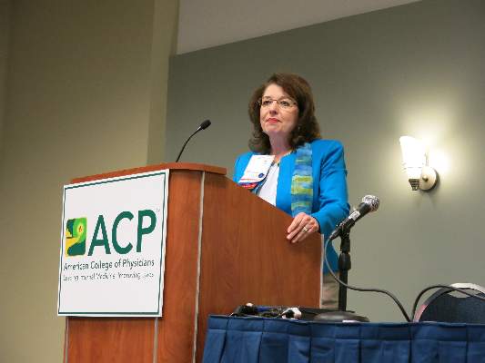
Take Advantage of Early Registration for AATS Focus on Thoracic Surgery: Novel Techniques in Lung Cancer — Program Now Online
The preliminary scientific program, along with registration and housing information, is now online for AATS Focus on Thoracic Surgery: Novel Techniques in Lung Cancer.
November 21 – 22, 2014
Renaissance Boston Waterfront Hotel
Boston, MA, USA
Take advantage of early registration rates and register before October 23rd!
Program Directors
G. Alec Patterson, Washington University
David J. Sugarbaker, Baylor College of Medicine
Program Committee Members:
Thomas A. D’Amico, Duke University
Shaf Keshavjee, University of Toronto
James D. Luketich, University of Pittsburgh
Bryan F. Meyers, Washington University
Scott J. Swanson, Brigham and Women’s Hospital
Overview
Lung cancer is the most common cause of cancer-related deaths and second most common malignancy in the Western world. Recent innovations in diagnosis, staging and management of early stage and locally advanced non-small cell lung cancer have changed the paradigm for patient surgical management. There has also been a virtual explosion of technological developments enabling widespread use of minimally invasive techniques, which current and future thoracic surgeons will need to embrace in order to improve patient care.
Join AATS in Boston for the 2014 Focus On Thoracic Surgery whose program will focus on recent developments in Novel Technologies in Lung Cancer. The faculty includes internationally recognized experts in the field, including those knowledgeable about the use of new technology in lung cancer surgical management.
Program Highlights
Imaging and Staging
Surgical Alternatives
Simulation, Education, and Training
Optimal Management of Lung Metastases
Credentialing/ Efficiencies
Intraoperative Management
Video Sessions - Advanced Minimally Invasive Techniques
Honored Guest Speaker
Larry R. KaiserDean, Temple University
Hepatitis C Infection Rates Rising in Women, Young Children
Rates of hepatitis C virus detection increased in U.S. women aged 15-44 years and in children less than 2 years old from 2011 to 2014, according to investigators from the Centers for Disease Control and Prevention.
A report published July 21 in Morbidity and Mortality Weekly Report revealed hepatitis C virus (HCV) detection rates (detection via antibody or RNA positivity) that were determined using data collected from Quest Diagnostics for Kentucky and for the United States. In Kentucky, the rate of HCV detection in women aged 15-44 increased 213% over the study period, from 275 to 862 cases per 100,000 people, and for the entire U.S., the rate of detection increased by 22% from 139 to 169 cases per 100,000 people (65[28]:705-10).
The rate of HCV testing for children under 2 years old in Kentucky increased 151% over the study period, rising from 403 to 1,011 per 100,000 people, and the rate of children born to HCV-positive mothers increased from 0.71% to 1.59%. Nationwide, the HCV testing rate for children under 2 years old increased 14% from 310 to 353 per 100,000, and the rate of children born to HCV-positive mothers increased from 0.19% to 0.32%.
“These findings underscore the importance of providing primary prevention services and following current recommendations to identify persons at risk for HCV infection and test accordingly; doing so among pregnant women would improve early identification of HCV-infected infants and linkage of the mother and infant to care and treatment,” the CDC investigators said.
FROM MMWR