User login
Efficacy of malaria vaccine declines over time
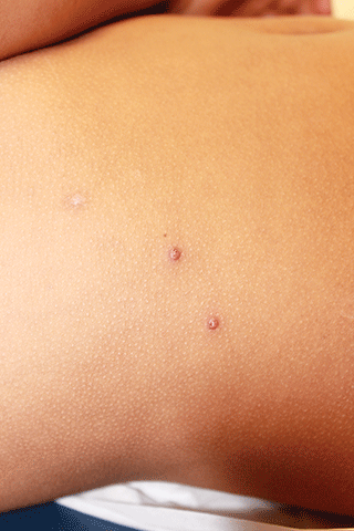
Photo by Caitlin Kleiboer
Results from a phase 2 study of the malaria vaccine RTS,S (also known as RTS,S/AS01 or Mosquirix) suggest its efficacy decreases over time, and this decline is fastest in children living in areas with higher-than-average rates of malaria.
Researchers say the results suggest the benefits of the vaccine are likely to vary across different populations and highlight the need for more research to
determine the most effective way of using RTS,S, which last year became the first malaria vaccine to receive a green light from the European Medicines Agency.
“We found that 3-dose vaccination with RTS,S was initially protective, but this was offset by a rebound in later years among children exposed to higher-than-average levels of malaria-carrying mosquitoes,” said Philip Bejon, PhD, of the Kenya Medical Research Institute–Wellcome Trust Programme in Kilifi, Kenya.
Dr Bejon and his colleagues reported these results in NEJM.
The researchers followed 447 children who had received 3 doses of either RTS,S or a rabies (control) vaccine when they were 5 months to 17 months old.
After 7 years, there were 312 children still involved in the study. During the first year, the risk of getting malaria in the vaccinated children was 35.9% less than in the control group. After 7 years, this protection fell to 3.6%.
And in children exposed to higher-than-average rates of malaria, there were slightly more cases of malaria in the vaccinated group than the control group—1002 and 992 cases, respectively—5 years after vaccination.
This “rebound” effect, which has been seen in previous studies, is thought to occur because children initially protected by the vaccine develop their natural immunity against malaria more slowly than unvaccinated children.
Results from a phase 3 study showed that 3 doses of RTS,S reduced the risk of malaria in young children by 28% over 4 years, but this improved to 36% when children were given a fourth dose 18 months after the first dose. Longer-term follow up of these children is ongoing.
“Overall, our study shows that RTS,S can benefit children but suggests that a fourth dose may be important for sustaining this protection over the long term and to protect against a potential rebound,” said Ally Olotu, PhD, of the Kenya Medical Research Institute–Wellcome Trust Programme.
“Results from 3 sites involved in the original phase 3 study that are continuing follow up, and the WHO’s planned pilot program, will tell us more about the vaccine’s efficacy in different settings and help determine which populations would benefit most from receiving it as part of a wider vaccination strategy.” ![]()
MACRA Rule Offers Little Clarity for Hospitalists
Last year, Congress put an end to the Sustainable Growth Rate (SGR), which had become a yearly battle fought on behalf of and by physicians to prevent significant last-minute cuts to Medicare reimbursement. Many hoped its replacement would provide more stability and certainty.
However, that replacement, the Medicare Access and CHIP Reauthorization Act of 2015 (MACRA), has been anything but clear. On April 27, 2016, the Centers for Medicare & Medicaid Services (CMS) issued a Notice of Proposed Rulemaking in what it called a “first step” in implementing MACRA. CMS accepted feedback and input on the proposed rule through June 27, 2016.
The Society of Hospital Medicine worked to provide comment on what it sees as the biggest concerns of hospitalists.
For example, it remains unclear what quality markers CMS will use to evaluate hospitalists under MACRA, says Rush University Medical Center’s Suparna Dutta, MD, MPH, a hospitalist, assistant professor of medicine, and member of the SHM Public Policy Committee (PPC). “The biggest piece is, what will be used universally for all hospitalists and attributed to the work that we do?”
MACRA represents “a milestone” in efforts to “advance a healthcare system that rewards better care, smarter spending, and healthier people,” U.S. Department of Health & Human Services Secretary Sylvia M. Burwell said in a statement issued the day the proposed rule was announced.
What it is designed to do, says Ron Greeno, MD, MHM, president-elect of SHM, PPC chair, and senior advisor for medical affairs at TeamHealth, is push physicians to move toward alternative payment models.
To achieve this, MACRA creates a framework called the Quality Payment Program, which offers physicians two paths for value-over-volume-based payments: MIPS, for Merit-Based Incentive Payment System, and APMs, for Advanced Alternative Payment Models. The benchmark period for both pathways begins Jan. 1, 2017, and MACRA reimbursement would begin Jan. 1, 2019.
Under MIPS, current quality measurement programs are streamlined into a single payment adjustment, including the Physician Value-Based Modifier, the Electronic Health Record (EHR) Incentive Program and the Physician Quality Reporting System (PQRS).
Physicians will not assume risk on the MIPS pathway, but payment adjustments will be based on their MIPS score, which grows each year through 2022 and ranges that year from +9% to -9%. It will be budget neutral: The top half of scorers will see increases in payments, while the bottom half will see cuts. Additional adjustments will be given to top performers through 2024.
However, as Dr. Dutta and fellow PPC member Lauren Doctoroff, MD, FHM, a hospitalist at Beth Israel Deaconess Medical Center and instructor at Harvard Medical School, wrote for The Hospitalist in March 2016, it is not yet clear how MIPS scores will be calculated for hospitalists.
“The problem is that there is not a typical hospitalist in terms of the work that we do,” Dr. Dutta says. “It depends on the hospital and the types of responsibilities the hospitalists have and the types of patients they care for.”
CMS says 50% of the MIPS score will come from six reported measures that reflect different specialties and practices; 25% will come from technology use, with a focus on interoperability and information exchange; 15% will come from clinical improvement practices, like care coordination; and 10% will be based on cost, chosen from among 40 episode-specific measures.
The new hospitalist billing code, which has not yet been implemented, should be a tremendous help under MACRA, Dr. Dutta says. “As CMS plans on using peer-comparison groups for quality and cost measures, it is really important that we now have a specialty billing code for hospitalists, which should ensure we have a fair and valid comparison pool for any metrics we are measured on for MIPS.”
The second path may be much harder for hospitalists to achieve since it requires that physicians share in risk and reward and participate in alternative payment models like Next Generation ACO or the Comprehensive Primary Care Plus model.
Most hospitalists will not be candidates for taking on risks under APM since physicians need to achieve a threshold for taking on more than nominal financial risk, Dr. Dutta says, noting SHM’s efforts to better understand the implications.
“It depends on the the percentage of patients you’re seeing in an APM, and you might hit your threshold if your market has a lot of Medicare ACOs or risk-sharing, but it’s not something hospitalists can consistently plan on,” Dr. Dutta says.
Most hospitalists have little control over whether their facility participates in an APM, Dr. Dutta says, but allowing the APM to which a patient belongs count toward the care provided by hospitalists—though a patient may align with several APMs—may help reach these thresholds.
Feedback from SHM to CMS also included asking to allow the Bundled Payments for Care Improvement Initiative (BPCI) to qualify for APM and seeking clarification into whether hospitalists can tap into cost and quality metrics hospitals are already reporting to CMS.
“Hospitals are collecting a certain amount of data because they have to for Medicare, and that might be a good indicator of what hospitalists are doing,” Dr. Dutta says. This includes services like DVT prophylaxis after surgery in hospitals where hospitalists provide a majority of post-operative care or safety measures like CLABSI (central line–associated bloodstream infection) rates.
To stay up to date with MACRA, visit SHM’s MACRA website and follow @SHMadvocacy on Twitter. TH
Corrected version July 13, 2016.
Kelly April Tyrrell is a freelance writer in Madison, Wis.
Telomere length linked to neutrophil recovery in AML

Image by Volker Brinkmann
Researchers say they have discovered a way to predict which children with acute myeloid leukemia (AML) are at the highest risk of delayed neutrophil recovery.
The team examined the role of telomeres in neutrophil recovery and found that the length of a patient’s telomeres can indicate the rate of recovery following chemotherapy.
The group reported their findings in the Journal of Clinical Oncology.
“We were interested in telomere length as a marker of blood count recovery because defects in telomere maintenance are known risks for bone marrow failure and aplastic anemia,” said study author Maria Monica Gramatges, MD, PhD, of Baylor College of Medicine in Houston, Texas.
“We know that up to 15% to 20% of children can take 2 months or longer to recover their blood counts after a course of AML chemotherapy. Our goal was to understand if these children had an underlying genetic predisposition associated with an impaired capacity for recovery.”
Dr Gramatges and her colleagues hypothesized that short telomere length could be associated with a delay in neutrophil recovery.
So they obtained bone marrow samples from AML patients who recovered as expected (within 30 days) after each chemotherapy course (n=62), and from AML patients who experienced significant delays in recovery after chemotherapy (n=53).
The team then measured telomere length on each subject and categorized the group by quartile, from shortest to longest.
Subjects in the quartile with the shortest telomere lengths took the longest to recover, especially during the last 2 courses of chemotherapy. In an adjusted analysis, lower telomere content was significantly associated with prolonged neutropenia after the fourth (P=0.002) and fifth courses of chemotherapy (P=0.009).
The researchers said these results support the hypothesis that telomeres are an indicator of capacity for neutrophil recovery following chemotherapy.
Dr Gramatges hopes the results of this study will be helpful in further understanding which children are at a higher risk for prolonged myelosuppression and how to target those children with modified treatments, improved supportive care, and closer monitoring in order to prevent potential complications such as severe infections.
“A significant proportion of children with AML suffer from treatment-related toxicities, with some succumbing to complications of the therapies we give, rather than from the actual cancer itself,” Dr Gramatges said.
“We hope this research will help us identify those who are at a higher risk for delayed recovery and use this knowledge to reduce the morbidity and mortality associated with AML treatment.” ![]()
Hidradenitis Suppurativa Video Roundtable
This 4-part video series is moderated by Jeffrey M. Weinberg, MD, and features discussion among dermatologists on this chronic inflammatory condition that has a significant impact on the quality of life of patients. Moderated by Jeffrey M. Weinberg, MD, this series covers pathogenesis, comorbidities, diagnosis, treatment, and patient education.
This video roundtable was produced by the Custom Programs division of Frontline Medical Communications. The editorial staff of Dermatology News was not involved in developing the video roundtable.
Participants include:
- Robert G. Micheletti, MD
- George Han, MD, PhD
- Mary Ruth Buchness, MD
Disclosure: The faculty received modest honoraria from Frontline Medical Communications for their time participating in this roundtable, and maintained complete editorial control over all content presented.
Dr. Weinberg discloses that he has received honoraria from AbbVie Inc.
Dr. Buchness discloses that she is on the speakers’ bureau for AbbVie Inc.
Dr. Micheletti and Dr. Han have nothing to disclose.

Supreme Court will not hear pharmacy religious liberty case
The U.S. Supreme Court has refused to decide whether pharmacists with strongly held religious beliefs can be forced to dispense emergency contraception to patients.
Justices did not explain their June 28 denial of Stormans, Inc. vs. Wiesman, but the decision was made over the objection of Chief Justice John G. Roberts Jr., Associate Justice Samuel Alito Jr., and Associate Justice Clarence Thomas. In his dissent, Associate Justice Alito wrote the court should have heard the case to ensure that novel and concededly “unnecessary burden on religious objectors” does not trample fundamental rights.

“If this is a sign of how religious liberty claims will be treated in the years ahead, those who value religious freedom have cause for great concern,” he wrote in his dissent.
At issue in the case is a 2007 rule by Washington state that a family-owned pharmacy in Olympia must provide Plan B contraception to patients. The “delivery rule” creates “a duty for pharmacists to deliver lawfully prescribed drugs or devices in a timely manner and does not allow for conscience-based decisions not to dispense the drug. The Stormans family, who own Ralph’s Thriftway, sued the state over the regulation, alleging violations of the free exercise, equal protection, and due process clauses of the Constitution. The business owners equate emergency contraception to abortion, and they argue that dispensing the medication violates their religious beliefs.
The 9th U.S. Circuit Court of Appeals sided with the state, calling the regulations “neutral and generally applicable.” The plaintiffs appealed to the Supreme Court. The denial by the high court allows the 9th Circuit decision to stand.
Nearly 20 court briefs were issued to the Supreme Court in the case, including briefs by the American Association of Pro-Life Obstetricians and Gynecologists and 4,609 individual health care professionals in support of the pharmacy.
“By effectively prohibiting exemptions for religious reasons, the state of Washington’s regulations depart radically from widely established norms within the health care industry protecting the individual conscience rights of health care professionals,” the health care professionals wrote in their brief. “Such norms favoring the freedom of conscience are particularly well established where, as here, the practitioner’s right to decline care applies to particular treatments, not to individual patients or classes of persons; and where, as here, declining treatment for reasons of religious conscience has no practical impact on quality or availability of care.”
On Twitter @legal_med
The U.S. Supreme Court has refused to decide whether pharmacists with strongly held religious beliefs can be forced to dispense emergency contraception to patients.
Justices did not explain their June 28 denial of Stormans, Inc. vs. Wiesman, but the decision was made over the objection of Chief Justice John G. Roberts Jr., Associate Justice Samuel Alito Jr., and Associate Justice Clarence Thomas. In his dissent, Associate Justice Alito wrote the court should have heard the case to ensure that novel and concededly “unnecessary burden on religious objectors” does not trample fundamental rights.
“If this is a sign of how religious liberty claims will be treated in the years ahead, those who value religious freedom have cause for great concern,” he wrote in his dissent.
At issue in the case is a 2007 rule by Washington state that a family-owned pharmacy in Olympia must provide Plan B contraception to patients. The “delivery rule” creates “a duty for pharmacists to deliver lawfully prescribed drugs or devices in a timely manner and does not allow for conscience-based decisions not to dispense the drug. The Stormans family, who own Ralph’s Thriftway, sued the state over the regulation, alleging violations of the free exercise, equal protection, and due process clauses of the Constitution. The business owners equate emergency contraception to abortion, and they argue that dispensing the medication violates their religious beliefs.
The 9th U.S. Circuit Court of Appeals sided with the state, calling the regulations “neutral and generally applicable.” The plaintiffs appealed to the Supreme Court. The denial by the high court allows the 9th Circuit decision to stand.
Nearly 20 court briefs were issued to the Supreme Court in the case, including briefs by the American Association of Pro-Life Obstetricians and Gynecologists and 4,609 individual health care professionals in support of the pharmacy.
“By effectively prohibiting exemptions for religious reasons, the state of Washington’s regulations depart radically from widely established norms within the health care industry protecting the individual conscience rights of health care professionals,” the health care professionals wrote in their brief. “Such norms favoring the freedom of conscience are particularly well established where, as here, the practitioner’s right to decline care applies to particular treatments, not to individual patients or classes of persons; and where, as here, declining treatment for reasons of religious conscience has no practical impact on quality or availability of care.”
On Twitter @legal_med

Republicans Propose "A Better Way" to Regulate Healthcare
WASHINGTON - U.S. House of Representatives Speaker Paul Ryan unveiled a Republican healthcare agenda on Wednesday that would repeal Obamacare but keep some of its more popular provisions.
The proposal is part of Ryan's blueprint, titled "A Better Way," which offers a Republican alternative to the Democratic Party on policy issues ahead of the Nov. 8 election.
Earlier this month, Ryan, the country's highest-ranking elected Republican, released initiatives on national security and combating poverty. Proposals on regulation, tax reform and constitutional authority are expected in the coming weeks.
Republicans have challenged President Barack Obama's signature healthcare law, the Affordable Care Act, since it was enacted in 2010 after a bitter fight in Congress.
"Obamacare has limited choices for patients, driven up costs for consumers, and buried employers and health care providers under thousands of new regulations," a draft of the Ryan plan said. "This law cannot be fixed."
But Ryan's proposal would keep some popular aspects of the law, including not allowing people with pre-existing conditions to be denied coverage and permitting children to stay on their parents' coverage until age 26.
The Obama administration says some 20 million Americans have become insured as a result of the Affordable Care Act.
The Ryan plan recycles long-held Republican proposals like allowing consumers to buy health insurance across state lines, expanding the use of health savings accounts and giving states block grants to run the Medicaid program for the poor.
For people who do not get insurance through their jobs, the Republican plan would establish a refundable tax credit. Obamacare, by contrast, provides subsidies to some lower-income people to buy insurance if they do not qualify for Medicaid.
The Republican proposal would gradually increase the Medicare eligibility age, which currently is 65, to match that of the Social Security pension plan, which is 67 for people born in 1960 or later.
Like Obamacare's so-called Cadillac tax on expensive healthcare plans offered by employers, the Republican proposal would cap the tax deductibility of employer-based plans.
The Republican plan includes medical liability reform that would put a cap on non-economic damages awarded in lawsuits, a measure aimed at cutting overall healthcare costs.
Under Obamacare, many states expanded the number of people eligible for Medicaid. The Republican plan would allow states that decided to expand Medicaid before this year to keep the expansion, while preventing any new states from doing so.
Cisplatin-based chemo may be linked to hearing loss
In male patients with adult-onset germ cell tumors, cisplatin-based chemotherapy may be associated with hearing loss, according to the results of the large, multicenter Platinum Study.
For every 100-mg/m2 increase in cumulative cisplatin dose, a 3.2-dB decline in overall hearing threshold occurred, Robert Frisina, PhD, of the University of South Florida, Tampa, and his associates reported (J Clin Oncol. 2016 Jun. doi: 10.1200/JCO.2016.66.8822).

A total of 488 men with adult-onset germ cell tumors who were treated with cisplatin-based chemotherapy were consented into this study, and completed questionnaires concerning neurotoxic symptoms, lifestyle habits, and medication use. Each patient underwent bone-conduction and speech-conducting threshold testing. Pure-tone air conduction thresholds were obtained bilaterally at speech frequency range (0.25 to 12 kHz). Classification of hearing loss and assessment of severity followed standardized criteria as defined by the American Speech-Language-Hearing Association. Median age at cancer diagnosis was 31 years; the median interval between chemotherapy and audiometric testing was 4.25 years. Median cumulative cisplatin dose was 400 mg/m2. Increasing cumulative cisplatin dose was associated with increasing (worse) hearing thresholds at 4 kHz (P = .021), 6 kHz (P = .0017), 8 kHz (P less than .001), 10 kHz (P less than .001), and 12 kHz (P = .0013) after correcting for age.
Cumulative cisplatin doses above 300 mg/m2 were associated with more severe hearing loss, compared with doses less than 300 mg/m2 (odds ratio, 1.59; 95% confidence interval, 1.14-2.21; P = .0066).
Conductive hearing loss in the middle ear was not associated with drug exposure dosage levels. Hypertension was identified as a risk factor for hearing loss as impaired overall hearing threshold was significantly associated with hypertension when correcting for age and cisplatin dose (n = 60, P = .0066).
“Because alterations in the highly successful [germ cell tumor] regimens are unlikely, our results point to the importance of ongoing research aimed at the identification of genetic variants associated with cisplatin-related ototoxicity,” investigators wrote. They also suggested that cancer patients treated with cisplatin should be careful to avoid noise exposure, ototoxic drugs, and other factors that could further increase damage.
This study was funded by the National Cancer Institute. Dr. Frisina reported holding patents related to hearing loss products. Six other investigators reported serving in advisory roles, receiving financial compensation or honoraria from multiple pharmaceutical and biomedical companies.
On Twitter @jessnicolecraig
In male patients with adult-onset germ cell tumors, cisplatin-based chemotherapy may be associated with hearing loss, according to the results of the large, multicenter Platinum Study.
For every 100-mg/m2 increase in cumulative cisplatin dose, a 3.2-dB decline in overall hearing threshold occurred, Robert Frisina, PhD, of the University of South Florida, Tampa, and his associates reported (J Clin Oncol. 2016 Jun. doi: 10.1200/JCO.2016.66.8822).
A total of 488 men with adult-onset germ cell tumors who were treated with cisplatin-based chemotherapy were consented into this study, and completed questionnaires concerning neurotoxic symptoms, lifestyle habits, and medication use. Each patient underwent bone-conduction and speech-conducting threshold testing. Pure-tone air conduction thresholds were obtained bilaterally at speech frequency range (0.25 to 12 kHz). Classification of hearing loss and assessment of severity followed standardized criteria as defined by the American Speech-Language-Hearing Association. Median age at cancer diagnosis was 31 years; the median interval between chemotherapy and audiometric testing was 4.25 years. Median cumulative cisplatin dose was 400 mg/m2. Increasing cumulative cisplatin dose was associated with increasing (worse) hearing thresholds at 4 kHz (P = .021), 6 kHz (P = .0017), 8 kHz (P less than .001), 10 kHz (P less than .001), and 12 kHz (P = .0013) after correcting for age.
Cumulative cisplatin doses above 300 mg/m2 were associated with more severe hearing loss, compared with doses less than 300 mg/m2 (odds ratio, 1.59; 95% confidence interval, 1.14-2.21; P = .0066).
Conductive hearing loss in the middle ear was not associated with drug exposure dosage levels. Hypertension was identified as a risk factor for hearing loss as impaired overall hearing threshold was significantly associated with hypertension when correcting for age and cisplatin dose (n = 60, P = .0066).
“Because alterations in the highly successful [germ cell tumor] regimens are unlikely, our results point to the importance of ongoing research aimed at the identification of genetic variants associated with cisplatin-related ototoxicity,” investigators wrote. They also suggested that cancer patients treated with cisplatin should be careful to avoid noise exposure, ototoxic drugs, and other factors that could further increase damage.
This study was funded by the National Cancer Institute. Dr. Frisina reported holding patents related to hearing loss products. Six other investigators reported serving in advisory roles, receiving financial compensation or honoraria from multiple pharmaceutical and biomedical companies.
On Twitter @jessnicolecraig
FROM THE JOURNAL OF CLINICAL ONCOLOGY
Key clinical point: In male patients with adult-onset germ cell tumors, cisplatin-based chemotherapy may be associated with hearing loss.
Major finding: For every 100-mg/m2 increase in cumulative cisplatin dose, a 3.2-dB decline in overall hearing threshold occurred.
Data source: A multicenter study of 488 men with adult-onset germ cell tumors.
Disclosures: This study was funded by the National Cancer Institute. Dr. Frisina reported holding patents related to hearing loss. Six other investigators reported serving in advisory roles, receiving financial compensation or honoraria from multiple pharmaceutical and biomedical companies.

Sticks and stones and words
“Sticks and stones may break my bones, but words will never hurt me.” This mantra was the retort of choice for thousands of apparently resilient premillennial children. But you and I, and just about everyone else, know that words can be very hurtful. A recent article in the journal Eating and Weight Disorders entitled, “ ‘Don’t eat so much’: How parent comments relate to female weight satisfaction” (Eat Weight Disord. 2016 Jun 6. [Epub ahead of print]) reminds us that the pain can last forever.

In a retrospective study of 501 young women aged 20-35 years, the investigators asked whether the women could recall their parents making any comments about their weight when they were young children. What the authors discovered was that even among young women who were of normal weight, those who could recall their parents making a comment about their weight were more dissatisfied with their body weight than the young women who could not recall such a comment. However, if the comment had been about eating habits and not weight, then there was no significant association with weight dissatisfaction.
Before we rush out to send all of the parents of weight-dissatisfied young women on a guilt trip, let’s remember that this was a retrospective study. Let’s consider the not unlikely explanation that there may be something built into the psyche of weight-dissatisfied young women that sharpens their memory for negative comments from friends and family.
Regardless of how we interpret the findings from this study, it is probably safe to say that telling a young girl that she is overweight doesn’t help and should be avoided. This is just another example of how poorly chosen words can be hurtful. But it is also an example of how words alone are seldom shapers of positive behaviors. You can’t talk a picky eater into eating spinach anymore than you can talk the child in the middle of a tantrum into settling down. Good manners are best learned by modeling the behavior of respected adults and not by being subjected to a series of parental lectures. Telling a child she is overweight won’t solve the problem.
So what is the parent of an obese child to do? Unfortunately, many parents of obese children don’t perceive their child as being significantly overweight. But let’s assume we have cleared that hurdle of denial. If telling the child she is overweight is the wrong thing to do, then her parents are forced into using strategies that are subliminal, applied slowly and patiently – silently.

These strategies could include gradually decreasing the child’s screen time, hoping that it will be replaced by calorie-burning activities; changing the food available for all the inhabitants of the home to increase the likelihood that healthier choices will dominate; and decreasing serving sizes. It is critical that these changes are done so slowly that they go unnoticed by the child. If the child questions the changes, then the response should be that they are being done to help the entire family to be healthier, and that they are not being targeted at any one individual. Of course, the big problem is getting the rest of the family to buy into the changes so that the overweight child doesn’t become a scapegoat.
Shielding the overweight child from the blame game is much easier if the parents have been careful to avoid labeling from the moment they realized or accepted that the child had a weight problem. Here is where we pediatricians can play a critical role in our choice of words, and the setting in which we discuss the child’s weight with the parents. We must point out to the parents that their words can create a hurt that may not ever go away.
Dr. Wilkoff practiced primary care pediatrics in Brunswick, Maine, for nearly 40 years. He has authored several books on behavioral pediatrics, including “How to Say No to Your Toddler.”
“Sticks and stones may break my bones, but words will never hurt me.” This mantra was the retort of choice for thousands of apparently resilient premillennial children. But you and I, and just about everyone else, know that words can be very hurtful. A recent article in the journal Eating and Weight Disorders entitled, “ ‘Don’t eat so much’: How parent comments relate to female weight satisfaction” (Eat Weight Disord. 2016 Jun 6. [Epub ahead of print]) reminds us that the pain can last forever.
In a retrospective study of 501 young women aged 20-35 years, the investigators asked whether the women could recall their parents making any comments about their weight when they were young children. What the authors discovered was that even among young women who were of normal weight, those who could recall their parents making a comment about their weight were more dissatisfied with their body weight than the young women who could not recall such a comment. However, if the comment had been about eating habits and not weight, then there was no significant association with weight dissatisfaction.
Before we rush out to send all of the parents of weight-dissatisfied young women on a guilt trip, let’s remember that this was a retrospective study. Let’s consider the not unlikely explanation that there may be something built into the psyche of weight-dissatisfied young women that sharpens their memory for negative comments from friends and family.
Regardless of how we interpret the findings from this study, it is probably safe to say that telling a young girl that she is overweight doesn’t help and should be avoided. This is just another example of how poorly chosen words can be hurtful. But it is also an example of how words alone are seldom shapers of positive behaviors. You can’t talk a picky eater into eating spinach anymore than you can talk the child in the middle of a tantrum into settling down. Good manners are best learned by modeling the behavior of respected adults and not by being subjected to a series of parental lectures. Telling a child she is overweight won’t solve the problem.
So what is the parent of an obese child to do? Unfortunately, many parents of obese children don’t perceive their child as being significantly overweight. But let’s assume we have cleared that hurdle of denial. If telling the child she is overweight is the wrong thing to do, then her parents are forced into using strategies that are subliminal, applied slowly and patiently – silently.
These strategies could include gradually decreasing the child’s screen time, hoping that it will be replaced by calorie-burning activities; changing the food available for all the inhabitants of the home to increase the likelihood that healthier choices will dominate; and decreasing serving sizes. It is critical that these changes are done so slowly that they go unnoticed by the child. If the child questions the changes, then the response should be that they are being done to help the entire family to be healthier, and that they are not being targeted at any one individual. Of course, the big problem is getting the rest of the family to buy into the changes so that the overweight child doesn’t become a scapegoat.
Shielding the overweight child from the blame game is much easier if the parents have been careful to avoid labeling from the moment they realized or accepted that the child had a weight problem. Here is where we pediatricians can play a critical role in our choice of words, and the setting in which we discuss the child’s weight with the parents. We must point out to the parents that their words can create a hurt that may not ever go away.
Dr. Wilkoff practiced primary care pediatrics in Brunswick, Maine, for nearly 40 years. He has authored several books on behavioral pediatrics, including “How to Say No to Your Toddler.”

Pharma jousts statistically for an ankylosing spondylitis edge
Now that the interleukin-17 inhibitor secukinumab and tumor necrosis factor inhibitors are competing options for treatment of patients with ankylosing spondylitis, the companies that make those drugs must feel pressure to find some sort of advantage for their agents.
How else to explain the remarkable pair of similar post hoc analyses presented in June at the European Congress of Rheumatology in London? One of the analyses was funded by Novartis – the company that markets secukinumab (Cosentyx) – and included several Novartis employees as coauthors. The second study, presented immediately afterward in the main session at the meeting devoted to ankylosing spondylitis (AS) treatments, had backing from AbbVie, which markets adalimumab (Humira), the largest-selling tumor necrosis factor inhibitor worldwide, and had several AbbVie employees as coauthors.
Both analyses used a “matching adjusted indirect comparison,” a fairly new way to compare the performance of interventions studied in two totally independent trials by propensity matching patients from each of the two trials. It’s purportedly a way to make a legitimate comparison in the absence of head-to-head data.
Making the two reports even more surreal was their use of essentially the same data.
The first report came from Walter P. Maksymowych, MD, an AS clinician and researcher from the University of Alberta, who with his coauthors used data collected on secukinumab in the MEASURE 1 pivotal trial and on adalimumab in the ATLAS pivotal trial. He spent much of his presentation describing the methods behind the indirect comparison, and I don’t think I can be blamed for calling the results of this Novartis-sponsored analysis predictable: overall better performance by secukinumab, compared “indirectly” with adalimumab for clinical responses and patient quality of life.

The second report, the one sponsored by AbbVie, came from Keith A. Betts, PhD, a biostatistician who works for the Analysis Group, an international consulting firm. He also used the ATLAS database as the source for adalimumab outcomes, and differed marginally from Dr. Maksymowych by taking data on secukinumab patients from both the MEASURE 1 and MEASURE 2 pivotal trials. Although Dr. Betts also used the matching adjusted indirect comparison approach and broadened his data source modestly, his results showed a distinctly different outcome: similar efficacy for the two drugs. Dr. Betts also included a cost efficacy analysis, and in this part adalimumab showed superior performance after he factored in the cost per responding AS patient.
During the combined discussion period following the two talks, both presenters defended the legitimacy of their approaches, although Dr. Maksymowych conceded that these indirect comparisons are “hypothesis generating rather than producing a definitive answer.” But a couple of active European AS researchers rose to comment from the floor and discredit the whole process.
“These two presentations show why I am not a proponent of indirect comparisons. The statistical models squeeze the data until they confess,” said Robert Landewé, MD, an AS specialist at the University of Amsterdam. “This is now a commercial rather than a scientific clash between two important drugs. I challenge these companies to perform a head-to-head trial. Indirect comparisons are not good,” he concluded, to a round of audience applause.
“There are so many methodological issues,” said Désirée van der Heijde, MD, another Dutch AS clinician and researcher who rose to critique both studies. “The only thing you can rely on is head-to-head trials.”
I later spoke with Dr. Maksymowych, and he expressed some pessimism about the prospects for a fully-powered, head-to-head trial of an interleukin-17 inhibitor and tumor necrosis factor inhibitor because it would need to enroll so many patients. “Randomized studies of active comparators need to be huge because it’s hard to show improvements when the response rates are high,” he said. Plus, he added, it isn’t entirely about a drug’s efficacy against AS spinal symptoms anyway.
“We also have to think about the impact of treatment on other aspects of this disease, such as psoriasis and colitis, as well as radiographic disease progression,” he said. These aspects of the activity of both classes of drugs have not received much study in AS patients until now.
In other words, the battle between treatment options for AS has just begun, and seems likely to be fought on many fronts.
On Twitter @mitchelzoler
Now that the interleukin-17 inhibitor secukinumab and tumor necrosis factor inhibitors are competing options for treatment of patients with ankylosing spondylitis, the companies that make those drugs must feel pressure to find some sort of advantage for their agents.
How else to explain the remarkable pair of similar post hoc analyses presented in June at the European Congress of Rheumatology in London? One of the analyses was funded by Novartis – the company that markets secukinumab (Cosentyx) – and included several Novartis employees as coauthors. The second study, presented immediately afterward in the main session at the meeting devoted to ankylosing spondylitis (AS) treatments, had backing from AbbVie, which markets adalimumab (Humira), the largest-selling tumor necrosis factor inhibitor worldwide, and had several AbbVie employees as coauthors.
Both analyses used a “matching adjusted indirect comparison,” a fairly new way to compare the performance of interventions studied in two totally independent trials by propensity matching patients from each of the two trials. It’s purportedly a way to make a legitimate comparison in the absence of head-to-head data.
Making the two reports even more surreal was their use of essentially the same data.
The first report came from Walter P. Maksymowych, MD, an AS clinician and researcher from the University of Alberta, who with his coauthors used data collected on secukinumab in the MEASURE 1 pivotal trial and on adalimumab in the ATLAS pivotal trial. He spent much of his presentation describing the methods behind the indirect comparison, and I don’t think I can be blamed for calling the results of this Novartis-sponsored analysis predictable: overall better performance by secukinumab, compared “indirectly” with adalimumab for clinical responses and patient quality of life.
The second report, the one sponsored by AbbVie, came from Keith A. Betts, PhD, a biostatistician who works for the Analysis Group, an international consulting firm. He also used the ATLAS database as the source for adalimumab outcomes, and differed marginally from Dr. Maksymowych by taking data on secukinumab patients from both the MEASURE 1 and MEASURE 2 pivotal trials. Although Dr. Betts also used the matching adjusted indirect comparison approach and broadened his data source modestly, his results showed a distinctly different outcome: similar efficacy for the two drugs. Dr. Betts also included a cost efficacy analysis, and in this part adalimumab showed superior performance after he factored in the cost per responding AS patient.
During the combined discussion period following the two talks, both presenters defended the legitimacy of their approaches, although Dr. Maksymowych conceded that these indirect comparisons are “hypothesis generating rather than producing a definitive answer.” But a couple of active European AS researchers rose to comment from the floor and discredit the whole process.
“These two presentations show why I am not a proponent of indirect comparisons. The statistical models squeeze the data until they confess,” said Robert Landewé, MD, an AS specialist at the University of Amsterdam. “This is now a commercial rather than a scientific clash between two important drugs. I challenge these companies to perform a head-to-head trial. Indirect comparisons are not good,” he concluded, to a round of audience applause.
“There are so many methodological issues,” said Désirée van der Heijde, MD, another Dutch AS clinician and researcher who rose to critique both studies. “The only thing you can rely on is head-to-head trials.”
I later spoke with Dr. Maksymowych, and he expressed some pessimism about the prospects for a fully-powered, head-to-head trial of an interleukin-17 inhibitor and tumor necrosis factor inhibitor because it would need to enroll so many patients. “Randomized studies of active comparators need to be huge because it’s hard to show improvements when the response rates are high,” he said. Plus, he added, it isn’t entirely about a drug’s efficacy against AS spinal symptoms anyway.
“We also have to think about the impact of treatment on other aspects of this disease, such as psoriasis and colitis, as well as radiographic disease progression,” he said. These aspects of the activity of both classes of drugs have not received much study in AS patients until now.
In other words, the battle between treatment options for AS has just begun, and seems likely to be fought on many fronts.
On Twitter @mitchelzoler

Pediatric Dermatology Consult - July 2016
By Ellen S. Haddock, MBA, and Lawrence F. Eichenfield, MD
Molluscum
Molluscum typically presents as smooth, flesh-colored, flat-topped papules 2-8 mm in diameter with central whitish area, which is composed of the causative molluscum pox virus. While central depressions called umbilications are common, they may not be present in early molluscum lesions.1,2 Lesions most often occur on the trunk and arms, but can occur anywhere.3-5 Individuals usually have multiple lesions, which may be clustered especially in areas of skin-to-skin contact.5,6 Molluscum lesions can be itchy.
Molluscum is a benign viral skin infection caused by the molluscum contagiosum virus, which is a member of the poxvirus family. Molluscum infections are common, affecting 5%-11% of children.5,7 Molluscum most often affects children younger than 8-years-old,5 with an average age of 5.8 years.8 The infection is spread through skin-to-skin contact with other individuals and by autoinoculation, which means that the infection can be spread from one area of an individual's skin to another when he or she scratches a lesion and then touches another area. It also can be spread by contact with fomites like towels and sponges.1 An association between public swimming pool use and molluscum infection has been reported, but this may have more to do with shared towels and equipment like kick-boards than transmission through the water itself.9 Molluscum sometimes is spread through contact sports like wrestling6 and between children sharing a bath.9 In adults, in whom molluscum is much less common because of acquired immunity,10 molluscum may be sexually transmitted or associated with HIV; however, this is rarely the case in children.1
Children with atopic dermatitis have increased risk of molluscum infection in part because breaks in their skin and pruritus facilitate autoinoculation through scratching.11 Not uncommonly, molluscum lesions become inflamed, with tenderness, erythema, and crust. In a study by Berger et al., 22% of patients had inflamed molluscum lesions.3 The appearance of inflamed molluscum lesions may raise concern about bacterial infection, but more often, the inflammation is a sign that the immune system is reacting to the viral infection and has almost "won the battle."10 After inflamed molluscum lesions develop, the total number of molluscum lesions typically declines,3 and some consider inflamed molluscum lesions to be a "beginning of the end sign," indicating that the infection may soon resolve.10 If the child is afebrile, lesions are itchy and painless, skin culture is negative, and there is no lymphangitis or spreading erythema, the inflammation is more likely a sign of impending resolution than bacterial secondary infection, and the urge to prescribe antibiotics should be resisted.10
As seen in this case, some patients with molluscum (5%) develop a diffuse, monomorphous, papular, or papulovesicular eruption that is an id reaction.3 This may appear to be eczema-like, lichenoid in appearance, or mimicking Gianotti-Crosti syndrome (papular acrodermatitis of childhood).3 It typically affects the arms and legs, is bilateral, and may be pruritic. The id reaction may occur in conjunction with inflamed molluscum, as is true in this case. The diffuse eruption can sometimes be mistaken for a sudden increase in the number of molluscum lesions, but the papular dermatitis lesions do not have the flat-topped dome-shape nor white centers.3 On average, the id reaction lasts about 6 weeks, after which both it and the primary molluscum lesions typically resolve.3 Although not seen in this case, more than a third of molluscum patients develop a pruritic, erythematous, eczematous area around molluscum lesions, termed molluscum dermatitis or eczema molluscatum, which may be more prominent than the molluscum itself.3 The eczematous patch typically surrounds molluscum lesions but also may occur at distant sites.12 This reaction is especially common in patients with atopic dermatitis, 51% of whom develop it.3 It is considered a hypersensitivity reaction and may be asymptomatic or minimally pruritic.12 Molluscum dermatitis suggests that the immune system has taken notice of the infection and is fighting it13; however, it does not necessarily indicate impending resolution.3
Differential diagnosis
The differential diagnosis for molluscum includes herpes simplex, warts, and milia.14 Like molluscum, herpes simplex lesions can have central umbilication, but the lesions are vesicular rather than solid. Warts typically have a rough, jagged surface in contrast to the smooth surface of molluscum lesions. Milia tend to be smaller and not flat topped. They are more common in infants and adults than in children and primarily affect the face.
Inflamed molluscum lesions and molluscum dermatitis can be mistaken for atopic dermatitis, and molluscum-associated id reactions may exacerbate atopic dermatitis. Inflamed molluscum and molluscum with id reaction could be confused with scabies, which may become crusted and also may be accompanied by id reaction. Presence of serpiginous linear burrows would suggest scabies rather than molluscum, and the diagnosis of scabies can be confirmed by scraping a burrow and looking for a mite or its feces under a microscope.
Prognosis and treatment
Molluscum infections typically resolve spontaneously in months to years (average duration, 13 months),14 so treatment may not be required. The goal of treatment is to accelerate the resolution of the infection, but some studies have found that common treatments may not shorten the time to resolution.11 However, if there is substantial pruritus, lesions are cosmetically undesirable, or a child has atopic dermatitis and is at increased risk for autoinoculation, treatment may be warranted.15 Furthermore, molluscum lesions can scar, so prevention of autoinoculation may help minimize scarring.16

Few high-quality studies of the efficacy of molluscum treatments exist, and a 2009 Cochrane review found insufficient evidence to recommend any therapy for molluscum. The most common treatment used by pediatric dermatologists is cantharidin,17 and this treatment also is available to primary care practitioners. This option is preferred over other destructive methods such as curettage or liquid nitrogen cryotherapy because it is not painful or traumatic and is well tolerated by pediatric patients.8 Parent and physician satisfaction with the therapy is high; 78%-95% of parents would use cantharidin treatment again for molluscum recurrence.4,8,18 Originally extracted from the blister beetle but now synthesized commercially,19 cantharidin causes vesiculation at the dermoepidermal junction6 by destroying intercellular connections.4 Vesiculation of the skin causes extrusion of the molluscum body, which facilitates resolution of the lesion.19 The cantharidin formulation is applied directly to molluscum lesions with the wooden end of a cotton-tipped applicator.4 Patients may be directed to wash it off after 4-6 hours. Blistering is an expected, desired outcome. A minority of patients may experience mild temporary pain (7%), more significant blistering (2.5%), burning (less than 1%), pruritus (less than 1%), or irritation (less than 1%).4 There is a risk of scarring and pigmentary changes, but these risks also exist for untreated lesions.19 Cantharidin treatment is repeated approximately every 4 weeks, and 90% of cases resolve after an average of 2.1 treatments.18 Topical retinoids can be used in an attempt to trigger an irritant response by the immune system, and they are the preferred therapy for facial lesions, but they are inconsistently effective.4 Randomized controlled trials found that imiquimod, a previously popular treatment is not effective,20 and the evidence for cimetidine is contradictory.21,22 Molluscum dermatitis and id reaction can be treated with medium strength topical steroids.
References
- Viral diseases of the skin, in "Hurwitz Clinical Pediatric Dermatology," 4 ed. (New York: Elsevier, 2011, pp. 348-69). .
- Molluscum, in "Red Book Report of the Committee on Infectious Diseases," 2015.
Ms. Haddock is a medical student at the University of California, San Diego, and a research associate at Rady Children's Hospital-San Diego. Dr. Eichenfield is chief of pediatric and adolescent dermatology at Rady Children's Hospital-San Diego and professor of dermatology and pediatrics at the University of California, San Diego. Dr. Eichenfield and Ms. Haddock state they have no relevant financial disclosures.
By Ellen S. Haddock, MBA, and Lawrence F. Eichenfield, MD
Molluscum
Molluscum typically presents as smooth, flesh-colored, flat-topped papules 2-8 mm in diameter with central whitish area, which is composed of the causative molluscum pox virus. While central depressions called umbilications are common, they may not be present in early molluscum lesions.1,2 Lesions most often occur on the trunk and arms, but can occur anywhere.3-5 Individuals usually have multiple lesions, which may be clustered especially in areas of skin-to-skin contact.5,6 Molluscum lesions can be itchy.
Molluscum is a benign viral skin infection caused by the molluscum contagiosum virus, which is a member of the poxvirus family. Molluscum infections are common, affecting 5%-11% of children.5,7 Molluscum most often affects children younger than 8-years-old,5 with an average age of 5.8 years.8 The infection is spread through skin-to-skin contact with other individuals and by autoinoculation, which means that the infection can be spread from one area of an individual's skin to another when he or she scratches a lesion and then touches another area. It also can be spread by contact with fomites like towels and sponges.1 An association between public swimming pool use and molluscum infection has been reported, but this may have more to do with shared towels and equipment like kick-boards than transmission through the water itself.9 Molluscum sometimes is spread through contact sports like wrestling6 and between children sharing a bath.9 In adults, in whom molluscum is much less common because of acquired immunity,10 molluscum may be sexually transmitted or associated with HIV; however, this is rarely the case in children.1
Children with atopic dermatitis have increased risk of molluscum infection in part because breaks in their skin and pruritus facilitate autoinoculation through scratching.11 Not uncommonly, molluscum lesions become inflamed, with tenderness, erythema, and crust. In a study by Berger et al., 22% of patients had inflamed molluscum lesions.3 The appearance of inflamed molluscum lesions may raise concern about bacterial infection, but more often, the inflammation is a sign that the immune system is reacting to the viral infection and has almost "won the battle."10 After inflamed molluscum lesions develop, the total number of molluscum lesions typically declines,3 and some consider inflamed molluscum lesions to be a "beginning of the end sign," indicating that the infection may soon resolve.10 If the child is afebrile, lesions are itchy and painless, skin culture is negative, and there is no lymphangitis or spreading erythema, the inflammation is more likely a sign of impending resolution than bacterial secondary infection, and the urge to prescribe antibiotics should be resisted.10
As seen in this case, some patients with molluscum (5%) develop a diffuse, monomorphous, papular, or papulovesicular eruption that is an id reaction.3 This may appear to be eczema-like, lichenoid in appearance, or mimicking Gianotti-Crosti syndrome (papular acrodermatitis of childhood).3 It typically affects the arms and legs, is bilateral, and may be pruritic. The id reaction may occur in conjunction with inflamed molluscum, as is true in this case. The diffuse eruption can sometimes be mistaken for a sudden increase in the number of molluscum lesions, but the papular dermatitis lesions do not have the flat-topped dome-shape nor white centers.3 On average, the id reaction lasts about 6 weeks, after which both it and the primary molluscum lesions typically resolve.3 Although not seen in this case, more than a third of molluscum patients develop a pruritic, erythematous, eczematous area around molluscum lesions, termed molluscum dermatitis or eczema molluscatum, which may be more prominent than the molluscum itself.3 The eczematous patch typically surrounds molluscum lesions but also may occur at distant sites.12 This reaction is especially common in patients with atopic dermatitis, 51% of whom develop it.3 It is considered a hypersensitivity reaction and may be asymptomatic or minimally pruritic.12 Molluscum dermatitis suggests that the immune system has taken notice of the infection and is fighting it13; however, it does not necessarily indicate impending resolution.3
Differential diagnosis
The differential diagnosis for molluscum includes herpes simplex, warts, and milia.14 Like molluscum, herpes simplex lesions can have central umbilication, but the lesions are vesicular rather than solid. Warts typically have a rough, jagged surface in contrast to the smooth surface of molluscum lesions. Milia tend to be smaller and not flat topped. They are more common in infants and adults than in children and primarily affect the face.
Inflamed molluscum lesions and molluscum dermatitis can be mistaken for atopic dermatitis, and molluscum-associated id reactions may exacerbate atopic dermatitis. Inflamed molluscum and molluscum with id reaction could be confused with scabies, which may become crusted and also may be accompanied by id reaction. Presence of serpiginous linear burrows would suggest scabies rather than molluscum, and the diagnosis of scabies can be confirmed by scraping a burrow and looking for a mite or its feces under a microscope.
Prognosis and treatment
Molluscum infections typically resolve spontaneously in months to years (average duration, 13 months),14 so treatment may not be required. The goal of treatment is to accelerate the resolution of the infection, but some studies have found that common treatments may not shorten the time to resolution.11 However, if there is substantial pruritus, lesions are cosmetically undesirable, or a child has atopic dermatitis and is at increased risk for autoinoculation, treatment may be warranted.15 Furthermore, molluscum lesions can scar, so prevention of autoinoculation may help minimize scarring.16
Few high-quality studies of the efficacy of molluscum treatments exist, and a 2009 Cochrane review found insufficient evidence to recommend any therapy for molluscum. The most common treatment used by pediatric dermatologists is cantharidin,17 and this treatment also is available to primary care practitioners. This option is preferred over other destructive methods such as curettage or liquid nitrogen cryotherapy because it is not painful or traumatic and is well tolerated by pediatric patients.8 Parent and physician satisfaction with the therapy is high; 78%-95% of parents would use cantharidin treatment again for molluscum recurrence.4,8,18 Originally extracted from the blister beetle but now synthesized commercially,19 cantharidin causes vesiculation at the dermoepidermal junction6 by destroying intercellular connections.4 Vesiculation of the skin causes extrusion of the molluscum body, which facilitates resolution of the lesion.19 The cantharidin formulation is applied directly to molluscum lesions with the wooden end of a cotton-tipped applicator.4 Patients may be directed to wash it off after 4-6 hours. Blistering is an expected, desired outcome. A minority of patients may experience mild temporary pain (7%), more significant blistering (2.5%), burning (less than 1%), pruritus (less than 1%), or irritation (less than 1%).4 There is a risk of scarring and pigmentary changes, but these risks also exist for untreated lesions.19 Cantharidin treatment is repeated approximately every 4 weeks, and 90% of cases resolve after an average of 2.1 treatments.18 Topical retinoids can be used in an attempt to trigger an irritant response by the immune system, and they are the preferred therapy for facial lesions, but they are inconsistently effective.4 Randomized controlled trials found that imiquimod, a previously popular treatment is not effective,20 and the evidence for cimetidine is contradictory.21,22 Molluscum dermatitis and id reaction can be treated with medium strength topical steroids.
References
- Viral diseases of the skin, in "Hurwitz Clinical Pediatric Dermatology," 4 ed. (New York: Elsevier, 2011, pp. 348-69). .
- Molluscum, in "Red Book Report of the Committee on Infectious Diseases," 2015.
Ms. Haddock is a medical student at the University of California, San Diego, and a research associate at Rady Children's Hospital-San Diego. Dr. Eichenfield is chief of pediatric and adolescent dermatology at Rady Children's Hospital-San Diego and professor of dermatology and pediatrics at the University of California, San Diego. Dr. Eichenfield and Ms. Haddock state they have no relevant financial disclosures.
An otherwise healthy 9-year-old girl presented for evaluation of multiple small, skin-colored bumps on her belly, arms, knees, and buttocks. She first noticed a few bumps on her belly 4 months ago. Some of the original bumps have resolved, leaving only two of the originals remaining, but a few weeks ago she developed many additional itchy bumps on her arms, knees, and buttocks. On physical exam, she has two erythematous, flat-topped papules on her abdomen with white centers. (See photo.) A hypopigmented macule also is present on the abdomen. On her legs, arms, and buttocks she has multiple skin-colored to pink papules without white centers.