User login
FDA approves long-acting hemophilia B therapy
The US Food and Drug Administration (FDA) has approved Idelvion (Coagulation Factor IX [Recombinant], Albumin Fusion Protein) for the treatment of hemophilia B.
The product—a fusion protein linking recombinant coagulation factor IX with recombinant albumin—is intended for use in children and adults for routine prophylaxis to prevent or reduce the frequency of bleeding episodes, for on-demand control and prevention of bleeding episodes, and for the perioperative management of bleeding.
Appropriate patients age 12 and older can go up to 14 days between Idelvion infusions. This dosing interval has been achieved while maintaining high levels of factor IX activity—above 5% over 14 days at 75 IU/kg.
Idelvion is the first FDA-approved recombinant factor IX therapy that can extend the dosing interval up to 14 days.
Idelvion is expected to be available in the US later this month. The product is being developed by CSL Behring. For more details on the drug, see the full prescribing information.
Phase 3 trial
The FDA approved Idelvion based on results of the PROLONG-9FP clinical development program. PROLONG-9FP includes phase 1, 2, and 3 studies evaluating the safety and efficacy of Idelvion in adults and children (ages 1 to 61) with hemophilia B.
Data from the phase 3 study were recently published in Blood. This study included 63 previously treated male patients with severe hemophilia B. They had a mean age of 33 (range, 12 to 61).
The patients were divided into 2 groups. Group 1 (n=40) received routine prophylaxis with Idelvion once every 7 days for 26 weeks, followed by a 7-, 10- or 14-day prophylaxis regimen for a mean of 50, 38, or 51 weeks, respectively.
Group 2 received on-demand treatment with Idelvion for bleeding episodes for 26 weeks (n=23) and then switched to a 7-day prophylaxis regimen for a mean of 45 weeks (n=19).
The median annualized bleeding rate (ABR) was 2.0 in the prophylaxis arm (group 1) and 23.5 in the on-demand treatment arm (group 2). The median spontaneous ABRs were 0.0 and 17.0, respectively.
For patients in group 2, there was a significant reduction in median ABRs when patients switched from on-demand treatment to prophylaxis—19.22 and 1.58, respectively (P<0.0001). And there was a significant reduction in median spontaneous ABRs—15.43 and 0.00, respectively (P<0.0001).
Overall, 98.6% of bleeding episodes were treated successfully, including 93.6% that were treated with a single injection of Idelvion.
None of the patients developed inhibitors or experienced thromboembolic events, anaphylaxis, or life-threatening adverse events (AEs).
There were 347 treatment-emergent AEs reported in 54 (85.7%) patients. The most common were nasopharyngitis (25.4%), headache (23.8%), arthralgia (4.3%), and influenza (11.1%).
Eleven mild/moderate AEs in 5 patients (7.9%) were considered possibly related to Idelvion. Two patients discontinued treatment due to AEs—1 with hypersensitivity and 1 with headache. ![]()
U.S. flu activity falls for first time since early January
Influenza-like illness (ILI) activity in the 2015-2016 U.S. flu season declined for the first time since early January, according to the Centers for Disease Control and Prevention.
The proportion of outpatient visits for ILI was reported at 3.2% for the week ending Feb. 20, but the CDC has adjusted that figure to 3.3%, which makes the 3.2% reported for this most recent week (week 20 of the season, ending Feb. 27, 2016) a decrease from the week before.
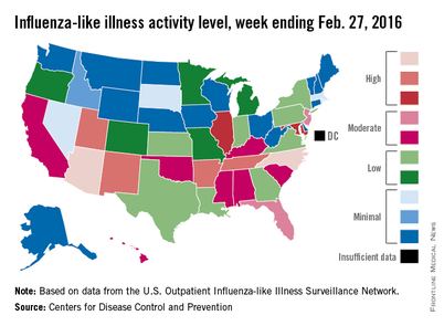
Despite that drop, two states were at level 10 on the CDC’s 1-10 scale of ILI activity for the first time this season. Arizona had already reached level 10, and joining it there last week was North Carolina, moving up from level 8 the week before. Other states in the “high” range of activity were Arkansas, New Mexico, Tennessee, and Utah at level 9, and Illinois and Maryland at level 8, the CDC reported March 4. Puerto Rico, which had been at level 10 for several weeks, moved down to level 8.
States in the “moderate” range of activity for the week ending Feb. 27 were Florida and New Jersey at level 7 and Alabama, California, Hawaii, Kentucky, Mississippi, Oklahoma, and South Carolina at level 6. Altogether, there were 35 states at level 2 or higher, according to data from the CDC’s Outpatient Influenza-like Illness Surveillance Network.
Four pediatric ILI-related deaths were reported to the CDC during week 20, but three actually occurred during week 19. There have been 18 ILI-related deaths so far during the 2015-2016 season, the CDC said.
Influenza-like illness (ILI) activity in the 2015-2016 U.S. flu season declined for the first time since early January, according to the Centers for Disease Control and Prevention.
The proportion of outpatient visits for ILI was reported at 3.2% for the week ending Feb. 20, but the CDC has adjusted that figure to 3.3%, which makes the 3.2% reported for this most recent week (week 20 of the season, ending Feb. 27, 2016) a decrease from the week before.
Despite that drop, two states were at level 10 on the CDC’s 1-10 scale of ILI activity for the first time this season. Arizona had already reached level 10, and joining it there last week was North Carolina, moving up from level 8 the week before. Other states in the “high” range of activity were Arkansas, New Mexico, Tennessee, and Utah at level 9, and Illinois and Maryland at level 8, the CDC reported March 4. Puerto Rico, which had been at level 10 for several weeks, moved down to level 8.
States in the “moderate” range of activity for the week ending Feb. 27 were Florida and New Jersey at level 7 and Alabama, California, Hawaii, Kentucky, Mississippi, Oklahoma, and South Carolina at level 6. Altogether, there were 35 states at level 2 or higher, according to data from the CDC’s Outpatient Influenza-like Illness Surveillance Network.
Four pediatric ILI-related deaths were reported to the CDC during week 20, but three actually occurred during week 19. There have been 18 ILI-related deaths so far during the 2015-2016 season, the CDC said.

Guidelines: Combine topical, oral therapy for most effective acne treatment
WASHINGTON – Monotherapy is not recommended in treating moderate-severe acne, and antibiotics should always be coupled with topical therapy, according to the latest guidelines from the American Academy of Dermatology.
And although it may be hard – even nearly impossible – to discontinue antibiotics completely, patients should be reevaluated every 3-4 months to determine whether reducing the dosage may be possible while maintaining effectiveness, the document says.
AAD published the guideline on Feb 17. At the academy’s annual meeting, a panel met to discuss its practical application.
Topical therapy
Benzoyl peroxide is a first-line agent that not only effectively fights Propionibacterium acnes, but also discourages the development of antibiotic resistance. Topical antibiotics also decrease P. acnes populations and exert a mild anti-inflammatory effect; however, monotherapy with a topical antibiotic is strongly discouraged. These should be used in combination with another agent such as a retinoid, benzoyl peroxide, adapalene, azelaic acid, or dapsone. This approach decreases the chance of antibiotic resistance, attacks the acne on several fronts, and provides for a maintenance transition.
Systemic antibiotics
Tetracycline-class antibiotics are still the best option for moderate-severe acne. A Cochrane review found that minocycline and doxycycline are equally effective (Cochrane Skin Group Nov 2011. doi: 10.1002/14651858.CD002086.pub2).
The incidence of adverse events associated with each is low, although minocycline may be marginally more troublesome. Low doses seem to be as effective as traditional doses, but pulsed therapy is inadequate. To prevent antibiotic resistance, limit both dose and length of therapy as much as possible. This can best be accomplished by adding a topical agent – either benzoyl peroxide or a retinoid – to the regimen.
“This is critical,” Dr. Jonette E. Keri of the University of Miami said at the meeting. “When antibiotics are eventually discontinued, the retinoid will fulfill the need for maintenance therapy.”
Hormonal agents
Four combination oral contraceptives are Food and Drug Administration–approved for acne treatment. Each of them decreases androgens by interrupting the pathway of testosterone production. There are no data suggesting that one is better than the other; patient preferences and their individual clinical picture should drive choice. Because of the cardiovascular risks associated with these combination OCs, they should not be prescribed for anyone with a personal or family history of clotting disorders or thromboembolic events. Smoking should also be a contraindication.
Oral contraceptives can be tried alone or as part of a comprehensive treatment regimen, including one containing antibiotics. Rifampin and griseofulvin are the only antibiotics known to decrease the contraceptive effect of the medications.
The tincture of time is an important part of this therapy, said Dr. Diane M. Thiboutot, professor of dermatology at Pennsylvania State University, Hershey. “You can’t rush it. It may take three cycles to see any real improvement in acne, and patients should be aware of this.”
Isotretinoin
Oral isotretinoin is a highly effective treatment for severe, recalcitrant acne. It decreases sebum production, acne lesion count, and scarring. Despite concerns about depression and suicidality, isotretinoin treatment can actually improve mood in most patients, said Dr. Megha M. Tollefson of the Mayo Clinic, Rochester, Minn.
“A very well-done Swedish study published in 2010 in BMJ found a slightly increased risk of suicide in the first 6 months after treatment started, but that risk was already rising before treatment started, so it could [be unrelated] to the drug,” she said. “And, in those who got isotretinoin, the [suicide] rate after that was actually decreased, compared to the general population.”
Female patients need education on isotretinoin’s teratogenic potential. After discussions, they should sign the SMART or iPLEDGE agreements about using effective birth control while taking the drug. Unfortunately, Dr. Tollefson said, “We continue to see hundreds of isotretinoin-exposed pregnancies each year.”
A recent study found that up to 30% of women did not comply with the birth control measures they agreed to while taking the drug (J Am Acad Dermatol. 2011 Oct. doi: 10.1016/j.jaad.2013.08.034).
The link between isotretinoin and inflammatory bowel disease is not well founded, Dr. Tollefson said. Studies have been contradictory, and most evidence is based on case report and association studies. There is, however, some evidence suggesting an innate connection between acne and inflammatory bowel disease, she noted.
Diet
Emerging evidence suggests that high glycemic diets may be associated with acne, but these studies are small. However, those randomized to a low glycemic index diet showed decreased sebum production and inflammation.
A small case-control study in 2012 suggested a link between milk and acne. AAD makes no recommendation based on this. Milk remains an important source of calcium and vitamin D for Americans, especially children, the panel said.
Dr. Tollefson had no financial disclosures. Dr. Keri said she has been a consultant for Hoffmann-LaRoche.
AT AAD 2016
FDA approves ibrutinib as first-line CLL therapy

Photo courtesy of Janssen
The US Food and Drug Administration (FDA) has approved the BTK inhibitor ibrutinib (Imbruvica) as a first-line treatment for patients with chronic lymphocytic leukemia (CLL).
This means ibrutinib is now FDA-approved to treat CLL patients regardless of their treatment history, including patients with 17p deletion.
Ibrutinib is also FDA-approved to treat Waldenström’s macroglobulinemia, and the drug was granted accelerated approval to treat patients with mantle cell lymphoma who have received at least 1 prior therapy.
Ibrutinib is jointly developed and commercialized by Pharmacyclics LLC, an AbbVie company, and Janssen Biotech, Inc. For more details on the drug, see the full prescribing information, available at imbruvica.com.
RESONATE-2 trial
The latest FDA approval for ibrutinib is based on results from the phase 3 RESONATE-2 trial (PCYC-1115), which were presented at the 2015 ASH Annual Meeting and simultaneously published in NEJM.
RESONATE-2 enrolled 269 treatment-naïve patients with CLL or small lymphocytic lymphoma who were 65 or older.
Patients were randomized to receive ibrutinib (n=136) at 420 mg once a day until progression or unacceptable toxicity, or chlorambucil (n=133) on days 1 and 15 of each 28-day cycle for up to 12 cycles. The starting dose for chlorambucil in cycle 1 was 0.5 mg/kg and was increased based on tolerability in cycle 2 by increments of 0.1 mg/kg to a maximum of 0.8 mg/kg.
The primary endpoint of the study was progression-free survival (PFS), as assessed by an independent review committee (IRC) according to the International Workshop on Chronic Lymphocytic Leukemia (iWCLL) 2008 criteria, with modification for treatment-related lymphocytosis.
Key secondary endpoints included overall response rate (based on the same iWCLL criteria), overall survival (OS), and safety.
Ibrutinib significantly prolonged PFS, as determined by the IRC, reducing the risk of progression or death by 84% compared to chlorambucil. The hazard ratio was 0.16 (P<0.001). The median PFS was not reached in the ibrutinib arm but was 18.9 months for the chlorambucil arm.
Ibrutinib significantly prolonged OS as well, although the median OS was not reached in either treatment arm. The OS rate at 24 months was 98% with ibrutinib and 85% with chlorambucil. The relative risk of death with ibrutinib was 84% lower than that with chlorambucil. The hazard ratio was 0.16 (P=0.001).
Ibrutinib was associated with a significantly higher IRC-assessed overall response rate compared to chlorambucil—82% and 35%, respectively (P<0.0001). Five patients (4%) in the ibrutinib arm achieved a complete response, as did 2 patients (2%) in the chlorambucil arm.
The median duration of treatment was 17.4 months in the ibrutinib arm and 7.1 months in the chlorambucil arm.
The most common adverse events of any grade—in the ibrutinib and chlorambucil arms, respectively—were diarrhea (42% and 17%), fatigue (30% and 38%), cough (22% and 15%), nausea (22% and 39%), peripheral edema (19% and 9%), dry eye (17% and 5%), arthralgia (16% and 7%), neutropenia (16% and 23%), and vomiting (13% and 20%).
Adverse events of grade 3 or higher—in the ibrutinib and chlorambucil arms, respectively—were neutropenia (10% and 18%), anemia (6% and 8%), hypertension (4% and 0%), pneumonia (4% and 2%), diarrhea (4% and 0%), maculopapular rash (3% and 2%), decreased platelet count (3% and 1%), abdominal pain (3% and 1%), hyponatremia (3% and 0%), thrombocytopenia (2% and 6%), febrile neutropenia (2% and 2%), upper respiratory tract infection (2% and 2%), pleural effusion (2% and 1%), cellulitis (2% and 0%), fatigue (1% and 5%), syncope (1% and 2%), and hemolytic anemia (0% and 2%). ![]()
CMS Introduces Billing Code for Hospitalists: What You Need to Know
The Centers for Medicare & Medicaid Services (CMS) recently announced the approval of a dedicated specialty billing code for hospitalists that will soon be ready for official use. This is a monumental step for hospital medicine, which continues to be the fastest growing medical specialty in the U.S., with more than 48,000 practitioners identifying as hospitalists.
The Hospitalist recently discussed the implications of this decision with Ron Greeno, MD, MHM, chief strategy officer for IPC Healthcare and chair of SHM’s Public Policy Committee (PPC), and Josh Boswell, director of government relations at SHM, to answer questions raised by SHM members.
Question: What are the benefits to hospitalists using the code?
Dr. Greeno: As we transition from fee-for-service to quality-based payment models, using this code will become critical to ensure hospitalists are reimbursed and evaluated fairly. Under the current code structure, hospitalists are missing opportunities to be rewarded and may be penalized unnecessarily because they are required to identify with internal medicine, family medicine, or another specialty that most closely resembles their daily practice. What current measures do not account for is that hospitalists’ patients are inherently more complex than those seen by practitioners in these other—most often outpatient—specialties. We as hospitalists face unique challenges and work with patients from all demographics, often with severe illnesses, making it nearly impossible to rely on benchmarks used for these other specialties.
There are a few prime examples of this that illustrate the need for the new code. Under the current system, some quality-based patient satisfaction measures under MACRA, on which hospitalists are being evaluated, pertain to the outpatient setting, including waiting room quality and office staff–irrelevant measurements for hospitalists. Hospitalists are also often incorrectly penalized under meaningful use due to complications brought on by observation status and its classification as an outpatient stay. This can cause both quality and cost measures to be extremely flawed and can misrepresent the performance and cost of hospitalists and hospital medicine groups. In the current billing structure, there is no way to accurately identify hospitalists and enable a definite fix to these problems.
To get what we want (fair measurement using relevant metrics), we must be able to identify as a separate group, and fortunately, now we can. There will be benefits we don’t even know about yet. We have to wait and see how healthcare policy continues to evolve and change moving forward. What we do know is that having this code will help us shape MACRA and future healthcare policy so that it works better for hospitalists as the specialty continues to grow in scope and impact.
Q: When will the new code go into effect?
Boswell: While there is not a set date at this time, CMS has reported that it can take up to a year, mostly due to technical changes that need to be made within their own systems. The code has already been officially approved; we just need to wait a bit longer to actually use it.
Q: What happens to hospitalists if they do not use the code?
Dr. Greeno: Some hospitalists might be nervous about the change after having billed a certain way for so long. While there is no absolute requirement for hospitalists to use the new code, the bottom line is that if hospitalists do not adopt the new code, they risk not receiving fair evaluations. Using this code should provide hospitalists with greater insight into their own performance—the data will be much more accurate and meaningful. This will allow hospitalists to hone in on areas needing improvement and provide them with more confidence that they are being compared using accurate benchmarks.
I want to stress that hospitalists, or in some cases their hospital medicine groups, will need to physically change their specialty affiliation when the code becomes effective. Otherwise, they risk not reaping the benefits associated with the new code and will continue to be evaluated using less-than-optimal benchmarks. The ball is in their court to make the change when the code is available, and SHM will serve as a resource to help ensure they know what to do and when.
Q: Where can someone go to find the code? Will it be available on the CMS website?
Boswell: When the code does become available for use, it will be communicated through various channels at SHM and also through the Medicare Learning Network, the site that houses education, information, and resources for healthcare professionals. It will also likely be distributed through additional Medicare circulars and newsletters.
As more details from CMS become available, we will have more specific information to share with members, including information on our website, webinars with billing and coding experts, email communication, and more. Continue to watch your email and social media channels for the latest updates and information.
Q: What role did SHM play in bringing this code to fruition?
Boswell: We can say with confidence that this effort was driven entirely by SHM. To start, a formal application needs to be filed in order for a code to even be considered. After determining that the benefits associated with this code far outweighed the costs and then receiving the support of our board of directors, SHM’s staff and PPC members collaborated to draft a brief and made the argument for the addition of a hospitalist billing code based on the individual elements CMS requires for consideration.
Due to the fact hospital medicine doesn’t have a board certification, while solid, our argument was far from a slam dunk. After submitting the application, SHM continuously followed up with and pressured CMS through various channels and utilized our grassroots network of hospitalists on the Hill to put this code on legislators’ radars—the result was pressure getting applied from interested members of Congress as well. If it weren’t for the persistent advocacy efforts of SHM and its members over the past several years, this code would not have even been considered, let alone approved.
This is a significant development—to our knowledge, this is the first medical specialty to be granted a code without also having a board certification. We’re thrilled that what we have been advocating for on behalf of our members is now a reality!
For the latest information on the new hospitalist billing code and other important healthcare policy updates, continue to check for SHM emails and follow SHM’s social media channels, including @SHMLive and @SHMAdvocacy on Twitter.
Sign up for the network to get the latest news in healthcare policy and discover opportunities to advocate for yourself and fellow hospitalists. TH
Brett Radler is SHM’s communications coordinator.
Vaccines committee approves recommended influenza strains for 2016-2017 vaccine
The Food and Drug Administration’s Vaccines and Related Biological Products Advisory Committee unanimously approved recommendations regarding the trivalent and quadrivalent influenza vaccines to be distributed during the 2016-2017 flu season.
The 14-member committee voted that the components of the trivalent influenza vaccine for the upcoming flu season should include an A/California/7/2009 (H1N1) pdm09-like virus; an A/Hong Kong/4801/2014 (H3N2)-like virus; and a B/Brisbane/60/2008-like virus of the B/Victoria lineage.

Additionally, the quadrivalent influenza vaccine should include a B/Phuket/3073/2013-like virus of the B/Yamagata lineage as “the second influenza B strain in the vaccine.”
All four components correspond with the recommendations of the World Health Organization, which announced its proposed components for influenza vaccines in the Northern Hemisphere on Feb. 25.
“I’m comfortable trying to follow the footprint of the virus we’ve seen today, quite elegantly put out in front of us,” said committee member Dr. Sarah Long, professor of pediatrics at Drexel University in Philadelphia, adding that she was “very pleased with what happened in the last year” regarding the predictions of dominant virus strains and the effectiveness of the eventual vaccine.
The proposed vaccine for next season differs from the one distributed during the 2015-16 flu season. While both vaccines contain the identical California strain of influenza A and the Phuket strain of influenza B, the 2015-2016 vaccine included an A/Switzerland/9715293/2013 (H3N2)-like virus in its trivalent form, and a B/Brisbane/60/2008-like virus of the B/Victoria lineage in its quadrivalent form.
“Timely vaccine supply requires close collaboration and communication between multiple stakeholders to ensure sufficient provision of [a] well-matched vaccine,” said Matthew Downham, Ph.D., associate director of biopharmaceutical development, AstraZeneca. Timely strain selection will ensure “vaccine availability and usage” for the most widespread and effective coverage.
While the FDA is not obligated to follow the recommendations of the Vaccines and Related Biological Products Advisory Committee, it generally does.
None of the committee members reported any relevant financial disclosures, nor were there any waivers for conflicts of interest.
The Food and Drug Administration’s Vaccines and Related Biological Products Advisory Committee unanimously approved recommendations regarding the trivalent and quadrivalent influenza vaccines to be distributed during the 2016-2017 flu season.
The 14-member committee voted that the components of the trivalent influenza vaccine for the upcoming flu season should include an A/California/7/2009 (H1N1) pdm09-like virus; an A/Hong Kong/4801/2014 (H3N2)-like virus; and a B/Brisbane/60/2008-like virus of the B/Victoria lineage.
Additionally, the quadrivalent influenza vaccine should include a B/Phuket/3073/2013-like virus of the B/Yamagata lineage as “the second influenza B strain in the vaccine.”
All four components correspond with the recommendations of the World Health Organization, which announced its proposed components for influenza vaccines in the Northern Hemisphere on Feb. 25.
“I’m comfortable trying to follow the footprint of the virus we’ve seen today, quite elegantly put out in front of us,” said committee member Dr. Sarah Long, professor of pediatrics at Drexel University in Philadelphia, adding that she was “very pleased with what happened in the last year” regarding the predictions of dominant virus strains and the effectiveness of the eventual vaccine.
The proposed vaccine for next season differs from the one distributed during the 2015-16 flu season. While both vaccines contain the identical California strain of influenza A and the Phuket strain of influenza B, the 2015-2016 vaccine included an A/Switzerland/9715293/2013 (H3N2)-like virus in its trivalent form, and a B/Brisbane/60/2008-like virus of the B/Victoria lineage in its quadrivalent form.
“Timely vaccine supply requires close collaboration and communication between multiple stakeholders to ensure sufficient provision of [a] well-matched vaccine,” said Matthew Downham, Ph.D., associate director of biopharmaceutical development, AstraZeneca. Timely strain selection will ensure “vaccine availability and usage” for the most widespread and effective coverage.
While the FDA is not obligated to follow the recommendations of the Vaccines and Related Biological Products Advisory Committee, it generally does.
None of the committee members reported any relevant financial disclosures, nor were there any waivers for conflicts of interest.
FROM AN FDA ADVISORY COMMITTEE MEETING

The Rheumatoid Arthritis Support Network
The Rheumatoid Arthritis Support Network (RASN) has the goal of providing up-to-date information and resources for rheumatoid arthritis patients so that they know their options and fully understand their diagnosis.
The RASN provides information on what causes RA, its symptoms, treatment options, positive lifestyles changes that people with RA can make, and resources, such as finding a rheumatologist, support groups, up-to-date literature, blogs about RA, and apps for pain management.
For more information, visit the RASN website (www.rheumatoidarthritis.org), send an email ([email protected]), or call 800-405-4043.
VIDEO: The New Gastroenterologist offers insights, lifestyle info for young specialists
PHILADELPHIA – Are there things you wish you’d learned as a gastroenterology fellow? How to build a reputation as a good speaker, how to grow a successful clinical practice, and even how to choose the best retirement fund options for your personal goals are the kinds of tips and insights you’ll find in The New Gastroenterologist.
The newest publication from the American Gastroenterological Association, The New Gastroenterologist offers practical clinical information, lifestyle features, interviews with leaders in the field, and details on where to find research funding.
“Our goal is to provide unique content that speaks to all the needs that young gastroenterologists have, and to have it all in one place,” says The New Gastroenterologist Editor-in-Chief Dr. Bryson Katona.
The video associated with this article is no longer available on this site. Please view all of our videos on the MDedge YouTube channel
On Twitter @whitneymcknight

Two new drugs
Two new drugs have arrived to challenge our prescription pad or electronic record, depending on which you use. Empagliflozin (Jardiance), a drug used to modify glucose metabolism in type 2 diabetes in patients with preexisting cardiovascular disease, demonstrated a decrease in cardiovascular mortality. The other drug, Entresto, appears to provide an added benefit in heart failure therapy and was compared with a standard ACE inhibitor.
The results of EMPA-REG OUTCOME, reported at the European Association for the Study of Diabetes meeting in Stockholm, showed empagliflozin to be the first drug to decrease the mortality and morbidity of cardiovascular disease in diabetes. It is one of a group of new sodium-glucose cotransporter 2 (SGLT-2) blockers being tested in type 2 diabetes with established cardiovascular disease. Patients were randomized to placebo or empagliflozin while receiving standard medical and cardiovascular medications. After 3 years of follow-up, patients receiving the drug experienced a lower cardiovascular mortality rate, compared with placebo patients (3.7% vs. 5.9%, a 38% reduction) in addition to a decrease in hospitalization for heart failure and death from any cause. No effect was observed on the incidence of myocardial infarction or stroke. In addition, the drug also lowered blood glucose and blood pressure and led to some significant weight loss. The drug also was shown to decrease vascular resistance and albuminuria (N Engl J Med. 2015. 373:2117-2).

Furthermore, an analysis of EMPA-REG OUTCOME presented in November at the American Society of Nephrology meeting in San Diego, showed a profound benefit on the new onset and progression of chronic renal disease in diabetes patients. The importance of these results needs emphasis. Up until recently, the Food and Drug Administration has given a pass to diabetes drugs in regard to cardiovascular endpoints; approval has been based on their primary effect on lowering blood glucose. Some drugs in the past, such as rosiglitazone, actually have shown an increase in mortality in some diabetes patients. At long last, FDA approval for diabetes drugs hinges on acceptable outcomes in cardiovascular endpoints. The addition of a drug that can actually affect cardiovascular mortality and morbidity, the major risk factor of diabetes, provides an important addition to therapy.
The other drug that provides a choice of drugs for the treatment of heart failure is Entresto, a combination of sacubitril, a neprilysin inhibitor, and the angiotensin receptor inhibitor valsartan, approved in July 2015. In PARADIGM-HF, the compound was compared to enalapril in the treatment of patients with class II, III, and IV heart failure who were also receiving beta-blockers. Entresto-treated patients reported a 21.8% incidence of the primary outcome measure, cardiovascular death and hospitalization for heart failure, compared with the enalapril alone incidence of 26.5% (P less than .001) (N Engl J Med. 2014;371:993-1004). Investigators initially excluded 11.4% of the recruited patients from the study who could not tolerate Entresto or enalapril therapy. The drugs were well tolerated without any adverse reactions during therapy. Entresto was more effective than enalapril in regards to the occurrence of heart failure and death from any cause over a 27-month average follow-up.
The observations in this study emphasize how much the mortality of heart failure has decreased over the last decade. Cardiovascular deaths have decreased to roughly 7% per year and rehospitalization occurs in about 8% in the first year. Both drugs provide an important incremental benefit in heart failure patients.
Dr. Goldstein, medical editor of Cardiology News, is professor of medicine at Wayne State University and division head emeritus of cardiovascular medicine at Henry Ford Hospital, both in Detroit. He is on data safety monitoring committees for the National Institutes of Health and several pharmaceutical companies.
Two new drugs have arrived to challenge our prescription pad or electronic record, depending on which you use. Empagliflozin (Jardiance), a drug used to modify glucose metabolism in type 2 diabetes in patients with preexisting cardiovascular disease, demonstrated a decrease in cardiovascular mortality. The other drug, Entresto, appears to provide an added benefit in heart failure therapy and was compared with a standard ACE inhibitor.
The results of EMPA-REG OUTCOME, reported at the European Association for the Study of Diabetes meeting in Stockholm, showed empagliflozin to be the first drug to decrease the mortality and morbidity of cardiovascular disease in diabetes. It is one of a group of new sodium-glucose cotransporter 2 (SGLT-2) blockers being tested in type 2 diabetes with established cardiovascular disease. Patients were randomized to placebo or empagliflozin while receiving standard medical and cardiovascular medications. After 3 years of follow-up, patients receiving the drug experienced a lower cardiovascular mortality rate, compared with placebo patients (3.7% vs. 5.9%, a 38% reduction) in addition to a decrease in hospitalization for heart failure and death from any cause. No effect was observed on the incidence of myocardial infarction or stroke. In addition, the drug also lowered blood glucose and blood pressure and led to some significant weight loss. The drug also was shown to decrease vascular resistance and albuminuria (N Engl J Med. 2015. 373:2117-2).
Furthermore, an analysis of EMPA-REG OUTCOME presented in November at the American Society of Nephrology meeting in San Diego, showed a profound benefit on the new onset and progression of chronic renal disease in diabetes patients. The importance of these results needs emphasis. Up until recently, the Food and Drug Administration has given a pass to diabetes drugs in regard to cardiovascular endpoints; approval has been based on their primary effect on lowering blood glucose. Some drugs in the past, such as rosiglitazone, actually have shown an increase in mortality in some diabetes patients. At long last, FDA approval for diabetes drugs hinges on acceptable outcomes in cardiovascular endpoints. The addition of a drug that can actually affect cardiovascular mortality and morbidity, the major risk factor of diabetes, provides an important addition to therapy.
The other drug that provides a choice of drugs for the treatment of heart failure is Entresto, a combination of sacubitril, a neprilysin inhibitor, and the angiotensin receptor inhibitor valsartan, approved in July 2015. In PARADIGM-HF, the compound was compared to enalapril in the treatment of patients with class II, III, and IV heart failure who were also receiving beta-blockers. Entresto-treated patients reported a 21.8% incidence of the primary outcome measure, cardiovascular death and hospitalization for heart failure, compared with the enalapril alone incidence of 26.5% (P less than .001) (N Engl J Med. 2014;371:993-1004). Investigators initially excluded 11.4% of the recruited patients from the study who could not tolerate Entresto or enalapril therapy. The drugs were well tolerated without any adverse reactions during therapy. Entresto was more effective than enalapril in regards to the occurrence of heart failure and death from any cause over a 27-month average follow-up.
The observations in this study emphasize how much the mortality of heart failure has decreased over the last decade. Cardiovascular deaths have decreased to roughly 7% per year and rehospitalization occurs in about 8% in the first year. Both drugs provide an important incremental benefit in heart failure patients.
Dr. Goldstein, medical editor of Cardiology News, is professor of medicine at Wayne State University and division head emeritus of cardiovascular medicine at Henry Ford Hospital, both in Detroit. He is on data safety monitoring committees for the National Institutes of Health and several pharmaceutical companies.

Is expectant management a safe alternative to immediate delivery in patients with PPROM close to term?
Preterm premature rupture of membranes (PPROM) refers to rupture of membranes prior to the onset of labor before 37 weeks’ gestation. It accounts for one-third of all preterm births.1 Pregnancy complications associated with PPROM include intrauterine infection (chorioamnionitis), preterm labor, and placental abruption. Should such complications develop, immediate delivery is indicated. When to recommend elective delivery in the absence of complications, however, remains controversial.
The American College of Obstetricians and Gynecologists (ACOG) currently recommends elective delivery at or after 34 weeks’ gestation,2 because the prevailing evidence suggests that the risk of pregnancy-related complications (especially ascending infection) exceeds the risks of iatrogenic prematurity at this gestational age. However, ACOG acknowledges that this recommendation is based on “limited and inconsistent scientific evidence.”2 To address deficiencies in the literature, investigators designed the PPROMT (preterm prelabor rupture of the membranes close to term) trial to study women with ruptured membranes before the onset of labor between 34 and 37 weeks’ gestation.
PPROMT study designMorris and colleagues present results of their multicenter, international, randomized controlled trial (RCT) of expectant management versus planned delivery in pregnancies complicated by PPROM at 34 0/7 through 36 6/7 weeks’ gestation carried out in 65 centers across 11 countries. A total of 1,839 women not requiring urgent delivery were randomly assigned to either immediate delivery (n = 924) or expectant management (n = 915).
No difference was noted in the primary outcome of neonatal sepsis between the immediate birth (n = 23 [2%]) and expectant management groups (n = 29 [3%]; relative risk [RR], 0.8; 95% confidence interval [CI], 0.5–1.3). This also was true in the subgroup of women who were colonized with group B streptococcus (RR, 0.9; 95% CI, 0.2–4.5).
There also was no difference in the secondary outcome measure, a composite metric including sepsis, ventilation for 24 or more hours, or death (73 [8%] in the immediate delivery group vs 61 [7%] in the expectant management group; RR, 1.2; 95% CI, 0.9–1.6). However, infants born to women randomly assigned to immediate delivery, versus expectant management, had a significantly higher rate of respiratory distress syndrome (RR, 1.6; 95% CI, 1.1–2.3) and mechanical ventilation (RR, 1.4; 95% CI, 1.0–1.8). In addition, the immediate-delivery infants had a longer median stay in the special care nursery/neonatal intensive care unit (4.0 days, interquartile range [IQR], 0.0–10.0 vs 2.0 days, IQR, 0.0–7.0) and total hospital stay (6.0 days, IQR, 3.0–10.0 vs 4.0 days, IQR, 3.0–8.0). As expected, women in the expectant management group had a significantly longer hospital stay than women in the immediate delivery group, because 75% (688/912) were managed as inpatients. Interestingly, women in the immediate delivery group had a higher cesarean delivery rate than those in the expectant management group (239 [26%] vs 169 [19%], respectively; RR, 1.4; 95% CI, 1.2–1.7), although no explanation was offered.
Strengths and limitationsMajor strengths of this study include the large sample size and superior study design. It is by far the largest RCT to address this question. Because this was a pragmatic RCT, certain practices (such as the choice of latency antibiotic regimen) varied across centers, although randomization would be expected to minimize the effect of such variables on study outcome.
A major limitation is that participant recruitment occurred over a period of more than 10 years, during which time antenatal and neonatal intensive care unit practices likely would have changed.
What this evidence means for practiceFew clinical studies have the potential to significantly change obstetric management. This report by Morris and colleagues is one such study. It was well designed, well executed, and powered to look at the most clinically relevant outcome, namely, neonatal sepsis. While these study results do call into question the current American College of Obstetricians and Gynecologists recommendations to electively deliver patients with PPROM at or after 34 weeks’ gestation, additional discussion is needed at the national level before these recommendations can be changed.
—Denis A. Vaughan, MBBCh, BAO, MRCPI, and Errol R. Norwitz, MD, PhD
Share your thoughts! Send your Letter to the Editor to [email protected]. Please include your name and the city and state in which you practice.
- Goldenberg RL, Rouse DJ. Prevention of premature birth. N Engl J Med. 1998;339(5):313–320.
- American College of Obstetricians and Gynecologists Committee on Practice Bulletins–Obstetrics. ACOG Practice Bulletin No. 160: premature rupture of membranes. Obstet Gynecol. 2016;127(1):192–194.
Preterm premature rupture of membranes (PPROM) refers to rupture of membranes prior to the onset of labor before 37 weeks’ gestation. It accounts for one-third of all preterm births.1 Pregnancy complications associated with PPROM include intrauterine infection (chorioamnionitis), preterm labor, and placental abruption. Should such complications develop, immediate delivery is indicated. When to recommend elective delivery in the absence of complications, however, remains controversial.
The American College of Obstetricians and Gynecologists (ACOG) currently recommends elective delivery at or after 34 weeks’ gestation,2 because the prevailing evidence suggests that the risk of pregnancy-related complications (especially ascending infection) exceeds the risks of iatrogenic prematurity at this gestational age. However, ACOG acknowledges that this recommendation is based on “limited and inconsistent scientific evidence.”2 To address deficiencies in the literature, investigators designed the PPROMT (preterm prelabor rupture of the membranes close to term) trial to study women with ruptured membranes before the onset of labor between 34 and 37 weeks’ gestation.
PPROMT study designMorris and colleagues present results of their multicenter, international, randomized controlled trial (RCT) of expectant management versus planned delivery in pregnancies complicated by PPROM at 34 0/7 through 36 6/7 weeks’ gestation carried out in 65 centers across 11 countries. A total of 1,839 women not requiring urgent delivery were randomly assigned to either immediate delivery (n = 924) or expectant management (n = 915).
No difference was noted in the primary outcome of neonatal sepsis between the immediate birth (n = 23 [2%]) and expectant management groups (n = 29 [3%]; relative risk [RR], 0.8; 95% confidence interval [CI], 0.5–1.3). This also was true in the subgroup of women who were colonized with group B streptococcus (RR, 0.9; 95% CI, 0.2–4.5).
There also was no difference in the secondary outcome measure, a composite metric including sepsis, ventilation for 24 or more hours, or death (73 [8%] in the immediate delivery group vs 61 [7%] in the expectant management group; RR, 1.2; 95% CI, 0.9–1.6). However, infants born to women randomly assigned to immediate delivery, versus expectant management, had a significantly higher rate of respiratory distress syndrome (RR, 1.6; 95% CI, 1.1–2.3) and mechanical ventilation (RR, 1.4; 95% CI, 1.0–1.8). In addition, the immediate-delivery infants had a longer median stay in the special care nursery/neonatal intensive care unit (4.0 days, interquartile range [IQR], 0.0–10.0 vs 2.0 days, IQR, 0.0–7.0) and total hospital stay (6.0 days, IQR, 3.0–10.0 vs 4.0 days, IQR, 3.0–8.0). As expected, women in the expectant management group had a significantly longer hospital stay than women in the immediate delivery group, because 75% (688/912) were managed as inpatients. Interestingly, women in the immediate delivery group had a higher cesarean delivery rate than those in the expectant management group (239 [26%] vs 169 [19%], respectively; RR, 1.4; 95% CI, 1.2–1.7), although no explanation was offered.
Strengths and limitationsMajor strengths of this study include the large sample size and superior study design. It is by far the largest RCT to address this question. Because this was a pragmatic RCT, certain practices (such as the choice of latency antibiotic regimen) varied across centers, although randomization would be expected to minimize the effect of such variables on study outcome.
A major limitation is that participant recruitment occurred over a period of more than 10 years, during which time antenatal and neonatal intensive care unit practices likely would have changed.
What this evidence means for practiceFew clinical studies have the potential to significantly change obstetric management. This report by Morris and colleagues is one such study. It was well designed, well executed, and powered to look at the most clinically relevant outcome, namely, neonatal sepsis. While these study results do call into question the current American College of Obstetricians and Gynecologists recommendations to electively deliver patients with PPROM at or after 34 weeks’ gestation, additional discussion is needed at the national level before these recommendations can be changed.
—Denis A. Vaughan, MBBCh, BAO, MRCPI, and Errol R. Norwitz, MD, PhD
Share your thoughts! Send your Letter to the Editor to [email protected]. Please include your name and the city and state in which you practice.
- Goldenberg RL, Rouse DJ. Prevention of premature birth. N Engl J Med. 1998;339(5):313–320.
- American College of Obstetricians and Gynecologists Committee on Practice Bulletins–Obstetrics. ACOG Practice Bulletin No. 160: premature rupture of membranes. Obstet Gynecol. 2016;127(1):192–194.
- Goldenberg RL, Rouse DJ. Prevention of premature birth. N Engl J Med. 1998;339(5):313–320.
- American College of Obstetricians and Gynecologists Committee on Practice Bulletins–Obstetrics. ACOG Practice Bulletin No. 160: premature rupture of membranes. Obstet Gynecol. 2016;127(1):192–194.