User login
Vaccination: An option not to be ignored

We have previously discussed in the Journal the recrudescence of pertussis in adults and the challenges to its diagnosis, which include the misperception that it is a rare disease. 1 Adult pertussis infection is usually due to the waning effect of childhood vaccination, and in adults it is more often an extreme annoyance than a life-threatening illness.
In this issue, Dr. Camille Sabella2 discusses measles, an infection thought to be all but eradicated in the United States by vaccination, predominantly using the live-attenuated measles virus contained in the measles-mumps-rubella (MMR) vaccine.
But measles outbreaks are seemingly on the rise, and because measles is extremely contagious, it poses a real risk to closed communities such as college dormitories, churches, and health care facilities. Measles infection can have significant adverse outcomes, particularly in immunosuppressed patients.
Although outbreaks have been attributed to virus imported from locations outside the United States, the spread of infection has been blamed on an increased number of unvaccinated children and adults. The reasons for decreased vaccination rates are many, and include parental fears that the vaccine will cause problems such as autism.
This autism link is a goblin that refuses to go away, despite strongly worded debunking by the US Centers for Disease Control and Prevention, the Institute of Medicine,1 and many peer-reviewed publications. The very recent retraction by the Lancet4—based on ethical and nondisclosure concerns—of the 1998 paper by Wakefield et al5 (which suggested a link in 12 children between MMR vaccine and chronic gastrointestinal problems and autism spectrum disorders) may further diffuse this concern. But I fear it will not.
So at present, we should reeducate ourselves on the clinical features, natural history, and potential complications of this eradicable disease, particularly if we treat patients who work in closed communities or have defects in cellular immunity.
- Sabella C. Pertussis: old foe, persistent problem. Cleve Clin J Med 2005; 72:601–608.
- Sabella C. Measles: not just a childhood rash. Cleve Clin J Med 2010; 77:207–213.
- Board on Population Health and Public Health Practice, Institute of Medicine of the National Academies. Immunization safety review: vaccines and autism. http://www.iom.edu/Reports/2004/Immunization-Safety-Review-Vaccines-and-Autism.aspx. Accessed February 11, 2010.
- The Editors of The Lancet. Retraction—ileal-lymphoid-nodular hyperplasia, non-specific colitis, and pervasive developmental disorder in children. Lancet (published online February 2, 2010) DOI:10.1016/S0140-6736(10)60175-4. Accessed February 11, 2010.
- Wakefield AJ, Murch SH, Anthony A, et al Ileal-lymphoid-nodular hyperplasia, non-specific colitis, and pervasive developmental disorder in children. Lancet 1998; 351:637–641.
We have previously discussed in the Journal the recrudescence of pertussis in adults and the challenges to its diagnosis, which include the misperception that it is a rare disease. 1 Adult pertussis infection is usually due to the waning effect of childhood vaccination, and in adults it is more often an extreme annoyance than a life-threatening illness.
In this issue, Dr. Camille Sabella2 discusses measles, an infection thought to be all but eradicated in the United States by vaccination, predominantly using the live-attenuated measles virus contained in the measles-mumps-rubella (MMR) vaccine.
But measles outbreaks are seemingly on the rise, and because measles is extremely contagious, it poses a real risk to closed communities such as college dormitories, churches, and health care facilities. Measles infection can have significant adverse outcomes, particularly in immunosuppressed patients.
Although outbreaks have been attributed to virus imported from locations outside the United States, the spread of infection has been blamed on an increased number of unvaccinated children and adults. The reasons for decreased vaccination rates are many, and include parental fears that the vaccine will cause problems such as autism.
This autism link is a goblin that refuses to go away, despite strongly worded debunking by the US Centers for Disease Control and Prevention, the Institute of Medicine,1 and many peer-reviewed publications. The very recent retraction by the Lancet4—based on ethical and nondisclosure concerns—of the 1998 paper by Wakefield et al5 (which suggested a link in 12 children between MMR vaccine and chronic gastrointestinal problems and autism spectrum disorders) may further diffuse this concern. But I fear it will not.
So at present, we should reeducate ourselves on the clinical features, natural history, and potential complications of this eradicable disease, particularly if we treat patients who work in closed communities or have defects in cellular immunity.
- Sabella C. Pertussis: old foe, persistent problem. Cleve Clin J Med 2005; 72:601–608.
- Sabella C. Measles: not just a childhood rash. Cleve Clin J Med 2010; 77:207–213.
- Board on Population Health and Public Health Practice, Institute of Medicine of the National Academies. Immunization safety review: vaccines and autism. http://www.iom.edu/Reports/2004/Immunization-Safety-Review-Vaccines-and-Autism.aspx. Accessed February 11, 2010.
- The Editors of The Lancet. Retraction—ileal-lymphoid-nodular hyperplasia, non-specific colitis, and pervasive developmental disorder in children. Lancet (published online February 2, 2010) DOI:10.1016/S0140-6736(10)60175-4. Accessed February 11, 2010.
- Wakefield AJ, Murch SH, Anthony A, et al Ileal-lymphoid-nodular hyperplasia, non-specific colitis, and pervasive developmental disorder in children. Lancet 1998; 351:637–641.
Measles: Not just a childhood rash
Although measles is generally considered a disease of children, it affects people of all ages. While the incidence of measles in the United States is significantly lower than in 1963, when an effective measles vaccine was first introduced, recent increases in the number of sporadic cases and community outbreaks in adults show that measles is still a significant health problem.
PATHOGENESIS OF MEASLES
Measles is a highly contagious viral infection, whose manifestations have been recognized since the 7th century. The measles virus is an RNA virus of the Paramyxoviridae family. It is very difficult to isolate from clinical specimens, requiring special cell lines for in vitro propagation.
After acquisition, the measles virus establishes localized infection of the respiratory epithelium and then spreads to the regional lymphatics. A primary viremia then occurs, in which the virus replicates at the site of inoculation and in the reticuloendothelial tissues. A secondary viremia follows, in which the virus infects and replicates in the skin, conjunctiva, respiratory tract, and other distant organs.
The measles rash is thought to be due to a hypersensitivity reaction.1 Cell-mediated responses are the main line of defense against measles, as evidenced by the fact that people with cell-mediated deficiencies develop severe measles infection.2 Immunity to wild-type measles is believed to be lifelong.3,4
MEASLES IS HIGHLY CONTAGIOUS
Measles is one of the most contagious infectious diseases, with a secondary attack rate of at least 90% in susceptible household contacts. 4 The fact that emergency departments and physicians’ offices have become sites of measles transmission in recent years underscores the transmissibility of the virus.5–7
Although the virus is very labile, it can remain infective in respiratory droplets from the air for many hours. Thus, measles virus spreads from person to person by direct contact with droplets from respiratory secretions of infected persons.
The period of maximal contagion is the late prodrome, ie, 2 to 4 days before the onset of the rash. People who are generally in good health are contagious through 4 days after the onset of the rash, whereas people with compromised immunity can continue to shed the virus for the entire duration of the illness.
Airborne transmission precautions are required for 4 days after the onset of the rash in hospitalized, non-immunocompromised patients with measles, and for the duration of the illness for immunocompromised patients.
In the absence of widespread measles vaccination, measles infection peaks in late winter and early spring.
EPIDEMIOLOGIC TRENDS: CAUSE FOR CONCERN
Since an effective vaccine became available in 1963, the annual incidence of measles cases in the United States has decreased by more than 99%. A significant resurgence from 1989 to 1991 affected mainly unvaccinated preschoolers and resulted in more than 55,000 cases and 130 deaths.8 This resurgence prompted widespread, intensive immunization efforts and the recommendation that a second dose of measles vaccine be given to school-aged children. This led to the effective elimination of endemic transmission of measles in the United States.9
Although 90% of these cases either were directly imported or were associated with importation from other countries,10 the reason for the large number of cases was clearly the greater transmission after importation of the virus into the United States. This transmission was the direct result of the fact that 91% of the cases occurred in unvaccinated people or people whose vaccination status was not known or was not documented. A high proportion— at least 61 (47%)—of the 131 measles cases in 2008 were in school-aged children whose parents chose not to have them vaccinated. Although no deaths were reported in these 131 patients, 15 required hospitalization.
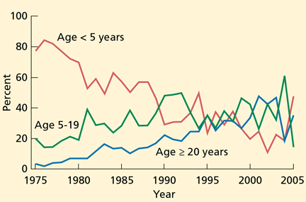
Although most reported measles cases are still in young and school-aged children, recent cases and outbreaks have also occurred in isolated communities of adults. Approximately 25% of the cases reported in 2008 were in people age 20 and older. Most adults who contracted measles had unknown or undocumented vaccination status. Similarly, a small measles outbreak occurred in Indiana in 2005, when an adolescent US citizen traveling in Europe became infected in Romania and exposed 500 people at a church gathering upon her return. Thirty-four cases of measles were reported from this exposure, and many were in adults.11
The recent increase in the number of cases reported and the continued reports of outbreaks highlight the fact that measles outbreaks can occur in communities with a high number of unvaccinated people, and underscore the need for high overall measles vaccination coverage to limit the spread of this infection.12
CLINICAL FEATURES OF MEASLES
The first sign of measles is a distinct prodrome, which occurs after an incubation period of 10 to 12 days. The prodrome is characterized by fever, malaise, anorexia, conjunctivitis, coryza, and cough and may resemble an upper respiratory tract infection; it lasts 2 to 4 days.
Towards the end of the prodrome, the body temperature can rise to as high as 40°C, and Koplik spots, pathognomonic for measles, appear. Koplik spots, bluish-gray specks on an erythematous base, usually appear on the buccal mucosa opposite the second molars 1 to 2 days before the onset of the rash, and last for 1 to 2 days after the onset of the rash. Thus, it is not unusual for Koplik spots to have disappeared at the time the diagnosis of measles is entertained.
The classic measles rash is an erythematous maculopapular eruption that begins on the head and face and spreads to involve the entire body. It usually persists for 4 to 5 days and is most confluent on the face and upper body. The rash fades in order of appearance, and may desquamate. People with measles appear ill, especially 1 to 2 days after the rash appears.
The entire course of measles usually lasts 7 to 10 days in patients with a healthy immune system. The cough, a manifestation of tracheobronchitis, is usually the last symptom to resolve. Patients are contagious 2 to 4 days before the onset of the rash, and remain so through 4 days after the onset of the rash.
COMPLICATIONS
Respiratory complications
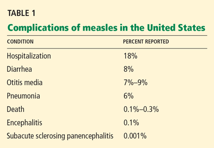
Pneumonia is responsible for 60% of deaths associated with measles.13 Although radiographic evidence of pneumonia is found in measles patients with no complications, symptomatic pneumonia occurs in 1% to 6% of patients. It is the result of either direct invasion by the virus or secondary bacterial infection,17 most often with Staphylococcus aureus and Streptococcus pneumoniae. Other respiratory complications include otitis media, sinusitis, and laryngotracheobronchitis.
Neurologic complications
Acute measles encephalitis is more common in adults than in children. Occuring in 1 in 1,000 to 2,000 patients,18 it is characterized by the resurgence of fever during the convalescent phase of the illness, along with headaches, seizures, and altered consciousness. These manifestations may be mild or severe, but they lead to permanent neurologic sequelae in a substantial proportion of affected patients. It is not clear whether acute measles encephalitis represents direct invasion of the virus or a postinfectious process from a hypersensitivity to the virus.19
Subacute sclerosing panencephalitis is a rare, chronic, degenerative central nervous system disease that occurs secondary to persistent infection with a defective measles virus.20 The prevalence is estimated at 1 per 100,000 cases. Signs and symptoms appear an average of 7 years after the initial infection and include personality changes, myoclonic seizures, and motor disturbances. Often, coma and death follow.
This condition occurs particularly in those who had measles at a very young age, ie, before the age of 2 years, and it occurs despite a vigorous host-immune response to the virus. Patients have high titers of measles-specific antibody in the sera and cerebrospinal fluid.
Other complications
Diarrhea and stomatitis account for much of the sickness and death from measles in developing countries.
Subclinical hepatitis occurs in at least 30% of adult measles patients.
Less common complications include thrombocytopenia, appendicitis, ileocolitis, pericarditis, myocarditis, and hypocalcemia.
MEASLES DURING PREGNANCY
Measles during pregnancy may be severe, mainly due to primary measles pneumonia.21 Measles is associated with a risk of miscarriage and prematurity, but congenital anomalies of the fetus have not been described, as they have for rubella infection.22
MEASLES IN COMPROMISED IMMUNITY
Measles patients with deficiencies of cellmediated immunity have a prolonged, severe, and often fatal course.2,23,24 This includes patients with:
- Human immunodeficiency virus (HIV) infection
- Congenital immunodeficiencies
- Disorders requiring chemotherapeutic and immunosuppressive therapy.
These patients are particularly susceptible to acute progressive encephalitis and measles pneumonitis. Case-fatality rates of 70% in cancer patients and 40% in HIV-infected patients have been reported.24
The diagnosis of measles may be difficult in patients without cell-mediated immunity, as 25% to 40% of them do not develop the characteristic rash.2,23 The absence of rash supports the theory that the rash is a hypersensitivity reaction to the virus.
MODIFIED AND ATYPICAL MEASLES
Modified measles
A modified form of measles can occur in people with some degree of passive immunity to the virus, including those previously vaccinated. It occurs mostly in patients who recently received immunoglobulin products, or in young infants who have residual maternal antibody. A modified measles illness can also follow vaccination with live-virus vaccine (see later discussion).
The clinical manifestations vary, and the illness may not have the classic features of prodrome, rash, and Koplik spots.
Atypical measles
Atypical measles is an unusual form that can occur when a person previously vaccinated with a killed-virus measles vaccine (used from 1963 to 1967) is exposed to wild-type measles.25 Features include a shorter prodrome (1 to 2 days), followed by appearance of a rash that begins on the distal extremities and spreads centripetally, usually sparing the neck, face, and head. The rash may be petechial, maculopapular, urticarial, vesicular, or a combination. The rash is accompanied by high fever and edema of the extremities. Complications such as pneumonia and hepatitis may occur.
The course of atypical measles is more prolonged than with classic measles, but because these patients are thought to have partial protection against the virus, they do not transmit it and are not considered contagious.26
DIAGNOSIS OF MEASLES
The classic clinical features are usually enough to distinguish measles from other febrile illnesses with similar clinical manifestions, such as rubella, dengue, parvovirus B19 infection, erythema multiforme, Stevens-Johnson syndrome, and streptococcal scarlet fever. The distinctive measles prodrome, Koplik spots, the progression of the rash from the head and neck to the trunk and the extremities, and the severity of disease are distinctive features of measles.
Laboratory tests to confirm the diagnosis are often used in areas where measles is rare, and laboratory confirmation is currently recommended in the United States. Because viral isolation is technically difficult and is not widely available, serologic testing is the method most commonly used. The measles-specific immunoglobulin M (IgM) antibody assay, the test used most often, is almost 100% sensitive when done 2 to 3 days after the onset of the rash.27,28 Measles IgM antibody peaks at 4 weeks after the infection and disappears by 6 to 8 weeks.
It is important to remember that false-positive measles IgM antibody may occur with other viral infections, such as parvovirus B19 and rubella. Because measles-specific IgG antibody is produced with the onset of infection and peaks at 4 weeks, a fourfold rise in the IgG titer is useful in confirming the diagnosis. Measles IgG antibody after infection is sustained for life.
Reverse transcription-polymerase chain reaction testing can also detect measles virus in the blood and urine when direct evidence of the virus is necessary, such as in immunocompromised patients.29
TREATMENT IS SUPPORTIVE
Treatment of measles mainly involves supportive measures, such as fluids and antipyretics. Antiviral agents such as ribavirin and interferon have in vitro activity against the measles virus and have been used to treat severe measles infection in immunocompromised patients. However, their clinical efficacy is unproven.30
Routine use of antibacterial agents to prevent secondary bacterial infection is not recommended.
CURRENT RECOMMENDATIONS FOR ACTIVE IMMUNIZATION
Active immunization for measles has been available since 1963. Between 1963 and 1967, both killed-virus and live-virus vaccines were available. As atypical measles cases became recognized, the killed-virus vaccine was withdrawn.
The vaccine currently available in the United States is a live-attenuated strain prepared in chicken embryo cell culture and combined with mumps and rubella vaccine (MMR) or mumps, rubella, and varicella vaccine (MMRV).
Two doses of live-virus measles vaccine are recommended for all healthy children before they begin school, with the first dose given at 12 to 15 months of age. A second dose is needed because the failure rate with one dose is 5%. More than 99% of people who receive two doses separated by 4 weeks develop serologic evidence of measles.
Waning immunity after vaccination occurs very rarely, with approximately 5% of children developing secondary vaccine failure 10 to 15 years after vaccination.3,31
Although rates of vaccination in the United States are high, cases of measles continue to occur in unvaccinated infants and in children who are either too young to be vaccinated or whose parents claimed exemption because of religious or personal beliefs.
Because of the occurrence of measles cases in adolescents, young adults, and adults, potentially susceptible people should be identified and vaccinated according to current guidelines. People should be considered susceptible unless they have documentation of at least two doses of measles vaccine given at least 28 days apart, physician-diagnosed measles, laboratory evidence of immunity to measles, or were born before 1957. All adults who are susceptible should receive at least one dose of measles vaccine.10 Adults at higher risk of contracting measles include:
- Students in high school and college
- International travelers
- Health care personnel.
For these adults, two doses of measles vaccine, at least 28 days apart, are recommended.32
Postexposure prophylaxis
Measles vaccination given to susceptible contacts within 72 hours of exposure as postexposure prophylaxis may protect against infection and induces protection against subsequent exposures to measles.33,34 Vaccination is the intervention of choice for susceptible individuals older than 12 months of age who are exposed to measles and who do not have a contraindication to measles vaccination.35 Active rather than passive immunization is also the strategy of choice for controlling measles outbreaks.
Passive immunization with intramuscular immune globulin within 6 days of exposure can be used in selected circumstances to prevent transmission or to modify the clinical course of the infection.36 Immune globulin therapy is recommended for susceptible individuals who are exposed to measles and who are at high risk of developing severe or fatal measles. This includes individuals who are being treated with immunosuppressive agents, those with HIV infection, pregnant women, and infants less than 1 year of age. Immune globulin should not be used to control measles outbreaks.
ADVERSE EFFECTS OF MEASLES VACCINE
Live-virus measles vaccine has an excellent safety record. A transient fever, which may be accompanied by a measles-like rash, occurs in 5% to 15% of people 5 to 12 days after vaccination. The rash may be discrete or confluent and is self-limited.
Although measles vaccine is a live-attenuated vaccine, vaccinated people do not transmit the virus to susceptible contacts and are not considered contagious, even if they develop a vaccine-associated rash. Thus, the vaccine can be safely given to close contacts of immunocompromised and other susceptible people. Encephalitis is exceedingly rare following vaccination.
There is no scientific evidence that the risk of autism is higher in children who receive measles or MMR vaccine than in unvaccinated children.37 An Institute of Medicine report in 2001 rejected a causal relationship between MMR vaccine and autism spectrum disorders.38
CONTRAINDICATIONS TO MEASLES VACCINATION
Measles vaccine is contraindicated for:
- People who have cell-mediated immune deficiencies (except patients wtih HIV infection—see discussion just below)
- Pregnant women
- Those who had a severe allergic reaction to a vaccine component after a previous dose
- Those with moderate or severe acute illness
- Those who have recently received immune globulin products.
HIV-infected patients with severe immunosuppression should not receive the liveattenuated measles vaccine. However, because patients with HIV are at risk of severe measles, and because the vaccine has been shown to be safe in HIV patients who do not have severe immunosuppression, the vaccine is recommended for those with asymptomatic or mildly symptomatic HIV infection who do not have evidence of severe immunosuppression. 39
After receiving immune globulin
Anyone who has recently received immune globulin should not receive measles vaccine until sufficient time has passed, since passively acquired antibodies interfere with the immune response to live-virus vaccines. How long to wait depends on the type of immune globulin, the indication, the amount, and the route of administration. In general, the waiting period is:
- At least 3 months after intramuscular immune globulin or tetanus, hepatitis A, or hepatitis B prophylaxis
- At least 4 months after intramuscular immune globulin for rabies, or 6 months after intravenous immune globulin for cytomegalovirus (dose, 150 mg/kg)
- At least 8 months after intravenous immune globulin as replacement or therapy for immune deficiencies (dose, 400 mg/kg), or after intravenous immune globulin for immune thrombocytopenic purpura (400 mg/kg)
- At least 10 months after intravenous immune globulin for immune thrombocytopenic pupura at a dose of 1 g/kg.39
Egg allergy is not a contraindication
Although measles vaccine is produced in chick embryo cell culture, the vaccine has been shown to be safe in people with egg allergy, so they may be vaccinated without first being tested for egg allergy.39,40
- Lachmann PJ. Immunopathology of measles. Proc R Soc Med 1974; 67:1120–1122.
- Enders JF, McCarthy K, Mitus A, Cheatham WJ. Isolation of measles virus at autopsy in case of giant cell pneumonia without rash. N Engl J Med 1959; 261:875–881.
- Markowitz LE, Preblud SR, Fine PE, Orenstein WA. Duration of live measles vaccine-induced immunity. Pediatr Infect Dis J 1990; 9:101–110.
- Stokes J, Reilly CM, Buynak EB, Hilleman MR. Immunologic studies of measles. Am J Hyg 1961; 74:293–303.
- Farizo KM, Stehr-Green PA, Simpson DM, Markowitz LE. Pediatric emergency room visits: a risk factor for acquiring measles. Pediatrics 1991; 87:74–79.
- Bloch AB, Orenstein W, Ewing WM, et al. Measles outbreak in a pediatric practice: airborne transmission in an office setting. Pediatrics 1985; 75:676–683.
- Remington PL, Hall WN, Davis IH, et al. Airborne transmission of measles in a physician’s office. JAMA 1985; 253:1574–1577.
- US Centers for Disease Control and Prevention. Reported vaccine-preventable diseases—United States, 1993, and the Childhood Immunization Initiative. MMWR 1994; 43:57–60.
- Orenstein WA, Papania MJ, Wharton ME. Measles elimination in the United States. J Infect Dis 2004; 189 (suppl 1):S1–S3.
- US Centers for Disease Control and Prevention. Update: Measles—United States, January–July 2008. MMWR 2008; 57:893–896.
- Parker AA, Staggs W, Dayan GH, et al. Implications of a 2005 measles outbreak in Indiana for sustained elimination of measles in the United States. N Engl J Med 2006; 355:447–455.
- Mulholland EK. Measles in the United States, 2006. N Engl J Med 2006; 355:440–443.
- Barkin RM. Measles mortality. Analysis of the primary cause of death. Am J Dis Child 1975; 129:307–309.
- Barkin RM. Measles mortality: a retrospective look at the vaccine era. Am J Epidemiol 1975; 102:341–349.
- US Centers for Disease Control and Prevention. Public-sector vaccination efforts in response to the resurgence of measles among preschool-aged children—United States, 1989–1991. MMWR 1992; 41:522–525.
- Gremillion DH, Crawford GE. Measles pneumonia in young adults. an analysis of 106 cases. Am J Med 1981; 71:539–542.
- Quiambao BP, Gatchalian SR, Halonen P, et al. Coinfection is common in measles-associated pneumonia. Pediatr Infect Dis J 1998; 17:89–93.
- Perry RT, Halsey NA. The clinical significance of measles: a review. J Infect Dis 2004; 189( suppl 1):S4–S16.
- Johnson RT, Griffin DE, Hirsch RL, et al. Measles encephalomyelitis—clinical and immunologic studies. N Engl J Med 1984; 310:137–141.
- Sever JL. Persistent measles infection of the central nervous system: subacute sclerosing panencephalitis. Rev Infect Dis 1983; 5:467–473.
- Atmar RL, Englund JA, Hammill H. Complications of measles during pregnancy. Clin Infect Dis 1992; 14:217–226.
- Gershon AA. Chickenpox, measles, and mumps. In:Remington J, Klein J, eds. Infectious Diseases of the Fetus and Newborn Infant. Elsevier Saunders: Philadelphia, 2006:693–738.
- Mitus A, Enders JF, Craig JM, Holloway A. Persistence of measles virus and depression of antibody formation in patients with giant-cell pneumonia after measles. N Engl J Med 1959; 261:882–889.
- Kaplan LJ, Daum RS, Smaron M, McCarthy CA. Severe measles in immunocompromised patients JAMA 1992; 267:1237–1241.
- Frey HM, Krugman S. Atypical measles syndrome: unusual hepatic, pulmonary, and immunologic aspects. Am J Med Sci 1981; 281:51–55.
- Fulginiti VA, Eller JJ, Downie AW, Kempe CH. Altered reactivity to measles virus. Atypical measles in children previously immunized with inactivated measles virus vaccines. JAMA 1967; 202:1075–1080.
- Mayo DR, Brennan T, Cormier DP, Hadler J, Lamb P. Evaluation of a commercial measles virus immunoglobulin M enzyme immunoassay. J Clin Microbiol 1991; 29:2865–2867.
- Bellini WJ, Helfand RF. The challenges and strategies for laboratory diagnosis of measles in an international setting. J Infect Dis 2003; 187( suppl 1):S283–S290.
- Riddell MA, Chibo D, Kelly HA, Catton MG, Birch CJ. Investigation of optimal specimen type and sampling time for detection of measles virus RNA during a measles epidemic. J Clin Microbiol 2001; 39:375–376.
- Forni Al, Schluger NW, Roberts RB. Severe measles pneumonitis in adults: evaluation of clinical characteristics and therapy with intravenous ribavirin. Clin Infect Dis 1994; 19:454–462.
- Anders JF, Jacobsen RM, Poland GA, Jacobsen SJ, Wollan PC. Secondary failure rates of measles vaccines: a metaanalysis of published studes. Pediar Infect Dis J 1996; 15:62–66.
- US Centers for Disease Control and Prevention. Measles—United States, January 1–April 25, 2008. MMWR 2008; 57:494–498.
- Berkovitz S, Starr S. Use of live-measles-virus vaccine to abort an expected outbreak of measles within a closed population. N Engl J Med 1963; 269:75–77.
- Ruuskanen O, Salmi TT, Halonen P. Measles vaccination after exposure to natural measles. J Pediatr 1978; 93:43–46.
- Strebel PM, Papania MJ, Halsey NA. Measles vaccine. In:Plotkin SA, Orenstein WA, eds. Vaccines. Saunders: New York, 2004:389–440.
- US Centers for Disease Control and Prevention. Measles, mumps and rubella—vaccine use and strategies for elimination of measles, rubella, and congenital rubella syndrome and control of mumps: Recommendations of the Advisory Committee on Immunization Practices (ACIP). MMWR 1998; 47( RR-8):1–57. http://www.cdc.gov/mmwr/PDF/rr/rr4708.pdf. Accessed December 30, 2009.
- Madsen KM, Hviid A, Vestergaard M, et al. A population-based study of measles, mumps, and rubella vaccination and autism. N Engl J Med 2002; 347:1477–1482.
- Straton K, Gable A, Shetty P, McCormick M; for the Institute of Medicine. Immunization Safety Review: Measles-Mumps-Rubella Vaccine and Autism. National Academy Press: Washington, DC, 2001.
- Pickerington LK, Baker CJ, Long SS, McMillan JA, editors. Red Book: 2009 Report of the Committee on Infectious Diseases. 28th ed. Elk Grove Village, IL: American Academy of Pediatrics, 2009:444–455.
- James JM, Burks AW, Roberson PK, Sampson HA. Safe administration of the measles vaccine to children allergic to eggs. N Engl J Med 1995; 332:1262–1266.
Although measles is generally considered a disease of children, it affects people of all ages. While the incidence of measles in the United States is significantly lower than in 1963, when an effective measles vaccine was first introduced, recent increases in the number of sporadic cases and community outbreaks in adults show that measles is still a significant health problem.
PATHOGENESIS OF MEASLES
Measles is a highly contagious viral infection, whose manifestations have been recognized since the 7th century. The measles virus is an RNA virus of the Paramyxoviridae family. It is very difficult to isolate from clinical specimens, requiring special cell lines for in vitro propagation.
After acquisition, the measles virus establishes localized infection of the respiratory epithelium and then spreads to the regional lymphatics. A primary viremia then occurs, in which the virus replicates at the site of inoculation and in the reticuloendothelial tissues. A secondary viremia follows, in which the virus infects and replicates in the skin, conjunctiva, respiratory tract, and other distant organs.
The measles rash is thought to be due to a hypersensitivity reaction.1 Cell-mediated responses are the main line of defense against measles, as evidenced by the fact that people with cell-mediated deficiencies develop severe measles infection.2 Immunity to wild-type measles is believed to be lifelong.3,4
MEASLES IS HIGHLY CONTAGIOUS
Measles is one of the most contagious infectious diseases, with a secondary attack rate of at least 90% in susceptible household contacts. 4 The fact that emergency departments and physicians’ offices have become sites of measles transmission in recent years underscores the transmissibility of the virus.5–7
Although the virus is very labile, it can remain infective in respiratory droplets from the air for many hours. Thus, measles virus spreads from person to person by direct contact with droplets from respiratory secretions of infected persons.
The period of maximal contagion is the late prodrome, ie, 2 to 4 days before the onset of the rash. People who are generally in good health are contagious through 4 days after the onset of the rash, whereas people with compromised immunity can continue to shed the virus for the entire duration of the illness.
Airborne transmission precautions are required for 4 days after the onset of the rash in hospitalized, non-immunocompromised patients with measles, and for the duration of the illness for immunocompromised patients.
In the absence of widespread measles vaccination, measles infection peaks in late winter and early spring.
EPIDEMIOLOGIC TRENDS: CAUSE FOR CONCERN
Since an effective vaccine became available in 1963, the annual incidence of measles cases in the United States has decreased by more than 99%. A significant resurgence from 1989 to 1991 affected mainly unvaccinated preschoolers and resulted in more than 55,000 cases and 130 deaths.8 This resurgence prompted widespread, intensive immunization efforts and the recommendation that a second dose of measles vaccine be given to school-aged children. This led to the effective elimination of endemic transmission of measles in the United States.9
Although 90% of these cases either were directly imported or were associated with importation from other countries,10 the reason for the large number of cases was clearly the greater transmission after importation of the virus into the United States. This transmission was the direct result of the fact that 91% of the cases occurred in unvaccinated people or people whose vaccination status was not known or was not documented. A high proportion— at least 61 (47%)—of the 131 measles cases in 2008 were in school-aged children whose parents chose not to have them vaccinated. Although no deaths were reported in these 131 patients, 15 required hospitalization.
Although most reported measles cases are still in young and school-aged children, recent cases and outbreaks have also occurred in isolated communities of adults. Approximately 25% of the cases reported in 2008 were in people age 20 and older. Most adults who contracted measles had unknown or undocumented vaccination status. Similarly, a small measles outbreak occurred in Indiana in 2005, when an adolescent US citizen traveling in Europe became infected in Romania and exposed 500 people at a church gathering upon her return. Thirty-four cases of measles were reported from this exposure, and many were in adults.11
The recent increase in the number of cases reported and the continued reports of outbreaks highlight the fact that measles outbreaks can occur in communities with a high number of unvaccinated people, and underscore the need for high overall measles vaccination coverage to limit the spread of this infection.12
CLINICAL FEATURES OF MEASLES
The first sign of measles is a distinct prodrome, which occurs after an incubation period of 10 to 12 days. The prodrome is characterized by fever, malaise, anorexia, conjunctivitis, coryza, and cough and may resemble an upper respiratory tract infection; it lasts 2 to 4 days.
Towards the end of the prodrome, the body temperature can rise to as high as 40°C, and Koplik spots, pathognomonic for measles, appear. Koplik spots, bluish-gray specks on an erythematous base, usually appear on the buccal mucosa opposite the second molars 1 to 2 days before the onset of the rash, and last for 1 to 2 days after the onset of the rash. Thus, it is not unusual for Koplik spots to have disappeared at the time the diagnosis of measles is entertained.
The classic measles rash is an erythematous maculopapular eruption that begins on the head and face and spreads to involve the entire body. It usually persists for 4 to 5 days and is most confluent on the face and upper body. The rash fades in order of appearance, and may desquamate. People with measles appear ill, especially 1 to 2 days after the rash appears.
The entire course of measles usually lasts 7 to 10 days in patients with a healthy immune system. The cough, a manifestation of tracheobronchitis, is usually the last symptom to resolve. Patients are contagious 2 to 4 days before the onset of the rash, and remain so through 4 days after the onset of the rash.
COMPLICATIONS
Respiratory complications
Pneumonia is responsible for 60% of deaths associated with measles.13 Although radiographic evidence of pneumonia is found in measles patients with no complications, symptomatic pneumonia occurs in 1% to 6% of patients. It is the result of either direct invasion by the virus or secondary bacterial infection,17 most often with Staphylococcus aureus and Streptococcus pneumoniae. Other respiratory complications include otitis media, sinusitis, and laryngotracheobronchitis.
Neurologic complications
Acute measles encephalitis is more common in adults than in children. Occuring in 1 in 1,000 to 2,000 patients,18 it is characterized by the resurgence of fever during the convalescent phase of the illness, along with headaches, seizures, and altered consciousness. These manifestations may be mild or severe, but they lead to permanent neurologic sequelae in a substantial proportion of affected patients. It is not clear whether acute measles encephalitis represents direct invasion of the virus or a postinfectious process from a hypersensitivity to the virus.19
Subacute sclerosing panencephalitis is a rare, chronic, degenerative central nervous system disease that occurs secondary to persistent infection with a defective measles virus.20 The prevalence is estimated at 1 per 100,000 cases. Signs and symptoms appear an average of 7 years after the initial infection and include personality changes, myoclonic seizures, and motor disturbances. Often, coma and death follow.
This condition occurs particularly in those who had measles at a very young age, ie, before the age of 2 years, and it occurs despite a vigorous host-immune response to the virus. Patients have high titers of measles-specific antibody in the sera and cerebrospinal fluid.
Other complications
Diarrhea and stomatitis account for much of the sickness and death from measles in developing countries.
Subclinical hepatitis occurs in at least 30% of adult measles patients.
Less common complications include thrombocytopenia, appendicitis, ileocolitis, pericarditis, myocarditis, and hypocalcemia.
MEASLES DURING PREGNANCY
Measles during pregnancy may be severe, mainly due to primary measles pneumonia.21 Measles is associated with a risk of miscarriage and prematurity, but congenital anomalies of the fetus have not been described, as they have for rubella infection.22
MEASLES IN COMPROMISED IMMUNITY
Measles patients with deficiencies of cellmediated immunity have a prolonged, severe, and often fatal course.2,23,24 This includes patients with:
- Human immunodeficiency virus (HIV) infection
- Congenital immunodeficiencies
- Disorders requiring chemotherapeutic and immunosuppressive therapy.
These patients are particularly susceptible to acute progressive encephalitis and measles pneumonitis. Case-fatality rates of 70% in cancer patients and 40% in HIV-infected patients have been reported.24
The diagnosis of measles may be difficult in patients without cell-mediated immunity, as 25% to 40% of them do not develop the characteristic rash.2,23 The absence of rash supports the theory that the rash is a hypersensitivity reaction to the virus.
MODIFIED AND ATYPICAL MEASLES
Modified measles
A modified form of measles can occur in people with some degree of passive immunity to the virus, including those previously vaccinated. It occurs mostly in patients who recently received immunoglobulin products, or in young infants who have residual maternal antibody. A modified measles illness can also follow vaccination with live-virus vaccine (see later discussion).
The clinical manifestations vary, and the illness may not have the classic features of prodrome, rash, and Koplik spots.
Atypical measles
Atypical measles is an unusual form that can occur when a person previously vaccinated with a killed-virus measles vaccine (used from 1963 to 1967) is exposed to wild-type measles.25 Features include a shorter prodrome (1 to 2 days), followed by appearance of a rash that begins on the distal extremities and spreads centripetally, usually sparing the neck, face, and head. The rash may be petechial, maculopapular, urticarial, vesicular, or a combination. The rash is accompanied by high fever and edema of the extremities. Complications such as pneumonia and hepatitis may occur.
The course of atypical measles is more prolonged than with classic measles, but because these patients are thought to have partial protection against the virus, they do not transmit it and are not considered contagious.26
DIAGNOSIS OF MEASLES
The classic clinical features are usually enough to distinguish measles from other febrile illnesses with similar clinical manifestions, such as rubella, dengue, parvovirus B19 infection, erythema multiforme, Stevens-Johnson syndrome, and streptococcal scarlet fever. The distinctive measles prodrome, Koplik spots, the progression of the rash from the head and neck to the trunk and the extremities, and the severity of disease are distinctive features of measles.
Laboratory tests to confirm the diagnosis are often used in areas where measles is rare, and laboratory confirmation is currently recommended in the United States. Because viral isolation is technically difficult and is not widely available, serologic testing is the method most commonly used. The measles-specific immunoglobulin M (IgM) antibody assay, the test used most often, is almost 100% sensitive when done 2 to 3 days after the onset of the rash.27,28 Measles IgM antibody peaks at 4 weeks after the infection and disappears by 6 to 8 weeks.
It is important to remember that false-positive measles IgM antibody may occur with other viral infections, such as parvovirus B19 and rubella. Because measles-specific IgG antibody is produced with the onset of infection and peaks at 4 weeks, a fourfold rise in the IgG titer is useful in confirming the diagnosis. Measles IgG antibody after infection is sustained for life.
Reverse transcription-polymerase chain reaction testing can also detect measles virus in the blood and urine when direct evidence of the virus is necessary, such as in immunocompromised patients.29
TREATMENT IS SUPPORTIVE
Treatment of measles mainly involves supportive measures, such as fluids and antipyretics. Antiviral agents such as ribavirin and interferon have in vitro activity against the measles virus and have been used to treat severe measles infection in immunocompromised patients. However, their clinical efficacy is unproven.30
Routine use of antibacterial agents to prevent secondary bacterial infection is not recommended.
CURRENT RECOMMENDATIONS FOR ACTIVE IMMUNIZATION
Active immunization for measles has been available since 1963. Between 1963 and 1967, both killed-virus and live-virus vaccines were available. As atypical measles cases became recognized, the killed-virus vaccine was withdrawn.
The vaccine currently available in the United States is a live-attenuated strain prepared in chicken embryo cell culture and combined with mumps and rubella vaccine (MMR) or mumps, rubella, and varicella vaccine (MMRV).
Two doses of live-virus measles vaccine are recommended for all healthy children before they begin school, with the first dose given at 12 to 15 months of age. A second dose is needed because the failure rate with one dose is 5%. More than 99% of people who receive two doses separated by 4 weeks develop serologic evidence of measles.
Waning immunity after vaccination occurs very rarely, with approximately 5% of children developing secondary vaccine failure 10 to 15 years after vaccination.3,31
Although rates of vaccination in the United States are high, cases of measles continue to occur in unvaccinated infants and in children who are either too young to be vaccinated or whose parents claimed exemption because of religious or personal beliefs.
Because of the occurrence of measles cases in adolescents, young adults, and adults, potentially susceptible people should be identified and vaccinated according to current guidelines. People should be considered susceptible unless they have documentation of at least two doses of measles vaccine given at least 28 days apart, physician-diagnosed measles, laboratory evidence of immunity to measles, or were born before 1957. All adults who are susceptible should receive at least one dose of measles vaccine.10 Adults at higher risk of contracting measles include:
- Students in high school and college
- International travelers
- Health care personnel.
For these adults, two doses of measles vaccine, at least 28 days apart, are recommended.32
Postexposure prophylaxis
Measles vaccination given to susceptible contacts within 72 hours of exposure as postexposure prophylaxis may protect against infection and induces protection against subsequent exposures to measles.33,34 Vaccination is the intervention of choice for susceptible individuals older than 12 months of age who are exposed to measles and who do not have a contraindication to measles vaccination.35 Active rather than passive immunization is also the strategy of choice for controlling measles outbreaks.
Passive immunization with intramuscular immune globulin within 6 days of exposure can be used in selected circumstances to prevent transmission or to modify the clinical course of the infection.36 Immune globulin therapy is recommended for susceptible individuals who are exposed to measles and who are at high risk of developing severe or fatal measles. This includes individuals who are being treated with immunosuppressive agents, those with HIV infection, pregnant women, and infants less than 1 year of age. Immune globulin should not be used to control measles outbreaks.
ADVERSE EFFECTS OF MEASLES VACCINE
Live-virus measles vaccine has an excellent safety record. A transient fever, which may be accompanied by a measles-like rash, occurs in 5% to 15% of people 5 to 12 days after vaccination. The rash may be discrete or confluent and is self-limited.
Although measles vaccine is a live-attenuated vaccine, vaccinated people do not transmit the virus to susceptible contacts and are not considered contagious, even if they develop a vaccine-associated rash. Thus, the vaccine can be safely given to close contacts of immunocompromised and other susceptible people. Encephalitis is exceedingly rare following vaccination.
There is no scientific evidence that the risk of autism is higher in children who receive measles or MMR vaccine than in unvaccinated children.37 An Institute of Medicine report in 2001 rejected a causal relationship between MMR vaccine and autism spectrum disorders.38
CONTRAINDICATIONS TO MEASLES VACCINATION
Measles vaccine is contraindicated for:
- People who have cell-mediated immune deficiencies (except patients wtih HIV infection—see discussion just below)
- Pregnant women
- Those who had a severe allergic reaction to a vaccine component after a previous dose
- Those with moderate or severe acute illness
- Those who have recently received immune globulin products.
HIV-infected patients with severe immunosuppression should not receive the liveattenuated measles vaccine. However, because patients with HIV are at risk of severe measles, and because the vaccine has been shown to be safe in HIV patients who do not have severe immunosuppression, the vaccine is recommended for those with asymptomatic or mildly symptomatic HIV infection who do not have evidence of severe immunosuppression. 39
After receiving immune globulin
Anyone who has recently received immune globulin should not receive measles vaccine until sufficient time has passed, since passively acquired antibodies interfere with the immune response to live-virus vaccines. How long to wait depends on the type of immune globulin, the indication, the amount, and the route of administration. In general, the waiting period is:
- At least 3 months after intramuscular immune globulin or tetanus, hepatitis A, or hepatitis B prophylaxis
- At least 4 months after intramuscular immune globulin for rabies, or 6 months after intravenous immune globulin for cytomegalovirus (dose, 150 mg/kg)
- At least 8 months after intravenous immune globulin as replacement or therapy for immune deficiencies (dose, 400 mg/kg), or after intravenous immune globulin for immune thrombocytopenic purpura (400 mg/kg)
- At least 10 months after intravenous immune globulin for immune thrombocytopenic pupura at a dose of 1 g/kg.39
Egg allergy is not a contraindication
Although measles vaccine is produced in chick embryo cell culture, the vaccine has been shown to be safe in people with egg allergy, so they may be vaccinated without first being tested for egg allergy.39,40
- Lachmann PJ. Immunopathology of measles. Proc R Soc Med 1974; 67:1120–1122.
- Enders JF, McCarthy K, Mitus A, Cheatham WJ. Isolation of measles virus at autopsy in case of giant cell pneumonia without rash. N Engl J Med 1959; 261:875–881.
- Markowitz LE, Preblud SR, Fine PE, Orenstein WA. Duration of live measles vaccine-induced immunity. Pediatr Infect Dis J 1990; 9:101–110.
- Stokes J, Reilly CM, Buynak EB, Hilleman MR. Immunologic studies of measles. Am J Hyg 1961; 74:293–303.
- Farizo KM, Stehr-Green PA, Simpson DM, Markowitz LE. Pediatric emergency room visits: a risk factor for acquiring measles. Pediatrics 1991; 87:74–79.
- Bloch AB, Orenstein W, Ewing WM, et al. Measles outbreak in a pediatric practice: airborne transmission in an office setting. Pediatrics 1985; 75:676–683.
- Remington PL, Hall WN, Davis IH, et al. Airborne transmission of measles in a physician’s office. JAMA 1985; 253:1574–1577.
- US Centers for Disease Control and Prevention. Reported vaccine-preventable diseases—United States, 1993, and the Childhood Immunization Initiative. MMWR 1994; 43:57–60.
- Orenstein WA, Papania MJ, Wharton ME. Measles elimination in the United States. J Infect Dis 2004; 189 (suppl 1):S1–S3.
- US Centers for Disease Control and Prevention. Update: Measles—United States, January–July 2008. MMWR 2008; 57:893–896.
- Parker AA, Staggs W, Dayan GH, et al. Implications of a 2005 measles outbreak in Indiana for sustained elimination of measles in the United States. N Engl J Med 2006; 355:447–455.
- Mulholland EK. Measles in the United States, 2006. N Engl J Med 2006; 355:440–443.
- Barkin RM. Measles mortality. Analysis of the primary cause of death. Am J Dis Child 1975; 129:307–309.
- Barkin RM. Measles mortality: a retrospective look at the vaccine era. Am J Epidemiol 1975; 102:341–349.
- US Centers for Disease Control and Prevention. Public-sector vaccination efforts in response to the resurgence of measles among preschool-aged children—United States, 1989–1991. MMWR 1992; 41:522–525.
- Gremillion DH, Crawford GE. Measles pneumonia in young adults. an analysis of 106 cases. Am J Med 1981; 71:539–542.
- Quiambao BP, Gatchalian SR, Halonen P, et al. Coinfection is common in measles-associated pneumonia. Pediatr Infect Dis J 1998; 17:89–93.
- Perry RT, Halsey NA. The clinical significance of measles: a review. J Infect Dis 2004; 189( suppl 1):S4–S16.
- Johnson RT, Griffin DE, Hirsch RL, et al. Measles encephalomyelitis—clinical and immunologic studies. N Engl J Med 1984; 310:137–141.
- Sever JL. Persistent measles infection of the central nervous system: subacute sclerosing panencephalitis. Rev Infect Dis 1983; 5:467–473.
- Atmar RL, Englund JA, Hammill H. Complications of measles during pregnancy. Clin Infect Dis 1992; 14:217–226.
- Gershon AA. Chickenpox, measles, and mumps. In:Remington J, Klein J, eds. Infectious Diseases of the Fetus and Newborn Infant. Elsevier Saunders: Philadelphia, 2006:693–738.
- Mitus A, Enders JF, Craig JM, Holloway A. Persistence of measles virus and depression of antibody formation in patients with giant-cell pneumonia after measles. N Engl J Med 1959; 261:882–889.
- Kaplan LJ, Daum RS, Smaron M, McCarthy CA. Severe measles in immunocompromised patients JAMA 1992; 267:1237–1241.
- Frey HM, Krugman S. Atypical measles syndrome: unusual hepatic, pulmonary, and immunologic aspects. Am J Med Sci 1981; 281:51–55.
- Fulginiti VA, Eller JJ, Downie AW, Kempe CH. Altered reactivity to measles virus. Atypical measles in children previously immunized with inactivated measles virus vaccines. JAMA 1967; 202:1075–1080.
- Mayo DR, Brennan T, Cormier DP, Hadler J, Lamb P. Evaluation of a commercial measles virus immunoglobulin M enzyme immunoassay. J Clin Microbiol 1991; 29:2865–2867.
- Bellini WJ, Helfand RF. The challenges and strategies for laboratory diagnosis of measles in an international setting. J Infect Dis 2003; 187( suppl 1):S283–S290.
- Riddell MA, Chibo D, Kelly HA, Catton MG, Birch CJ. Investigation of optimal specimen type and sampling time for detection of measles virus RNA during a measles epidemic. J Clin Microbiol 2001; 39:375–376.
- Forni Al, Schluger NW, Roberts RB. Severe measles pneumonitis in adults: evaluation of clinical characteristics and therapy with intravenous ribavirin. Clin Infect Dis 1994; 19:454–462.
- Anders JF, Jacobsen RM, Poland GA, Jacobsen SJ, Wollan PC. Secondary failure rates of measles vaccines: a metaanalysis of published studes. Pediar Infect Dis J 1996; 15:62–66.
- US Centers for Disease Control and Prevention. Measles—United States, January 1–April 25, 2008. MMWR 2008; 57:494–498.
- Berkovitz S, Starr S. Use of live-measles-virus vaccine to abort an expected outbreak of measles within a closed population. N Engl J Med 1963; 269:75–77.
- Ruuskanen O, Salmi TT, Halonen P. Measles vaccination after exposure to natural measles. J Pediatr 1978; 93:43–46.
- Strebel PM, Papania MJ, Halsey NA. Measles vaccine. In:Plotkin SA, Orenstein WA, eds. Vaccines. Saunders: New York, 2004:389–440.
- US Centers for Disease Control and Prevention. Measles, mumps and rubella—vaccine use and strategies for elimination of measles, rubella, and congenital rubella syndrome and control of mumps: Recommendations of the Advisory Committee on Immunization Practices (ACIP). MMWR 1998; 47( RR-8):1–57. http://www.cdc.gov/mmwr/PDF/rr/rr4708.pdf. Accessed December 30, 2009.
- Madsen KM, Hviid A, Vestergaard M, et al. A population-based study of measles, mumps, and rubella vaccination and autism. N Engl J Med 2002; 347:1477–1482.
- Straton K, Gable A, Shetty P, McCormick M; for the Institute of Medicine. Immunization Safety Review: Measles-Mumps-Rubella Vaccine and Autism. National Academy Press: Washington, DC, 2001.
- Pickerington LK, Baker CJ, Long SS, McMillan JA, editors. Red Book: 2009 Report of the Committee on Infectious Diseases. 28th ed. Elk Grove Village, IL: American Academy of Pediatrics, 2009:444–455.
- James JM, Burks AW, Roberson PK, Sampson HA. Safe administration of the measles vaccine to children allergic to eggs. N Engl J Med 1995; 332:1262–1266.
KEY POINTS
- Measles is one of the most contagious infectious diseases, with a secondary attack rate of at least 90% in susceptible household contacts.
- Since 1993, most reported cases of measles have been directly or indirectly linked to international travel, and many have occurred in adults.
- Acute measles encephalitis, a neurologic complication of measles, is more common in adults than in children and is characterized by the resurgence of fever during the convalescent phase, along with headaches, seizures, and altered consciousness.
News Roundup: New and Noteworthy Information
Untreated poor vision may be a contributing factor to late-life dementia and Alzheimer’s disease, a study in the February 11 online American Journal of Epidemiology found. Using data from the Health and Retirement Study and Medicare files from 1992 to 2005, researchers tracked the visual health of 625 elderly subjects with normal cognition for an average span of 8.5 years. Subjects with very good or excellent vision at baseline had a 63% reduced risk of dementia. Those with poorer vision who did not seek ophthalmologic treatment had a 9.5-fold increased risk of developing Alzheimer’s disease and a fivefold greater risk of cognitive impairment without dementia. Poorer vision without a previous eye procedure increased the risk of Alzheimer’s disease fivefold. In study subjects ages 90 years or older, 77.9% who maintained normal cognition had at least one previous eye procedure, compared with 51% of those who developed Alzheimer’s disease.
Behavioral signs of autism are not present from birth to age 6 months, but emerge with significantly declining trajectories over time, according to a study in the March issue of the Journal of the American Academy of Child and Adolescent Psychiatry. In a prospective longitudinal study, researchers compared 25 infants, who were later diagnosed with an autism spectrum disorder (ASD), with 25 gender-matched, low-risk control children, later determined to have typical development. Subjects were evaluated via videos taken at 6, 12, 18, 24, and 36 months. Researchers rated children based on frequency of gaze to faces, social smiles, and directed vocalizations. Both groups were similar at 6 months of age, but those with ASD declined significantly by 12 months of age. “Although repeated evaluation documented loss of skills in most infants with ASD, most parents did not report a regression in their child’s development,” the study authors wrote. “More children may present with a regressive course than previously thought, but parent report methods do not capture this phenomenon well.”
Treating children who have intractable epilepsy with a ketogenic diet appears to have no long-term adverse effects, researchers reported in the February 1 online Epilepsia. The investigators recruited questionnaires and laboratory reports from patients who were treated with the diet at Johns Hopkins Hospital between November 1993 and December 2008. Of the 101 responders (median age, 13), 96% would recommend the diet to others; however, just slightly more than half would have started the diet before trying anticonvulsants. The respondents’ mean total cholesterol was normal at 158 mg/dL, although most lipid levels were abnormal during the diet.
Elderly individuals who experience spontaneous alterations in cognition, attention, and arousal are 4.6 times more likely to have dementia, according to research published in the January 19 Neurology. In a study of 511 subjects (mean age, 78.1) at the Washington University Alzheimer Disease Research Center, investigators assessed patients for dementia using the Clinical Dementia Rating (CDR) and a neuropsychologic test battery. Participants also filled out the Mayo Fluctuations Questionnaire to assess cognitive changes and the Mayo Sleep Questionnaire to determine daytime alertness levels. Those presenting with disorganized, illogical thinking were 7.8 times more likely to have a CDR rating greater than 0. The incidence of a CDR rating of 0.5 was 13.4 times greater in patients with fluctuations than in those without, and a CDR 1 rating was associated with a 34-fold increase in patients with fluctuations.
An increase in brain magnesium enhances both short-term synaptic facilitation and longer-term potentiation and improves learning and memory functions, according to data published in the January 28 Neuron. In a study of young and old rats, researchers found that increasing brain magnesium using magnesium-L-threonate (MgT), a novel magnesium compound, enhanced learning ability, working memory, and short- and long-term memory in rats. “MgT treated rats had higher density of synaptophysin-/synaptobrevin-positive puncta in DG and CA1 subregions of the hypocampus that were correlated with memory improvement,” the authors wrote. “Functionally, magnesium increased the number of functional presynaptic release sites, while it reduced their release probability.” In addition, the researchers noted that when combined with upregulation of NR2B-containing NMDA receptors, MgT further enhanced synaptic plasticity.
A defect in the peroxisome proliferator-activated receptor-gamma (PPAR-gamma) signaling in Cftr-deficient mice can be corrected with rosiglitazone, which helped reduce the severity of the cystic fibrosis phenotype, investigators reported in the February 14 online Nature Medicine. Cystic fibrosis is caused by mutations in the cystic fibrosis transmembrane conductance regulator. Treatment with the synthetic PPAR-gamma ligand rosiglitazone partially normalized the altered gene expression patterned, reducing disease severity. Although the drug had no effect on chloride secretion in the colon, it increased expression of the genes encoding carbonic anhydrases 4 and 2, increased bicarbonate secretion, and reduced mucus retention.
Aquatic Antagonists: Cubozoan Jellyfish (Chrionex fleckeri and Carukia barnesi)
Pityriasis Lichenoides Chronica in Black Patients
Perineural Involvement of Squamous Cell Carcinoma Presenting With Formication
When is it safe to forego a CT in kids with head trauma?
Use these newly derived and validated clinical prediction rules to decide which kids need a CT scan after head injury.1
STRENGTH OF RECOMMENDATION
A: Based on consistent, good-quality patient-oriented evidence.
Kuppermann N, Holmes JF, Dayan PS, et al. Identification of children at very low risk of clinically-important brain injuries after head trauma: a prospective cohort study. Lancet. 2009;374:1160-1170.
ILLUSTRATIVE CASE
An anxious mother rushes into your office carrying her 22-month-old son, who fell and hit his head an hour ago. The child has an egg-sized lump on his forehead. Upon questioning his mom about the incident, you learn that the boy fell from a seated position on a chair, which was about 2 feet off the ground. He did not lose consciousness and has no palpable skull fracture—and has been behaving normally ever since. Nonetheless, his mother wants to know if she should take the boy to the emergency department (ED) for a computed tomography (CT) head scan, “just to be safe.” What should you tell her?
Traumatic brain injury (TBI) is a leading cause of childhood morbidity and mortality. In the United States, pediatric head trauma is responsible for 7200 deaths, 60,000 hospitalizations, and more than 600,000 ED visits annually. 2 CT is the diagnostic standard when significant injury from head trauma is suspected, and more than half of all children brought to EDs as a result of head trauma undergo CT scanning. 3
CT is not risk free
CT scans are not benign, however. In addition to the risks associated with sedation, diagnostic radiation is a carcinogen. It is estimated that between 1 in 1000 and 1 in 5000 head CT scans results in a lethal malignancy, and the younger the child, the greater the risk. 4 Thus, when a child incurs a head injury, it is vital to weigh the potential benefit of imaging (discovering a serious, but treatable, injury) and the risk (CT-induced cancer).
Clinical prediction rules for head imaging in children have traditionally been less reliable than those for adults, especially for preverbal children. Guidelines agree that for children with moderate or severe head injury or with a Glasgow Coma Scale (GCS) score ≤13, CT is definitely recommended. 5 The guidelines are less clear regarding the necessity of CT imaging for children with a GCS of 14 or 15.
Eight head trauma clinical prediction rules for kids existed as of December 2008, and they differed considerably in population characteristics, predictors, outcomes, and performance. Only 2 of the 8 prediction rules were derived from high-quality studies, and none were validated in a population separate from their derivation group. 6 A high-quality, high-performing, validated rule was needed to identify children at low risk for serious, treatable head injury—for whom head CT would be unnecessary.
STUDY SUMMARY: Large study yields 2 validated age-based rules
Researchers from the Pediatric Emergency Care Applied Research Network (PECARN) conducted a prospective cohort study to first derive, and then to validate, clinical prediction rules to identify children at very low risk for clinically important traumatic brain injury (ciTBI). They defined ciTBI as death as a result of TBI, need for neurosurgical intervention, intubation of >24 hours, or hospitalization for >2 nights for TBI.
Twenty-five North American EDs enrolled patients younger than 18 years with GCS scores of 14 or 15 who presented within 24 hours of head trauma. Patients were excluded if the mechanism of injury was trivial (ie, ground-level falls or walking or running into stationary objects with no signs or symptoms of head trauma other than scalp abrasions or lacerations). Also excluded were children who had incurred a penetrating trauma, had a known brain tumor or preexisting neurologic disorder that complicated assessment, or had undergone imaging for the head injury at an outside facility. Of 57,030 potential participants, 42,412 patients qualified for the study.
Because the researchers set out to develop 2 pediatric clinical prediction rules—1 for children <2 years of age (preverbal) and 1 for kids ≥2—they divided participants into these age groups. Both groups were further divided into derivation cohorts (8502 preverbal patients and 25,283 patients ≥2 years) and validation cohorts (2216 and 6411 patients, respectively).
Based on their clinical assessment, emergency physicians obtained CT scans for a total of 14,969 children and found ciTBIs in 376—35% and 0.9% of the 42,412 study participants, respectively. Sixty patients required neurosurgery. Investigators ascertained outcomes for the 65% of participants who did not undergo CT imaging via telephone, medical record, and morgue record follow-up; 96 patients returned to a participating health care facility for subsequent care and CT scanning as a result. Of those 96, 5 patients were found to have a TBI. One child had a ciTBI and was hospitalized for 2 nights for a cerebral contusion.
The investigators used established prediction rule methods and Standards for the Reporting of Diagnostic Accuracy Studies (STARD) guidelines to derive the rules. They assigned a relative cost of 500 to 1 for failure to identify a patient with ciTBI vs incorrect classification of a patient who did not have a ciTBI.
Negative finding=0 of 6 predictors
The rules that were derived and validated on the basis of this study are more detailed than previous pediatric prediction rules. For children <2 years, the new standard features 6 factors: altered mental status, palpable skull fracture, loss of consciousness (LOC) for ≥5 seconds, nonfrontal scalp hematoma, severe injury mechanism, and acting abnormally (according to the parents).
The prediction rule for children ≥2 years has 6 criteria, as well, with some key differences. While it, too, includes altered mental status and severe injury mechanism, it also includes clinical signs of basilar skull fracture, any LOC, a history of vomiting, and severe headache. The criteria are further defined, as follows:
Altered mental status: GCS <15, agitation, somnolence, repetitive questions, or slow response to verbal communication.
Severe injury mechanism: Motor vehicle crash with patient ejection, death of another passenger, or vehicle rollover; pedestrian or bicyclist without a helmet struck by a motor vehicle; falls of >3 feet for children <2 years and >5 feet for children ≥2; or head struck by a high-impact object.
Clinical signs of basilar skull fracture: Retroauricular bruising—Battle’s sign (peri-orbital bruising)—raccoon eyes, hemotympanum, or cerebrospinal fluid otorrhea or rhinorrhea.
In both prediction rules, a child is considered negative and, therefore, not in need of a CT scan, only if he or she has none of the 6 clinical predictors of ciTBI.
New rules are highly predictive
In the validation cohorts, the rule for children <2 years had a 100% negative predictive value for ciTBI (95% confidence interval [CI], 99.7-100) and a sensitivity of 100% (95% CI, 86.3-100). The rule for the older children had a negative predictive value of 99.95% (95% CI, 99.81-99.99) and a sensitivity of 96.8% (95% CI, 89-99.6).
In a child who has no clinical predictors, the risk of ciTBI is negligible—and, considering the risk of malignancy from CT scanning, imaging is not recommended. Recommendations for how to proceed if a child has any predictive factors depend on the clinical scenario and age of the patient. In children with a GCS score of 14 or with other signs of altered mental status or palpable skull fracture in those <2 years, or signs of basilar skull fracture in kids ≥2, the risk of ciTBI is slightly greater than 4%. CT is definitely recommended.
In children with a GCS score of 15 and a severe mechanism of injury or any other isolated prediction factor (LOC >5 seconds, non-frontal hematoma, or not acting normally according to a parent in kids <2; any history of LOC, severe headache, or history of vomiting in patients ≥2), the risk of ciTBI is less than 1%. For these children, either CT or observation may be appropriate, as determined by other factors, including clinician experience and patient/parent preference. CT scanning should be given greater consideration in patients who have multiple findings, worsening symptoms, or are <3 months old.
WHAT’S NEW: Rules shed light on hazy areas
These new PECARN rules perform much better than previous pediatric clinical predictors and differ in several ways from the 8 older pediatric head CT imaging rules. The key provisions are the same—if a child has a change in mental status with palpable or visible signs of skull fracture, proceed to imaging. However, this study clarifies which of the other predictors are most important. A severe mechanism of injury is important for all ages. For younger, preverbal children, a nonfrontal hematoma and a parental report of abnormal behavior are important predictors; vomiting or a LOC for <5 seconds is not. For children ≥2 years, vomiting, headache, and any LOC are important; a hematoma is not.
CAVEATS: Clinical decision making is still key
The PECARN rules should guide, rather than dictate, clinical decision making. They use a narrow definition of “clinically important” TBI outcomes—basically death, neurosurgery to prevent death, or prolonged observation to prevent neurosurgery. There are other important, albeit less dire, clinical decisions associated with TBI for which a brain CT may be useful—determining if a high school athlete can safely complete the football season or whether a child should receive anticonvulsant medication to decrease the likelihood of posttraumatic seizures.
We worry, too, that some providers may be tempted to use the rules for after-hours telephone triage. However, clinical assessment of the presence of signs of skull fracture, basilar or otherwise, requires in-person assessment by an experienced clinician.
CHALLENGES TO IMPLEMENTATION: Over- (or under-) reliance on the rules
The PECARN decision rules should simplify head trauma assessment in children. Physicians should first check for altered mental status and signs of skull fracture and immediately send the patient for imaging if either is present. Otherwise, physicians should continue the assessment—looking for the other clinical predictors and ordering a brain CT if 1 or more are found. However, risk of ciTBI is only 1% when only 1 prediction criterion is present. These cases require careful consideration of the potential benefit and risk.
Some emergency physicians may resist using a checklist approach, even one as useful as the PECARN decision guide, and continue to rely solely on their clinical judgment. And some parents are likely to insist on a CT scan for reassurance that there is no TBI, despite the absence of any clinical predictors.
Acknowledgements
The PURLs Surveillance System is supported in part by Grant Number UL1RR024999 from the National Center for Research Resources; the grant is a Clinical Translational Science Award to the University of Chicago. The content is solely the responsibility of the authors and does not necessarily represent the official views of either the National Center for Research Resources or the National Institutes of Health.
The authors wish to thank Sarah-Anne Schumann, MD, Department of Medicine, University of Chicago, for her guidance in the preparation of this manuscript.
PURLs methodology
This study was selected and evaluated using FPIN’s Priority Updates from the Research Literature (PURL) Surveillance System methodology. The criteria and findings leading to the selection of this study as a PURL can be accessed at www.jfponline.com/purls
Click here to view PURL METHODOLOGY
1. Kuppermann N, Holmes JF, Dayan PS, et al. Identification of children at very low risk of clinically-important brain injuries after head trauma: a prospective cohort study. Lancet. 2009;374:1160-1170.
2. National Center for Injury Prevention and Control. Traumatic brain injury in the United States: assessing outcomes in children. CDC; 2006. Available at: http://www.cdc.gov/ncipc/tbi/tbi_report/index.htm . Accessed December 3, 2009.
3. Klassen TP, Reed MH, Stiell IG, et al. Variation in utilization of computed tomography scanning for the investigation of minor head trauma in children: a Canadian experience. Acad Emerg Med. 2000;7:739-744.
4. Brenner DJ. Estimating cancer risks from pediatric CT: going from the qualitative to the quantitative. Pediatr Radiol. 2002;32:228-231.
5. National Guideline Clearing House, ACR Appropriateness Criteria, 2008. Available at: www.guidelines.gov/summary/summary.aspx?doc_id=13670&nbr=007004&string=head+AND+trauma . Accessed December 3, 2009.
6. Maguire JL, Boutis K, Uleryk EM, et al. Should a head-injured child receive a head CT scan? A systematic review of clinical prediction rules. Pediatrics. 2009;124:e145-e154.
Use these newly derived and validated clinical prediction rules to decide which kids need a CT scan after head injury.1
STRENGTH OF RECOMMENDATION
A: Based on consistent, good-quality patient-oriented evidence.
Kuppermann N, Holmes JF, Dayan PS, et al. Identification of children at very low risk of clinically-important brain injuries after head trauma: a prospective cohort study. Lancet. 2009;374:1160-1170.
ILLUSTRATIVE CASE
An anxious mother rushes into your office carrying her 22-month-old son, who fell and hit his head an hour ago. The child has an egg-sized lump on his forehead. Upon questioning his mom about the incident, you learn that the boy fell from a seated position on a chair, which was about 2 feet off the ground. He did not lose consciousness and has no palpable skull fracture—and has been behaving normally ever since. Nonetheless, his mother wants to know if she should take the boy to the emergency department (ED) for a computed tomography (CT) head scan, “just to be safe.” What should you tell her?
Traumatic brain injury (TBI) is a leading cause of childhood morbidity and mortality. In the United States, pediatric head trauma is responsible for 7200 deaths, 60,000 hospitalizations, and more than 600,000 ED visits annually. 2 CT is the diagnostic standard when significant injury from head trauma is suspected, and more than half of all children brought to EDs as a result of head trauma undergo CT scanning. 3
CT is not risk free
CT scans are not benign, however. In addition to the risks associated with sedation, diagnostic radiation is a carcinogen. It is estimated that between 1 in 1000 and 1 in 5000 head CT scans results in a lethal malignancy, and the younger the child, the greater the risk. 4 Thus, when a child incurs a head injury, it is vital to weigh the potential benefit of imaging (discovering a serious, but treatable, injury) and the risk (CT-induced cancer).
Clinical prediction rules for head imaging in children have traditionally been less reliable than those for adults, especially for preverbal children. Guidelines agree that for children with moderate or severe head injury or with a Glasgow Coma Scale (GCS) score ≤13, CT is definitely recommended. 5 The guidelines are less clear regarding the necessity of CT imaging for children with a GCS of 14 or 15.
Eight head trauma clinical prediction rules for kids existed as of December 2008, and they differed considerably in population characteristics, predictors, outcomes, and performance. Only 2 of the 8 prediction rules were derived from high-quality studies, and none were validated in a population separate from their derivation group. 6 A high-quality, high-performing, validated rule was needed to identify children at low risk for serious, treatable head injury—for whom head CT would be unnecessary.
STUDY SUMMARY: Large study yields 2 validated age-based rules
Researchers from the Pediatric Emergency Care Applied Research Network (PECARN) conducted a prospective cohort study to first derive, and then to validate, clinical prediction rules to identify children at very low risk for clinically important traumatic brain injury (ciTBI). They defined ciTBI as death as a result of TBI, need for neurosurgical intervention, intubation of >24 hours, or hospitalization for >2 nights for TBI.
Twenty-five North American EDs enrolled patients younger than 18 years with GCS scores of 14 or 15 who presented within 24 hours of head trauma. Patients were excluded if the mechanism of injury was trivial (ie, ground-level falls or walking or running into stationary objects with no signs or symptoms of head trauma other than scalp abrasions or lacerations). Also excluded were children who had incurred a penetrating trauma, had a known brain tumor or preexisting neurologic disorder that complicated assessment, or had undergone imaging for the head injury at an outside facility. Of 57,030 potential participants, 42,412 patients qualified for the study.
Because the researchers set out to develop 2 pediatric clinical prediction rules—1 for children <2 years of age (preverbal) and 1 for kids ≥2—they divided participants into these age groups. Both groups were further divided into derivation cohorts (8502 preverbal patients and 25,283 patients ≥2 years) and validation cohorts (2216 and 6411 patients, respectively).
Based on their clinical assessment, emergency physicians obtained CT scans for a total of 14,969 children and found ciTBIs in 376—35% and 0.9% of the 42,412 study participants, respectively. Sixty patients required neurosurgery. Investigators ascertained outcomes for the 65% of participants who did not undergo CT imaging via telephone, medical record, and morgue record follow-up; 96 patients returned to a participating health care facility for subsequent care and CT scanning as a result. Of those 96, 5 patients were found to have a TBI. One child had a ciTBI and was hospitalized for 2 nights for a cerebral contusion.
The investigators used established prediction rule methods and Standards for the Reporting of Diagnostic Accuracy Studies (STARD) guidelines to derive the rules. They assigned a relative cost of 500 to 1 for failure to identify a patient with ciTBI vs incorrect classification of a patient who did not have a ciTBI.
Negative finding=0 of 6 predictors
The rules that were derived and validated on the basis of this study are more detailed than previous pediatric prediction rules. For children <2 years, the new standard features 6 factors: altered mental status, palpable skull fracture, loss of consciousness (LOC) for ≥5 seconds, nonfrontal scalp hematoma, severe injury mechanism, and acting abnormally (according to the parents).
The prediction rule for children ≥2 years has 6 criteria, as well, with some key differences. While it, too, includes altered mental status and severe injury mechanism, it also includes clinical signs of basilar skull fracture, any LOC, a history of vomiting, and severe headache. The criteria are further defined, as follows:
Altered mental status: GCS <15, agitation, somnolence, repetitive questions, or slow response to verbal communication.
Severe injury mechanism: Motor vehicle crash with patient ejection, death of another passenger, or vehicle rollover; pedestrian or bicyclist without a helmet struck by a motor vehicle; falls of >3 feet for children <2 years and >5 feet for children ≥2; or head struck by a high-impact object.
Clinical signs of basilar skull fracture: Retroauricular bruising—Battle’s sign (peri-orbital bruising)—raccoon eyes, hemotympanum, or cerebrospinal fluid otorrhea or rhinorrhea.
In both prediction rules, a child is considered negative and, therefore, not in need of a CT scan, only if he or she has none of the 6 clinical predictors of ciTBI.
New rules are highly predictive
In the validation cohorts, the rule for children <2 years had a 100% negative predictive value for ciTBI (95% confidence interval [CI], 99.7-100) and a sensitivity of 100% (95% CI, 86.3-100). The rule for the older children had a negative predictive value of 99.95% (95% CI, 99.81-99.99) and a sensitivity of 96.8% (95% CI, 89-99.6).
In a child who has no clinical predictors, the risk of ciTBI is negligible—and, considering the risk of malignancy from CT scanning, imaging is not recommended. Recommendations for how to proceed if a child has any predictive factors depend on the clinical scenario and age of the patient. In children with a GCS score of 14 or with other signs of altered mental status or palpable skull fracture in those <2 years, or signs of basilar skull fracture in kids ≥2, the risk of ciTBI is slightly greater than 4%. CT is definitely recommended.
In children with a GCS score of 15 and a severe mechanism of injury or any other isolated prediction factor (LOC >5 seconds, non-frontal hematoma, or not acting normally according to a parent in kids <2; any history of LOC, severe headache, or history of vomiting in patients ≥2), the risk of ciTBI is less than 1%. For these children, either CT or observation may be appropriate, as determined by other factors, including clinician experience and patient/parent preference. CT scanning should be given greater consideration in patients who have multiple findings, worsening symptoms, or are <3 months old.
WHAT’S NEW: Rules shed light on hazy areas
These new PECARN rules perform much better than previous pediatric clinical predictors and differ in several ways from the 8 older pediatric head CT imaging rules. The key provisions are the same—if a child has a change in mental status with palpable or visible signs of skull fracture, proceed to imaging. However, this study clarifies which of the other predictors are most important. A severe mechanism of injury is important for all ages. For younger, preverbal children, a nonfrontal hematoma and a parental report of abnormal behavior are important predictors; vomiting or a LOC for <5 seconds is not. For children ≥2 years, vomiting, headache, and any LOC are important; a hematoma is not.
CAVEATS: Clinical decision making is still key
The PECARN rules should guide, rather than dictate, clinical decision making. They use a narrow definition of “clinically important” TBI outcomes—basically death, neurosurgery to prevent death, or prolonged observation to prevent neurosurgery. There are other important, albeit less dire, clinical decisions associated with TBI for which a brain CT may be useful—determining if a high school athlete can safely complete the football season or whether a child should receive anticonvulsant medication to decrease the likelihood of posttraumatic seizures.
We worry, too, that some providers may be tempted to use the rules for after-hours telephone triage. However, clinical assessment of the presence of signs of skull fracture, basilar or otherwise, requires in-person assessment by an experienced clinician.
CHALLENGES TO IMPLEMENTATION: Over- (or under-) reliance on the rules
The PECARN decision rules should simplify head trauma assessment in children. Physicians should first check for altered mental status and signs of skull fracture and immediately send the patient for imaging if either is present. Otherwise, physicians should continue the assessment—looking for the other clinical predictors and ordering a brain CT if 1 or more are found. However, risk of ciTBI is only 1% when only 1 prediction criterion is present. These cases require careful consideration of the potential benefit and risk.
Some emergency physicians may resist using a checklist approach, even one as useful as the PECARN decision guide, and continue to rely solely on their clinical judgment. And some parents are likely to insist on a CT scan for reassurance that there is no TBI, despite the absence of any clinical predictors.
Acknowledgements
The PURLs Surveillance System is supported in part by Grant Number UL1RR024999 from the National Center for Research Resources; the grant is a Clinical Translational Science Award to the University of Chicago. The content is solely the responsibility of the authors and does not necessarily represent the official views of either the National Center for Research Resources or the National Institutes of Health.
The authors wish to thank Sarah-Anne Schumann, MD, Department of Medicine, University of Chicago, for her guidance in the preparation of this manuscript.
PURLs methodology
This study was selected and evaluated using FPIN’s Priority Updates from the Research Literature (PURL) Surveillance System methodology. The criteria and findings leading to the selection of this study as a PURL can be accessed at www.jfponline.com/purls
Click here to view PURL METHODOLOGY
1. Kuppermann N, Holmes JF, Dayan PS, et al. Identification of children at very low risk of clinically-important brain injuries after head trauma: a prospective cohort study. Lancet. 2009;374:1160-1170.
2. National Center for Injury Prevention and Control. Traumatic brain injury in the United States: assessing outcomes in children. CDC; 2006. Available at: http://www.cdc.gov/ncipc/tbi/tbi_report/index.htm . Accessed December 3, 2009.
3. Klassen TP, Reed MH, Stiell IG, et al. Variation in utilization of computed tomography scanning for the investigation of minor head trauma in children: a Canadian experience. Acad Emerg Med. 2000;7:739-744.
4. Brenner DJ. Estimating cancer risks from pediatric CT: going from the qualitative to the quantitative. Pediatr Radiol. 2002;32:228-231.
5. National Guideline Clearing House, ACR Appropriateness Criteria, 2008. Available at: www.guidelines.gov/summary/summary.aspx?doc_id=13670&nbr=007004&string=head+AND+trauma . Accessed December 3, 2009.
6. Maguire JL, Boutis K, Uleryk EM, et al. Should a head-injured child receive a head CT scan? A systematic review of clinical prediction rules. Pediatrics. 2009;124:e145-e154.
Copyright © 2010 The Family Physicians Inquiries Network.
All rights reserved.
ACIP immunization update
The Advisory Committee on Immunization Practices (ACIP) made a number of major new recommendations last year. These new recommendations address:
- expanded use of hepatitis A virus (HAV) vaccine
- preferences for combination vaccines
- timing of poliovirus vaccine doses
- resumption of the normal Haemophilus influenzae Type b (Hib) schedule, as shortages have resolved
- the use of a new bivalent human papilloma virus (HPV2) vaccine in women and quadrivalent (HPV4) vaccine in men
- a reduced-dose schedule for rabies postexposure prophylaxis
- proof of immunity against mumps, measles, and rubella for health care workers
- recommendations for meningococcal vaccine boosters.
Adoptive families need more protection against HAV
Each year, approximately 18,000 children are adopted from foreign countries, almost all of them born in countries with high or intermediate rates of HAV, 85% of them under 5 years of age.1 Identifying adoptees with an acute HAV infection is problematic, because in this age group, fewer than 10% of infected children manifest jaundice.1 The Centers for Disease Control and Prevention (CDC) has recorded a small number of cases of acute HAV infection traced back to exposure to adoptees, and there is some evidence that 1% to 6% of new international adoptees have acute, and infectious, HAV.1
In response to these data, the most recent ACIP recommendation expands indications for HAV vaccine to include anyone who will be in close personal contact—living in the same household or providing regular babysitting—with an adoptee from any country with high or intermediate endemic rates of HAV. The vaccine should be given within the first 60 days of the adoptee’s arrival in the United States.1The first dose of the 2-dose series should be given as soon as the adoption is planned, ideally 2 or more weeks before exposure to the adoptee.
This new recommendation adds to earlier expansions of indications for HAV vaccine, which include universal use in children, use in postexposure prophylaxis, and preexposure protection for travelers.2,3
ACIP still prefers combination vaccines, with caveats
Increasing numbers of vaccine products with multiple antigens have reduced the number of injections needed to complete the recommended childhood immunization schedule. These new products also create a situation in which parents and physicians have to choose between using the combination products or staying with component vaccines that contain fewer antigens, but necessitate a larger number of injections.
When ACIP considered this dilemma, committee members gave the general preference to combination vaccines. At the same time, the committee acknowledged that many considerations—storage, costs, number of injections, vaccine availability, vaccination status, likelihood of improved coverage, likelihood of return visits, patient preference, and the potential for adverse events—factor into the decision.4
MMRV is a special case. One combination product received special attention because of the potential for increased rates of febrile seizures. Combined measles, mumps, rubella, and varicella (MMRV) vaccine is currently in short supply, but when the supply improves it will provide 1 less injection to immunize against 4 childhood viral infections at each of 2 visits. However, there is good evidence that in children 1 to 2 years of age who are receiving the first dose of MMRV, there is an additional incidence of febrile seizures of 1 in every 2300 to 2600, compared with children receiving separate doses of MMR and varicella vaccines.5 There is no increased risk for older children or for the second dose.
ACIP considered this risk and recommends discussing the benefits and risks of MMR and varicella separately vs using the MMRV combination vaccine. The committee notes: “Use of MMR and varicella vaccines avoids [the] increased risk for fever and febrile seizures following MMRV vaccine.”5
IPV combination dosing is clarified
The inclusion of inactivated poliovirus (IPV) antigen into new combination vaccine products has caused some confusion over the recommended dosing schedule of polio vaccine. ACIP has now clarified that for the recommended 4-dose IPV schedule, the fourth dose should be administered after age 4 and at least 6 months after dose 3. In addition, the minimal intervals (4 weeks) in the first 6 months of life should be used only for those traveling overseas.6
Resume normal Hib schedule
With the licensure of a new Hib product (Hiberix, GlaxoSmithKline) for the booster dose of Hib starting at age 15 months, the supply of Hib vaccine has stabilized. Supply is now adequate to resume all 4 doses in the routine schedule and to recall all children who had their booster dose deferred. Children can be vaccinated with Hib through the age of 59 months (prior to their fifth birthday).7
2 HPV vaccines are now available
With the licensure of an HPV2 vaccine for use in women in the United States (Cervarix, GlaxoSmithKline), 2 HPV vaccine products are now available for use.8 An HPV4 vaccine (Gardasil, Merck & Co.) was licensed in 2006. The TABLE compares the composition, dosing schedules, and precaution for these 2 products. Each requires 3 doses, but the age ranges and dosing schedules are slightly different. The HPV4 vaccine contains antigens against HPV types 16 and 18, which cause 70% of cervical cancers and precancerous lesions, and types 6 and 11, which cause 90% of anogenital warts.9
The HPV2 vaccine contains antigens for HPV types 16 and 18 only and does not protect against warts. The bivalent product appears to produce a higher level of antibody response and may provide better cross protection against other HPV types. ACIP compared effectiveness studies of both vaccines and decided to show no preference for either vaccine for the prevention of cervical cancer and precancerous lesions.
TABLE
HPV vaccines: A side-by-side comparison
| HPV4 | HPV2 | |
|---|---|---|
| Year licensed | 2006 | 2009 |
| Virus-like particle types | 6, 11, 16, 18 | 16,18 |
| Hypersensitivity-related contraindication | Yeast | Latex |
| Schedule | 0, 2, 6 months | 0, 1, 6 months |
| Age range | 9-26 years | 10-25 years |
The recommendation is for routine vaccination with an HPV product for all adolescent girls ages 11 to 12, with catch-up through age 26. If a female wants protection against anogenital warts, HPV4 is recommended. It is preferable to complete a 3-dose series with the same product, but if this is not possible, a series can be completed with the other product. The HPV4 vaccine is made using yeast, and prefilled HPV2 syringes contain latex. Hypersensitivity to these substances is a contraindication to their use. Patients who receive either vaccine should be observed for 15 minutes after the injection to prevent injury from syncope.
HPV4 in men. The HPV4 vaccine has now been licensed in the United States for use in males ages 9 to 26 to prevent anogenital warts. It may also protect against HPV-caused cancers (oral, genital, and anal), but the proof of that is still lacking. ACIP debated whether to recommend HPV4 for boys routinely at age 11 to 12 and decided against this. Instead the group voted for a “permissive” recommendation that states HPV4 may be given to adolescents and young men ages 9 to 26 to prevent warts and that protection is better if it is administered before exposure.10 This allows vaccine use in young males to be provided in the Vaccines for Children Program, but falls short of including it in the routine vaccine schedules.
The reasons for not recommending HPV4 routinely in young men were the cost and the perception that anogenital warts are primarily a cosmetic problem, although it was acknowledged that they can cause serious psychological morbidity. ACIP acknowledged that using HPV4 in men might lead to more protection for women because viral spread would be reduced, but stated that much more protection for women would be gained from a higher level of vaccination among women. As the evidence of protection against HPV-related cancers in men is gathered, ACIP will probably revisit this recommendation.
For a more detailed discussion of the issues posed by these 2 vaccines, see “The case for HPV immunization” in the Journal of Family Practice, December 2009.11
Rabies vaccine: 4 doses are sufficient
Due to a threatened shortage of rabies vaccine, ACIP commissioned a study to determine if a 4-dose series might be as effective as the licensed 5-dose series. The results showed that a reduced-dose series achieved equivalent antibody levels, so ACIP voted to recommend 4 doses of vaccine at days 0, 3, 7, and 14 postexposure.12 The vaccine should be part of a 3-pronged approach to prevent rabies after an exposure, along with rabies immune globulin administration and wound cleaning.13 The 4-dose schedule differs from the rabies vaccine package inserts and the FDA licensure information.
Tougher immunity criteria for health care personnel
Prior to 2009, criteria for proof of immunity to measles, mumps, or rubella among health care workers included serologic testing, history of 2 vaccines after age 1, physician-diagnosed disease, or being born prior to 1957. The new criteria require laboratory confirmation of a physician diagnosis and add a footnote to the “born before 1957” criterion that states: Institutions with unvaccinated health care workers who lack laboratory evidence of immunity should consider vaccinating them with 2 doses of MMR (for measles and mumps) and 1 dose of MMR (for rubella). In an outbreak, the new standards recommend inoculating unvaccinated health care personnel who do not have serological proof of immunity with 2 doses for outbreaks of measles or mumps and 1 dose during an outbreak of rubella.14,15
Meningococcal booster for those at high risk
ACIP now recommends quadrivalent meningococcal conjugate vaccine (MCV4) for all teens ages 11 to 18 years and for anyone 2 to 55 years of age who is at increased risk for meningococcal disease.16 MCV4 is licensed as a single dose.
Because of the high risk for meningococcal disease among certain groups of people, as well as limited data on duration of protection, ACIP now recommends that individuals previously vaccinated with either MCV4 or meningococcal polysaccharide vaccine (MPSV4) who are at prolonged increased risk be revaccinated with MCV4.
Those who were previously vaccinated at 7 years of age or older should be revaccinated 5 years after their previous meningococcal vaccine; individuals who were previously vaccinated at ages 2 to 6 years should be revaccinated 3 years after their previous meningococcal vaccine.
Individuals at prolonged risk for meningococcal disease are those with complement component deficiencies or anatomic or functional asplenia, microbiologists who routinely work with Neisseria meningitides, and travelers to countries where meningococcal disease is hyperendemic or epidemic.
College freshmen living in dormitories who were previously vaccinated with MCV4 do not need to be revaccinated. However, college freshmen living in dormitories who were vaccinated with MPSV4 ≥5 years previously should be vaccinated with MCV4.
New pneumococcal vaccine with more coverage
A new pneumococcal conjugate vaccine (PCV13) for infants and children will be licensed soon. It will replace the PCV7 vaccine now recommended routinely. ACIP will make recommendations on how to introduce PCV13 into a schedule for infants and children who are in the middle of a PCV7 series, and for catch-up vaccination for children who have completed a PCV7 series.
The new vaccine will provide added protection against an additional 6 types of pneumococcal bacteria, and will replace the older product immediately after licensure. It is unclear what will become of unused supplies of PCV7. Physicians who need to order PCV7 in this interim period before the new vaccine is licensed will be faced with difficult choices. The options include ordering only small quantities or trying to get an advance commitment from the manufacturers to take back any unused vaccine.
1. Centers for Disease Control and Prevention. Updated recommendations from the ACIP for use of hepatitis A vaccine in close contacts of newly arriving international adoptees. MMWR Morbid Mortal Wkly Rep. 2009;58:1006-1007.Available at: www.cdc.gov/mmwr/preview/mmwrhtml/mm5836a4.htm. Accessed January 19, 2010.
2. Centers for Disease Control and Prevention. Update: prevention of hepatitis A after exposure to hepatitis A virus and in international travelers. Updated recommendations of the ACIP. MMWR Morbid Mortal Wkly Rep. 2007;56:1080-1084.Available at: www.cdc.gov/mmwr/preview/mmwrhtml/mm5641a3.htm. Accessed January 19, 2010.
3. Centers for Disease Control and Prevention. Prevention of hepatitis A through active or passive immunization: recommendation of the ACIP. MMWR Recomm Rep. 2006;55(RR-7):1-23.Available at: www.cdc.gov/mmwr/preview/mmwrhtml/rr5507a1.htm. Accessed January 19, 2010.
4. ACIP provisional recommendations for the use of combination vaccines. August 28, 2009. Available at: www.cdc.gov/vaccines/recs/provisional/downloads/combo-vax-Aug2009-508.pdf. Accessed January 18, 2010.
5. ACIP provisional recommendations for use of measles, mumps, rubella and varicella (MMRV) vaccine. October 20, 2009. Available at: www.cdc.gov/vaccines/recs/provisional/downloads/mmrv-oct2009-508.pdf. Accessed January 19, 2010.
6. Centers for Disease Control and Prevention. Update recommendations of the ACIP regarding routine poliovirus vaccination. MMWR Morbid Mortal Wkly Rep. 2009;58:829-830.Available at: www.cdc.gov/mmwr/preview/mmwrhtml/mm5830a3.htm?s-cid=mr. Accessed January 20, 2010.
7. Centers for Disease Control and Prevention. Provider letter, July 30, 2009. Available at: www.cdc.gov/vaccines/vac-gen/shortages/downloads/Hib-hcp-ltr-7-30-09.pdf. Accessed February 15, 2010.
8. ACIP provisional recommendations for HPV vaccine. December 1, 2009. Available at: www.cdc.gov/vaccines/recs/provisional/downloads/hpv-vac-dec2009-508.pdf. Accessed January 18, 2010.
9. Centers for Disease Control and Prevention. Quadrivalent human papillomavirus vaccine: recommendations of the Advisory Committee on Immunization Practices (ACIP). MMWR Morb Mortal Wkly Rep. 2007;56(RR-2):1-24.Available at: http://www.cdc.gov/mmwr/pdf/rr/rr5602.pdf. Accessed February 2, 2010.
10. Meeting of the Advisory Committee on Immunization Practices. October 21-22, 2009, Atlanta, GA. Available at: www.cdc.gov/vaccines/recs/ACIP/livemeeting-Oct09.htm#hpv. Accessed January 21, 2010.
11. Campos-Outcalt D. The case for HPV immunization. J Fam Pract. 2009;58:660-664.
12. ACIP provisional recommendations for the prevention of human rabies. July 10, 2009. Available at: www.cdc.gov/vaccines/recs/provisional/downloads/rabies-July2009-508.pdf. Accessed January 28, 2010.
13. Centers for Disease Control and Prevention. Human rabies prevention—United States, 2008: Recommendations of the ACIP. MMWR Morbid Mortal Wkly Rep. 2008;57(early release):1-26, 28.Available at: www.cdc.gov/mmwr/preview/mmwrhtml/rr57e507a1.htm. Accessed January 28, 2010.
14. Advisory Committee on Immunization Practices summary report. June 24-26, 2009. Available at: http://www.cdc.gov/vaccines/recs/ACIP/downloads/min-jun09.pdf. Accessed January 28, 2010.
15. ACIP provisional recommendations for measles-mumps-rubella (MMR) “evidence of immunity” requirements for healthcare personnel. August 28, 2009. Available at:www.cdc.gov/vaccines/recs/provisional/downloads/mmr-evidence-immunity-Aug2009-508.pdf. Accessed January 28, 2010.
16. Centers for Disease Control and Prevention. Updated recommendation from ACIP for revaccination of persons at prolonged increased risk for meningococcal disease. MMWR Morb Mortal Wkly Rep. 2009;58:1042-1043.Available at: www.cdc.gov/mmwr/preview/mmwrhtml/mm5837a4.htm. Accessed January 20, 2010.
The Advisory Committee on Immunization Practices (ACIP) made a number of major new recommendations last year. These new recommendations address:
- expanded use of hepatitis A virus (HAV) vaccine
- preferences for combination vaccines
- timing of poliovirus vaccine doses
- resumption of the normal Haemophilus influenzae Type b (Hib) schedule, as shortages have resolved
- the use of a new bivalent human papilloma virus (HPV2) vaccine in women and quadrivalent (HPV4) vaccine in men
- a reduced-dose schedule for rabies postexposure prophylaxis
- proof of immunity against mumps, measles, and rubella for health care workers
- recommendations for meningococcal vaccine boosters.
Adoptive families need more protection against HAV
Each year, approximately 18,000 children are adopted from foreign countries, almost all of them born in countries with high or intermediate rates of HAV, 85% of them under 5 years of age.1 Identifying adoptees with an acute HAV infection is problematic, because in this age group, fewer than 10% of infected children manifest jaundice.1 The Centers for Disease Control and Prevention (CDC) has recorded a small number of cases of acute HAV infection traced back to exposure to adoptees, and there is some evidence that 1% to 6% of new international adoptees have acute, and infectious, HAV.1
In response to these data, the most recent ACIP recommendation expands indications for HAV vaccine to include anyone who will be in close personal contact—living in the same household or providing regular babysitting—with an adoptee from any country with high or intermediate endemic rates of HAV. The vaccine should be given within the first 60 days of the adoptee’s arrival in the United States.1The first dose of the 2-dose series should be given as soon as the adoption is planned, ideally 2 or more weeks before exposure to the adoptee.
This new recommendation adds to earlier expansions of indications for HAV vaccine, which include universal use in children, use in postexposure prophylaxis, and preexposure protection for travelers.2,3
ACIP still prefers combination vaccines, with caveats
Increasing numbers of vaccine products with multiple antigens have reduced the number of injections needed to complete the recommended childhood immunization schedule. These new products also create a situation in which parents and physicians have to choose between using the combination products or staying with component vaccines that contain fewer antigens, but necessitate a larger number of injections.
When ACIP considered this dilemma, committee members gave the general preference to combination vaccines. At the same time, the committee acknowledged that many considerations—storage, costs, number of injections, vaccine availability, vaccination status, likelihood of improved coverage, likelihood of return visits, patient preference, and the potential for adverse events—factor into the decision.4
MMRV is a special case. One combination product received special attention because of the potential for increased rates of febrile seizures. Combined measles, mumps, rubella, and varicella (MMRV) vaccine is currently in short supply, but when the supply improves it will provide 1 less injection to immunize against 4 childhood viral infections at each of 2 visits. However, there is good evidence that in children 1 to 2 years of age who are receiving the first dose of MMRV, there is an additional incidence of febrile seizures of 1 in every 2300 to 2600, compared with children receiving separate doses of MMR and varicella vaccines.5 There is no increased risk for older children or for the second dose.
ACIP considered this risk and recommends discussing the benefits and risks of MMR and varicella separately vs using the MMRV combination vaccine. The committee notes: “Use of MMR and varicella vaccines avoids [the] increased risk for fever and febrile seizures following MMRV vaccine.”5
IPV combination dosing is clarified
The inclusion of inactivated poliovirus (IPV) antigen into new combination vaccine products has caused some confusion over the recommended dosing schedule of polio vaccine. ACIP has now clarified that for the recommended 4-dose IPV schedule, the fourth dose should be administered after age 4 and at least 6 months after dose 3. In addition, the minimal intervals (4 weeks) in the first 6 months of life should be used only for those traveling overseas.6
Resume normal Hib schedule
With the licensure of a new Hib product (Hiberix, GlaxoSmithKline) for the booster dose of Hib starting at age 15 months, the supply of Hib vaccine has stabilized. Supply is now adequate to resume all 4 doses in the routine schedule and to recall all children who had their booster dose deferred. Children can be vaccinated with Hib through the age of 59 months (prior to their fifth birthday).7
2 HPV vaccines are now available
With the licensure of an HPV2 vaccine for use in women in the United States (Cervarix, GlaxoSmithKline), 2 HPV vaccine products are now available for use.8 An HPV4 vaccine (Gardasil, Merck & Co.) was licensed in 2006. The TABLE compares the composition, dosing schedules, and precaution for these 2 products. Each requires 3 doses, but the age ranges and dosing schedules are slightly different. The HPV4 vaccine contains antigens against HPV types 16 and 18, which cause 70% of cervical cancers and precancerous lesions, and types 6 and 11, which cause 90% of anogenital warts.9
The HPV2 vaccine contains antigens for HPV types 16 and 18 only and does not protect against warts. The bivalent product appears to produce a higher level of antibody response and may provide better cross protection against other HPV types. ACIP compared effectiveness studies of both vaccines and decided to show no preference for either vaccine for the prevention of cervical cancer and precancerous lesions.
TABLE
HPV vaccines: A side-by-side comparison
| HPV4 | HPV2 | |
|---|---|---|
| Year licensed | 2006 | 2009 |
| Virus-like particle types | 6, 11, 16, 18 | 16,18 |
| Hypersensitivity-related contraindication | Yeast | Latex |
| Schedule | 0, 2, 6 months | 0, 1, 6 months |
| Age range | 9-26 years | 10-25 years |
The recommendation is for routine vaccination with an HPV product for all adolescent girls ages 11 to 12, with catch-up through age 26. If a female wants protection against anogenital warts, HPV4 is recommended. It is preferable to complete a 3-dose series with the same product, but if this is not possible, a series can be completed with the other product. The HPV4 vaccine is made using yeast, and prefilled HPV2 syringes contain latex. Hypersensitivity to these substances is a contraindication to their use. Patients who receive either vaccine should be observed for 15 minutes after the injection to prevent injury from syncope.
HPV4 in men. The HPV4 vaccine has now been licensed in the United States for use in males ages 9 to 26 to prevent anogenital warts. It may also protect against HPV-caused cancers (oral, genital, and anal), but the proof of that is still lacking. ACIP debated whether to recommend HPV4 for boys routinely at age 11 to 12 and decided against this. Instead the group voted for a “permissive” recommendation that states HPV4 may be given to adolescents and young men ages 9 to 26 to prevent warts and that protection is better if it is administered before exposure.10 This allows vaccine use in young males to be provided in the Vaccines for Children Program, but falls short of including it in the routine vaccine schedules.
The reasons for not recommending HPV4 routinely in young men were the cost and the perception that anogenital warts are primarily a cosmetic problem, although it was acknowledged that they can cause serious psychological morbidity. ACIP acknowledged that using HPV4 in men might lead to more protection for women because viral spread would be reduced, but stated that much more protection for women would be gained from a higher level of vaccination among women. As the evidence of protection against HPV-related cancers in men is gathered, ACIP will probably revisit this recommendation.
For a more detailed discussion of the issues posed by these 2 vaccines, see “The case for HPV immunization” in the Journal of Family Practice, December 2009.11
Rabies vaccine: 4 doses are sufficient
Due to a threatened shortage of rabies vaccine, ACIP commissioned a study to determine if a 4-dose series might be as effective as the licensed 5-dose series. The results showed that a reduced-dose series achieved equivalent antibody levels, so ACIP voted to recommend 4 doses of vaccine at days 0, 3, 7, and 14 postexposure.12 The vaccine should be part of a 3-pronged approach to prevent rabies after an exposure, along with rabies immune globulin administration and wound cleaning.13 The 4-dose schedule differs from the rabies vaccine package inserts and the FDA licensure information.
Tougher immunity criteria for health care personnel
Prior to 2009, criteria for proof of immunity to measles, mumps, or rubella among health care workers included serologic testing, history of 2 vaccines after age 1, physician-diagnosed disease, or being born prior to 1957. The new criteria require laboratory confirmation of a physician diagnosis and add a footnote to the “born before 1957” criterion that states: Institutions with unvaccinated health care workers who lack laboratory evidence of immunity should consider vaccinating them with 2 doses of MMR (for measles and mumps) and 1 dose of MMR (for rubella). In an outbreak, the new standards recommend inoculating unvaccinated health care personnel who do not have serological proof of immunity with 2 doses for outbreaks of measles or mumps and 1 dose during an outbreak of rubella.14,15
Meningococcal booster for those at high risk
ACIP now recommends quadrivalent meningococcal conjugate vaccine (MCV4) for all teens ages 11 to 18 years and for anyone 2 to 55 years of age who is at increased risk for meningococcal disease.16 MCV4 is licensed as a single dose.
Because of the high risk for meningococcal disease among certain groups of people, as well as limited data on duration of protection, ACIP now recommends that individuals previously vaccinated with either MCV4 or meningococcal polysaccharide vaccine (MPSV4) who are at prolonged increased risk be revaccinated with MCV4.
Those who were previously vaccinated at 7 years of age or older should be revaccinated 5 years after their previous meningococcal vaccine; individuals who were previously vaccinated at ages 2 to 6 years should be revaccinated 3 years after their previous meningococcal vaccine.
Individuals at prolonged risk for meningococcal disease are those with complement component deficiencies or anatomic or functional asplenia, microbiologists who routinely work with Neisseria meningitides, and travelers to countries where meningococcal disease is hyperendemic or epidemic.
College freshmen living in dormitories who were previously vaccinated with MCV4 do not need to be revaccinated. However, college freshmen living in dormitories who were vaccinated with MPSV4 ≥5 years previously should be vaccinated with MCV4.
New pneumococcal vaccine with more coverage
A new pneumococcal conjugate vaccine (PCV13) for infants and children will be licensed soon. It will replace the PCV7 vaccine now recommended routinely. ACIP will make recommendations on how to introduce PCV13 into a schedule for infants and children who are in the middle of a PCV7 series, and for catch-up vaccination for children who have completed a PCV7 series.
The new vaccine will provide added protection against an additional 6 types of pneumococcal bacteria, and will replace the older product immediately after licensure. It is unclear what will become of unused supplies of PCV7. Physicians who need to order PCV7 in this interim period before the new vaccine is licensed will be faced with difficult choices. The options include ordering only small quantities or trying to get an advance commitment from the manufacturers to take back any unused vaccine.
1. Centers for Disease Control and Prevention. Updated recommendations from the ACIP for use of hepatitis A vaccine in close contacts of newly arriving international adoptees. MMWR Morbid Mortal Wkly Rep. 2009;58:1006-1007.Available at: www.cdc.gov/mmwr/preview/mmwrhtml/mm5836a4.htm. Accessed January 19, 2010.
2. Centers for Disease Control and Prevention. Update: prevention of hepatitis A after exposure to hepatitis A virus and in international travelers. Updated recommendations of the ACIP. MMWR Morbid Mortal Wkly Rep. 2007;56:1080-1084.Available at: www.cdc.gov/mmwr/preview/mmwrhtml/mm5641a3.htm. Accessed January 19, 2010.
3. Centers for Disease Control and Prevention. Prevention of hepatitis A through active or passive immunization: recommendation of the ACIP. MMWR Recomm Rep. 2006;55(RR-7):1-23.Available at: www.cdc.gov/mmwr/preview/mmwrhtml/rr5507a1.htm. Accessed January 19, 2010.
4. ACIP provisional recommendations for the use of combination vaccines. August 28, 2009. Available at: www.cdc.gov/vaccines/recs/provisional/downloads/combo-vax-Aug2009-508.pdf. Accessed January 18, 2010.
5. ACIP provisional recommendations for use of measles, mumps, rubella and varicella (MMRV) vaccine. October 20, 2009. Available at: www.cdc.gov/vaccines/recs/provisional/downloads/mmrv-oct2009-508.pdf. Accessed January 19, 2010.
6. Centers for Disease Control and Prevention. Update recommendations of the ACIP regarding routine poliovirus vaccination. MMWR Morbid Mortal Wkly Rep. 2009;58:829-830.Available at: www.cdc.gov/mmwr/preview/mmwrhtml/mm5830a3.htm?s-cid=mr. Accessed January 20, 2010.
7. Centers for Disease Control and Prevention. Provider letter, July 30, 2009. Available at: www.cdc.gov/vaccines/vac-gen/shortages/downloads/Hib-hcp-ltr-7-30-09.pdf. Accessed February 15, 2010.
8. ACIP provisional recommendations for HPV vaccine. December 1, 2009. Available at: www.cdc.gov/vaccines/recs/provisional/downloads/hpv-vac-dec2009-508.pdf. Accessed January 18, 2010.
9. Centers for Disease Control and Prevention. Quadrivalent human papillomavirus vaccine: recommendations of the Advisory Committee on Immunization Practices (ACIP). MMWR Morb Mortal Wkly Rep. 2007;56(RR-2):1-24.Available at: http://www.cdc.gov/mmwr/pdf/rr/rr5602.pdf. Accessed February 2, 2010.
10. Meeting of the Advisory Committee on Immunization Practices. October 21-22, 2009, Atlanta, GA. Available at: www.cdc.gov/vaccines/recs/ACIP/livemeeting-Oct09.htm#hpv. Accessed January 21, 2010.
11. Campos-Outcalt D. The case for HPV immunization. J Fam Pract. 2009;58:660-664.
12. ACIP provisional recommendations for the prevention of human rabies. July 10, 2009. Available at: www.cdc.gov/vaccines/recs/provisional/downloads/rabies-July2009-508.pdf. Accessed January 28, 2010.
13. Centers for Disease Control and Prevention. Human rabies prevention—United States, 2008: Recommendations of the ACIP. MMWR Morbid Mortal Wkly Rep. 2008;57(early release):1-26, 28.Available at: www.cdc.gov/mmwr/preview/mmwrhtml/rr57e507a1.htm. Accessed January 28, 2010.
14. Advisory Committee on Immunization Practices summary report. June 24-26, 2009. Available at: http://www.cdc.gov/vaccines/recs/ACIP/downloads/min-jun09.pdf. Accessed January 28, 2010.
15. ACIP provisional recommendations for measles-mumps-rubella (MMR) “evidence of immunity” requirements for healthcare personnel. August 28, 2009. Available at:www.cdc.gov/vaccines/recs/provisional/downloads/mmr-evidence-immunity-Aug2009-508.pdf. Accessed January 28, 2010.
16. Centers for Disease Control and Prevention. Updated recommendation from ACIP for revaccination of persons at prolonged increased risk for meningococcal disease. MMWR Morb Mortal Wkly Rep. 2009;58:1042-1043.Available at: www.cdc.gov/mmwr/preview/mmwrhtml/mm5837a4.htm. Accessed January 20, 2010.
Splitting treatment: How to limit liability risk when you share a patient’s care
Dear Drs. Mossman and Weston:
In my psychiatric practice, I sometimes provide pharmacotherapy for patients treated by psychotherapists who practice independently. Am I liable for what these therapists do or don’t do—for example, not contacting me if a patient is suicidal or experiences a medication side effect? How much communication should occur between us? Sometimes—after a patient signs a release—I call the therapist and leave messages, but my calls are not returned. What should I do?—Submitted by “Dr. B”
Pharmacologic advances and altered reimbursement patterns have drastically changed how psychiatrists understand and manage mental problems. Not long ago, insight-oriented psychotherapy was the primary treatment—and often the only one—psychiatrists provided for outpatients. Nowadays, most visits to psychiatrists involve little or no in-depth psychotherapy,1 and many patients receive “joint treatment”—a psychiatrist performs the diagnostic and medical assessment and prescribes medications where appropriate, and a nonphysician provides other treatment services.
Psychiatrists need to be clear about their responsibilities for patients whom they “share” with other mental health professionals. In this article, we’ll discuss:
- forces that promote split treatment
- types of split-treatment relationships
- how to limit liability risk when you split treatment with an nonphysician mental health practitioner.
Dollars and cents reasons
Since the 1980s, psychiatrists have spent less time with their patients, provided less psychotherapy, and prescribed medications more frequently.2 An estimated 70% of outpatient visits to psychiatrists involve no psychotherapy.1
Market conditions are a major factor in these changes. Cost-containment policies and reduced private insurance payments for psychotherapy visits have incentivized psychiatrists to collaborate with less-well-paid psychotherapists. Combining medication and psychotherapy may be the best and most cost-effective treatment for mentally ill patients, but psychiatrists get paid more for three 15-minute “med checks” than for one 45-minute psychotherapy session.3-5
Although managed care payment patterns may be “perversely influencing” psychiatry (as one psychiatrist puts it)6 other factors contributing to the decline of psychotherapy include:
- new medications with fewer side effects
- aggressive pharmaceutical company promotions of psychotropics
- greater public acceptance of mental illness and its treatment
- an increasingly cohort of psychiatrists trained by teachers and mentors who emphasized biologic therapies.1
- Submit your malpractice-related questions to Dr. Mossman at [email protected].
- Include your name, address, and practice location. If your question is chosen for publication, your name can be withheld by request.
Forms of split treatment
Psychiatrists engage in several types of professional relationships that split the care provided to mentally ill patients (Table 1),7 and Dr. B has asked us to focus on one type of split-care relationship: a physician and psychotherapist treat the same patient, ideally collaborating to provide good clinical care.
Split, collaborative care is common throughout medicine. Most of us see medical specialists who treat different illnesses, but each doctor is responsible for the care he or she provides. An allergist knows what orthopedic surgery is, but we don’t expect our allergist to provide follow-up after arthroscopic surgery—and neither does our orthopedist.
The same considerations apply when a psychiatrist’s patient sees an independent nonphysician therapist. The psychiatrist provides the same care that a patient receiving only pharmacotherapy would need. The psychiatrist should not expect the collaborating therapist to monitor the patient’s pharmacotherapy—for example, by checking lab tests or asking about medication side effects—although the therapist is welcome to tell the psychiatrist about pharmacotherapy matters or encourage the patient to do so.
Table 1
Types of split-care relationships
| Type | Characteristics |
|---|---|
| Consultative | Develops when one licensed practitioner asks for the opinion of another |
| Informal (‘curbside’) | One practitioner describes clinical characteristics to another; patient’s identity is unknown to consultant |
| Formal | Consultant documents findings after reviewing record and/or examining patient; consultant knows patient’s identity |
| Supervisory | Mandated relationship between 2 professionals in which supervisee should follow supervisor’s recommendations |
| Collaborative | Credentialed professionals share ongoing responsibility for care and complementary aspects of a patient’s treatment |
| Same agency | Practitioners have distinct treatment roles (eg, therapist and prescriber) in several patients’ care and may have regular contact with each other; shared medical record |
| Independent | Practitioners lack regular contact; scope of treatment duties is established for the individual shared patient |
| Source: Reference 7 | |
Limiting liability
Psychiatrists who share patients with independent nonphysicians can take several steps to promote better care and limit potential liability.
Delegation. Do not delegate essential aspects of medical care. For example, tell young patients starting antidepressants (and minors’ legal guardians) about the risk of increased suicidal ideation, and provide close monitoring. Although it is acceptable for a patient to tell his or her therapist about worsening suicidal thoughts, instruct the patient to inform you as well.
Check them out. Before agreeing to split care, find out if the potential collaborator is credentialed, and respectfully inquire about his or her training and clinical approaches.8 Because unlicensed or uncredentialed therapists might not be held to the same practice standards as physicians and often have little or no malpractice insurance, psychiatrists who work with them may be assuming most of the clinical and legal liability.9 If a court is looking for a way to compensate an injured patient, it may hold the psychiatrist accountable for not knowing the therapist’s qualifications, failing to supervise the therapist, or failing to inform the patient of the therapist’s lack of qualifications.7,10
Establish the collaboration. Psychiatrists have a duty to ensure that their patients receive good care. Split treatment can help patients—who get 2 pairs of eyes monitoring them, plus 2 clinicians’ combined areas of skill—if the clinicians work together satisfactorily. Some psychiatrists recommend using initial consultation forms8 or contracts to spell out mutual expectations and establish important components of the relationship (Table 2).11,12 Other psychiatrists are comfortable with brief discussions with potential collaborators that cover:
- how the clinicians will divide treatment responsibilities
- circumstances when they will communicate
- patient coverage during each other’s vacations
- availability to patients during crises
- types of problems that would prompt the patient to contact the psychiatrist or therapist first.
Table 2
7 C’s of effective collaborative treatment
| Clarity about the relationship (collaborative, supervisory) and what each provider will do |
| Contract. A written agreement about duties and roles may help to dispel an assumption that the psychiatrist had supervisory responsibilities |
| Communication that occurs routinely between caregivers, even when treatment is going well |
| Consent by the patient, which requires that the patient knows, understands, and accepts each provider’s role |
| Comprehensive review of what’s happened in therapy, especially if the psychiatrist sees the patient infrequently |
| Credentialing. If the parties don’t know each other well, they should check out each other’s background |
| Consultation. Seek when problems arise in treatment or with the above arrangements |
| Source: References 11,12 |
Be sure to document these discussions as well as written consent for initial and ongoing communication in the patient’s medical record. Major treatment advances or setbacks, nonadherence, or termination of treatment by/with one clinician should prompt contact with the other clinician. Collaborating clinicians should communicate regularly even when treatment is going well, not only when big changes occur.8
Back to Dr. B
What should you do if a patient seeks pharmacotherapy and the therapist hasn’t contacted you? First, you probably should speak with your patient about the absence of interclinician communication, explain that it is important, and get the patient’s written permission to initiate contact. After contacting the therapist, you will be in a better position to determine how often you should see the patient and how often you need to share information with the therapist.
If you are uncomfortable sharing care with some or all nonphysician therapists, tell your patients. You might refer prospective patients to psychotherapists with whom you’re comfortable providing collaborative care or to other psychiatrists who accept split relationships.
Ideally, get patients’ written consent to share confidential information before you agree to participate in a shared treatment relationship. If patients refuse, you will not have access to all treatment information. This may adversely affect the quality of care and increase your liability risk.
In some cases, your discomfort with a split-treatment situation may make you decide to decline or terminate the treatment relationship. This is permissible if you give the patient proper notice, suggest other psychiatrists who might see the patient, and remain available for urgent matters for a reasonable time—usually 30 to 60 days—to allow the patient to contact another psychiatrist.10 When you discuss potential providers, explain that you don’t know these clinicians (if that’s the case) or whether they will agree to treat the patient.12
1. Mojtabai R, Olfson M. National trends in psychotherapy by office-based psychiatrists. Arch Gen Psychiatry. 2008;65:962-970.
2. Olfson M, Marcus SC, Pincus HA. Trends in office-based psychiatric practice. Am J Psychiatry. 1999;156:451-457.
3. Rosack J. Researchers learning why treatment combo is best. Psychiatr News. 2004;39(12):37.-
4. Thase ME, Greenhouse JB, Frank E, et al. Treatment of major depression with psychotherapy or psychotherapy-pharmacotherapy combinations. Arch Gen Psychiatry. 1997;54:1009-1015.
5. Dewan M. Are psychiatrists cost-effective? An analysis of integrated versus split treatment. Am J Psychiatry. 1999;156:324-326.
6. Clemens NA, MacKenzie KR, Griffith JL, et al. Psychotherapy by psychiatrists in a managed care environment: must it be an oxymoron? A forum from the APA commission on psychotherapy by psychiatrists. J Psychother Pract Res. 2001;10:53-62.
7. Sederer LI, Ellison J, Keyes C. Guidelines for prescribing psychiatrists in consultative, collaborative, and supervisory relationships. Psychiatr Serv. 1998;49:1197-1202.
8. Ellison JM. Teaching collaboration between pharmacotherapist and psychotherapist. Acad Psychiatry. 2005;29:195-202.
9. Sederer LI, Ellison J, Badaracco M, et al. Guidelines for prescribing psychiatrists in consultative, collaborative, or supervisory relationships. Cambridge, MA: Risk Management Foundation of the Harvard Medical Institutions; 1998. Available at: http://www.rmf.harvard.edu/files/documents/psychPr.pdf. Accessed January 4, 2010.
10. Melonas JM. Split treatment: does managed care change the risk to psychiatrists? Psychiatric Practice and Managed Care. 1999;5:5.-Available at: http://www.psychprogram.com/Risk_Management/news_splittreatment1.htm. Accessed January 4, 2010.
11. Gutheil TG, Simon RI. Abandonment of patients in split treatment. Harv Rev Psychiatry. 2003;11:175-179.
12. Reid WH. Organization liability: beyond respondeat superior. J Psychiatr Pract. 2004;4:258-262.

Douglas Mossman, MD
Christina G. Weston, MD
Dr. Mossman is director, Glenn M. Weaver Institute of Law and Psychiatry, University of Cincinnati College of Law, and adjunct professor of clinical psychiatry and training director, division of forensic psychiatry, University of Cincinnati College of Medicine. Dr. Weston is assistant professor and associate director, division of child and adolescent psychiatry, Wright State University Boonshoft School of Medicine, Dayton, OH.
Dear Drs. Mossman and Weston:
In my psychiatric practice, I sometimes provide pharmacotherapy for patients treated by psychotherapists who practice independently. Am I liable for what these therapists do or don’t do—for example, not contacting me if a patient is suicidal or experiences a medication side effect? How much communication should occur between us? Sometimes—after a patient signs a release—I call the therapist and leave messages, but my calls are not returned. What should I do?—Submitted by “Dr. B”
Pharmacologic advances and altered reimbursement patterns have drastically changed how psychiatrists understand and manage mental problems. Not long ago, insight-oriented psychotherapy was the primary treatment—and often the only one—psychiatrists provided for outpatients. Nowadays, most visits to psychiatrists involve little or no in-depth psychotherapy,1 and many patients receive “joint treatment”—a psychiatrist performs the diagnostic and medical assessment and prescribes medications where appropriate, and a nonphysician provides other treatment services.
Psychiatrists need to be clear about their responsibilities for patients whom they “share” with other mental health professionals. In this article, we’ll discuss:
- forces that promote split treatment
- types of split-treatment relationships
- how to limit liability risk when you split treatment with an nonphysician mental health practitioner.
Dollars and cents reasons
Since the 1980s, psychiatrists have spent less time with their patients, provided less psychotherapy, and prescribed medications more frequently.2 An estimated 70% of outpatient visits to psychiatrists involve no psychotherapy.1
Market conditions are a major factor in these changes. Cost-containment policies and reduced private insurance payments for psychotherapy visits have incentivized psychiatrists to collaborate with less-well-paid psychotherapists. Combining medication and psychotherapy may be the best and most cost-effective treatment for mentally ill patients, but psychiatrists get paid more for three 15-minute “med checks” than for one 45-minute psychotherapy session.3-5
Although managed care payment patterns may be “perversely influencing” psychiatry (as one psychiatrist puts it)6 other factors contributing to the decline of psychotherapy include:
- new medications with fewer side effects
- aggressive pharmaceutical company promotions of psychotropics
- greater public acceptance of mental illness and its treatment
- an increasingly cohort of psychiatrists trained by teachers and mentors who emphasized biologic therapies.1
- Submit your malpractice-related questions to Dr. Mossman at [email protected].
- Include your name, address, and practice location. If your question is chosen for publication, your name can be withheld by request.
Forms of split treatment
Psychiatrists engage in several types of professional relationships that split the care provided to mentally ill patients (Table 1),7 and Dr. B has asked us to focus on one type of split-care relationship: a physician and psychotherapist treat the same patient, ideally collaborating to provide good clinical care.
Split, collaborative care is common throughout medicine. Most of us see medical specialists who treat different illnesses, but each doctor is responsible for the care he or she provides. An allergist knows what orthopedic surgery is, but we don’t expect our allergist to provide follow-up after arthroscopic surgery—and neither does our orthopedist.
The same considerations apply when a psychiatrist’s patient sees an independent nonphysician therapist. The psychiatrist provides the same care that a patient receiving only pharmacotherapy would need. The psychiatrist should not expect the collaborating therapist to monitor the patient’s pharmacotherapy—for example, by checking lab tests or asking about medication side effects—although the therapist is welcome to tell the psychiatrist about pharmacotherapy matters or encourage the patient to do so.
Table 1
Types of split-care relationships
| Type | Characteristics |
|---|---|
| Consultative | Develops when one licensed practitioner asks for the opinion of another |
| Informal (‘curbside’) | One practitioner describes clinical characteristics to another; patient’s identity is unknown to consultant |
| Formal | Consultant documents findings after reviewing record and/or examining patient; consultant knows patient’s identity |
| Supervisory | Mandated relationship between 2 professionals in which supervisee should follow supervisor’s recommendations |
| Collaborative | Credentialed professionals share ongoing responsibility for care and complementary aspects of a patient’s treatment |
| Same agency | Practitioners have distinct treatment roles (eg, therapist and prescriber) in several patients’ care and may have regular contact with each other; shared medical record |
| Independent | Practitioners lack regular contact; scope of treatment duties is established for the individual shared patient |
| Source: Reference 7 | |
Limiting liability
Psychiatrists who share patients with independent nonphysicians can take several steps to promote better care and limit potential liability.
Delegation. Do not delegate essential aspects of medical care. For example, tell young patients starting antidepressants (and minors’ legal guardians) about the risk of increased suicidal ideation, and provide close monitoring. Although it is acceptable for a patient to tell his or her therapist about worsening suicidal thoughts, instruct the patient to inform you as well.
Check them out. Before agreeing to split care, find out if the potential collaborator is credentialed, and respectfully inquire about his or her training and clinical approaches.8 Because unlicensed or uncredentialed therapists might not be held to the same practice standards as physicians and often have little or no malpractice insurance, psychiatrists who work with them may be assuming most of the clinical and legal liability.9 If a court is looking for a way to compensate an injured patient, it may hold the psychiatrist accountable for not knowing the therapist’s qualifications, failing to supervise the therapist, or failing to inform the patient of the therapist’s lack of qualifications.7,10
Establish the collaboration. Psychiatrists have a duty to ensure that their patients receive good care. Split treatment can help patients—who get 2 pairs of eyes monitoring them, plus 2 clinicians’ combined areas of skill—if the clinicians work together satisfactorily. Some psychiatrists recommend using initial consultation forms8 or contracts to spell out mutual expectations and establish important components of the relationship (Table 2).11,12 Other psychiatrists are comfortable with brief discussions with potential collaborators that cover:
- how the clinicians will divide treatment responsibilities
- circumstances when they will communicate
- patient coverage during each other’s vacations
- availability to patients during crises
- types of problems that would prompt the patient to contact the psychiatrist or therapist first.
Table 2
7 C’s of effective collaborative treatment
| Clarity about the relationship (collaborative, supervisory) and what each provider will do |
| Contract. A written agreement about duties and roles may help to dispel an assumption that the psychiatrist had supervisory responsibilities |
| Communication that occurs routinely between caregivers, even when treatment is going well |
| Consent by the patient, which requires that the patient knows, understands, and accepts each provider’s role |
| Comprehensive review of what’s happened in therapy, especially if the psychiatrist sees the patient infrequently |
| Credentialing. If the parties don’t know each other well, they should check out each other’s background |
| Consultation. Seek when problems arise in treatment or with the above arrangements |
| Source: References 11,12 |
Be sure to document these discussions as well as written consent for initial and ongoing communication in the patient’s medical record. Major treatment advances or setbacks, nonadherence, or termination of treatment by/with one clinician should prompt contact with the other clinician. Collaborating clinicians should communicate regularly even when treatment is going well, not only when big changes occur.8
Back to Dr. B
What should you do if a patient seeks pharmacotherapy and the therapist hasn’t contacted you? First, you probably should speak with your patient about the absence of interclinician communication, explain that it is important, and get the patient’s written permission to initiate contact. After contacting the therapist, you will be in a better position to determine how often you should see the patient and how often you need to share information with the therapist.
If you are uncomfortable sharing care with some or all nonphysician therapists, tell your patients. You might refer prospective patients to psychotherapists with whom you’re comfortable providing collaborative care or to other psychiatrists who accept split relationships.
Ideally, get patients’ written consent to share confidential information before you agree to participate in a shared treatment relationship. If patients refuse, you will not have access to all treatment information. This may adversely affect the quality of care and increase your liability risk.
In some cases, your discomfort with a split-treatment situation may make you decide to decline or terminate the treatment relationship. This is permissible if you give the patient proper notice, suggest other psychiatrists who might see the patient, and remain available for urgent matters for a reasonable time—usually 30 to 60 days—to allow the patient to contact another psychiatrist.10 When you discuss potential providers, explain that you don’t know these clinicians (if that’s the case) or whether they will agree to treat the patient.12
1. Mojtabai R, Olfson M. National trends in psychotherapy by office-based psychiatrists. Arch Gen Psychiatry. 2008;65:962-970.
2. Olfson M, Marcus SC, Pincus HA. Trends in office-based psychiatric practice. Am J Psychiatry. 1999;156:451-457.
3. Rosack J. Researchers learning why treatment combo is best. Psychiatr News. 2004;39(12):37.-
4. Thase ME, Greenhouse JB, Frank E, et al. Treatment of major depression with psychotherapy or psychotherapy-pharmacotherapy combinations. Arch Gen Psychiatry. 1997;54:1009-1015.
5. Dewan M. Are psychiatrists cost-effective? An analysis of integrated versus split treatment. Am J Psychiatry. 1999;156:324-326.
6. Clemens NA, MacKenzie KR, Griffith JL, et al. Psychotherapy by psychiatrists in a managed care environment: must it be an oxymoron? A forum from the APA commission on psychotherapy by psychiatrists. J Psychother Pract Res. 2001;10:53-62.
7. Sederer LI, Ellison J, Keyes C. Guidelines for prescribing psychiatrists in consultative, collaborative, and supervisory relationships. Psychiatr Serv. 1998;49:1197-1202.
8. Ellison JM. Teaching collaboration between pharmacotherapist and psychotherapist. Acad Psychiatry. 2005;29:195-202.
9. Sederer LI, Ellison J, Badaracco M, et al. Guidelines for prescribing psychiatrists in consultative, collaborative, or supervisory relationships. Cambridge, MA: Risk Management Foundation of the Harvard Medical Institutions; 1998. Available at: http://www.rmf.harvard.edu/files/documents/psychPr.pdf. Accessed January 4, 2010.
10. Melonas JM. Split treatment: does managed care change the risk to psychiatrists? Psychiatric Practice and Managed Care. 1999;5:5.-Available at: http://www.psychprogram.com/Risk_Management/news_splittreatment1.htm. Accessed January 4, 2010.
11. Gutheil TG, Simon RI. Abandonment of patients in split treatment. Harv Rev Psychiatry. 2003;11:175-179.
12. Reid WH. Organization liability: beyond respondeat superior. J Psychiatr Pract. 2004;4:258-262.
The Infant/Toddler With Delayed Development
Start with identification of a possible delay by reviewing developmental milestones. Most general pediatricians do this routinely. For example, being able to grab and transfer an object is a milestone at about 5 months of age. Some infants develop this fine motor skill at 4 months or 6 months, so sometimes delays are within a reasonable time frame.
Be aware of who is at high-risk for delayed development. Consider pregnancy, labor and delivery, and birth history. Did the infant have any acquired illnesses during the first few months of life? There could be a setup in utero for subtle presentations in the first few months of life.
If you suspect that an infant or toddler is experiencing developmental delay, close monitoring is warranted. Have the patient return sooner and more frequently than you would otherwise with routine well-child visits.
If a suspected delay becomes more prominent, consider referral to a subspecialist. When to refer a patient for further evaluation can be subjective, but it is better to err on the safe side. As a general rule, if the child is more than 3 months behind in any developmental area, referral is warranted.
A concern for me is that some pediatricians tend to underplay a possible developmental delay for too long. Telling concerned parents to “just give it some time” can be dangerous, particularly if suspicions about a true clinical delay are ultimately confirmed.
Early diagnosis and identification of the cause for the delay increase the likelihood it can be corrected or treated more effectively.
Seizure, congenital brain abnormality, and metabolic disorder are potential etiologies for developmental delay.
Remember to assess the four general areas of infant and toddler development: gross motor skills, fine motor skills, language, and social interaction. The delay or delays demonstrated by an individual child guides the patient's management. For example, an infant with gross motor deficits could benefit from consultation with a physical therapist. A toddler with fine motor delay could improve with the assistance of an occupational therapist.
Speech therapy might be warranted, as well, depending on the age of the child. At around 18 months, for example, most toddlers display significant gains in language and socialization skills. Always keep autism in mind with speech and socialization delays—this is a big area of concern today.
Ask yourself if the child has a global delay or a specific delay. The more global the delay, the more I worry about brain involvement. If an infant presents with low muscle tone, try to determine if the brain, spinal cord, nerve and/or muscle systems are involved. Deficits in each system necessitate different treatment approaches.
When performing a physical examination, look for an asymmetrical head or dysmorphic appearance. If the child does not look like the rest of the family—for example, has a small jaw, a small head, or rotational ears—the developmental delay could have a genetic basis.
We know a lot more about genetic causes now than we did even just 5 years ago, in part because microarray assessment allows us to detect genetic microdeletions.
Although some general pediatricians can and do order diagnostic tests and imaging to confirm delayed development, many rely on a subspecialist for further work-up of the child. Often subspecialists do an MRI scan after initial screening and examination. Metabolic laboratory tests also are useful for screening and diagnosis.
Be careful about what I call “scam” treatments. For example, some families might consider trying bariatric oxygen, stem cell treatments, or vitamin therapies. While omega-3 fish oil supplements may not hurt the child, they are not necessarily helpful either.
Follow these patients regularly for progress to guide families and determine appropriate treatment over time.