User login
Physician Pay-for-Performance Comes to the Hospital
In January 2004, the Mercy Inpatient Medicine Service embarked on a quality-based incentive program, or “pay-for-performance.” This was spurred on by Blue Cross of Massachusetts, which contracted with all hospitals in the state to receive a substantial financial bonus for agreed-upon quality indicators.
Prior to the start date, representatives from Mercy Medical Center negotiated with Blue Cross, arriving at the following quality indicators and targets for all hospital patients for the period of January to December 2004:
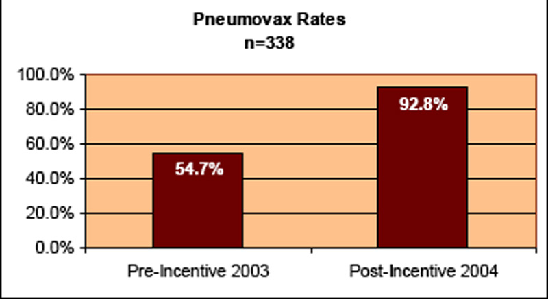
- For pneumonia patients, 45% rate of pneumococcal vaccine screening and administration;
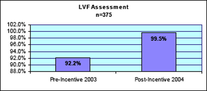
- For heart failure patients, 85% rate of documentation of ejection fraction; and
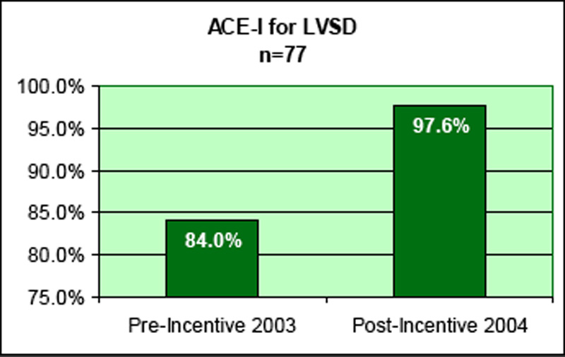
- For heart failure patients with ejection fraction <40%, prescription of an ACE-inhibitor (or documentation of a contraindication) upon discharge.
The Mercy Inpatient Medicine Service, which consisted of 10 full-time hospitalists in 2004, cared for approximately 75% of all medical inpatients at the hospital. We created a quality-based incentive program for this hospital medicine group that mirrored the incentives mentioned above for the entire hospital. We postulated that superior performance by the hospitalists would “raise the bar” for the remainder of the patients, some of whom were cared for by a separate hospital medicine group and some by traditional PCPs.
Mechanics of the Pay for Performance Program
The quality-based bonus was structured so that it would be paid out at 6-month intervals in equal parts to all full-time hospitalists. The magnitude of the bonus was set in excess of 7.5% of the hospitalists’ base salary, an amount that we felt would be large enough to influence behavior change.
From the outset of the bonus period (2004), as medical director of the group, I convened a weekly meeting with hospitalists and representatives from the Quality Improvement department, nursing, and case management. The meeting was being held amid the backdrop of a renewed institutional emphasis on all the JCAHO Core Measures and other quality/patient safety initiatives. The purpose of the meeting was to review hospital performance on all JCAHO Core Measures.
A nurse from the Quality Improvement department reviewed all charts of pneumonia and heart failure patients. For cases where pneumovax was not screened/administered, ejection fraction not assessed, and ACE-I not given, a hospitalist would review the chart and provide feedback to the physician-of-record regarding the nature of the problem. Further, I provided regular feedback to the hospital medicine group regarding their performance on the 3 indicators.
Results
The results in Figures 1–3 are for the hospitalist group, with results for the hospital as a whole slightly lower, but still in excess of the targets agreed upon with Blue Cross.
Summary
Mercy’s experience in 2004 is among the first hospitalist pay-for-performance programs reported. This quality-based incentive resulted in marked improvement in 3 quality indicators, resulting in the hospital’s attainment of the bonus paid by Blue Cross. The hospitalists were satisfied with the incentive program, because they felt appropriately rewarded for high-quality care. It is interesting to note that less-aggressive measures had been undertaken in the past to improve these indicators, but with little success. In 2004, with financial rewards at stake, more robust processes—such as a weekly meeting, ongoing chart review, and direct feedback to physicians—were put in place to ensure quality improvement.
Win Whitcomb, MD, cofounded the Society of Hospital Medicine with John Nelson, MD, in 1996. He is director of Performance Improvement at Mercy Medical Center, Springfield, MA, and can be reached at [email protected].
In January 2004, the Mercy Inpatient Medicine Service embarked on a quality-based incentive program, or “pay-for-performance.” This was spurred on by Blue Cross of Massachusetts, which contracted with all hospitals in the state to receive a substantial financial bonus for agreed-upon quality indicators.
Prior to the start date, representatives from Mercy Medical Center negotiated with Blue Cross, arriving at the following quality indicators and targets for all hospital patients for the period of January to December 2004:
- For pneumonia patients, 45% rate of pneumococcal vaccine screening and administration;
- For heart failure patients, 85% rate of documentation of ejection fraction; and
- For heart failure patients with ejection fraction <40%, prescription of an ACE-inhibitor (or documentation of a contraindication) upon discharge.
The Mercy Inpatient Medicine Service, which consisted of 10 full-time hospitalists in 2004, cared for approximately 75% of all medical inpatients at the hospital. We created a quality-based incentive program for this hospital medicine group that mirrored the incentives mentioned above for the entire hospital. We postulated that superior performance by the hospitalists would “raise the bar” for the remainder of the patients, some of whom were cared for by a separate hospital medicine group and some by traditional PCPs.
Mechanics of the Pay for Performance Program
The quality-based bonus was structured so that it would be paid out at 6-month intervals in equal parts to all full-time hospitalists. The magnitude of the bonus was set in excess of 7.5% of the hospitalists’ base salary, an amount that we felt would be large enough to influence behavior change.
From the outset of the bonus period (2004), as medical director of the group, I convened a weekly meeting with hospitalists and representatives from the Quality Improvement department, nursing, and case management. The meeting was being held amid the backdrop of a renewed institutional emphasis on all the JCAHO Core Measures and other quality/patient safety initiatives. The purpose of the meeting was to review hospital performance on all JCAHO Core Measures.
A nurse from the Quality Improvement department reviewed all charts of pneumonia and heart failure patients. For cases where pneumovax was not screened/administered, ejection fraction not assessed, and ACE-I not given, a hospitalist would review the chart and provide feedback to the physician-of-record regarding the nature of the problem. Further, I provided regular feedback to the hospital medicine group regarding their performance on the 3 indicators.
Results
The results in Figures 1–3 are for the hospitalist group, with results for the hospital as a whole slightly lower, but still in excess of the targets agreed upon with Blue Cross.
Summary
Mercy’s experience in 2004 is among the first hospitalist pay-for-performance programs reported. This quality-based incentive resulted in marked improvement in 3 quality indicators, resulting in the hospital’s attainment of the bonus paid by Blue Cross. The hospitalists were satisfied with the incentive program, because they felt appropriately rewarded for high-quality care. It is interesting to note that less-aggressive measures had been undertaken in the past to improve these indicators, but with little success. In 2004, with financial rewards at stake, more robust processes—such as a weekly meeting, ongoing chart review, and direct feedback to physicians—were put in place to ensure quality improvement.
Win Whitcomb, MD, cofounded the Society of Hospital Medicine with John Nelson, MD, in 1996. He is director of Performance Improvement at Mercy Medical Center, Springfield, MA, and can be reached at [email protected].
SHM Member in the Spotlight

SHM member David Feinbloom, MD, testified before the Massachusetts Joint Committee on Healthcare Financing and Economic Development and Emerging Technologies on May 5, 2005. Dr. Feinbloom was part of a panel of Massachusetts’ healthcare and information systems leaders advocating for additional funding of a statewide initiative to install Computerized Physician Order Entry (CPOE) systems and other advanced information technologies in each hospital across Massachusetts. Dr. Feinbloom is the director of clinical resource management, Department of Medicine, and physician liaison for Clinical Information Systems Development at Beth Israel Deaconess Medical Center in Boston. Under the leadership of John Halamka, MD, MS, and chief information office of Harvard Medical School and BIDMC, the medical center is a nationally recognized leader in medical information technology.
“The goal of the hearing was to share views about the implementation of advanced technologies like CPOE, one of a series of initiatives to create a statewide medical information technology infrastructure,” says Dr. Feinbloom. “Ultimately, this will include applications such as e-prescribing, online physician-patient communications, and regional data sharing networks, which will improve quality, patient satisfaction, and reduce costs.” He says that currently a parallel initiative for related technologies has a $50 million commitment from Blue Cross and Blue Shield. An additional $210 million is needed to bring inpatient CPOE to all of the hospitals in the state. “We wanted to make sure that the committee understood that despite the seemingly high initial outlay of capital, there is a projected savings of $275 million annually.” The dramatic savings, Dr. Feinbloom says, come from efficiencies in patient throughput, reductions in medication errors and adverse drug events, and improved utilization of inpatient resources. The Massachusetts Technology Collaborative and New England Healthcare Institute are coordinating statewide efforts to remove barriers to inpatient CPOE.
Currently, only 5% to 10% percent of hospitals nationwide have CPOE systems, but that is destined to change, says Dr. Feinbloom, especially if hospitalists lead the charge. “Hospitalists are the natural choice to champion these initiatives,” Dr. Feinbloom says. “We are the experts on inpatient care and hospital systems, and we understand how important information technology is for managing complicated patients during an acute hospitalization. In addition, these technologies have proven indispensable for communicating among care providers and managing the transition from the inpatient to the outpatient setting—a process that is notorious for errors.”
For more information on CPOE implementation or funding, contact Dr. Feinbloom at [email protected].
SHM’s Jamie Newman, MD, Appointed Editor of The Hospitalist

After several months of planning, interviewing, and lively debate, the SHM editorial board has appointed Jamie Newman, MD, as editor of its publication, The Hospitalist, effective September 1, 2005. The Hospitalist is the nation’s only society-sanctioned news publication for hospitalists, and it contains the latest in hospital medicine news and industry issues, along with updates on society-sponsored programs, activities, and educational opportunities.
Dr. Newman is assistant professor of Internal Medicine and Medical History at the Mayo Clinic College of Medicine in Rochester, MN, where he has been a member of the Hospital Internal Medicine Program since 2001. Prior to joining the Mayo Clinic College, Dr. Newman practiced medicine in several single and multispecialty group practices. From 1989 to 2001 he taught at the University of Texas Medical Branch in Galveston, where he received several teaching awards. He also was the host of a local medical radio show, “Onward and Upward.” He has been an editor and a writer of numerous papers and book chapters.
“We’re very pleased to have Dr. Newman accept the position of editor for The Hospitalist,” said Jim Pile, MD, outgoing editor of the publication. “He is highly respected, well-published, and has a broad view of the industry and where hospitalists fit in. SHM’s Editorial Board was particularly impressed by his creativity, drive, and energy, and felt that he would bring a new vision that would steer The Hospitalist in a positive direction.”
Dr. Newman was one of a number of very qualified and interesting candidates vying for the editor position. “We were very fortunate to have so many excellent candidates to interview for this high-profile and important job,” said Editorial Board member Peter Lindenauer, MD. “I think that says a lot about just how much The Hospitalist has grown and is valued by our members.”
“I’m happy to have this opportunity to serve SHM and the field of hospital medicine through my contributions as editor of The Hospitalist,” said Dr. Newman. “With the continued rapid expansion of hospital medicine, The Hospitalist must provide vital and relevant information to a wide range of practitioners, from solo to group practice, from small local hospital to tertiary referral center, all with one common goal: improving the care of the hospitalized patient.”
As SHM readies its launch of the new Journal of Hospital Medicine in January 2006, Dr. Newman says it is more important than ever to distinguish the role of The Hospitalist from that of the journal. “While the Journal of Hospital Medicine will provide authors a place for peer reviewed publication, The Hospitalist will continue to present timely information in other areas that impact our industry, from government regulation to patient safety to education and medical innovation,” explained Dr. Newman. “Members can stay informed about these things as well as activities of the society and other members, and hopefully be entertained as well. I plan to create new sections on palliative care, geriatrics, communication, and quality, as well as a surprise or two.”
Dr. Newman earned his undergraduate degree from Johns Hopkins University and obtained his MD from Mayo Medical School. A member of SHM since 2001, Dr. Newman has served in a variety of leadership roles, including the Communication Committee and the Medical History Forum.
Launched in 1997, The Hospitalist newsletter is published 6 times a year and provided free to approximately 10,000 hospitalists in the United States. In September 2005, The Hospitalist will be issued 10 times per year.
To contact Dr. Newman about story or column ideas for the Hospitalist, please email him at [email protected].
Palliative Care Services Offer New Horizons for Hospitalists
Howard Epstein, a hospitalist at Regions Hospital in St. Paul, MN, spent nearly 2 years planning an inpatient palliative care consultation service for Regions before its launch in January of this year. A multidisciplinary advisory committee met monthly to help with the planning, and Dr. Epstein, the embryonic program’s medical director, went before the hospital’s administration to make the clinical and financial case for supporting it.
“What we’re trying to do is to take the basic interdisciplinary approach pioneered in hospice and move it upstream,” to help relieve suffering in seriously ill patients before they need or qualify for hospice care, he explains. “I just knew I wanted to incorporate it into a hospitalist model,” and into the Hospitalist Services Division’s weekly block schedule.
After the service was launched, it became clear that the schedule did not allow for the significant time commitment required to do palliative care, so a new approach is planned for July. Dr. Epstein and 6 other hospitalists participating in the palliative care service will divide up weekly blocks of time. Half of their duties while on service will be devoted to palliative care and the other half to covering 1 hospital unit as a hospitalist, rather than the usual 2 units for hospitalists at Regions.
The palliative care service at Regions, which includes a half-time chaplain and social worker and a full-time nurse practitioner, responds to consultation requests by doctors and nurses from all of the hospital’s adult services. The service also admits patients from HealthPartners’ affiliated hospice program when they are hospitalized at Regions for short-term symptom management or respite care.
Key to long-term success lies in documenting improved clinical outcomes, patient, family, and provider satisfaction, financial savings, and enhanced patient throughput. “I’m optimistic we’ll be able to demonstrate significant value, but if we can’t, we’ll be hard-pressed to get continued support,” he says. This challenge, he adds, is similar to what the hospitalist service at Regions faced when it was launched in 1998.
Palliative care is not a new concept in medicine, but it has enjoyed dramatic growth in recent years. The American Hospital Association estimates that 17% of community hospitals and 26% of academic teaching hospitals in the United States now have either a palliative care consultation service or a dedicated unit, although the former is more common because it can be established with a smaller fiscal outlay.
Palliative care aims to relieve suffering, broadly defined, for patients living with chronic, advanced illnesses. State-of-the-art pain management is a major emphasis for the interdisciplinary palliative care team, but so are addressing the patient and family’s emotional, psychological and spiritual concerns related to the illness and offering guidance for making informed treatment decisions that reflect their values and goals for care. Palliative care services generally target all patients with advanced illness from the point of diagnosis, simultaneous with any other medical treatment regimens.
Two Medical Fields Growing Together
“I believe hospitalist practices and palliative care services are of necessity growing closer together,” says Susan Block, codirector of the Harvard Medical School Center for Palliative Care in Boston, MA. The Harvard Center provides intensive palliative care training for clinicians who also have an interest in teaching.
“If you run a palliative care consult service or a palliative care unit, you are operating much like a hospitalist, with a focus on hospital systems and workload issues, communication, and getting people out of the hospital,” Dr. Block says. At the same time, most hospitalists deal with end-of-life issues and the challenges of relieving symptoms such as pain, delirium, or anxiety every day, whether they view their role in those terms or not.
Stephanie Grossman, a hospitalist at Emory Healthcare in Atlanta, GA, says she discovered personal satisfaction as a young physician in having meaningful conversations about care goals with seriously ill patients and leading family conferences, despite the time pressures of the job. The head of the hospital medicine service at Emory, Mark Williams, MD, told Dr. Grossman there was a name for what she enjoyed doing: palliative care. He encouraged her, along with colleague Melissa Mahoney, to obtain additional training in developing such a program, starting with a 2003 conference in San Diego sponsored by the Center to Advance Palliative Care.
“I came back from San Diego feeling swept away by how it really was possible to develop a financially feasible program,” says Dr. Grossman. Back at Emory, she and Dr. Mahoney joined a palliative care task force formed by Dr. Williams, with representatives from geriatrics, nursing, social work, chaplaincy, finance, and administration. This group provided input for a business plan for the palliative care service that will start in September at Emory Crawford Long and Emory University hospitals.
The 2 hospitalists, who have become certified in palliative medicine, will divide a full-time position as codirectors of the inpatient palliative care service in alternating monthly blocks, along with additional teaching responsibilities. Their 4-year plan is to add additional staffing as the program grows and to work with a geriatrician to develop a palliative care fellowship program. The palliative care team, including a nurse, social worker, and chaplain, will conduct daily palliative care rounds and biweekly interdisciplinary case conferences at the 2 hospitals.
“We have a nurse practitioner involved to help us coordinate between the 2 sites. We’ll go to various departments and do some grand rounds to introduce and market the program,” Dr. Grossman notes. In addition to practicing a style of medicine that offers deeper personal interactions with patients, she is excited to be part of creating a new program. However, she emphasizes the importance of having an executive champion within the hospital who understands financing, institutional politics, and how to recruit other champions. “We’ve been lucky to enjoy the support of Dr. Williams and [Emory Chief Operating Officer] Pete Basler. Dr. Mahoney and I have been working with hospitalists for several years, but our work has all been clinical,” she says.
Another challenge for hospitalists interested in pursuing palliative care include the need to make sure their new responsibilities are not just an add-on to a full-time job. The hospital needs to commit resources for planning and implementing a palliative care program, including a percentage of the hospitalist’s time, Dr. Block says. “Zero FTEs is not viable in the long run,” she adds.
Physician billing for palliative care consults can help offset the costs of running a service, but it is unlikely to break even on billing alone, says Eva Chittenden, a hospitalist and palliative care physician at the University of California-San Francisco, which has operated a palliative care service since 1999. Dr. Chittenden is also part of the Palliative Care Leadership Center at UCSF, which offers 2-day intensive training programs 4 times a year for hospital teams that want to start or strengthen inpatient palliative care services.
In most cases, palliative care requires financial support from the hospital, although it’s not difficult to justify that support by showing cost avoidance, reduced lengths of stay, and improved clinical outcomes, with the help of tools developed by the Center to Advance Palliative Care, Dr. Chittenden says. Program development also challenges the hospitalist’s leadership and marketing skills.
A Process of Growing Involvement
“What often happens with hospitalists is that they start out exploring palliative care, and it becomes very compelling,” Dr. Block adds. “The more competent you get at it, the more compelling it becomes. They find deeper meaning in their work. And then they’re hooked.”
A hospitalist can seek additional training and then incorporate palliative care tools, concepts, and perspectives into his or her daily work. An interest in palliative care may lead to involvement with the hospital ethics committee, a seat on a palliative care advisory committee, or a role in standards or protocol development, as well as pursuit of specialty certification in hospice and palliative medicine.
Although hospitalists may be obvious candidates to participate in more formal palliative care program development, “incorporating palliative care into a routine hospitalist practice is not a trivial thing,” Dr. Block adds. For starters, it requires additional training. “Most hospitalists don’t have the competencies to practice expert palliative care if they don’t seek them out,” she says. But the opportunities are increasing, with growing palliative care fellowship opportunities nationwide.
Two hospitalists at Chandler Regional Medical Center in Chandler, AZ, are among the 4 physicians who serve on that facility’s 12-member interdisciplinary palliative care team, attending weekly team meetings to review active cases and brainstorm program development. Both have attended national palliative care conferences, reports the palliative care service’s nurse practitioner, Donna Nolde. The service consulted on 89 patients in March, and about 70% of those referrals came from hospitalists.
“As hospitalists, we often deal with issues of death and dying,” especially when working in the ICU or with referring oncologists, notes Chandler’s Mahmood Shahlapour. “We can sometimes step back and see the big picture when other doctors have trouble letting go.” Dr. Shahlapour believes palliative care is a logical extension of good internal medicine and will eventually become a bigger part of the training of internists.
An atypical path to palliative care is that of Glenda Hickman, MD, who was a hospitalist for the Denver, CO–based HealthONE system until one of the system’s hospitals asked her to take on the role of freelance palliative care consultant. Hickman, who also works part-time for Hospice of Metro Denver as a team medical director and picks up lecturing and teaching assignments, accepts consultations from 3 HealthONE hospitals and bills third-party payers for her consultations. Her husband is office manager and biller for her home-based business, and she carries a cell phone and pager to promptly answer referrals.
“I had a reputation for the touchy-feely aspects of medicine at the hospitals where I worked,” Dr. Hickman relates. “Dying patients would often get referred to me.” Based on her interests, Dr. Hickman sought training in palliative care, but she found it difficult to juggle with her full-time job as a hospitalist. “The heart of palliative care in a hospital is talking with patients and families. These conversations take a long time,” she says. When the hospital asked for her help in meeting its JCAHO requirements in pain management and palliative care, Dr. Hickman was willing to explore a model for how she could hang out a shingle as a solo practitioner.
Business is growing, although the workload fluctuates widely. However, while Dr. Hickman works alongside social workers and chaplains at the hospitals, the biggest drawback has been the lack of a formal, interdisciplinary team. “This is high-maintenance, high-emotion work. It can be a big drain, and I don’t have a designated team with which to share the burden. My goal is to run a full palliative care team for the hospital,” she says, and there are signs that HealthONE eventually may move in that direction.
“It’s not that hospitalists can’t do palliative care. I did. I was so drawn to it and to trying to do it right, which meant I was trying to do 2 jobs at once,” she adds. Hospitalists can also participate by recognizing when their patients need the extra attention of a palliative care specialist. “Identifying who those patients are is a huge skill by itself.”
Resources for Getting Started in Palliative Care
The Center to Advance Palliative Care (CAPC) at Mount Sinai School of Medicine in New York City offers a comprehensive national resource for palliative care development in hospitals, including how to make the financial case. Its next national seminar is October 17 to 19 in San Diego, CA. CAPC also supports 6 regional Palliative Care Leadership Centers, including one with a hospitalist emphasis at the University of California-San Francisco, scheduled to run through June 2006. For more information on CAPC’s resources and leadership centers, call 202-201-2670 or visit www.capc.org.
Larry Beresford can be contacted at [email protected].
Other Helpful Resources
- For information on the Education in Palliative and End-of-Life Care (EPEC) curriculum, visit www.epec.net.
- The American Board of Hospice and Palliative Medicine will offer its next specialty certifying examination in November of 2005. For eligibility or other questions, call 301-439-8001 or visit www.abhpm.org.
- The American Association of Hospice and Palliative Medicine offers education and training resources, including an annual assembly scheduled for February 8 to 11, 2006, in Nashville, TN; visit www.aahpm.org.
- Harvard’s Center for Palliative Care offers a 2-week intensive training course, with an emphasis on teaching, in April and November every year. For information, call 617-724-9509, send email to [email protected], or visit www.hms.harvard.edu/cdi/pallcare/.
- The Veterans Administration also offers palliative care resources, fellowship opportunities and other information; visit www.hospice.va.gov.
A New Debut

Here’s a question for you: When was the first issue of The Hospitalist published?
The answer: Winter (early) 1997.
In the late ’90s, The Hospitalist was little more than a bifold eight-page newsletter mailed to members of the then-National Association of Inpatient Physicians. The newsletter contained one page of classified advertising.
Contrast that first issue of The Hospitalist to the one you’re reading now: a whopping 45 pages of editorial content, with about the same number of advertising pages to match!
Nine years and dozens of changes later, The Hospitalist is about to transform itself again—a transformation that will put it at the forefront of professionally published, business-to- business titles. Sounds exciting, to be sure. But I bet you’re wondering, “Why another change?”
Simply put, The Hospitalist must reflect the progressive growth of SHM and of hospital medicine overall.
Partnering for the Future
In January SHM endeavored to elevate SHM’s publishing program on the national publishing stage and so partnered with John Wiley & Sons. SHM brought hospital medicine knowledge and expertise to the table; Wiley brought editorial and publishing knowledge and expertise.
Together, the two organizations determined that The Hospitalist needed to further grow—in format, frequency, and editorial excellence—to meet the needs of members and to respond to industry interest in The Hospitalist as the most trusted, credible source of news, issues, and trends in hospital medicine. SHM and Wiley also partnered to launch the first peer-reviewed hospital medicine journal (the Journal of Hospital Medicine—coming in January).
These initiatives, along with the commitment to maintaining strict ethical publishing guidelines (meaning—among other things—separation of editorial content and advertising) in both titles, pack the one-two punch that SHM members deserve.
What You’ll See
Beginning with the September 2005 issue, you’ll notice quite a few improvements in The Hospitalist. Here’s my short list of what you can expect:
- Size change: The publication will grow from its current iteration (a standard or 8.5” x 11” size) to a “tabloid” (or 11” by 17” size);
- Frequency change: We’ll begin publishing The Hospitalist on a monthly basis. Instead of six issue per year, you’ll receive 12; • The “look:” The design of the publication will evolve from a text-driven format to a more diverse, visually appealing format;
- Who’s who: The editorial staff will grow from a sole physician editor to a two-editor team: A professional editor from SHM publishing partner John Wiley & Sons—that’s me—will pair with Physician Editor Jamie Newman, MD, from the Mayo Clinic Rochester, to create each issue. In addition, we’ll utilize hospital medicine experts from around the country, as well as tap professional health-care writers to report on and investigate the most pressing issues in hospital medicine; and
- Content changes: Each month we’ll continue to present the quality articles you’ve come to expect from us. Our singular editorial goal is to be the voice of hospital medicine. And, thanks to the format changes, we’ll be able to offer you even more each issue. In the coming months, look for such enhancements as
- Balance: a blend of clinical, management/leadership, administrative, and policy articles in each issue—all written and edited with one question in mind, “Why is this article meaningful to you, the hospitalist reader?”;
- More strategies on how to get information on subjects: extended references, resources, and other bonus guidance in each article;
- Fast reads: “bottom line” sections that condense articles into at-a-glance summaries and shorter articles;
- SHM focus: organized, easy-to read news specific to SHM members and activities;
- Voices, voices: hospitalists from all parts of the country used as sources and in quoted material throughout the publication; and
- Much more!
Growth and Change = Great
Every once in a while you get lucky enough to be involved in a project that’s good as it is—but that teeters on the cusp of catapulting into something truly great. That’s how I feel about the impending growth and change of The Hospitalist. Incoming Physician Editor Jamie Newman and I welcome your ideas on how we can best shape the future of The Hospitalist. E-mail us at [email protected] or [email protected]. Onward!
Editor Lisa Dionne has been involved in magazine publishing for more than 14 years. She’s helped create, edit, and write award-winning editorial content for audiences ranging from long-term care physicians to EMS personnel to children.
Patient-Centered, Measurable-Quality, True Teamwork
I have mentioned in previous columns and often in public speeches that hospitalists will actively create and implement the hospital of the future. This is not an idle promise but an inevitability, and SHM is working actively with other key players to make this happen.
This new era will have as its hallmark 3 essential features:
- Care will be patient-centered;
- Quality standards will be met, measured, and documented; and
- Hospital care will be delivered by empowered teams of health professionals.
If you think this is already the standard of care, you either practice in a utopian hospital that I am not aware of or your definitions are different than mine. Let me walk you through what I foresee.
Patient-Centered Care
Today’s hospitals are set up, for the most part, to accommodate the physician rather than the patient. We live in an on-demand world where the “consumers” expect to have as much information as they want and to have it when they want it. When a patient at your hospital has a test performed, does someone stop by within the hour with the results and an explanation of what the results mean to the patient? When the patient first arrives, does someone on the healthcare team sit down and ask the patient what his or her expectations are for this hospitalization (e.g., cure, diagnosis, pain relief, a good death)? Does the patient have immediate access to caregivers (e.g., cell phone or pager numbers) and has he or she been genuinely told to “call me if I can help”? Do patients even know when their doctor will be back to see them?
Do their doctors talk to them in terms meant for the patient and not for grand rounds? Do they use words like “blood clot” rather than “deep vein thrombosis”? Do they understand that health literacy is a major problem for many of our patients?
If patients have a hospitalist as their care manager and if this hospitalist works in a state-of-the-art hospital medicine group, then maybe this is already happening. But hospitalists will need to make this happen, if this is ever to be a universal focus of our hospital care.
Quality Is Job 1
For the 30 years I have been a physician we have “talked” about quality. Any quality we have in our system is largely a function of the altruism and hard work of the healthcare professionals and advances in science. It certainly is not baked into the culture, the data collection, or the compensation structure. Today doctors are paid for doing more, not for doing better. In fact, there is almost no incentive for even knowing how good a job you do. Has anyone ever been denied payment or denied the ability to manage diabetes because they have never checked a glycohemoglobin?
This will change, and hospitalists need to be at the center of this revolution. There are forces amassing that may nudge this movement forward. Leapfrog and other forces in the business community are demanding that something happen, and soon. The National Quality Forum (NQF) is trying to set and enforce standards. There are rumblings of pay for performance (P4P), although I think this will translate more into paying less for poor performance. JCAHO, IHI, AHRQ, and so many others have projects centered around patient safety and quality improvement that just from sheer effort alone things will change.
At the same time, patients seem to have noticed all this commotion and wonder why all of a sudden there are so many concerns about both the safety of their hospital and the quality they receive in it. This is definitely on the radar screen.
Hospitalists will be both the measured and the measurers. We will need to work with others to decide what data will help tell us how well we are doing, what changes in the systems need to be installed to improve quality, and how to harness all the resources available to be better tomorrow than we are today.
True Teamwork
And this leads me to the third leg of this triad. Hospitalists and other physicians can’t do this alone, and we can’t do this under the old model where the doctor has the steering wheel and everyone else waits to see where the ship is going. This may evolve into more of an orchestra, where the physician is the conductor and others are the virtuoso musicians.
Current legal limitations aside, we markedly underutilize the perspective and expertise of nurses, pharmacists, social workers, therapists, and case managers. These other team members often know important information about our patients and about the hospital and available resources. If we had a seamless way to incorporate this knowledge base, patients would get better care, the entire team would feel empowered, and job satisfaction would improve for everyone.
Critical-Care Collaborative
For the past year, senior leaders from SHM have met with senior leaders from the American College of Chest Physicians (ACCP), the American Association of Critical Care Nurses (AACN), the American Society of Health System Pharmacists (ASHP), and representatives of respiratory therapy to examine our current management of acutely ill patients in the hospital and to develop plans to move to a new health system that is patient centered and team driven. AACN has started giving out Beacon Awards to hospital units that demonstrate these values. SHM will partner with ASHP in 2005 on joint research of teams of hospitalists and pharmacists. The Collaborative will look at any examples of collaborative efforts that are in place and working today.
Those of you who think this is pie in the sky should look back at some history. About 15 to 20 years ago, about 10 physicians got together and decided to make California smoke free. Our goal was to remove cigarettes from restaurants, businesses, and the work place by 2000. The doubters said we were up against a multibillion dollar industry and trying to outlaw an addictive substance. Today about the only public place you can still smoke a cigarette in California is out in front of a hospital.
Grand goals can happen when motivated people are willing to make them a priority. Making the patient the focus of health care, creating an environment where teams can flourish, and raising expectations for delivering a quality experience in every hospital will happen in my lifetime. And hospitalists (along with other key stakeholders) will be at the center of this effort. This is the right commitment at the right time. SHM will do our share, and we hope each of you will make this one of your core professional values.
Rewards at the Bedside
Enter text here
Enter text here
Enter text here
Practice Profile
Contact
Richard Rohr, MD
Milford Hospital
300 Seaside Avenue
Milford, CT 06460
Phone: 203-876-4000
Staff
Christine Chen, MD
Andrew Chow, MD
Renee Giometti, MD
Richard Rohr, MD
Michael Rudolph, MD
Keith Swan, MD
Yelena Titko, MD
The Milford Hospital Hospitalist Service program started in 1996 with 1 physician hired to provide coordination for inpatient medical care on weekdays. The hospital had previously offered only night coverage provided by moonlighting cardiology fellows. Milford Hospital has 100 beds, does not participate in any medical teaching programs, and competes with 5 teaching hospitals located within 10 miles. The community has traditionally preferred treatment in the local area, but concern about quality of medical services led many local residents to seek treatment at larger hospitals. The hospital had studied the hospitalist concept from its inception, but the medical staff did not immediately embrace the idea and feared encroachment upon their incomes. After several years of steadily increasing the role of the daytime care coordinator, the administration decided to convert the moonlighting positions in 2001 to 5 full-time employed physicians who provide 24-hour coverage in the facility. The medical staff has gradually become more comfortable with the hospitalist concept, although the internists still prefer to treat their own established patients. The community also recognizes the higher level of medical care provided, and the average daily census has nearly doubled since starting the program.
The service has been scheduled with 2 daytime physicians for 8 hours on each weekday, 1 daytime physician for 8 hours on Saturday and Sunday, and 1 physician for 16 hours every night. The staffing pattern was developed to accommodate an active joint replacement service with significant consultation needs on weekdays. The orthopedic service is expanding, and other surgeons have recognized the importance of immediate consultation, particularly as their malpractice premiums rise. The hospital administration has recognized the need for additional staffing, and the service will operate with 2 daytime physicians and 1 nighttime physician every day of the week starting in July 2005. An additional position has been created to meet the personnel needs.
The physicians are employed directly by the hospital and participate in the hospital’s benefit programs, including pension, disability insurance, life insurance, and a malpractice liability trust. There is presently no incentive plan, but the program has achieved a high level of effort from the staff. This is largely due to the culture of the hospital, which is highly collegial and patient-focused. Employees at all levels of the organization are treated well, and staff retention levels are quite high. Out of the first 10 full-time hospitalists hired, 4 are still in the program, 5 have pursued additional training, and 1 left to join her husband in California. One physician hired from a leading academic residency program found it difficult to adjust to a community hospital and resigned prior to year‘s end. The service has not experienced other personnel problems.
The program met its early staffing needs with physicians who had recently completed residency in internal medicine and were waiting to start a fellowship in 1 year. This type of staffing allowed the service to get started but required constant training in billing, continuity of care, and medical staff relations. As the program has become established in the region, it has attracted physicians who have previous experience with traditional private practice but have chosen to concentrate on inpatient care. This has allowed the program director to concentrate on advanced skill building with the staff and to spend less time on recruiting and scheduling.
Physicians are required to work 1,800 hours each year and may work additional hours for extra payment. The service admitted 600 patients (one-fourth of all medical admissions) in 2004 and provided consultation on 800 patients. The service also manages intensive care patients and handles emergencies throughout the hospital; there were 800 critical care visits last year. The staff performed a total of 6,200 billable patient visits in the past year and provided assistance to private physicians on 1,000 additional admissions. The number of billed admissions and visits has been constrained by limited weekend staffing. The service presently carries no more than 12 patients on weekends and up to 20 patients on weekdays. With the new staffing pattern, the service will round on up to 24 patients, with additional patients seen on a 1-time basis. Hospitalists normally admit patients who are not affiliated with a private physician on the hospital staff. When the hospital medicine service reaches its patient cap, private physicians must admit unaffiliated patients in rotation, as they did before the hospitalists were available. This accommodation will remain in place until the hospital medicine service is able to meet the entire demand for inpatient internal medicine services.
Postdischarge care coordination has been a major challenge. Approximately one-third of patients are discharged to nursing homes. Most of the others are affiliated with primary care physicians located in other communities who are not members of the Milford Hospital medical staff. Communication with these physicians has been improved by an electronic record management system that allows automated fax transmission of discharge summaries. Limited outpatient services are available for Medicaid patients and for those without insurance. The hospital medicine service does not provide outpatient care.
Another challenge involves care for critically ill patients. Although there are several physicians with training in pulmonary disease on the private staff, the hospital had not developed effective critical-care services. There are 2 hospitalists with critical-care training, and we have been working with the other staffers to improve their competence in critical care. The hospitalists provide 24-hour response to unstable patients throughout the hospital and have dramatically reduced unexpected mortality.
Future development will focus on improving hospitalist productivity with information technology. The hospital has undertaken installation of an integrated clinical-information system, which will include direct physician order entry and deployment of wireless technology. It is expected that many of the difficulties experienced by other hospitals with physician order entry will be ameliorated by hospitalist involvement, as the staff is comfortable with computer use. We also expect that the hospitalists will develop leadership roles within the medical staff and develop skills in quality improvement.
Rewards at the Bedside
At our exciting and energizing annual meeting in Chicago, I had the honor and privilege of sharing my goals for SHM for the coming year: to promote palliative care and research in SHM and hospital medicine. Research will maintain SHM’s role as the leader in defining hospital medicine, and palliative care will keep us connected to our fundamental mission, which is to provide the highest-quality care to our patients. Over the next year I will share my vision for why I think these initiatives are key for SHM and our field and how SHM will promote them.
Recently, family of Mrs. T., a 62-year-old woman with metastatic pancreatic cancer, asked me not to tell Mrs. T that she was dying of her disease. Mrs. T had been admitted with severe pain and nausea. She could not eat and had been losing weight. Her pain had gotten suddenly worse. We treated her pain and nausea aggressively and achieved good control of both. Before I saw Mrs. T, her husband and 2 daughters took me aside and told me that although they realized that she was dying of her disease, they feared that she would lose hope if I told her how sick she was. “Let her think that she can get chemo in the future,” they implored. I asked why. “So she will think she can get better,” they answered. I asked Mrs. T.’s family what they thought she thought was going on. “She knows she is sick but believes the chemo can make her better,” they said.
This was not the first time I had encountered such a request, but it always makes me uncomfortable. What if they are right? Couldn’t I hurt her by giving her bad news she doesn’t want to hear? But if they were wrong, wouldn’t I be denying her the opportunity to say good-bye and bring closure? I took the middle road. I promised the family I wouldn’t say anything the patient didn’t ask me to tell her, but I would offer her the opportunity to ask. My first question to her was a typical open-ended one: “How are things going for you?” Mrs. T. said, “Well my daughter spent the night with me in my room last night.” I nodded. “I think she did it because she thought I might die over night,” she said. Perhaps as I suspected, she understood more than her family thought. She worried about her husband and how he would do without her. She wanted chemotherapy but realized that the best it could do was prolong her life a few months. When I asked her what she hoped for, she told me she has hope in God. I explained that I had spoken with her family and told them what I had told her. I said that they were concerned about her and were afraid that this knowledge would be too much for her. We talked some more, and then she asked me to have her husband come to her. There were things they needed to talk about. Next she spoke with her daughters. All were grateful for the opportunity to talk openly about their grief, sadness, and love for each other. Mrs. T. died 2 days later.
I share this story because it reaffirmed for me the crucial role that I play as a hospitalist in the care of people with serious and terminal illness and the importance of palliative care in providing the highest quality of care to these people. Caring for Mrs. T. raised many issues: effective treatment of pain and nausea, discussion of prognosis, respect for cultural differences, exploration of spiritual issues, and a request from the family to withhold the truth. I felt like I was trying to delicately balance respecting the family’s wish while honoring my responsibility to the patient to tell her the truth. In the end, a careful conversation allowed me to bridge the gap and get Mrs. T. and her family talking. My conversation with her husband several days after she died reaffirmed the importance of raising these issues with Mrs. T.
As hospitalists, we encounter these situations regularly, maybe even daily. Half of Americans die in hospitals, and 98% of Medicare beneficiaries who die spend at least some time in a hospital in the year before death (School, 2000, #1006). We are the physicians who care for the seriously ill and the dying. The question is not whether we will take care of these patients; rather, when we do, will we be ready and able? A survey of hospitalists found that we recognize the importance of palliative care to our practice, but that we feel that our training did not adequately prepare us to provide this care (Plauth, 2001, #763). Our core curriculum, which you will see in early 2006 as a supplement to the 1st volume of the Journal of Hospital Medicine includes a chapter on palliative care.
In many ways palliative care is easy, and in many ways it is difficult. Yes, it takes time. Conversations like the ones with Mrs. T. cannot happen in 5 minutes. But an investment of time up front to talk with patients and their families about preferences for care can save many hours down the road. And yes, these discussions are challenging. It’s not just patients who don’t like to talk about death and dying, their families and physicians don’t like it either. But we can learn how to conduct these discussions better and can practice phrases that will help them go more smoothly. Pain and nausea can be difficult to control. Yet, palliative care experts report that pain can be relieved with simple medications like morphine in more than 95% of cases. As hospitalists, we can fulfill our sacred duty to the sickest patients by learning these critical palliative care skills.
SHM has provided learning about palliative care at most annual meetings and will continue to do so in the future. We will also work on providing many more educational materials targeted specifically at hospitalists. Many CME courses across the country focused on palliative care, including those sponsored by the American Academy of Hospice and Palliative Medicine (www.aahpm.org), which also offers many educational resources on its Web site. Many hospitalists have already participated in Education in Palliative and End-of-Life Care (EPEC) (epec.net), a comprehensive, well-regarded curriculum in palliative care that you can purchase on the web. There are textbooks in palliative care, including the Oxford Textbook of Palliative Medicine, 3rd edition, edited by Derek Doyle, Geoffrey Hanks, Nathan I. Cherny, and Kenneth Calman, and Palliative and Supportive Oncology, 2nd edition, edited by Ann Berger, Russell Portenoy and David Weissman.
With skills in palliative care, we can make a profound difference in the lives of our patients and their families. What is it worth to be able to say good-bye and “I love you” to your family? Mrs. T.’s husband was profoundly grateful for the opportunity to say those things and more to his wife before she died. And although his wife died in the hospital, he was exceedingly satisfied with the care she received. Relieving symptoms for seriously ill patients and talking with them about profound and important issues is not only good for patients; it is good for us, too. Providing high quality care to seriously and terminally ill patients can provide a deep sense of fulfillment and satisfaction through the real and intimate engagement with our fellow human beings. It also allows us to use our humanism and to connect directly to the reason that many of us chose medicine as a career— to help people. In this way it can also protect us against burnout.
I encourage every hospitalist to embrace palliative care for the benefits it will bestow on our patients and their families, for the benefits it will bestow on us, and for the benefits it will bestow on our entire field. As president of SHM, I will do all I can to help SHM promote palliative care, beginning with the newly appointed Palliative Care Task Force. Ultimately, palliative care is about demonstrating caring for our patients and our families. It reminds us that life is precious and that it is important to choose how we spend our time. This last lesson is, perhaps, the greatest gift of palliative care— learning to make the most of the time we have, embodied in this anonymous poem I received from a friend via email:
Sing like no one is listening.
Dance like no one is watching.
Work like you don’t need the money.
Love like you’ve never been hurt.