User login
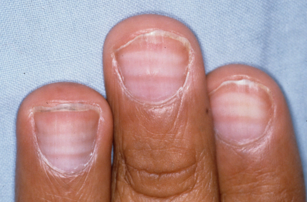
Abnormal fingernails
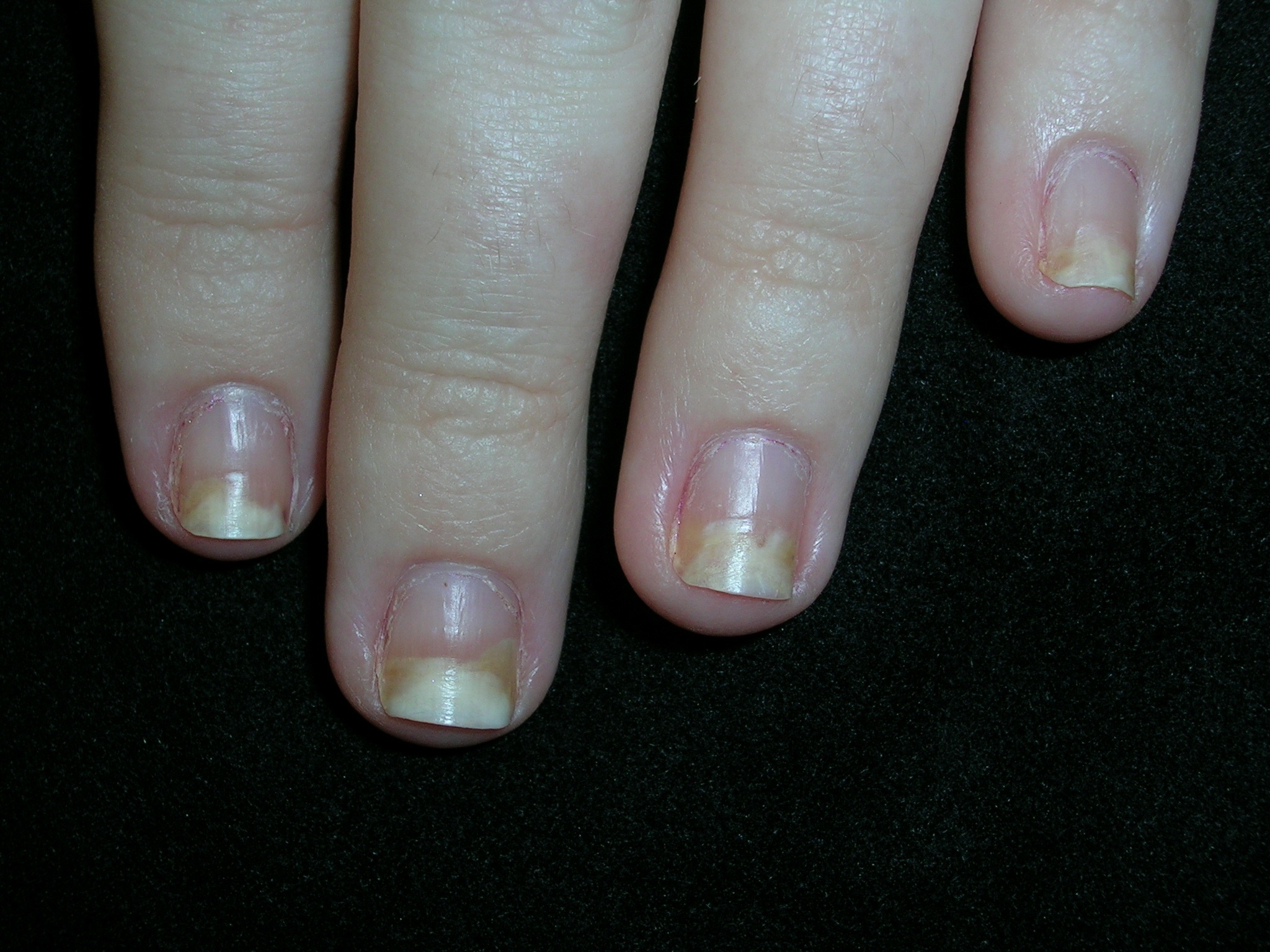
The family physician (FP) diagnosed plaque psoriasis on the elbows along with nail psoriasis, characterized by onycholysis.
In this case, the FP clipped off a portion of the fingernail and sent it for a fungal culture. He prescribed clobetasol ointment for the elbows and told the patient to apply it twice daily for 2 weeks and then on weekends only, as needed. He also counseled her to quit smoking, even though she claimed that she hardly smoked.
The patient returned in 2 weeks and was delighted that her elbows were 90% clear. The fungal culture was negative and the FP and patient discussed options for the treatment of the nail psoriasis.
Application of a high-potency topical corticosteroid under the free edge of the distal nail plate can slow the progression of distal onycholysis. Other topical therapies that have shown some effectiveness include 1% 5-fluorouracil solution or 5% cream applied twice daily to the matrix area for 6 months, as well as topical calcipotriol, anthralin, tazarotene, or cyclosporine. Systemic therapies for psoriasis may improve nail manifestations, but would have been overkill for this patient. She decided to use clobetasol cream for her nails.
Photos and text for Photo Rounds Friday courtesy of Richard P. Usatine, MD. This case was adapted from: Mayeaux EJ. Psoriatic nails. In: Usatine R, Smith M, Mayeaux EJ, et al, eds. The Color Atlas of Family Medicine. New York, NY: McGraw-Hill; 2009:838-841.
To learn more about The Color Atlas of Family Medicine, see:
• http://www.amazon.com/Color-Atlas-Family-Medicine/dp/0071474641
You can now get The Color Atlas of Family Medicine as an app for mobile devices including the iPhone and iPad by clicking this link:
Spot on fingernail
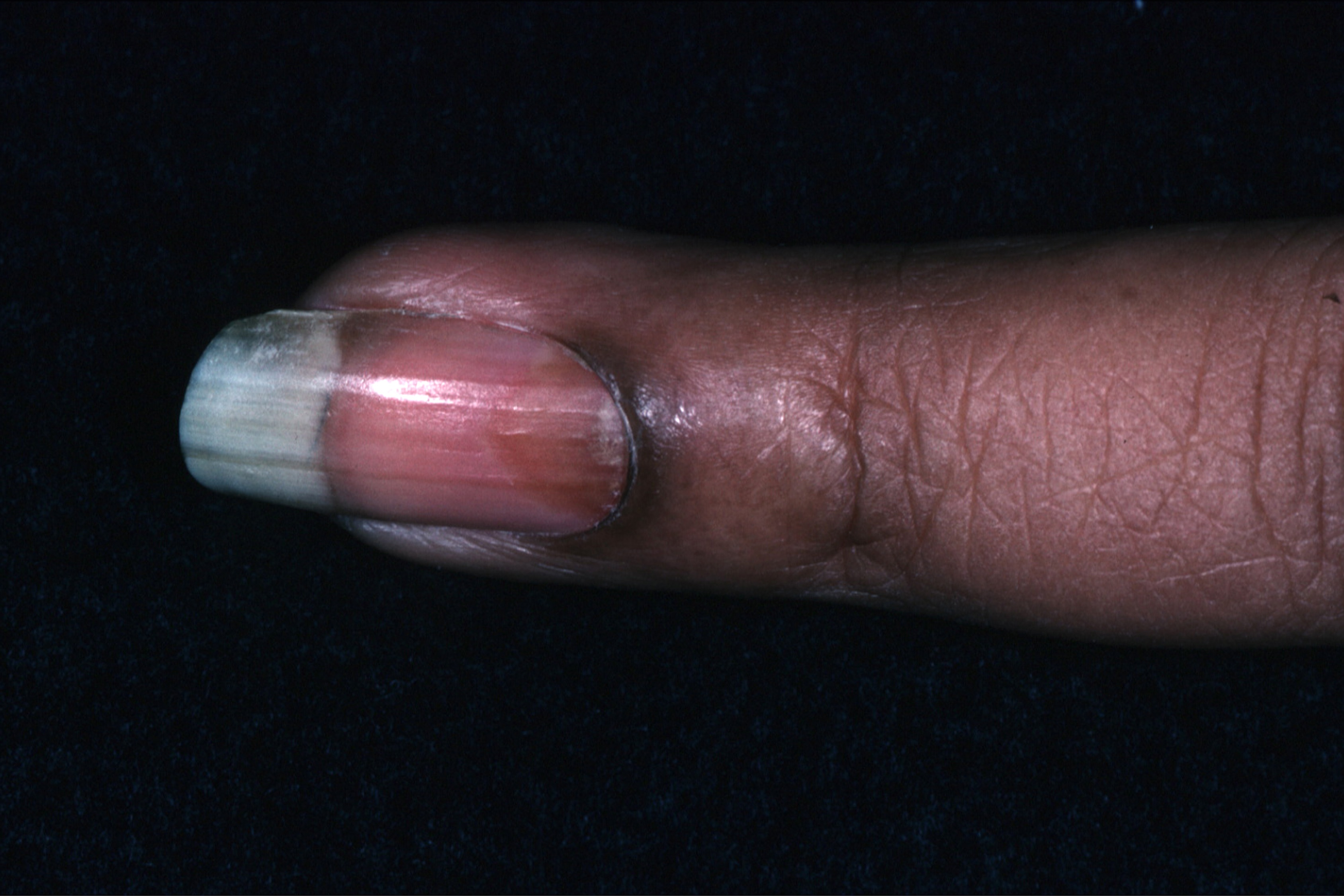
The FP suspected psoriatic arthritis in light of the nail finding (oil drop sign due to localized onycholysis), bilaterally symmetric small joint arthralgias, and morning stiffness. Given the lack of cutaneous findings, the FP ordered hand films, a rheumatoid factor test, and an erythrocyte sedimentation rate (ESR).
Most times, nail involvement coexists with cutaneous psoriasis, although the skin surrounding the affected nails need not be involved. Psoriatic nail disease without overt cutaneous disease occurs in 1% to 5% of cases.
The diagnosis of nail psoriasis is usually straightforward when characteristic nail findings coexist with cutaneous psoriasis. Nail bed psoriasis produces localized onycholysis, which often appears like a drop of oil on a piece of paper (oil drop sign).
The patient in this case was not worried about her fingernail, but the joint pain was making it difficult to type at work. The physician started her on ibuprofen with meals to treat the probable arthritis.
During a follow-up visit one week later, the patient said that her hand pain and stiffness was about 50% better. The hand films were negative, other than some signs of synovitis on the DIP joint of the involved fingernail. In addition, the rheumatoid factor was negative and the ESR was mildly elevated.
The physician counseled the patient to lose weight and to stop smoking for general health reasons, and because smoking and obesity are 2 risk factors for psoriasis. The FP referred the patient to a rheumatologist to clarify the diagnosis and consider the use of more potent medications such as methotrexate.
Photos and text for Photo Rounds Friday courtesy of Richard P. Usatine, MD. This case was adapted from: Mayeaux EJ. Psoriatic nails. In: Usatine R, Smith M, Mayeaux EJ, et al, eds. The Color Atlas of Family Medicine. New York, NY: McGraw-Hill; 2009:838-841.
To learn more about The Color Atlas of Family Medicine, see:
• http://www.amazon.com/Color-Atlas-Family-Medicine/dp/0071474641
You can now get The Color Atlas of Family Medicine as an app for mobile devices including the iPhone and iPad by clicking this link:
Acute abdominal pain in an elderly patient
Nausea, Vomiting, and Weakness for 4 days prompted a 76-year-old woman to seek care at our hospital. She was admitted for possible large bowel obstruction and severe dehydration. Her medical history was significant for a metastatic lung cancer to the mediastinal lymph nodes and to the left hip (for which she underwent a hip replacement 4 months earlier), anemia, and diverticulosis.
On Day 1 of her hospital stay, the patient became hypotensive and developed labored breathing. She also had mottled skin and cool fingertips with poor capillary refill. Her abdomen was distended, firm, and diffusely tympanic with diffuse pain to deep palpation and absent bowel sounds.
Her laboratory values revealed leukocytosis (with a significant left shift), metabolic acidosis, and an elevated lactic acid level. Her upright chest x-ray (FIGURE) is shown. The patient was transferred to the intensive care unit for further management.
FIGURE
Upright chest x-ray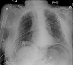
WHAT IS YOUR DIAGNOSIS?
HOW WOULD YOU TREAT THIS PATIENT?
Diagnosis: Pneumoperitoneum
This patient had free air under her diaphragm (due to a viscus perforation) and concomitant septic shock. Free air in the peritoneal cavity—pneumoperitoneum— indicates visceral perforation in 85% to 95% of cases.1,2 A ruptured intra-abdominal viscus is considered a surgical emergency. Pneumoperitoneum is often linked to peptic ulcer disease and is seen in 50% of cases of bowel perforations.3 This condition has a higher prevalence in the elderly and carries a higher mortality rate (up to 30% compared with 19% in a younger population).4
A picture that shifts according to the patient’s age
Physical findings suggestive of visceral perforation include sharp abdominal pain with a rigid abdominal wall. Patients will usually lie still because of the peritoneal irritation. Tachycardia and tachypnea are seen early in the disease process, while hypotension and fever usually develop within 4 to 6 hours.5
Elderly patients, however, can present with milder or nonspecific symptoms. Rather than pain, they may complain of the urge to defecate. Physical exam findings such as tachycardia or fever can also be absent due to autonomic dysregulation or medication. Furthermore, laboratory analysis is commonly within normal limits, making the diagnosis even more challenging in this population.5,6
Imaging confirms the Dx
The standard imaging test used to confirm pneumoperitoneum is a standing chest x-ray that will detect free air in almost 80% of cases.7 The sensitivity is influenced by the location of the perforation: Free air will be seen in 69% of gastroduodenal perforations, 30% to 41% of distal small bowel perforations, and 37% to 46% of large bowel perforations.1 Abdominal computed tomography scans have been reported to be more sensitive (up to 100%), especially in identifying small pneumoperitoneum.8,9
Surgery is the next step
Management of pneumoperitoneum includes a prompt surgical consult for a possible emergent laparotomy, nasogastric suctioning, supportive measures for blood pressure, and broad-spectrum antibiotics such as a fourth-generation penicillin or a third-generation cephalosporin plus metronidazole.10
The end of the fight
Given the high mortality rate and the atypical presentation of perforated viscus in the elderly, it is important to maintain a high index of suspicion in this population and to intervene rapidly to improve the outcome.
In the case of our patient, the family followed her wishes and declined surgery. She was aggressively managed with broad-spectrum antibiotics, IV fluids, and vasopressors—but unfortunately died 2 days later.
CORRESPONDENCE Balaji Yegneswaran, MD, University of Pittsburgh Medical Center, 651, Scaife Hall, Pittsburgh, PA 15261; [email protected]
1. Winek TG, Mosely HS, Grout G. Pneumoperitoneum and its association with ruptured abdominal viscus. Arch Surg. 1988;123:709-712.
2. Roh JJ, Thompson S, Harned RK, et al. Value of pneumoperitoneum in the diagnosis of visceral perforation. Am J Surg. 1983;146:830-833.
3. Borum ML. Peptic-ulcer disease in the elderly. Clin Geriatr Med. 1999;15:457-471.
4. Blomgren LG. Perforated peptic ulcer: long-term results after simple closure in the elderly. World J Surg. 1997;21:412-415.
5. Hendrickson M, Naparst TR. Abdominal surgical emergencies in the elderly. Emerg Med Clin N Am. 2003;21:937-969.
6. Kane E, Fried G, McSherry CK. Perforated peptic ulcer in the elderly. J Am Geriatr Soc. 1981;29:224-227.
7. Chen CH, Yang CC, Yen YH. Role of upright chest radiography and ultrasonography in demonstrating free air of perforated peptic ulcers. Hepatogastroenterology. 2001;48:1082-1084.
8. Stapakis JC, Thickman D. Diagnosis of pneumoperitoneum: abdominal CT vs upright chest film. J Comput Assist Tomogr. 1992;16:713-716.
9. Chen CH, Huang HS, Yang CC. The features of perforated peptic ulcers in conventional computed tomography. Hepatogastroenterology. 2001;48:1393-1396.
10. Gorbach SL. Intraabdominal infections. Clin Infect Dis. 1993;17:961-965.
Nausea, Vomiting, and Weakness for 4 days prompted a 76-year-old woman to seek care at our hospital. She was admitted for possible large bowel obstruction and severe dehydration. Her medical history was significant for a metastatic lung cancer to the mediastinal lymph nodes and to the left hip (for which she underwent a hip replacement 4 months earlier), anemia, and diverticulosis.
On Day 1 of her hospital stay, the patient became hypotensive and developed labored breathing. She also had mottled skin and cool fingertips with poor capillary refill. Her abdomen was distended, firm, and diffusely tympanic with diffuse pain to deep palpation and absent bowel sounds.
Her laboratory values revealed leukocytosis (with a significant left shift), metabolic acidosis, and an elevated lactic acid level. Her upright chest x-ray (FIGURE) is shown. The patient was transferred to the intensive care unit for further management.
FIGURE
Upright chest x-ray
WHAT IS YOUR DIAGNOSIS?
HOW WOULD YOU TREAT THIS PATIENT?
Diagnosis: Pneumoperitoneum
This patient had free air under her diaphragm (due to a viscus perforation) and concomitant septic shock. Free air in the peritoneal cavity—pneumoperitoneum— indicates visceral perforation in 85% to 95% of cases.1,2 A ruptured intra-abdominal viscus is considered a surgical emergency. Pneumoperitoneum is often linked to peptic ulcer disease and is seen in 50% of cases of bowel perforations.3 This condition has a higher prevalence in the elderly and carries a higher mortality rate (up to 30% compared with 19% in a younger population).4
A picture that shifts according to the patient’s age
Physical findings suggestive of visceral perforation include sharp abdominal pain with a rigid abdominal wall. Patients will usually lie still because of the peritoneal irritation. Tachycardia and tachypnea are seen early in the disease process, while hypotension and fever usually develop within 4 to 6 hours.5
Elderly patients, however, can present with milder or nonspecific symptoms. Rather than pain, they may complain of the urge to defecate. Physical exam findings such as tachycardia or fever can also be absent due to autonomic dysregulation or medication. Furthermore, laboratory analysis is commonly within normal limits, making the diagnosis even more challenging in this population.5,6
Imaging confirms the Dx
The standard imaging test used to confirm pneumoperitoneum is a standing chest x-ray that will detect free air in almost 80% of cases.7 The sensitivity is influenced by the location of the perforation: Free air will be seen in 69% of gastroduodenal perforations, 30% to 41% of distal small bowel perforations, and 37% to 46% of large bowel perforations.1 Abdominal computed tomography scans have been reported to be more sensitive (up to 100%), especially in identifying small pneumoperitoneum.8,9
Surgery is the next step
Management of pneumoperitoneum includes a prompt surgical consult for a possible emergent laparotomy, nasogastric suctioning, supportive measures for blood pressure, and broad-spectrum antibiotics such as a fourth-generation penicillin or a third-generation cephalosporin plus metronidazole.10
The end of the fight
Given the high mortality rate and the atypical presentation of perforated viscus in the elderly, it is important to maintain a high index of suspicion in this population and to intervene rapidly to improve the outcome.
In the case of our patient, the family followed her wishes and declined surgery. She was aggressively managed with broad-spectrum antibiotics, IV fluids, and vasopressors—but unfortunately died 2 days later.
CORRESPONDENCE Balaji Yegneswaran, MD, University of Pittsburgh Medical Center, 651, Scaife Hall, Pittsburgh, PA 15261; [email protected]
1. Winek TG, Mosely HS, Grout G. Pneumoperitoneum and its association with ruptured abdominal viscus. Arch Surg. 1988;123:709-712.
2. Roh JJ, Thompson S, Harned RK, et al. Value of pneumoperitoneum in the diagnosis of visceral perforation. Am J Surg. 1983;146:830-833.
3. Borum ML. Peptic-ulcer disease in the elderly. Clin Geriatr Med. 1999;15:457-471.
4. Blomgren LG. Perforated peptic ulcer: long-term results after simple closure in the elderly. World J Surg. 1997;21:412-415.
5. Hendrickson M, Naparst TR. Abdominal surgical emergencies in the elderly. Emerg Med Clin N Am. 2003;21:937-969.
6. Kane E, Fried G, McSherry CK. Perforated peptic ulcer in the elderly. J Am Geriatr Soc. 1981;29:224-227.
7. Chen CH, Yang CC, Yen YH. Role of upright chest radiography and ultrasonography in demonstrating free air of perforated peptic ulcers. Hepatogastroenterology. 2001;48:1082-1084.
8. Stapakis JC, Thickman D. Diagnosis of pneumoperitoneum: abdominal CT vs upright chest film. J Comput Assist Tomogr. 1992;16:713-716.
9. Chen CH, Huang HS, Yang CC. The features of perforated peptic ulcers in conventional computed tomography. Hepatogastroenterology. 2001;48:1393-1396.
10. Gorbach SL. Intraabdominal infections. Clin Infect Dis. 1993;17:961-965.
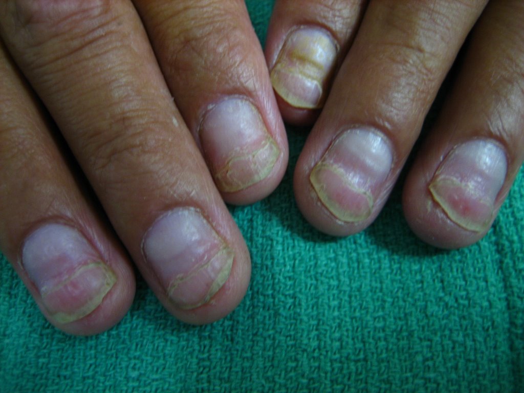
Difficult to cut toenails
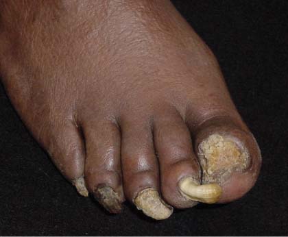
The FP suspected onychomycosis along with some mild tinea pedis. She decided to confirm her suspicion by scraping the nails with the most flaking and looking for fungus with a potassium hydroxide (KOH) preparation. The KOH was negative.
Concerned that the results might be a false negative, the FP cut off a piece of the second toenail and sent it for culture. (A nail with this configuration is called a ram’s horn nail [onychogryphosis], and can be secondary to a fungal infection.) While waiting for the fungal culture, the FP told the patient to apply a topical antifungal for his tinea pedis. The fungal culture came back positive for trichophyton rubrum, the most common cause of onychomycosis. The patient did not have hepatitis C or any other liver disease and recent liver function tests were normal. The FP prescribed terbinafine 250 mg daily for 3 months after making sure that this medication would not adversely interact with the patient’s other medications. The FP told the patient that his nails would improve over the coming months, but that the full effect would not be seen for 10 months.
Photos and text for Photo Rounds Friday courtesy of Richard P. Usatine, MD. This case was adapted from: Mayeaux EJ. Onychomycosis. In: Usatine R, Smith M, Mayeaux EJ, et al, eds. The Color Atlas of Family Medicine. New York, NY: McGraw-Hill; 2009:829-833.
To learn more about The Color Atlas of Family Medicine, see:
• http://www.amazon.com/Color-Atlas-Family-Medicine/dp/0071474641
You can now get The Color Atlas of Family Medicine as an app for mobile devices including the iPhone and iPad by clicking this link:
Dark lines on nails
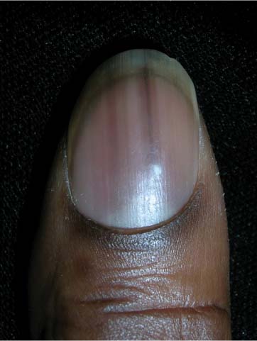
The family physician (FP) recognized that this was a case of longitudinal melanonychia (LM) from racial melanosis with pseudo-Hutchinson’s sign. (The real Hutchinson’s sign is the presence of dark pigment on the proximal nail fold in conjunction with melanoma of the nail unit.)
The FP told the patient that no treatment was needed, but that if the nail pigment started to change or one line became increasingly dark or wide, she should come in for further evaluation. LM is more common in darkly pigmented individuals. It occurs in 77% of African Americans older than 20 years and in almost all of those older than 50 years. It also occurs in 10% to 20% of people who are of Japanese descent, and is common in the Hispanic population. LM is unusual in Caucasians.
Photos and text for Photo Rounds Friday courtesy of Richard P. Usatine, MD. This case was adapted from: Mayeaux EJ. Pigmented nail disorders. In: Usatine R, Smith M, Mayeaux EJ, et al, eds. The Color Atlas of Family Medicine. New York, NY: McGraw-Hill; 2009: 822-825.
To learn more about The Color Atlas of Family Medicine, see:
• http://www.amazon.com/Color-Atlas-Family-Medicine/dp/0071474641
You can now get The Color Atlas of Family Medicine as an app for mobile devices including the iPhone and iPad by clicking this link:
Abnormal thumb
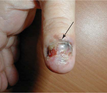
The FP suspected subungual melanoma of the thumb and referred the patient to a surgical oncologist, who confirmed the FP’s suspicions. Subungual melanoma is a type of acrolentiginous melanoma.
The arrow in the image points to hyperpigmentation on the proximal nail fold (Hutchinson’s sign), which is strongly indicative of melanoma. The ulceration suggested that the bad prognosis was going to get even worse.
The initial biopsy showed that the melanoma was greater than 1 mm in depth, so a sentinel node biopsy was planned for the time of surgery. The oncologist thought that the patient might need a partial amputation of the thumb, so a consultation with a hand surgeon was planned.
Subungual melanoma arises on the hand in 45% to 60% of cases, and most of those occur in the thumb. On the foot, subungual melanoma usually occurs in the great toe. The median age at which subungual melanoma is usually diagnosed is in the sixth and seventh decades.
Text for Photo Rounds Friday is courtesy of Richard P. Usatine, MD. Photo is courtesy of Dr. Dubin at http://www.skinatlas.com. This case was adapted from: Mayeaux EJ. Pigmented nail disorders. In: Usatine R, Smith M, Mayeaux EJ, et al, eds. The Color Atlas of Family Medicine. New York, NY: McGraw-Hill; 2009:822-825.
To learn more about The Color Atlas of Family Medicine, see:
• http://www.amazon.com/Color-Atlas-Family-Medicine/dp/0071474641
You can now get The Color Atlas of Family Medicine as an app for mobile devices including the iPhone and iPad by clicking this link:
Band on index finger
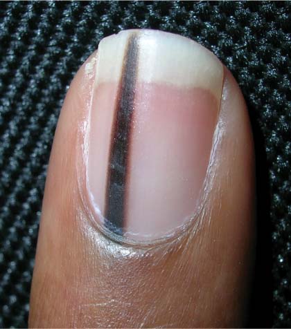
The physician recognized this pigmented band as longitudinal melanonychia (LM), but was concerned about the cause. LM may involve one or several digits and vary in color (light brown to black) and width (2-4 mm). LM is more common in more darkly pigmented individuals.
LM can be caused by chronic trauma, nevi, certain drugs, and endocrine disorders. Subungual melanoma, a rare tumor that can be lethal if it goes undetected, must always be considered in patients with LM.
In this case, the physician performed a biopsy of the nail matrix, which revealed that this was a benign nevus, and not a subungual melanoma.
Photos and text for Photo Rounds Friday courtesy of Richard P. Usatine, MD. This case was adapted from: Mayeaux EJ. Pigmented nail disorders. In: Usatine R, Smith M, Mayeaux EJ, et al, eds. The Color Atlas of Family Medicine. New York, NY: McGraw-Hill; 2009:822-825.
To learn more about The Color Atlas of Family Medicine, see:
• http://www.amazon.com/Color-Atlas-Family-Medicine/dp/0071474641
You can now get The Color Atlas of Family Medicine as an app for mobile devices including the iPhone and iPad by clicking this link:
Breast swelling and erythema in a teen
A 19-YEAR-OLD NULLIPAROUS CAUCASIAN WOMAN came into our family practice clinic; she was concerned about redness and swelling that had developed in her left breast 2 weeks earlier. She’d gone to a local emergency department 8 days ago with similar complaints, and was treated with azithromycin and cephalexin for presumptive mastitis and possible cat scratch disease. Despite the antibiotics, she said that her symptoms had worsened; she’d developed a dry cough, dyspnea, general malaise, and a fever of 100.6°F.
The patient was a former smoker and had asthma as a child. She said her mother had been diagnosed with premenopausal breast cancer at age 31. She also indicated that she had a cat and that it might have scratched her arm prior to the onset of symptoms.
On exam, the patient was afebrile but tachycardic at 140 beats per minute. She had left axillary lymphadenopathy. Her left breast was indurated, erythematous, and generally edematous (FIGURE 1). There was no nipple discharge or evidence of trauma to the skin. The patient had decreased breath sounds at the bases.
A breast ultrasound (obtained to evaluate for possible abscess) showed only diffuse edema—but no abscess. A chest x-ray (FIGURE 2). showed an anterior and middle mediastinal soft tissue mass with a left-sided pleural effusion.
The patient was admitted to the hospital for further evaluation.
FIGURE 1
Induration, erythema, and diffuse edema of left breast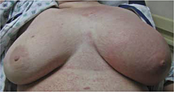
FIGURE 2
Chest x-ray reveals left pleural effusion, mediastinal mass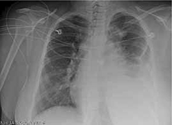
WHAT IS YOUR DIAGNOSIS?
HOW WOULD YOU TREAT THIS PATIENT?
Diagnosis: Diffuse large B-cell lymphoma
This patient had Stage IV-B diffuse large B-cell lymphoma (DLBCL), diagnosed by computed tomography (CT)-guided biopsy of the mediastinal mass. DLBCL is the most common histological subtype of non-Hodgkin lymphoma (NHL), accounting for approximately 30% of NHL cases.1,2
Patients with DLBCL typically present with a rapidly enlarging mass in the neck or abdomen. Thirty percent of patients will have systemic “B” symptoms, including fever, weight loss, and night sweats.3 DLBCL can be highly invasive and may cause compression of the airway and lymphatic or circulatory vessels. While dysphagia, hoarseness, breast swelling, chest pain, and cough can be among the presenting symptoms, superior vena cava syndrome is the most common complication and occurs in 30% of patients.3 The lymphatic obstruction seen in lymphoproliferative disease can produce lymphedema.
In this patient, diffuse lymphadenopathy in the subpectoral and axillary region caused lymphatic obstruction and breast edema. Extra-nodal disease occurs in up to one-third of cases.4 DLBCL can be involved in virtually any tissue, including breast, bone, testes, skin, liver, central nervous system, uterus, and gastrointestinal tract.5
The National Cancer Institute reports that between 2004 and 2008, the US age-adjusted incidence rate for non-Hodgkin lymphoma was 19.8 per 100,000 men and women per year.6 Incidence varies by ethnicity (Caucasians have the highest rate) and increases with age (median age at diagnosis is 66 years with a male predominance).6
Consider infection early on in the differential
The differential diagnosis for unilateral breast swelling and lymphadenopathy includes infection and malignancies. Potential infectious etiologies include acute bacterial mastitis, cat scratch disease, and breast abcess. Malignant causes include inflammatory breast cancer and primary malignant lymphoma. Primary malignant lymphoma of the breast is rare, with an estimated prevalence of 0.04% to 1.1% of all breast tumors and 1.7% to 2.2% of all extranodal non-Hodgkin lymphomas.7
Initial evaluation should be directed toward the most likely etiologies of the symptoms and should include breast ultrasound, blood cultures, and serologies—such as Bartonella—as history dictates.
Once more common causes of unilateral breast swelling are excluded, less common etiologies, such as duct ectasia, mammary adenosis or fat necrosis, and blunt force trauma, should be considered.
Due to the age of our patient, mastitis was initially high on the differential; however, when she did not respond to antibiotic therapy, other causes for her symptoms had to be considered. An ultrasound (noted earlier) ruled out a breast abscess. Blood cultures and serologies were negative. Punch skin biopsies of the patient’s breast showed no evidence of carcinoma, making inflammatory breast cancer less likely.
Imaging studies to start, then biopsy
Many imaging modalities can aid in the diagnosis of DLBCL and help determine the extent of involvement for staging of the disease. These modalities include plain radiograph; CT, magnetic resonance imaging, and positron emission tomography (PET) scans; and lymphangiograms. Confirmatory diagnosis of DLBCL is best made by excisional tissue biopsy. Bone marrow biopsy is also used to help determine staging and management after initial diagnosis.8
Survival hinges on chemotherapy
Survival without treatment in patients with aggressive DLBCL can be measured in months. Combination chemotherapy with cyclophosphamide, doxorubicin, vincristine, and prednisone (the CHOP regimen) increases the disease-free survival rate to between 35% and 45% at 4 years.9 The addition of rituximab has further increased survival in adult patients.10 While the optimal number of treatment cycles remains unclear, 6 to 8 cycles are typically given prior to PET imaging to assess for response.
Consider radiation. Some patients benefit from the addition of radiation to their chemotherapy regimen—particularly those with bulky disease.1 After completion of the planned treatment of DLBCL, a one-month follow-up physical exam with labs and a 2-month follow-up PET/CT scan should be obtained to evaluate response.
It took many scans and tests to arrive at a Dx
The patient’s CT scan showed a large anterior mediastinal mass with central necrosis, diffuse lymphadenopathy, a large left-sided pleural effusion, and multiple pulmonary nodules. Cytologic evaluation of pleural fluid did not show evidence of carcinoma. A fine needle aspiration of the substernal mass under radiologic guidance was performed, and histology was consistent with DLBCL. Further testing confirmed that disease was present both above and below the diaphragm, leading to the diagnosis of Stage IV-B DLBCL.
Our patient. The patient was treated with a chemotherapy regimen of rituximab-cyclophosphamide, vincristine, prednisone, and doxorubicin every 2 weeks for a total of 5 cycles.
Vincristine was discontinued after 2 weeks when the patient developed a Grade 2 peripheral neuropathy at her fingertips. The patient is currently in remission.
CORRESPONDENCE
Larissa Buccolo, MD, Naval Hospital Family Practice Clinic, 2080 Child Street, Jacksonville, FL 32214; [email protected]
1. Nguyen LN, Ha CS, Hess M, et al. The outcome of combined-modality treatments for stage I and II primary large B-cell lymphoma of the mediastinum. Int J Radiat Oncol Biol Phys. 2000;47:1281-1285.
2. Moller MB, Pedersen NT, Christensen BE. Diffuse large B-cell lymphoma: clinical implications of extranodal versus nodal presentation—a population-based study of 1575 cases. Br J Haematol. 2004;124:151-159.
3. Armitage JO, Weisenburger DD. New approach to classifying non-Hodgkin’s lymphoma: clinical features of the major histologic subtypes. Non-Hodgkin’s Lymphoma Classification Project. J Clin Oncol. 1998;16:2780-2795.
4. Brogi E, Harris NL. Lymphomas of the breast: pathology and clinical behavior. Semin Oncol. 1999;22:357-364.
5. Aviles A, Neri N, Huerta-Guzman J. Large bowel lymphoma: an analysis of prognostic factors and therapy in 53 patients. J Surg Oncol. 2002;80:111-115.
6. National Cancer Institute. Surveillance epidemiology and end results. SEER stat fact sheets: non-Hodgkin lymphoma. Available at: (http://seer.cancer.gov/statfacts/html/nhl.html.) Accessed September 28, 2011.
7. Gholam D, Bibeau F, El Welshi A, et al. Primary breast lymphoma. Leuk Lymphoma. 2003;44:1173-1178.
8. Fisher RI, Gaynor ER, Dahlberg S, et al. Comparison of a standard regimen (CHOP) with three intensive chemotherapy regimens for advanced non-Hodgkin’s lymphoma. N Engl J Med. 1993;328:1002-1006.
9. Pond GD, Castellini RA, Horning S, et al. Non-Hodgkin lymphoma: influence of lymphography, CT, and bone marrow biopsy on staging and management. Radiology. 1989;170:159-164.
10. Sehn LH, Donaldson J, Chhanabhai M, et al. Introduction of combined CHOP plus rituximab therapy dramatically improved outcome of diffuse large B-cell lymphoma in British Columbia. J Clin Oncol. 2005;23:5027-5033.
A 19-YEAR-OLD NULLIPAROUS CAUCASIAN WOMAN came into our family practice clinic; she was concerned about redness and swelling that had developed in her left breast 2 weeks earlier. She’d gone to a local emergency department 8 days ago with similar complaints, and was treated with azithromycin and cephalexin for presumptive mastitis and possible cat scratch disease. Despite the antibiotics, she said that her symptoms had worsened; she’d developed a dry cough, dyspnea, general malaise, and a fever of 100.6°F.
The patient was a former smoker and had asthma as a child. She said her mother had been diagnosed with premenopausal breast cancer at age 31. She also indicated that she had a cat and that it might have scratched her arm prior to the onset of symptoms.
On exam, the patient was afebrile but tachycardic at 140 beats per minute. She had left axillary lymphadenopathy. Her left breast was indurated, erythematous, and generally edematous (FIGURE 1). There was no nipple discharge or evidence of trauma to the skin. The patient had decreased breath sounds at the bases.
A breast ultrasound (obtained to evaluate for possible abscess) showed only diffuse edema—but no abscess. A chest x-ray (FIGURE 2). showed an anterior and middle mediastinal soft tissue mass with a left-sided pleural effusion.
The patient was admitted to the hospital for further evaluation.
FIGURE 1
Induration, erythema, and diffuse edema of left breast
FIGURE 2
Chest x-ray reveals left pleural effusion, mediastinal mass
WHAT IS YOUR DIAGNOSIS?
HOW WOULD YOU TREAT THIS PATIENT?
Diagnosis: Diffuse large B-cell lymphoma
This patient had Stage IV-B diffuse large B-cell lymphoma (DLBCL), diagnosed by computed tomography (CT)-guided biopsy of the mediastinal mass. DLBCL is the most common histological subtype of non-Hodgkin lymphoma (NHL), accounting for approximately 30% of NHL cases.1,2
Patients with DLBCL typically present with a rapidly enlarging mass in the neck or abdomen. Thirty percent of patients will have systemic “B” symptoms, including fever, weight loss, and night sweats.3 DLBCL can be highly invasive and may cause compression of the airway and lymphatic or circulatory vessels. While dysphagia, hoarseness, breast swelling, chest pain, and cough can be among the presenting symptoms, superior vena cava syndrome is the most common complication and occurs in 30% of patients.3 The lymphatic obstruction seen in lymphoproliferative disease can produce lymphedema.
In this patient, diffuse lymphadenopathy in the subpectoral and axillary region caused lymphatic obstruction and breast edema. Extra-nodal disease occurs in up to one-third of cases.4 DLBCL can be involved in virtually any tissue, including breast, bone, testes, skin, liver, central nervous system, uterus, and gastrointestinal tract.5
The National Cancer Institute reports that between 2004 and 2008, the US age-adjusted incidence rate for non-Hodgkin lymphoma was 19.8 per 100,000 men and women per year.6 Incidence varies by ethnicity (Caucasians have the highest rate) and increases with age (median age at diagnosis is 66 years with a male predominance).6
Consider infection early on in the differential
The differential diagnosis for unilateral breast swelling and lymphadenopathy includes infection and malignancies. Potential infectious etiologies include acute bacterial mastitis, cat scratch disease, and breast abcess. Malignant causes include inflammatory breast cancer and primary malignant lymphoma. Primary malignant lymphoma of the breast is rare, with an estimated prevalence of 0.04% to 1.1% of all breast tumors and 1.7% to 2.2% of all extranodal non-Hodgkin lymphomas.7
Initial evaluation should be directed toward the most likely etiologies of the symptoms and should include breast ultrasound, blood cultures, and serologies—such as Bartonella—as history dictates.
Once more common causes of unilateral breast swelling are excluded, less common etiologies, such as duct ectasia, mammary adenosis or fat necrosis, and blunt force trauma, should be considered.
Due to the age of our patient, mastitis was initially high on the differential; however, when she did not respond to antibiotic therapy, other causes for her symptoms had to be considered. An ultrasound (noted earlier) ruled out a breast abscess. Blood cultures and serologies were negative. Punch skin biopsies of the patient’s breast showed no evidence of carcinoma, making inflammatory breast cancer less likely.
Imaging studies to start, then biopsy
Many imaging modalities can aid in the diagnosis of DLBCL and help determine the extent of involvement for staging of the disease. These modalities include plain radiograph; CT, magnetic resonance imaging, and positron emission tomography (PET) scans; and lymphangiograms. Confirmatory diagnosis of DLBCL is best made by excisional tissue biopsy. Bone marrow biopsy is also used to help determine staging and management after initial diagnosis.8
Survival hinges on chemotherapy
Survival without treatment in patients with aggressive DLBCL can be measured in months. Combination chemotherapy with cyclophosphamide, doxorubicin, vincristine, and prednisone (the CHOP regimen) increases the disease-free survival rate to between 35% and 45% at 4 years.9 The addition of rituximab has further increased survival in adult patients.10 While the optimal number of treatment cycles remains unclear, 6 to 8 cycles are typically given prior to PET imaging to assess for response.
Consider radiation. Some patients benefit from the addition of radiation to their chemotherapy regimen—particularly those with bulky disease.1 After completion of the planned treatment of DLBCL, a one-month follow-up physical exam with labs and a 2-month follow-up PET/CT scan should be obtained to evaluate response.
It took many scans and tests to arrive at a Dx
The patient’s CT scan showed a large anterior mediastinal mass with central necrosis, diffuse lymphadenopathy, a large left-sided pleural effusion, and multiple pulmonary nodules. Cytologic evaluation of pleural fluid did not show evidence of carcinoma. A fine needle aspiration of the substernal mass under radiologic guidance was performed, and histology was consistent with DLBCL. Further testing confirmed that disease was present both above and below the diaphragm, leading to the diagnosis of Stage IV-B DLBCL.
Our patient. The patient was treated with a chemotherapy regimen of rituximab-cyclophosphamide, vincristine, prednisone, and doxorubicin every 2 weeks for a total of 5 cycles.
Vincristine was discontinued after 2 weeks when the patient developed a Grade 2 peripheral neuropathy at her fingertips. The patient is currently in remission.
CORRESPONDENCE
Larissa Buccolo, MD, Naval Hospital Family Practice Clinic, 2080 Child Street, Jacksonville, FL 32214; [email protected]
1. Nguyen LN, Ha CS, Hess M, et al. The outcome of combined-modality treatments for stage I and II primary large B-cell lymphoma of the mediastinum. Int J Radiat Oncol Biol Phys. 2000;47:1281-1285.
2. Moller MB, Pedersen NT, Christensen BE. Diffuse large B-cell lymphoma: clinical implications of extranodal versus nodal presentation—a population-based study of 1575 cases. Br J Haematol. 2004;124:151-159.
3. Armitage JO, Weisenburger DD. New approach to classifying non-Hodgkin’s lymphoma: clinical features of the major histologic subtypes. Non-Hodgkin’s Lymphoma Classification Project. J Clin Oncol. 1998;16:2780-2795.
4. Brogi E, Harris NL. Lymphomas of the breast: pathology and clinical behavior. Semin Oncol. 1999;22:357-364.
5. Aviles A, Neri N, Huerta-Guzman J. Large bowel lymphoma: an analysis of prognostic factors and therapy in 53 patients. J Surg Oncol. 2002;80:111-115.
6. National Cancer Institute. Surveillance epidemiology and end results. SEER stat fact sheets: non-Hodgkin lymphoma. Available at: (http://seer.cancer.gov/statfacts/html/nhl.html.) Accessed September 28, 2011.
7. Gholam D, Bibeau F, El Welshi A, et al. Primary breast lymphoma. Leuk Lymphoma. 2003;44:1173-1178.
8. Fisher RI, Gaynor ER, Dahlberg S, et al. Comparison of a standard regimen (CHOP) with three intensive chemotherapy regimens for advanced non-Hodgkin’s lymphoma. N Engl J Med. 1993;328:1002-1006.
9. Pond GD, Castellini RA, Horning S, et al. Non-Hodgkin lymphoma: influence of lymphography, CT, and bone marrow biopsy on staging and management. Radiology. 1989;170:159-164.
10. Sehn LH, Donaldson J, Chhanabhai M, et al. Introduction of combined CHOP plus rituximab therapy dramatically improved outcome of diffuse large B-cell lymphoma in British Columbia. J Clin Oncol. 2005;23:5027-5033.
Lines across fingernails
The FP diagnosed Mees’ lines in this patient.
Mees’ lines spread transversely across the entire breadth of the nail and are somewhat rounded, with a contour similar to the distal lunula. Mees’ lines tend to occur on several nails at once. A history of a systemic insult is correlated with the onset of the lines, such as chemotherapy, heart failure, or arsenic poisoning. In this case, each transverse white band across the nail correlated with a round of chemotherapy.
The FP explained the correlation between the white bands and the chemotherapy and told the patient that since the chemotherapy was complete, the white bands would grow out over time (probably in a couple of months).
Text for Photo Rounds Friday courtesy of Richard P. Usatine, MD. Photo courtesy of Jeff Meffert, MD. This case was adapted from: Mayeaux EJ. Normal nail variants. In: Usatine R, Smith M, Mayeaux EJ, et al, eds. The Color Atlas of Family Medicine. New York, NY: McGraw-Hill; 2009:819-821.
To learn more about The Color Atlas of Family Medicine, see:
• http://www.amazon.com/Color-Atlas-Family-Medicine/dp/0071474641
You can now get The Color Atlas of Family Medicine as an app for mobile devices including the iPhone and iPad by clicking this link:
Indentation across fingernails
The FP recognized the nail condition as Beau's lines. Given that fingernails grow approximately 2 to 3 mm per month, the physiologic stressor causing the Beau’s lines was likely the cholecystitis and cholecystectomy 2 months earlier. The physician reassured the patient that the lines would grow out and likely be gone by 4 months, at the latest.
The FP also used the visit as an opportunity to reinforce the importance of a good diet, exercise, and weight loss for the patient's general health.
Text for Photo Rounds Friday courtesy of Richard P. Usatine, MD. Photo courtesy of Suraj Reddy, MD. This case was adapted from: Mayeaux EJ. Normal nail variants. In: Usatine R, Smith M, Mayeaux EJ, et al, eds. The Color Atlas of Family Medicine. New York, NY: McGraw-Hill; 2009:819-821.
To learn more about The Color Atlas of Family Medicine, see:
• http://www.amazon.com/Color-Atlas-Family-Medicine/dp/0071474641
You can now get The Color Atlas of Family Medicine as an app for mobile devices including the iPhone and iPad by clicking this link: