User login
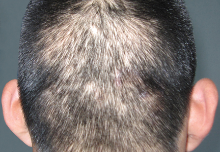
Isolated Linear Lichen Planopilaris: Extremely Rare When Limited to the Scalp
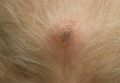
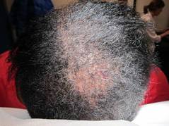
An Illustrative Case Report of Secondary Syphilis With Alopecia Syphilitica, Perianal Condyloma Latum, and Granuloma Annulare–Like Lesions
Lung Cancer–Associated Scalp Hair Loss: A Rare Cause of Secondary Alopecia Neoplastica
Cosmetic Corner: Dermatologists Weigh in on Shampoos and Conditioners
To improve patient care and outcomes, leading dermatologists offered their recommendations on the top shampoos and conditioners. Consideration must be given to:
Johnson & Johnson Consumer Companies, Inc
Recommended by Marian Northington, MD, Birmingham, Alabama
Procter & Gamble
“Head & Shoulders shampoo and conditioner is by far one of the best formulations on the market in terms of a therapeutic shampoo. It is not only gentle and cleanses but clears dandruff quickly. There are a lot of new products in this line to help with different hair and scalp needs.”—Marta I. Rendon, MD, Miami, Florida
• Série Expert Intense Repair Conditioner
L'Oréal Paris Professional
“This is an excellent shampoo for dry and color-treated hair because it is extremely hydrating and moisturizing.”—Marian Northington, MD, Birmingham, Alabama
Aveda Corporation
“This shampoo is free of sodium lauryl sulfate, which can be a contact allergen for some people, and adds moisture to the hair with ingredients such as aloe.”—Anthony M. Rossi, MD, New York, New York
Guthy-Renker
“This product is a 5-in-1 formula that takes the place of your shampoo, conditioner, deep conditioner, detangler, and leave-in conditioner. It cleanses thoroughly without stripping hair and scalp of their natural oils.”—Basil M. Hantash, MD, PhD, Turlock, California
Cutis invites readers to send us their recommendations. Facial moisturizers, scar treatments, and over-the-counter antifungals will be featured in upcoming editions of Cosmetic Corner. Please e-mail your recommendation(s) to [email protected].
Disclaimer: Opinions expressed herein do not necessarily reflect those of Cutis or Frontline Medical Communications Inc. and shall not be used for product endorsement purposes. Any reference made to a specific commercial product does not indicate or imply that Cutis or Frontline Medical Communications Inc. endorses, recommends, or favors the product mentioned. No guarantee is given to the effects of recommended products.
Diagnosing hair loss in black patients starts with right questions
NEW YORK – To get to the root cause of hair loss in African American patients, ask them about their chemical styling and physical styling practices, advised Dr. Roopal V. Kundu.
"Ask them about the hair products they use; how they style their hair; what kind of relaxers they use; what kind of heat, how much of it, and how often they use it; and find out if they’re wearing extensions or weaves," she said to a packed room during the American Academy of Dermatology summer meeting.
"And get their hair stylist’s input," said Dr. Kundu, director of the Center for Ethnic Skin at Northwestern University, Chicago.
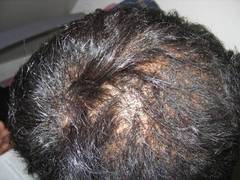
Central centrifugal cicatricial alopecia (CCCA) is one of the most common types of hair loss in African American women, Dr. Kundu said. The reason for the prevalence of CCCA in this population remains unclear, but data from several studies point not only to genetic and ethnic predispositions, but also to hair styling practices.
Data from one study showed that women with CCCA were significantly more likely to report recent traumatic hairstyling than were those who did not have the condition (J. Am. Acad. Dermatol. 2009;60:574-8).
"Educate your patients [about the effect of hairstyling practices on hair loss] as early as possible," and suspect CCCA in patients who present with a lot of breakage in their hair, even if they have no sign of alopecia or scalp inflammation, Dr. Kundu said.
Keep chemical and physical hairstyling practices in mind when examining and diagnosing hair loss in black women, she noted.
Chemical relaxers, also known as perms, are one of the most common treatments used by African American women. The relaxers are used to loosen tight curls and are applied every 6-8 weeks to new growth only. There are two types of relaxers: lye (professional product) or no lye (at-home use). The lye relaxers have more potential for irritation, Dr. Kundu said.
Physical styling, which usually pulls on hair, also can be a factor in hair loss, she explained. Some popular types of physical styling include:
• Braids, which often involve added synthetic hair and are left in place for weeks to months.
• Twisting, which involves interlocking two pieces of hair to create a loose or bound-down, three-dimensional hair style.
• Cornrows, which are braids that lie flat against the scalp, with or without added hair, that can cause excessive tension on the hair.
• Locks or dreads, which are matted coils of hair.
• Weaves, which can be clipped, glued, or sewn into place.
In addition, thermal styling (with tools such as flatirons) is a popular treatment that can damage the hair because of the high temperature of the iron, Dr. Kundu said.
When examining a skin of color patient who presents with hair loss, note their hairstyle first, Dr. Kundu emphasized. "Ask them to take their hairpiece off [if they are wearing one]. If the hairstyle doesn’t allow you to examine the scalp, the patient needs to come back between hairstyles," she said.
Grab a dermatoscope and examine the scalp, Dr. Kundu said. Look for hair loss, hair breakage, and traction. She cited a recent study suggesting that early CCCA should be considered in the differential diagnosis of these patients (Arch. Dermatol. 2012;148:1047-52).
"And don’t dismiss scalp dysesthesia* in a black woman," Dr. Kundu emphasized. "I really take this to heart," she said.
Biopsy the patient, "and always go for the periphery to see if there is active inflammation," since the central area of hair loss is usually scarred, she noted.
Dr. Kundu said that she usually prescribes potent to superpotent topical steroids, such as fluocinonide daily, and then reduces the steroid to 1-3 times a week for maintenance. She also performs intralesional injections every 6 weeks, and stops if she sees no efficacy after 3-4 serial injections.
When prescribing minoxidil, she advised taking the patient’s facial hair into consideration, and warns them about additional facial hair growth.

For patients with significant inflammation, Dr. Kundu said she first turns to doxycycline (starting at 100 mg twice a day and tapering to anti-inflammatory doses of 50 mg daily) for a total of 3-6 months.
Meanwhile, advise women with hair loss or hair loss concerns to modify their grooming practices to avoid hairstyles that pull on the scalp, she said. Also advise them to let their hair go natural completely for at least a year if possible. "It’s a hard job to do for some patients," she acknowledged, so at least encourage them to apply chemical relaxers less frequently, every 10-12 weeks instead, she said.
In addition, ask these patients to refrain from using heat on their hair more than once or twice per week, and suggest air-drying instead, she said.
Dr. Kundu had no relevant financial conflicts to disclose.
On Twitter @naseemsmiller
*CORRECTION, 10/21/13: An earlier version of this article misstated the effects of dysesthesia.
NEW YORK – To get to the root cause of hair loss in African American patients, ask them about their chemical styling and physical styling practices, advised Dr. Roopal V. Kundu.
"Ask them about the hair products they use; how they style their hair; what kind of relaxers they use; what kind of heat, how much of it, and how often they use it; and find out if they’re wearing extensions or weaves," she said to a packed room during the American Academy of Dermatology summer meeting.
"And get their hair stylist’s input," said Dr. Kundu, director of the Center for Ethnic Skin at Northwestern University, Chicago.
Central centrifugal cicatricial alopecia (CCCA) is one of the most common types of hair loss in African American women, Dr. Kundu said. The reason for the prevalence of CCCA in this population remains unclear, but data from several studies point not only to genetic and ethnic predispositions, but also to hair styling practices.
Data from one study showed that women with CCCA were significantly more likely to report recent traumatic hairstyling than were those who did not have the condition (J. Am. Acad. Dermatol. 2009;60:574-8).
"Educate your patients [about the effect of hairstyling practices on hair loss] as early as possible," and suspect CCCA in patients who present with a lot of breakage in their hair, even if they have no sign of alopecia or scalp inflammation, Dr. Kundu said.
Keep chemical and physical hairstyling practices in mind when examining and diagnosing hair loss in black women, she noted.
Chemical relaxers, also known as perms, are one of the most common treatments used by African American women. The relaxers are used to loosen tight curls and are applied every 6-8 weeks to new growth only. There are two types of relaxers: lye (professional product) or no lye (at-home use). The lye relaxers have more potential for irritation, Dr. Kundu said.
Physical styling, which usually pulls on hair, also can be a factor in hair loss, she explained. Some popular types of physical styling include:
• Braids, which often involve added synthetic hair and are left in place for weeks to months.
• Twisting, which involves interlocking two pieces of hair to create a loose or bound-down, three-dimensional hair style.
• Cornrows, which are braids that lie flat against the scalp, with or without added hair, that can cause excessive tension on the hair.
• Locks or dreads, which are matted coils of hair.
• Weaves, which can be clipped, glued, or sewn into place.
In addition, thermal styling (with tools such as flatirons) is a popular treatment that can damage the hair because of the high temperature of the iron, Dr. Kundu said.
When examining a skin of color patient who presents with hair loss, note their hairstyle first, Dr. Kundu emphasized. "Ask them to take their hairpiece off [if they are wearing one]. If the hairstyle doesn’t allow you to examine the scalp, the patient needs to come back between hairstyles," she said.
Grab a dermatoscope and examine the scalp, Dr. Kundu said. Look for hair loss, hair breakage, and traction. She cited a recent study suggesting that early CCCA should be considered in the differential diagnosis of these patients (Arch. Dermatol. 2012;148:1047-52).
"And don’t dismiss scalp dysesthesia* in a black woman," Dr. Kundu emphasized. "I really take this to heart," she said.
Biopsy the patient, "and always go for the periphery to see if there is active inflammation," since the central area of hair loss is usually scarred, she noted.
Dr. Kundu said that she usually prescribes potent to superpotent topical steroids, such as fluocinonide daily, and then reduces the steroid to 1-3 times a week for maintenance. She also performs intralesional injections every 6 weeks, and stops if she sees no efficacy after 3-4 serial injections.
When prescribing minoxidil, she advised taking the patient’s facial hair into consideration, and warns them about additional facial hair growth.
For patients with significant inflammation, Dr. Kundu said she first turns to doxycycline (starting at 100 mg twice a day and tapering to anti-inflammatory doses of 50 mg daily) for a total of 3-6 months.
Meanwhile, advise women with hair loss or hair loss concerns to modify their grooming practices to avoid hairstyles that pull on the scalp, she said. Also advise them to let their hair go natural completely for at least a year if possible. "It’s a hard job to do for some patients," she acknowledged, so at least encourage them to apply chemical relaxers less frequently, every 10-12 weeks instead, she said.
In addition, ask these patients to refrain from using heat on their hair more than once or twice per week, and suggest air-drying instead, she said.
Dr. Kundu had no relevant financial conflicts to disclose.
On Twitter @naseemsmiller
*CORRECTION, 10/21/13: An earlier version of this article misstated the effects of dysesthesia.
AT THE AAD SUMMER ACADEMY 2013

Efinaconazole Solution 10%: Topical Antifungal Therapy for Toenail Onychomycosis
Use images and analogies to explain hair disorders
NEW YORK – Dr. Paradi Mirmirani has an elegant way of explaining hair loss to her postmenopausal patients.
"Think about [hair production] like an orchestra. When your body is making hair, a whole group of musicians are coming together to make music. But when you’re postmenopausal, the estrogen isn’t there, the head violinist isn’t there, and it’s not going to be the same, but you’re still making music. It’s not going to be same sound, but it’s still there," she said.
Dr. Mirmirani of the University of California, San Francisco, is well recognized for her research in hair disorders, and during her presentation at the American Academy of Dermatology’s summer meeting, she shared her tips on diagnosing and treating different types of hair loss and alopecia in women:
• Telogen effluvium (I’m shedding gobs of hair!)
Find out whether the patient has a history of weight loss or is on new oral contraceptives, Dr. Mirmirani said. Use a hair shaft contrast card and search the scalp for scarring or scaling. Also, perform a "pull test" (for bulb) and "tug test" (for shaft), she advised. For laboratory data, she recommended ferritin and TSH, and possibly tests for antinuclear antibodies and vitamin D levels, and a biopsy of the area. Treat the underlying problem, not the hair, and assure patients that they will not go bald, she said.
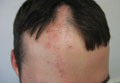
• Traction alopecia (I’ve got a bald spot!)
Start by asking these patients what they do for hair care and styling, said Dr. Mirmirani. On exam, look for a telltale "fringe sign," which she and her colleagues described in a paper as "the presence of retained hairs along the frontal and/or temporal rim." (J. Clin. Exp. Dermatol. Res. 2011;2:117).
She recommended ordering ferritin and TSH tests for these patients, and possibly vitamin D. Also, treat any inflammation; consider nonspecific hair growth treatments such as minoxidil, and surgical hair restoration, and remind patients to treat their hair gently, she said.
• Alopecia areata
Ask alopecia areata patients about a personal or family history of atopic disorders, Dr. Mirmirani advised. Treatment options include intralesional corticosteroids (10 mg/cc to 2 cc total); topical corticosteroids; topical minoxidil 5% twice daily; short-contact anthralin (up to 30 minutes); topical immunotherapy; or psoralen + ultraviolet A (PUVA) treatment. Finally, remind patients that alopecia areata is an autoimmune condition that is not contagious, Dr. Mirmirani said. Compare it to an unwelcome house guest, she suggested.
• Female pattern hair loss (midlife hair crisis)
Start care for these patients by ordering (only if virilized) free and total T, dehydroepiandrosterone sulfate, and prolactin tests, Dr. Mirmirani said. Her recommended treatment protocol: minoxidil 2% or 5% solution twice daily, or 5% foam once daily; finasteride (1 mg) or spironolactone. Also, consider hair restoration or cosmetics that bring the scalp’s color closer to hair color and make hair loss less apparent, she suggested. This is when to remind patients that "the orchestra is still playing" (hair may still be produced) although estrogen (the first violinist) is absent, so it may not be quite the same.
• Acquired trichorrhexis nodosa (My hair just won’t grow!)
Acquired trichorrhexis nodosa is the most commonly reported hair shaft defect, Dr. Mirmirani said. It often results from excessive chemical processes and heat applied to hair. She described her typical acquired trichorrhexis nodosa patient as a 24-year-old black woman who washes her every 2 weeks and straightens every 6 weeks, her hair has been breaking off in the back, started after a recent color, with no symptoms.
In these patients, and exam usually shows that the overall hair density is good, no alopecia, a localized area of short hair with blunt ends, and a positive tug test (hair breaks off easily). The scalp often has mild scaling, but no pustules.
Make a hair mount, and show the patient her hair under the microscope; "it’s the easiest and most satisfying thing you can do," said Dr. Mirmirani.
Advise patients to use gentle hair care, trim unhealthy hair, and avoid heat and chemicals, she said. Wigs are fine for these patients, and most will recover the condition of their hair within a year or 2, she added.
• Cicatricial alopecia
Start with a biopsy around the margin of early active area, and then culture the pustules, said Dr. Mirmirani. Dermatopathology can show whether sebaceous glands are absent, and the degree of inflammatory infiltrate.
Explain to these patients that the hair roots or bulbs have been damaged, she added. Tell them regrowth is not possible, but they can relieve the signs and symptoms of the condition and prevent it from spreading. Describe it as "like a wildfire; we want to contain it, and halt the spread," she suggested.
Treat the predominantly lymphocytic patients with anti-inflammatories (intralesionals and topicals; antibiotics and antimalarials; systemic anti-inflammatory therapies), said Dr. Mirmirani. For lichen planopilaris and frontal fibrosing alopecia, try PPAR-gamma agonists; use topical minoxidil or finasteride to promote nonspecific hair growth; or try cosmetic or surgical therapies, she said.
Treat folliculitis decalvans with antibacterial treatments and staph eradication, and treat dissecting cellulitis with intralesionals/anti-inflammatory drugs; perform incision and drainage; use isotretinoin and antitumor-necrosis factor; and consider laser hair removal, she noted.
Dr. Mirmirani said that, in her experience, many dermatologists dread seeing hair-loss patients because of a lack of training in how to care for them. She shared several educational resources, including a reference book she coauthored, "Cicatricial Alopecia: An Approach to Diagnosis and Management," (New York: Springer, 2011) to help clinicians and residents better understand and treat hair disorders, especially the rare kinds. She also recommended the North American Hair Research Society and the Cicatricial Alopecia Research Foundation as useful resources.
Dr. Mirmirani has been an investigator and/or consultant for Johnson & Johnson, as well as Procter & Gamble.
On Twitter @NaseemSMiller
NEW YORK – Dr. Paradi Mirmirani has an elegant way of explaining hair loss to her postmenopausal patients.
"Think about [hair production] like an orchestra. When your body is making hair, a whole group of musicians are coming together to make music. But when you’re postmenopausal, the estrogen isn’t there, the head violinist isn’t there, and it’s not going to be the same, but you’re still making music. It’s not going to be same sound, but it’s still there," she said.
Dr. Mirmirani of the University of California, San Francisco, is well recognized for her research in hair disorders, and during her presentation at the American Academy of Dermatology’s summer meeting, she shared her tips on diagnosing and treating different types of hair loss and alopecia in women:
• Telogen effluvium (I’m shedding gobs of hair!)
Find out whether the patient has a history of weight loss or is on new oral contraceptives, Dr. Mirmirani said. Use a hair shaft contrast card and search the scalp for scarring or scaling. Also, perform a "pull test" (for bulb) and "tug test" (for shaft), she advised. For laboratory data, she recommended ferritin and TSH, and possibly tests for antinuclear antibodies and vitamin D levels, and a biopsy of the area. Treat the underlying problem, not the hair, and assure patients that they will not go bald, she said.
• Traction alopecia (I’ve got a bald spot!)
Start by asking these patients what they do for hair care and styling, said Dr. Mirmirani. On exam, look for a telltale "fringe sign," which she and her colleagues described in a paper as "the presence of retained hairs along the frontal and/or temporal rim." (J. Clin. Exp. Dermatol. Res. 2011;2:117).
She recommended ordering ferritin and TSH tests for these patients, and possibly vitamin D. Also, treat any inflammation; consider nonspecific hair growth treatments such as minoxidil, and surgical hair restoration, and remind patients to treat their hair gently, she said.
• Alopecia areata
Ask alopecia areata patients about a personal or family history of atopic disorders, Dr. Mirmirani advised. Treatment options include intralesional corticosteroids (10 mg/cc to 2 cc total); topical corticosteroids; topical minoxidil 5% twice daily; short-contact anthralin (up to 30 minutes); topical immunotherapy; or psoralen + ultraviolet A (PUVA) treatment. Finally, remind patients that alopecia areata is an autoimmune condition that is not contagious, Dr. Mirmirani said. Compare it to an unwelcome house guest, she suggested.
• Female pattern hair loss (midlife hair crisis)
Start care for these patients by ordering (only if virilized) free and total T, dehydroepiandrosterone sulfate, and prolactin tests, Dr. Mirmirani said. Her recommended treatment protocol: minoxidil 2% or 5% solution twice daily, or 5% foam once daily; finasteride (1 mg) or spironolactone. Also, consider hair restoration or cosmetics that bring the scalp’s color closer to hair color and make hair loss less apparent, she suggested. This is when to remind patients that "the orchestra is still playing" (hair may still be produced) although estrogen (the first violinist) is absent, so it may not be quite the same.
• Acquired trichorrhexis nodosa (My hair just won’t grow!)
Acquired trichorrhexis nodosa is the most commonly reported hair shaft defect, Dr. Mirmirani said. It often results from excessive chemical processes and heat applied to hair. She described her typical acquired trichorrhexis nodosa patient as a 24-year-old black woman who washes her every 2 weeks and straightens every 6 weeks, her hair has been breaking off in the back, started after a recent color, with no symptoms.
In these patients, and exam usually shows that the overall hair density is good, no alopecia, a localized area of short hair with blunt ends, and a positive tug test (hair breaks off easily). The scalp often has mild scaling, but no pustules.
Make a hair mount, and show the patient her hair under the microscope; "it’s the easiest and most satisfying thing you can do," said Dr. Mirmirani.
Advise patients to use gentle hair care, trim unhealthy hair, and avoid heat and chemicals, she said. Wigs are fine for these patients, and most will recover the condition of their hair within a year or 2, she added.
• Cicatricial alopecia
Start with a biopsy around the margin of early active area, and then culture the pustules, said Dr. Mirmirani. Dermatopathology can show whether sebaceous glands are absent, and the degree of inflammatory infiltrate.
Explain to these patients that the hair roots or bulbs have been damaged, she added. Tell them regrowth is not possible, but they can relieve the signs and symptoms of the condition and prevent it from spreading. Describe it as "like a wildfire; we want to contain it, and halt the spread," she suggested.
Treat the predominantly lymphocytic patients with anti-inflammatories (intralesionals and topicals; antibiotics and antimalarials; systemic anti-inflammatory therapies), said Dr. Mirmirani. For lichen planopilaris and frontal fibrosing alopecia, try PPAR-gamma agonists; use topical minoxidil or finasteride to promote nonspecific hair growth; or try cosmetic or surgical therapies, she said.
Treat folliculitis decalvans with antibacterial treatments and staph eradication, and treat dissecting cellulitis with intralesionals/anti-inflammatory drugs; perform incision and drainage; use isotretinoin and antitumor-necrosis factor; and consider laser hair removal, she noted.
Dr. Mirmirani said that, in her experience, many dermatologists dread seeing hair-loss patients because of a lack of training in how to care for them. She shared several educational resources, including a reference book she coauthored, "Cicatricial Alopecia: An Approach to Diagnosis and Management," (New York: Springer, 2011) to help clinicians and residents better understand and treat hair disorders, especially the rare kinds. She also recommended the North American Hair Research Society and the Cicatricial Alopecia Research Foundation as useful resources.
Dr. Mirmirani has been an investigator and/or consultant for Johnson & Johnson, as well as Procter & Gamble.
On Twitter @NaseemSMiller
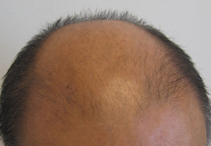
Female hair loss differs by age
SAN FRANCISCO – Three distinct stages of pattern hair loss in women are related to the age of onset, and are not necessarily androgen related.
Between puberty and age 40 years, hair miniaturization in females tends to be caused by androgenetic alopecia, a common hereditary thinning or balding induced by androgens in genetically susceptible people of both sexes. By contrast, women in their 60s or older may develop hair thinning from age-related, or "senescent" alopecia, which is distinct from androgenetic alopecia because senescent alopecia is not mediated by dihydrotestosterone, Dr. Vera H. Price said at the annual meeting of the Pacific Dermatologic Association.
However, a newly identified stage that often occurs between 45 and 55 years of age is gaining popularity in the lexicon of hair loss. In this stage, called "female pattern hair loss," the role of androgens is less clear-cut, and other hormonal and nonhormonal factors may play a role, said Dr. Price, professor of dermatology at the University of California, San Francisco.
All three stages show similar histopathology, with follicular downsizing, normal sebaceous glands, and no significant inflammation. However, recognizing and understanding the three stages help inform management, said Dr. Price.
Treatment with minoxidil is suitable for all three stages of hair loss, for example, but androgen blockade via off-label treatment with finasteride is not helpful in senescent alopecia. "I don’t use it after 60 years of age in women or men," she said.
Millions of men who have taken finasteride 5 mg/day for benign prostatic hypertrophy have not regrown scalp hair, evidence that senescent alopecia is not dihydrotestosterone mediated, she noted.
• Androgenetic alopecia. To screen for suspected androgenetic alopecia in women, check for menstrual irregularities, infertility, hirsutism, severe cystic acne, galactorrhea, and virilization. "If none are present, you do not have to do a single hormonal test" because it’s not androgenetic alopecia causing the hair loss, Dr. Price said.
If any one of those conditions is present, however, check levels of testosterone, dehydroepiandrosterone sulfate (DHEAS), and prolactin.
In all hair loss patients, get a complete blood count and check for normal levels of thyroid-stimulating hormone (TSH), ferritin, and 25-hydroxyvitamin D; the latter two are required for a normal hair cycle. Be sure to order the ferritin level specifically, because ordering "iron studies" won’t include a ferritin level, she added.
• Female pattern hair loss. The pathogenesis behind female pattern hair loss is not well understood. As women go through menopause, hair growth parameters change. The hair growth rate slows, the anagen/telogen ratio decreases, and hair diameters become smaller, Dr. Price explained.
Hormonal and molecular factors that may influence female pattern hair loss need to be better defined, including the possible roles of estrogen, estrogen receptor (ER)-beta, aromatase, 5-alpha-reductase, dihydrotestosterone, and androgen receptors, she said.
"When we understand that, we’ll understand this group a little more clearly," she added. "I’ve always been puzzled a little bit when the onset is in this age group."
• Senescent alopecia. "I call it ‘wisdom-related’ alopecia," quipped Dr. Price. The gene expression profiles of androgenetic alopecia and senescent alopecia differ. In the former, hair growth cycle genes are differentially expressed. In the latter, systemic senescent/aging genes are differentially expressed, suggesting these are two distinct disorders.
• Management. For any stage of hair loss, minoxidil (Rogaine) may help if used properly over an extended period of time, said Dr. Price. Apply the 2% or 5% solution every single day directly to a dry scalp, not right after a shower, and spread it gently across the scalp, she advised. Give minoxidil time to absorb without spraying, moussing, or blow-drying the hair while it absorbs. Once-daily minoxidil foam 5% also has been approved for women and is less oily and absorbed faster.
Of note, androgen blockade via off-label oral finasteride 1 mg/day for confirmed androgenetic alopecia is contraindicated in women who may be or may become pregnant because it will cause hypospadias in a male fetus, said Dr. Price. Prescribe concurrent oral contraception in premenopausal women or make sure the woman is postmenopausal when using finasteride.
"Do I use it in women? I do, but I’m very careful," she said. "They have to be post hysterectomy or post tubal ligation. I want to be certain that they’re not going to conceive."
In appropriate patients, minoxidil and finasteride could be used together if the patient can afford it, she added.
Spironolactone, an androgen receptor inhibitor, also has been used in a dose of 200 mg/day to retard hair thinning due to androgenetic alopecia. Data show that spironolactone 50-200 mg/day can be used successfully to treat acne and hirsutism, but there are no evidence-based studies showing that it helps hair regrowth.
"I use very little spironolactone for androgenetic alopecia," Dr. Price said. "It will not grow hair. I think it’s used a lot because people aren’t familiar with minoxidil or don’t know about using finasteride if there’s no possibility of pregnancy."
Dr. Price reported having financial associations with Allergan and Follica, neither of which was pertinent to this topic.
On Twitter @sherryboschert
EXPERT ANALYSIS FROM THE PDA ANNUAL MEETING