User login
Ipilimumab plus surgery boosted advanced melanoma survival
NATIONAL HARBOR, MD – Patients with stage IV melanoma treated with a combination of ipilimumab and surgical resection had a high rate of melanoma-specific and overall survival, a retrospective study of a single-center case series has shown.
"To our knowledge, this is the first report of 5-year melanoma-specific survival data on patients who have undergone surgical resection and ipilimumab treatment, and the data suggests that surgical resection and ipilimumab treatment may result in long-term survival in select metastatic melanoma patients," Dr. Junko Ozao-Choy said at the annual Society of Surgical Oncology Cancer Symposium.
Among 44 patients treated with the CTLA-4 (cytotoxic T-lymphocyte antigen 4) inhibitor ipilimumab (Yervoy) and surgical resection, the 5-year melanoma-specific survival (MSS) rate was 51% and the median overall survival duration was 60 months, reported Dr. Ozao-Choy of the John Wayne Cancer Institute at Saint John’s Health Center in Santa Monica, Calif.
For 24 patients who received ipilimumab before resection, the 5-year MSS was 61% at a median of 60 months, and for 18 of 20 patients treated with ipilimumab after surgery, the 5-year MSS was 42% at a median of 47 months, but this difference was not significant (data were incomplete for 2 patients in the latter group), she noted.
In a recent study of retrospective data on patients with metastatic melanoma treated at her center, the 4-year survival of patients who underwent resection of metastatic lesions with or without systemic medical therapy was 20.8%, compared with 7% for those who underwent systemic medical therapy alone. The study investigators concluded that more than half of patients with metastatic melanoma were eligible for metastasectomy (Ann. Surg. Oncol. 2012;19:2547-55).
Dr. Ozao-Choy and her colleagues reviewed the center’s records on patients with metastatic melanoma who underwent resection and had received ipilimumab, looking at disease-specific survival from the date of diagnosis of stage IV disease.
The groups were well balanced in terms of age, sex, mean Breslow thickness scores, and nodal status. However, significantly more patients who received ipilimumab before surgery had brain metastases (13 of 24 vs. 3 of 18, P = .001). In a univariate analysis, patients with brain metastases had a significantly worse 5-year MSS (31% vs. 60%, P = .049).
The only other significant variables associated in the univariate analysis with better survival were prior immunotherapy, with 69% of patients who had received any immunotherapy having a 5-year MSS of 69%, compared with 29% for those with no immunotherapy (P = .01), and previous number of resections, with more resections being associated with better survival (P = .01).
Neither previous treatment with Bacillus Calmette-Guérin vaccine, previous chemotherapy, T stage, N stage, or timing of ipilimumab were significantly associated with MSS.
In a multivariate analysis (which controlled for demographic and disease factors), only the previous number of resections remained a significant predictor of MSS (P = .01).
In the audience response segment following the presentation, Dr. Daniel G. Coit, a surgical oncologist at Memorial Sloan-Kettering Cancer Center in New York City, pointed out that the previous number of resections is a not an adequate independent predictor for survival. "One of the inescapable truths is that if you have to have more than one operation, you have to still be alive. ... Of necessity, older people live longer than younger people; people who die at an older age live longer than people who die at a younger age," he said.
The study was internally funded. Dr. Ozao-Choy and Dr. Coit reported having no relevant financial disclosures.
AT SSO 2013
Major finding: The 5-year melanoma-specific survival rate was 51%, and the median overall survival duration was 60 months for patients with advanced melanoma treated with ipilimumab and resection.
Data source: A retrospective study of a single-center case series of 44 patients.
Disclosures: The study was internally funded. Dr. Ozao-Choy and Dr. Coit reported having no relevant financial disclosures.
Agencies continue push for indoor tanning regulations
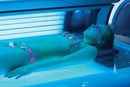
MIAMI BEACH – Calls for a ban on the use of tanning beds by minors in the United States have thus far gone unheeded, but medical organizations are increasingly supporting such a ban – and with good reason, according to Alan Geller of the Harvard School of Public Health, Boston.
The data linking tanning bed use and melanoma are consistent and convincing. A 2010 University of Minnesota case-control study, for example, demonstrated that melanoma risk was significantly increased among users, compared with nonusers, of UVB-enhanced tanning devices (adjusted odds ratio, 2.86) and primarily UVA-emitting devices (AOR, 4.44), Mr. Geller said at the annual meeting of the American Academy of Dermatology.
The risk increased as tanning bed use increased (Cancer Epidemiol. Biomarkers Prev. 2010;19:1557-68).
A more recent study demonstrated that with every visit to a tanning bed, the risk of melanoma increased by 1.8% – and the risk was even greater among those who started tanning at a younger age (BMJ 2012;345:e4757).
"We are clearly in the throes of a modern-day epidemic, particularly among teenage white girls and young women between the ages of 18 and 25," Mr. Geller said, noting that study after study shows that about a third of white teenage girls and about 20% of all teenage girls use a tanning bed by the age of 17.
And yet only five states restrict the use of tanning beds by those under age 18. Others have parental consent restrictions, but these have been shown to have no effect on tanning bed use by minors. That means that in 45 states, children aged 15 years and younger are free to visit tanning salons with no restrictions, he said, noting that a Washington University in St. Louis survey released in February showed that 65% of Missouri tanning salon owners would allow preteens aged 10-12 years to use their tanning beds – and that 43% of tanning salon employees believe indoor tanning poses no health risks.
Data show that 7% of girls use tanning beds by age 14 years. This doubles from age 14 to 15, and doubles again from age 15 to 17, he said, noting that girls are about five to six times more likely than boys to use tanning beds.
Of particular concern, not only are girls using tanning beds early, but they are using them more often.
A Centers for Disease Control and Prevention survey showed that while the rate of use (20% among all girls) has remained constant in recent years, the "prom phenomenon" – the occasional use of tanning beds before a special event – is no longer the norm; the average yearly number of uses of tanning beds among those surveyed was 28.
"We’re way past the prom phenomenon," Mr. Geller said, noting that one reason for this is that tanning salons "do a wonderful job of selling giant packages of use for very little money."
"When people are beginning to think of some kind of restrictions on the tanning bed industry, that would be one we could surely consider," he said, noting that based on the data showing a 1.8% increase in melanoma risk with each tanning bed use, the risk would be 54%-90% in a teen who starts tanning at age 18 and quits at age 19.
That’s a conservative estimate, because most teens start before age 18 and don’t stop at age 19, he said.
Surveillance, Epidemiology, and End Results (SEER) data from the National Cancer Institute show that the risk of melanoma has doubled among women aged 20-24 years since the 1980s, while the risk in men has declined in some age groups, and remained the same in others.
"You have to ask what’s happened during that time," Mr. Geller said, adding that there is concern about the late effects of tanning bed use, especially given that sun exposure time hasn’t changed in that age group over time.
As for what can be done from a public health perspective to reduce tanning bed use, Mr. Geller said a number of research, legislative, and public health campaigns are underway.
"We know from doing qualitative work, that indoor tanning is largely socially driven. "When [girls] are not tanning, they talk about tanning, they blog about tanning," he said, explaining that "the tanning culture involves some kind of socially driven bonds."
The key is to figure out how to break up those bonds.
"If one girl in a social group quits tanning, will this have an effect on the others? We don’t know," he said, adding that this is among the areas that require further research.
Researchers are also studying the effects of antitanning campaigns and legislation in other countries, a number of which have restricted access to tanning beds for minors. A recent web-based advertising campaign in Denmark targeted teens, and, along with legislation restricting access, resulted in a substantial drop in tanning bed use there, he said.
The results of campaigns and legislative efforts like these are being closely monitored so that the lessons learned about if and how they work can be incorporated into efforts here.
Lessons from the campaign against smoking launched three decades ago also are being incorporated into the current effort to reduce tanning, he said.
Although the link between tanning and melanoma isn’t quite as strong as the link between smoking and lung cancer, the seven key principles that made the antismoking campaign a success can be adapted for this purpose. These are surveillance, taxation, legal strategies, public health advertising campaigns, educational programs, legislation, and "some move to mandate enforcement," he said.
Some progress has been made with respect to these principles. For example, state-by-state surveillance and scoring of states’ level of compliance with existing regulations are underway, a 10% tax has been imposed on tanning salons, cost-efficacy studies are being planned, and lawsuits have been filed in multiple states. However, most of these efforts are in their infancy, Mr. Geller said.
For now, what exists across the United States is a "patchwork quilt of pretty crummy regulations," he said.
While intense pressure is on the Food and Drug Administration to ban tanning bed use by those under age 18 – including pressure from the American Academy of Dermatology – and while the agency is cognizant of the risks and has acknowledged a need for more regulations, "politics have prevailed, and at this point we don’t have the ban," he said.
The FDA website does, however, indicate plans for revising regulations and strengthening warning labels to make consumers more aware of the risks, he noted.
"This is good, but I think it’s a really faulty response to everything that we know about the link between tanning beds and melanoma," he said.
Despite the slow progress toward a ban for those under age 18, there have been some successes in the antitanning campaign. For one, numerous organizations have taken up the cause, including the World Health Organization, the American Academy of Pediatrics, the American Medical Association, the Society of Surgical Oncology, and the Canadian Pediatric Society.
Also, thanks to a Federal Trade Commission crackdown in 2010, the tanning industry is no longer allowed to claim that tanning has certain health benefits, such as reducing the risks of some types of cancers. And in 2012, the U.S. Preventive Services Task Force issued its first guidelines on tanning, stating that the evidence is strong enough to recommend that women aged 10-24 years who have fair skin should avoid prime-time sun exposure and tanning beds.
Additionally, a wellness provision of the Patient Protection and Affordable Care Act that will go into effect in May provides for full reimbursement to health care providers for counseling about skin cancer prevention and tanning bed reduction.
"We want to study this because we think this will have a huge effect on increasing the rate of counseling," he said.
Mr. Geller reported having no disclosures.
EXPERT ANALYSIS FROM THE AAD ANNUAL MEETING
New data improve characterization of pediatric melanoma
MIAMI BEACH – Data from several recently published studies have shed new light on the behavior and natural history of melanoma in children and adolescents – and much of the news is good, according to Dr. Sheilagh M. Maguiness.
For example, the findings suggest that the risk of malignant melanoma arising in large congenital nevi is lower than previously thought, at about 2%. Also, outside of the neonatal period the prognosis is excellent for most children diagnosed with melanoma, said Dr. Maguiness of Boston Children’s Hospital.
Data from three of the studies, taken together, show that 36 deaths occurred in 278 cases involving melanoma during childhood or adolescence, for a mortality rate of 13%.
"There was only one death in a child under 10, and that was in the setting of a large congenital nevus," she said at the annual meeting of the American Academy of Dermatology.
Furthermore, the presentation of melanoma in adolescents is similar to that in adults, and the outcomes seem to parallel – and perhaps exceed – those of adults with similar stage tumors, she noted.
The studies do little, however, to clear up controversy about the value of sentinel lymph node biopsy for predicting outcomes in children with melanoma, she said.
Pediatric melanoma represents only about 1%-3% of all melanomas and about 2% of all pediatric malignancies, and it is best considered based on the timing of presentation – presentation during the congenital period, during childhood up to the age of 10 years, and during adolescence – because findings during these stages differ substantially, she said.
Melanoma during the congenital and neonatal period is extremely rare. Four cases involving transplacental metastases from maternal malignant melanoma, nine cases involving large or giant congenital melanocytic nevi, and seven de novo cases have been reported in the literature.
"Unfortunately, when melanoma presents this early, it really does have a poor prognosis, with greater than 50% of the patients dying from their disease," Dr. Maguiness said.
The outlook is much better for older children with melanoma, but any discussion of these cases must include consideration of cases that arise in large congenital melanocytic nevi (LCMN). Controversy has existed over the actual risk of melanoma in these cases, with reports citing risks of anywhere from 7% to 40%, but the studies reviewed by Dr. Maguiness demonstrate that the risk is actually quite low.
A retrospective analysis of data on this topic published in March in the Journal of the American Academy of Dermatology, for example, showed that melanoma developed in only 2% of 2,578 cases of LCMN in 14 studies (J. Am. Acad. Dermatol. 2013;68:493-8.e14).
Other "very, very valuable points" coming out of this data include a finding that 14% of the melanomas occurred viscerally or noncutaneously, and a finding that the mortality rate is high in these patients at about 55%.
"And one of the most important points that I had never appreciated before is a finding that in over 90% of the cases where melanoma developed in these children, satellite nevi were present. Historically we’ve known that the risk of melanoma arising within the satellite nevi itself is quite low – it almost never occurs – but (the finding) that they confer an elevated risk of development of cutaneous melanoma is very interesting," she said.
These findings suggest that a careful examination and regular follow-up is crucial in patients with LCMN, she said.
Findings from three large single-institution studies also have provided interesting new information about pediatric melanoma, she said.
Researchers at the University of Texas Health Science Center at Houston reviewed data on 109 patients under age 19 years with melanoma, including 25 under age 10 years. Seven of 82 patients with adequate follow-up died from their disease, and none of those were in the group under age 10, Dr. Maguiness said.
Among other notable findings from this study: Patients were more likely to be nonwhite, 44% had spitzoid histology, and 52% of those under age 10 who underwent sentinel lymph node biopsy had positive results, compared with 26% in the adolescent cohort (Ann. Surg. 2011;253:1211-15).
The rate of positivity decreased by 13% for each year of increasing age, and because those under age 10 did paradoxically well, the authors suggested that sentinel lymph node positivity does not predict prognosis or outcome in childhood melanoma, Dr. Maguiness noted.
However, investigators from the Moffitt Cancer Center in Tampa concluded, conversely, that sentinel lymph node biopsy does predict outcomes in pediatric melanoma.
Of 126 patients with pediatric melanoma in that retrospective review, 62 underwent sentinel lymph node biopsy and 29% had positive findings. Overall, 19 melanoma-related deaths occurred, for a rate of 16%.
Six patients under the age of 12 were included in the study, but these patients did not undergo sentinel lymph node biopsy. All survived, but there were several late recurrences – after 5 years – even in the node-negative patients, she said.
When the investigators looked at recurrence-free survival, they found that node-positive patients had significantly worse recurrence-free survival and melanoma-specific survival than that of node-negative patients (60% vs.94% and 78% vs. 97%, respectively) at a median follow-up of 5 years (Ann. Surg. Oncol. 2012;19:3888-95).
A study by investigators at the University of California, San Francisco, focused more on historical data, finding that many of the 70 patients included in the study had putative risk factors for melanoma. For example, 20% had numerous nevi, 27% had a positive family history, and 25% had a history of sunburn. Also, this study was the only one of the three to address the presence of LCMN, and only three patients had melanoma arising in a nevus, providing further evidence of a low risk of melanoma in LCMN.
The diagnosis of pediatric melanoma was delayed by about a year in more than 60% of the patients.
The investigators noted that primary lesion characteristics differed from those seen in adults, and they concluded that the conventional ABCDE criteria used to help in the diagnosis of melanoma did not capture melanoma in about 60% of the childhood cases and 40% of the adolescent cases.
Lesions in this study were much more likely to be amelanotic in children, and of uniform color in adolescents. Lesional evolution was nearly universal, and bleeding, bumps, variable diameter, and de novo development were common.
On histopathology, a majority of tumors were not superficial spreading type; more were unclassified spitzoid and other histopathologic subtypes, she noted.
Ten patients (14%) died from their melanoma, and of these, 7 had amelanotic melanoma. Only 1 patient under age 10 years died, and that was in the setting of a large congenital melanocytic nevus.
Based on their findings, the investigators suggested pediatric-specific ABCD criteria (A = amelanotic, B = bleeding, bumps, C = color uniformity, and D = de novo and any diameter) to be used along with the conventional ABCDE criteria to facilitate earlier recognition and treatment of pediatric melanoma (J. Am. Acad. Dermatol. 2013 [doi:10.1016/j.jaad.2012.12.953]).
Although there are some conflicting findings in these three studies – including differing conclusions with respect to the value of sentinel lymph node biopsy for predicting outcomes – there also are some consistent findings, Dr. Maguiness said.
Prepubertal melanoma tends to involve thicker tumors, and darker skin types are overrepresented. Also, lesions in all ages in the pediatric population tend to be amelanotic with spitzoid histology, and tend to have higher rates of positive sentinel lymph node biopsies, compared with adult cases. Prepubertal cases have the highest rates of node positivity, she said.
"So, in conclusion, the risk of malignant melanoma within large congenital nevi seems to be lower than we thought, and the diagnosis of malignant melanoma of childhood has excellent prognosis – speaking to the unique natural history and biology of these tumors, which we probably don’t fully understand," she said, adding that adolescent presentations of melanoma seem to be similar to those in adults, with a slightly better overall prognosis.
Dr. Maguiness reported having no disclosures.
EXPERT ANALYSIS FROM THE AAD ANNUAL MEETING
New therapies bolster the melanoma armamentarium
Community Oncology Editor Dr. Jame Abraham spoke with Dr. Michael Postow at the Oncology Practice Summit in Las Vegas about new immuno- and targeted therapies for melanoma, their side effects, and early phase studies of emerging therapies.
The Oncology Practice Summit was the 8th annual meeting of Community Oncology, the journal of clinical issues in community practice. Dr. Abraham was a co-chair of the Summit, which was hosted this year by Community Oncology as well as The Journal of Supportive Oncology, and The Oncology Report.
Early Detection of Melanoma Via Patient and Physician Screening
Skin Cancer Screenings: What Is Our Role as Dermatology Residents?
Vismodegib shrinks BCCs, reduces Mohs defect size
MIAMI BEACH – Vismodegib treatment for 3 months prior to Mohs surgery for operable basal cell carcinomas shrunk tumors by 46% and reduced the Mohs defect size by 38%, based on data from an open-label intervention trial. The findings were reported at the annual meeting of the American Academy of Dermatology.
The estimated Mohs surgical defect area (based on tumor size) in the first five patients treated in the single-arm study decreased by a mean of 1.4 cm2 from baseline, and actual Mohs surgical defect size decreased by 1.1 cm2 after a mean of 3.4 months of treatment with vismodegib. The changes were statistically significant.
The Mohs defect size was considered a secondary endpoint, because actual defect size can be influenced by skin tension and lesion location, said Dr. Mina Ally, a research fellow at Stanford (Calif.) University.
Patients included in the study were five adults with a total of seven basal cell carcinomas (BCCs) of varying histologic subtypes. One occurred on the shoulder, and the rest occurred on the face; one was a recurrence. All patients were treated for at least 3 months at a vismodegib dosage of 150 mg daily, and all required only a single Mohs stage of excision.
Vismodegib, an oral hedgehog pathway inhibitor approved for the indefinite treatment of advanced and metastatic basal cell carcinomas (that result from aberrant hedgehog pathway signaling), was generally safe, said Dr. Ally.
"All patients experienced mild grade 1 side effects, including muscle cramps, hair loss, and taste loss, and we only needed to discontinue the medication early – after 2 months – in one patient due to a grade 2 elevation in liver enzymes," she said, noting that all of the adverse events resolved after treatment discontinuation.
Further sectioning of Mohs specimens revealed no evidence of residual BCC in three cases and residual BCC in one case. The diagnosis was equivocal in the remaining cases.
"This was because we were seeing an increased number of aberrant follicular structures after (vismodegib) treatment, which were sometimes difficult to differentiate from residual BCC," Dr. Ally explained.
Further staining using a panel that included pleckstrin homology-like domain, family A, member 1, a hair follicle stem cell marker, helped differentiate the follicular structures from BCC, she noted.
None of the patients experienced tumor recurrence during a median 5 months of follow-up.
The findings are of interest because about 1 million people in the United States are affected by BCCs each year, said Dr. Ally. While vismodegib appears to represent a useful adjuvant therapy when given for 3 months prior to Mohs surgery in some cases, certain challenges must be overcome, she said.
"Suppression of the Hedgehog pathway does appear to alter normal follicular development, and this was causing us difficulty in analyzing our histology specimens," Dr. Ally noted. "Future challenges really lie in interpreting these histology specimens and being able to differentiate these follicular structures from residual BCC, which can confound tumor margin clearance."
Dr. Ally reported having no disclosures.
AT THE AAD ANNUAL MEETING
Major finding: Vismodegib reduced tumor size by an average of 46% after 3 months.
Data source: Interim results from an open-label single-arm intervention study.
Disclosures: Dr. Ally reported having no disclosures.
Imaging agent approved for locating lymph nodes
A radioactive diagnostic imaging agent called Lymphoseek (technetium Tc 99m tilmanocept) Injection has been approved for locating lymph nodes in patients who have breast cancer or melanoma and are undergoing surgery to remove tumor-draining lymph nodes, the Food and Drug Administration announced.
Lymphoseek is the first new drug for lymph-node mapping to be approved in more than 30 years. Other FDA-approved drugs used for lymph-node mapping include sulfur colloid and isosulfan blue.
"To use Lymphoseek, doctors inject the drug into the tumor area and later, using a handheld radiation detector, find lymph nodes that have taken up Lymphoseek’s radioactivity," said Dr. Shaw Chen, deputy director of the Office of Drug Evaluation IV in the FDA’s Center for Drug Evaluation and Research.
Lymphoseek is marketed by Navidea Biopharmaceuticals. The manufacturer’s website notes that the ability to rapidly locate and biopsy sentinel nodes enables surgical management to be tailored specifically to each patient’s burden of disease.
In two clinical trials, 332 patients with melanoma or breast cancer were injected with Lymphoseek and blue dye. Surgeons subsequently removed suspected lymph nodes for pathologic examination. Confirmed lymph nodes were examined for their content of blue dye and Lymphoseek. The combination of Lymphoseek and blue dye localized most lymph nodes, although a notable number of nodes were localized only by Lymphoseek.
The most common side effects identified in clinical trials were pain and irritation at the injection site.
According to the manufacturer, a clinical trial involving patients with head and neck cancer is completing enrollment and is expected to be the subject of a future New Drug Application amendment. An initial Marketing Authorization Application filing in the European Union is anticipated by the end of 2012.
Aspirin may protect older women from melanoma
Postmenopausal women who used aspirin regularly had a significantly lower risk of melanoma in an observational study from the Women’s Health Initiative including nearly 60,000 women, according to results published online in Cancer on March 11.
The longer the women took aspirin, the lower their risk for melanoma. Acetaminophen and other NSAIDs had no protective effects.
"These findings suggest that aspirin may have a chemopreventive effect against the development of melanoma, and further clinical investigation is warranted," senior investigator and dermatologist Jean Tang of Stanford (Calif.) University and her colleagues concluded (Cancer 2013 March 11 [doi: 10.1002/cncr.27817]).
The analysis included 59,806 white, postmenopausal women aged 50-79 years who completed questionnaires about their aspirin and NSAID use as part of the Women’s Health Initiative, a federally funded effort to address the most common causes of death, disability, and impaired quality of life in postmenopausal women.
At baseline, 25% of the women reported regular aspirin use (defined as use of aspirin at least twice in the past 2 weeks). Of these, 75% reported using regular or extrastrength aspirin. A total of 15% of the women reported nonaspirin NSAID use at least twice in the prior 2 weeks. Ibuprofen and naproxen were the most commonly used nonaspirin NSAIDS. Approximately 60% of the women reported no regular use of aspirin or other NSAIDs.
The women were asked again about their aspirin and NSAID use at a second clinical visit after 3 years; 59% of aspirin users and 38% of NSAID users reported the same levels of medication use.
A total of 548 incident melanomas occurred during a median follow-up of 12 years: 289 melanomas in situ, 255 invasive melanomas, and 4 cases classified as unknown. The risk of melanoma was 21% lower in regular aspirin users compared with women who reported no aspirin use (hazard ratio, 0.79). Increased duration of aspirin use (less than 1 year, 1-4 years, and 5 years or more) was associated with an 11% lower risk of melanoma for each categorical increase.
In addition, the risk of melanoma was 30% lower in women who used aspirin regularly for 5 or more years (HR, 0.70). The incidence of melanoma per 100,000 person-years was 69.8, 87.9, and 87.1 for aspirin users, nonaspirin NSAID users, and NSAID and aspirin nonusers, respectively.
Dr. Tang and her colleagues controlled for skin cancer histories, sun exposure, sunscreen use, and other potential confounders in their analysis. At baseline, "the most important predictors of melanoma risk, prior nonmelanoma skin cancer and melanoma history, did not differ among the groups," they noted.
Although previous studies have shown a protective effect for aspirin and other NSAIDs against gastric, colorectal, and breast cancers, the results for melanoma have been mixed, possibly because negative studies used too low a dose of aspirin or the dosing was too infrequent, the researchers noted.
Similarly, infrequent use of nonaspirin NSAIDs may account for their lack of benefit in the trial, or "aspirin may have properties that other non[aspirin] NSAIDs lack," the investigators noted.
"Our finding that increased duration of aspirin use was associated with lower risk of melanoma after extended follow-up is consistent with studies of other malignancies, particularly colorectal cancer, that reported benefit after several years of aspirin use and extended follow-up. ... Cardiovascular benefits and potential antitumor effects of aspirin are particularly intriguing in the context of preventive medicine, but they must be weighed against well-known risks, such as gastrointestinal bleeding," they wrote.
The study was sponsored in part by the National Heart, Lung, and Blood Institute and the National Institute of Arthritis and Musculoskeletal and Skin Diseases. Dr. Tang is a consultant for Genentech.
FROM CANCER
Major Finding: The risk of melanoma was 21% lower in white, postmenopausal women who took aspirin regularly.
Data Source: Women’s Health Initiative observational study involving approximately 60,000 postmenopausal women.
Disclosures: The senior investigator is a consultant for Genentech. The study was sponsored in part by the National Heart, Lung, and Blood Institute and the National Institute of Arthritis and Musculoskeletal and Skin Diseases.
Innovations in noninvasive procedures keep dermatology on cutting edge
Noninvasive procedural dermatology has evolved at a dizzying pace, and continues to do so.
In addition to an array of procedures for skin tightening, skin resurfacing, and fat reduction, emerging technologies such as complex feedback devices, nanotechnology, and stem cell–based therapies promise to keep dermatology at the forefront of the cosmetic and esthetic realm, according to Dr. Murad Alam.
In an article featured in the March issue of Seminars in Cutaneous Medicine and Surgery, Dr. Alam of Northwestern University, Chicago, makes several predictions about the future of these technologies (Semin. Cutan. Med. Surg. 2013;32:61-63).
For example, like modern vehicles equipped with computer chips that can change steering and braking in response to environmental conditions, dermatologic devices will soon include technology that uses precise feedback to make automated setting changes, he said.
"Over time, the reduced cost of microelectronics, feedback controls, and computing power is simplifying the capacity of devices to analyze intraoperative information and adjust the procedure to compensate. For instance, certain laser and energy devices already have tips that are able to sense the temperature in the microenvironment and adjust power output to maintain site-specific temperature within a narrow band," he explained.
This technology could increase effectiveness and improve the safety of devices by reducing the level of operator time and expertise needed, and by making setting changes faster than humanly possible.
Autonomous nanotechnology devices are another advance described by Dr. Alam.
Miniaturization will become more feasible and affordable, and eventually devices will become "so exceedingly small that they will be mostly disposable and deployed in large numbers to the treatment site," he said.
The concept of hundreds of minuscule machines deployed to resurface skin or repair a wound may sound like science fiction, but the rapid advances in nanotechnology could make it a reality that could lead to the creation of new procedures such as ways to treat scars that can’t be corrected using currently available technologies, he added.
Dr. Alam’s other predictions for the future of noninvasive procedural dermatology included:
• Optimization of minimally invasive procedures for fat reduction and skin tightening, which currently provide only mild to modest results and longevity.
• The use of stem cells for augmentation of tissue layers, which could provide genuine rejuvenation rather than simply repair and concealment.
• The improvement of artificial dermal substitutes that can develop many of the functions of live skin, and can be grafted without inducing contractures.
• The development of rapid treatments for pigmentation using nanotechnology and cellular therapies, which will allow for precise melanocyte and melanosome transfer and automatic recoloration of discolored skin.
While these technologies continue to emerge, plenty of others have already established their places in the dermatology arena. The many and varied applications of one of these – low-level laser therapy, or LLLT – are described in another article in the March issue of Seminars in Cutaneous Medicine and Surgery (Semin. Cutan. Med. Surg. 2013;32:42-54).
"LLLT involves exposing cells or tissue to low levels of red and near-infrared light. ... Recently, medical treatment with LLLT at various intensities has been found to stimulate or inhibit an assortment of cellular processes," wrote Dr. Pinar Avci of Massachusetts General Hospital, Boston, and his colleagues, noting that the mechanism associated with the cellular photobiostimulation by LLLT is not yet fully understood, but appears to have a wide range of effects at the molecular, cellular, and tissue levels.
Describing LLLT as "possibly the ultimate noninvasive approach to treating the skin," the researchers highlighted numerous existing or emerging applications for the technology, outlined below.
Skin rejuvenation
Many modalities developed to reverse the dermal and epidermal signs of photoaging and chronological aging depend on the removal of the epidermis and the induction of a controlled form of skin wounding to promote collagen biosynthesis and dermal matrix remodeling. Examples include retinoic acid, dermabrasion, chemical peels, and ablative laser resurfacing.
These modalities require intensive posttreatment care and prolonged down time, and are associated with a risk of numerous complications, the researchers said.
LLLT represents an alternative that is known to increase microcirculation and vascular perfusion in the skin. Data from previous studies have shown that LLLT increased collagen and improved wrinkles and skin laxity with less down time and risk than that of other treatments.
In one study, for example, 300 patients treated with only a light-emitting diode (LED) LLLT device set at 590 nm, 0.10 J/cm2, were compared with 600 patients who received the LED therapy in combination with a thermal-based photorejuvenation procedure. Of those who received LED therapy alone, 90% reported softer skin and less roughness and fine lines. The changes ranged from subtle to significant.
Those who received thermal photorejuvenation laser treatment reported a reduction in posttreatment erythema and an overall impression of increased efficacy with the additional LED treatment – an effect that could be attributed to anti-inflammatory effects of LLLT, they noted.
In another study, more than 90% of 90 patients receiving eight LED treatments over 4 weeks experienced favorable results, improving by at least one Fitzpatrick photoaging category. In addition, 65% of patients experienced global improvement in facial texture, fine lines, background erythema, and pigmentation, with results peaking 4-6 months after completion.
Acne treatment
LLLT in the red to near-infrared (NIR) spectral range (630-1000 nm) and with nonthermal power (less than 200 mW) has been shown in several studies to improve acne vulgaris. In one study, a significant reduction in active acne lesions occurred after 12 twice-weekly sessions using 630 nm red-spectrum LLLT with a fluence of 12 J/cm2 in conjunction with 2% topical clindamycin. No significant effects were seen using an 890-nm laser. Other studies have demonstrated that the combination of blue and red light is synergistic for treating acne.
Photoprotection
Recent suggestions that infrared exposure might have protective effects against ultraviolet light–induced skin damage are based on the theory that the exposure might trigger protective or repair responses to UV irradiation. While controversial, this view is supported by data suggesting potential mechanisms of action. For example, some data suggest a role of p53, a sensor of gene integrity involved in cell apoptosis and repair mechanisms. In one study, the response to infrared (IR) irradiation was shown to be p53 dependent, suggesting the IR irradiation prepares cells to resist and/or repair further UV-induced DNA damage. Data from another study showed that IR irradiation induced the protective protein ferritin, which is involved in skin repair.
Data from yet another study suggested that nerve growth factor (NGF) production induced by LLLT using the helium neon semiconductor laser diode (HeNe, 633 nm) might explain the photoprotective effects of LLLT. In that study, NGF – a major paracrine maintenance factor for melanocyte survival in skin, was shown to protect melanocytes from UV-induced apoptosis by upregulating the level of Bcl-2 (an antiapoptotic protein) in the cells.
Herpesvirus lesion treatment
New therapies are needed to shorten recurrent herpesvirus episodes and reduce related pain and inflammation. LLLT has been suggested as an alternative to current medications. In one study of 50 patients with recurrent perioral HSV infection, LLLT at 690 nm, 80 mW/cm2, 48 J/cm2 daily for 2 weeks during recurrence-free periods decreased the frequency of herpes labialis episodes, the authors said.
In another study with similar parameters, patients achieved a significant prolongation of remission intervals from 30 to 73 days.
The mechanism of action remains unclear, but an indirect effect of LLLT on cellular and humoral components of the immune system may be involved in antiviral responses, as opposed to a direct virus-inactivating effect, the researchers noted.
Vitiligo treatment
Modest efficacy seen with the low-energy HeNe laser (632 nm, 25 mW/cm2) for the treatment of 18 vitiligo patients led to speculation that LLLT could serve as an alternative effective treatment for this typically treatment-resistant condition. (Repigmentation was observed in 64%, and some follicular repigmentation was observed in the remaining patients).
In a subsequent study of local administration of the HeNe laser light at 3 J/cm2, 1.0 mW, 632.8 nm in patients with segmental type vitiligo, marked perilesional and perifollicular repigmentation of more than 50% was observed in 60% of patients.
"Both NGF and (basic fibroblast growth factor) stimulate melanocyte migration, and deficiencies of these mediators may participate in the development of vitiligo," the researchers wrote.
Depigmentation
During tests of red and blue light for acne, researchers unexpectedly found that patients treated with both red and blue light experienced an overall decrease in melanin.
Based on instrumental measurement results, blue light exposure (415 nm, 40 mW/cm2, 48 J/cm2), increased the melanin level by 6.7, whereas red light exposure (633 nm, 80 mW/cm2, 96 J/cm2) decreased the melanin level by 15.5.
"This finding may have some relationship with the laser’s brightening effect of the skin tone, which 14 of 24 patients spontaneously reported after the treatment period. However, as of today, no other studies investigated or reported a similar decrease in melanin levels after red light irradiations," the researchers said.
Hypertrophic scar and keloid eradication
LLLT has shown promise for preventing hypertrophic and keloid scars in patients who undergo scar revision by surgery or CO2 laser. The use of daily near-infrared LED (NIR-LED) treatment on one of the two bilateral sites safely reduced the risk of scar development in that lesion, compared with the untreated lesion in three patients with bilateral scars. One underwent surgical revision/excision for preauricular linear keloids that developed after a face-lift procedure, one underwent CO2 resurfacing for hypertrophic acne scars on the chest, and one underwent CO2 resurfacing after excision of hypertrophic scars on the back. No significant treatment-related adverse effects were reported.
LLLT may work in these types of scars through an inhibitory effect on interleukin-6 mRNA levels and the modulating of platelet derived growth factor, transforming growth factor–beta, interleukins, and MMPs, which are associated with abnormal wound healing, the researchers noted.
Burn treatment
LED exposure was shown to provide benefit for the treatment of acute sunburn in a study of 10 patients.
Treatment once or twice daily for 3 days on half of the affected area decreased symptoms of burning, redness, swelling, and peeling compared with the untreated half. Decreased MMP-1 was noted on the treated side through immunofluorescence staining in one patient, and real-time polymerase chain reaction gene expression analysis showed a significant decrease in MMP-1 gene expression at both 4 and 24 hours after the UV injury on the treated side.
In another study, LED treatment was effective compared with control for speeding the healing process of laser treatment–related burns. In nine patients with second-degree burns from nonablative laser devices, LED therapy once daily for 1 week was associated with 50% faster healing based on both patient and physician accounts.
LED treatment also was shown in a pilot study to accelerate re-epithelialization of a forearm injury induced by a CO2 laser; identical test sites were treated with daily dressing changes and polysporin ointment, but the site with faster re-epithelialization had also received the LED treatment (a computer pattern generator was used to deliver the identical CO2 treatment to both sites).
Psoriasis
LLLT also shows promise for the treatment of plaque psoriasis. In a preliminary study, the combined use of sequential NIR (830 nm) and visible red light (630 nm) led to resolution of psoriasis in patients who were resistant to conventional therapy. The patients received treatment in two 20-minute sessions, 48 hours apart, for 4 or 5 weeks. No adverse side effects occurred.
Despite the variety of potential applications for the technology, LLLT remains somewhat controversial due to "uncertainties about the fundamental molecular and cellular mechanisms responsible for transducing signals from the photons incident on the cells to the biological effects that take place in the irradiated tissue" and because "there are significant variations in terms of dosimetry parameter: wavelength, irradiance or power density, pulse structure, coherence, polarization, energy, fluence, irradiation time, contact versus noncontact application, and repetition regimen," the researchers said.
They noted, however, that problems that have been experienced with LLLT – as well as negative study results – could be the result of inappropriate parameters, skin preparation, and/or device maintenance.
"LLLT appears to have a wide range of applications in dermatology, especially in indications where stimulation of healing, reduction of inflammation, reduction of cell death, and skin rejuvenation are required," they noted, but added that the lack of agreement on important parameters (particularly whether red, NIR, or a combination of both is optimal for a given application) has created a credibility gap that must be overcome before LLLT is routinely applied in every dermatologist’s office.
Once LLLT does become so widely accepted, however, it may be time for dermatologists to move on to the next big thing, Dr. Alam said in his article on the future of procedural dermatology.
"For several decades, dermatologists have worked closely with start-up companies to commercialize new devices and technologies ... when new toxins, fillers, and energy devices have been marketed, dermatologists have been early adopters," he said. "Over time, each device or procedure has diminished in cost and exclusivity as other physicians and nonphysicians have entered the market, and dermatologists have moved on to greener pastures. In all likelihood, this cycle will continue," he noted. "Dermatologists cannot prevent the dissemination of stable technologies, but they can continue to innovate and create new ones."
In fact, he added, continued success in this arena will rest largely on clinicians’ ability to nurture innovation to ensure a healthy pipeline of novel technologies.
The authors of the articles had no financial conflicts to disclose.
FROM SEMINARS IN CUTANEOUS MEDICINE AND SURGERY

