User login
Terlipressin Shows Some Benefit in Hepatorenal Syndrome
SAN DIEGO – Terlipressin, widely used in Europe for hepatorenal syndrome but not yet approved in the United States, would be a useful option for this difficult-to-treat condition, said transplant hepatologist Dr. James Burton at the annual meeting of the Society of Hospital Medicine.
Currently, U.S. clinicians do their best with albumin and vasoconstrictors such as octreotide and midodrine to counteract the intense splanchnic vasodilatation that causes the condition (J. Clin. Gastroenterol. 2009;43:680-5).
Hepatorenal syndrome is a complication of advanced cirrhosis that can lead to renal failure. Liver transplantation is "the only treatment we currently have available to us that is very effective," especially for rapidly progressing type 1 HRS [hepatorenal syndrome]," said Dr. Burton, associate professor of medicine and the medical director of liver transplantation at the University of Colorado Hospital, Aurora.
The European vasopressin analogue terlipressin (Lucassin) has a long half-life that allows for bolus dosing every 4-6 hours. Several studies found evidence that the drug might be beneficial.
In one study, 56 patients with type 1 HRS were randomized to terlipressin (1 mg intravenously every 6 hours) plus albumin. Another 56 patients received placebo plus albumin.
Serum creatinine level decreased to 1.5 mg/dL or less in 14 (25%) terlipressin patients, but in only 7 (12.5%) placebo patients by day 14. This difference was not statistically significant, however. The two groups had similar adverse event rates (Gastroenterology 2008;134:1360-8).
A significant benefit was found in a randomized study of 46 patients. Serum creatinine fell below 1.5 mg/dL or decreased 50% from baseline in 10 (43.5%) patients treated with albumin and terlipressin (1-2 mg/4 hours), vs. 2 (8.7%) patients treated with albumin alone (P = .017). Ten terlipressin patients and four albumin-only patients had cardiovascular complications (Gastroenterology 2008;134:1352-9).
In a third study, which was also randomized, 24 HRS patients received albumin and terlipressin (3 mg/24 hours with escalation based on response) and 17 received standard U.S. treatment – albumin plus midodrine and octreotide, also escalated according to response.
Serum creatinine in that study fell below 1.5 mg/dL in 13 (54%) terlipressin patients and renal function improved in three-quarters. Creatinine fell below 1.5 mg/dL in just 2 (12%) of the standard-treatment patients, and renal function improved in about a third (Angeli et al. The Liver Meeting 2011, American Association for the Study of Liver Diseases).
However, in all three studies, people didn’t live any longer when they were given terlipressin, and this is what is holding up FDA approval, said Dr. Burton, who noted that he and his colleagues are working on a new study "to try to show a survival benefit" for the drug.
He said he hopes the FDA approves terlipressin "because it will be a very effective treatment for hepatorenal syndrome. It would be amazing and wonderful if I could prevent people from going on dialysis" as they wait for a new liver.
Dr. Burton said he has no relevant financial disclosures.
EXPERT ANALYSIS FROM THE ANNUAL MEETING OF THE SOCIETY OF HOSPITAL MEDICINE
Dialysis: How, When, and at What Dose?
Q: I work as a hospitalist PA in a large teaching hospital. In our ICU we had a patient diagnosed with acute kidney injury after a coronary artery bypass graft. We consulted nephrology and they decided to start dialysis. There was quite a discussion about whether to use hemodialysis every other day or continuous renal replacement therapy. What is the basis for this question? Is there science behind the answer, or is it determined by nephrologist preference?
The development of intermittent hemodialysis (IHD) revolutionized the care of patients with acute renal failure and allowed the medical establishment means to give these patients a chance to recover from their illness. However, IHD had (and continues to have) many downsides, and mortality in acute renal failure remains high. Thus, there is an ongoing search for the best renal replacement therapy; this search led to modern continuous therapies. Three main questions have arisen from this:
- Which Modality is Best?
- What is the optimal dose for dialysis?
- When should we initiate therapy?
Continue reading for the answers...
Which Modality is Best? IHD is a shorter treatment (2 to 4 hours), typically performed three times per week but as often as daily. Fluid and electrolyte clearance is rapid, making IHD very efficient but increasing the risk for complications, such as hemodynamic instability. Furthermore, the abrupt fluid and electrolyte shifts associated with IHD do not mimic native kidney function. Providing slower treatments delivered continuously over 24 hours has many benefits.
Continuous renal replacement therapy (CRRT) provides clearance of large amounts of fluid and electrolytes over 24 hours, with minimal hemodynamic disturbances. This allows for more gradual shifts in volume and electrolyte levels, reducing the potential for ischemic damage to the kidney and other organs. Also, CRRT more closely replicates normal renal function than IHD.
CRRT is now extremely safe and efficient, although it has been difficult to prove its superiority to IHD in regard to mortality. While there may be no actual benefit to CRRT, it is also likely that its benefit is observed only in certain subsets of patients with renal failure. For example, we do have compelling evidence of increased intracranial pressure during IHD; CRRT is much safer for patients at risk for this development.1 It is also possible that we need to further improve CRRT systems and delivery in order to see a benefit.
Because current data favor neither CRRT nor IHD, most experts recommend choosing a therapy based on patient characteristics. For instance, hemodynamically unstable patients commonly receive generous amounts of fluid daily (antibiotics, nutrition, etc), and thus are often better suited for CRRT because it is more likely to remove higher volumes of fluid successfully, and less likely to contribute to hemodynamic instability than IHD. Conversely, patients with acute electrolyte deviations may benefit more from the rapid electrolyte removal IHD provides.
Additionally, stable patients may be more suitable candidates for IHD because of location (CRRT requires intensive care monitoring) and other variables.2,3 Results from multiple studies have suggested that CRRT may also provide renal protection and consequently improve renal recovery. However, this evidence is not conclusive; the possibility needs further evaluation.1
Continue reading for the optimal dose for dialysis...
What is the optimal dose for dialysis? This question, too, is plagued by inconclusive research findings. Paganini was the first to raise it formally in patients with acute renal failure; his research team found improved survival with higher doses, but had excluded the sickest and healthiest patients (according to probability of survival) from the study.4 This was followed by two additional studies with results that also seemed to support higher doses.1,5
Then in 2008, in a a similar randomized controlled trial, Tolwani et al6 found no survival benefit with higher versus lower dosing; participants in this study were not excluded based on severity of illness. Also, Tolwani’s research team identified failure to achieve prescribed doses as one factor complicating dose comparison.6
Finally, two large randomized controlled trials were performed to evaluate dosage, one in the US7 and one in Australia and New Zealand.8 Neither research team was able to confirm survival benefits with higher-dose renal replacement therapy, and there were inconsistencies between doses used in the study and standard practice in the US. In fact, the low-dose group received dialysis exceeding what is current practice by more than 30%.
The current prevailing opinion is that we should reach a minimum dose: a Kt/V of 1.2, three times a week, for IHD; or a CRRT dose of 20 mL/kg/h. At this time, higher doses do not confer a clear survival benefit. It remains unknown whether certain patients may benefit from a higher dose. Further research is needed.
Continue reading to find out when to initiate therapy...
When should we initiate therapy? While some study results suggest that early initiation is better, this remains unconfirmed.1 In theory, renal replacement therapy should be initiated early because it improves metabolic control and corrects fluid overload, facilitating management of hemodynamics and ventilation, and reducing the potential for complications caused by uremia-induced physiologic dysfunction. However, it is still unknown which patients would benefit most from early initiation, and the appropriate triggers for when to initiate therapy remain unclear.
So, how do you choose a renal replacement therapy? This is often a matter of opinion, based on evaluation of risks and benefits specific to the patient and clinical expertise with the renal replacement therapies available.
Patient safety must be a primary consideration. IHD and CRRT have dramatically improved in safety and efficacy, but it must yet be proven beyond a doubt which is superior. Outcomes may depend on how the chosen therapy is used to treat specific patient needs—not which therapy is chosen. Further research is needed to identify the best and safest way to provide renal replacement therapy.
Catherine Wells, DNP, ACNP-BC, CNN-NP, Division of Nephrology, University of Mississippi, Jackson
Continue for references...
REFERENCES
1. Prowle JR, Bellomo R. Continuous renal replacement therapy: recent advances and future research. Nat Rev Nephrol. 2010;6(9):521-529.
2. Abi Antoun T, Palevsky PM. Selection of modality of renal replacement therapy. Semin Dial. 2009; 22(2):108-113.
3. Vanholder R, Van Biesen W, Lameire N. What is the renal replacement method of choice for intensive care patients? J Am Soc Nephrol. 2001;12 suppl 17:S40-S43.
4. Augustine JJ, Sandy D, Seifert TH, Paganini EP. A randomized controlled trial comparing intermittent with continuous dialysis in patients with ARF. Am J Kidney Dis. 2004;44(6):1000-1007.
5. Ronco C, Bellomo R, Homel P, et al. Effects of different doses in continuous veno-venous haemofiltration on outcomes of acute renal failure: a prospective randomised trial. Lancet. 2000;356(9223):26-30.
6. Tolwani AJ, Campbell RC, Stofan BS, et al. Standard versus high-dose CVVHDF for ICU-related acute renal failure. J Am Soc Nephrol. 2008;19(6):1233-1238.
7. Palevsky PM, Zhang JH, O’Connor TZ, et al; VA/NIH Acute Renal Failure Trial Network. Intensity of renal support in critically ill patients with acute kidney injury. N Engl J Med. 2008;359(1):7-20.
8. Bellomo R, Cass A, Cole L, et al; RENAL Replacement Therapy Study Investigators. Intensity of continuous renal-replacement therapy in critically ill patients. N Engl J Med. 2009;361(17):1627-1683.
"Hemorrhoids" turn out to be cancer … and more
“Hemorrhoids” turn out to be cancer
A 49-YEAR-OLD WOMAN, whose husband was on active duty with the US Army, went to an army community hospital in March complaining of hemorrhoids, back pain, and itching, burning, and pain with bowel movements. A guaiac-based fecal occult blood test was positive; no further testing was done to rule out rectal cancer.
The woman was discharged with pain medication but returned the following day, reporting intense anal pain despite taking the medication and bright red blood in her stools. The symptoms were attributed to hemorrhoids, and the patient was given a toilet “donut” and topical medication. Although her records noted a referral to a general surgeon, the referral wasn’t arranged or scheduled.
The patient returned to the hospital in April, May, and June with continuing complaints that included unrelieved constipation. A laxative was prescribed, but no further testing was done, nor was the patient referred to a surgeon.
In August, she went to the emergency department because of rectal bleeding for the previous 2 weeks, abdominal pain, blood in her urine, and difficulty breathing. Once again the symptoms were blamed on hemorrhoids even though the patient questioned the diagnosis.
The patient continued to see various providers at the army community hospital for the rest of the year, during which time she turned 50. None of them recommended a colonoscopy despite standard recommendations to begin colorectal cancer screening at 50 years of age and the woman’s symptoms, which suggested colorectal cancer.
In March of the following year, the patient consulted a bariatric surgeon in private practice, who recommended evaluating the patient’s bloody stools and offered to perform a diagnostic colonoscopy if authorized. The army hospital didn’t immediately authorize the procedure, and it wasn’t performed.
In late September, the patient consulted a surgeon at the hospital, by which time bright red blood was squirting from her anal region and appeared in the toilet water after every bowel movement. She had never undergone a full colon evaluation.
Less than a week after the surgery consult, the patient’s husband was transferred to another military base. Her doctors said that a surgeon at the new base would be told about her medical condition, but that didn’t happen.
Five months later, a surgery consultation at the new military base found a rectal lesion extending 8 cm into the rectum from the anal verge. Pathology confirmed stage IIIC mucinous adenocarcinoma that had spread to the lymph nodes. Two years later, after several surgeries, chemotherapy, and radiation, the patient died at 53 years of age.
PLAINTIFF’S CLAIM If testing to rule out rectal cancer, such as a colonoscopy, had been performed earlier, the cancer would have been diagnosed at a curable stage.
THE DEFENSE No information about the defense is available.
VERDICT $2.15 million Tennessee settlement.
COMMENT Recurrent, unrelenting symptoms should prompt the alert clinician to explore alternative diagnoses.
For want of diagnosis and treatment, kidney function is lost
A FEBRILE ILLNESS prompted a patient to visit his primary care physician. After 3 months of treatment by the primary care doctor, the patient sought a second opinion and treatment from a federally funded community health clinic, where he was treated for 2 more months. During that time, the patient developed signs and symptoms of impaired kidney function, which laboratory results confirmed.
The clinic staff didn’t address the possible loss of kidney function. Three days after his last examination at the clinic, the patient went to a hospital emergency department, where he was promptly diagnosed with subacute bacterial endocarditis. His kidney function could not be restored.
PLAINTIFF’S CLAIM The primary care physician and the staff at the clinic were negligent in failing to diagnose and treat the kidney issues. Also, they didn’t recognize and treat the signs and symptoms of subacute bacterial endocarditis.
THE DEFENSE The primary care physician claimed that the patient’s injuries resulted solely from negligence on the part of the clinic staff. He maintained that the patient’s kidney function was normal when the man left his care. The federal government, on behalf of the clinic staff, claimed that the primary care physician was at least 50% responsible for the patient’s injuries.
VERDICT $1.45 million Texas settlement.
COMMENT Subacute bacterial endocarditis can be a challenging diagnosis because of the subtlety and variety of presentations. Remember the zebras when confronted with unexplained symptoms and signs.
Neuropathy blamed on belated diabetes diagnosis
A PATIENT IN A FAMILY PRACTICE was treated by several of the doctors and a physician assistant in the group over about a decade. After the patient developed neuropathy in his arms and legs, he was diagnosed with type 2 diabetes.
PLAINTIFF’S CLAIM Earlier diagnosis of the diabetes would have prevented development of neuropathy. High blood glucose levels identified on tests weren’t addressed.
THE DEFENSE Only 3 tests had shown excessive levels of glucose; the patient had many comorbidities that required attention. A special diet had been prescribed that would have helped control glucose levels. This was an appropriate initial step to address a diagnosis of type 2 diabetes.
VERDICT $285,000 New York settlement.
COMMENT It’s easy to overlook or postpone treatment of apparently less urgent issues such as glucose intolerance. Clear documentation and explicit discussion with patients might help mitigate the risk of adverse judgments.
Too many narcotic prescriptions
A WOMAN TREATED FOR CHRONIC SINUSITIS by an ear, nose, and throat physician received prescriptions for oxycodone, acetaminophen and oxycodone, and methadone for years to relieve headaches and facial pain. She died at 40 years of age from a methadone overdose. The physician admitted in a deposition that he’d kept on prescribing the medications even after the patient’s health insurer informed him that she was obtaining narcotics from multiple providers.
PLAINTIFF’S CLAIM No information about the plaintiff’s claim is available.
THE DEFENSE No information about the defense is available.
VERDICT $1.05 million New Jersey settlement.
COMMENT Strict tracking and oversight of opioid administration is essential. Clear documentation and regular follow-up remain very important.
Delayed Tx turns skin breakdown into a long-term problem
A NEARLY IMMOBILE WOMAN was discharged from a hospital—where she’d been treated for congestive heart failure, hypertension, diabetes, altered mental status, severe arthritis, and gout—and transported by ambulance to her home. Discharge diagnoses included possible obstructive sleep apnea and hypercapnia. Because the patient needed a great deal of help with activities of daily living, her physician ordered home health services.
Twelve days after discharge, a representative from the home health agency performed an initial assessment in the patient’s home, at which time the patient’s daughter reported that her mother had developed some skin breakdown on her buttocks that required care. The home health nurse allegedly told the daughter that the agency would need an order from her mother’s physician before starting home treatment for the skin breakdown.
The daughter phoned the physician every day for the next few days to get treatment authorization, but the doctor didn’t return her calls. The home health agency didn’t seek authorization from the doctor.
When the home health nurse returned to the patient’s home a week later to begin care, the daughter again mentioned the areas of skin breakdown, which by that time had become pressure sores. The nurse didn’t treat the pressure sores. The home health agency tried to contact the patient’s physician, who didn’t return their calls.
The agency finally received an order to treat the pressure sores 6 days after the home health nurse had begun caring for the patient, by which time the sores were infected and considerably larger. Healing required more than a year of treatment.
PLAINTIFF’S CLAIM As a result of the delay in treating the pressure sores, the patient’s condition was worse that it otherwise would have been.
THE DEFENSE The defendants denied any negligence.
VERDICT Alabama defense verdict.
COMMENT Better communication and coordination of care between home health providers and a patient’s medical home are important to provide optimal care—and avoid lawsuits.
Fast-Acting Erectile Dysfunction Drug Approved
The fast-acting erectile dysfunction medication avanafil was approved by the Food and Drug Administration April 30 on the basis of findings from multiple phase-III trials demonstrating the safety and efficacy of the phosphodiesterase type 5 inhibitor, according to a statement from the agency.
The first new entry in the erectile dysfunction (ED) marketplace in nearly 10 years, avanafil (Stendra) joins the ranks of other PDE5 inhibitors approved for the treatment of ED and is expected to be a formidable competitor in the marketplace because it is a faster-acting agent associated with fewer side effects than the currently available options, including sildenafil citrate (Viagra), vardenafil (Levitra), and tadalafil (Cialis), according to Dr. Irwin Goldstein, director of the sexual medicine program at Alvarado Hospital in San Diego.
Dr. Goldstein was the lead investigator of the pivotal phase-III Research Evaluating an Investigational Medication for Erectile Dysfunction (REVIVE) trial in which 646 men with a history of ED for at least 6 months were randomized to 50-mg, 100-mg, or 200-mg of avanafil or placebo (J. Sex. Med. 2012;9:1122-33).
"We observed significant improvements in erectile function and low rates of side effects common to [the PDE5] drug class," Dr. Goldstein said in an interview. "Importantly, the majority of patients who attempted intercourse within 15 minutes of dosing were successful at all dosage levels," he said, noting that successful attempts were reported 64%, 67%, and 71% of the time with 50-mg, 100-mg, and 200-mg doses, respectively, compared with 27% for placebo.
In addition to the REVIVE trial, the avanafil development program included the REVIVE-Diabetes trial comprising 390 men with ED and diabetes and the REVIVE-RP trial comprising 298 men with ED following radical prostatectomy, as well as a year-long safety study of 712 continuation patients from the REVIVE and REVIVE-Diabetes trials, according to a statement issued by Vivus, manufacturer of the drug.
The combined highlights of the various studies showed statistically significant improvements relative to placebo in measures of erectile function, vaginal penetration, and successful intercourse at all three dosages, according the FDA.
The most common side effects, reported in more than 2% of the patients, were headache, facial flushing, nasal congestion, and back pain; there were no drug-related serious adverse events reported, the FDA said.
In the continuation study of patients who received up to 40 additional weeks of treatment, initially at the 100-mg dose and eventually maintained, increased, or decreased based on individual patient response, the side effects did not appear to worsen over time.
Dr. Goldstein disclosed financial relationships with BioSante, Boehringer Ingelheim, Medtronic, Pfizer, VIVUS, Alagin, Shionogi, Slate, Abbott, Ascend, Auxilium, Coloplast, and Warner Chilcott.
Hyponatremia: What to Worry About in the Hospital
MIAMI BEACH – When you get a call in the hospital to evaluate a patient with postoperative hyponatremia, you need to worry primarily about three causes, according to an expert.
Increased free water intake, sodium loss that exceeds free water loss, and – "what we see all the time" – an inability to excrete free water will be your main concerns with these inpatients, Dr. Rachel E. Thompson said.
Initially rule out the most serious potential consequences of hyponatremia. "Assess mental status first to rule out increased intracranial pressure," said Dr. Thompson, founder and director of the Medicine Consult Service at Harborview Medical Center in Seattle. She spoke at a meeting on perioperative medicine sponsored by the University of Miami. "We have rigid skulls. There is very little space for tissue expansion." Left unchecked, intracranial hypertension can lead to brain damage, herniation, and respiratory arrest. "You can die from this."
It’s also important to distinguish neurology patients from others, Dr. Thompson said. Neurologic surgeons often want patients to have sodium levels above 140 mEq/L. "However, in other populations, if there is not brain damage that is going to be worsened by this, we are a lot more laissez faire. We might allow it to go lower, and people don’t get too worried about it until you’re in the mid to low 120s."
Dr. Thompson cited a neurosurgery patient as a case example. You get a call about a 66-year-old woman currently in the rehabilitation unit following endovascular coiling for aneurysmal subarachnoid hemorrhage repair two weeks earlier. The rehabilitation specialist reports her sodium is low at 126 mEq/L (it was 142 mEq/L when she left the neurosurgery unit 7 days earlier).
Dr. Thompson electronically polled meeting attendees regarding the next appropriate step. Of the total 91 responses, 33% would evaluate the patient’s volume status, 29% would evaluate mental status, 24% would check serum osmolality, and the remaining 14% would recheck the sodium level.
"I like that you want to see the volume status, and another third of you want to check the mental status. That means you are going to go see the patient. That is great," Dr. Thompson said.
Early assessment of mental status is key in Dr. Thompson’s four-step practical model for perioperative providers caring for hyponatremia patients:
• Step 1. Go to the bedside to assess mental status. Does it reflect what you expect for that sodium level? Assess acuity and severity, check which fluids are running, determine if the patient is hypovolemic, and find out if the patient had any recent vomiting or diarrhea that could facilitate volume loss.
• Step 2. Review medications. Did the patient take any diuretics or medications known to cause syndrome of inappropriate antidiuretic hormone (SIADH)? For more information on this syndrome, see (Nephron. Clin. Pract. 2011;119:c62-73).
• Step 3. Order appropriate studies. You might repeat the sodium level and check serum osmolality; also measure urine osmolality and sodium. Which brings us to the final step ...
• Step 4. "The urine is going to be where your answers are," Dr. Thompson said. If the urine is dilute, think primary polydipsia or "tea and toast diet syndrome." If the urine is concentrated, causes include chronic kidney disease, thiazides, hypothyroidism, or glucocorticoid deficiency.
It is important to be thorough and closely monitor hyponatremia patients either way, Dr. Thompson said.
Hyponatremia comes with risks. In one retrospective cohort study, hospital-acquired hyponatremia was associated with increased in-hospital mortality (adjusted odds ratio, 1.66); a 64% adjusted increase in length of stay; and a greater likelihood of discharge to a short- or long-term care facility (OR, 1.64) compared to unaffected patients (Arch. Intern. Med. 2010;8:294-302). The researchers found 38% of more than 55,000 inpatients at a single center over 7 years had hospital-acquired hyponatremia, defined as a decrease in serum sodium to below 138 mEq/L following normal levels at admission.
Although management of inpatients with hyponatremia might appear complex, there are two main treatment choices. "You either give fluids or you restrict fluids," Dr. Thompson said.
Give fluids to patients who are hypovolemic, have cerebral salt wasting, or display acute mental status changes. Go slow with treatment, she added, with a goal correction of no more than 0.5 mEq/L per hour or 12 mEq/L every 24 hours. Some clinicians recommend a more conservative increase of 6 mEq/L per 24 hours, she added.
Restrict fluids to patients with SIADH or primary polydipsia. Restriction to 1L to 2L free water is generally appropriate, Dr. Thompson said.
Sodium chloride tablets and vasopressin antagonists are two additional treatment options. Consider administration of sodium chloride tablets to patients with cerebral salt wasting, Dr. Thompson said. Some clinicians are reluctant to treat with salt tablets. "I was one of the people who used to think this was a terrible thing, but it actually works in cerebral salt wasting." The tablets also are indicated as a treatment for chronic SIADH, she said.
Vasopressin antagonists can be given I.V. or oral and are considered a treatment of last resort, she added. "Save them for the severely symptomatic patient. Yes, the sodium comes up, but there are no data on better outcomes in the acute setting." At an estimated daily cost of $260-$550, "they are extremely expensive." She cautioned that patients need to be watched for rapid overcorrection with these agents.
Dr. Thompson did not have any relevant disclosures.
EXPERT ANALYSIS FROM A MEETING ON PERIOPERATIVE MEDICINE SPONSORED BY THE UNIVERSITY OF MIAMI
Renal Denervation Outcomes for Resistant Hypertension Persist at 3 Years
CHICAGO – Selective renal denervation for the treatment of resistant hypertension continues to rack up impressively large and durable blood pressure reductions and a solid safety profile 3 years post-procedure, according to the latest update from the Symplicity HTN-1 study, an open-label, uncontrolled investigation.
At baseline the 153 participants in the study conducted in Australia, the United States, and Europe had a mean office blood pressure of 175/98 mm Hg, despite being on an average of 5.1 antihypertensive medications. At 36 months post-denervation they maintained an average in-office blood pressure reduction of -33/-19 mm Hg, according to Dr. Paul A. Sobotka, professor of medicine at Ohio State University, Columbus.
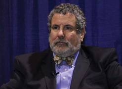
The response rate rose over time. One month post-denervation, 31% of HTN-1 participants were nonresponders as defined by failure to reduce their office systolic blood pressure by at least 10 mm Hg compared to baseline. By 1 year follow-up, the nonresponder rate had fallen to 21%. At 2 years, it was 10%. And at 36 months it was zero.
In other words, the response rate climbed from 69% at 1 month to 100% at 3 years. Thus, it would be premature to repeat renal denervation or switch to alternative therapy because of blood pressure nonresponse at 6 months, the cardiologist said.
"The assumption had been that nonresponse represented inadequate treatment related either to the device or to the operator. That would not appear to be the case at this time. I assume that the nonresponder rate is primarily related to a patient-based characteristic. But so far, we can identify no patient characteristic or drug characteristic that predicts early nonresponse and later response. It’s a wonderful area to look into further," he continued.
The blood pressure reduction was similar regardless of patient age, diabetes status, or baseline renal function.
"What’s particularly gratifying to me is that elderly patients seem to have a significant reduction in systolic blood pressure and narrowing of pulse pressure. To the extent that [elevations of these parameters] are risk factors for the development of cerebrovascular disease, this therapy may have particular value in the elderly population," Dr. Sobotka noted.
With regard to long-term safety, there have been no hypotensive events requiring hospitalization and no change over time in mean electrolyte levels or estimated glomerular filtration rate.

"The absence of a significant reduction in eGFR in the presence of a 30 mm Hg decrease in systolic blood pressure is virtually unheralded in hypertension therapy. One would have expected that the reduction in blood pressure should have been accompanied by a significant reduction in eGFR, which was not seen. The eGFR looks to be stable over 3 years," Dr. Sobotka observed.
Bilateral selective renal denervation is a minimally invasive endovascular procedure targeting the sympathetic nerves running to and from the kidney. It is made possible by the fact that the renal afferent and efferent sympathetic nerves are located in the adventitia of the renal artery wall, well within reach of radiofrequency energy delivered by a special catheter.
Chronic activation of renal sympathetic outflow is a prominent feature in untreated essential hypertension. Animal studies have demonstrated that severing the renal sympathetic nerves reverses or prevents hypertension.
An estimated 15%-20% of patients diagnosed with hypertension are classified as having treatment-resistant hypertension as defined by a systolic blood pressure of at least 160 mm Hg despite three or more antihypertensive drugs.
The mean procedure time was 38 minutes, with an average of four radiofrequency ablations per artery delivered by the proprietary Medtronic Symplicity renal denervation system. Intravenous narcotics and sedatives were used for pain during ablation. Minor complications occurred in 4 of 153 patients. These consisted of three minor access site complications and one renal artery dissection that occurred prior to ablation and was stented with no further consequences.
In response to audience questions about the possibility of nerve sprouting eventually limiting the effectiveness of renal denervation, Dr. Sobotka said that studies in kidney transplant recipients indicate that while there is some regrowth of efferent fibers with partial neurologic activity, the outbound afferent fibers have little or no ability to reconnect.
"We haven’t reexamined neurologic function of the kidneys post-denervation. That needs to be looked at. But to give a pedestrian response, I’d say the blood pressure response is so significant that it’s hard to imagine that something at a neurologic level has regrown that has any clinical importance," the cardiologist explained.
The Symplicity HTN-1 study was an open-label and uncontrolled. In contrast, the Simplicity HTN-2 study features a randomized prospective crossover design. The 6-month results of HTN-2 have been published (Lancet 2010;376:1903-9). At the Chicago ACC meeting, HTN-2 chief investigator Dr. Murray Esler presented the 1-year findings, focusing on the 35 control subjects who crossed over to renal denervation after 6 months of usual care.
The control subjects had a baseline blood pressure of 178/98 mm Hg despite being on an average of five antihypertensive drugs. After 6 months of usual care, their blood pressure had increased to 190/100 mm Hg. But 6 months after undergoing the ablation procedure, their average office blood pressure was 166/92 mm Hg.
One patient was hospitalized for post-procedure hypotension. This individual was treated with intravenous fluids and a reduction in antihypertensive medication and was discharged without further incident. Such occurrences have been quite rare among the roughly 4,000 patients treated worldwide to date, according to Dr. Esler, associated director of the Baker IDI Heart and Diabetes Institute, Melbourne.
Two patients in the control arm had a total of three hypertensive episodes requiring hospitalization during the 6-month usual care phase prior to crossover to renal denervation.
"These patients, of course, ended up as very good treatment responders," he said.
These severe hypertensive episodes while on usual care, along with the natural tendency of blood pressure to rise from baseline to crossover in the control group despite medication, underscore the potential costs in delaying renal denervation, Dr. Esler commented.
A pivotal Phase-3 Symplicity HTN-3 trial is underway in the United States, where renal denervation remains investigational. The study, led by Dr. George Bakris, president of the American Society of Hypertension and professor of medicine at the University of Chicago, involves more than 500 patients with resistant hypertension. It features a blinded crossover study design. The Symplicity system is commercially available in Australia, Europe, and Asia.
Dr. Sobotka is a paid advisor to Medtronic. Dr. Esler has received research grants from and is a paid consultant to Medtronic and Ardian.
CHICAGO – Selective renal denervation for the treatment of resistant hypertension continues to rack up impressively large and durable blood pressure reductions and a solid safety profile 3 years post-procedure, according to the latest update from the Symplicity HTN-1 study, an open-label, uncontrolled investigation.
At baseline the 153 participants in the study conducted in Australia, the United States, and Europe had a mean office blood pressure of 175/98 mm Hg, despite being on an average of 5.1 antihypertensive medications. At 36 months post-denervation they maintained an average in-office blood pressure reduction of -33/-19 mm Hg, according to Dr. Paul A. Sobotka, professor of medicine at Ohio State University, Columbus.
The response rate rose over time. One month post-denervation, 31% of HTN-1 participants were nonresponders as defined by failure to reduce their office systolic blood pressure by at least 10 mm Hg compared to baseline. By 1 year follow-up, the nonresponder rate had fallen to 21%. At 2 years, it was 10%. And at 36 months it was zero.
In other words, the response rate climbed from 69% at 1 month to 100% at 3 years. Thus, it would be premature to repeat renal denervation or switch to alternative therapy because of blood pressure nonresponse at 6 months, the cardiologist said.
"The assumption had been that nonresponse represented inadequate treatment related either to the device or to the operator. That would not appear to be the case at this time. I assume that the nonresponder rate is primarily related to a patient-based characteristic. But so far, we can identify no patient characteristic or drug characteristic that predicts early nonresponse and later response. It’s a wonderful area to look into further," he continued.
The blood pressure reduction was similar regardless of patient age, diabetes status, or baseline renal function.
"What’s particularly gratifying to me is that elderly patients seem to have a significant reduction in systolic blood pressure and narrowing of pulse pressure. To the extent that [elevations of these parameters] are risk factors for the development of cerebrovascular disease, this therapy may have particular value in the elderly population," Dr. Sobotka noted.
With regard to long-term safety, there have been no hypotensive events requiring hospitalization and no change over time in mean electrolyte levels or estimated glomerular filtration rate.
"The absence of a significant reduction in eGFR in the presence of a 30 mm Hg decrease in systolic blood pressure is virtually unheralded in hypertension therapy. One would have expected that the reduction in blood pressure should have been accompanied by a significant reduction in eGFR, which was not seen. The eGFR looks to be stable over 3 years," Dr. Sobotka observed.
Bilateral selective renal denervation is a minimally invasive endovascular procedure targeting the sympathetic nerves running to and from the kidney. It is made possible by the fact that the renal afferent and efferent sympathetic nerves are located in the adventitia of the renal artery wall, well within reach of radiofrequency energy delivered by a special catheter.
Chronic activation of renal sympathetic outflow is a prominent feature in untreated essential hypertension. Animal studies have demonstrated that severing the renal sympathetic nerves reverses or prevents hypertension.
An estimated 15%-20% of patients diagnosed with hypertension are classified as having treatment-resistant hypertension as defined by a systolic blood pressure of at least 160 mm Hg despite three or more antihypertensive drugs.
The mean procedure time was 38 minutes, with an average of four radiofrequency ablations per artery delivered by the proprietary Medtronic Symplicity renal denervation system. Intravenous narcotics and sedatives were used for pain during ablation. Minor complications occurred in 4 of 153 patients. These consisted of three minor access site complications and one renal artery dissection that occurred prior to ablation and was stented with no further consequences.
In response to audience questions about the possibility of nerve sprouting eventually limiting the effectiveness of renal denervation, Dr. Sobotka said that studies in kidney transplant recipients indicate that while there is some regrowth of efferent fibers with partial neurologic activity, the outbound afferent fibers have little or no ability to reconnect.
"We haven’t reexamined neurologic function of the kidneys post-denervation. That needs to be looked at. But to give a pedestrian response, I’d say the blood pressure response is so significant that it’s hard to imagine that something at a neurologic level has regrown that has any clinical importance," the cardiologist explained.
The Symplicity HTN-1 study was an open-label and uncontrolled. In contrast, the Simplicity HTN-2 study features a randomized prospective crossover design. The 6-month results of HTN-2 have been published (Lancet 2010;376:1903-9). At the Chicago ACC meeting, HTN-2 chief investigator Dr. Murray Esler presented the 1-year findings, focusing on the 35 control subjects who crossed over to renal denervation after 6 months of usual care.
The control subjects had a baseline blood pressure of 178/98 mm Hg despite being on an average of five antihypertensive drugs. After 6 months of usual care, their blood pressure had increased to 190/100 mm Hg. But 6 months after undergoing the ablation procedure, their average office blood pressure was 166/92 mm Hg.
One patient was hospitalized for post-procedure hypotension. This individual was treated with intravenous fluids and a reduction in antihypertensive medication and was discharged without further incident. Such occurrences have been quite rare among the roughly 4,000 patients treated worldwide to date, according to Dr. Esler, associated director of the Baker IDI Heart and Diabetes Institute, Melbourne.
Two patients in the control arm had a total of three hypertensive episodes requiring hospitalization during the 6-month usual care phase prior to crossover to renal denervation.
"These patients, of course, ended up as very good treatment responders," he said.
These severe hypertensive episodes while on usual care, along with the natural tendency of blood pressure to rise from baseline to crossover in the control group despite medication, underscore the potential costs in delaying renal denervation, Dr. Esler commented.
A pivotal Phase-3 Symplicity HTN-3 trial is underway in the United States, where renal denervation remains investigational. The study, led by Dr. George Bakris, president of the American Society of Hypertension and professor of medicine at the University of Chicago, involves more than 500 patients with resistant hypertension. It features a blinded crossover study design. The Symplicity system is commercially available in Australia, Europe, and Asia.
Dr. Sobotka is a paid advisor to Medtronic. Dr. Esler has received research grants from and is a paid consultant to Medtronic and Ardian.
FROM THE ANNUAL SCIENTIFIC SESSION OF THE AMERICAN COLLEGE OF CARDIOLOGY
Major Finding: At baseline the 153 participants had a mean office blood pressure of 175/98 mm Hg despite being on an average of 5.1 antihypertensive medications. At 36 months post-denervation they maintained an average in-office blood pressure reduction of -33/-19 mm Hg.
Data Source: Findings from Symplicity HTN-1 are based on an open-label, uncontrolled study. One-year data from a controlled, crossover study were also reported.
Disclosures: Dr. Sobotka is a paid advisor to Medtronic. Dr. Esler has received research grants from and is a paid consultant to Medtronic and Ardian.
Renal Disease Hypertension Goal Reset Below 140/90
CHICAGO – A U.S. foundation that makes recommendations for the management of patients with chronic kidney disease recently set a blood pressure goal of less than 140/90 mm Hg for these patients, upping the target from the 130/80 mm Hg level established in 2003 by the seventh report of the Joint National Committee on Prevention, Detection, and Evaluation, and Treatment of High Blood Pressure (JNC 7) (Hypertension 2003;42:1206-52).
The new goal, made by a panel assembled by the Kidney Disease: Improving Global Outcomes (KDIGO) foundation, will appear in a journal article within the next few weeks, Dr. George L. Bakris said at the meeting.

"We knew when we wrote JNC7 that a target systolic blood pressure of less than 130 mm Hg was not that defensible, but a number of us were concerned that if we set a higher target, most physicians would be content with 145 mm Hg; that’s what drove setting 130 mm Hg," said Dr. Bakris, a member of the JNC 7 writing group, as well as a member of the panel that wrote the new KDIGO guidelines.
Clear-cut, albeit limited, evidence from randomized, controlled trials addresses the appropriate target systolic blood pressure in patients with chronic kidney disease. Three controlled studies addressed the issue, none included patients with diabetic nephropathy, and all three enrolled patients with stage 3 chronic kidney disease, defined as a glomerular filtration rate of 30-59 mL/min per 1.73 m2.
For example, results from the REIN (Ramipril Efficacy in Nephropathy)-2 trial showed that intensive blood pressure lowering to less than 130/80 mm Hg was no more effective than was treatment to a conventional target, diastolic pressure less than 90 mm Hg, for preventing progression of patients to end-stage renal disease (Lancet 2005;365:939-46). And results from AASK (African-American Study of Kidney Disease and Hypertension) showed that intensive blood pressure reduction produced no improvement in the outcome of lost renal function, end-stage renal disease, or death compared with conventional blood pressure control (JAMA 2002;288:2421-31).
A recent observational study had similar findings. Results from the Kidney Early Evaluation Program showed that in more than 16,000 American adults with kidney disease, patients with a baseline systolic blood pressure of less than 130 mm Hg did no better than did patients with a baseline pressure of 130-139 mm Hg for progression to end-stage renal disease during 3 years of follow-up (Arch. Int. Med. 2012;172:41-7).
These results show that you "don’t need to get the [blood pressure] numbers down to the floor to get benefit," said Dr. Bakris, who is a professor of medicine and director of the hypertension center at the University of Chicago.
"A goal of less than 140/90 mm Hg is for everyone with chronic kidney disease, including patients with proteinuria. That may be a little surprising, but the evidence [supporting a lower goal in patients with proteinuria] is soft. This doesn’t mean that you can’t go lower than 130 mm Hg, but the evidence doesn’t really support it."
In addition, while ACE inhibitors and angiotensin-receptor blockers (ARBs) are "definitely indicated" as primary antihypertensive agents for patients with kidney disease, "there is no evidence that using these two classes in combination in patients with advanced kidney disease or in normotensive patients with kidney disease will change the natural history of the disease. The evidence that a combination of an ACE inhibitor and an ARB will reduce proteinuria is pretty good, but we don’t know whether that will lead to benefit in terms of slowing progression," Dr. Bakris said.
Dr. Bakris said that he has received research grants from Forest, Novartis, and Relypsa, that he has been a consultant to Takeda, Servier, Abbott, CVRx, Johnson & Johnson, and Eli Lilly, and that he has been a speaker for Takeda and Novartis.
CHICAGO – A U.S. foundation that makes recommendations for the management of patients with chronic kidney disease recently set a blood pressure goal of less than 140/90 mm Hg for these patients, upping the target from the 130/80 mm Hg level established in 2003 by the seventh report of the Joint National Committee on Prevention, Detection, and Evaluation, and Treatment of High Blood Pressure (JNC 7) (Hypertension 2003;42:1206-52).
The new goal, made by a panel assembled by the Kidney Disease: Improving Global Outcomes (KDIGO) foundation, will appear in a journal article within the next few weeks, Dr. George L. Bakris said at the meeting.
"We knew when we wrote JNC7 that a target systolic blood pressure of less than 130 mm Hg was not that defensible, but a number of us were concerned that if we set a higher target, most physicians would be content with 145 mm Hg; that’s what drove setting 130 mm Hg," said Dr. Bakris, a member of the JNC 7 writing group, as well as a member of the panel that wrote the new KDIGO guidelines.
Clear-cut, albeit limited, evidence from randomized, controlled trials addresses the appropriate target systolic blood pressure in patients with chronic kidney disease. Three controlled studies addressed the issue, none included patients with diabetic nephropathy, and all three enrolled patients with stage 3 chronic kidney disease, defined as a glomerular filtration rate of 30-59 mL/min per 1.73 m2.
For example, results from the REIN (Ramipril Efficacy in Nephropathy)-2 trial showed that intensive blood pressure lowering to less than 130/80 mm Hg was no more effective than was treatment to a conventional target, diastolic pressure less than 90 mm Hg, for preventing progression of patients to end-stage renal disease (Lancet 2005;365:939-46). And results from AASK (African-American Study of Kidney Disease and Hypertension) showed that intensive blood pressure reduction produced no improvement in the outcome of lost renal function, end-stage renal disease, or death compared with conventional blood pressure control (JAMA 2002;288:2421-31).
A recent observational study had similar findings. Results from the Kidney Early Evaluation Program showed that in more than 16,000 American adults with kidney disease, patients with a baseline systolic blood pressure of less than 130 mm Hg did no better than did patients with a baseline pressure of 130-139 mm Hg for progression to end-stage renal disease during 3 years of follow-up (Arch. Int. Med. 2012;172:41-7).
These results show that you "don’t need to get the [blood pressure] numbers down to the floor to get benefit," said Dr. Bakris, who is a professor of medicine and director of the hypertension center at the University of Chicago.
"A goal of less than 140/90 mm Hg is for everyone with chronic kidney disease, including patients with proteinuria. That may be a little surprising, but the evidence [supporting a lower goal in patients with proteinuria] is soft. This doesn’t mean that you can’t go lower than 130 mm Hg, but the evidence doesn’t really support it."
In addition, while ACE inhibitors and angiotensin-receptor blockers (ARBs) are "definitely indicated" as primary antihypertensive agents for patients with kidney disease, "there is no evidence that using these two classes in combination in patients with advanced kidney disease or in normotensive patients with kidney disease will change the natural history of the disease. The evidence that a combination of an ACE inhibitor and an ARB will reduce proteinuria is pretty good, but we don’t know whether that will lead to benefit in terms of slowing progression," Dr. Bakris said.
Dr. Bakris said that he has received research grants from Forest, Novartis, and Relypsa, that he has been a consultant to Takeda, Servier, Abbott, CVRx, Johnson & Johnson, and Eli Lilly, and that he has been a speaker for Takeda and Novartis.
EXPERT OPINION FROM THE ANNUAL MEETING OF THE AMERICAN COLLEGE OF CARDIOLOGY
Hemodialysis, Injection Drug Users Vulnerable to Recurrent Endocarditis
LONDON – Hemodialysis and injection drug use were major risk factors for recurrent episodes of infective endocarditis among patients enrolled in a large international prospective study.
Repeat infective endocarditis (IE) is a serious complication of patients who survive an initial episode. In previous studies, the incidence has ranged from 2% to 31%. "Repeat IE is associated with significant mortality. It is an uncommon complication but can be highly relevant among specific groups of patients," Dr. Laura Alagna of San Raffaele Hospital, Milan, said at the European Congress of Clinical Microbiology and Infectious Diseases.
The findings come from The International Collaboration on Endocarditis-Prospective Cohort Study (ICE-PCS), a contemporary cohort of more than 5,000 patients with infective endocarditis (IE) from 64 centers in 28 countries worldwide. Patients included in the current analysis were those enrolled from June 2000 to December 2006, with a diagnosis of definite IE on native or prosthetic valves who had at least a 1-year follow-up.
Of 1,857 patients who met inclusion criteria, 1,783 had one episode of IE and 91 (5%) had a repeat IE. Of those, 17 had a presumed relapse, defined clinically as infection with the same pathogen isolated within 6 months of the initial episode. The other 74 had a presumed new infection, defined clinically as a repeat episode with a different pathogen or the same pathogen isolated greater than 6 months from the initial episode.
On bivariate analysis, being from North America, infection with Staphylococcus aureus, hemodialysis dependence, intravenous drug use (IDU), HIV infection, history of previous IE, and non-nosocomial health care as a presumed source of infection were all associated with repeat IE. However, on multivariate analysis, only four independent risk factors emerged: North American location (odds ratio, 1.96), hemodialysis (OR, 2.54), IDU (OR, 2.89), and history of previous IE (2.76).
At 1-year follow-up, survival was significantly lower for those with repeat IE, 80% compared to 91% for those with only one episode (P = .0034), Dr. Alagna reported.
Molecular analysis with pulsed-gel electrophoresis performed in 12 of the repeat IE patients demonstrated concordance with the clinical definition in 10, including eight confirmed relapses and two confirmed new infections. Of the other two patients, one had S. aureus with the same molecular pattern isolated more than a year after the first episode. That patient was hemodialysis-dependent and had other risk factors. In the other discordant patient, S. bovis with the same molecular pattern was isolated after nearly 9 months. In that patient, a cardiac device had not been removed during the first episode, she explained.
"The clinical classification of repeat IE is satisfactory, but can be improved with molecular analysis," she concluded.
Dr. Alagna stated that she had no disclosures.
FROM THE EUROPEAN CONGRESS OF CLINICAL MICROBIOLOGY AND INFECTIOUS DISEASES
Major Finding: On multivariate analysis, independent risk factors for repeat infective endocarditis were North American location (odds ratio, 1.96), hemodialysis (OR, 2.54), IDU (OR, 2.89), and history of previous IE (2.76).
Data Source: The findings come from The International Collaboration on Endocarditis-Prospective Cohort Study (ICE-PCS), a contemporary cohort of more than 5,000 patients with infective endocarditis (IE) from 64 centers in 28 countries worldwide. Patients included in the current analysis were those enrolled from June 2000 to December 2006, with a diagnosis of definite IE on native or prosthetic valves who had at least a 1-year follow-up.
Disclosures: Dr. Alagna stated that she had no disclosures.
FDA Panel: Approve Mirabegron for Overactive Bladder
SILVER SPRING, MD. – Physicians may soon have a first-in-class alternative to current treatments for overactive bladder, which may not always work and can have unpleasant side effects.
A Food and Drug Administration panel on April 5 voted 7-4 with 1 abstention that the overall benefit-risk assessment supports the approval of mirabegron for the treatment of overactive bladder.
Pharmacologic treatment options to date include muscarinic receptor antagonists, which can affect the salivary glands, intestines, and eyes, resulting in side effects such as dry mouth, constipation, and blurred vision, respectively.
Mirabegron is a first-in-class agonist of beta 3-adrenoceptors. Astellas Pharma developed the drug for the treatment of overactive bladder with symptoms of urge urinary incontinence, urgency, and urinary frequency. In the United States, the proposed dose is 50 mg once daily with or without food. However, a dose of 25 mg is reserved for patients with severe renal or moderate hepatic impairment. The formulation is an oral controlled-absorption system (extended-release) tablets. The drug is already approved in Japan.
"The committee has a sense that there is marginal efficacy, but overall the majority feels that the benefits outweigh the risks. This is seen as a new agent, which can potentially be used for individuals who have not had success with other available agents," summarized Dr. Julia V. Johnson, who is the chair of the advisory committee for reproductive health drugs. "With some caution and with some reservation the overall approval of the medication comes from this committee."
The committee also voted 8-4 that the data do provide substantial evidence of benefit for mirabegron in the treatment of overactive bladder. However, many members agreed that there was statistically-significant improvement in symptoms but questioned whether this would translate to a meaningful clinical difference.
The committee also voted 9-3 that adequate safety had been demonstrated for the drug. Committee members did express reservations about the paucity of data with regard to neoplasms, hepatotoxicity events, and hypersensitivity reactions.
The primary sources of data for the evaluation are three randomized placebo-controlled phase III trials. Supportive data included data from a single long-term (active-controlled) study, three phase II studies, and a phase II study in patients with symptomatic benign prostatic hyperplasia.
The three phase III trials lasted for 12 weeks with individuals randomized to receive mirabegron either 25 mg daily, or 50 mg daily, or placebo. Coprimary end points included change from baseline in the average number of micturitions in 24 hours and the change from baseline in the average number of incontinence episodes in 24 hours.
Mirabegron at a dose of 50 mg achieved the primary efficacy objectives in three phase III studies. Treatment with 50 mg mirabegron reduced the mean number of micturitions by 0.55/day, compared with placebo. Likewise, mirabegron reduced the mean number of incontinence episodes per day by 0.40, compared with placebo. Lastly, the mean volume per void increased by 11.9 mL with the drug, compared with placebo.
Committee members expressed some concern about increases in both blood pressure and pulse rate associated with the drug. There was also an increased incidence of urologic adverse events, particularly urinary tract infections of mild severity. The committee members were particularly concerned about an increased incidence of neoplasm serious adverse events – new malignant events – in the group on 100 mg mirabegron in the 1-year study, as well as hepatotoxicity events and hypersensitivity reactions. However, they noted that there was insufficient data to draw firm conclusions about these serious adverse events.
FROM A MEETING OF THE FDA'S REPRODUCTIVE HEALTH DRUGS ADVISORY COMMITTEE
Angioedema Hospitalizations Rising, Especially in African Americans
ORLANDO – The rate of U.S. hospitalizations for angioedema doubled among African Americans during 2000-2009 based on a national sampling of inpatients, and the rise appears largely driven by adverse drug reactions to angiotensin-converting enzyme inhibitors.
"The rate of angioedema due to an adverse drug reaction rose from about 40% in 2000 to about 60% in 2009," said Dr. Robert Y. Lin while presenting a poster at the annual meeting of the American Academy of Allergy, Asthma, and Immunology. Adverse drug reactions "are not the only reason [that angioedema occurs], but I think it’s driving the increase."

Angiotensin-converting enzyme (ACE) inhibitors are the drugs most responsible for causing angioedema, and it seems this drug class is especially responsible for the rapid increase in the number of angioedema cases among African Americans, he said.
Among African Americans, hospitalizations for angioedema jumped from 9 cases per 100,000 in 2000 to 18 cases per 100,000 in 2009. In contrast, among non–African American residents of the United States, hospitalizations for angioedema rose from 2.7 cases per 100,000 in 2000 to 3.6 cases per 100,000 in 2009(see table).
African Americans seem particularly susceptible to developing angioedema triggered by ACE inhibitors, noted Dr. Lin. In addition, diseases that often lead to treatment with an ACE inhibitor, such as hypertension, chronic kidney disease, and heart failure, are especially prevalent among African Americans.
Angiotensin-receptor blockers, which are not linked to causing angioedema, might be a better alternative for African American patients, said Dr. Lin, a professor of medicine at New York Medical College and chief of the division of allergy and immunology at New York Downtown Hospital.
The angioedema danger from ACE inhibitors is especially insidious because it appears to be a class effect that is not dose or duration related. The side effect can occur suddenly at any time during the course of treatment. "The lag period between when you start treatment and when you see a reaction can be so variable. It can be 2 weeks, or it can be 2 years. A patient can be fine on an ACE inhibitor for months or years and then suddenly have an angioedema reaction. We don’t know why," he said in an interview.
Although angioedema is rarely fatal—the mortality rate was 0.4% in the more than 128,000 hospitalized cases that Dr. Lin tallied in his analysis—it led to an intubation rate of 8% and average hospitalization charges of about $10,000 per case. With nearly 13,000 Americans hospitalized for angioedema annually during 2000-2009, that means the annual cost was roughly $130 million.
Dr. Lin identified angioedema cases in U.S. hospitalization records collected by the Nationwide Inpatient Sample, a database maintained by the U.S. Agency for Healthcare Research and Quality. He identified angioedema cases by their ICD code. During the study period, patients hospitalized with angioedema averaged 61 years old, 61% were women, and 37% were African American. The percent of all patients with angioedema who were African American rose from 32% of cases in 2000 to 41% in 2009.
Throughout the decade studied, 56% of the angioedema cases appeared secondary to an adverse drug reaction; 61% of the angioedema patients had hypertension. Adverse drug reactions linked to hypertension or cardiovascular disease occurred in 29% of the angioedema patients overall throughout the decade studied, rising from 22% of cases in 2000 to 36% of cases in 2009. Adverse drug reactions linked to drugs used to treat hypertension or cardiovascular disease occurred overall in 38% of African American patients hospitalized for angioedema, and hit a high of 44% of African Americans hospitalized for angioedema in 2009.
Dr. Lin said that he had no disclosures.
The observations made in this report from a credible database are disturbing. These findings do not definitively prove that increasing ACE inhibitor use in African Americans is causing the rise in hospitalizations for angioedema, but this is a plausible speculation. Another factor causing the increased hospitalizations may be increasing recognition of this effect in patients treated with an ACE inhibitor. The link between ACE inhibitor use and the increase in hospitalizations for angioedema in African Americans would be clearer if data were available to show that definitively the rise in hospitalizations occurred in parallel with increased use of these drugs.
We have long known that African Americans have more problems with angioedema triggered by ACE inhibitor treatment than do whites. Even without this possible adverse effect, use of ACE inhibitors as monotherapy has fallen out of favor in African Americans because, in general, they receive less blood-pressure-lowering benefit. However, ACE inhibitors have been useful in African American patients with chronic kidney disease. They have become a drug of choice for patients with diabetes, although this application is a bit of an extrapolation as no data specifically show the superiority of ACE inhibitors to other antihypertensive drugs in African American patients with diabetes. For example, in the results of the ALLHAT (Antihypertensive and Lipid Lowering Treatment to Prevent Heart Attack Trial), treatment with an ACE inhibitor showed no benefit over the other drug classes tested in patients with diabetes, regardless of racial or ethnic groups.
The incremental benefits that ACE inhibitors provide also appear obtainable from angiotensin-receptor blocker (ARB) drugs. One substantial difference, however, is cost. Losartan is the only ARB currently available in generic formulations. Many ACE inhibitors are available as generic formulations and hence are less expensive. Insurers and managed care organizations often put up significant barriers to prescribing an ARB before first trying treatment with an ACE inhibitor. Aside from this economic barrier, ARBs are not as effective as ACE inhibitors for preventing or reducing the peripheral edema caused by treatment with a calcium antagonist.
Based on the overall better tolerability of ARBs, which do not cause cough or angioedema, an emerging sense in the field is to question whether treatment with an ACE inhibitor warrants unqualified recommendation over an ARB for African American patients, even those patients who have a compelling indication for ACE inhibitor treatment.
The report by Dr. Lin gives credence to the idea that greater caution is warranted when considering treatment with an ACE inhibitor in African American patients. This caution stems not from concern about the efficacy that ACE inhibitors might have for blood pressure reduction, but because use of these drugs may increase the chance for harm.
Dr. John M. Flack is a professor of medicine and physiology and chairman of the department of medicine at Wayne State University in Detroit. He is also specialist-in-chief for internal medicine at the Detroit Medical Center. Dr. Flack has been a consultant to or on a steering committee for Cardiodynamics, Daiichi-Sankyo, Novartis, and GlaxoSmithKline. He has been on the speakers bureaus of Daiichi-Sankyo, Novartis, and Pfizer, and he has received research support from Covance, Cardiodynamics, Daiichi-Sankyo, Sanofi Aventis, Mannkind, and Novartis. Dr. Flack made these comments in an interview.
ORLANDO – The rate of U.S. hospitalizations for angioedema doubled among African Americans during 2000-2009 based on a national sampling of inpatients, and the rise appears largely driven by adverse drug reactions to angiotensin-converting enzyme inhibitors.
"The rate of angioedema due to an adverse drug reaction rose from about 40% in 2000 to about 60% in 2009," said Dr. Robert Y. Lin while presenting a poster at the annual meeting of the American Academy of Allergy, Asthma, and Immunology. Adverse drug reactions "are not the only reason [that angioedema occurs], but I think it’s driving the increase."
Angiotensin-converting enzyme (ACE) inhibitors are the drugs most responsible for causing angioedema, and it seems this drug class is especially responsible for the rapid increase in the number of angioedema cases among African Americans, he said.
Among African Americans, hospitalizations for angioedema jumped from 9 cases per 100,000 in 2000 to 18 cases per 100,000 in 2009. In contrast, among non–African American residents of the United States, hospitalizations for angioedema rose from 2.7 cases per 100,000 in 2000 to 3.6 cases per 100,000 in 2009(see table).
African Americans seem particularly susceptible to developing angioedema triggered by ACE inhibitors, noted Dr. Lin. In addition, diseases that often lead to treatment with an ACE inhibitor, such as hypertension, chronic kidney disease, and heart failure, are especially prevalent among African Americans.
Angiotensin-receptor blockers, which are not linked to causing angioedema, might be a better alternative for African American patients, said Dr. Lin, a professor of medicine at New York Medical College and chief of the division of allergy and immunology at New York Downtown Hospital.
The angioedema danger from ACE inhibitors is especially insidious because it appears to be a class effect that is not dose or duration related. The side effect can occur suddenly at any time during the course of treatment. "The lag period between when you start treatment and when you see a reaction can be so variable. It can be 2 weeks, or it can be 2 years. A patient can be fine on an ACE inhibitor for months or years and then suddenly have an angioedema reaction. We don’t know why," he said in an interview.
Although angioedema is rarely fatal—the mortality rate was 0.4% in the more than 128,000 hospitalized cases that Dr. Lin tallied in his analysis—it led to an intubation rate of 8% and average hospitalization charges of about $10,000 per case. With nearly 13,000 Americans hospitalized for angioedema annually during 2000-2009, that means the annual cost was roughly $130 million.
Dr. Lin identified angioedema cases in U.S. hospitalization records collected by the Nationwide Inpatient Sample, a database maintained by the U.S. Agency for Healthcare Research and Quality. He identified angioedema cases by their ICD code. During the study period, patients hospitalized with angioedema averaged 61 years old, 61% were women, and 37% were African American. The percent of all patients with angioedema who were African American rose from 32% of cases in 2000 to 41% in 2009.
Throughout the decade studied, 56% of the angioedema cases appeared secondary to an adverse drug reaction; 61% of the angioedema patients had hypertension. Adverse drug reactions linked to hypertension or cardiovascular disease occurred in 29% of the angioedema patients overall throughout the decade studied, rising from 22% of cases in 2000 to 36% of cases in 2009. Adverse drug reactions linked to drugs used to treat hypertension or cardiovascular disease occurred overall in 38% of African American patients hospitalized for angioedema, and hit a high of 44% of African Americans hospitalized for angioedema in 2009.
Dr. Lin said that he had no disclosures.
FROM THE ANNUAL MEETING OF THE AMERICAN ACADEMY OF ALLERGY, ASTHMA, AND IMMUNOLOGY
Major Finding: Among African Americans, the incidence of angioedema hospitalizations rose from 9 cases per 100,000 in 2000 to 18 cases per 100,000 in 2009.
Data Source: Review of a sample of U.S. hospitalizations during 2000-2009 from the Nationwide Inpatient Sample.
Disclosures: Dr. Lin said that he had no disclosures.