User login
Progesterone Therapy Ineffective in Women With Recurrent Miscarriages
Women with histories of miscarriage do not experience any significant increase in successful births by undergoing progesterone therapy during the first trimester of pregnancy, according to a new study published Nov. 25 in the New England Journal of Medicine.
“Our results do not support earlier findings of a Cochrane analysis that suggested a benefit of progesterone therapy in the first trimester of pregnancy,” wrote Dr. Arri Coomarasamy, professor of gynecology at the University of Birmingham in Edgbaston, England, and his associates, referencing the 2013 review of 2,158 women across 14 randomized trials, which found that progesterone therapy administered in the first trimester can increase the likelihood of having a successful birth without risk of adverse effects on either the mother or infant.
For the current study, known as the PROMISE trial, the investigators randomized 836 women into cohorts of 404 (progesterone therapy) and 432 (placebo). The progesterone therapy regimen consisted of vaginal suppositories of 400 mg micronized progesterone, inserted twice daily, through 12 weeks of gestation. Those in the placebo cohort also were given vaginal suppositories twice a day for the same duration of the pregnancy (N Engl J Med. 2015;373:2141-8).
Of these women, 826 (98.8%) had follow-up interviews – 398 in the progesterone cohort and 428 in the placebo cohort. Primary outcome (live birth after at least 24 weeks of gestation) occurred in 262 (65.8%) of the women in the progesterone cohort and 271 (63.3%) of the women in the placebo cohort (P = .045). Investigators concluded that there was no significant statistical difference in outcomes between progesterone therapy and placebo based on these results.
The PROMISE study was funded by the United Kingdom National Institute of Health Research. Dr. Coomarasamy did not report any relevant financial disclosures.
FROM THE NEW ENGLAND JOURNAL OF MEDICINE
Progesterone therapy ineffective in women with recurrent miscarriages
Women with histories of miscarriage do not experience any significant increase in successful births by undergoing progesterone therapy during the first trimester of pregnancy, according to a new study published Nov. 25 in the New England Journal of Medicine.
“Our results do not support earlier findings of a Cochrane analysis that suggested a benefit of progesterone therapy in the first trimester of pregnancy,” wrote Dr. Arri Coomarasamy, professor of gynecology at the University of Birmingham in Edgbaston, England, and his associates, referencing the 2013 review of 2,158 women across 14 randomized trials, which found that progesterone therapy administered in the first trimester can increase the likelihood of having a successful birth without risk of adverse effects on either the mother or infant.
For the current study, known as the PROMISE trial, the investigators randomized 836 women into cohorts of 404 (progesterone therapy) and 432 (placebo). The progesterone therapy regimen consisted of vaginal suppositories of 400 mg micronized progesterone, inserted twice daily, through 12 weeks of gestation. Those in the placebo cohort also were given vaginal suppositories twice a day for the same duration of the pregnancy (N Engl J Med. 2015;373:2141-8).
Of these women, 826 (98.8%) had follow-up interviews – 398 in the progesterone cohort and 428 in the placebo cohort. Primary outcome (live birth after at least 24 weeks of gestation) occurred in 262 (65.8%) of the women in the progesterone cohort and 271 (63.3%) of the women in the placebo cohort (P = .045). Investigators concluded that there was no significant statistical difference in outcomes between progesterone therapy and placebo based on these results.
The PROMISE study was funded by the United Kingdom National Institute of Health Research. Dr. Coomarasamy did not report any relevant financial disclosures.
FROM THE NEW ENGLAND JOURNAL OF MEDICINE
Key clinical point: Women who have experienced previous recurrent miscarriages are not significantly more likely to have a successful birth if they undergo progesterone therapy during the first trimester of their pregnancy.
Major finding: Of 398 women in the progesterone cohort, 262 (65.8%) had live births; in the placebo cohort, 271 of the 428 women (63.3%) had live births, indicating no significant difference between the two.
Data source: A multicenter, double-blind, placebo-controlled, randomized trial of 826 pregnant women with history of recurrent miscarriages.
Disclosures: The study was funded by the United Kingdom National Institute of Health Research. Dr. Coomarasamy did not report any relevant financial disclosures.
Breastfeeding cuts risk of progression from GDM to type 2 diabetes
Women with gestational diabetes reduced their risk of progressing to type 2 diabetes by more than half if they exclusively breastfed their infants, compared with women who fed their babies exclusively with formula, according to a new study.
Researchers also found that breastfeeding for more than 2 months was associated with a lower incidence of progression to type 2 diabetes.
“In our study, both higher lactation intensity and duration showed strong, graded protective associations with type 2 diabetes mellitus incidence, independent of risk factors (sociodemographic characteristics, prenatal metabolic status and course, perinatal outcomes, and lifestyle behaviors) that were not explained by weight loss,” Erica P. Gunderson, Ph.D., of Kaiser Permanente Northern California, and her associates wrote.

The researchers initially evaluated 1,035 women who had been diagnosed with gestational diabetes mellitus (GDM) and had a singleton delivery at 35 weeks’ gestation or later, but followed only the 1,010 women without type 2 diabetes at 6-9 weeks postpartum. Of the 959 women evaluated at the end of the 2-year follow-up, 11.8% had progressed to type 2 diabetes (Ann Intern Med. 2015 Nov 24. doi:10.7326/M15-0807).
Data on breastfeeding intensity and duration were collected through feeding diaries, phone calls, in-person exams, and monthly mailed questionnaires. The four categories of intensity included exclusive lactation, mostly lactation (about 1-6 ounces of formula per 24 hours), mostly formula (more than 17 ounces per 24 hours), and mixed feeding (7-17 ounces of formula per 24 hours). These categories were compared to exclusive formula feeding.
Compared with mothers who exclusively fed their infants formula, mothers who exclusively breastfed had a 54% lower risk of developing type 2 diabetes (hazard ratio, 0.46). The risk of developing type 2 diabetes was 46% lower for women who mostly breastfed, and women who mostly used formula or who mixed formula and breastfeeding had a 36% reduced risk (HR, 0.54 and 0.64, respectively; P trend = 0.016).
Similarly, duration of breastfeeding correlated with the reduced risk of developing diabetes. Compared with mothers who breastfed their infants from 0 to 2 months, those who breastfed for longer than 10 months had a 57% lower risk of developing type 2 diabetes. Women who breastfed their infants for 5-10 months had a 50% lower risk, and those who breastfed from 2 to 5 months had a 45% lower risk, compared with those breastfeeding up to 2 months.
“Potential mechanisms to explain the lower incidence of [diabetes mellitus] with higher intensity and duration of lactation include preservation of pancreatic beta cells, less inflammation, and improved endothelial function; however, biochemical evidence is sparse,” the researchers wrote, noting the effect prolactin has during pregnancy on increasing pancreatic beta-cell mass and function.
In analyzing the relationship between development of type 2 diabetes and lactation duration and intensity, the researchers controlled for a range of confounders. These included maternal body mass index, gestational diabetes treatment, prepregnancy weight, maternal delivery weight, delivery method, and newborn outcomes (birth weight, length, gestational age, Apgar score, hospital stay duration, medical diagnoses, and NICU admission). Researchers also accounted for lifestyle behaviors, depression, medication use, maternal education, parity, age, race/ethnicity, and health history.
The research was funded by the National Institutes of Health, Kaiser Permanente Northern California, and the W.K. Kellogg Foundation. Dr. Gunderson has received grant funding from the American Diabetes Association. Other authors reported receiving industry grants outside of this study.
Women with gestational diabetes reduced their risk of progressing to type 2 diabetes by more than half if they exclusively breastfed their infants, compared with women who fed their babies exclusively with formula, according to a new study.
Researchers also found that breastfeeding for more than 2 months was associated with a lower incidence of progression to type 2 diabetes.
“In our study, both higher lactation intensity and duration showed strong, graded protective associations with type 2 diabetes mellitus incidence, independent of risk factors (sociodemographic characteristics, prenatal metabolic status and course, perinatal outcomes, and lifestyle behaviors) that were not explained by weight loss,” Erica P. Gunderson, Ph.D., of Kaiser Permanente Northern California, and her associates wrote.
The researchers initially evaluated 1,035 women who had been diagnosed with gestational diabetes mellitus (GDM) and had a singleton delivery at 35 weeks’ gestation or later, but followed only the 1,010 women without type 2 diabetes at 6-9 weeks postpartum. Of the 959 women evaluated at the end of the 2-year follow-up, 11.8% had progressed to type 2 diabetes (Ann Intern Med. 2015 Nov 24. doi:10.7326/M15-0807).
Data on breastfeeding intensity and duration were collected through feeding diaries, phone calls, in-person exams, and monthly mailed questionnaires. The four categories of intensity included exclusive lactation, mostly lactation (about 1-6 ounces of formula per 24 hours), mostly formula (more than 17 ounces per 24 hours), and mixed feeding (7-17 ounces of formula per 24 hours). These categories were compared to exclusive formula feeding.
Compared with mothers who exclusively fed their infants formula, mothers who exclusively breastfed had a 54% lower risk of developing type 2 diabetes (hazard ratio, 0.46). The risk of developing type 2 diabetes was 46% lower for women who mostly breastfed, and women who mostly used formula or who mixed formula and breastfeeding had a 36% reduced risk (HR, 0.54 and 0.64, respectively; P trend = 0.016).
Similarly, duration of breastfeeding correlated with the reduced risk of developing diabetes. Compared with mothers who breastfed their infants from 0 to 2 months, those who breastfed for longer than 10 months had a 57% lower risk of developing type 2 diabetes. Women who breastfed their infants for 5-10 months had a 50% lower risk, and those who breastfed from 2 to 5 months had a 45% lower risk, compared with those breastfeeding up to 2 months.
“Potential mechanisms to explain the lower incidence of [diabetes mellitus] with higher intensity and duration of lactation include preservation of pancreatic beta cells, less inflammation, and improved endothelial function; however, biochemical evidence is sparse,” the researchers wrote, noting the effect prolactin has during pregnancy on increasing pancreatic beta-cell mass and function.
In analyzing the relationship between development of type 2 diabetes and lactation duration and intensity, the researchers controlled for a range of confounders. These included maternal body mass index, gestational diabetes treatment, prepregnancy weight, maternal delivery weight, delivery method, and newborn outcomes (birth weight, length, gestational age, Apgar score, hospital stay duration, medical diagnoses, and NICU admission). Researchers also accounted for lifestyle behaviors, depression, medication use, maternal education, parity, age, race/ethnicity, and health history.
The research was funded by the National Institutes of Health, Kaiser Permanente Northern California, and the W.K. Kellogg Foundation. Dr. Gunderson has received grant funding from the American Diabetes Association. Other authors reported receiving industry grants outside of this study.
FROM ANNALS OF INTERNAL MEDICINE
Key clinical point: Longer or exclusive breastfeeding reduces maternal risk of type 2 diabetes in women with prior GDM.
Major finding: Breastfeeding for longer than 10 months reduced risk of progression from GDM to type 2 diabetes by 57%, and exclusively breastfeeding reduced the risk by 54%.
Data source: The findings are based on a prospective, observational cohort starting with 1,035 women diagnosed with GDM and followed for 2 years.
Disclosures: The research was funded by the National Institutes of Health, Kaiser Permanente Northern California, and the W.K. Kellogg Foundation. Dr. Gunderson has received grant funding from the American Diabetes Association. Other authors reported receiving industry grants outside of this study.

New ACOG recommendations encourage exercise during pregnancy
Women with uncomplicated pregnancies should be encouraged to engage in aerobic and strength-conditioning exercises, including running, stationary cycling, and modified yoga, according to new recommendations from the American College of Obstetricians and Gynecologists.
Even women who were not active before becoming pregnant can start exercising during pregnancy, provided they consult with their physicians and ramp up their exercise gradually.
“Pregnancy should not be looked at as a state of confinement,” Dr. Raul Artal, chairman emeritus of the department of ob.gyn. at Saint Louis University and the lead author of the ACOG recommendations, said in a statement. “In fact, it is an ideal time for lifestyle modification. That is because more than any other time in her life, a pregnant woman has the most available access to medical care and supervision.”

The opinion from ACOG’s Committee on Obstetric Practice, which was released on Nov. 23, outlines safe and unsafe exercises during pregnancy and in the postpartum period, updating recommendations previously issued in 2002 and reaffirmed in 2009.
The updated recommendations differ from the relatively limited ones published by ACOG in 2002. Previously, ACOG recommended exercise only for women who were recreational or competitive athletes prior to pregnancy. And even in those cases, women were cautioned to “modify their usual exercise routines as medically indicated” and to avoid strenuous exercise in the second and third trimesters, especially women with “a history of or risk for preterm labor or fetal growth restriction.” Women who were not regularly physically active prior to their pregnancies were advised to consult with their physicians before beginning any exercise regimen (Obstet Gynecol 2002;99:171-3).
Now, women are encouraged to engage in some amount of physical activity during pregnancy, but physicians should conduct a thorough clinical evaluation before prescribing a specific exercise program to ensure the patient doesn’t have a medical reason to avoid exercise, according to ACOG.
The updated opinion advises that women should have an eventual goal of moderate-intensity exercise for at least 20-30 minutes per day on most or all days of the week. The “talk test” – whether a woman can carry on a conversation while exercising – is a good way to ensure that the exercise is not too intense.
In the updated recommendations, ACOG designates walking, swimming, stationary cycling, low-impact aerobics, modified yoga or pilates, running, jogging, racquet sports, and strength training as “safe” during pregnancy. But pregnant women should avoid contact sports, scuba diving, sky diving, “hot yoga” or “hot pilates,” and “activities with a high risk of falling” such as gymnastics and skiing, among others (Obstet Gynecol. 2015;126:e135-42).
Some absolute contraindications to aerobic exercise during pregnancy include hemodynamically significant heart disease, restrictive lung disease, incompetent cervix or cerclage, and multiple gestation at risk of premature labor, among others.
Obesity in pregnancy
ACOG also released updated clinical management guidelines for obesity in pregnancy, calling on ob.gyns. to calculate body mass index at the first prenatal visit and offer diet and exercise counseling.
The guidelines advise that obese women can have improved pregnancy outcomes by losing even just a little bit of weight prior to pregnancy, and losing weight between pregnancies can decrease the chances of obese women having “large-for-gestational-age neonates in a subsequent pregnancy” (Obstet Gynecol 2015;126:e112-26).
In addition, ACOG advises physicians to consider allowing a longer first stage of labor in obese women before performing a cesarean delivery for labor arrest. Mechanical thromboprophylaxis is recommended both before and after a cesarean in obese women.
While stillbirth rates are higher in obese pregnant women, ACOG did not make a recommendation for or against routine antenatal fetal surveillance in these women because of a lack of evidence showing clear improvement in pregnancy outcomes with surveillance.
Women with uncomplicated pregnancies should be encouraged to engage in aerobic and strength-conditioning exercises, including running, stationary cycling, and modified yoga, according to new recommendations from the American College of Obstetricians and Gynecologists.
Even women who were not active before becoming pregnant can start exercising during pregnancy, provided they consult with their physicians and ramp up their exercise gradually.
“Pregnancy should not be looked at as a state of confinement,” Dr. Raul Artal, chairman emeritus of the department of ob.gyn. at Saint Louis University and the lead author of the ACOG recommendations, said in a statement. “In fact, it is an ideal time for lifestyle modification. That is because more than any other time in her life, a pregnant woman has the most available access to medical care and supervision.”
The opinion from ACOG’s Committee on Obstetric Practice, which was released on Nov. 23, outlines safe and unsafe exercises during pregnancy and in the postpartum period, updating recommendations previously issued in 2002 and reaffirmed in 2009.
The updated recommendations differ from the relatively limited ones published by ACOG in 2002. Previously, ACOG recommended exercise only for women who were recreational or competitive athletes prior to pregnancy. And even in those cases, women were cautioned to “modify their usual exercise routines as medically indicated” and to avoid strenuous exercise in the second and third trimesters, especially women with “a history of or risk for preterm labor or fetal growth restriction.” Women who were not regularly physically active prior to their pregnancies were advised to consult with their physicians before beginning any exercise regimen (Obstet Gynecol 2002;99:171-3).
Now, women are encouraged to engage in some amount of physical activity during pregnancy, but physicians should conduct a thorough clinical evaluation before prescribing a specific exercise program to ensure the patient doesn’t have a medical reason to avoid exercise, according to ACOG.
The updated opinion advises that women should have an eventual goal of moderate-intensity exercise for at least 20-30 minutes per day on most or all days of the week. The “talk test” – whether a woman can carry on a conversation while exercising – is a good way to ensure that the exercise is not too intense.
In the updated recommendations, ACOG designates walking, swimming, stationary cycling, low-impact aerobics, modified yoga or pilates, running, jogging, racquet sports, and strength training as “safe” during pregnancy. But pregnant women should avoid contact sports, scuba diving, sky diving, “hot yoga” or “hot pilates,” and “activities with a high risk of falling” such as gymnastics and skiing, among others (Obstet Gynecol. 2015;126:e135-42).
Some absolute contraindications to aerobic exercise during pregnancy include hemodynamically significant heart disease, restrictive lung disease, incompetent cervix or cerclage, and multiple gestation at risk of premature labor, among others.
Obesity in pregnancy
ACOG also released updated clinical management guidelines for obesity in pregnancy, calling on ob.gyns. to calculate body mass index at the first prenatal visit and offer diet and exercise counseling.
The guidelines advise that obese women can have improved pregnancy outcomes by losing even just a little bit of weight prior to pregnancy, and losing weight between pregnancies can decrease the chances of obese women having “large-for-gestational-age neonates in a subsequent pregnancy” (Obstet Gynecol 2015;126:e112-26).
In addition, ACOG advises physicians to consider allowing a longer first stage of labor in obese women before performing a cesarean delivery for labor arrest. Mechanical thromboprophylaxis is recommended both before and after a cesarean in obese women.
While stillbirth rates are higher in obese pregnant women, ACOG did not make a recommendation for or against routine antenatal fetal surveillance in these women because of a lack of evidence showing clear improvement in pregnancy outcomes with surveillance.
FROM OBSTETRICS AND GYNECOLOGY

Endometriosis and Pain: Expert Answers to 6 Questions Targeting Your Management Options
IN THIS ARTICLE
• When is laparoscopy indicated?
• Excision versus ablation
• How to reduce the risk for postoperative recurrence
Endometriosis has always posed a treatment challenge. In the early 19th century, before the widespread advent of surgery, the disease was managed by applying leeches to the cervix. In fact, as Nezhat and colleagues note in their comprehensive survey of the 4,000-year history of endometriosis, “leeches were considered a mainstay in treating any condition associated with menstruation.”1
In the 21st century, the picture is clearer, though still not crystal clear. The optimal approach to endometriosis depends on many factors, foremost the patient’s chief complaint: pain or infertility (or both).
This article focuses on medical and surgical management of pain. Six experts address such questions as: When is laparoscopy indicated? Is excision or ablation of lesions preferred? What is the role of hysterectomy in eliminating pain? And what can be done about the problem of recurrence?
1. WHAT ARE THE OPTIONS FOR EMPIRIC THERAPY?
One reason for the diagnostic delay with endometriosis, which still averages about six years, is that definitive diagnosis is achieved only through laparoscopic investigation and histologic confirmation. For many women who experience pain thought to be associated with endometriosis, however, clinicians begin empiric treatment with medical agents as a way to avert the need for surgery, if at all possible.
“There is no cure for endometriosis,” says John R. Lue, MD, MPH, “but there are many ways that endometriosis can be treated” and the impact of the disease reduced in a patient’s life. (Editor’s Note: See below for biographical information on each clinician interviewed in this article.)
Medical and hormonal options include
• NSAIDs, often used with combined oral contraceptives (OCs). NSAIDs are not a long-term treatment option because of their effect on cyclo-oxygenase (COX) 1 and 2 enzymes, says Dr. Lue. COX-1 protects the gastrointestinal (GI) system, and prolonged use of NSAIDs can cause adverse GI effects.
• Cyclic combined OCs “are recommended as firstline therapy in the absence of contraindications,” says Dr. Lue, and are often used in combination with NSAIDs. However, the failure rate may be as high as 20% to 25%.2 “If pain persists after a trial of three to six months of cyclic OCs, consider switching to continuous low-dose combined OCs for an additional six months,” Dr. Lue adds. When combined OCs were compared with placebo in the treatment of dysmenorrhea, they reduced baseline pain scores by 45% to 52%, compared with 14% to 17% for placebo (P < .001).2 They also reduced the volume of endometriomas by 48%, compared with 32% for placebo (P = .04). According to Linda C. Giudice, MD, PhD, “In women with severe dysmenorrhea who have been treated with cyclic combined OCs, a switch to continuous combined OCs reduced pain scores by 58% within six months and by 75% at two years” (P < .001).2
• Depot medroxyprogesterone acetate (DMPA) or the levonorgestrel-releasing intrauterine system (LNG-IUS). These agents suppress the hypothalamic-pituitary-ovarian (HPO) axis to different degrees. DMPA suppresses the HPO completely, preventing ovulation. The LNG-IUS does not fully suppress the HPO but acts directly on endometrial tissue, with antiproliferative effects on eutopic and endometriotic implants, says Dr. Lue. The LNG-IUS also is effective at suppressing disease after surgical treatment, says Dr. Giudice.2
• Gonadotropin-releasing hormone (GnRH) agonist therapy, with estrogen and/or progestin add-back therapy to temper the associated loss in bone mineral density, “may be effective—if only temporarily—as it inhibits the HPO axis and blocks ovarian function, thereby greatly reducing systemic estrogen levels and inducing artificial menopause,” says Dr. Lue.
• Norethindrone acetate, a synthetic progestational agent, is occasionally used as empiric therapy for endometriosis because of its ability to inhibit ovulation. It has antiandrogenic and antiestrogenic effects.
• Aromatase inhibitors. Dr. Lue points to considerable evidence that endometriotic implants are an autocrine source of estrogen.3 “This locally produced estrogen results from overexpression of the enzyme P450 aromatase by endometriotic tissue,” he says. Consequently, in postmenopausal women, “aromatase inhibitors may be used orally in a daily pill form to curtail endometriotic implant production of estrogen and subsequent implant growth.”4 In women of reproductive age, aromatase inhibitors are combined with an HPO-suppressive therapy, such as norethindrone acetate. These strategies represent off-label use of aromatase inhibitors.
• Danazol, a synthetic androgen, has been used in the past to treat dysmenorrhea and dyspareunia. Because of its severe androgenic effects, however, it is not widely used today.
“For those using medical approaches, endometriosis-related pain may be reduced by using hormonal treatments to modify reproductive tract events, thereby decreasing local peritoneal inflammation and cytokine production,” says Pamela Stratton, MD. Because endometriosis is a “central sensitivity syndrome,” multidisciplinary approaches, such as physical therapy, may be beneficial to treat myofascial dysfunction and sensitization. “Chronic pain conditions that overlap with endometriosis-associated pain—such as migraines, irritable bowel syndrome, or painful bladder syndrome—should be identified and treated. Mood changes of depression and anxiety common to women with endometriosis-associated pain also warrant treatment,” she says.
Continue on to find out when laparoscopy is indicated >>
2. WHEN IS LAPAROSCOPY INDICATED?
When medical and hormonal treatments fail to control a patient’s pain, laparoscopy is indicated to confirm the diagnosis of endometriosis. During that procedure, it is also advisable to treat any endometriosis that is present, provided the surgeon is highly experienced in such treatment.
Proper treatment is preferable—even if it requires expert consultation. “No treatment and referral to a more experienced surgeon are better than incomplete treatment by an inexperienced surgeon,” says Ceana Nezhat, MD. “Not all GYN surgeons have the expertise to treat advanced endometriosis.”
Dr. Stratton agrees about the importance of thorough treatment of endometriosis at the time of diagnostic laparoscopy: “At the laparoscopy, the patient benefits if all potential sources of pain are investigated and addressed.” At surgery, the surgeon should look for and treat any lesions suspicious for endometriosis, as well as any other finding that might contribute to pain, she says. “For example, routinely inspecting the appendix for endometriosis or other lesions, and removing affected appendices is reasonable; also, lysis and, where possible, excision of adhesions is an important strategy.”
If a medical approach fails for a patient, “then surgery is indicated to confirm the diagnosis and treat the disease,” agrees Tommaso Falcone, MD.
“Surgery is very effective in treating the pain associated with endometriosis,” Dr. Falcone adds. “Randomized clinical trials have shown that up to 90% of patients who obtain pain relief from surgery will have an effect lasting one year.6 If patients do not get relief, then the association of the pain with endometriosis should be questioned and other causes sought.”
The most common anatomic sites of implants
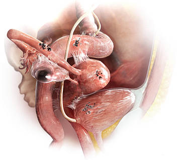
“The most common accepted theory for pathogenesis of endometriosis suggests that implants develop when debris from retrograde menstruation attaches to the pelvic peritoneum,” says Dr. Stratton.7 “Thus, the vast majority of lesions occur in the dependent portions of the pelvis, which include the ovarian fossae (posterior broad ligament under the ovaries), cul de sac, and the uterosacral ligaments.8 The bladder peritoneum, ovarian surface, uterine peritoneal surface, fallopian tube, and pelvic sidewall are also frequent sites. The colon and appendix are less common sites, and small bowel lesions are rare.”
“However, pain location does not correlate with lesion location,” Dr. Stratton notes. “For this reason, the goal at surgery is to treat all lesions, even ones that are not in sites of pain.”
Continue to find out how disease should be staged >>
3. HOW SHOULD DISEASE BE STAGED?
Most surgeons with expertise in treating endometriosis attempt to stage the disease at the time of initial laparoscopy, even though a patient’s pain does not always correlate with the stage of disease.
“The staging system for endometriosis is a means to systematically catalogue where lesions are located,” says Dr. Stratton. The most commonly used classification system was developed by the American Society for Reproductive Medicine (ASRM). It takes into account such characteristics as how deep an implant lies, the extent to which it obliterates the posterior cul de sac, and the presence and extent of adhesions. Although the classification system is broken down into four stages ranging from minimal to severe disease, it is fairly complex. For example, it assigns a score for each lesion as well as the size and location of that lesion, notes Dr. Stratton. The presence of an endometrioma automatically renders the disease as stage III or IV, and an obliterated cul de sac means the endometriosis is graded as stage IV.
“This system enables us to communicate with each other about patients and may guide future surgeries for assessment of lesion recurrence or the planning of treatment for lesions the surgeon was unable to treat at an initial surgery,” says Dr. Stratton.
“Women with uterosacral nodularity, fixed pelvic organs, or severe pain with endometriomas may have deep infiltrating lesions. These lesions, in particular, are not captured well with the current staging system,” says Dr. Stratton. Because they appear to be innervated, “the greatest benefit to the patient is achieved by completely excising these lesions.” Preoperative imaging may help confirm the existence, location, and extent of these deep lesions and help the surgeon plan her approach “based on clinical and imaging findings.”
“Severity of pain or duration of surgical effect does not correlate with stage or extent of disease,” Dr. Stratton says.9 “In fact, patients with the least amount of disease noted at surgery experience pain sooner, suggesting that the central nervous system may have been remodeled prior to surgery or that the pain is in part due to some other cause.10 This observation underscores the principle that, while endometriosis may initiate pain, the pain experience is determined by engagement of the central nervous system.”
For more information on the ASRM revised classification of endometriosis, visit www.fertstert.org/article/S0015-0282(97)81391-X/pdf.
Continue to learn whether excision or ablation is preferable >>
4. WHICH IS PREFERABLE: EXCISION OR ABLATION?
In a prospective, randomized, double-blind study, Healey and colleagues compared pain levels following laparoscopic treatment of endometriosis with either excision or ablation. Preoperatively, women in the study completed a questionnaire rating various types of pain using visual analogue scales. They then were randomly assigned to treatment of endometriosis via excision or ablation. Postoperatively, they again completed a questionnaire about pain levels at three, six, nine, and 12 months. Investigators found no significant difference in pain scores at 12 months.11
Five-year follow-up of the same population yielded slightly different findings, however. Although there was a reduction in all pain scores at five years in both the excision and ablation groups, a significantly greater reduction in dyspareunia was observed in the excision group at five years.12
In an accompanying editorial, Dr. Falcone and a coauthor called excision versus ablation of ovarian, bowel, and peritoneal endometriosis one of the “great debates” in the surgical management of endometriosis.13 “When there is deep involvement of adjacent organs, there is general consensus that excision is best for optimal surgical outcome,” they write. “However, for disease involving the peritoneum alone, there are proponents for either option.”13
“This is a very controversial issue,” says Dr. Falcone, “and the debate can sometimes be somewhat inflammatory…. It is hard to understand how a comparative trial could even be accomplished between excision and ablation. In my experience, deep disease typically occurs on the pelvic sidewall over the ureter or in the cul de sac on the bowel or infiltrating the bladder peritoneum. Therefore, ablation would increase the risk of damaging any of these structures. With superficial disease away from critical structures, it should be fine to ablate. Everywhere else and with deep disease, you need to excise or leave disease behind.”
“Endometriomas are a special situation,” Dr. Falcone adds. “Excision of the cyst has been shown in randomized controlled trials (RCTs) to be associated with less risk for recurrence.14 Therefore, it should be the treatment of choice. However, in patients interested in future fertility, we must take into consideration the potential damage to ovarian reserve associated with excision.”
Endometriosis of the ovaries has unique manifestations. “My approach to ovarian cysts depends on their classification,” says Dr. Nezhat.15 In general, primary endometriomas (type 1) are small, superficial cysts that contain dark “chocolate” fluid. They tend to be firmly adherent to the ovarian tissue and difficult to remove surgically.
Secondary endometriomas (type 2) are follicular or luteal cysts that have been involved or invaded by cortical endometriotic implants or by primary endometrioma. Secondary endometriomas are further classified by the relationship between cortical endometriosis and the cyst wall. Type 2A endometriomas are usually large, with a capsule that is easily separated from ovarian tissue. Type 2B endometriomas have some features of functional cysts but show deep involvement with surface endometriosis. Type 2C endometriomas are similar, showing extensive surface endometrial implants but with deep penetration of the endometriosis into the cyst wall.15
“For type 1 endometriomas, I biopsy the cyst to ensure the lesion is benign, then vaporize the endometrioma,” Dr. Nezhat says. “In cases of type 2A and 2B endometriomas, the cyst capsule is easily enucleated and removed. Type 2C endometriomas are biopsied as well, and then I proceed with vaporizing the fibrotic area with a low-power energy source, such as neutral argon plasma, avoiding excessive coagulation and thermal injury.” Recent literature supports the idea of evaluation and biopsy of fibrotic endometriomas to confirm benign conditions, followed by ablation without compromising ovarian function.16
“Excision and ablation both have indications,” Dr. Nezhat asserts. “It depends on the location and depth of penetration of implants, as well as the patient’s ultimate goal. For example, if the patient desires future fertility and has endometriosis on the ovary, removal by excision could damage ovarian function. The same holds true for endometriosis on the fallopian tubes. It’s better in such cases to ablate.”
“Ablation is different from coagulation, which is not recommended,” Dr. Nezhat explains. “Ablation vaporizes the diseased area layer by layer, like peeling an onion, until the disease is eradicated. It is similar to dermatologic skin resurfacing. Vaporization is preferable for endometriosis on the tubes and ovaries in patients who desire pregnancy. The choice between excision and ablation depends on the location, depth of penetration, and the patient’s desire for fertility.”
Either way—and regardless of the primary indication for surgery (pain versus infertility)—a minimally invasive gynecologic surgeon is expected to have the ability to perform both techniques, Dr. Nezhat says.
Continue to find out if hysterectomy is definitive treatment >>
5. IS HYSTERECTOMY DEFINITIVE TREATMENT?
“Not necessarily,” says Dr. Nezhat. “Hysterectomy by itself doesn’t take care of endometriosis unless the patient has adenomyosis. If a patient has endometriosis, the first step is complete treatment of the disease to restore the anatomy. Then the next step might be hysterectomy to give a better long-term result, especially in cases of adenomyosis. Removal of the ovaries at the time of hysterectomy has to be individualized.”
“The implication that hysterectomy ‘cures’ endometriosis is false yet is stated in some textbooks,” says Dr. Nezhat. “Even at the time of hysterectomy, the first step should be complete treatment of endometriosis and restoration of anatomy, followed by the hysterectomy. Leaving endometriosis behind, believing it will go away by itself or not cause future issues, is a gross misperception.”
Removal of the ovaries at hysterectomy?
“There are few comparative studies on the long-term follow-up of patients who have undergone hysterectomy with or without removal of both ovaries,” says Dr. Falcone. “The conventional dogma has been that, in women undergoing definitive surgery for endometriosis, both ovaries should be removed, even if they are normal. I personally believe that this was because hysterectomy was often performed without excision of the endometriosis. So the uterus was removed and disease was left behind. In these cases, recurrent symptoms were due to persistent disease.”
“We reported our experience at the Cleveland Clinic with a seven-year follow-up,” Dr. Falcone continues. “Hysterectomy was performed with excision of all visible disease. Ovaries were conserved if normal and removed if not. We looked at the reoperation-free frequency over time. In women undergoing hysterectomy with excision of visible disease but ovarian preservation, the reoperation-free percentages at two, five, and seven years were 95%, 86%, and 77%, respectively, versus 96%, 91%, and 91% in those without ovarian preservation. So, overall, there was an advantage over time for removal of the ovaries. However, in the subset of women between ages 30 and 39, there was no difference in the long-term recurrence rate if the ovaries were left in. For this reason, in women younger than 40, we recommend keeping normal ovaries if all disease is removed.”17
Continue on to find out if the risk for postoperative recurrence can be reduced >>
6. CAN THE RISK FOR POSTOPERATIVE RECURRENCE BE REDUCED?
“The main problem with surgery is the recurrence rate,” Dr. Falcone says. “Studies have shown that the recurrence rate of pain at seven years may be as high as 50%.”17 Furthermore, “the recurrence of pain may not be associated with visualized endometriosis at laparoscopy.”
“Incomplete removal of lesions may be associated with an increase in pain after surgery,” says Dr. Stratton.18 “Incomplete removal of lesions may occur because of varying technical skill or specific lesion characteristics. The lesions may be difficult to remove because of their location. Lesions may not be recognized because their appearance can vary from subtle (red or clear or white) to classic (blue-black). The depth of the lesion may not be appreciated until surgery is under way, and a surgeon may not be adequately prepared to treat deep lesions when they are identified.”
Adenomyosis is another reason pain may persist or recur after surgery.19 “Adenomyosis appears as either diffuse or focal thickening of the junctional zone between the endometrium and myometrium of the uterus on T2-weighted MRI,” says Dr. Stratton. “After excision of endometriosis, chronic pelvic pain is significantly more likely to persist in women who have a junctional zone thickness of more than 11 mm on MRI.”
The frequent recurrence of pain after surgery makes the disease a long-term challenge.
“Pelvic pain caused by endometriosis is a chronic problem that requires a multiyear management plan, involving both surgery and hormonal therapy,” says Robert L. Barbieri, MD. “To reduce the number of surgical procedures in the lifetime of a woman with endometriosis and pain, I suggest hormonal medical therapy following conservative surgery for endometriosis.”
“Definitive surgery, such as hysterectomy or hysterectomy plus bilateral salpingo-oophorectomy (BSO), typically results in prolonged symptom relief,” Dr. Barbieri says. “Following hysterectomy, hormonal therapy may not be needed. Following BSO, low-dose hormonal therapy is often needed to reduce the severity of menopausal symptoms.”
After surgical treatment of endometriosis associated with pain, Dr. Barbieri presents the patient with the following menu of hormonal options:
• No hormonal therapy
• Estrogen-progestin contraceptives, either cyclic or continuous
• The LNG-IUS
• Norethindrone acetate (5 mg/d)
• DMPA (150 mg every three months)
• Leuprolide acetate depot (3.75 mg IM monthly)
• Nafarelin nasal spray (200 µg bid)
• Danazol (200 mg bid).
“I explain the common adverse effects with each approach and have the patient select what she determines to be her best option,” says Dr. Barbieri. “In my experience, conservative surgery followed by hormonal therapy is effective in more than 75% of women.”
“The evidence to support postoperative hormonal therapy is modest,” Dr. Barbieri notes. “The best evidence is available for use of the LNG-IUS, estrogen-progestin contraceptives, and GnRH agonists.”20-22
In addition, he notes, “major professional societies have highlighted the option of postoperative hormonal therapy to reduce the risk for recurrent pain and repetitive surgical procedures in the future.”23,24
When pain recurs after surgery for endometriosis, it pays to consider what type of pain it is, says Dr. Barbieri.
“There are two major types of pain—nociceptive and neuropathic,” he says. “Nociceptive pain is caused by an injury, acute or chronic. Neuropathic pain is caused by ‘activation’ of neural circuits, sometimes in the absence of an ongoing injury. Many women with endometriosis and chronic pain have both nociceptive and neuropathic pain. Consequently, it is important to consider the use of a multidisciplinary pain practice in the management of chronic pain syndromes. Multidisciplinary pain practices have special expertise in the management of neuropathic pain. Standard conservative surgical intervention is unlikely to improve pain caused by neuropathic mechanisms. Likewise, opioid analgesics are not recommended for the treatment of neuropathic pain.”
REFERENCES
1. Nezhat C, Nezhat F, Nezhat C. Endometriosis: ancient disease, ancient treatments. Fertil Steril. 2012;98(6S):S1-S62.
2. Giudice LC. Clinical practice. Endometriosis. N Engl J Med. 2010;362(25):2389-2398.
3. Pavone ME, Bulun SE. Aromatase inhibitors for the treatment of endometriosis: a review. Fertil Steril. 2012;98(6):1370-1379.
4. Nothnick WB. The emerging use of aromatase inhibitors for endometriosis treatment. Reprod Biol Endocrinol. 2011;9:87.
5. Chwalisz K, Garg R, Brenner RM, et al. Selective progesterone receptor modulators (SPRMs): a novel therapeutic concept in endometriosis. Ann N Y Acad Sci. 2002;955:373-393, 396-406.
6. Duffy JM, Arambage K, Correa FJ, et al. Laparoscopic surgery for endometriosis. Cochrane Database Syst Rev. 2014;(4):CD011031.
7. Bulun SE. Endometriosis. N Engl J Med. 2009;360(3):268-279.
8. Stegmann BJ, Sinaii N, Liu S, et al. Using location, color, size, and depth to characterize and identify endometriosis lesions in a cohort of 133 women. Fertil Steril. 2008;89(6):1632-1636.
9. Hsu AL, Sinaii N, Segars J, et al. Relating pelvic pain location to surgical findings of endometriosis. Obstet Gynecol. 2011;118(2 pt 1):223-230.
10. Stratton P, Berkley KJ. Chronic pelvic pain and endometriosis: translational evidence of the relationship and implications. Hum Reprod Update. 2011;17(3):327-346.
11. Healey M, Ang WC, Cheng C. Surgical treatment of endometriosis: a prospective randomized double-blinded trial comparing excision and ablation. Fertil Steril. 2010;94(7):2536-2540.
12. Healey M, Chang C, Kaur H. To excise or ablate endometriosis? A prospective randomized double blinded trial after 5-year follow-up. JMIG. 2014;21(6):999-1004.
13. Falcone T, Wilson JR. Surgical management of endometriosis: excision or ablation. JMIG. 2014;21(6):969.
14. Hart RJ, Hickey M, Maouris P, Buckett W. Excisional surgery versus ablative surgery for ovarian endometriomata. Cochrane Database Syst Rev. 2008;(2):CD004992.
15. Nezhat C, Nezhat F, Nezhat CH, Seidman D. Classification of endometriosis: improving the classification of endometriotic ovarian cysts. Hum Reprod. 1994;9(12):2212-2216.
16. Roman H, Auber M, Mokdad C, et al. Ovarian endometrioma ablation using plasma energy versus cystectomy: a step toward better preservation of the ovarian parenchyma in women wishing to conceive. Fertil Steril. 2011;96(6):1396-1400.
17. Shakiba K, Bena JF, McGill KM, et al. Surgical treatment of endometriosis: a 7-year follow-up on the requirement for further surgery. Obstet Gynecol. 2008;111(6):1285-1292.
18. McAllister SL, McGinty KA, Resuehr D, Berkley KJ. Endometriosis-induced vaginal hyperalgesia in the rat: role of the ectopic growths and their innervation. Pain. 2009;147(1-3):255-264.
19. Parker JD, Leondires M, Sinaii N, et al. Persistence of dysmenorrhea and nonmenstrual pain after optimal endometriosis surgery may indicate adenomyosis. Fertil Steril. 2006;86(3):711-715.
20. Abou-Setta AM, Al-Inany HG, Farquar CM. Levonorgestrel-releasing intrauterine device for symptomatic endometriosis following surgery. Cochrane Database Syst Rev. 2006;(1):CD005072.
21. Seracchioli R, Mabrouk M, Manuzzi L, et al. Postoperative use of oral contraceptive pills for prevention of anatomic relapse or symptom recurrence following surgery. Hum Reprod. 2009;24(11):2729-2735.
22. Hornstein MD, Hemmings R, Yuzpe AA, Heinrichs WL. Use of nafarelin versus placebo after reductive laparoscopic surgery for endometriosis. Fertil Steril. 1997;68(5):860-864.
23. Practice Committee of the American Society for Reproductive Medicine. Treatment of pain associated with endometriosis: a committee opinion. Fertil Steril. 2014;101(4):927-935.
24. Dunselman GA, Vermeulen N, Becker C, et al. ESHRE guideline: management of women with endometriosis. Hum Reprod. 2014;29(3):400-412.
IN THIS ARTICLE
• When is laparoscopy indicated?
• Excision versus ablation
• How to reduce the risk for postoperative recurrence
Endometriosis has always posed a treatment challenge. In the early 19th century, before the widespread advent of surgery, the disease was managed by applying leeches to the cervix. In fact, as Nezhat and colleagues note in their comprehensive survey of the 4,000-year history of endometriosis, “leeches were considered a mainstay in treating any condition associated with menstruation.”1
In the 21st century, the picture is clearer, though still not crystal clear. The optimal approach to endometriosis depends on many factors, foremost the patient’s chief complaint: pain or infertility (or both).
This article focuses on medical and surgical management of pain. Six experts address such questions as: When is laparoscopy indicated? Is excision or ablation of lesions preferred? What is the role of hysterectomy in eliminating pain? And what can be done about the problem of recurrence?
1. WHAT ARE THE OPTIONS FOR EMPIRIC THERAPY?
One reason for the diagnostic delay with endometriosis, which still averages about six years, is that definitive diagnosis is achieved only through laparoscopic investigation and histologic confirmation. For many women who experience pain thought to be associated with endometriosis, however, clinicians begin empiric treatment with medical agents as a way to avert the need for surgery, if at all possible.
“There is no cure for endometriosis,” says John R. Lue, MD, MPH, “but there are many ways that endometriosis can be treated” and the impact of the disease reduced in a patient’s life. (Editor’s Note: See below for biographical information on each clinician interviewed in this article.)
Medical and hormonal options include
• NSAIDs, often used with combined oral contraceptives (OCs). NSAIDs are not a long-term treatment option because of their effect on cyclo-oxygenase (COX) 1 and 2 enzymes, says Dr. Lue. COX-1 protects the gastrointestinal (GI) system, and prolonged use of NSAIDs can cause adverse GI effects.
• Cyclic combined OCs “are recommended as firstline therapy in the absence of contraindications,” says Dr. Lue, and are often used in combination with NSAIDs. However, the failure rate may be as high as 20% to 25%.2 “If pain persists after a trial of three to six months of cyclic OCs, consider switching to continuous low-dose combined OCs for an additional six months,” Dr. Lue adds. When combined OCs were compared with placebo in the treatment of dysmenorrhea, they reduced baseline pain scores by 45% to 52%, compared with 14% to 17% for placebo (P < .001).2 They also reduced the volume of endometriomas by 48%, compared with 32% for placebo (P = .04). According to Linda C. Giudice, MD, PhD, “In women with severe dysmenorrhea who have been treated with cyclic combined OCs, a switch to continuous combined OCs reduced pain scores by 58% within six months and by 75% at two years” (P < .001).2
• Depot medroxyprogesterone acetate (DMPA) or the levonorgestrel-releasing intrauterine system (LNG-IUS). These agents suppress the hypothalamic-pituitary-ovarian (HPO) axis to different degrees. DMPA suppresses the HPO completely, preventing ovulation. The LNG-IUS does not fully suppress the HPO but acts directly on endometrial tissue, with antiproliferative effects on eutopic and endometriotic implants, says Dr. Lue. The LNG-IUS also is effective at suppressing disease after surgical treatment, says Dr. Giudice.2
• Gonadotropin-releasing hormone (GnRH) agonist therapy, with estrogen and/or progestin add-back therapy to temper the associated loss in bone mineral density, “may be effective—if only temporarily—as it inhibits the HPO axis and blocks ovarian function, thereby greatly reducing systemic estrogen levels and inducing artificial menopause,” says Dr. Lue.
• Norethindrone acetate, a synthetic progestational agent, is occasionally used as empiric therapy for endometriosis because of its ability to inhibit ovulation. It has antiandrogenic and antiestrogenic effects.
• Aromatase inhibitors. Dr. Lue points to considerable evidence that endometriotic implants are an autocrine source of estrogen.3 “This locally produced estrogen results from overexpression of the enzyme P450 aromatase by endometriotic tissue,” he says. Consequently, in postmenopausal women, “aromatase inhibitors may be used orally in a daily pill form to curtail endometriotic implant production of estrogen and subsequent implant growth.”4 In women of reproductive age, aromatase inhibitors are combined with an HPO-suppressive therapy, such as norethindrone acetate. These strategies represent off-label use of aromatase inhibitors.
• Danazol, a synthetic androgen, has been used in the past to treat dysmenorrhea and dyspareunia. Because of its severe androgenic effects, however, it is not widely used today.
“For those using medical approaches, endometriosis-related pain may be reduced by using hormonal treatments to modify reproductive tract events, thereby decreasing local peritoneal inflammation and cytokine production,” says Pamela Stratton, MD. Because endometriosis is a “central sensitivity syndrome,” multidisciplinary approaches, such as physical therapy, may be beneficial to treat myofascial dysfunction and sensitization. “Chronic pain conditions that overlap with endometriosis-associated pain—such as migraines, irritable bowel syndrome, or painful bladder syndrome—should be identified and treated. Mood changes of depression and anxiety common to women with endometriosis-associated pain also warrant treatment,” she says.
Continue on to find out when laparoscopy is indicated >>
2. WHEN IS LAPAROSCOPY INDICATED?
When medical and hormonal treatments fail to control a patient’s pain, laparoscopy is indicated to confirm the diagnosis of endometriosis. During that procedure, it is also advisable to treat any endometriosis that is present, provided the surgeon is highly experienced in such treatment.
Proper treatment is preferable—even if it requires expert consultation. “No treatment and referral to a more experienced surgeon are better than incomplete treatment by an inexperienced surgeon,” says Ceana Nezhat, MD. “Not all GYN surgeons have the expertise to treat advanced endometriosis.”
Dr. Stratton agrees about the importance of thorough treatment of endometriosis at the time of diagnostic laparoscopy: “At the laparoscopy, the patient benefits if all potential sources of pain are investigated and addressed.” At surgery, the surgeon should look for and treat any lesions suspicious for endometriosis, as well as any other finding that might contribute to pain, she says. “For example, routinely inspecting the appendix for endometriosis or other lesions, and removing affected appendices is reasonable; also, lysis and, where possible, excision of adhesions is an important strategy.”
If a medical approach fails for a patient, “then surgery is indicated to confirm the diagnosis and treat the disease,” agrees Tommaso Falcone, MD.
“Surgery is very effective in treating the pain associated with endometriosis,” Dr. Falcone adds. “Randomized clinical trials have shown that up to 90% of patients who obtain pain relief from surgery will have an effect lasting one year.6 If patients do not get relief, then the association of the pain with endometriosis should be questioned and other causes sought.”
The most common anatomic sites of implants
“The most common accepted theory for pathogenesis of endometriosis suggests that implants develop when debris from retrograde menstruation attaches to the pelvic peritoneum,” says Dr. Stratton.7 “Thus, the vast majority of lesions occur in the dependent portions of the pelvis, which include the ovarian fossae (posterior broad ligament under the ovaries), cul de sac, and the uterosacral ligaments.8 The bladder peritoneum, ovarian surface, uterine peritoneal surface, fallopian tube, and pelvic sidewall are also frequent sites. The colon and appendix are less common sites, and small bowel lesions are rare.”
“However, pain location does not correlate with lesion location,” Dr. Stratton notes. “For this reason, the goal at surgery is to treat all lesions, even ones that are not in sites of pain.”
Continue to find out how disease should be staged >>
3. HOW SHOULD DISEASE BE STAGED?
Most surgeons with expertise in treating endometriosis attempt to stage the disease at the time of initial laparoscopy, even though a patient’s pain does not always correlate with the stage of disease.
“The staging system for endometriosis is a means to systematically catalogue where lesions are located,” says Dr. Stratton. The most commonly used classification system was developed by the American Society for Reproductive Medicine (ASRM). It takes into account such characteristics as how deep an implant lies, the extent to which it obliterates the posterior cul de sac, and the presence and extent of adhesions. Although the classification system is broken down into four stages ranging from minimal to severe disease, it is fairly complex. For example, it assigns a score for each lesion as well as the size and location of that lesion, notes Dr. Stratton. The presence of an endometrioma automatically renders the disease as stage III or IV, and an obliterated cul de sac means the endometriosis is graded as stage IV.
“This system enables us to communicate with each other about patients and may guide future surgeries for assessment of lesion recurrence or the planning of treatment for lesions the surgeon was unable to treat at an initial surgery,” says Dr. Stratton.
“Women with uterosacral nodularity, fixed pelvic organs, or severe pain with endometriomas may have deep infiltrating lesions. These lesions, in particular, are not captured well with the current staging system,” says Dr. Stratton. Because they appear to be innervated, “the greatest benefit to the patient is achieved by completely excising these lesions.” Preoperative imaging may help confirm the existence, location, and extent of these deep lesions and help the surgeon plan her approach “based on clinical and imaging findings.”
“Severity of pain or duration of surgical effect does not correlate with stage or extent of disease,” Dr. Stratton says.9 “In fact, patients with the least amount of disease noted at surgery experience pain sooner, suggesting that the central nervous system may have been remodeled prior to surgery or that the pain is in part due to some other cause.10 This observation underscores the principle that, while endometriosis may initiate pain, the pain experience is determined by engagement of the central nervous system.”
For more information on the ASRM revised classification of endometriosis, visit www.fertstert.org/article/S0015-0282(97)81391-X/pdf.
Continue to learn whether excision or ablation is preferable >>
4. WHICH IS PREFERABLE: EXCISION OR ABLATION?
In a prospective, randomized, double-blind study, Healey and colleagues compared pain levels following laparoscopic treatment of endometriosis with either excision or ablation. Preoperatively, women in the study completed a questionnaire rating various types of pain using visual analogue scales. They then were randomly assigned to treatment of endometriosis via excision or ablation. Postoperatively, they again completed a questionnaire about pain levels at three, six, nine, and 12 months. Investigators found no significant difference in pain scores at 12 months.11
Five-year follow-up of the same population yielded slightly different findings, however. Although there was a reduction in all pain scores at five years in both the excision and ablation groups, a significantly greater reduction in dyspareunia was observed in the excision group at five years.12
In an accompanying editorial, Dr. Falcone and a coauthor called excision versus ablation of ovarian, bowel, and peritoneal endometriosis one of the “great debates” in the surgical management of endometriosis.13 “When there is deep involvement of adjacent organs, there is general consensus that excision is best for optimal surgical outcome,” they write. “However, for disease involving the peritoneum alone, there are proponents for either option.”13
“This is a very controversial issue,” says Dr. Falcone, “and the debate can sometimes be somewhat inflammatory…. It is hard to understand how a comparative trial could even be accomplished between excision and ablation. In my experience, deep disease typically occurs on the pelvic sidewall over the ureter or in the cul de sac on the bowel or infiltrating the bladder peritoneum. Therefore, ablation would increase the risk of damaging any of these structures. With superficial disease away from critical structures, it should be fine to ablate. Everywhere else and with deep disease, you need to excise or leave disease behind.”
“Endometriomas are a special situation,” Dr. Falcone adds. “Excision of the cyst has been shown in randomized controlled trials (RCTs) to be associated with less risk for recurrence.14 Therefore, it should be the treatment of choice. However, in patients interested in future fertility, we must take into consideration the potential damage to ovarian reserve associated with excision.”
Endometriosis of the ovaries has unique manifestations. “My approach to ovarian cysts depends on their classification,” says Dr. Nezhat.15 In general, primary endometriomas (type 1) are small, superficial cysts that contain dark “chocolate” fluid. They tend to be firmly adherent to the ovarian tissue and difficult to remove surgically.
Secondary endometriomas (type 2) are follicular or luteal cysts that have been involved or invaded by cortical endometriotic implants or by primary endometrioma. Secondary endometriomas are further classified by the relationship between cortical endometriosis and the cyst wall. Type 2A endometriomas are usually large, with a capsule that is easily separated from ovarian tissue. Type 2B endometriomas have some features of functional cysts but show deep involvement with surface endometriosis. Type 2C endometriomas are similar, showing extensive surface endometrial implants but with deep penetration of the endometriosis into the cyst wall.15
“For type 1 endometriomas, I biopsy the cyst to ensure the lesion is benign, then vaporize the endometrioma,” Dr. Nezhat says. “In cases of type 2A and 2B endometriomas, the cyst capsule is easily enucleated and removed. Type 2C endometriomas are biopsied as well, and then I proceed with vaporizing the fibrotic area with a low-power energy source, such as neutral argon plasma, avoiding excessive coagulation and thermal injury.” Recent literature supports the idea of evaluation and biopsy of fibrotic endometriomas to confirm benign conditions, followed by ablation without compromising ovarian function.16
“Excision and ablation both have indications,” Dr. Nezhat asserts. “It depends on the location and depth of penetration of implants, as well as the patient’s ultimate goal. For example, if the patient desires future fertility and has endometriosis on the ovary, removal by excision could damage ovarian function. The same holds true for endometriosis on the fallopian tubes. It’s better in such cases to ablate.”
“Ablation is different from coagulation, which is not recommended,” Dr. Nezhat explains. “Ablation vaporizes the diseased area layer by layer, like peeling an onion, until the disease is eradicated. It is similar to dermatologic skin resurfacing. Vaporization is preferable for endometriosis on the tubes and ovaries in patients who desire pregnancy. The choice between excision and ablation depends on the location, depth of penetration, and the patient’s desire for fertility.”
Either way—and regardless of the primary indication for surgery (pain versus infertility)—a minimally invasive gynecologic surgeon is expected to have the ability to perform both techniques, Dr. Nezhat says.
Continue to find out if hysterectomy is definitive treatment >>
5. IS HYSTERECTOMY DEFINITIVE TREATMENT?
“Not necessarily,” says Dr. Nezhat. “Hysterectomy by itself doesn’t take care of endometriosis unless the patient has adenomyosis. If a patient has endometriosis, the first step is complete treatment of the disease to restore the anatomy. Then the next step might be hysterectomy to give a better long-term result, especially in cases of adenomyosis. Removal of the ovaries at the time of hysterectomy has to be individualized.”
“The implication that hysterectomy ‘cures’ endometriosis is false yet is stated in some textbooks,” says Dr. Nezhat. “Even at the time of hysterectomy, the first step should be complete treatment of endometriosis and restoration of anatomy, followed by the hysterectomy. Leaving endometriosis behind, believing it will go away by itself or not cause future issues, is a gross misperception.”
Removal of the ovaries at hysterectomy?
“There are few comparative studies on the long-term follow-up of patients who have undergone hysterectomy with or without removal of both ovaries,” says Dr. Falcone. “The conventional dogma has been that, in women undergoing definitive surgery for endometriosis, both ovaries should be removed, even if they are normal. I personally believe that this was because hysterectomy was often performed without excision of the endometriosis. So the uterus was removed and disease was left behind. In these cases, recurrent symptoms were due to persistent disease.”
“We reported our experience at the Cleveland Clinic with a seven-year follow-up,” Dr. Falcone continues. “Hysterectomy was performed with excision of all visible disease. Ovaries were conserved if normal and removed if not. We looked at the reoperation-free frequency over time. In women undergoing hysterectomy with excision of visible disease but ovarian preservation, the reoperation-free percentages at two, five, and seven years were 95%, 86%, and 77%, respectively, versus 96%, 91%, and 91% in those without ovarian preservation. So, overall, there was an advantage over time for removal of the ovaries. However, in the subset of women between ages 30 and 39, there was no difference in the long-term recurrence rate if the ovaries were left in. For this reason, in women younger than 40, we recommend keeping normal ovaries if all disease is removed.”17
Continue on to find out if the risk for postoperative recurrence can be reduced >>
6. CAN THE RISK FOR POSTOPERATIVE RECURRENCE BE REDUCED?
“The main problem with surgery is the recurrence rate,” Dr. Falcone says. “Studies have shown that the recurrence rate of pain at seven years may be as high as 50%.”17 Furthermore, “the recurrence of pain may not be associated with visualized endometriosis at laparoscopy.”
“Incomplete removal of lesions may be associated with an increase in pain after surgery,” says Dr. Stratton.18 “Incomplete removal of lesions may occur because of varying technical skill or specific lesion characteristics. The lesions may be difficult to remove because of their location. Lesions may not be recognized because their appearance can vary from subtle (red or clear or white) to classic (blue-black). The depth of the lesion may not be appreciated until surgery is under way, and a surgeon may not be adequately prepared to treat deep lesions when they are identified.”
Adenomyosis is another reason pain may persist or recur after surgery.19 “Adenomyosis appears as either diffuse or focal thickening of the junctional zone between the endometrium and myometrium of the uterus on T2-weighted MRI,” says Dr. Stratton. “After excision of endometriosis, chronic pelvic pain is significantly more likely to persist in women who have a junctional zone thickness of more than 11 mm on MRI.”
The frequent recurrence of pain after surgery makes the disease a long-term challenge.
“Pelvic pain caused by endometriosis is a chronic problem that requires a multiyear management plan, involving both surgery and hormonal therapy,” says Robert L. Barbieri, MD. “To reduce the number of surgical procedures in the lifetime of a woman with endometriosis and pain, I suggest hormonal medical therapy following conservative surgery for endometriosis.”
“Definitive surgery, such as hysterectomy or hysterectomy plus bilateral salpingo-oophorectomy (BSO), typically results in prolonged symptom relief,” Dr. Barbieri says. “Following hysterectomy, hormonal therapy may not be needed. Following BSO, low-dose hormonal therapy is often needed to reduce the severity of menopausal symptoms.”
After surgical treatment of endometriosis associated with pain, Dr. Barbieri presents the patient with the following menu of hormonal options:
• No hormonal therapy
• Estrogen-progestin contraceptives, either cyclic or continuous
• The LNG-IUS
• Norethindrone acetate (5 mg/d)
• DMPA (150 mg every three months)
• Leuprolide acetate depot (3.75 mg IM monthly)
• Nafarelin nasal spray (200 µg bid)
• Danazol (200 mg bid).
“I explain the common adverse effects with each approach and have the patient select what she determines to be her best option,” says Dr. Barbieri. “In my experience, conservative surgery followed by hormonal therapy is effective in more than 75% of women.”
“The evidence to support postoperative hormonal therapy is modest,” Dr. Barbieri notes. “The best evidence is available for use of the LNG-IUS, estrogen-progestin contraceptives, and GnRH agonists.”20-22
In addition, he notes, “major professional societies have highlighted the option of postoperative hormonal therapy to reduce the risk for recurrent pain and repetitive surgical procedures in the future.”23,24
When pain recurs after surgery for endometriosis, it pays to consider what type of pain it is, says Dr. Barbieri.
“There are two major types of pain—nociceptive and neuropathic,” he says. “Nociceptive pain is caused by an injury, acute or chronic. Neuropathic pain is caused by ‘activation’ of neural circuits, sometimes in the absence of an ongoing injury. Many women with endometriosis and chronic pain have both nociceptive and neuropathic pain. Consequently, it is important to consider the use of a multidisciplinary pain practice in the management of chronic pain syndromes. Multidisciplinary pain practices have special expertise in the management of neuropathic pain. Standard conservative surgical intervention is unlikely to improve pain caused by neuropathic mechanisms. Likewise, opioid analgesics are not recommended for the treatment of neuropathic pain.”
REFERENCES
1. Nezhat C, Nezhat F, Nezhat C. Endometriosis: ancient disease, ancient treatments. Fertil Steril. 2012;98(6S):S1-S62.
2. Giudice LC. Clinical practice. Endometriosis. N Engl J Med. 2010;362(25):2389-2398.
3. Pavone ME, Bulun SE. Aromatase inhibitors for the treatment of endometriosis: a review. Fertil Steril. 2012;98(6):1370-1379.
4. Nothnick WB. The emerging use of aromatase inhibitors for endometriosis treatment. Reprod Biol Endocrinol. 2011;9:87.
5. Chwalisz K, Garg R, Brenner RM, et al. Selective progesterone receptor modulators (SPRMs): a novel therapeutic concept in endometriosis. Ann N Y Acad Sci. 2002;955:373-393, 396-406.
6. Duffy JM, Arambage K, Correa FJ, et al. Laparoscopic surgery for endometriosis. Cochrane Database Syst Rev. 2014;(4):CD011031.
7. Bulun SE. Endometriosis. N Engl J Med. 2009;360(3):268-279.
8. Stegmann BJ, Sinaii N, Liu S, et al. Using location, color, size, and depth to characterize and identify endometriosis lesions in a cohort of 133 women. Fertil Steril. 2008;89(6):1632-1636.
9. Hsu AL, Sinaii N, Segars J, et al. Relating pelvic pain location to surgical findings of endometriosis. Obstet Gynecol. 2011;118(2 pt 1):223-230.
10. Stratton P, Berkley KJ. Chronic pelvic pain and endometriosis: translational evidence of the relationship and implications. Hum Reprod Update. 2011;17(3):327-346.
11. Healey M, Ang WC, Cheng C. Surgical treatment of endometriosis: a prospective randomized double-blinded trial comparing excision and ablation. Fertil Steril. 2010;94(7):2536-2540.
12. Healey M, Chang C, Kaur H. To excise or ablate endometriosis? A prospective randomized double blinded trial after 5-year follow-up. JMIG. 2014;21(6):999-1004.
13. Falcone T, Wilson JR. Surgical management of endometriosis: excision or ablation. JMIG. 2014;21(6):969.
14. Hart RJ, Hickey M, Maouris P, Buckett W. Excisional surgery versus ablative surgery for ovarian endometriomata. Cochrane Database Syst Rev. 2008;(2):CD004992.
15. Nezhat C, Nezhat F, Nezhat CH, Seidman D. Classification of endometriosis: improving the classification of endometriotic ovarian cysts. Hum Reprod. 1994;9(12):2212-2216.
16. Roman H, Auber M, Mokdad C, et al. Ovarian endometrioma ablation using plasma energy versus cystectomy: a step toward better preservation of the ovarian parenchyma in women wishing to conceive. Fertil Steril. 2011;96(6):1396-1400.
17. Shakiba K, Bena JF, McGill KM, et al. Surgical treatment of endometriosis: a 7-year follow-up on the requirement for further surgery. Obstet Gynecol. 2008;111(6):1285-1292.
18. McAllister SL, McGinty KA, Resuehr D, Berkley KJ. Endometriosis-induced vaginal hyperalgesia in the rat: role of the ectopic growths and their innervation. Pain. 2009;147(1-3):255-264.
19. Parker JD, Leondires M, Sinaii N, et al. Persistence of dysmenorrhea and nonmenstrual pain after optimal endometriosis surgery may indicate adenomyosis. Fertil Steril. 2006;86(3):711-715.
20. Abou-Setta AM, Al-Inany HG, Farquar CM. Levonorgestrel-releasing intrauterine device for symptomatic endometriosis following surgery. Cochrane Database Syst Rev. 2006;(1):CD005072.
21. Seracchioli R, Mabrouk M, Manuzzi L, et al. Postoperative use of oral contraceptive pills for prevention of anatomic relapse or symptom recurrence following surgery. Hum Reprod. 2009;24(11):2729-2735.
22. Hornstein MD, Hemmings R, Yuzpe AA, Heinrichs WL. Use of nafarelin versus placebo after reductive laparoscopic surgery for endometriosis. Fertil Steril. 1997;68(5):860-864.
23. Practice Committee of the American Society for Reproductive Medicine. Treatment of pain associated with endometriosis: a committee opinion. Fertil Steril. 2014;101(4):927-935.
24. Dunselman GA, Vermeulen N, Becker C, et al. ESHRE guideline: management of women with endometriosis. Hum Reprod. 2014;29(3):400-412.
A surge in congenital syphilis reveals gaps in obstetric practice
The rate of congenital syphilis increased to 11.6 cases per 100,000 live births in 2014 in the United States—the highest rate documented since 2001, according to a new report from the Centers for Disease Control and Prevention (CDC).1 The increase in the rate of congenital syphilis reflects a rise in the rate of primary and secondary syphilis among US women, from 0.9 to 1.1 cases per 100,000 women, during the same period.1
The rate of congenital syphilis had declined between 2008 and 2012, from 10.5 cases to 8.4 cases per 100,000 live births.
Congenital syphilis occurs when an infected mother transmits the disease to her fetus during pregnancy. Among the adverse effects of congenital syphilis are deformities, stillbirth, and early infant death. “However, among mothers identified with syphilis who deliver past 20 weeks’ gestation, treatment with penicillin at least 30 days before delivery is 98% effective at preventing [congenital syphilis],” the CDC report notes.1
For the purposes of the CDC report, congenital syphilis includes “both infants and stillbirths with clinical evidence” of the disease, “as well as those infants and stillbirths born to mothers with untreated or inadequately treated syphilis, regardless of the infant’s manifestation of clinical disease.”1
CDC recommendations
The CDC notes that most of the increases in the rates of maternal and congenital syphilis likely stem from inadequate prenatal care.
“A large percentage of [congenital syphilis] cases continue to be attributable to a lack of prenatal care; even among those receiving some prenatal care, the detection and treatment of maternal syphilis is often not early enough….At particular risk are those who are uninsured or underinsured and those with substance use issues.”1
Among the recommendations of the CDC:
- Screen all pregnant women for syphilis at their first prenatal visit.
- Screen women at elevated risk for syphilis, as well as those who live in “high-morbidity geographic areas” at the beginning of the third trimester and again at delivery.
- In cases where prenatal care has been lacking, screen the woman for syphilis using a rapid plasma reagin (RPR) card and treat the patient who tests positive at the time the pregnancy is confirmed.
- Do not discharge an infant from the hospital unless the syphilis serologic status of the mother has been tested at least once during pregnancy and, preferably, again at delivery (in high-risk cases).
- Test any woman who delivers a stillborn infant for syphilis.
Reference
1. Bowen V, Su J, Torrone E, Kidd S, Weinstock H. Increase in incidence of congenital syphilis—United States, 2008–2014. MMWR. 2015;64(44):1241–1245.
The rate of congenital syphilis increased to 11.6 cases per 100,000 live births in 2014 in the United States—the highest rate documented since 2001, according to a new report from the Centers for Disease Control and Prevention (CDC).1 The increase in the rate of congenital syphilis reflects a rise in the rate of primary and secondary syphilis among US women, from 0.9 to 1.1 cases per 100,000 women, during the same period.1
The rate of congenital syphilis had declined between 2008 and 2012, from 10.5 cases to 8.4 cases per 100,000 live births.
Congenital syphilis occurs when an infected mother transmits the disease to her fetus during pregnancy. Among the adverse effects of congenital syphilis are deformities, stillbirth, and early infant death. “However, among mothers identified with syphilis who deliver past 20 weeks’ gestation, treatment with penicillin at least 30 days before delivery is 98% effective at preventing [congenital syphilis],” the CDC report notes.1
For the purposes of the CDC report, congenital syphilis includes “both infants and stillbirths with clinical evidence” of the disease, “as well as those infants and stillbirths born to mothers with untreated or inadequately treated syphilis, regardless of the infant’s manifestation of clinical disease.”1
CDC recommendations
The CDC notes that most of the increases in the rates of maternal and congenital syphilis likely stem from inadequate prenatal care.
“A large percentage of [congenital syphilis] cases continue to be attributable to a lack of prenatal care; even among those receiving some prenatal care, the detection and treatment of maternal syphilis is often not early enough….At particular risk are those who are uninsured or underinsured and those with substance use issues.”1
Among the recommendations of the CDC:
- Screen all pregnant women for syphilis at their first prenatal visit.
- Screen women at elevated risk for syphilis, as well as those who live in “high-morbidity geographic areas” at the beginning of the third trimester and again at delivery.
- In cases where prenatal care has been lacking, screen the woman for syphilis using a rapid plasma reagin (RPR) card and treat the patient who tests positive at the time the pregnancy is confirmed.
- Do not discharge an infant from the hospital unless the syphilis serologic status of the mother has been tested at least once during pregnancy and, preferably, again at delivery (in high-risk cases).
- Test any woman who delivers a stillborn infant for syphilis.
Reference
1. Bowen V, Su J, Torrone E, Kidd S, Weinstock H. Increase in incidence of congenital syphilis—United States, 2008–2014. MMWR. 2015;64(44):1241–1245.
Reference
1. Bowen V, Su J, Torrone E, Kidd S, Weinstock H. Increase in incidence of congenital syphilis—United States, 2008–2014. MMWR. 2015;64(44):1241–1245.
VIDEO: How to handle pregnancy and breast feeding in rheumatoid arthritis
San Francisco – Should you give rheumatoid arthritis patients tumor necrosis factor blockers during pregnancy? What can you substitute for methotrexate?
And what in the world do you do about breast feeding?
“The management of rheumatoid arthritis in pregnancy seems to be evolving,” explained Dr. Megan Clowse, a specialist in rheumatology and pregnancy at Duke University, Durham, N.C. “The old strategy of just stopping all the medications and letting the woman flare and using some prednisone really doesn’t seem to actually be achieving the outcomes that we need to get.”
Dr. Clowse answered a host of pregnancy-related questions and more in a pearl-filled interview at the annual meeting of the American College of Rheumatology.
The video associated with this article is no longer available on this site. Please view all of our videos on the MDedge YouTube channel
AT THE ACR ANNUAL MEETING

U.S congenital syphilis rates on the rise
Since 2012, congenital syphilis rates in newborns have increased significantly, and in 2014 the disease incidence reached its highest rate since 2001, according to the Centers for Disease Control and Prevention.
From 1991 to 2005, congenital syphilis (CS) rates in the United States decreased, but increased slightly from 2005 to 2008 to 10.5/100,000 live births, said Dr. Virginia Bowen of the CDC’s Epidemic Intelligence Service (MMWR. 2015 Nov 13;64[44]:1241-5) and her coauthors. After 2008, CS rates decreased again, reaching a low of 8.4/100,000 live births in 2012. In 2013, the rate increased to 9.1/100,000 births, and grew to 11.6/100,000 births in 2014. This growth corresponds to a concurrent growth in primary and secondary syphilis in women, the rate of which increased from 0.9/100,000 in 2012 to 1.1/100,000 in 2014, a 22% increase.

Every region in the United States saw an increase in CS rates from 2012 to 2014, but was highest in the West, where the rate more than doubled, increasing from 5.5/100,000 births to 12.8/100,000. All racial/ethnic groups saw an increase as well, with the greatest increase in whites; however, CS rates in non-Hispanic blacks remained more than 10 times higher than in whites, and 3 times higher than in Hispanics.
“Health departments, in partnership with prenatal care providers and other local organizations, should work together to address barriers to obtaining early and adequate prenatal care for the majority of vulnerable pregnant women. Women who are uninsured or underinsured and women with substance use issues have been found to be at increased risk for receiving inadequate or no prenatal care, placing them at increased risk for CS,” the CDC investigators said.
Find the full report in the MMWR.
Since 2012, congenital syphilis rates in newborns have increased significantly, and in 2014 the disease incidence reached its highest rate since 2001, according to the Centers for Disease Control and Prevention.
From 1991 to 2005, congenital syphilis (CS) rates in the United States decreased, but increased slightly from 2005 to 2008 to 10.5/100,000 live births, said Dr. Virginia Bowen of the CDC’s Epidemic Intelligence Service (MMWR. 2015 Nov 13;64[44]:1241-5) and her coauthors. After 2008, CS rates decreased again, reaching a low of 8.4/100,000 live births in 2012. In 2013, the rate increased to 9.1/100,000 births, and grew to 11.6/100,000 births in 2014. This growth corresponds to a concurrent growth in primary and secondary syphilis in women, the rate of which increased from 0.9/100,000 in 2012 to 1.1/100,000 in 2014, a 22% increase.
Every region in the United States saw an increase in CS rates from 2012 to 2014, but was highest in the West, where the rate more than doubled, increasing from 5.5/100,000 births to 12.8/100,000. All racial/ethnic groups saw an increase as well, with the greatest increase in whites; however, CS rates in non-Hispanic blacks remained more than 10 times higher than in whites, and 3 times higher than in Hispanics.
“Health departments, in partnership with prenatal care providers and other local organizations, should work together to address barriers to obtaining early and adequate prenatal care for the majority of vulnerable pregnant women. Women who are uninsured or underinsured and women with substance use issues have been found to be at increased risk for receiving inadequate or no prenatal care, placing them at increased risk for CS,” the CDC investigators said.
Find the full report in the MMWR.
FROM MORBIDITY & MORTALITY WEEKLY REPORT

Assessing longer-term effects of SSRI exposure in pregnancy
Over the last 2 decades, the medical community has accumulated more information regarding fetal exposure to selective serotonin reuptake inhibitors than almost any medicine that women take during pregnancy.
Data on the risks for major congenital malformations and even specific malformations, such as cardiac defects, are so vast that they can be confusing for clinicians and researchers alike to digest. Yet the consensus among experts in reproductive safety is that the risk of teratogenesis following fetal exposure to SSRIs is small in terms of absolute risks (N Engl J Med. 2007;356:2732-3.).

But women and their partners are worried about more than the risk of malformations. They also are concerned about how fetal exposure to SSRIs will impact long-term neurobehavioral sequelae. It turns out that is a much more challenging question, and the data available to inform our answers still are relatively sparse.
Research gaps
There have been several reports in the literature that suggest there are modest effects on motor function in children whose mothers used SSRIs during pregnancy, but these reports have not been particularly systematic and have excluded some relevant variables, such as the presence or absence of a psychiatric disorder during pregnancy.
In what is considered a seminal examination of this topic performed more than a decade ago, Dr. Irena Nulman and her colleagues at the Motherisk program in Toronto demonstrated the importance of accounting for postpartum maternal depression and mood. Among a group of children exposed to fluoxetine (Prozac) or tricyclic antidepressants throughout pregnancy, there were no differences in IQ or language development, compared with unexposed children between 15 and 71 months of age. However, children with lower cognitive and language achievement were more likely to have mothers with higher ratings of postpartum mood disturbance (Am J Psychiatry. 2002 Nov;159[11]:1889-95.).
The charge to the field is to tackle this next clinical research frontier: the longer-term effects of fetal exposure to antidepressants and maternal psychiatric illness. Only by factoring in both of these important variables will we be able to get a true model of risk for this very critical issue of longer-term behavioral function.
Recently, researchers attempted to look at this question using data from the Norwegian Mother and Child Cohort study, a large population-based, prospective pregnancy cohort study. Children were assessed at age 3 years and motor development was assessed by maternal reports of fine and gross motor development using items from the Ages and Stages Questionnaire (ASQ). Maternal ASQ scores were compared with data from another Norwegian study in which clinicians assessed motor development using the gross and fine motor Mullen Scales of Early Learning (MSEL).
The strength of the study is that it included a very large number of children – more than 50,000 – although it is noteworthy that only a small percentage of women actually used SSRIs during pregnancy. A total of 381 women reported the use of SSRIs during pregnancy, with about half of the women reporting prolonged use (defined as exposure at two points of observation in the study).

The findings are noteworthy. The study showed that children with prolonged SSRI exposure were more likely to exhibit delays in fine motor development, compared with children with no fetal exposure to SSRIs (odds ratio, 1.42; 95% confidence interval, 1.07-1.87). The association persisted even after adjusting for symptoms of anxiety and depression before and during pregnancy. The severity of maternal depression provided only a partial explanation for the association, and depression after pregnancy appeared to have no impact on the estimated effect of SSRIs during pregnancy (BJOG. 2015 Sep 15. doi: 10.1111/1471-0528.13582).
Personal medicine
So how does the clinician translate these findings into practice? While the study indicates that prolonged prenatal exposure to SSRIs was associated with delayed motor development at age 3 years, the association was very weak, and only a few children had clinically significant impairment in motor development. The finding should be reassuring to many women who are faced with this decision.
The researchers should be commended for trying to at least begin to tackle this critical frontier with respect to the risk-benefit decision in using these medicines during pregnancy. The problem is that in the absence of robust data quantifying the exposure to maternal psychiatric illness, the quality of the data still is somewhat limited.
So far the data indicate that there is not a major signal for neurobehavioral dysregulation associated with fetal exposure to SSRIs. That being said, to adequately answer the question for the field it will be important to conduct a single, prospective study where we can accurately quantify exposure to medicine and exposure to maternal psychiatric illness during pregnancy. That type of study will allow us to assess the relative exposures and come up with a much more refined risk estimate. We also will be able to fold in a critical piece – the role of genomics.
Genomics will play a significant role in determining risk because vulnerability to potential disruption in central nervous system development may be a factor in determining the development of the brain in utero. We know, for example, that prenatal stress affects neuromodulation in the brain, and that vulnerability to disruption will be variable from patient to patient. Prospective studies where we not only quantify what medicines women are taking and the severity of psychiatric illness, but also vulnerability to central nervous system disruption, if any, at the level of the genome will be a critical missing piece for future investigators.
Even similar exposure to medicine and psychiatric disorders may have variable effects on the developing brain. If we can identify that variability at the level of the genome, then we will be able to create a refined model of risk that will dictate personalized medicine with respect to reproductive psychopharmacology.
Dr. Cohen is the director of the Center for Women’s Mental Health at Massachusetts General Hospital in Boston, which provides information resources and conducts clinical care and research in reproductive mental health. He has been a consultant to manufacturers of psychiatric medications. To comment, email him at [email protected].
Over the last 2 decades, the medical community has accumulated more information regarding fetal exposure to selective serotonin reuptake inhibitors than almost any medicine that women take during pregnancy.
Data on the risks for major congenital malformations and even specific malformations, such as cardiac defects, are so vast that they can be confusing for clinicians and researchers alike to digest. Yet the consensus among experts in reproductive safety is that the risk of teratogenesis following fetal exposure to SSRIs is small in terms of absolute risks (N Engl J Med. 2007;356:2732-3.).
But women and their partners are worried about more than the risk of malformations. They also are concerned about how fetal exposure to SSRIs will impact long-term neurobehavioral sequelae. It turns out that is a much more challenging question, and the data available to inform our answers still are relatively sparse.
Research gaps
There have been several reports in the literature that suggest there are modest effects on motor function in children whose mothers used SSRIs during pregnancy, but these reports have not been particularly systematic and have excluded some relevant variables, such as the presence or absence of a psychiatric disorder during pregnancy.
In what is considered a seminal examination of this topic performed more than a decade ago, Dr. Irena Nulman and her colleagues at the Motherisk program in Toronto demonstrated the importance of accounting for postpartum maternal depression and mood. Among a group of children exposed to fluoxetine (Prozac) or tricyclic antidepressants throughout pregnancy, there were no differences in IQ or language development, compared with unexposed children between 15 and 71 months of age. However, children with lower cognitive and language achievement were more likely to have mothers with higher ratings of postpartum mood disturbance (Am J Psychiatry. 2002 Nov;159[11]:1889-95.).
The charge to the field is to tackle this next clinical research frontier: the longer-term effects of fetal exposure to antidepressants and maternal psychiatric illness. Only by factoring in both of these important variables will we be able to get a true model of risk for this very critical issue of longer-term behavioral function.
Recently, researchers attempted to look at this question using data from the Norwegian Mother and Child Cohort study, a large population-based, prospective pregnancy cohort study. Children were assessed at age 3 years and motor development was assessed by maternal reports of fine and gross motor development using items from the Ages and Stages Questionnaire (ASQ). Maternal ASQ scores were compared with data from another Norwegian study in which clinicians assessed motor development using the gross and fine motor Mullen Scales of Early Learning (MSEL).
The strength of the study is that it included a very large number of children – more than 50,000 – although it is noteworthy that only a small percentage of women actually used SSRIs during pregnancy. A total of 381 women reported the use of SSRIs during pregnancy, with about half of the women reporting prolonged use (defined as exposure at two points of observation in the study).
The findings are noteworthy. The study showed that children with prolonged SSRI exposure were more likely to exhibit delays in fine motor development, compared with children with no fetal exposure to SSRIs (odds ratio, 1.42; 95% confidence interval, 1.07-1.87). The association persisted even after adjusting for symptoms of anxiety and depression before and during pregnancy. The severity of maternal depression provided only a partial explanation for the association, and depression after pregnancy appeared to have no impact on the estimated effect of SSRIs during pregnancy (BJOG. 2015 Sep 15. doi: 10.1111/1471-0528.13582).
Personal medicine
So how does the clinician translate these findings into practice? While the study indicates that prolonged prenatal exposure to SSRIs was associated with delayed motor development at age 3 years, the association was very weak, and only a few children had clinically significant impairment in motor development. The finding should be reassuring to many women who are faced with this decision.
The researchers should be commended for trying to at least begin to tackle this critical frontier with respect to the risk-benefit decision in using these medicines during pregnancy. The problem is that in the absence of robust data quantifying the exposure to maternal psychiatric illness, the quality of the data still is somewhat limited.
So far the data indicate that there is not a major signal for neurobehavioral dysregulation associated with fetal exposure to SSRIs. That being said, to adequately answer the question for the field it will be important to conduct a single, prospective study where we can accurately quantify exposure to medicine and exposure to maternal psychiatric illness during pregnancy. That type of study will allow us to assess the relative exposures and come up with a much more refined risk estimate. We also will be able to fold in a critical piece – the role of genomics.
Genomics will play a significant role in determining risk because vulnerability to potential disruption in central nervous system development may be a factor in determining the development of the brain in utero. We know, for example, that prenatal stress affects neuromodulation in the brain, and that vulnerability to disruption will be variable from patient to patient. Prospective studies where we not only quantify what medicines women are taking and the severity of psychiatric illness, but also vulnerability to central nervous system disruption, if any, at the level of the genome will be a critical missing piece for future investigators.
Even similar exposure to medicine and psychiatric disorders may have variable effects on the developing brain. If we can identify that variability at the level of the genome, then we will be able to create a refined model of risk that will dictate personalized medicine with respect to reproductive psychopharmacology.
Dr. Cohen is the director of the Center for Women’s Mental Health at Massachusetts General Hospital in Boston, which provides information resources and conducts clinical care and research in reproductive mental health. He has been a consultant to manufacturers of psychiatric medications. To comment, email him at [email protected].

LT4 therapy for SCH may improve pregnancy outcomes
LAKE BUENA VISTA, FLA. – Levothyroxine therapy during pregnancy in women with subclinical hypothyroidism was associated with a decrease in low birth weight and low Apgar scores but not with a significant decrease in pregnancy loss in a large retrospective cohort study.
In 79 pregnant women with subclinical hypothyroidism (SCH) who received therapy with levothyroxine (LT4), and 285 with SCH who did not receive LT4 therapy, the frequency of low birth weight (less than 2,500 g) was 1.3% vs. 10%, respectively, and the frequency of Apgar scores of 7 or less at 5 minutes was 0% vs. 6.9%, Dr. S. Maraka reported at the International Thyroid Congress.
The rate of pregnancy loss was clinically, but not significantly, lower in the treated group (5.1% vs. 8.8%), and there was no difference between the groups with respect to 11 other maternal and neonatal outcomes, Dr. Maraka of the Mayo Clinic, Rochester, Minn., said at the meeting held by the American Thyroid Association, Asia-Oceania Thyroid Association, European Thyroid Association, and Latin American Thyroid Society.
Study subjects were women evaluated at the Mayo Clinic, Rochester, between January 2011 and December 2013, who had SCH during pregnancy. SCH was defined as thyroid stimulating hormone levels greater than 2.5 mIU/L during the first trimester, or greater than 3 mIU/L but no more than 10 mIU/L during the second and third trimesters. Those with a twin pregnancy or who used medications that might affect thyroid function were excluded.
The treated and untreated groups were similar with regard to age, history of pregnancy loss, and smoking status, but the treated group had higher body mass index and higher TSH levels, Dr. Maraka noted.
The findings that LT4 may improve pregnancy outcome in women with SCH are important because SCH has been associated with an increased risk of adverse pregnancy outcomes in some studies and it has been unclear whether LT4 therapy improves outcomes.
However, the association seen in the current study, which involves the largest cohort reporting pregnancy outcomes of women with SCH treated vs. untreated with LT4 therapy, requires confirmation in randomized trials before widespread use of LT4 therapy for SCH can be recommended, she concluded.
Dr. Maraka reported having no disclosures.
AT ITC 2015
Key clinical point: LT4 therapy during pregnancy in women with subclinical hypothyroidism was associated with a decrease in low birth weight and low Apgar scores, but not with a decrease in pregnancy loss in a large retrospective cohort study.
Major finding: The frequency of low birth weight was 1.3% vs. 10%, and the frequency of Apgar scores of 7 or less at 5 minutes was 0% vs. 6.9% in the treated vs. untreated patients.
Data source: A retrospective cohort study of 364 women.
Disclosures: Dr. Maraka reported having no disclosures.