User login
EHR treat-to-target prompts spur RA medication management decisions
ATLANTA – Opportunities to escalate or deescalate medications for patients with rheumatoid arthritis via an electronic health record at the point of care led rheumatologists at the Geisinger Medical Center in Danville, Pa., to increase the number of such decisions in their practice.
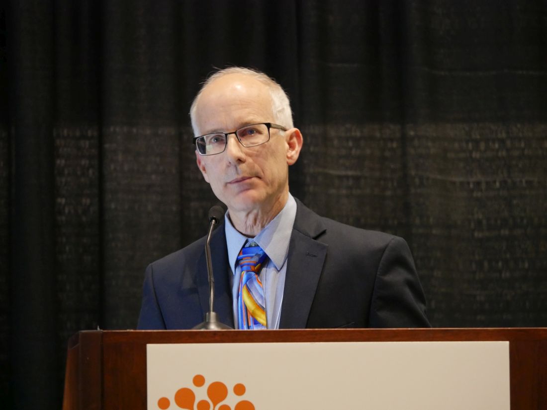
“Opportunities for escalation and de-escalation of therapy are common, even in a well-managed RA population,” Eric D. Newman, MD, director of the department of rheumatology at Geisinger, said in a presentation at the annual meeting of the American College of Rheumatology. “In our hands, over a third of the time, there was an opportunity to change therapy.”
Dr. Newman and colleagues developed a new treat-to-target tab for their (Patient Centric Electronic Redesign) PACER project, an EHR-adjacent system that captures patient and provider data and presents the information in different views, based on desired action items. The target in the study was low disease activity or remission, which was assessed using Clinical Disease Activity Index (CDAI) scores.
The treat-to-target tab offered three options to rheumatologists in real time when meeting with a patient: an escalation opportunity, which was defined as the patient’s two most recent CDAI scores showing moderate to high disease activity; and a deescalation opportunity, defined as a look-back up to 1 year during which at least two CDAI measures were within low to-moderate disease range. There was also a third “leave-alone” option to neither escalate nor deescalate therapy, but the rheumatologist was prompted to explain why if that option is selected, Dr. Newman noted.
In the first phase of releasing the treat-to-target tab, there was low adoption among the 17 rheumatologists at Geisinger: 82% of rheumatologists did not use the tab for escalation therapy, and 64% did not use the tab for deescalation therapy.
That prompted Dr. Newman and colleagues to develop a new version of the treat-to-target tab for phase 2 of the program. “Once they complete their CDAI, if there’s an opportunity, there would be a bright orange button that would glow right in front of them,” Dr. Newman said. “It was really hard to miss, and all they would have to do is click it and it would bring them right to the treat-to-target tab.”
To increase rheumatologists’ use of the new treat-to-target tab, the amount of time spent using the tab and making decisions is presented to them, he said. “It’s actually now part of our quality measure bundle, so every month, they get a leaderboard to see how they compare with their partners,” he noted. “It’s a great way to drive down variability and get everybody approaching the same sort of mean.”
Overall, between July 2018 and May 2019, there were 1,428 treat-to-target opportunities, consisting of 34.2% of RA office visits. Of these, 11.3% were escalation opportunities and 22.9% were deescalation opportunities, Dr. Newman said.
Between phase 1 and phase 2, the rheumatologists’ nonuse of the treat-to-target tab decreased from 82% to 36% for escalation opportunities, and decisions to escalate therapy increased from 10% to 46%. Similarly, nonuse of the treat-to-target tab for deescalation therapy decreased from 64% to 34%, and decisions to deescalate therapy increased from 5% to 17%.
In 49% of escalation opportunities and 80% of deescalation opportunities in phase 2 of the program, rheumatologists made the decision to leave the patient alone. The reasons for not escalating therapy for a patient included an inaccurate CDAI (34%), patient decision (15%), risks outweighing the benefits (15%), and other (37%). “This is interesting, because if you take 49% times 34%, that means that only 17% of the time they felt it didn’t represent what was going on in our department, which is not what we heard from them verbally before this project,” he said. For deescalation opportunities in which the rheumatologist left the patient alone, the most common reasons were hard-to-control disease (46%), patient preference (29%), and poor prognostic factors (25%).
“Keep in mind, some of these patients may have actually already been deescalated prior to this,” Dr. Newman noted. “We’ve actually done some previous work 3 years ago – we provided a visual signal to our physicians that there was a deescalation opportunity, so we may have already accounted for that portion of the population to some extent.”
Dr. Newman said a future goal of the treat-to-target system is to developed more specific treat-to-target strategies to improve the “signal-to-noise” ratio in the system. “Now [that] it’s fully embedded into our routine RA care delivery across our system, our next steps are going to be to use this tool to proactively drive value-concordant decision making and monitor the effect this has on both disease control as well as cost of care,” he said.
Dr. Newman reported no relevant financial disclosures.
SOURCE: Newman ED et al. Arthritis Rheumatol. 2019;71(suppl 10). Abstract 1862.
ATLANTA – Opportunities to escalate or deescalate medications for patients with rheumatoid arthritis via an electronic health record at the point of care led rheumatologists at the Geisinger Medical Center in Danville, Pa., to increase the number of such decisions in their practice.
“Opportunities for escalation and de-escalation of therapy are common, even in a well-managed RA population,” Eric D. Newman, MD, director of the department of rheumatology at Geisinger, said in a presentation at the annual meeting of the American College of Rheumatology. “In our hands, over a third of the time, there was an opportunity to change therapy.”
Dr. Newman and colleagues developed a new treat-to-target tab for their (Patient Centric Electronic Redesign) PACER project, an EHR-adjacent system that captures patient and provider data and presents the information in different views, based on desired action items. The target in the study was low disease activity or remission, which was assessed using Clinical Disease Activity Index (CDAI) scores.
The treat-to-target tab offered three options to rheumatologists in real time when meeting with a patient: an escalation opportunity, which was defined as the patient’s two most recent CDAI scores showing moderate to high disease activity; and a deescalation opportunity, defined as a look-back up to 1 year during which at least two CDAI measures were within low to-moderate disease range. There was also a third “leave-alone” option to neither escalate nor deescalate therapy, but the rheumatologist was prompted to explain why if that option is selected, Dr. Newman noted.
In the first phase of releasing the treat-to-target tab, there was low adoption among the 17 rheumatologists at Geisinger: 82% of rheumatologists did not use the tab for escalation therapy, and 64% did not use the tab for deescalation therapy.
That prompted Dr. Newman and colleagues to develop a new version of the treat-to-target tab for phase 2 of the program. “Once they complete their CDAI, if there’s an opportunity, there would be a bright orange button that would glow right in front of them,” Dr. Newman said. “It was really hard to miss, and all they would have to do is click it and it would bring them right to the treat-to-target tab.”
To increase rheumatologists’ use of the new treat-to-target tab, the amount of time spent using the tab and making decisions is presented to them, he said. “It’s actually now part of our quality measure bundle, so every month, they get a leaderboard to see how they compare with their partners,” he noted. “It’s a great way to drive down variability and get everybody approaching the same sort of mean.”
Overall, between July 2018 and May 2019, there were 1,428 treat-to-target opportunities, consisting of 34.2% of RA office visits. Of these, 11.3% were escalation opportunities and 22.9% were deescalation opportunities, Dr. Newman said.
Between phase 1 and phase 2, the rheumatologists’ nonuse of the treat-to-target tab decreased from 82% to 36% for escalation opportunities, and decisions to escalate therapy increased from 10% to 46%. Similarly, nonuse of the treat-to-target tab for deescalation therapy decreased from 64% to 34%, and decisions to deescalate therapy increased from 5% to 17%.
In 49% of escalation opportunities and 80% of deescalation opportunities in phase 2 of the program, rheumatologists made the decision to leave the patient alone. The reasons for not escalating therapy for a patient included an inaccurate CDAI (34%), patient decision (15%), risks outweighing the benefits (15%), and other (37%). “This is interesting, because if you take 49% times 34%, that means that only 17% of the time they felt it didn’t represent what was going on in our department, which is not what we heard from them verbally before this project,” he said. For deescalation opportunities in which the rheumatologist left the patient alone, the most common reasons were hard-to-control disease (46%), patient preference (29%), and poor prognostic factors (25%).
“Keep in mind, some of these patients may have actually already been deescalated prior to this,” Dr. Newman noted. “We’ve actually done some previous work 3 years ago – we provided a visual signal to our physicians that there was a deescalation opportunity, so we may have already accounted for that portion of the population to some extent.”
Dr. Newman said a future goal of the treat-to-target system is to developed more specific treat-to-target strategies to improve the “signal-to-noise” ratio in the system. “Now [that] it’s fully embedded into our routine RA care delivery across our system, our next steps are going to be to use this tool to proactively drive value-concordant decision making and monitor the effect this has on both disease control as well as cost of care,” he said.
Dr. Newman reported no relevant financial disclosures.
SOURCE: Newman ED et al. Arthritis Rheumatol. 2019;71(suppl 10). Abstract 1862.
REPORTING FROM ACR 2019
B-cell-poor RA responds better to tocilizumab than to rituximab
ATLANTA – Tocilizumab proved more effective than rituximab in B-cell-poor but not in B-cell-rich patients with RA who have had an inadequate response to disease-modifying antirheumatic drugs (DMARDs) and tumor necrosis factor inhibition in the randomized, open-label, 48-week, phase 4 R4-RA trial.

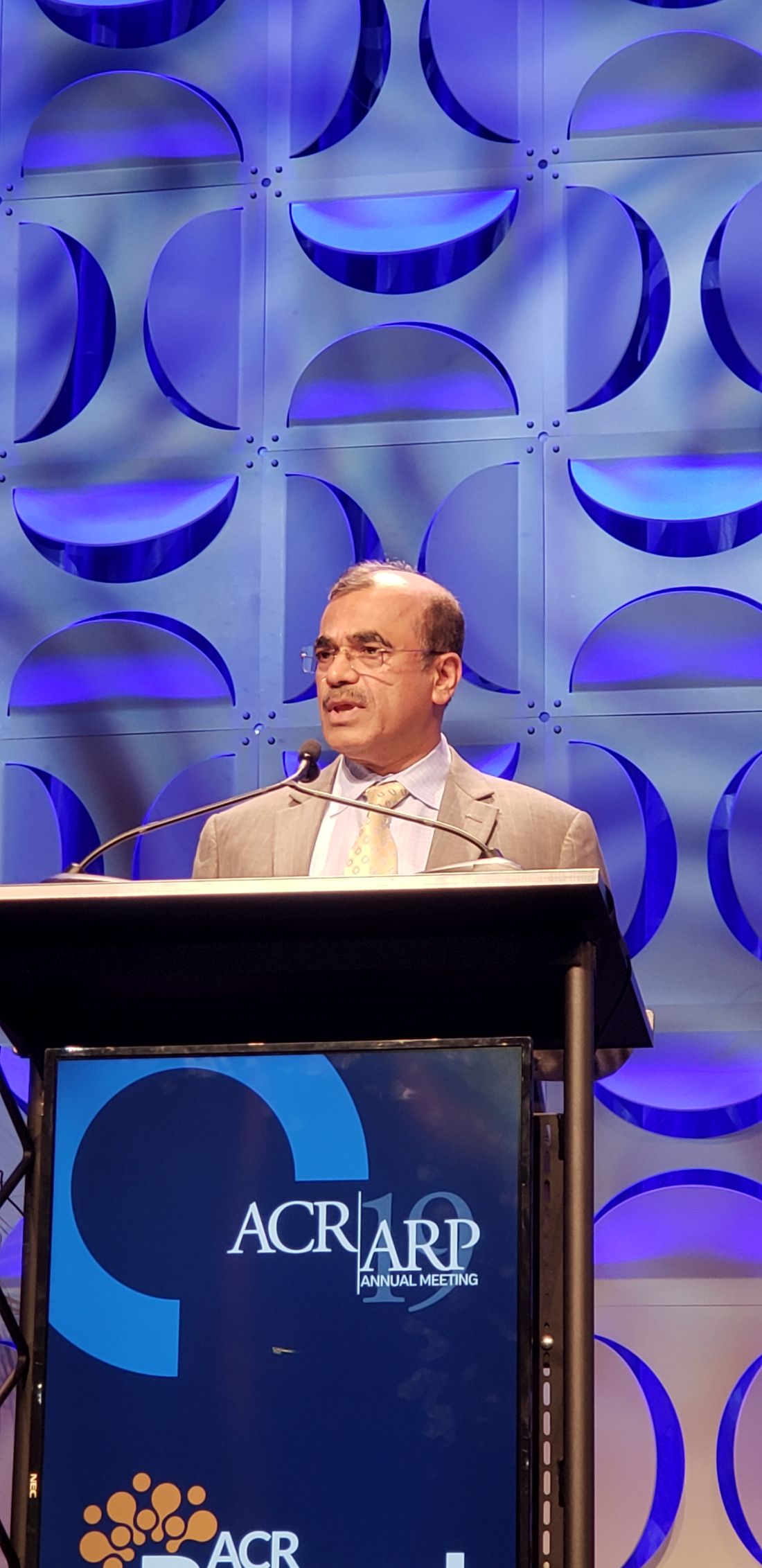
If validated, the findings of the trial – the first randomized, controlled, biopsy-driven trial in RA – could have “massive implications” for treatment selection and improved outcomes, Constantino Pitzalis, MD, reported during a press conference at the annual meeting of the American College of Rheumatology.
Of 164 RA patients who were failing or intolerant to conventional synthetic (cs) DMARD therapy and at least one tumor necrosis factor inhibitor (TNFi), 83 were randomized to receive rituximab and 81 received tocilizumab. Of those patients, 49.1% were considered B-cell poor (BCP) based on synovial tissue biopsies obtained at trial entry.
The BCP patients treated with tocilizumab were numerically more likely than those treated with rituximab to achieve the coprimary endpoint of Clinical Disease Activity Index (CDAI) improvement of at least 50% from baseline at 16 weeks (56.1% vs. 44.7%), and they were significantly more likely – twice as likely, in fact – to achieve the coprimary endpoint of CDAI improvement of at least 50% from baseline at 16 weeks as well as CDAI score less than 10, indicating a major treatment response (46.3% vs. 23.7%), said Dr. Pitzalis, head of the Centre for Experimental Medicine & Rheumatology at Queen Mary University of London.
BCP patients receiving tocilizumab also were significantly more likely to achieve a number of secondary endpoints, he noted.
In the B-cell-rich (BCR) population, no significant differences were seen in the majority of endpoints with tocilizumab versus rituximab, he said.
Study participants had a mean age of 55-56 years and were intolerant of or refractory to csDMARDs and at least one TNFi. They were recruited from 19 centers in Europe, were randomized and treated with standard doses of rituximab or tocilizumab, and were stratified based on histologic classification (BCP vs. BCR). Baseline characteristics were comparable among the treatment groups, Dr. Pitzalis said.
Adverse events occurred in 62 and 68 patients in the rituximab and tocilizumab groups, respectively, and serious adverse events occurred in 8 and 12 patients, respectively, thus 40% of all serious adverse events occurred in the rituximab group and 60% in the tocilizumab group. Infections and serious infections each occurred in three patients in each group, and two patients in each group discontinued treatment because of adverse events.
B cells are pivotal to RA pathogenesis, as demonstrated by the efficacy of the B-cell-depleting agent, rituximab. However, rituximab, which is licensed for use following failure of csDMARDs and TNFi therapy, is only effective for achieving a 50% improvement in ACR response criteria at 6 months in about 30% of such patients, Dr. Pitzalis noted. In a recent early RA cohort, he and his colleagues found synovial heterogeneity, with more than half of patients showing low or no synovial B-cell infiltration.
“So why would you give them rituximab?” he asked, explaining the rationale for the R4-RA trial: The hypothesis was that alternative biologic agents targeting alternative pathways may be more effective in BCP patients.
“The results showed quite clearly that rituximab was inferior to tocilizumab in this patient group,” he said. “We demonstrated that tocilizumab is more effective than rituximab in achieving low disease activity in patients who had, on synovial biopsy, low levels of B-cell infiltration.
“The study really highlights the importance of integrating molecular pathology into the clinical algorithms, because making the diagnosis is not sufficient. We really need to know what the pathology of the patient is so we can give the right drug to the right patients.”

Donald Thomas, MD, a rheumatologist in private practice in Silver Spring, Md., called the study “fascinating,” and noted during a question-and-answer period during the press conference that, during his lifetime, he “has probably wasted tens of thousands to hundreds of thousands of dollars,” using the trial-and-error approach to treatment.
“We can only treat by trial and error ... so being able to pinpoint therapy is just phenomenal,” he added, further noting in an interview after the press conference that finding the right treatment can be “such a struggle.”
“I literally have patients who have gone through 10 medicines and wasted thousands of dollars,” he said.
In response to a question from Dr. Thomas about the potential for rheumatologists to do their own synovial biopsies to help guide treatment in the event the findings are validated, Dr. Pitzalis said that is both feasible and an important goal.
“All the biopsies [in the study] were carried out by rheumatologists,” he noted. “We have trained over 150 rheumatologists worldwide, including at 15 centers in the United States. ... We want to empower the rheumatologists to do it.”
He added that “this is early data ... and will require validation in larger trials,” but said the point is that “we can’t continue to just give these drugs and see if the patient responds.”
R4-RA is funded by the National Institute of Health Research Efficacy and Mechanism Evaluation program. Dr. Pitzalis and some of the other study authors reported financial relationships with Roche/Genentech, which markets tocilizumab and rituximab, and other pharmaceutical companies.
SOURCE: Pitzalis C et al. Arthritis Rheumatol. 2019;71(suppl 10), Abstract 2911.
ATLANTA – Tocilizumab proved more effective than rituximab in B-cell-poor but not in B-cell-rich patients with RA who have had an inadequate response to disease-modifying antirheumatic drugs (DMARDs) and tumor necrosis factor inhibition in the randomized, open-label, 48-week, phase 4 R4-RA trial.
If validated, the findings of the trial – the first randomized, controlled, biopsy-driven trial in RA – could have “massive implications” for treatment selection and improved outcomes, Constantino Pitzalis, MD, reported during a press conference at the annual meeting of the American College of Rheumatology.
Of 164 RA patients who were failing or intolerant to conventional synthetic (cs) DMARD therapy and at least one tumor necrosis factor inhibitor (TNFi), 83 were randomized to receive rituximab and 81 received tocilizumab. Of those patients, 49.1% were considered B-cell poor (BCP) based on synovial tissue biopsies obtained at trial entry.
The BCP patients treated with tocilizumab were numerically more likely than those treated with rituximab to achieve the coprimary endpoint of Clinical Disease Activity Index (CDAI) improvement of at least 50% from baseline at 16 weeks (56.1% vs. 44.7%), and they were significantly more likely – twice as likely, in fact – to achieve the coprimary endpoint of CDAI improvement of at least 50% from baseline at 16 weeks as well as CDAI score less than 10, indicating a major treatment response (46.3% vs. 23.7%), said Dr. Pitzalis, head of the Centre for Experimental Medicine & Rheumatology at Queen Mary University of London.
BCP patients receiving tocilizumab also were significantly more likely to achieve a number of secondary endpoints, he noted.
In the B-cell-rich (BCR) population, no significant differences were seen in the majority of endpoints with tocilizumab versus rituximab, he said.
Study participants had a mean age of 55-56 years and were intolerant of or refractory to csDMARDs and at least one TNFi. They were recruited from 19 centers in Europe, were randomized and treated with standard doses of rituximab or tocilizumab, and were stratified based on histologic classification (BCP vs. BCR). Baseline characteristics were comparable among the treatment groups, Dr. Pitzalis said.
Adverse events occurred in 62 and 68 patients in the rituximab and tocilizumab groups, respectively, and serious adverse events occurred in 8 and 12 patients, respectively, thus 40% of all serious adverse events occurred in the rituximab group and 60% in the tocilizumab group. Infections and serious infections each occurred in three patients in each group, and two patients in each group discontinued treatment because of adverse events.
B cells are pivotal to RA pathogenesis, as demonstrated by the efficacy of the B-cell-depleting agent, rituximab. However, rituximab, which is licensed for use following failure of csDMARDs and TNFi therapy, is only effective for achieving a 50% improvement in ACR response criteria at 6 months in about 30% of such patients, Dr. Pitzalis noted. In a recent early RA cohort, he and his colleagues found synovial heterogeneity, with more than half of patients showing low or no synovial B-cell infiltration.
“So why would you give them rituximab?” he asked, explaining the rationale for the R4-RA trial: The hypothesis was that alternative biologic agents targeting alternative pathways may be more effective in BCP patients.
“The results showed quite clearly that rituximab was inferior to tocilizumab in this patient group,” he said. “We demonstrated that tocilizumab is more effective than rituximab in achieving low disease activity in patients who had, on synovial biopsy, low levels of B-cell infiltration.
“The study really highlights the importance of integrating molecular pathology into the clinical algorithms, because making the diagnosis is not sufficient. We really need to know what the pathology of the patient is so we can give the right drug to the right patients.”
Donald Thomas, MD, a rheumatologist in private practice in Silver Spring, Md., called the study “fascinating,” and noted during a question-and-answer period during the press conference that, during his lifetime, he “has probably wasted tens of thousands to hundreds of thousands of dollars,” using the trial-and-error approach to treatment.
“We can only treat by trial and error ... so being able to pinpoint therapy is just phenomenal,” he added, further noting in an interview after the press conference that finding the right treatment can be “such a struggle.”
“I literally have patients who have gone through 10 medicines and wasted thousands of dollars,” he said.
In response to a question from Dr. Thomas about the potential for rheumatologists to do their own synovial biopsies to help guide treatment in the event the findings are validated, Dr. Pitzalis said that is both feasible and an important goal.
“All the biopsies [in the study] were carried out by rheumatologists,” he noted. “We have trained over 150 rheumatologists worldwide, including at 15 centers in the United States. ... We want to empower the rheumatologists to do it.”
He added that “this is early data ... and will require validation in larger trials,” but said the point is that “we can’t continue to just give these drugs and see if the patient responds.”
R4-RA is funded by the National Institute of Health Research Efficacy and Mechanism Evaluation program. Dr. Pitzalis and some of the other study authors reported financial relationships with Roche/Genentech, which markets tocilizumab and rituximab, and other pharmaceutical companies.
SOURCE: Pitzalis C et al. Arthritis Rheumatol. 2019;71(suppl 10), Abstract 2911.
REPORTING FROM ACR 2019
Even low-dose steroids increase DMARD infection risk
ATLANTA – Concomitant use of even low-dose steroids increases the risk of serious infections with antirheumatic drugs, according to a review of 170,357 Medicare patients by investigators at the University of Pennsylvania, Philadelphia.

Infections are a well-known side effect of high-dose glucocorticoids, but there’s been debate about prednisone doses in the 5-10 mg/day range. Guidelines generally advise tapering RA patients off steroids after they start a biologic or methotrexate, but that doesn’t always happen because there’s a common perception that low-dose steroids are safe, said lead investigator Michael George, MD, assistant professor of medicine and epidemiology at the university.
“Many people continue low-dose steroids over the long term, but even low dose seems to be associated with infection. It’s a small risk, but it should be something you are aware of; for some patients, it might be quite important,” he said in an interview at the annual meeting of the American College of Rheumatology.
The team wanted to mimic real-world practice, so they compared infection incidence between the 53% of patients who were not on low-dose steroids with the 47% who were after at least 6 months of disease-modifying antirheumatic drug (DMARD) therapy. About 56% of patients were on methotrexate, with the rest on biologics or a targeted synthetic DMARD (tsDMARD). Average follow up was an additional 6 months, but some people were followed for several years; prednisone 5 mg/day or less was the most common dose.
There were 20,630 serious infections requiring hospitalization, most often urinary tract infection, pneumonia, bacteremia/septicemia, and skin or soft-tissue infections. The crude incidence was 11 per 100 person-years.
After propensity-score weighting to balance out about 50 potential confounders, the predicted 1-year incidence of infection was 9.3% among patients not on steroids. Among those on up to 5 mg/day of prednisone, it was 12.5%; among those on 5-10 mg/day, 17.2%; and among those on more than 10 mg/day, 23.9%.
Glucocorticoids were associated with a 37% increased rate of serious infections, even with doses at or below 5mg/day. The effect “was really similar” whether people were on a biologic, tsDMARD, or methotrexate, which was “surprising,” Dr. George said.
“When I see a patient now who is on long-term, low-dose prednisone, I don’t just say ‘okay, that’s probably safe.’ I think really hard about how much benefit they’re getting. For some people, that means I try to get them off it,” he said. For those who flare otherwise, “I might continue them on it, but recognize there is likely some risk.”
The magnitude of the infection risk was similar to that reported with tumor necrosis factors inhibitors, which might reassure patients who are reluctant to switch to a tumor necrosis factor inhibitor.
“Now I can say you’ve been taking 10 mg prednisone a day, and that’s probably at least as risky,” Dr. George said.
Frequency of office visits, hospitalizations, and ED visits, as well as prior infections, comorbidities, nursing-home admissions, and use of durable medical equipment were among the potential confounders controlled for in the analysis. They stood in for direct markers of RA severity, which weren’t available in the data. “We spent a lot of time trying to make sure our groups were as similar as possible in every way except prednisone use,” he said.
Patients were in their late 60s on average, 71% white, and 81% were women. People with other autoimmune rheumatic diseases, cancer, or HIV were excluded. Dr. George said the next step is to run the same analysis in a younger cohort.
The work was funded by the National Institutes of Health. Dr. George disclosed relationships with AbbVie and Bristol-Myers Squibb.
SOURCE: George M et al. ACR 2019, Abstract 848
ATLANTA – Concomitant use of even low-dose steroids increases the risk of serious infections with antirheumatic drugs, according to a review of 170,357 Medicare patients by investigators at the University of Pennsylvania, Philadelphia.
Infections are a well-known side effect of high-dose glucocorticoids, but there’s been debate about prednisone doses in the 5-10 mg/day range. Guidelines generally advise tapering RA patients off steroids after they start a biologic or methotrexate, but that doesn’t always happen because there’s a common perception that low-dose steroids are safe, said lead investigator Michael George, MD, assistant professor of medicine and epidemiology at the university.
“Many people continue low-dose steroids over the long term, but even low dose seems to be associated with infection. It’s a small risk, but it should be something you are aware of; for some patients, it might be quite important,” he said in an interview at the annual meeting of the American College of Rheumatology.
The team wanted to mimic real-world practice, so they compared infection incidence between the 53% of patients who were not on low-dose steroids with the 47% who were after at least 6 months of disease-modifying antirheumatic drug (DMARD) therapy. About 56% of patients were on methotrexate, with the rest on biologics or a targeted synthetic DMARD (tsDMARD). Average follow up was an additional 6 months, but some people were followed for several years; prednisone 5 mg/day or less was the most common dose.
There were 20,630 serious infections requiring hospitalization, most often urinary tract infection, pneumonia, bacteremia/septicemia, and skin or soft-tissue infections. The crude incidence was 11 per 100 person-years.
After propensity-score weighting to balance out about 50 potential confounders, the predicted 1-year incidence of infection was 9.3% among patients not on steroids. Among those on up to 5 mg/day of prednisone, it was 12.5%; among those on 5-10 mg/day, 17.2%; and among those on more than 10 mg/day, 23.9%.
Glucocorticoids were associated with a 37% increased rate of serious infections, even with doses at or below 5mg/day. The effect “was really similar” whether people were on a biologic, tsDMARD, or methotrexate, which was “surprising,” Dr. George said.
“When I see a patient now who is on long-term, low-dose prednisone, I don’t just say ‘okay, that’s probably safe.’ I think really hard about how much benefit they’re getting. For some people, that means I try to get them off it,” he said. For those who flare otherwise, “I might continue them on it, but recognize there is likely some risk.”
The magnitude of the infection risk was similar to that reported with tumor necrosis factors inhibitors, which might reassure patients who are reluctant to switch to a tumor necrosis factor inhibitor.
“Now I can say you’ve been taking 10 mg prednisone a day, and that’s probably at least as risky,” Dr. George said.
Frequency of office visits, hospitalizations, and ED visits, as well as prior infections, comorbidities, nursing-home admissions, and use of durable medical equipment were among the potential confounders controlled for in the analysis. They stood in for direct markers of RA severity, which weren’t available in the data. “We spent a lot of time trying to make sure our groups were as similar as possible in every way except prednisone use,” he said.
Patients were in their late 60s on average, 71% white, and 81% were women. People with other autoimmune rheumatic diseases, cancer, or HIV were excluded. Dr. George said the next step is to run the same analysis in a younger cohort.
The work was funded by the National Institutes of Health. Dr. George disclosed relationships with AbbVie and Bristol-Myers Squibb.
SOURCE: George M et al. ACR 2019, Abstract 848
REPORTING FROM ACR 2019
Methotrexate may affect joint erosions but not pain in patients with erosive hand OA
ATLANTA – , according to results from the small, prospective, double-blind, randomized, placebo-controlled ADEM trial.

“Our study failed to show the superiority of methotrexate over placebo on pain evolution, but our results on structural evolution and the presence of inflammatory parameters as predictors of erosive evolution in nonerosive diseases may lead us to discuss the place of methotrexate in early steps of the disease evolution, and underlines the importance of the part played by the interaction between synovitis and subchondral bone in erosive progression,” Christian Roux, MD, PhD, of the department of rheumatology at Côte d’Azur University, Nice, France, said in his presentation at the annual meeting of the American College of Rheumatology.
Dr. Roux and colleagues enrolled 64 patients in the ADEM trial, where patients with symptomatic erosive hand osteoarthritis (EHOA) were randomized to receive 10 mg of methotrexate (MTX) per week or placebo. At 3 months, researchers assessed patients for pain using the Visual Analog Scale (VAS) score for hand pain, and secondary outcome measures at 12 months included VAS score for hand pain, radiographic progression using Verbruggen-Veys Anatomical Phase Score and Gent University Scoring System, and MRI.
Patients were included in the study if they were between 45 and 85 years old with a VAS pain score greater than 40, had failed classic therapeutics (acetaminophen, topical NSAIDs, and symptomatic slow-acting drugs), and had at least one erosive lesion. At baseline, the MTX and placebo groups were not significantly different with regard to gender (91% vs. 97% female), mean body mass index (24.6 kg/m2 vs. 24.2 kg/m2) and mean age (67.5 years vs. 64.9 years). Radiologic data showed joint loss, erosive, and erosive plus remodeling measurements were also similar between groups at baseline.
The mean VAS score for patients in the MTX group decreased from 65.7 at baseline to 48.2 at 3 months (–17.5; P = .07), compared with a decrease from 63.9 to 55.5 (–8.4; P = .002). At 12 months, VAS scores for patients in the MTX group decreased to 47.5, compared with a decrease in the placebo group to 48.2. However, the between-group differences for VAS scores were not significant at 3 months (P = .2) and at 12 months (P = .6).
“We have different hypotheses on the failure of our study on our main outcome, which was pain,” he said. “The first is a low-dose of methotrexate, and the second may be ... a placebo effect, which is very, very important in osteoarthritis.”
Dr. Roux noted the results from the ADEM trial were similar to a recent study in which 90 patients with hand OA were randomized to receive etanercept or placebo. At 24 weeks, there was no statistically significant difference between VAS pain in the etanercept group (between group difference, −5.7; 95% confidence interval, −15.9 to 4.5; P = .27) and the placebo groups, and at 1 year (between-group difference, –8.5; 95% CI, −18.6 to 1.6; P = .10), although the results favored patients receiving anti-tumor necrosis factor therapy (Ann Rheum Dis. 2018;77:1757-64. doi: 10.1136/annrheumdis-2018-213202).
With regard to the Verbruggen-Veys score, joint degradation was not significantly higher in the placebo group (29.4%), compared with the MTX group (7.7%), but there was a significantly higher number of erosive joints progressing to a remodeling phase in the MTX group (27.2%), compared with the placebo group (15.2%) at 12 months.
Dr. Roux said two factors are likely predictors of erosive disease based on data in ADEM: the level of interleukin-6 at baseline (odds ratio, 1.04; 95% CI, 1.03-1.06; P less than .0001), and joints with synovitis at baseline (OR, 4.7; 95% CI, 1.25-17.90; P = .02).
“Our study has several limitations, but we like to see our study as a pilot study,” he added, noting that a study analyzing bone turnover in patients with different doses of methotrexate and a longer disease duration is needed.
The authors reported no conflicts of interest.
SOURCE: Ferraro S et al. Arthritis Rheumatol. 2019;71(suppl 10), Abstract 1759.
ATLANTA – , according to results from the small, prospective, double-blind, randomized, placebo-controlled ADEM trial.
“Our study failed to show the superiority of methotrexate over placebo on pain evolution, but our results on structural evolution and the presence of inflammatory parameters as predictors of erosive evolution in nonerosive diseases may lead us to discuss the place of methotrexate in early steps of the disease evolution, and underlines the importance of the part played by the interaction between synovitis and subchondral bone in erosive progression,” Christian Roux, MD, PhD, of the department of rheumatology at Côte d’Azur University, Nice, France, said in his presentation at the annual meeting of the American College of Rheumatology.
Dr. Roux and colleagues enrolled 64 patients in the ADEM trial, where patients with symptomatic erosive hand osteoarthritis (EHOA) were randomized to receive 10 mg of methotrexate (MTX) per week or placebo. At 3 months, researchers assessed patients for pain using the Visual Analog Scale (VAS) score for hand pain, and secondary outcome measures at 12 months included VAS score for hand pain, radiographic progression using Verbruggen-Veys Anatomical Phase Score and Gent University Scoring System, and MRI.
Patients were included in the study if they were between 45 and 85 years old with a VAS pain score greater than 40, had failed classic therapeutics (acetaminophen, topical NSAIDs, and symptomatic slow-acting drugs), and had at least one erosive lesion. At baseline, the MTX and placebo groups were not significantly different with regard to gender (91% vs. 97% female), mean body mass index (24.6 kg/m2 vs. 24.2 kg/m2) and mean age (67.5 years vs. 64.9 years). Radiologic data showed joint loss, erosive, and erosive plus remodeling measurements were also similar between groups at baseline.
The mean VAS score for patients in the MTX group decreased from 65.7 at baseline to 48.2 at 3 months (–17.5; P = .07), compared with a decrease from 63.9 to 55.5 (–8.4; P = .002). At 12 months, VAS scores for patients in the MTX group decreased to 47.5, compared with a decrease in the placebo group to 48.2. However, the between-group differences for VAS scores were not significant at 3 months (P = .2) and at 12 months (P = .6).
“We have different hypotheses on the failure of our study on our main outcome, which was pain,” he said. “The first is a low-dose of methotrexate, and the second may be ... a placebo effect, which is very, very important in osteoarthritis.”
Dr. Roux noted the results from the ADEM trial were similar to a recent study in which 90 patients with hand OA were randomized to receive etanercept or placebo. At 24 weeks, there was no statistically significant difference between VAS pain in the etanercept group (between group difference, −5.7; 95% confidence interval, −15.9 to 4.5; P = .27) and the placebo groups, and at 1 year (between-group difference, –8.5; 95% CI, −18.6 to 1.6; P = .10), although the results favored patients receiving anti-tumor necrosis factor therapy (Ann Rheum Dis. 2018;77:1757-64. doi: 10.1136/annrheumdis-2018-213202).
With regard to the Verbruggen-Veys score, joint degradation was not significantly higher in the placebo group (29.4%), compared with the MTX group (7.7%), but there was a significantly higher number of erosive joints progressing to a remodeling phase in the MTX group (27.2%), compared with the placebo group (15.2%) at 12 months.
Dr. Roux said two factors are likely predictors of erosive disease based on data in ADEM: the level of interleukin-6 at baseline (odds ratio, 1.04; 95% CI, 1.03-1.06; P less than .0001), and joints with synovitis at baseline (OR, 4.7; 95% CI, 1.25-17.90; P = .02).
“Our study has several limitations, but we like to see our study as a pilot study,” he added, noting that a study analyzing bone turnover in patients with different doses of methotrexate and a longer disease duration is needed.
The authors reported no conflicts of interest.
SOURCE: Ferraro S et al. Arthritis Rheumatol. 2019;71(suppl 10), Abstract 1759.
REPORTING FROM ACR 2019
CVD risk in black SLE patients 18 times higher than in whites
ATLANTA – Black race was the single greatest predictor of cardiovascular disease (CVD) events in systemic lupus erythematosus, with black patients having an 18-fold higher risk than white patients from 2 years before to 8 years after diagnosis, according to a review of 336 patients in the Georgia Lupus Registry that was presented at the annual meeting of the American College of Rheumatology.
The greatest risk was in the first 2 years after diagnosis, which has been reported before in white patients, but not before in a mostly (75%) black cohort.
Lupus is known to strike earlier and be more aggressive in black patients, so “we were expecting racial disparities in incident CVD, but” the magnitude of the increased risk “was very surprising. This study [identifies] a population that needs more attention, more targeted CVD prevention. We have to intervene early and be on top of everything,” especially for black patients, said lead investigator Shivani Garg, MD, an assistant professor of rheumatology at the University of Wisconsin–Madison.
Lipids, blood pressure, and the other usual CVD risk factors, as well as lupus itself, have to be optimally controlled; glucocorticoid use limited as much as possible; and there needs to be improved adherence to hydroxychloroquine, which has been shown to reduce CVD events in lupus patients, she said in an interview.
The 336 patients, mostly women (87%) from the Atlanta area, were diagnosed during 2002-2004 at a mean age of 40 years. Dr. Garg and associates reviewed CVD events – ischemic heart disease, stroke, transient ischemic attack, and peripheral vascular disease – and death over 16 years, beginning 2 years before diagnosis.
About 22% of subjects had a CVD event, most commonly within 2 years after diagnosis. The risk was 500% higher in black patients overall (adjusted hazard ratio, 6.4; 95% confidence interval, 2.4-17.5; P = .0003), and markedly higher in the first 10 years (aHR, 18; 95% CI, 2.2-141; P less than .0001). The findings were not adjusted for socioeconomic factors.
In the first 12 years of the study, the mean age at lupus diagnosis was 46 years and the first CVD event occurred at an average of 48 years. From 12 to 16 years follow-up, the mean age of diagnosis was 38 years, and the first CVD event occurred at 52 years.
Age older than 65 years (aHR, 7.9; 95% CI, 2.2-29) and the presence of disease-associated antibodies (aHR, 2.1; 95% CI, 1.01-4.4) increased CVD risk, which wasn’t surprising, but another predictor – discoid lupus – was unexpected (aHR, 3.2; 95% CI, 1.5-6.8). “A lot of times, we’ve considered discoid rash to be a milder form, but these patients have some kind of chronic, smoldering inflammation that is leading to atherosclerosis,” Dr. Garg said.
At diagnosis, 84% of the subjects had lupus hematologic disorders, 69% immunologic disorders, and 14% a discoid rash. CVD risk factor data were not collected.
There was no external funding, and the investigators reported no disclosures.
SOURCE: Garg S et al. Arthritis Rheumatol. 2019;71(suppl 10), Abstract 805.
REPORTING FROM ACR 2019
Guselkumab improves psoriatic arthritis regardless of prior TNFi use
ATLANTA – Guselkumab improved outcomes in psoriatic arthritis patients regardless of past treatment with tumor necrosis factor inhibitors in the phase 3 DISCOVER-1 trial.
The anti-interleukin-23p19 monoclonal antibody is approved in the United States for the treatment of moderate to severe plaque psoriasis (PsO).
Benefits in psoriatic arthritis (PsA) were seen in both biologic-naive and tumor necrosis factor inhibitor (TNFi)–treated patients and occurred with both 4- and 8-week dosing regimens, Atul Deodhar, MD, reported during a plenary session at the annual meeting of the American College of Rheumatology.
For example, the primary endpoint of ACR 20 response at 24 weeks was achieved in 58.6% and 52.8% of patients randomized to receive 100 mg of guselkumab delivered subcutaneously either at baseline and every 4 weeks or at baseline, week 4, and then every 8 weeks, respectively, compared with 22.2% of those randomized to receive placebo, said Dr. Deodhar, professor of medicine at Oregon Health & Science University, Portland.
Greater proportions of patients in the guselkumab groups achieved ACR 20 response at week 16; ACR 50 response at weeks 16 and 24; ACR 70 response at week 24; Psoriasis Area and Severity Index 75, 90, and 100 responses at week 24; and minimal disease activity response at week 24, he said, adding that improvements were also seen with guselkumab versus placebo for the controlled major secondary endpoints of change from baseline in Health Assessment Questionnaire–Disability Index score, Short Form 36 Health Survey score, and investigator global assessment (IGA) of PsO response.
The response rates with guselkumab versus placebo were seen regardless of prior TNFi use, he said.
The study included 381 patients with active PsA, defined as three or more swollen joints, three or more tender joints, and C-reactive protein of 0.3 mg/dL or greater despite standard therapies. About 30% were exposed to up to two TNFi therapies and 10% were nonresponders or inadequate responders to those therapies.
Concomitant use of select nonbiologic disease-modifying antirheumatic drugs, oral corticosteroids, and NSAIDs was allowed, and patients with less than 5% improvement in tender plus swollen joints at week 16 could initiate or increase the dose of the permitted medications while continuing study treatment, Dr. Deodhar said.
The mean body surface area with PsO involvement was 13.4%; 42.5% of patients had an IGA of 3-4 for skin involvement. Mean swollen and tender joint counts were 9.8 and 19.3, respectively, indicating a population with moderate to severe disease, he added.
Serious adverse events, serious infections, and death occurred in 2.4%, 0.5%, and 0.3% of patients, respectively.
“Both guselkumab regimens were safe and well tolerated through week 24,” Dr. Deodhar said, noting that the safety profile was consistent with that established in the treatment of PsO and described in the label.
DISCOVER-1 was funded by Janssen Research & Development. Dr. Deodhar reported relationships (advisory board activity, consulting, and/or research grant funding) with several pharmaceutical companies including Janssen. Several coauthors are employees of Janssen.
SOURCE: Deodhar A et al. Arthritis Rheumatol. 2019;71(suppl 10), Abstract 807.
Patients taking TNF inhibitors can safely receive Zostavax
ATLANTA – A group of patients using a tumor necrosis factor inhibitor safely received the live-attenuated varicella vaccine Zostavax without any cases of herpes zoster in the first 6 weeks after vaccination in the blinded, randomized, placebo-controlled Varicella Zoster Vaccine (VERVE) trial .

According to guidelines from the Centers for Disease Control and Prevention’s Advisory Committee on Immunization Practices, there is a theoretical concern that patients using a tumor necrosis factor inhibitor (TNFi) and other biologic therapies who receive a live-attenuated version of the varicella vaccine (Zostavax) could become infected with varicella from the vaccine. Patients with RA and psoriatic arthritis as well as other autoimmune and inflammatory conditions who are likely to receive TNFi therapy are also at risk for herpes zoster reactivation, Jeffrey Curtis, MD, professor of medicine in the division of clinical immunology and rheumatology of the University of Alabama at Birmingham, said in his presentation at the annual meeting of the American College of Rheumatology. There also exists a risk for patients receiving low-dose glucocorticoids.
“The challenge, of course, is there’s not a great definition and there certainly is not a well-standardized assay for how immunocompromised someone is, and so that led to the uncertainty in this patient population for this and other live-virus vaccines,” Dr. Curtis said.
Dr. Curtis and colleagues enrolled 627 participants from 33 centers into the VERVE trial. Participants were aged at least 50 years, were taking a TNFi, and had not previously received Zostavax.
Patients in both groups had a mean age of about 63 years and about two-thirds were women. The most common indications for TNFi use in the Zostavax group and the placebo group were RA (59.2% vs. 56.0%, respectively), psoriatic arthritis (24.3% vs. 23.9%), and ankylosing spondylitis (7.2% vs. 8.5%), while the anti-TNF agents used were adalimumab (38.1% vs. 27.4%), infliximab (28.4% vs. 34.2%), etanercept (19.0% vs. 23.5%), golimumab (10.0% vs. 8.1%), and certolizumab pegol (4.5% vs. 6.8%). In addition, some patients in the Zostavax and placebo groups were also taking concomitant therapies with TNFi, such as oral glucocorticoids (9.7% vs. 11.4%).
The researchers randomized participants to receive Zostavax or placebo (saline) and then followed them for 6 weeks, and looked for signs of wild-type or vaccine-strain varicella infection. If participants were suspected to have varicella, they were assessed clinically, underwent polymerase chain reaction testing, and rashes were photographed. At baseline and at 6 weeks, the researchers collected serum and peripheral blood mononuclear cells to determine patient immunity to varicella. After 6 months, participants were unmasked to the treatment arm of the study.
Dr. Curtis and colleagues found no confirmed varicella infection cases at 6 weeks. “To the extent that 0 cases out of 317 vaccinated people is reassuring, there were no cases, so that was exceedingly heartening as a result,” he said.
Out of 20 serious adverse events total in the groups, 15 events occurred before 6 months, including 8 suspected varicella cases in the Zostavax group and 7 in the placebo group. However, there were no positive cases of varicella – either wild type or vaccine type – after polymerase chain reaction tests. Overall, there were 268 adverse events in 195 participants, with 73 events (27.2%) consisting of injection-site reactions. The researchers also found no difference in the rate of disease flares, and found no differences in adverse reactions between groups, apart from a higher rate of injection-site reactions in the varicella group (19.4% vs. 4.2%).
With regard to immunogenicity, the humoral immune response was measured through IgG, which showed an immune response in the varicella group at 6 weeks (geometric mean fold ratio, 1.33; 95% confidence interval, 1.18-1.51), compared with the placebo group (GMFR, 1.02; 95% CI, 0.91-1.14); cell-mediated immune response was measured by interferon-gamma, which also showed an immune response in the live-vaccine group (GMFR, 1.49; 95% CI, 1.14-1.94), compared with participants who received placebo (GMFR, 1.14; 95% CI, 0.87-1.48). In preliminary 1-year data, IgG immune response was elevated in the varicella group (GMFR, 1.46; 95% CI, 1.08-1.99), but there was no elevated immune response for interferon-gamma (GMFR, 0.78; 95% CI, 0.49-1.25).
“I think the trial is encouraging not only for its result with the live zoster vaccine and TNF-treated patients, but also challenge the notion that, if you need to, a live-virus vaccine may in fact be able to be safely given to people with autoimmune and inflammatory diseases, even those treated with biologics like tumor necrosis factor inhibitors,” Dr. Curtis said.
As patients in VERVE consented to long-term follow-up in health plan claims and EHR data, it will be possible to follow these patients in the future to assess herpes zoster reactivation. Dr. Curtis also noted that a new trial involving the recombinant, adjuvanted zoster vaccine (Shingrix) is currently in development and should begin next year.
The VERVE trial was supported by the National Institute of Arthritis and Musculoskeletal and Skin Diseases. Dr. Curtis reported serving as a current member of the Center for Disease Control and Prevention’s Advisory Committee on Immunization Practices Herpes Zoster Work Group. He and some of the other authors reported financial relationships with many pharmaceutical companies.
SOURCE: Curtis J et al. Arthritis Rheumatol. 2019;71(suppl 10), Abstract 824.
ATLANTA – A group of patients using a tumor necrosis factor inhibitor safely received the live-attenuated varicella vaccine Zostavax without any cases of herpes zoster in the first 6 weeks after vaccination in the blinded, randomized, placebo-controlled Varicella Zoster Vaccine (VERVE) trial .
According to guidelines from the Centers for Disease Control and Prevention’s Advisory Committee on Immunization Practices, there is a theoretical concern that patients using a tumor necrosis factor inhibitor (TNFi) and other biologic therapies who receive a live-attenuated version of the varicella vaccine (Zostavax) could become infected with varicella from the vaccine. Patients with RA and psoriatic arthritis as well as other autoimmune and inflammatory conditions who are likely to receive TNFi therapy are also at risk for herpes zoster reactivation, Jeffrey Curtis, MD, professor of medicine in the division of clinical immunology and rheumatology of the University of Alabama at Birmingham, said in his presentation at the annual meeting of the American College of Rheumatology. There also exists a risk for patients receiving low-dose glucocorticoids.
“The challenge, of course, is there’s not a great definition and there certainly is not a well-standardized assay for how immunocompromised someone is, and so that led to the uncertainty in this patient population for this and other live-virus vaccines,” Dr. Curtis said.
Dr. Curtis and colleagues enrolled 627 participants from 33 centers into the VERVE trial. Participants were aged at least 50 years, were taking a TNFi, and had not previously received Zostavax.
Patients in both groups had a mean age of about 63 years and about two-thirds were women. The most common indications for TNFi use in the Zostavax group and the placebo group were RA (59.2% vs. 56.0%, respectively), psoriatic arthritis (24.3% vs. 23.9%), and ankylosing spondylitis (7.2% vs. 8.5%), while the anti-TNF agents used were adalimumab (38.1% vs. 27.4%), infliximab (28.4% vs. 34.2%), etanercept (19.0% vs. 23.5%), golimumab (10.0% vs. 8.1%), and certolizumab pegol (4.5% vs. 6.8%). In addition, some patients in the Zostavax and placebo groups were also taking concomitant therapies with TNFi, such as oral glucocorticoids (9.7% vs. 11.4%).
The researchers randomized participants to receive Zostavax or placebo (saline) and then followed them for 6 weeks, and looked for signs of wild-type or vaccine-strain varicella infection. If participants were suspected to have varicella, they were assessed clinically, underwent polymerase chain reaction testing, and rashes were photographed. At baseline and at 6 weeks, the researchers collected serum and peripheral blood mononuclear cells to determine patient immunity to varicella. After 6 months, participants were unmasked to the treatment arm of the study.
Dr. Curtis and colleagues found no confirmed varicella infection cases at 6 weeks. “To the extent that 0 cases out of 317 vaccinated people is reassuring, there were no cases, so that was exceedingly heartening as a result,” he said.
Out of 20 serious adverse events total in the groups, 15 events occurred before 6 months, including 8 suspected varicella cases in the Zostavax group and 7 in the placebo group. However, there were no positive cases of varicella – either wild type or vaccine type – after polymerase chain reaction tests. Overall, there were 268 adverse events in 195 participants, with 73 events (27.2%) consisting of injection-site reactions. The researchers also found no difference in the rate of disease flares, and found no differences in adverse reactions between groups, apart from a higher rate of injection-site reactions in the varicella group (19.4% vs. 4.2%).
With regard to immunogenicity, the humoral immune response was measured through IgG, which showed an immune response in the varicella group at 6 weeks (geometric mean fold ratio, 1.33; 95% confidence interval, 1.18-1.51), compared with the placebo group (GMFR, 1.02; 95% CI, 0.91-1.14); cell-mediated immune response was measured by interferon-gamma, which also showed an immune response in the live-vaccine group (GMFR, 1.49; 95% CI, 1.14-1.94), compared with participants who received placebo (GMFR, 1.14; 95% CI, 0.87-1.48). In preliminary 1-year data, IgG immune response was elevated in the varicella group (GMFR, 1.46; 95% CI, 1.08-1.99), but there was no elevated immune response for interferon-gamma (GMFR, 0.78; 95% CI, 0.49-1.25).
“I think the trial is encouraging not only for its result with the live zoster vaccine and TNF-treated patients, but also challenge the notion that, if you need to, a live-virus vaccine may in fact be able to be safely given to people with autoimmune and inflammatory diseases, even those treated with biologics like tumor necrosis factor inhibitors,” Dr. Curtis said.
As patients in VERVE consented to long-term follow-up in health plan claims and EHR data, it will be possible to follow these patients in the future to assess herpes zoster reactivation. Dr. Curtis also noted that a new trial involving the recombinant, adjuvanted zoster vaccine (Shingrix) is currently in development and should begin next year.
The VERVE trial was supported by the National Institute of Arthritis and Musculoskeletal and Skin Diseases. Dr. Curtis reported serving as a current member of the Center for Disease Control and Prevention’s Advisory Committee on Immunization Practices Herpes Zoster Work Group. He and some of the other authors reported financial relationships with many pharmaceutical companies.
SOURCE: Curtis J et al. Arthritis Rheumatol. 2019;71(suppl 10), Abstract 824.
REPORTING FROM ACR 2019
AAD-NPF Pediatric psoriasis guideline advises on physical and mental care
Psoriasis management in children involves attention not only to treatment of the physical condition but also psychosocial wellness and quality of life, according to

Psoriasis affects approximately 1% of children, either alone or associated with comorbid conditions such as psoriatic arthritis (PsA), wrote Alan Menter, MD, of Baylor University Medical Center, Dallas, and coauthors of the guideline.
In the guideline, published in the Journal of the American Academy of Dermatology, the multidisciplinary work group identified screening tools to measure disease severity, strategies for management of comorbidities, and the safety and effectiveness of topical, systemic, and phototherapy treatments.
To assess disease severity, the work group recommended not only the use of body surface area (BSA), similar to measurement of severity in adults, but also the use of the Children’s Dermatology Life Quality Index, a 10-question quality of life survey, as BSA alone does not account for the potential negative impact of the disease on quality of life in terms of physical, emotional, social, and psychological function.
“For example, a child with psoriasis limited to the face or the entire scalp does not have severe disease based on BSA definitions, but if this involvement causes shame, social withdrawal, or bullying, it satisfies criteria for severe disease based on impact beyond the skin,” they said.
The work group stated that a variety of conditions may trigger or exacerbate psoriasis in children, including infections, cutaneous trauma, or physiological, emotional, and environmental stressors.
The majority of children with PsA develop joint inflammation before skin disease, the work group wrote. In addition, children with psoriasis are at increased risk for rheumatoid arthritis, so clinicians may need to distinguish between a combination of psoriasis and musculoskeletal issues and cases of either psoriatic or rheumatoid arthritis in young patients.
The cardiovascular risk factors associated with metabolic syndrome are greater in children with psoriasis, compared with children without psoriasis, the work group noted. In addition, pediatric psoriasis patients have a higher prevalence of obesity than children without psoriasis, and they recommended that children with psoriasis be monitored for the development of obesity, and that obese children with psoriasis should be referred for weight management.
The work group noted that data are insufficient in children to support the link between psoriasis and cardiovascular disease that has been documented in adults with psoriasis. However, “patients with pediatric psoriasis should have American Academy of Pediatrics (AAP)–recommended age-related cardiovascular screening regardless of the presence of signs or symptoms,” they said.
The guideline also recommends screening for dyslipidemia and hypertension according to AAP guidelines and educating pediatric psoriasis patients about the risk of diabetes and regularly screening for diabetes and insulin resistance in those who are obese. Overweight children with psoriasis may be screened at the provider’s discretion, they wrote. Patients with signs of inflammatory bowel disease, which also is associated with psoriasis in adults, should be considered for referral to a gastroenterologist, they noted.
Children with psoriasis should be screened regularly for mental health conditions regardless of age, and they should be asked about substance abuse, according to the guideline, and those with concerns should be referred for additional assessment and management.
The guideline divides treatment of psoriasis in children into three categories: topical, phototherapy and photochemotherapy, and systemic treatments (nonbiologic or biologic).
For topicals, the guideline recommendations include corticosteroids as an off-label therapy, as well as ultra-high-potency topical corticosteroids as monotherapy. Overall, “selection of a therapeutic routine (potency, delivery vehicle, frequency of application) should take into account sites of involvement, type and thickness of psoriasis, age of the patient, total BSA of application, anticipated occlusion, and disease acuity, among other patient-, disease-, and drug-related factors,” the authors wrote. Other topical options included in the recommendations: calcineurin inhibitors, topical vitamin D analogues, tazarotene (off label), anthralin, and coal tar.
Phototherapy has a history of use in psoriasis treatment and remains part of the current recommendations, although data in children are limited, and data on the use of phototherapy for pustular psoriasis in children are insufficient to make specific treatment and dosing recommendations, the work group noted. The researchers also noted that in-office phototherapy may not be feasible for many patients, but that in-home ultraviolet light equipment or natural sunlight in moderation could be recommended as an alternative.
The use of systemic, nonbiologic treatments for pediatric psoriasis should be “based on baseline severity of disease, subtype of psoriasis, speed of disease progression, lack of response to more conservative therapies such as topical agents and phototherapy (when appropriate), impaired physical or psychological functioning or [quality of life] due to disease extent, and the presence of comorbidities such as PsA,” the workgroup said.
Options for systemic treatment include methotrexate, cyclosporine (notably for pustular as well as plaque and erythrodermic psoriasis), and systemic retinoids. In addition, fumaric acid esters may be an option for children with moderate to severe psoriasis, with recommended clinical and laboratory monitoring.
The increasing safety and efficacy data on biologics in pediatric psoriasis patients support their consideration among first-line systemic treatments, the work group suggested. “Etanercept and ustekinumab are now [Food and Drug Administration] approved for patients with psoriasis 4 years and older and 12 years and older, respectively,” they said, and infliximab and adalimumab have been used off label in children.
The work group concluded that research and knowledge gaps about pediatric psoriasis persist and include mechanism of disease onset, development of comorbidities, and identification of ideal dosing for various treatments.
Finally, the work group emphasized the importance of collaboration between dermatologists and primary care providers for managing psoriasis in children, as well as the importance of patient education.
“Dermatologists should be mindful of the unique aspects of the emotional development of children and the social dynamics of having a visible difference,” they wrote. “Shared decision making with the patient (if age appropriate) and the caregivers is a useful approach, particularly as related to the use of off-label medications to treat severe disease,” they said.
“This is the first time that pediatric psoriasis has been discussed as an independent topic within the guideline,” said one of the guideline authors, Dawn M.R. Davis, MD, of the Mayo Clinic, Rochester, Minn., in an interview. “Children have unique physiology and psychosocial aspects to their care relative to adults. In addition, psoriasis has some clinical manifestations that are oftentimes distinctly seen in children,” she commented. “Creation of a guideline specific to children allows us to summarize the similarities and differences of disease presentation and management. It also allows an opportunity to clarify what research data (especially therapeutics) have been studied in children and their uses, safety profiles, and dosing,” she noted.
Psoriasis can be a psychosocially debilitating disease, she emphasized. “In children, for example, isolated or prominent facial involvement is common, which can be embarrassing and impact relationships.”
The take-home message for clinicians, Dr. Davis said, is to keep in mind the multisystemic nature of psoriasis. “It is not limited to the skin,” she said. “Treating a patient with psoriasis necessitates practicing whole-person care” and considering the multiple comorbidities that impact quality of life and overall health in children, as well as adults with psoriasis, she commented. “Dermatologists can empower patients and their caregivers by educating them on the multifocal, complex nature of the disease.” She added, “We have much to learn regarding psoriasis in the pediatric population. More research into therapeutics, topical and systemic, is necessary to optimize patient care.”
The guideline was based on studies published in the PubMed and MEDLINE databases from January 2011 through December 31, 2017.
Dr. Menter and Craig A. Elmets, MD, professor of dermatology, at the University of Alabama, Birmingham, were cochairs of the work group. The pediatric guideline is the latest in a multipart series of AAD-NPF guidelines on psoriasis being published this year in the Journal of the American Academy of Dermatology.
Many of the guideline authors, including lead author Dr. Menter, disclosed relationships with multiple companies; however, a minimum 51% of workgroup members had no relevant conflicts of interest in accordance with AAD policy. There was no funding source. Dr. Davis disclosed serving as an investigator for Regeneron, with no compensation.
SOURCE: Menter et al. J Am Acad Dermatol. 2019. doi: 10.1016/j.jaad.2019.08.049.
Psoriasis management in children involves attention not only to treatment of the physical condition but also psychosocial wellness and quality of life, according to
Psoriasis affects approximately 1% of children, either alone or associated with comorbid conditions such as psoriatic arthritis (PsA), wrote Alan Menter, MD, of Baylor University Medical Center, Dallas, and coauthors of the guideline.
In the guideline, published in the Journal of the American Academy of Dermatology, the multidisciplinary work group identified screening tools to measure disease severity, strategies for management of comorbidities, and the safety and effectiveness of topical, systemic, and phototherapy treatments.
To assess disease severity, the work group recommended not only the use of body surface area (BSA), similar to measurement of severity in adults, but also the use of the Children’s Dermatology Life Quality Index, a 10-question quality of life survey, as BSA alone does not account for the potential negative impact of the disease on quality of life in terms of physical, emotional, social, and psychological function.
“For example, a child with psoriasis limited to the face or the entire scalp does not have severe disease based on BSA definitions, but if this involvement causes shame, social withdrawal, or bullying, it satisfies criteria for severe disease based on impact beyond the skin,” they said.
The work group stated that a variety of conditions may trigger or exacerbate psoriasis in children, including infections, cutaneous trauma, or physiological, emotional, and environmental stressors.
The majority of children with PsA develop joint inflammation before skin disease, the work group wrote. In addition, children with psoriasis are at increased risk for rheumatoid arthritis, so clinicians may need to distinguish between a combination of psoriasis and musculoskeletal issues and cases of either psoriatic or rheumatoid arthritis in young patients.
The cardiovascular risk factors associated with metabolic syndrome are greater in children with psoriasis, compared with children without psoriasis, the work group noted. In addition, pediatric psoriasis patients have a higher prevalence of obesity than children without psoriasis, and they recommended that children with psoriasis be monitored for the development of obesity, and that obese children with psoriasis should be referred for weight management.
The work group noted that data are insufficient in children to support the link between psoriasis and cardiovascular disease that has been documented in adults with psoriasis. However, “patients with pediatric psoriasis should have American Academy of Pediatrics (AAP)–recommended age-related cardiovascular screening regardless of the presence of signs or symptoms,” they said.
The guideline also recommends screening for dyslipidemia and hypertension according to AAP guidelines and educating pediatric psoriasis patients about the risk of diabetes and regularly screening for diabetes and insulin resistance in those who are obese. Overweight children with psoriasis may be screened at the provider’s discretion, they wrote. Patients with signs of inflammatory bowel disease, which also is associated with psoriasis in adults, should be considered for referral to a gastroenterologist, they noted.
Children with psoriasis should be screened regularly for mental health conditions regardless of age, and they should be asked about substance abuse, according to the guideline, and those with concerns should be referred for additional assessment and management.
The guideline divides treatment of psoriasis in children into three categories: topical, phototherapy and photochemotherapy, and systemic treatments (nonbiologic or biologic).
For topicals, the guideline recommendations include corticosteroids as an off-label therapy, as well as ultra-high-potency topical corticosteroids as monotherapy. Overall, “selection of a therapeutic routine (potency, delivery vehicle, frequency of application) should take into account sites of involvement, type and thickness of psoriasis, age of the patient, total BSA of application, anticipated occlusion, and disease acuity, among other patient-, disease-, and drug-related factors,” the authors wrote. Other topical options included in the recommendations: calcineurin inhibitors, topical vitamin D analogues, tazarotene (off label), anthralin, and coal tar.
Phototherapy has a history of use in psoriasis treatment and remains part of the current recommendations, although data in children are limited, and data on the use of phototherapy for pustular psoriasis in children are insufficient to make specific treatment and dosing recommendations, the work group noted. The researchers also noted that in-office phototherapy may not be feasible for many patients, but that in-home ultraviolet light equipment or natural sunlight in moderation could be recommended as an alternative.
The use of systemic, nonbiologic treatments for pediatric psoriasis should be “based on baseline severity of disease, subtype of psoriasis, speed of disease progression, lack of response to more conservative therapies such as topical agents and phototherapy (when appropriate), impaired physical or psychological functioning or [quality of life] due to disease extent, and the presence of comorbidities such as PsA,” the workgroup said.
Options for systemic treatment include methotrexate, cyclosporine (notably for pustular as well as plaque and erythrodermic psoriasis), and systemic retinoids. In addition, fumaric acid esters may be an option for children with moderate to severe psoriasis, with recommended clinical and laboratory monitoring.
The increasing safety and efficacy data on biologics in pediatric psoriasis patients support their consideration among first-line systemic treatments, the work group suggested. “Etanercept and ustekinumab are now [Food and Drug Administration] approved for patients with psoriasis 4 years and older and 12 years and older, respectively,” they said, and infliximab and adalimumab have been used off label in children.
The work group concluded that research and knowledge gaps about pediatric psoriasis persist and include mechanism of disease onset, development of comorbidities, and identification of ideal dosing for various treatments.
Finally, the work group emphasized the importance of collaboration between dermatologists and primary care providers for managing psoriasis in children, as well as the importance of patient education.
“Dermatologists should be mindful of the unique aspects of the emotional development of children and the social dynamics of having a visible difference,” they wrote. “Shared decision making with the patient (if age appropriate) and the caregivers is a useful approach, particularly as related to the use of off-label medications to treat severe disease,” they said.
“This is the first time that pediatric psoriasis has been discussed as an independent topic within the guideline,” said one of the guideline authors, Dawn M.R. Davis, MD, of the Mayo Clinic, Rochester, Minn., in an interview. “Children have unique physiology and psychosocial aspects to their care relative to adults. In addition, psoriasis has some clinical manifestations that are oftentimes distinctly seen in children,” she commented. “Creation of a guideline specific to children allows us to summarize the similarities and differences of disease presentation and management. It also allows an opportunity to clarify what research data (especially therapeutics) have been studied in children and their uses, safety profiles, and dosing,” she noted.
Psoriasis can be a psychosocially debilitating disease, she emphasized. “In children, for example, isolated or prominent facial involvement is common, which can be embarrassing and impact relationships.”
The take-home message for clinicians, Dr. Davis said, is to keep in mind the multisystemic nature of psoriasis. “It is not limited to the skin,” she said. “Treating a patient with psoriasis necessitates practicing whole-person care” and considering the multiple comorbidities that impact quality of life and overall health in children, as well as adults with psoriasis, she commented. “Dermatologists can empower patients and their caregivers by educating them on the multifocal, complex nature of the disease.” She added, “We have much to learn regarding psoriasis in the pediatric population. More research into therapeutics, topical and systemic, is necessary to optimize patient care.”
The guideline was based on studies published in the PubMed and MEDLINE databases from January 2011 through December 31, 2017.
Dr. Menter and Craig A. Elmets, MD, professor of dermatology, at the University of Alabama, Birmingham, were cochairs of the work group. The pediatric guideline is the latest in a multipart series of AAD-NPF guidelines on psoriasis being published this year in the Journal of the American Academy of Dermatology.
Many of the guideline authors, including lead author Dr. Menter, disclosed relationships with multiple companies; however, a minimum 51% of workgroup members had no relevant conflicts of interest in accordance with AAD policy. There was no funding source. Dr. Davis disclosed serving as an investigator for Regeneron, with no compensation.
SOURCE: Menter et al. J Am Acad Dermatol. 2019. doi: 10.1016/j.jaad.2019.08.049.
FROM THE JOURNAL OF THE AMERICAN ACADEMY OF DERMATOLOGY
EULAR releases recommendations for management of Sjögren’s syndrome
The European League Against Rheumatism has released its first recommendations for the management of Sjögren’s syndrome with topical and systemic treatments.

First author Manuel Ramos-Casals, MD, PhD, of Hospital Clinic de Barcelona Institut Clinic de Medicina i Dermatologia and colleagues on the multinational, multispecialty EULAR Sjögren Syndrome Task Force Group said the EULAR recommendations “synthesize current thinking” of management for Sjögren’s syndrome, which is complicated by individualized treatment that does not address the systemic disease and for which therapeutic decisions are often made based on expert opinion and personal experience because of a lack of data.
“Sjögren’s syndrome is generally treated through symptom management,” noted Sara McCoy, MD, who is director of the University of Wisconsin Health Sjögren’s Syndrome Clinic and was not involved in the recommendations, which were published in Annals of the Rheumatic Diseases. “Although we have measures to address these symptoms, we do not have any approved therapy that ameliorates the driving cause.”
There is limited high-quality evidence for many of the therapies discussed in the recommendations: In total, 9 randomized controlled trials, 18 prospective studies, and 5 case-control studies were included as evidence in the recommendations, and many involved a small number of patients.
The first overarching recommendation says that patients with Sjögren’s syndrome should be managed at centers of expertise using a multidisciplinary approach, the authors wrote. Two other overarching recommendations focus on how to manage certain patient cases. In cases of dryness, first-line therapy should include topical therapies for symptomatic relief, while clinicians should consider systemic therapies for patients with active systemic disease. All three overarching recommendations had a high level of agreement among task force members.
The task force also offered 12 specific recommendations on disease management, which take an “algorithmic approach” to Sjögren’s syndrome management that is guided by the EULAR Sjögren’s Syndrome Disease Activity Index (ESSDAI), Dr. McCoy said in an interview.
“The EULAR recommendations structure a clear approach to treatment of Sjögren’s manifestations, ranging from oral and ocular sicca management, to treatment of uncommon but severe systemic involvement of Sjögren’s,” she said.
Before treating patients for oral dryness symptoms, clinicians should evaluate salivary function at baseline using unstimulated whole salivary flows. First-line therapeutic approaches for oral dryness can include nonpharmacologic stimulation in cases of mild salivary gland dysfunction, pharmacologic stimulation in cases of moderate dysfunction, and substitution of saliva in cases of severe dysfunction, the authors said.
Ocular dryness should be assessed using ocular staining score followed by ocular surface–disease index, and once identified, it can be managed with artificial tears and ocular gels or ointments as first-line therapy. Topical immunosuppressive-containing drops and autologous serum eye drops are options for more severe or refractory ocular dryness.
Patients who have fatigue or pain associated with Sjögren’s syndrome should have their fatigue or pain scored with the EULAR Sjögren’s Syndrome Patient Reported Index (ESSPRI), and any concomitant disease should also be evaluated. Clinicians should consider acetaminophen and NSAIDs for treatment of acute musculoskeletal pain and other pain-modifying agents, such as hydroxychloroquine in cases of articular pain, despite a lack of evidence showing its efficacy in trials.
For systemic disease, the task force advised matching treatment to the organ(s) involved and using the ESSDAI to evaluate disease severity., They said that “patients presenting with at least moderate activity in one clinical domain, or with a global moderate disease activity score” of 5 or higher can be considered for systemic therapy.
Although many clinicians use glucocorticoids (GCs) for managing Sjögren’s syndrome, there is no evidence of the treatment’s efficacy in systemic disease. The task force recommended using glucocorticoids such as methylprednisolone at the minimum dose and withdrawing use “as soon as possible” in patients with inactive disease. “Based on the potential development of chronic damage in patients with uncontrolled systemic disease, some patients may require long-term therapy with GCs, especially those with severe organ impairments,” Dr. Ramos-Casals and colleagues wrote. “In these patients, the addition of immunosuppressive agents as GC-sparing agents is justified, always weighing the potential benefits and risks.” Because of a lack of head-to-head studies of different immunosuppressive agents, the task force did not recommend one particular agent over another.
One recommendation that contained a high level of evidence was for the use of B-cell targeted therapies for patients with severe, refractory systemic disease. Efficacy of rituximab was mixed in studies totaling more than 400 patients, but generally showed a positive outcome in at least one of the following areas: global response, organ-specific response, ESSDAI reduction, and prednisone reduction. The task force recommended that rituximab “may be considered” in “patients with severe, refractory systemic disease” and may be best for those with symptoms linked to cryoglobulinemic-associated vasculitis, with the potential of using belimumab (Benlysta) as rescue therapy. While systemic organ-specific therapeutic approaches vary, the general rule should be to treat with GCs first, followed by immunosuppressive agents and biologics as second-line therapy in cases refractory to GCs, either sequentially or in combination, the authors said.
For patients with B-cell lymphoma, clinicians should use individualized treatment based on the World Health Organization 2016 histologic subtype and the stage of the disease. While some clinicians employ a “watchful waiting” approach to treating low-grade hematologic neoplasia, “the decision to treat low-grade lymphomas or not must be discussed in a multidisciplinary committee, taking into account the fact that they are linked to the disease activity and are the ultimate stage of autoimmune B-cell activation,” the authors wrote.
The task force also defined a number of future research agenda goals for Sjögren’s syndrome treatment management, including developing personalized therapeutic approaches, finding predictors of biological response to agents used in treatment, and identifying biomarkers of poor outcomes.
In addition, EULAR acknowledged some research gaps exist in the guidelines for particular symptoms of Sjögren’s syndrome, such as in the case of fatigue. “The EULAR recommendations mention the common association of fatigue with Sjögren’s syndrome, though acknowledge that fatigue is more commonly seen in the same demographic cohort as Sjögren’s,” Dr. McCoy said. “Aside from exercise, there remains little available to address this common and burdensome symptom amongst Sjögren’s patients. Overall, this is a pertinent gap in knowledge in our Sjögren’s patients.”
The Sjögren’s syndrome recommendations were funded by EULAR. Many task force members reported relationships with industry in the form of advisory board and speaker’s bureau memberships, consultancies, and grants. Dr. McCoy reported serving on a data monitoring committee for Bristol-Myers Squibb unrelated to the data in the EULAR recommendations.
SOURCE: Ramos-Casals M et al. Ann Rheum Dis. 2019 Oct 31. doi: 10.1136/annrheumdis-2019-216114
The European League Against Rheumatism has released its first recommendations for the management of Sjögren’s syndrome with topical and systemic treatments.
First author Manuel Ramos-Casals, MD, PhD, of Hospital Clinic de Barcelona Institut Clinic de Medicina i Dermatologia and colleagues on the multinational, multispecialty EULAR Sjögren Syndrome Task Force Group said the EULAR recommendations “synthesize current thinking” of management for Sjögren’s syndrome, which is complicated by individualized treatment that does not address the systemic disease and for which therapeutic decisions are often made based on expert opinion and personal experience because of a lack of data.
“Sjögren’s syndrome is generally treated through symptom management,” noted Sara McCoy, MD, who is director of the University of Wisconsin Health Sjögren’s Syndrome Clinic and was not involved in the recommendations, which were published in Annals of the Rheumatic Diseases. “Although we have measures to address these symptoms, we do not have any approved therapy that ameliorates the driving cause.”
There is limited high-quality evidence for many of the therapies discussed in the recommendations: In total, 9 randomized controlled trials, 18 prospective studies, and 5 case-control studies were included as evidence in the recommendations, and many involved a small number of patients.
The first overarching recommendation says that patients with Sjögren’s syndrome should be managed at centers of expertise using a multidisciplinary approach, the authors wrote. Two other overarching recommendations focus on how to manage certain patient cases. In cases of dryness, first-line therapy should include topical therapies for symptomatic relief, while clinicians should consider systemic therapies for patients with active systemic disease. All three overarching recommendations had a high level of agreement among task force members.
The task force also offered 12 specific recommendations on disease management, which take an “algorithmic approach” to Sjögren’s syndrome management that is guided by the EULAR Sjögren’s Syndrome Disease Activity Index (ESSDAI), Dr. McCoy said in an interview.
“The EULAR recommendations structure a clear approach to treatment of Sjögren’s manifestations, ranging from oral and ocular sicca management, to treatment of uncommon but severe systemic involvement of Sjögren’s,” she said.
Before treating patients for oral dryness symptoms, clinicians should evaluate salivary function at baseline using unstimulated whole salivary flows. First-line therapeutic approaches for oral dryness can include nonpharmacologic stimulation in cases of mild salivary gland dysfunction, pharmacologic stimulation in cases of moderate dysfunction, and substitution of saliva in cases of severe dysfunction, the authors said.
Ocular dryness should be assessed using ocular staining score followed by ocular surface–disease index, and once identified, it can be managed with artificial tears and ocular gels or ointments as first-line therapy. Topical immunosuppressive-containing drops and autologous serum eye drops are options for more severe or refractory ocular dryness.
Patients who have fatigue or pain associated with Sjögren’s syndrome should have their fatigue or pain scored with the EULAR Sjögren’s Syndrome Patient Reported Index (ESSPRI), and any concomitant disease should also be evaluated. Clinicians should consider acetaminophen and NSAIDs for treatment of acute musculoskeletal pain and other pain-modifying agents, such as hydroxychloroquine in cases of articular pain, despite a lack of evidence showing its efficacy in trials.
For systemic disease, the task force advised matching treatment to the organ(s) involved and using the ESSDAI to evaluate disease severity., They said that “patients presenting with at least moderate activity in one clinical domain, or with a global moderate disease activity score” of 5 or higher can be considered for systemic therapy.
Although many clinicians use glucocorticoids (GCs) for managing Sjögren’s syndrome, there is no evidence of the treatment’s efficacy in systemic disease. The task force recommended using glucocorticoids such as methylprednisolone at the minimum dose and withdrawing use “as soon as possible” in patients with inactive disease. “Based on the potential development of chronic damage in patients with uncontrolled systemic disease, some patients may require long-term therapy with GCs, especially those with severe organ impairments,” Dr. Ramos-Casals and colleagues wrote. “In these patients, the addition of immunosuppressive agents as GC-sparing agents is justified, always weighing the potential benefits and risks.” Because of a lack of head-to-head studies of different immunosuppressive agents, the task force did not recommend one particular agent over another.
One recommendation that contained a high level of evidence was for the use of B-cell targeted therapies for patients with severe, refractory systemic disease. Efficacy of rituximab was mixed in studies totaling more than 400 patients, but generally showed a positive outcome in at least one of the following areas: global response, organ-specific response, ESSDAI reduction, and prednisone reduction. The task force recommended that rituximab “may be considered” in “patients with severe, refractory systemic disease” and may be best for those with symptoms linked to cryoglobulinemic-associated vasculitis, with the potential of using belimumab (Benlysta) as rescue therapy. While systemic organ-specific therapeutic approaches vary, the general rule should be to treat with GCs first, followed by immunosuppressive agents and biologics as second-line therapy in cases refractory to GCs, either sequentially or in combination, the authors said.
For patients with B-cell lymphoma, clinicians should use individualized treatment based on the World Health Organization 2016 histologic subtype and the stage of the disease. While some clinicians employ a “watchful waiting” approach to treating low-grade hematologic neoplasia, “the decision to treat low-grade lymphomas or not must be discussed in a multidisciplinary committee, taking into account the fact that they are linked to the disease activity and are the ultimate stage of autoimmune B-cell activation,” the authors wrote.
The task force also defined a number of future research agenda goals for Sjögren’s syndrome treatment management, including developing personalized therapeutic approaches, finding predictors of biological response to agents used in treatment, and identifying biomarkers of poor outcomes.
In addition, EULAR acknowledged some research gaps exist in the guidelines for particular symptoms of Sjögren’s syndrome, such as in the case of fatigue. “The EULAR recommendations mention the common association of fatigue with Sjögren’s syndrome, though acknowledge that fatigue is more commonly seen in the same demographic cohort as Sjögren’s,” Dr. McCoy said. “Aside from exercise, there remains little available to address this common and burdensome symptom amongst Sjögren’s patients. Overall, this is a pertinent gap in knowledge in our Sjögren’s patients.”
The Sjögren’s syndrome recommendations were funded by EULAR. Many task force members reported relationships with industry in the form of advisory board and speaker’s bureau memberships, consultancies, and grants. Dr. McCoy reported serving on a data monitoring committee for Bristol-Myers Squibb unrelated to the data in the EULAR recommendations.
SOURCE: Ramos-Casals M et al. Ann Rheum Dis. 2019 Oct 31. doi: 10.1136/annrheumdis-2019-216114
FROM ANNALS OF THE RHEUMATIC DISEASES
Probiotics with Lactobacillus reduce loss in spine BMD for postmenopausal women
, according to recent research published in The Lancet Rheumatology.

“The menopausal and early postmenopausal lumbar spine bone loss is substantial in women, and by using a prevention therapy with bacteria naturally occurring in the human gut microbiota we observed a close to complete protection against lumbar spine bone loss in healthy postmenopausal women,” Per-Anders Jansson, MD, chief physician at the University of Gothenburg (Sweden), and colleagues wrote in their study.
Dr. Jansson and colleagues performed a double-blind trial at four centers in Sweden in which 249 postmenopausal women were randomized during April-November 2016 to receive probiotics consisting of three Lactobacillus strains or placebo once per day for 12 months. Participants were healthy women, neither underweight nor overweight, and were postmenopausal, which was defined as being 2-12 years or less from last menstruation. The Lactobacillus strains, L. paracasei 8700:2 (DSM 13434), L. plantarum Heal 9 (DSM 15312), and L. plantarum Heal 19 (DSM 15313), were equally represented in a capsule at a dose of 1 x 10¹⁰ colony-forming unit per capsule. The researchers measured the lumbar spine bone mineral density (LS-BMD) at baseline and at 12 months, and also evaluated the safety profile of participants in both the probiotic and placebo groups.
Overall, 234 participants (94%) had data available for analysis at the end of the study. There was a significant reduction in LS-BMD loss for participants who received the probiotic treatment, compared with women in the control group (mean difference, 0.71%; 95% confidence interval, 0.06%-1.35%), while there was a significant loss in LS-BMD for participants in the placebo group (percentage change, –0.72%; 95% CI, –1.22% to –0.22%) compared with loss in the probiotic group (percentage change, –0.01%; 95% CI, –0.50% to 0.48%). Using analysis of covariance, the researchers found the probiotic group had reduced LS-BMD loss after adjustment for factors such as study site, age at baseline, BMD at baseline, and number of years from menopause (mean difference, 7.44 mg/cm2; 95% CI, 0.38 to 14.50).
In a subgroup analysis of women above and below the median time since menopause at baseline (6 years), participants in the probiotic group who were below the median time saw a significant protective effect of Lactobacillus treatment (mean difference, 1.08%; 95% CI, 0.20%-1.96%), compared with women above the median time (mean difference, 0.31%; 95% CI, –0.62% to 1.23%).
Researchers also examined the effects of probiotic treatment on total hip and femoral neck BMD as secondary endpoints. Lactobacillus treatment did not appear to affect total hip (–1.01%; 95% CI, –1.65% to –0.37%) or trochanter BMD (–1.13%; 95% CI, –2.27% to 0.20%), but femoral neck BMD was reduced in the probiotic group (–1.34%; 95% CI, –2.09% to –0.58%), compared with the placebo group (–0.88%; 95% CI, –1.64% to –0.13%).
Limitations of the study included examining only one dose of Lactobacillus treatment and no analysis of the effect of short-chain fatty acids on LS-BMD. The researchers noted that “recent studies have shown that short-chain fatty acids, which are generated by fermentation of complex carbohydrates by the gut microbiota, are important regulators of both bone formation and resorption.”
The researchers also acknowledged that the LS-BMD effect size for the probiotic treatment over the 12 months was a lower magnitude, compared with first-line treatments for osteoporosis in postmenopausal women using bisphosphonates. “Further long-term studies should be done to evaluate if the bone-protective effect becomes more pronounced with prolonged treatment with the Lactobacillus strains used in the present study,” they said.
In a related editorial, Shivani Sahni, PhD, of Harvard Medical School, Boston, and Connie M. Weaver, PhD, of Purdue University, West Lafayette, Ind., reiterated that the effect size of probiotics is “of far less magnitude” than such treatments as bisphosphonates and expressed concern about the reduction of femoral neck BMD in the probiotic group, which was not explained in the study (Lancet Rheumatol. 2019 Nov;1[3]:e135-e137. doi: 10.1016/S2665-9913(19)30073-6). There is a need to learn the optimum dose of probiotics as well as which Lactobacillus strains should be used in future studies, as the strains chosen by Jansson et al. were based on results in mice.
In the meantime, patients might be better off choosing dietary interventions with proven bone protection and no documented negative effects on the hip, such as prebiotics like soluble corn fiber and dried prunes, in tandem with drug therapies, Dr. Sahni and Dr. Weaver said.
“Although Jansson and colleagues’ results are important, more work is needed before such probiotics are ready for consumers,” they concluded.
This study was funded by Probi, which employs two of the study’s authors. Three authors reported being coinventors of a patent involving the effects of probiotics in osteoporosis treatment, and one author is listed as an inventor on a pending patent application on probiotic compositions and uses. Dr. Sahni reported receiving grants from Dairy Management. Dr. Weaver reported no relevant conflicts of interest.
SOURCE: Jansson P-A et al. Lancet Rheumatol. 2019 Nov;1(3):e154-e162. doi: 10.1016/S2665-9913(19)30068-2
, according to recent research published in The Lancet Rheumatology.
“The menopausal and early postmenopausal lumbar spine bone loss is substantial in women, and by using a prevention therapy with bacteria naturally occurring in the human gut microbiota we observed a close to complete protection against lumbar spine bone loss in healthy postmenopausal women,” Per-Anders Jansson, MD, chief physician at the University of Gothenburg (Sweden), and colleagues wrote in their study.
Dr. Jansson and colleagues performed a double-blind trial at four centers in Sweden in which 249 postmenopausal women were randomized during April-November 2016 to receive probiotics consisting of three Lactobacillus strains or placebo once per day for 12 months. Participants were healthy women, neither underweight nor overweight, and were postmenopausal, which was defined as being 2-12 years or less from last menstruation. The Lactobacillus strains, L. paracasei 8700:2 (DSM 13434), L. plantarum Heal 9 (DSM 15312), and L. plantarum Heal 19 (DSM 15313), were equally represented in a capsule at a dose of 1 x 10¹⁰ colony-forming unit per capsule. The researchers measured the lumbar spine bone mineral density (LS-BMD) at baseline and at 12 months, and also evaluated the safety profile of participants in both the probiotic and placebo groups.
Overall, 234 participants (94%) had data available for analysis at the end of the study. There was a significant reduction in LS-BMD loss for participants who received the probiotic treatment, compared with women in the control group (mean difference, 0.71%; 95% confidence interval, 0.06%-1.35%), while there was a significant loss in LS-BMD for participants in the placebo group (percentage change, –0.72%; 95% CI, –1.22% to –0.22%) compared with loss in the probiotic group (percentage change, –0.01%; 95% CI, –0.50% to 0.48%). Using analysis of covariance, the researchers found the probiotic group had reduced LS-BMD loss after adjustment for factors such as study site, age at baseline, BMD at baseline, and number of years from menopause (mean difference, 7.44 mg/cm2; 95% CI, 0.38 to 14.50).
In a subgroup analysis of women above and below the median time since menopause at baseline (6 years), participants in the probiotic group who were below the median time saw a significant protective effect of Lactobacillus treatment (mean difference, 1.08%; 95% CI, 0.20%-1.96%), compared with women above the median time (mean difference, 0.31%; 95% CI, –0.62% to 1.23%).
Researchers also examined the effects of probiotic treatment on total hip and femoral neck BMD as secondary endpoints. Lactobacillus treatment did not appear to affect total hip (–1.01%; 95% CI, –1.65% to –0.37%) or trochanter BMD (–1.13%; 95% CI, –2.27% to 0.20%), but femoral neck BMD was reduced in the probiotic group (–1.34%; 95% CI, –2.09% to –0.58%), compared with the placebo group (–0.88%; 95% CI, –1.64% to –0.13%).
Limitations of the study included examining only one dose of Lactobacillus treatment and no analysis of the effect of short-chain fatty acids on LS-BMD. The researchers noted that “recent studies have shown that short-chain fatty acids, which are generated by fermentation of complex carbohydrates by the gut microbiota, are important regulators of both bone formation and resorption.”
The researchers also acknowledged that the LS-BMD effect size for the probiotic treatment over the 12 months was a lower magnitude, compared with first-line treatments for osteoporosis in postmenopausal women using bisphosphonates. “Further long-term studies should be done to evaluate if the bone-protective effect becomes more pronounced with prolonged treatment with the Lactobacillus strains used in the present study,” they said.
In a related editorial, Shivani Sahni, PhD, of Harvard Medical School, Boston, and Connie M. Weaver, PhD, of Purdue University, West Lafayette, Ind., reiterated that the effect size of probiotics is “of far less magnitude” than such treatments as bisphosphonates and expressed concern about the reduction of femoral neck BMD in the probiotic group, which was not explained in the study (Lancet Rheumatol. 2019 Nov;1[3]:e135-e137. doi: 10.1016/S2665-9913(19)30073-6). There is a need to learn the optimum dose of probiotics as well as which Lactobacillus strains should be used in future studies, as the strains chosen by Jansson et al. were based on results in mice.
In the meantime, patients might be better off choosing dietary interventions with proven bone protection and no documented negative effects on the hip, such as prebiotics like soluble corn fiber and dried prunes, in tandem with drug therapies, Dr. Sahni and Dr. Weaver said.
“Although Jansson and colleagues’ results are important, more work is needed before such probiotics are ready for consumers,” they concluded.
This study was funded by Probi, which employs two of the study’s authors. Three authors reported being coinventors of a patent involving the effects of probiotics in osteoporosis treatment, and one author is listed as an inventor on a pending patent application on probiotic compositions and uses. Dr. Sahni reported receiving grants from Dairy Management. Dr. Weaver reported no relevant conflicts of interest.
SOURCE: Jansson P-A et al. Lancet Rheumatol. 2019 Nov;1(3):e154-e162. doi: 10.1016/S2665-9913(19)30068-2
FROM THE LANCET RHEUMATOLOGY