User login
VIDEO: The return of Kaposi’s sarcoma
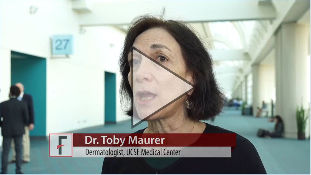
SAN DIEGO – Dermatologists, who served as crucial sentinels during the early years of the AIDS epidemic, should be alert for dermatologic signs and symptoms of HIV infection, according to Toby Maurer, MD, professor of clinical dermatology at the University of California, San Francisco.
“We’re now seeing a lot of HIV-infected patients presenting once again with skin symptoms,” including new-onset psoriasis, poorly controlled seborrheic dermatitis, and even Kaposi’s sarcoma, she said in a video interview at the annual meeting of the American Academy of Dermatology.
The upswing in cases of Kaposi’s sarcoma “comes as a shock to many dermatologists; they thought Kaposi’s sarcoma was a thing of the past,” added Dr. Maurer, who presented on HIV-associated skin conditions at the meeting.
“My whole plea is to remember that HIV has not gone away, that it keeps showing up, and that the skin symptoms absolutely show up,” she said. “It’s not on the radar as much as it should be.”
In her presentation, Dr. Maurer, who is also chief of dermatology at San Francisco General Hospital, said HIV and HIV medications have a variety of impacts on skin. For example, psoriasis gets worse when patients are off medication and better when they’re on it, she said, while molluscum contagiosum and herpes simplex can actually worsen when patients start HIV drugs. And, she said, a late start of AIDS drugs can worsen eczema.
In the interview, she discussed the impact of starting antiretrovirals late into the infection, when CD4 counts are low, on skin conditions, as well as possible reasons behind the increase in Kaposi’s sarcoma, and interactions between systemic dermatologic medications and some antiretrovirals.
Dr. Maurer reports no relevant disclosures.
REPORTING FROM AAD 18
VIDEO: Considering systemic disease in dermatology patients
SAN DIEGO – Be mindful of what lies below the skin.
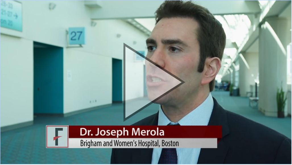
That was the message of Joseph Merola, MD, during a session on “rheumatology for the dermatologist” at the annual meeting of the American Academy of Dermatology.
“The idea is really to start to try to get our dermatology colleagues thinking more systemically and outside of just the skin,” said Dr. Merola, a rheumatologist and dermatologist who is codirector of the center for skin and related musculoskeletal diseases at Brigham and Women’s Hospital, Boston.
“ in up to 30% of patients,” he noted.
He urged his colleagues to ask patients functional questions; for example, those pertaining to sicca symptoms; and how to parse out whether a patient’s joint pain is inflammatory or non-inflammatory.
In a video interview, Dr. Merola also discussed lab tests used to evaluate patients with lupus, the value of a simple urine test, and recent work on the development of the first international classification criteria set for discoid type skin lupus.

Dr. Merola had no relevant disclosures.
SAN DIEGO – Be mindful of what lies below the skin.
That was the message of Joseph Merola, MD, during a session on “rheumatology for the dermatologist” at the annual meeting of the American Academy of Dermatology.
“The idea is really to start to try to get our dermatology colleagues thinking more systemically and outside of just the skin,” said Dr. Merola, a rheumatologist and dermatologist who is codirector of the center for skin and related musculoskeletal diseases at Brigham and Women’s Hospital, Boston.
“ in up to 30% of patients,” he noted.
He urged his colleagues to ask patients functional questions; for example, those pertaining to sicca symptoms; and how to parse out whether a patient’s joint pain is inflammatory or non-inflammatory.
In a video interview, Dr. Merola also discussed lab tests used to evaluate patients with lupus, the value of a simple urine test, and recent work on the development of the first international classification criteria set for discoid type skin lupus.
Dr. Merola had no relevant disclosures.
REPORTING FROM AAD 18
VIDEO: PPACMAN aims to advance the combined rheum-derm clinic approach in the community
SAN DIEGO – A new endeavor that aims to promote the concept of the combined clinic approach to caring for psoriatic patients is now underway.
PPACMAN (Psoriasis and Psoriatic Arthritis Clinics Multicenter Advancement Network) is made up of dermatologists and rheumatologists who play a key role in the management of psoriatic disease and are interested in combined clinics, with the mission “to nucleate psoriatic disease combined clinics and centers to advance a multilevel approach to psoriatic patients, increase disease awareness, and accelerate management,” according to Joseph Merola, MD, codirector of the center for skin and related musculoskeletal diseases at Brigham and Women’s Hospital, Boston.
There are now about 12 centers in North America with formal rheumatology-dermatology clinics for patients with psoriasis and psoriatic arthritis, including the one at Brigham and Women’s, where Dr. Merola and his colleagues have seen the “myriad benefits that come with having a combined clinic,” he said in a video interview at the annual meeting of the American Academy of Dermatology. The idea behind starting PPACMAN was to help form new clinics at academic centers but, also, “to start to catalyze local-regional partnerships in the community so we could get dermatologists and rheumatologists in the community to start interacting, communicating, [and] sharing patients,” he explained.
“The group is really very much focused on this mission of getting combined ... treatment models out there,” added Dr. Merola, president and chair of the board of PPACMAN, which is a 501c3 nonprofit organization.
In the interview, he discusses other benefits of the combined clinic model and other elements of the PPACMAN mission, including education and the potential for shared EMR templates.
The video associated with this article is no longer available on this site. Please view all of our videos on the MDedge YouTube channel
REPORTING FROM AAD 18

Ira Turner, MD
Gut grief
The video associated with this article is no longer available on this site. Please view all of our videos on the MDedge YouTube channel
This video was filmed at Metabolic & Endocrine Disease Summit (MEDS). Click here to learn more.
The video associated with this article is no longer available on this site. Please view all of our videos on the MDedge YouTube channel
This video was filmed at Metabolic & Endocrine Disease Summit (MEDS). Click here to learn more.
The video associated with this article is no longer available on this site. Please view all of our videos on the MDedge YouTube channel
This video was filmed at Metabolic & Endocrine Disease Summit (MEDS). Click here to learn more.
VIDEO: With new therapies available, it’s the ‘decade of eczema,’ researcher says
SAN DIEGO – A long dry spell in the development of new atopic dermatitis (AD) medications came to an end in 2016 with the approval of a topical treatment, and last year brought the first biologic for AD to the market. With more targets and potential treatments being studied, “it’s the decade of eczema,” according to a leading researcher.
And I think we’ll have many more approvals in the next few years,” said Emma Guttman, MD, PhD, said in a video interview at the annual meeting of the American Academy of Dermatology, where she was presenting a talk on the translational revolution in atopic dermatitis.
In the interview, she also discussed research showing that children with AD don’t have the same distribution of lesions as adults, and a study of young children, which found that during an early stage of the disease, when compared with adults, they showed much higher increases in Th17 similar to that seen in psoriasis. It will be interesting to see if “some drugs that work for psoriasis may work in children,” said Dr. Guttman, professor of dermatology and director of the laboratory of inflammatory skin diseases at the Icahn School of Medicine at Mount Sinai, New York.
Dr. Guttman disclosed research support, consulting, or lecture fees from Regeneron, Sanofi, Pfizer, and other companies developing AD treatments.
REPORTING FROM AAD 18
VIDEO: U.S. melanoma incidence hits all-time high
SAN DIEGO –
“Nobody’s quite sure why the [melanoma] rates are still rising so dramatically,” Darrell S. Rigel, MD, said in a video interview during the annual meeting of the American Academy of Dermatology. The increase remains even after adjustment of incidence rates for the increasing mean age of U.S. adults.
The American Cancer Society reported that the estimated annual incidence rate for invasive melanoma will be 91,270 cases for 2018, following what the society called a rapid rise in the rate for the past 30 years. Add to that the estimate of more than 87,000 U.S. cases of in situ melanoma for an overall annual U.S. rate of 178,560, Dr. Rigel said.
Melanoma has a latency of 5-20 years, “so what we’re seeing right now are the effects of what happened 5, 10, or 20 years ago,” said Dr. Rigel, a dermatologist at New York University.
During a talk at the meeting, Dr. Rigel said that based on current incident levels he projected a lifetime U.S. incidence rate of invasive melanoma of one case for every 40 adults by the end of this decade, and a lifetime incidence rate for either invasive or in situ melanoma of one case for every 20 adults by 2020.
A positive trend is that for the first time, the number of melanoma deaths has started to fall, with an estimated 9,320 deaths from melanoma in 2018 according to American Cancer Society statistics, down from a peak of 10,130 melanoma deaths in 2016, Dr. Rigel said.
REPORTING FROM AAD 18

VIDEO: Bioimpedance provides accurate assessment of Mohs surgical margins
SAN DIEGO – In assessing tumor-free margins during Mohs micrographic surgery for skin cancer, of histologic sections, in a single-center, pilot study of bioimpedance in 151 specimens from 50 consecutive patients.
The video associated with this article is no longer available on this site. Please view all of our videos on the MDedge YouTube channel
If the finding of high diagnostic accuracy using bioimpedance spectroscopy is confirmed in larger numbers of patients and specimens run at multiple sites, this approach could “potentially revolutionize what happens with the way Mohs sections are processed in the future” by potentially shaving many minutes off the duration of a standard procedure, Darrell S. Rigel, MD, said in a video interview during the annual meeting of the American Academy of Dermatology.
Usually, it takes 10-20 minutes to process and examine Mohs specimens at each stage of the surgical procedure to determine whether additional excision must remove residual cancer cells, said Dr. Rigel, a dermatologist at New York University. In contrast, assessment for residual cancer cells in the surgical field takes less than a minute using bioimpedance spectroscopy, which relies on differences in electrical conductivity between benign and cancerous cells to identify cancer cells remaining at the surgical margins.
The results of the study were presented in a poster at the meeting, by a research associate of Dr. Rigel’s, Ryan Svoboda, MD, of the National Society for Cutaneous Medicine, New York.
The researchers used a bioimpedance spectroscopy device made by NovaScan to assess 151 histology slides prepared during Mohs micrographic surgery on patients with nonmelanoma skin cancer, and compared the findings against the gold standard of histological slide examination. By this criterion, bioimpedance spectroscopy identified 105 true negatives and 2 false negatives, and 43 true positives and 1 false positive. Calculations showed that this equated to 95.6% sensitivity, 99.1% specificity, a 97.7% positive predictive value, and a 98.1% negative predictive value.
These may be underestimates of the accuracy of bioimpedance spectroscopy because the calculations presume that conventional histology is always correct, but Dr. Rigel noted that sometimes the histological diagnosis is wrong.
SOURCE: Svoboda R et al. Poster 7304.
REPORTING FROM AAD 18
Key clinical point: Bioimpedance spectroscopy showed excellent diagnostic accuracy for cancer cells on Mohs surgical margins.
Major finding: Bioimpedance spectroscopy had a sensitivity of 95.6% and specificity of 99.1% compared with Mohs histology.
Study details: A single-center pilot study with 151 Mohs surgical specimens taken from 50 patients.
Disclosures: The study was funded by NovaScan, the company developing the device tested in the study. Dr. Rigel has been a consultant to NovaScan and to Castle Biosciences, DermTech, Ferndale, Myriad, and Neutrogena, and has received research support from Castle and Neutrogena.
Source: Svoboda R et al. Poster 7304.
VIDEO: SPF 100 sunscreen outperformed SPF 50 in Vail study
SAN DIEGO – a finding that might interest consumers and prompt the Food and Drug Administration to continue to allow sunscreens to have labels listing sun protection factors greater than 50.*
“Our study results show pretty definitively that SPF 100 did significantly better than SPF 50 in a real world environment,” Darrell S. Rigel, MD, said at the annual meeting of the American Academy of Dermatology.
Dr. Rigel cited data that he and his associates recently published from 199 adults skiing on a sunny March day in Colorado. Participants applied a blinded sunscreen rated at SPF 50 to one side of their face all day and an SPF 100 sunscreen to the other side all day, and the researchers then ran a blinded assessment of images taken of each side at the end of the day. The sunburn on the SPF 50 side exceeded the other side in 55% of skiers, the two sides matched in 40%, and in 5% the sunburn was worse on the SPF 100 side (J Am Acad Dermatol. 2017 Dec 29. doi: 10.1016/j.jaad.2017.12.062).
“The SPF 50 side of the face was 11 times more likely to be sunburned than the SPF 100 side,” and for all the secondary endpoints and different ways of analyzing the data, the SPF 50 was not as effective as SPF 100, Dr. Rigel said in a video interview. Erythema appeared on 41% of the SPF 50–treated sides of participants faces, compared with 14% of the sides treated with SPF 100 sunscreen.
The results followed-up on a report from Dr. Rigel and his associates from 8 years ago that ran a similar comparison of two sunscreen potencies, SPF 85 and SPF 50, in 56 skiers, with similar results showing greater sunburn protection from the higher SPF sunscreen (J Am Acad Dermatol. 2010 Feb;62[2]:348-9). In 2011, the FDA proposed a new rule for SPF labeling that would cap the maximum SPF potency possible of 50, which created a label 50+ to designate unspecified SPF above 50. According to Dr. Rigel, the FDA rejected his 2010 study as documentation of incremental benefit above SPF 50 because of several flaws the agency found with that study, including not tracking sunscreen use by weight. He specifically designed the new, 199-subject study to address that and the FDA’s other concerns.
He and his associates decided to do the study because the FDA said in the monograph that, if the concerns were met, “they would accept the study as definitive,” said Dr. Rigel, a dermatologist at New York University.
The greater protection from SPF 100 sunscreen probably occurs because it’s “more forgiving” when used with inadequate application, he suggested. Allowing labeling that specifies SPF levels greater than 50 would help consumers pick sunscreen formulations that give greater protection, and it would encourage manufacturers to market sunscreens with higher SPF levels.
Dr. Rigel has been a consultant to Castle Biosciences, DermTech, Ferndale, Myriad, Neutrogena, and Novascan and has received research support from Castle and Neutrogena.
Correction, 2/22/18: Due to an editing error, an earlier version of this article implied incorrectly that sunscreen labels listing SPFs over 50 had been banned .
EXPERT ANALYSIS FROM AAD 18

VIDEO: Select atopic dermatitis patients need patch testing
SAN DIEGO – Patch testing may be in order for some patients with atopic dermatitis, according to Jonathan Silverberg, MD, PhD, of the department of dermatology, Northwestern University, Chicago.
Allergic contact dermatitis is a common comorbid condition in people with AD “and sometimes, can flare up the severity of the disease,” he said in a video interview at the American Academy of Dermatology annual meeting.
Patch testing can ferret out a trigger in atopic dermatitis patients with atypical disease distribution or refractory disease, and help avoid the need for systemic therapy, Dr. Silverman pointed out.
In the interview, he discussed these and other clinical scenarios, as well as how patch testing differs in these patients and what screening series to consider using.
Dr. Silverberg had no relevant financial disclosures.
SOURCE: Silverberg, J. et al, Session 061.
REPORTING FROM AAD 18