User login
Randomized trial finds DMARD therapy for RA has a beneficial effect on vascular inflammation, CV risk
Use of a tumor necrosis factor inhibitor (TNFi) or triple therapy with conventional, synthetic disease-modifying antirheumatic drugs (DMARDs) for rheumatoid arthritis have similar beneficial effects in reducing patients’ vascular inflammation and cardiovascular (CV) risk, according to results from a randomized, active comparator trial.
“The good news is, providers can rest assured that aggressive treatment for RA does reduce vascular inflammation and therefore cardiovascular risk,” lead author Daniel H. Solomon, MD, MPH, of Brigham and Women’s Hospital in Boston, told this news organization. “Part of the reason that treating people with potent disease-modifying agents is important is not only because of reductions in pain and improvements in function on the level of arthritis, but also because of the vascular impact.”

The small study, published in Annals of the Rheumatic Diseases, randomly assigned 115 patients with active RA despite methotrexate use to one of two treatment protocols for 24 weeks: addition of a TNFi or triple therapy with the addition of sulfasalazine and hydroxychloroquine. Participants had 18F-fluorodeoxyglucose (FDG)–PET/CT scans at baseline and 24 weeks to assess change in arterial inflammation, measured as an arterial target-to-background ratio (TBR) in the carotid arteries and aorta. The study achieved its outcomes despite a low 56.5% rate of adherence to 80% or more of randomized treatments.
Dr. Solomon said this is the first randomized trial comparing the effects of DMARDs on vascular inflammation in RA. The researchers hypothesized that TNFi would be superior to triple therapy for reducing vascular inflammation. “We found that they both reduced vascular inflammation on PET scanning to the same degree,” Dr. Solomon said.
Study results
In the TNFi group, the mean of the maximum of the TBR in the most diseased segment (MDS) of the index vessel declined from 2.72 to 2.47 for a delta of –0.24. In the triple-therapy patients, MDS declined from 2.62 to 2.43 for a delta of –0.19 (difference in deltas –0.02; 95% confidence interval, –0.19 to 0.15; P = .79).
Dr. Solomon explained the choice of FDG-PET/CT scanning to evaluate vascular inflammation in the study participants. “We know that FDG-PET/CT scanning correlates with CV risk, and we know that treatments like statins that impact CV risk reduce the inflammation as observed on FDG-PET/CT,” he said.
Although the study found no difference between the TNFi and triple therapy in terms of vascular outcomes, the conclusion is “a bit more nuanced,” Dr. Solomon said. “It tells us first that reducing inflammation with different strategies in rheumatoid arthritis can similarly impact vascular inflammation. That’s great news. These are aggressive treatment strategies, so if you can reduce vascular inflammation in a significant manner, that should result in reduced cardiovascular risk over time.”
Although the choice of TNFi or triple therapy may not matter for reducing CV risk, Dr. Solomon said, “It matters that you choose something that’s aggressive and that you use it in people who have active disease. That’s another part of the story: People who have active disease have worse vascular inflammation, which translates into a reduction in cardiovascular risk – but it’s not differentially reduced.”
Underlying mechanisms of CVD in RA
Commenting on the research for this news organization, Lihi Eder, MD, PhD, codirector of the cardio-rheumatology program at Women’s College Hospital in Toronto, said the study findings build on what’s known about some of the underlying mechanisms of cardiovascular diseases in RA and how to optimize treatments to reduce the risk.

“Importantly,” she said, “none of these treatment strategies was superior, suggesting that both treatment options are acceptable when considering cardiovascular risk reduction, in addition to controlling RA activity.”
The strengths of the study are its randomized, controlled design “conducted by a strong team of investigators,” and that it addressed questions relevant to routine practice, said Dr. Eder, who was not involved with the study.
The study’s use of FDG-PET/CT as a surrogate outcome is a limitation, she noted. “Although it would have been very challenging to perform a similar study that will include clinical events as a study outcome.” Another limitation, she said, was the low adherence rate to randomized treatments.
“Additional studies that will compare other modes of action [for example, interleukin-6 inhibitors, Janus kinase inhibitors, anti-CD20 monoclonal antibodies] could broaden our understanding regarding the inflammatory pathways driving CV risk in RA,” Dr. Eder added.
The study received funding from the National Institute of Arthritis and Musculoskeletal and Skin Diseases. AbbVie and Amgen supplied drugs used in the study. Dr. Solomon disclosed receiving research support from AbbVie, Amgen, CorEvitas, and Moderna, and royalties from UpToDate. Dr. Eder reports no relevant financial relationships.
A version of this article first appeared on Medscape.com.
Use of a tumor necrosis factor inhibitor (TNFi) or triple therapy with conventional, synthetic disease-modifying antirheumatic drugs (DMARDs) for rheumatoid arthritis have similar beneficial effects in reducing patients’ vascular inflammation and cardiovascular (CV) risk, according to results from a randomized, active comparator trial.
“The good news is, providers can rest assured that aggressive treatment for RA does reduce vascular inflammation and therefore cardiovascular risk,” lead author Daniel H. Solomon, MD, MPH, of Brigham and Women’s Hospital in Boston, told this news organization. “Part of the reason that treating people with potent disease-modifying agents is important is not only because of reductions in pain and improvements in function on the level of arthritis, but also because of the vascular impact.”
The small study, published in Annals of the Rheumatic Diseases, randomly assigned 115 patients with active RA despite methotrexate use to one of two treatment protocols for 24 weeks: addition of a TNFi or triple therapy with the addition of sulfasalazine and hydroxychloroquine. Participants had 18F-fluorodeoxyglucose (FDG)–PET/CT scans at baseline and 24 weeks to assess change in arterial inflammation, measured as an arterial target-to-background ratio (TBR) in the carotid arteries and aorta. The study achieved its outcomes despite a low 56.5% rate of adherence to 80% or more of randomized treatments.
Dr. Solomon said this is the first randomized trial comparing the effects of DMARDs on vascular inflammation in RA. The researchers hypothesized that TNFi would be superior to triple therapy for reducing vascular inflammation. “We found that they both reduced vascular inflammation on PET scanning to the same degree,” Dr. Solomon said.
Study results
In the TNFi group, the mean of the maximum of the TBR in the most diseased segment (MDS) of the index vessel declined from 2.72 to 2.47 for a delta of –0.24. In the triple-therapy patients, MDS declined from 2.62 to 2.43 for a delta of –0.19 (difference in deltas –0.02; 95% confidence interval, –0.19 to 0.15; P = .79).
Dr. Solomon explained the choice of FDG-PET/CT scanning to evaluate vascular inflammation in the study participants. “We know that FDG-PET/CT scanning correlates with CV risk, and we know that treatments like statins that impact CV risk reduce the inflammation as observed on FDG-PET/CT,” he said.
Although the study found no difference between the TNFi and triple therapy in terms of vascular outcomes, the conclusion is “a bit more nuanced,” Dr. Solomon said. “It tells us first that reducing inflammation with different strategies in rheumatoid arthritis can similarly impact vascular inflammation. That’s great news. These are aggressive treatment strategies, so if you can reduce vascular inflammation in a significant manner, that should result in reduced cardiovascular risk over time.”
Although the choice of TNFi or triple therapy may not matter for reducing CV risk, Dr. Solomon said, “It matters that you choose something that’s aggressive and that you use it in people who have active disease. That’s another part of the story: People who have active disease have worse vascular inflammation, which translates into a reduction in cardiovascular risk – but it’s not differentially reduced.”
Underlying mechanisms of CVD in RA
Commenting on the research for this news organization, Lihi Eder, MD, PhD, codirector of the cardio-rheumatology program at Women’s College Hospital in Toronto, said the study findings build on what’s known about some of the underlying mechanisms of cardiovascular diseases in RA and how to optimize treatments to reduce the risk.
“Importantly,” she said, “none of these treatment strategies was superior, suggesting that both treatment options are acceptable when considering cardiovascular risk reduction, in addition to controlling RA activity.”
The strengths of the study are its randomized, controlled design “conducted by a strong team of investigators,” and that it addressed questions relevant to routine practice, said Dr. Eder, who was not involved with the study.
The study’s use of FDG-PET/CT as a surrogate outcome is a limitation, she noted. “Although it would have been very challenging to perform a similar study that will include clinical events as a study outcome.” Another limitation, she said, was the low adherence rate to randomized treatments.
“Additional studies that will compare other modes of action [for example, interleukin-6 inhibitors, Janus kinase inhibitors, anti-CD20 monoclonal antibodies] could broaden our understanding regarding the inflammatory pathways driving CV risk in RA,” Dr. Eder added.
The study received funding from the National Institute of Arthritis and Musculoskeletal and Skin Diseases. AbbVie and Amgen supplied drugs used in the study. Dr. Solomon disclosed receiving research support from AbbVie, Amgen, CorEvitas, and Moderna, and royalties from UpToDate. Dr. Eder reports no relevant financial relationships.
A version of this article first appeared on Medscape.com.
FROM ANNALS OF THE RHEUMATIC DISEASES
Study implicates myelin plasticity in absence seizures
NASHVILLE, TENN. – that seems to provoke dysregulation of the insulating layer surrounding nerve fibers, perpetuating a cycle of increasing nerve damage and more frequent seizures later on.
“This study was the first to demonstrate that, at least in some forms of epilepsy, myelin plasticity is part of the maladaptive plasticity response that underlines epilepsy progression,” Juliet Knowles, MD, PhD, assistant professor at Stanford (Calif.) University, said in an interview. She reported the findings at the 2022 annual meeting of the American Epilepsy Society.

Dr. Knowles and colleagues made their discovery using laboratory mice. They used an imaging technique known as qMTI – quantitative magnetization transfer in conjunction with diffusion MRI – to map changes in myelin sheath thickness, or myelin plasticity, in major white matter tracks of the brain.
“Over the last decade we’ve come to understand that myelin, which is the insulating substance that coats the projections of brain cells or neurons, is more dynamic than we used to think,” she said. “In fact, throughout life, myelin’s structure in some regions of the brain can be changed in response to neuro activity. It’s a newly appreciated form of brain plasticity.”
However, she said, myelin plasticity has mostly been studied in healthy brains; “We don’t know very much about what role myelin plasticity might play in disease states like epilepsy,” Dr. Knowles said. The study’s goal was to investigate myelin plasticity specifically in absence seizures.
“We hypothesized that maybe absence seizures prompt activity-dependent myelin plasticity, but that maybe seizure-induced myelin plasticity alters the way that brain networks act in a way that contributes to the disease process,” she said.
Maladaptive myelin plasticity
The researchers found that absence seizures were infrequent when they first started, but then they rapidly progressed. “Over a couple of weeks, they’ll go from having very few seizures to having many seizures per hour,” Dr. Knowles said.
Using qMTI, the researchers found increased myelin sheath thickness across the longitudinal extent of the anterior corpus callosum, but they found myelin sheath thickness unchanged in brain regions where absence seizures weren’t prominent.
They also found that genetically blocking activity-dependent myelination markedly decreased seizure progression and decreased ictal somatosensory electroencephalography (EEG) coherence. Conversely, blocking myelin plasticity had no effect on ictal EEG coherence between visual cortices connected by the posterior corpus callosum.
The next step for the researchers is to develop MRI methods to use in human studies, Dr. Knowles said.
“We are working on developing an imaging approach in these same animal models that we hope we can use also to study in a detailed way white matter plasticity in humans with epilepsy and we’re also continuing our studies in animal models to try to identify ways to target maladaptive myelin plasticity, which ultimately we hope will inform treatment of people with epilepsy,” Dr. Knowles said.
Of mice and men
Although this study used mice, Chris Dulla, PhD, associate professor and director of the neuroscience graduate program at Tufts University in Boston, said the finding is “probably pretty transferable” to humans.

“This is the first study that really showed it,” he said of the link between myelin changes and seizure frequency. “I think people have suspected it, but that’s why this is kind of a big deal because this is one of the first studies to show it conclusively.”
He offered suggestions for validating the findings in humans. “The first thing would be to do imaging studies in people where you can examine to see if those white matter tracks are altered in a similar way in people with epilepsy,” he said. “I think now this study gives us good reason to undertake the work that it would take to ask that question and answer it in the human brain.”
Dr. Knowles and Dr. Dulla have no relevant relationships to disclose.
NASHVILLE, TENN. – that seems to provoke dysregulation of the insulating layer surrounding nerve fibers, perpetuating a cycle of increasing nerve damage and more frequent seizures later on.
“This study was the first to demonstrate that, at least in some forms of epilepsy, myelin plasticity is part of the maladaptive plasticity response that underlines epilepsy progression,” Juliet Knowles, MD, PhD, assistant professor at Stanford (Calif.) University, said in an interview. She reported the findings at the 2022 annual meeting of the American Epilepsy Society.
Dr. Knowles and colleagues made their discovery using laboratory mice. They used an imaging technique known as qMTI – quantitative magnetization transfer in conjunction with diffusion MRI – to map changes in myelin sheath thickness, or myelin plasticity, in major white matter tracks of the brain.
“Over the last decade we’ve come to understand that myelin, which is the insulating substance that coats the projections of brain cells or neurons, is more dynamic than we used to think,” she said. “In fact, throughout life, myelin’s structure in some regions of the brain can be changed in response to neuro activity. It’s a newly appreciated form of brain plasticity.”
However, she said, myelin plasticity has mostly been studied in healthy brains; “We don’t know very much about what role myelin plasticity might play in disease states like epilepsy,” Dr. Knowles said. The study’s goal was to investigate myelin plasticity specifically in absence seizures.
“We hypothesized that maybe absence seizures prompt activity-dependent myelin plasticity, but that maybe seizure-induced myelin plasticity alters the way that brain networks act in a way that contributes to the disease process,” she said.
Maladaptive myelin plasticity
The researchers found that absence seizures were infrequent when they first started, but then they rapidly progressed. “Over a couple of weeks, they’ll go from having very few seizures to having many seizures per hour,” Dr. Knowles said.
Using qMTI, the researchers found increased myelin sheath thickness across the longitudinal extent of the anterior corpus callosum, but they found myelin sheath thickness unchanged in brain regions where absence seizures weren’t prominent.
They also found that genetically blocking activity-dependent myelination markedly decreased seizure progression and decreased ictal somatosensory electroencephalography (EEG) coherence. Conversely, blocking myelin plasticity had no effect on ictal EEG coherence between visual cortices connected by the posterior corpus callosum.
The next step for the researchers is to develop MRI methods to use in human studies, Dr. Knowles said.
“We are working on developing an imaging approach in these same animal models that we hope we can use also to study in a detailed way white matter plasticity in humans with epilepsy and we’re also continuing our studies in animal models to try to identify ways to target maladaptive myelin plasticity, which ultimately we hope will inform treatment of people with epilepsy,” Dr. Knowles said.
Of mice and men
Although this study used mice, Chris Dulla, PhD, associate professor and director of the neuroscience graduate program at Tufts University in Boston, said the finding is “probably pretty transferable” to humans.
“This is the first study that really showed it,” he said of the link between myelin changes and seizure frequency. “I think people have suspected it, but that’s why this is kind of a big deal because this is one of the first studies to show it conclusively.”
He offered suggestions for validating the findings in humans. “The first thing would be to do imaging studies in people where you can examine to see if those white matter tracks are altered in a similar way in people with epilepsy,” he said. “I think now this study gives us good reason to undertake the work that it would take to ask that question and answer it in the human brain.”
Dr. Knowles and Dr. Dulla have no relevant relationships to disclose.
AT AES 2022

Newer brand-name drugs fuel spending on antiseizure medications
NASHVILLE, TENN. – , pointing to a major shift to newer, costlier, brand-name drugs – a trend in spending that may not be sustainable, the lead author of a study of drug costs said.
The study, presented at the 2022 annual meeting of the American Epilepsy Society, evaluated claims data for prescriptions for common antiseizure medications in the Medicare Part D and Medicaid databases from 2012 to 2020. The study excluded gabapentin and pregabalin because they’re frequently prescribed for other indications in addition to epileptic seizures.

“We found that third-generation medications, even though they accounted for the smallest percentage of claims in 2020, took up the most astronomical portion of the money that was spent,” lead author Deepti Zutshi, MD, an associate professor of neurology at Wayne State University in Detroit, said in an interview.
The study found that Medicare Part D spending on antiseizure medications increased from $1.16 billion in 2012 to $2.68 billion in 2020. In Medicaid, spending followed a similar trend, increasing from $973 million in 2012 to $1.05 billion in 2020.
Analyzing Medicare/Medicaid claims data
The study categorized drugs two ways: by brand or generic; and by first, second, or third generation, Dr. Zutshi said. First-generation drugs include medications such as phenobarbital, phenytoin, valproate, and carbamazepine. Second-generation medications were released in the early 2000s and include medications such as lamotrigine and levetiracetam. Examples of third-generation drugs include lacosamide, vigabatrin, clobazam, and perampanel.
Prescribers shifted significantly to third-generation treatments, Dr. Zutshi said. In Medicare Part D, the total spent on third-generation antiseizure medications went from $124 million in 2012 to $1.08 billion in 2020, representing a quadrupling in percentage of costs, from 10.7% to 40.4%. The total number of claims for third-generation antiseizure medications was 240,000 in 2012 (1.3%) and 1.1 million in 2020 (4.4%).
When looking at brand versus generic, the total spent on brand-name antiseizure medications increased nearly threefold from $546 million in 2012 to $1.62 million in 2020, with the share of all funding spent on brand-name antiseizure medications jumping from 46.8% to 60.2%. However, the proportion of total claims for branded antiseizure medications actually dropped, from 9.24% in 2012 to 6.62% in 2020.
Medicaid trends followed a similar pattern. Third-generation antiseizure medications accounted for 1.7% of total claims in 2012 and 6% in 2020. Spending on third-generation antiseizure medications grew nearly eight times: from $147 million, or 15.1% of funding spent on antiseizure medications, in 2012 to $1.15 billion in 2020, a 56.1% share of costs. The total spend of branded antiseizure medications in Medicaid was $605 million in 2012 and $1.46 billion in 2020 – a jump in the share of total spending from 62.2% to 71.3%. As in Medicare Part D, the percentage of total claims for branded antiseizure medications in Medicaid also dropped from 2012 to 2020, from 12.1% to 6.8%.
Why the substantial increase in spending?
“The reason we are prescribing these more expensive medications may be that the third-generation medications have better side-effect profiles, improved safety and outcomes in pregnancy, or that they have less drug interactions with other medications,” Dr. Zutshi said.
That’s desirable for older patients on Medicare who are more likely to have comorbidities and be on other medications, or women of child-bearing age on Medicaid, Dr. Zutshi said. “But I don’t think people realize what the cost is to Medicare and Medicaid,” she said, “so this was a bit of a shocking finding in our paper when we looked at this. I wasn’t expecting to see the substantial increase of spending focusing on just a few medications.”
Neurologists and other providers have to be more aware of individual patients’ needs as well as cost when prescribing branded or third-generation antiseizure medications, Dr. Zutshi said. “We have to do what’s best for all of our patients, but it has to be sustainable. If not, we could start losing the ability to prescribe these medications in these vulnerable population groups, so we have to use them judiciously,” Dr. Zutshi said.
Controlling costs versus managing seizures
Timothy E. Welty, PharmD, a professor of pharmacy at Drake University in Des Moines, Iowa, noted some potential issues with the study’s methodology, namely that, while it excluded gabapentin and pregabalin, it did include other antiseizure medications that are used for other indications without accounting for them. Additionally, the pharmacy claims data the study used didn’t cross match with any diagnostic data.

Controlling drug costs is noteworthy, he said, but managing seizures is equally important. “You have to think not only in terms of preventing seizures and what impact that has on health care costs specifically, but what impact that has on overall costs to society,” Dr. Welty said. “Doing the best we can to get their seizures under control as quickly as possible has great benefits for the patient outside of health care costs.”
He added, “We just really need to educate pharmacists and decision makers within third-party payers, be it Medicare, Medicaid, private insurance, whatever, on the advances that are being made in the use of seizure medications to treat epilepsy and stop seizures, but it’s a far broader issue than just how many dollars are we spending on seizure medication.”
Dr. Zutshi and Dr. Welty have no relevant disclosures to report.
NASHVILLE, TENN. – , pointing to a major shift to newer, costlier, brand-name drugs – a trend in spending that may not be sustainable, the lead author of a study of drug costs said.
The study, presented at the 2022 annual meeting of the American Epilepsy Society, evaluated claims data for prescriptions for common antiseizure medications in the Medicare Part D and Medicaid databases from 2012 to 2020. The study excluded gabapentin and pregabalin because they’re frequently prescribed for other indications in addition to epileptic seizures.
“We found that third-generation medications, even though they accounted for the smallest percentage of claims in 2020, took up the most astronomical portion of the money that was spent,” lead author Deepti Zutshi, MD, an associate professor of neurology at Wayne State University in Detroit, said in an interview.
The study found that Medicare Part D spending on antiseizure medications increased from $1.16 billion in 2012 to $2.68 billion in 2020. In Medicaid, spending followed a similar trend, increasing from $973 million in 2012 to $1.05 billion in 2020.
Analyzing Medicare/Medicaid claims data
The study categorized drugs two ways: by brand or generic; and by first, second, or third generation, Dr. Zutshi said. First-generation drugs include medications such as phenobarbital, phenytoin, valproate, and carbamazepine. Second-generation medications were released in the early 2000s and include medications such as lamotrigine and levetiracetam. Examples of third-generation drugs include lacosamide, vigabatrin, clobazam, and perampanel.
Prescribers shifted significantly to third-generation treatments, Dr. Zutshi said. In Medicare Part D, the total spent on third-generation antiseizure medications went from $124 million in 2012 to $1.08 billion in 2020, representing a quadrupling in percentage of costs, from 10.7% to 40.4%. The total number of claims for third-generation antiseizure medications was 240,000 in 2012 (1.3%) and 1.1 million in 2020 (4.4%).
When looking at brand versus generic, the total spent on brand-name antiseizure medications increased nearly threefold from $546 million in 2012 to $1.62 million in 2020, with the share of all funding spent on brand-name antiseizure medications jumping from 46.8% to 60.2%. However, the proportion of total claims for branded antiseizure medications actually dropped, from 9.24% in 2012 to 6.62% in 2020.
Medicaid trends followed a similar pattern. Third-generation antiseizure medications accounted for 1.7% of total claims in 2012 and 6% in 2020. Spending on third-generation antiseizure medications grew nearly eight times: from $147 million, or 15.1% of funding spent on antiseizure medications, in 2012 to $1.15 billion in 2020, a 56.1% share of costs. The total spend of branded antiseizure medications in Medicaid was $605 million in 2012 and $1.46 billion in 2020 – a jump in the share of total spending from 62.2% to 71.3%. As in Medicare Part D, the percentage of total claims for branded antiseizure medications in Medicaid also dropped from 2012 to 2020, from 12.1% to 6.8%.
Why the substantial increase in spending?
“The reason we are prescribing these more expensive medications may be that the third-generation medications have better side-effect profiles, improved safety and outcomes in pregnancy, or that they have less drug interactions with other medications,” Dr. Zutshi said.
That’s desirable for older patients on Medicare who are more likely to have comorbidities and be on other medications, or women of child-bearing age on Medicaid, Dr. Zutshi said. “But I don’t think people realize what the cost is to Medicare and Medicaid,” she said, “so this was a bit of a shocking finding in our paper when we looked at this. I wasn’t expecting to see the substantial increase of spending focusing on just a few medications.”
Neurologists and other providers have to be more aware of individual patients’ needs as well as cost when prescribing branded or third-generation antiseizure medications, Dr. Zutshi said. “We have to do what’s best for all of our patients, but it has to be sustainable. If not, we could start losing the ability to prescribe these medications in these vulnerable population groups, so we have to use them judiciously,” Dr. Zutshi said.
Controlling costs versus managing seizures
Timothy E. Welty, PharmD, a professor of pharmacy at Drake University in Des Moines, Iowa, noted some potential issues with the study’s methodology, namely that, while it excluded gabapentin and pregabalin, it did include other antiseizure medications that are used for other indications without accounting for them. Additionally, the pharmacy claims data the study used didn’t cross match with any diagnostic data.
Controlling drug costs is noteworthy, he said, but managing seizures is equally important. “You have to think not only in terms of preventing seizures and what impact that has on health care costs specifically, but what impact that has on overall costs to society,” Dr. Welty said. “Doing the best we can to get their seizures under control as quickly as possible has great benefits for the patient outside of health care costs.”
He added, “We just really need to educate pharmacists and decision makers within third-party payers, be it Medicare, Medicaid, private insurance, whatever, on the advances that are being made in the use of seizure medications to treat epilepsy and stop seizures, but it’s a far broader issue than just how many dollars are we spending on seizure medication.”
Dr. Zutshi and Dr. Welty have no relevant disclosures to report.
AT AES 2022
Novel PCI screening approach detects diffuse CAD
A novel approach for stratifying patients into one of two phenotypes for coronary artery disease (CAD) helped differentiate those who would benefit from percutaneous coronary intervention (PCI) from those who wouldn’t, researchers in Belgium reported in a subanalysis of a single-center, randomized clinical trial.
“What this study adds is that we are actually creating a refined definition of the appropriateness criteria for PCI,” lead study author Carlos Collet, MD, PhD, of the Cardiovascular Center at OLV Hospital in Aalst, Belgium, said in an interview. “We have been too long implanting stents in diffuse disease that actually have no benefit for the patient.”
The study found that patients with diffuse CAD were almost twice as likely to have residual angina 3 months after PCI than patients with focal CAD, with respective rates of 51.9% vs. 27.5% after PCI (P = .02).
The researchers analyzed 103 patients from the TARGET-FFR (Trial of Angiography vs. pressure-Ratio-Guided Enhancement Techniques–Fractional Flow Reserve) conducted at the Golden Jubilee National Hospital in Glasgow. Study patients completed the 7-item Seattle Angina Questionnaire at baseline and at 3 months after PCI, which provided the researchers information on outcomes.
The study, published in JACC: Cardiovascular Interventions, used median pullback pressure gradient (PPG) to define focal and diffuse CAD. The operators used the PressureWire X Guidewire (Abbott Vascular) to measure fractional flow reserve (FFR).
The procedure involved administering a 200-mcg bolus of intracoronary nitrate and then positioning the pressure wire sensor at the tip of the guide catheter equalized with aortic pressure. The pressure wire was then advanced to the position sensor in the distal third of the vessel. After hyperemia was induced, coronary flow reserve was assessed using bolus thermodilution. Manual FFR pullback maneuvers were done at a constant speed for 20-30 seconds. The PPG index was calculated post hoc from the manual FFR pullback recordings obtained pre-PCI.
In this study, patients with low PPG needed longer (48 mm vs. 37 mm; P = .015) and more (1.5 vs. 1.0; P = .036) stents during PCI, Dr. Collet and colleagues reported. They concluded that patients with low PPG can be treated with medical therapy.
“The beauty of the PPG is that everything happens before you implant the stent,” Dr. Collet said. “We’re starting to understand that we cannot treat diffuse disease with a focal disease therapy.”
The challenge with differentiating diffuse from focal CAD has been that it relies on visual assessment. “It’s subject to operator variability, and that’s the reason why there are no trials with focal or diffuse disease specifically because, until now, we didn’t have any metric that quantified the diffuseness or the focality of the disease,” Dr. Collet said.
The PPG itself isn’t novel, Dr. Collet said. “The novelty is that for first time we can quantify in a reproducible way the information from the pullback,” he added.

“What this study tells us is that once you have a patient with diffuse coronary artery disease, don’t try PCI because it will not help half of them,” Patrick W. Serruys, MD, PhD, a cardiologist at the National University of Ireland, Galway, and author of the accompanying editorial, said in an interview.
He noted that one limitation of the study was that Dr. Collet and colleagues used mechanical PPG to measure the pressure gradient. “We use now a surrogate, which is angiography,” Dr. Serruys said. “It’s not exactly the same as a measurement of pressure with the pressure wire, but we know from many, many studies that it’s quite a good surrogate.” Future research should focus on use of angiography without the pressure wire to evaluate the pressure gradient.
The ongoing PPG Global registry will aim to further validate findings from the subanalysis, Dr. Collet said, and the PPG Primetime study will evaluate deferring PCI in patients with low PPG.
Dr. Collet disclosed relationships with Biosensor, Coroventis Research, Medis Medical Imaging, Pie Medical Imaging, CathWorks, Boston Scientific, Siemens, HeartFlow, OpSens, Abbott Vascular and Philips Volcano. Dr. Serruys disclosed relationships with Sinomedical Sciences Technology, Sahajanand Medical Technological, Philips Volcano, Xeltis and HeartFlow.
A novel approach for stratifying patients into one of two phenotypes for coronary artery disease (CAD) helped differentiate those who would benefit from percutaneous coronary intervention (PCI) from those who wouldn’t, researchers in Belgium reported in a subanalysis of a single-center, randomized clinical trial.
“What this study adds is that we are actually creating a refined definition of the appropriateness criteria for PCI,” lead study author Carlos Collet, MD, PhD, of the Cardiovascular Center at OLV Hospital in Aalst, Belgium, said in an interview. “We have been too long implanting stents in diffuse disease that actually have no benefit for the patient.”
The study found that patients with diffuse CAD were almost twice as likely to have residual angina 3 months after PCI than patients with focal CAD, with respective rates of 51.9% vs. 27.5% after PCI (P = .02).
The researchers analyzed 103 patients from the TARGET-FFR (Trial of Angiography vs. pressure-Ratio-Guided Enhancement Techniques–Fractional Flow Reserve) conducted at the Golden Jubilee National Hospital in Glasgow. Study patients completed the 7-item Seattle Angina Questionnaire at baseline and at 3 months after PCI, which provided the researchers information on outcomes.
The study, published in JACC: Cardiovascular Interventions, used median pullback pressure gradient (PPG) to define focal and diffuse CAD. The operators used the PressureWire X Guidewire (Abbott Vascular) to measure fractional flow reserve (FFR).
The procedure involved administering a 200-mcg bolus of intracoronary nitrate and then positioning the pressure wire sensor at the tip of the guide catheter equalized with aortic pressure. The pressure wire was then advanced to the position sensor in the distal third of the vessel. After hyperemia was induced, coronary flow reserve was assessed using bolus thermodilution. Manual FFR pullback maneuvers were done at a constant speed for 20-30 seconds. The PPG index was calculated post hoc from the manual FFR pullback recordings obtained pre-PCI.
In this study, patients with low PPG needed longer (48 mm vs. 37 mm; P = .015) and more (1.5 vs. 1.0; P = .036) stents during PCI, Dr. Collet and colleagues reported. They concluded that patients with low PPG can be treated with medical therapy.
“The beauty of the PPG is that everything happens before you implant the stent,” Dr. Collet said. “We’re starting to understand that we cannot treat diffuse disease with a focal disease therapy.”
The challenge with differentiating diffuse from focal CAD has been that it relies on visual assessment. “It’s subject to operator variability, and that’s the reason why there are no trials with focal or diffuse disease specifically because, until now, we didn’t have any metric that quantified the diffuseness or the focality of the disease,” Dr. Collet said.
The PPG itself isn’t novel, Dr. Collet said. “The novelty is that for first time we can quantify in a reproducible way the information from the pullback,” he added.
“What this study tells us is that once you have a patient with diffuse coronary artery disease, don’t try PCI because it will not help half of them,” Patrick W. Serruys, MD, PhD, a cardiologist at the National University of Ireland, Galway, and author of the accompanying editorial, said in an interview.
He noted that one limitation of the study was that Dr. Collet and colleagues used mechanical PPG to measure the pressure gradient. “We use now a surrogate, which is angiography,” Dr. Serruys said. “It’s not exactly the same as a measurement of pressure with the pressure wire, but we know from many, many studies that it’s quite a good surrogate.” Future research should focus on use of angiography without the pressure wire to evaluate the pressure gradient.
The ongoing PPG Global registry will aim to further validate findings from the subanalysis, Dr. Collet said, and the PPG Primetime study will evaluate deferring PCI in patients with low PPG.
Dr. Collet disclosed relationships with Biosensor, Coroventis Research, Medis Medical Imaging, Pie Medical Imaging, CathWorks, Boston Scientific, Siemens, HeartFlow, OpSens, Abbott Vascular and Philips Volcano. Dr. Serruys disclosed relationships with Sinomedical Sciences Technology, Sahajanand Medical Technological, Philips Volcano, Xeltis and HeartFlow.
FROM JACC: CARDIOVASCULAR INTERVENTIONS
HDL cholesterol not linked to CHD risk in Blacks: REGARDS
High-density lipoprotein cholesterol may not be as effective a biomarker of cardiovascular disease risk as once thought, particularly in Black adults, according to results from a large biracial cohort study that also raised questions about the validity of high HDL cholesterol as a potentially protective factor in White and Black adults alike.
“I think this opens the door to suggest that every biomarker we use might have a race-specific association with disease outcome,” Nathalie Pamir, PhD, an associate professor at Oregon Health & Science University in Portland, said in an interview. “So, something as basic as HDL cholesterol – we’ve known about it since 1970 – has a race signature.”

Dr. Pamir and colleagues reported their findings from the REGARDS (Reasons for Geographic and Racial Differences in Stroke) cohort study that recruited 30,239 Black and White individuals aged 45 years and older from the contiguous United States from 2003 to 2007.
The study found that LDL cholesterol “modestly” predicted coronary heart disease (CHD) risk in Black and White adults. However, low HDL cholesterol, while associated with an increased risk in White patients (hazard ratio, 1.22; 95% confidence interval, 1.05-1.43), did not have a similar association in Blacks (HR, 0.94; 95% CI: 0.78-1.14). And high HDL cholesterol wasn’t found to be predictive in either group (HR, 0.96; 95% CI, 0.79-1.16 for White participants: HR, 0.91; 95% CI, 0.74-1.12 for Black participants).
Among 23,901 study participants who were CHD-risk free over a 10-year follow-up, 664 and 951 CHD events occurred in Black and White participants, respectively. The study cohort was 57.8% White and 58.4% women, with a mean age of 65 years.
The study noted that LDL cholesterol and triglycerides conferred similar risks for CHD in both White and Black participants.
Acknowledging that this study focused on Blacks, Dr. Pamir added that “we need to know about Asian Americans; we need to know about Hispanic Americans.”
Change of approach to lipid management called for
Dr. Pamir noted that the current understanding about HDL cholesterol and CHD risk comes from the Framingham heart study in the 1970s, whose population was 100% White.
Care algorithms derived from the Framingham study as well as the Multi-Ethnic Study of Atherosclerosis incorporate that association between HDL cholesterol and CHD risk, she noted, but these findings from REGARDS should change how cardiologists approach lipid management in Black and White patients.
“The conversation would go something like: High HDL cholesterol levels put you in a higher risk [bracket] but HDL cholesterol levels are not something we treat; we have no drugs for that,” Dr. Pamir said.
“The conversation would continue along the lines that: ‘You need to do more exercise, you need to change your diet, incorporate healthy fats, walnuts, and omega 3s.’
“But what might the conversation be for Black patients? ‘We don’t see the association that we see for White patients. Do adopt the good habits to exercise and dietary changes, but don’t get too worried about it.’ ”
The study report raises “caution” about using the Framingham, MESA, and other algorithms for evaluating CHD risk. Dr. Pamir explained what that means. “We might be underestimating risk, because what our study showed was that, when we looked at clinically high HDL cholesterol, about 60 mg/dL, it has no benefit for White and Black patients.”
She added, “So that pat on the back we get for patients that have high HDL-C levels? Maybe that pat on the back shouldn’t be there.”

In an invited commentary, Keith C. Ferdinand, MD, of Tulane University in New Orleans, wrote that using HDL cholesterol in risk calculations could inaccurately assess atherosclerotic cardiovascular risk in Black adults “and become a barrier to optimal care.”
In an interview, he said the REGARDS findings call for consideration of other biomarkers for evaluating CHD risk and point to the importance of socioeconomic factors in health outcomes.
“Physicians and other clinicians need to recognize the powerful impact of the social determinants of health and to also recognize the limits of HDL itself as either protective if it’s high or a definitive predictor of risk if it’s low, and focus on some more modern approaches, including coronary artery calcium scoring.”
He also said risk evaluation should include lipoprotein(a), which, he noted in the editorial, the European Atherosclerosis Society recommends measuring. “One of the reasons it’s underutilized is that we really don’t have a specific treatment for it,” he said of Lp(a) in the United States.
In his editorial comment, Dr. Ferdinand called for future research aimed at eliminating health disparities. “Regardless of the development of better tools for the assessment of risk, newer drugs to treat CVD, the use of coronary artery calcium, if we don’t apply evidence-based medicine equally across the population based on race, ethnicity, sex, gender, socioeconomic status, or geography, then the disparities are going to persist,” he said.
The National Institute of Neurological Disorders and Stroke and the National Institute on Aging provided funding for the study. Dr. Pamir has no relevant relationships to disclose. Dr. Ferdinand disclosed relationships with Boehringer Ingelheim, Novartis, Janssen, and Lilly.
High-density lipoprotein cholesterol may not be as effective a biomarker of cardiovascular disease risk as once thought, particularly in Black adults, according to results from a large biracial cohort study that also raised questions about the validity of high HDL cholesterol as a potentially protective factor in White and Black adults alike.
“I think this opens the door to suggest that every biomarker we use might have a race-specific association with disease outcome,” Nathalie Pamir, PhD, an associate professor at Oregon Health & Science University in Portland, said in an interview. “So, something as basic as HDL cholesterol – we’ve known about it since 1970 – has a race signature.”
Dr. Pamir and colleagues reported their findings from the REGARDS (Reasons for Geographic and Racial Differences in Stroke) cohort study that recruited 30,239 Black and White individuals aged 45 years and older from the contiguous United States from 2003 to 2007.
The study found that LDL cholesterol “modestly” predicted coronary heart disease (CHD) risk in Black and White adults. However, low HDL cholesterol, while associated with an increased risk in White patients (hazard ratio, 1.22; 95% confidence interval, 1.05-1.43), did not have a similar association in Blacks (HR, 0.94; 95% CI: 0.78-1.14). And high HDL cholesterol wasn’t found to be predictive in either group (HR, 0.96; 95% CI, 0.79-1.16 for White participants: HR, 0.91; 95% CI, 0.74-1.12 for Black participants).
Among 23,901 study participants who were CHD-risk free over a 10-year follow-up, 664 and 951 CHD events occurred in Black and White participants, respectively. The study cohort was 57.8% White and 58.4% women, with a mean age of 65 years.
The study noted that LDL cholesterol and triglycerides conferred similar risks for CHD in both White and Black participants.
Acknowledging that this study focused on Blacks, Dr. Pamir added that “we need to know about Asian Americans; we need to know about Hispanic Americans.”
Change of approach to lipid management called for
Dr. Pamir noted that the current understanding about HDL cholesterol and CHD risk comes from the Framingham heart study in the 1970s, whose population was 100% White.
Care algorithms derived from the Framingham study as well as the Multi-Ethnic Study of Atherosclerosis incorporate that association between HDL cholesterol and CHD risk, she noted, but these findings from REGARDS should change how cardiologists approach lipid management in Black and White patients.
“The conversation would go something like: High HDL cholesterol levels put you in a higher risk [bracket] but HDL cholesterol levels are not something we treat; we have no drugs for that,” Dr. Pamir said.
“The conversation would continue along the lines that: ‘You need to do more exercise, you need to change your diet, incorporate healthy fats, walnuts, and omega 3s.’
“But what might the conversation be for Black patients? ‘We don’t see the association that we see for White patients. Do adopt the good habits to exercise and dietary changes, but don’t get too worried about it.’ ”
The study report raises “caution” about using the Framingham, MESA, and other algorithms for evaluating CHD risk. Dr. Pamir explained what that means. “We might be underestimating risk, because what our study showed was that, when we looked at clinically high HDL cholesterol, about 60 mg/dL, it has no benefit for White and Black patients.”
She added, “So that pat on the back we get for patients that have high HDL-C levels? Maybe that pat on the back shouldn’t be there.”
In an invited commentary, Keith C. Ferdinand, MD, of Tulane University in New Orleans, wrote that using HDL cholesterol in risk calculations could inaccurately assess atherosclerotic cardiovascular risk in Black adults “and become a barrier to optimal care.”
In an interview, he said the REGARDS findings call for consideration of other biomarkers for evaluating CHD risk and point to the importance of socioeconomic factors in health outcomes.
“Physicians and other clinicians need to recognize the powerful impact of the social determinants of health and to also recognize the limits of HDL itself as either protective if it’s high or a definitive predictor of risk if it’s low, and focus on some more modern approaches, including coronary artery calcium scoring.”
He also said risk evaluation should include lipoprotein(a), which, he noted in the editorial, the European Atherosclerosis Society recommends measuring. “One of the reasons it’s underutilized is that we really don’t have a specific treatment for it,” he said of Lp(a) in the United States.
In his editorial comment, Dr. Ferdinand called for future research aimed at eliminating health disparities. “Regardless of the development of better tools for the assessment of risk, newer drugs to treat CVD, the use of coronary artery calcium, if we don’t apply evidence-based medicine equally across the population based on race, ethnicity, sex, gender, socioeconomic status, or geography, then the disparities are going to persist,” he said.
The National Institute of Neurological Disorders and Stroke and the National Institute on Aging provided funding for the study. Dr. Pamir has no relevant relationships to disclose. Dr. Ferdinand disclosed relationships with Boehringer Ingelheim, Novartis, Janssen, and Lilly.
FROM JOURNAL OF THE AMERICAN COLLEGE OF CARDIOLOGY
ACR and EULAR roll out updated antiphospholipid syndrome criteria
Draft document widens scope of signs, symptoms
PHILADELPHIA – A draft update of criteria for classifying antiphospholipid syndrome (APS) incorporates a much broader spectrum of disease signs and symptoms, such as kidney disease and more variables for pregnancy, and meets a higher level of specificity than the existing Sapporo criteria, although at the expense of lower sensitivity.

Three members of the core planning group that wrote the update, jointly commissioned by the American College of Rheumatology (ACR) and the European Alliance of Associations for Rheumatology (EULAR), reviewed the proposed criteria at the annual meeting of the ACR.
If ACR and EULAR adopt the new criteria, it would be an update to the Sapporo classification criteria for APS, which was last updated in 2006. The pending criteria consist of the following eight domains encompassing clinical findings and laboratory test results:
- Macrovascular – venous thromboembolism (VTE) with and without high VTE risk profile.
- Macrovascular – arterial thrombosis with and without a high cardiovascular disease risk profile.
- Microvascular – additional categories for kidney disease, pulmonary embolism, and other conditions for both suspected and established APS.
- Obstetric – expanded definitions to account for the absence or presence of preeclampsia or premature birth with or without fetal death.
- Cardiac valve – accounts for thickening and vegetation.
- Hematologic – includes thrombocytopenia (defined as the lowest platelet count, 20-130 x 109/L).
- Antiphospholipid (aPL) test – coagulation-based functional assay, assigning greater weight to persistent over one-time positive test results.
- aPL test by solid-phase assay – includes anticardiolipin enzyme-linked immunosorbent assay (aCL ELISA), and aCL/anti-beta 2 glycoprotein-I (aCL/anti-beta 2 GPI) tests, with greater weight assigned for moderate-to-high positive results depending on isotype, whether immunoglobulin G or M.
Changes from Sapporo criteria
The existing Sapporo criteria include two clinical categories, vascular thrombosis and pregnancy morbidity; and three laboratory categories, positive lupus anticoagulant, medium or high antibody titers, and high aCL/anti-beta 2 GPI measured by ELISA. All of these are included in the draft criteria under two domains.
“These novel clinical features will help us better stratify patients according to the risk factor profile,” Stéphane Zuily, MD, PhD, a vascular specialist and European co-principal investigator of the planning group, said in explaining the proposed updated domains.
“We well-defined the microvascular domain items further than the aPL nephropathy; we redefined pregnancy morbidities; we added cardiac valve disease and thrombocytopenia; and, through gathering novel laboratory features, we were able to quantify single, double, and triple aPL positivity based on different domains and weights,” said Dr. Zuily, professor of medicine at Lorraine University in Nancy, France.
Also noteworthy is the separation of aCL/anti-beta 2 GPI testing by IgG and IgM isotypes. “And we were also able to identify different thresholds in terms of aPL positivity,” Dr. Zuily said.
Rationale and methodology
Planning group member Medha Barbhaiya, MD, MPH, an attending physician at the Hospital for Special Surgery and assistant professor at Weill Cornell Medicine in New York, explained the rationale for the update. “The existing criteria were drafted in 1999 and updated in 2006 and require one clinical criterion, either vascular thrombosis event or pregnancy morbidity along with antiphospholipid antibodies,” she said.
Those 16-year-old criteria also ignored heterogeneous manifestations such as heart valve disease or thrombocytopenia, failed to stratify thrombotic events as risk factors, and used an outdated definition of pregnancy morbidity related to APS, she said.
“These findings helped to support our rationale for new criteria development, along with the fact that over the last 1 to 2 decades there have been important advancements in the methodology of classification criteria development,” she said. ACR and EULAR both endorsed the new methodology for developing the classification criteria, Dr. Barbhaiya added.
That methodology involved multidisciplinary international panels of experts and data-driven efforts, with the goal of identifying patients with a high likelihood of APS for research purposes. The planning group collected 568 cases from 29 international centers, dividing them into two validation cohorts of 284 cases each.
How classification criteria work
Doruk Erkan, MD, MPH, coprincipal investigator representing the United States on the planning group, an attending physician at the Hospital for Special Surgery in New York, and a professor at Weill Cornell Medicine in New York, explained how the classification system works. “If you have a patient that you are considering for APS classification, the story starts with entry criteria, which are one documented clinical criterion plus a positive aPL [antiphospholipid] test within 3 years of observation of the clinical criteria,” he said.
Once the entry criteria for APS are met, there are the clinical and laboratory domains. Dr. Erkan explained that weighted point values are assigned to individual categories under each domain. For example, in the macrovascular VTE domain, VTE with a high VTE risk profile is worth 1 point, but VTE without a high VTE risk profile is worth 3 points.
“APS classification will be achieved with at least three points from clinical domains and at least three points from the laboratory domains,” he said.
The planning group conducted a sensitivity and specificity analysis of the draft classification system using the two validation cohorts. “Our goal was very high specificity to improve the homogeneity in APS research, and we achieved this in both cohorts with 99% specificity,” Dr. Erkan said. That compares to sensitivity of 91% and 86% of the Sapporo criteria in the validation cohorts.
“Our sensitivity was 83% and 84% capturing a broad spectrum of patients assessed with APS suspicion,” he added, vs. 100% and 99% with the Sapporo criteria.
These criteria are not absolute and are structured to permit future modifications, Dr. Erkan said. “When this work is completed, another chapter will start,” he said. “If a case doesn’t meet APS classification criteria, the case may still be uncertain or equivocal rather than not APS. Uncertain or controversial cases should be studied separately to guide future updates of the new criteria.”
Comment: Why these updates are needed
April Jorge, MD, a rheumatologist at Massachusetts General Hospital in Boston and moderator of the session on the draft APS criteria, explained why these updated criteria are needed. “It’s very important to get the updated criteria because, as the speakers mentioned, the prior Sapporo criteria were limited to just large-vessel venous thrombosis or pregnancy complications, and so that makes it difficult to study the disease in other manifestations, such as kidney manifestations, if they’re not part of the criteria.”

She added, “I think there was a need for clear classification criteria that was thought to be highly specific for the disease so that future studies can be done in this population.”
Dr. Jorge called the 99% specificity described in the analysis “impressive” and “promising.”
Dr. Barbhaiya and Dr. Zuily have no relevant disclosures. Dr. Erkan disclosed relationships with Aurinia, Eli Lilly, Exagen, and GlaxoSmithKline. Dr. Jorge has no relevant disclosures.
Draft document widens scope of signs, symptoms
Draft document widens scope of signs, symptoms
PHILADELPHIA – A draft update of criteria for classifying antiphospholipid syndrome (APS) incorporates a much broader spectrum of disease signs and symptoms, such as kidney disease and more variables for pregnancy, and meets a higher level of specificity than the existing Sapporo criteria, although at the expense of lower sensitivity.
Three members of the core planning group that wrote the update, jointly commissioned by the American College of Rheumatology (ACR) and the European Alliance of Associations for Rheumatology (EULAR), reviewed the proposed criteria at the annual meeting of the ACR.
If ACR and EULAR adopt the new criteria, it would be an update to the Sapporo classification criteria for APS, which was last updated in 2006. The pending criteria consist of the following eight domains encompassing clinical findings and laboratory test results:
- Macrovascular – venous thromboembolism (VTE) with and without high VTE risk profile.
- Macrovascular – arterial thrombosis with and without a high cardiovascular disease risk profile.
- Microvascular – additional categories for kidney disease, pulmonary embolism, and other conditions for both suspected and established APS.
- Obstetric – expanded definitions to account for the absence or presence of preeclampsia or premature birth with or without fetal death.
- Cardiac valve – accounts for thickening and vegetation.
- Hematologic – includes thrombocytopenia (defined as the lowest platelet count, 20-130 x 109/L).
- Antiphospholipid (aPL) test – coagulation-based functional assay, assigning greater weight to persistent over one-time positive test results.
- aPL test by solid-phase assay – includes anticardiolipin enzyme-linked immunosorbent assay (aCL ELISA), and aCL/anti-beta 2 glycoprotein-I (aCL/anti-beta 2 GPI) tests, with greater weight assigned for moderate-to-high positive results depending on isotype, whether immunoglobulin G or M.
Changes from Sapporo criteria
The existing Sapporo criteria include two clinical categories, vascular thrombosis and pregnancy morbidity; and three laboratory categories, positive lupus anticoagulant, medium or high antibody titers, and high aCL/anti-beta 2 GPI measured by ELISA. All of these are included in the draft criteria under two domains.
“These novel clinical features will help us better stratify patients according to the risk factor profile,” Stéphane Zuily, MD, PhD, a vascular specialist and European co-principal investigator of the planning group, said in explaining the proposed updated domains.
“We well-defined the microvascular domain items further than the aPL nephropathy; we redefined pregnancy morbidities; we added cardiac valve disease and thrombocytopenia; and, through gathering novel laboratory features, we were able to quantify single, double, and triple aPL positivity based on different domains and weights,” said Dr. Zuily, professor of medicine at Lorraine University in Nancy, France.
Also noteworthy is the separation of aCL/anti-beta 2 GPI testing by IgG and IgM isotypes. “And we were also able to identify different thresholds in terms of aPL positivity,” Dr. Zuily said.
Rationale and methodology
Planning group member Medha Barbhaiya, MD, MPH, an attending physician at the Hospital for Special Surgery and assistant professor at Weill Cornell Medicine in New York, explained the rationale for the update. “The existing criteria were drafted in 1999 and updated in 2006 and require one clinical criterion, either vascular thrombosis event or pregnancy morbidity along with antiphospholipid antibodies,” she said.
Those 16-year-old criteria also ignored heterogeneous manifestations such as heart valve disease or thrombocytopenia, failed to stratify thrombotic events as risk factors, and used an outdated definition of pregnancy morbidity related to APS, she said.
“These findings helped to support our rationale for new criteria development, along with the fact that over the last 1 to 2 decades there have been important advancements in the methodology of classification criteria development,” she said. ACR and EULAR both endorsed the new methodology for developing the classification criteria, Dr. Barbhaiya added.
That methodology involved multidisciplinary international panels of experts and data-driven efforts, with the goal of identifying patients with a high likelihood of APS for research purposes. The planning group collected 568 cases from 29 international centers, dividing them into two validation cohorts of 284 cases each.
How classification criteria work
Doruk Erkan, MD, MPH, coprincipal investigator representing the United States on the planning group, an attending physician at the Hospital for Special Surgery in New York, and a professor at Weill Cornell Medicine in New York, explained how the classification system works. “If you have a patient that you are considering for APS classification, the story starts with entry criteria, which are one documented clinical criterion plus a positive aPL [antiphospholipid] test within 3 years of observation of the clinical criteria,” he said.
Once the entry criteria for APS are met, there are the clinical and laboratory domains. Dr. Erkan explained that weighted point values are assigned to individual categories under each domain. For example, in the macrovascular VTE domain, VTE with a high VTE risk profile is worth 1 point, but VTE without a high VTE risk profile is worth 3 points.
“APS classification will be achieved with at least three points from clinical domains and at least three points from the laboratory domains,” he said.
The planning group conducted a sensitivity and specificity analysis of the draft classification system using the two validation cohorts. “Our goal was very high specificity to improve the homogeneity in APS research, and we achieved this in both cohorts with 99% specificity,” Dr. Erkan said. That compares to sensitivity of 91% and 86% of the Sapporo criteria in the validation cohorts.
“Our sensitivity was 83% and 84% capturing a broad spectrum of patients assessed with APS suspicion,” he added, vs. 100% and 99% with the Sapporo criteria.
These criteria are not absolute and are structured to permit future modifications, Dr. Erkan said. “When this work is completed, another chapter will start,” he said. “If a case doesn’t meet APS classification criteria, the case may still be uncertain or equivocal rather than not APS. Uncertain or controversial cases should be studied separately to guide future updates of the new criteria.”
Comment: Why these updates are needed
April Jorge, MD, a rheumatologist at Massachusetts General Hospital in Boston and moderator of the session on the draft APS criteria, explained why these updated criteria are needed. “It’s very important to get the updated criteria because, as the speakers mentioned, the prior Sapporo criteria were limited to just large-vessel venous thrombosis or pregnancy complications, and so that makes it difficult to study the disease in other manifestations, such as kidney manifestations, if they’re not part of the criteria.”
She added, “I think there was a need for clear classification criteria that was thought to be highly specific for the disease so that future studies can be done in this population.”
Dr. Jorge called the 99% specificity described in the analysis “impressive” and “promising.”
Dr. Barbhaiya and Dr. Zuily have no relevant disclosures. Dr. Erkan disclosed relationships with Aurinia, Eli Lilly, Exagen, and GlaxoSmithKline. Dr. Jorge has no relevant disclosures.
AT ACR 2022
Denosumab may halt erosive hand OA progression
But pain outcomes questionable
PHILADELPHIA – A double dose of the antiosteoporosis biologic denosumab (Prolia) slowed progression and repaired joints in erosive hand osteoarthritis (OA) but showed no impact on pain levels until 2 years after patients received the first dose, the lead investigator of a Belgium-based randomized clinical trial reported at the annual meeting of the American College of Rheumatology.
“This is the first placebo-controlled, randomized clinical trial showing the efficacy of denosumab double-dosing regimen in structural modification of erosive hand osteoarthritis,” Ruth Wittoek, MD, PhD, a rheumatologist at Ghent (Belgium) University, said in presenting the results.

“Our primary endpoint was confirmed by a more robust secondary endpoint, both showing that denosumab stopped erosive progression and induced remodeling in patients with erosive hand OA,” she added. “Moreover, the double-dosing regimen was well-tolerated.”
However, during the question-and-answer period after her presentation, Dr. Wittoek acknowledged the study didn’t evaluate the impact denosumab had on cartilage and didn’t detect a signal for pain resolution until 96 weeks during the open-label extension phase. “I’m not quite sure if denosumab is sufficient to treat symptoms in osteoarthritis,” she said. “There were positive signals but, of course, having to wait 2 years for an effect is kind of hard for our patients.”
The trial randomized 100 adult patients 1:1 to denosumab 60 mg every 12 weeks – double the normal dose for osteoporosis – or placebo. The primary endpoint was changes in erosive progression and signs of repair based on x-ray at 48 weeks, after which all patients were switched to denosumab for the open-label study. To quantify changes, the investigators used the Ghent University Scoring System (GUSS), which uses a scale of 0-300 to quantify radiographic changes in erosive hand OA.
Dr. Wittoek said that the average change in GUSS at week 24 was +6 vs. –2.8 (P = .024) in the treatment and placebo groups, respectively, widening at week 48 to +10.1 and –7.9 (P = .003). By week 96, the variation was +18.8 for denosumab and +17 for placebo with switch to denosumab (P = .03).
“During the open-label extension the denosumab treatment group continued to increase to show remodeling while the former placebo treatment group, now also receiving denosumab, also showed signs of remodeling,” she said. “So, there was no more erosive progression.”
The secondary endpoint was the percentage of new erosive joint development at week 48: 1.8% in the denosumab group and 7% in placebo group (odds ratio, 0.23; 95% confidence interval, 0.10-0.50; P < .001). “Meaning the odds of erosive progression is 77% lower in the denosumab treatment group,” Dr. Wittoek said.
By week 96, those percentages were 0% and 0.7% in the respective treatment groups. “During the open-label extension, it was clear that denosumab blocked all new development of erosive joints,” she said.
Pain was one of the study’s exploratory endpoints, and the mean numeric rating scale showed no difference between treatment arms until the 96-week results, with a reduction by almost half in the denosumab group (from 4.2 at week 48 to 2.4) and a lesser reduction in the placebo-switched-to-denosumab arm (from 4.2 to 3.5; P = .028) between arms.
The placebo group was more susceptible to adverse events, namely musculoskeletal complaints and nervous system disorders, Dr. Wittoek noted. Infection rates, the most common adverse event, were similar between the two groups: 41 and 39 in the respective arms. Despite the double dose of denosumab, safety and tolerability in this trial was comparable to other trials, she said.
In comments submitted by e-mail, Dr. Wittoek noted that the extension study results will go out to 144 weeks. She also addressed the issues surrounding pain as an outcome.
“Besides disability, pain is also important from the patient’s perspective,” Dr. Wittoek said in the e-mailed comments. “However, pain and radiographic progression are undeniably coupled, but it’s unclear how.”
In erosive hand OA, structural progression and pain may not be related on a molecular level, she said. “Therefore, we don’t deny that pain levels should also be covered by treatment, but they should not be confused with structural modification; it is just another domain, not more nor less important.
The second year of the open-label extension study should clarify the pain outcomes, she said.
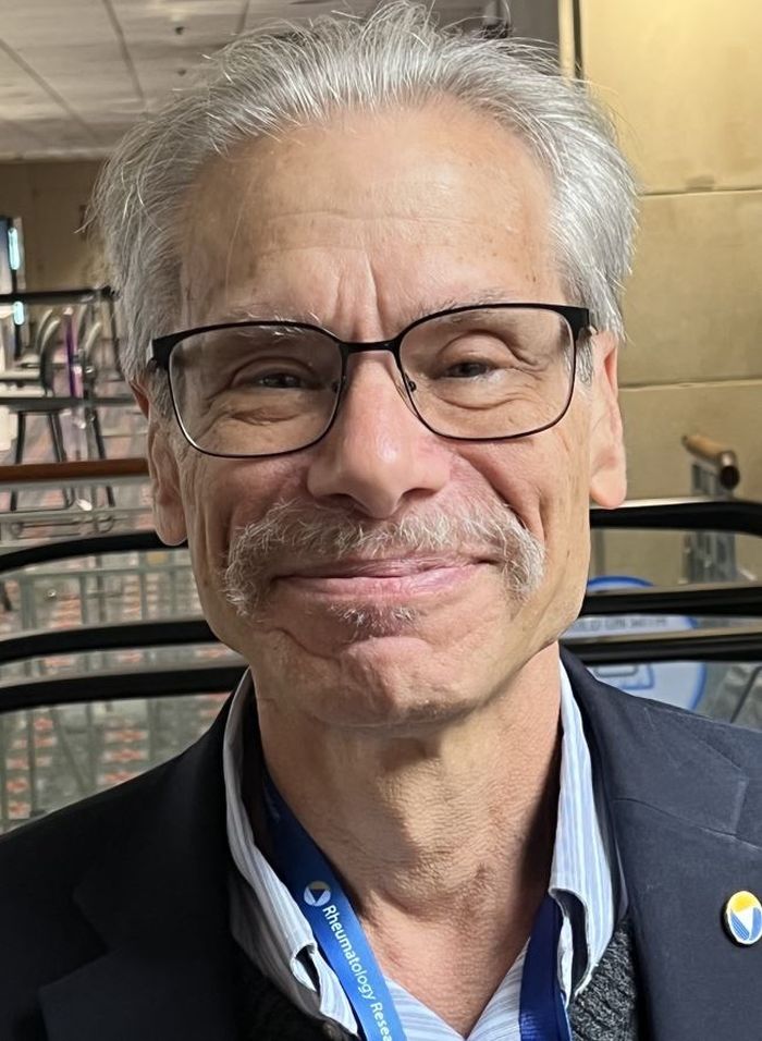
In an interview, David T. Felson, MD, MPH, professor and director of clinical epidemiology research at Boston University, questioned the delayed pain effect the study suggested. “It didn’t make any sense to me that there would be because both groups at that point got denosumab, so if there was going to be a pain effect that would’ve happened,” he said.
The pain effect is “really important,” he said. “We don’t use denosumab in rheumatoid arthritis to treat erosions because it doesn’t necessarily affect the pain and dysfunction of rheumatoid arthritis, and I’m not sure that isn’t going to be true in erosive hand osteoarthritis, but it’s possible.”
To clarify the pain outcomes, he said, “They’re going to have to work on the data.”
Amgen sponsored the trial but had no role in the design. Dr. Wittoek and Dr. Felson reported no relevant disclosures.
But pain outcomes questionable
But pain outcomes questionable
PHILADELPHIA – A double dose of the antiosteoporosis biologic denosumab (Prolia) slowed progression and repaired joints in erosive hand osteoarthritis (OA) but showed no impact on pain levels until 2 years after patients received the first dose, the lead investigator of a Belgium-based randomized clinical trial reported at the annual meeting of the American College of Rheumatology.
“This is the first placebo-controlled, randomized clinical trial showing the efficacy of denosumab double-dosing regimen in structural modification of erosive hand osteoarthritis,” Ruth Wittoek, MD, PhD, a rheumatologist at Ghent (Belgium) University, said in presenting the results.
“Our primary endpoint was confirmed by a more robust secondary endpoint, both showing that denosumab stopped erosive progression and induced remodeling in patients with erosive hand OA,” she added. “Moreover, the double-dosing regimen was well-tolerated.”
However, during the question-and-answer period after her presentation, Dr. Wittoek acknowledged the study didn’t evaluate the impact denosumab had on cartilage and didn’t detect a signal for pain resolution until 96 weeks during the open-label extension phase. “I’m not quite sure if denosumab is sufficient to treat symptoms in osteoarthritis,” she said. “There were positive signals but, of course, having to wait 2 years for an effect is kind of hard for our patients.”
The trial randomized 100 adult patients 1:1 to denosumab 60 mg every 12 weeks – double the normal dose for osteoporosis – or placebo. The primary endpoint was changes in erosive progression and signs of repair based on x-ray at 48 weeks, after which all patients were switched to denosumab for the open-label study. To quantify changes, the investigators used the Ghent University Scoring System (GUSS), which uses a scale of 0-300 to quantify radiographic changes in erosive hand OA.
Dr. Wittoek said that the average change in GUSS at week 24 was +6 vs. –2.8 (P = .024) in the treatment and placebo groups, respectively, widening at week 48 to +10.1 and –7.9 (P = .003). By week 96, the variation was +18.8 for denosumab and +17 for placebo with switch to denosumab (P = .03).
“During the open-label extension the denosumab treatment group continued to increase to show remodeling while the former placebo treatment group, now also receiving denosumab, also showed signs of remodeling,” she said. “So, there was no more erosive progression.”
The secondary endpoint was the percentage of new erosive joint development at week 48: 1.8% in the denosumab group and 7% in placebo group (odds ratio, 0.23; 95% confidence interval, 0.10-0.50; P < .001). “Meaning the odds of erosive progression is 77% lower in the denosumab treatment group,” Dr. Wittoek said.
By week 96, those percentages were 0% and 0.7% in the respective treatment groups. “During the open-label extension, it was clear that denosumab blocked all new development of erosive joints,” she said.
Pain was one of the study’s exploratory endpoints, and the mean numeric rating scale showed no difference between treatment arms until the 96-week results, with a reduction by almost half in the denosumab group (from 4.2 at week 48 to 2.4) and a lesser reduction in the placebo-switched-to-denosumab arm (from 4.2 to 3.5; P = .028) between arms.
The placebo group was more susceptible to adverse events, namely musculoskeletal complaints and nervous system disorders, Dr. Wittoek noted. Infection rates, the most common adverse event, were similar between the two groups: 41 and 39 in the respective arms. Despite the double dose of denosumab, safety and tolerability in this trial was comparable to other trials, she said.
In comments submitted by e-mail, Dr. Wittoek noted that the extension study results will go out to 144 weeks. She also addressed the issues surrounding pain as an outcome.
“Besides disability, pain is also important from the patient’s perspective,” Dr. Wittoek said in the e-mailed comments. “However, pain and radiographic progression are undeniably coupled, but it’s unclear how.”
In erosive hand OA, structural progression and pain may not be related on a molecular level, she said. “Therefore, we don’t deny that pain levels should also be covered by treatment, but they should not be confused with structural modification; it is just another domain, not more nor less important.
The second year of the open-label extension study should clarify the pain outcomes, she said.
In an interview, David T. Felson, MD, MPH, professor and director of clinical epidemiology research at Boston University, questioned the delayed pain effect the study suggested. “It didn’t make any sense to me that there would be because both groups at that point got denosumab, so if there was going to be a pain effect that would’ve happened,” he said.
The pain effect is “really important,” he said. “We don’t use denosumab in rheumatoid arthritis to treat erosions because it doesn’t necessarily affect the pain and dysfunction of rheumatoid arthritis, and I’m not sure that isn’t going to be true in erosive hand osteoarthritis, but it’s possible.”
To clarify the pain outcomes, he said, “They’re going to have to work on the data.”
Amgen sponsored the trial but had no role in the design. Dr. Wittoek and Dr. Felson reported no relevant disclosures.
AT ACR 2022
StopRA trial: Hydroxychloroquine doesn’t prevent or delay onset of rheumatoid arthritis
PHILADELPHIA – Hydroxychloroquine (HCQ) isn’t any more effective at preventing or delaying the onset of rheumatoid arthritis than placebo, based on interim results of a randomized clinical trial reported at the annual meeting of the American College of Rheumatology. Despite that futility, the percentage of patients who actually went on to develop clinical RA was lower than investigators expected, and the trial supports the use of a key biomarker for identifying RA.
While the StopRA trial was halted early because of futility of the treatment, investigators are continuing to mine the gathered data to deepen their understanding of disease progression and the potential of HCQ to improve symptoms in RA patients, said lead study author Kevin D. Deane, MD, PhD, of the University of Colorado at Denver, Aurora.

Overall, around 35% of the study participants on average developed RA, Dr. Deane said. “We were expecting somewhat more,” he said. “Teasing out who’s really going to progress to RA during a study and who’s not is going to be incredibly important.”
StopRA enrolled 144 adults who had elevated anti–cyclic citrullinated peptide antibodies (CCP3) levels of at least 40 units (about twice the normal level) but no history if inflammatory arthritis, randomizing them on a 1:1 basis to either HCQ (200-400 mg a day based on weight) or placebo for a 1-year treatment regimen.
The study identified participants through rheumatology clinics, testing of first-degree relatives with established RA, health fairs, blood donors, and biobanks. The interim findings are based on 2 years of follow-up after the last dose.
The study focused on HCQ because it has a relatively low risk profile with good safety and tolerability, is easy to administer, and is relatively low cost, Dr. Deane said.
StopRA study failed to meet its primary endpoint: to determine if 1 year of treatment with HCQ reduced the risk of developing inflammatory arthritis and classifiable RA at the end of 3 years in the study population. At the time of the interim analysis, 34% of patients in the HCQ arm and 36% in the placebo arm had developed RA (P = .844), Dr. Deane said. Baseline characteristics were balanced in both treatment arms.
The findings also support the use of CCP3 as a biomarker for RA, Dr. Deane said.
Now that the trial has been terminated, Dr. Deane said investigators are going to review the final data and perform secondary analyses for further clarity on the impact HCQ may have on RA.
“The future analysis should hopefully say if this treatment actually changes symptoms,” he said in an interview. “Because, if somebody felt better on the drug or had a milder form of rheumatoid arthritis once they developed it, that could potentially be a benefit.”
Dr. Deane noted the TREAT EARLIER trial similarly found that a 1-year course of methotrexate didn’t prevent the onset of clinical arthritis, but it did alter the disease course as measured in MRI-detected inflammation, related symptoms, and impairment.
“We’re hoping to look at those things and hopefully look at biologic changes over time,” Dr. Deane said of the extended analysis. “We’re not sure if the drug was associated with changes in biomarkers yet still didn’t halt progression to RA. That might be interesting, because those biomarkers might not be fundamentally related to the disease, but other mechanisms may be. That could give us some insights.”

Session moderator Ted Mikuls, MD, a professor of rheumatology at the University of Nebraska Medical Center, Omaha, said further mining of the study data is warranted.
“It’s common in a study like that, which took a lot of time and investment, to really take a deep dive into the data to make sure there aren’t signals that we’re missing,” he said in an interview.
One of the challenges with the study may have been patient enrollment, Dr. Mikuls noted. “I wonder about the study population in terms of where they recruit patients from. Who’s more likely to get RA? Is it patients who already have symptoms? Is it asymptomatic patients from biobanks? If it’s arthralgia joint pain patients, maybe by the time you have joint and autoantibody positivity it’s too late to have an intervention.”
The National Institute of Allergy and Infectious Diseases sponsored the study. Dr. Deane disclosed a relationship with Werfen. Dr. Mikuls has no relevant disclosures.
PHILADELPHIA – Hydroxychloroquine (HCQ) isn’t any more effective at preventing or delaying the onset of rheumatoid arthritis than placebo, based on interim results of a randomized clinical trial reported at the annual meeting of the American College of Rheumatology. Despite that futility, the percentage of patients who actually went on to develop clinical RA was lower than investigators expected, and the trial supports the use of a key biomarker for identifying RA.
While the StopRA trial was halted early because of futility of the treatment, investigators are continuing to mine the gathered data to deepen their understanding of disease progression and the potential of HCQ to improve symptoms in RA patients, said lead study author Kevin D. Deane, MD, PhD, of the University of Colorado at Denver, Aurora.
Overall, around 35% of the study participants on average developed RA, Dr. Deane said. “We were expecting somewhat more,” he said. “Teasing out who’s really going to progress to RA during a study and who’s not is going to be incredibly important.”
StopRA enrolled 144 adults who had elevated anti–cyclic citrullinated peptide antibodies (CCP3) levels of at least 40 units (about twice the normal level) but no history if inflammatory arthritis, randomizing them on a 1:1 basis to either HCQ (200-400 mg a day based on weight) or placebo for a 1-year treatment regimen.
The study identified participants through rheumatology clinics, testing of first-degree relatives with established RA, health fairs, blood donors, and biobanks. The interim findings are based on 2 years of follow-up after the last dose.
The study focused on HCQ because it has a relatively low risk profile with good safety and tolerability, is easy to administer, and is relatively low cost, Dr. Deane said.
StopRA study failed to meet its primary endpoint: to determine if 1 year of treatment with HCQ reduced the risk of developing inflammatory arthritis and classifiable RA at the end of 3 years in the study population. At the time of the interim analysis, 34% of patients in the HCQ arm and 36% in the placebo arm had developed RA (P = .844), Dr. Deane said. Baseline characteristics were balanced in both treatment arms.
The findings also support the use of CCP3 as a biomarker for RA, Dr. Deane said.
Now that the trial has been terminated, Dr. Deane said investigators are going to review the final data and perform secondary analyses for further clarity on the impact HCQ may have on RA.
“The future analysis should hopefully say if this treatment actually changes symptoms,” he said in an interview. “Because, if somebody felt better on the drug or had a milder form of rheumatoid arthritis once they developed it, that could potentially be a benefit.”
Dr. Deane noted the TREAT EARLIER trial similarly found that a 1-year course of methotrexate didn’t prevent the onset of clinical arthritis, but it did alter the disease course as measured in MRI-detected inflammation, related symptoms, and impairment.
“We’re hoping to look at those things and hopefully look at biologic changes over time,” Dr. Deane said of the extended analysis. “We’re not sure if the drug was associated with changes in biomarkers yet still didn’t halt progression to RA. That might be interesting, because those biomarkers might not be fundamentally related to the disease, but other mechanisms may be. That could give us some insights.”
Session moderator Ted Mikuls, MD, a professor of rheumatology at the University of Nebraska Medical Center, Omaha, said further mining of the study data is warranted.
“It’s common in a study like that, which took a lot of time and investment, to really take a deep dive into the data to make sure there aren’t signals that we’re missing,” he said in an interview.
One of the challenges with the study may have been patient enrollment, Dr. Mikuls noted. “I wonder about the study population in terms of where they recruit patients from. Who’s more likely to get RA? Is it patients who already have symptoms? Is it asymptomatic patients from biobanks? If it’s arthralgia joint pain patients, maybe by the time you have joint and autoantibody positivity it’s too late to have an intervention.”
The National Institute of Allergy and Infectious Diseases sponsored the study. Dr. Deane disclosed a relationship with Werfen. Dr. Mikuls has no relevant disclosures.
AT ACR 2022
Combination therapy shows mixed results for scleroderma-related lung disease
PHILADELPHIA – Combining the immunomodulatory agent mycophenolate with the antifibrotic pirfenidone led to more rapid improvement and showed a trend to be more effective than mycophenolate mofetil alone for treating the signs and symptoms of scleroderma-related interstitial lung disease, but the combination therapy came with an increase in side effects, according to results from the Scleroderma Lung Study III.
Dinesh Khanna, MBBS, MSc, of the University of Michigan, Ann Arbor, presented the results at the annual meeting of the American College of Rheumatology. He noted some problems with the study – namely its small size, enrolling only 51 patients, about one-third of its original goal. But he also said it showed a potential signal for efficacy and that the study itself could serve as a “template” for future studies of combination mycophenolate mofetil (MMF) plus pirfenidone therapy for scleroderma-related interstitial lung disease (SSc-ILD).

“The pirfenidone patients had quite a bit more GI side effects and photosensitivity, and those are known side effects,” Dr. Khanna said in an interview. “So the combination therapy had more side effects but trends to higher efficacy.”
The design of SLS-III, a phase 2 clinical trial, was a challenge, Dr. Khanna explained. The goal was to enroll 150 SSc-ILD patients who hadn’t had any previous treatment for their disease. Finding those patients proved difficult. “In fact, if you look at the recent history, 70% of the patients with early diffuse scleroderma are on MMF,” he said in his presentation. Compounding low study enrollment was the intervening COVID-19 pandemic, he added.
Testing a faster-acting combination
Nonetheless, the trial managed to enroll 27 patients in the combination therapy group and 24 in the MMF-plus-placebo group and compared their outcomes over 18 months. Study dosing was 1,500 mg MMF twice daily and pirfenidone 801 mg three times daily, titrated to the tolerable dose.
Despite the study’s being underpowered, Dr. Khanna said, it still reported some notable outcomes that merit further investigation. “I think what was intriguing in the study was the long-term benefit in the patient-reported outcomes and the structural changes,” he said in the interview.
Among those notable outcomes was a clinically significant change in forced vital capacity (FVC) percentage for the combination vs. the placebo groups: 2.24% vs. 2.09%. He also noted that the combination group saw a somewhat more robust improvement in FVC at six months: 2.59% (± 0.98%) vs. 0.92% (± 1.1%) in the placebo group.
The combination group showed greater improvements in high-resolution computed tomography-evaluated lung involvement and lung fibrosis and patient-reported outcomes, including a statistically significant 3.67-point greater improvement in PROMIS-29 physical function score (4.42 vs. 0.75).
The patients on combination therapy had higher rates of serious adverse events (SAEs), and seven discontinued one or both study drugs early, all in the combined arm. Four combination therapy patients had six SAEs, compared to two placebo patients with three SAEs. In the combination group, SAEs included chest pain, herpes zoster ophthalmicus, nodular basal cell cancer, marginal zone B cell lymphoma, renal crisis, and dyspnea. SAEs in the placebo group were colitis, COVID-19 and hypoxic respiratory failure.
Study design challenges
Nonetheless, Dr. Khanna said the SLS-III data are consistent with the SLS-II findings, with mean improvements in FVC of 2.24% and 2.1%, respectively.
“The next study may be able to replicate what we tried to do, keeping in mind that there are really no MMF-naive patients who are walking around,” Dr. Khanna said. “So the challenge is about the feasibility of recruiting within a trial vs. trying to show a statistical difference between the drug and placebo.”
This study could serve as a foundation for future studies of MMF in patients with SSc-ILD, Robert Spiera, MD, of the Hospital for Special Surgery in New York, said in an interview. “There are lessons to be learned both from the study but also from prior studies looking at MMF use in the background in patients treated with other drugs in clinical trials,” he said.
Dr. Spiera noted that the study had other challenges besides the difficulty in recruiting patients who hadn’t been on MMF therapy. “A great challenge is that the benefit with regard to the impact on the lungs from MMF seems most prominent in the first 6 months to a year to even 2 years that somebody is on the drug,” he said.
The other challenge with this study is that a large proportion of patients had limited systemic disease and relatively lower levels of skin disease compared with other studies of patients on MMF, Dr. Spiera said.
“The optimal treatment of scleroderma-associated lung disease remains a very important and not-adequately met need,” he said. “Particularly, we’re looking for drugs that are tolerable in a patient population that are very prone to GI side effects in general. This study and others have taught us a lot about trial design, and I think more globally this will allow us to move this field forward.”
Dr. Khanna disclosed relationships with Actelion, Boehringer Ingelheim, Bristol-Myers Squibb, CSL Behring, Horizon Therapeutics USA, Janssen Global Services, Prometheus Biosciences, Mitsubishi Tanabe Pharma Corp., Genentech/Roche, Theraly, and Pfizer. Genentech provided funding for the study and pirfenidone and placebo drugs at no cost.
Dr. Spiera disclosed relationships with GlaxoSmithKline, Boehringer-Ingelheim, Corbus Pharmaceutical, InflaRx, AbbVie/Abbott, Sanofi, Novartis, Chemocentryx, Roche and Vera.
PHILADELPHIA – Combining the immunomodulatory agent mycophenolate with the antifibrotic pirfenidone led to more rapid improvement and showed a trend to be more effective than mycophenolate mofetil alone for treating the signs and symptoms of scleroderma-related interstitial lung disease, but the combination therapy came with an increase in side effects, according to results from the Scleroderma Lung Study III.
Dinesh Khanna, MBBS, MSc, of the University of Michigan, Ann Arbor, presented the results at the annual meeting of the American College of Rheumatology. He noted some problems with the study – namely its small size, enrolling only 51 patients, about one-third of its original goal. But he also said it showed a potential signal for efficacy and that the study itself could serve as a “template” for future studies of combination mycophenolate mofetil (MMF) plus pirfenidone therapy for scleroderma-related interstitial lung disease (SSc-ILD).
“The pirfenidone patients had quite a bit more GI side effects and photosensitivity, and those are known side effects,” Dr. Khanna said in an interview. “So the combination therapy had more side effects but trends to higher efficacy.”
The design of SLS-III, a phase 2 clinical trial, was a challenge, Dr. Khanna explained. The goal was to enroll 150 SSc-ILD patients who hadn’t had any previous treatment for their disease. Finding those patients proved difficult. “In fact, if you look at the recent history, 70% of the patients with early diffuse scleroderma are on MMF,” he said in his presentation. Compounding low study enrollment was the intervening COVID-19 pandemic, he added.
Testing a faster-acting combination
Nonetheless, the trial managed to enroll 27 patients in the combination therapy group and 24 in the MMF-plus-placebo group and compared their outcomes over 18 months. Study dosing was 1,500 mg MMF twice daily and pirfenidone 801 mg three times daily, titrated to the tolerable dose.
Despite the study’s being underpowered, Dr. Khanna said, it still reported some notable outcomes that merit further investigation. “I think what was intriguing in the study was the long-term benefit in the patient-reported outcomes and the structural changes,” he said in the interview.
Among those notable outcomes was a clinically significant change in forced vital capacity (FVC) percentage for the combination vs. the placebo groups: 2.24% vs. 2.09%. He also noted that the combination group saw a somewhat more robust improvement in FVC at six months: 2.59% (± 0.98%) vs. 0.92% (± 1.1%) in the placebo group.
The combination group showed greater improvements in high-resolution computed tomography-evaluated lung involvement and lung fibrosis and patient-reported outcomes, including a statistically significant 3.67-point greater improvement in PROMIS-29 physical function score (4.42 vs. 0.75).
The patients on combination therapy had higher rates of serious adverse events (SAEs), and seven discontinued one or both study drugs early, all in the combined arm. Four combination therapy patients had six SAEs, compared to two placebo patients with three SAEs. In the combination group, SAEs included chest pain, herpes zoster ophthalmicus, nodular basal cell cancer, marginal zone B cell lymphoma, renal crisis, and dyspnea. SAEs in the placebo group were colitis, COVID-19 and hypoxic respiratory failure.
Study design challenges
Nonetheless, Dr. Khanna said the SLS-III data are consistent with the SLS-II findings, with mean improvements in FVC of 2.24% and 2.1%, respectively.
“The next study may be able to replicate what we tried to do, keeping in mind that there are really no MMF-naive patients who are walking around,” Dr. Khanna said. “So the challenge is about the feasibility of recruiting within a trial vs. trying to show a statistical difference between the drug and placebo.”
This study could serve as a foundation for future studies of MMF in patients with SSc-ILD, Robert Spiera, MD, of the Hospital for Special Surgery in New York, said in an interview. “There are lessons to be learned both from the study but also from prior studies looking at MMF use in the background in patients treated with other drugs in clinical trials,” he said.
Dr. Spiera noted that the study had other challenges besides the difficulty in recruiting patients who hadn’t been on MMF therapy. “A great challenge is that the benefit with regard to the impact on the lungs from MMF seems most prominent in the first 6 months to a year to even 2 years that somebody is on the drug,” he said.
The other challenge with this study is that a large proportion of patients had limited systemic disease and relatively lower levels of skin disease compared with other studies of patients on MMF, Dr. Spiera said.
“The optimal treatment of scleroderma-associated lung disease remains a very important and not-adequately met need,” he said. “Particularly, we’re looking for drugs that are tolerable in a patient population that are very prone to GI side effects in general. This study and others have taught us a lot about trial design, and I think more globally this will allow us to move this field forward.”
Dr. Khanna disclosed relationships with Actelion, Boehringer Ingelheim, Bristol-Myers Squibb, CSL Behring, Horizon Therapeutics USA, Janssen Global Services, Prometheus Biosciences, Mitsubishi Tanabe Pharma Corp., Genentech/Roche, Theraly, and Pfizer. Genentech provided funding for the study and pirfenidone and placebo drugs at no cost.
Dr. Spiera disclosed relationships with GlaxoSmithKline, Boehringer-Ingelheim, Corbus Pharmaceutical, InflaRx, AbbVie/Abbott, Sanofi, Novartis, Chemocentryx, Roche and Vera.
AT ACR 2022
Combination therapy may boost remission in JIA
Benefit endures at 3 years
PHILADELPHIA – Aggressive therapy using conventional synthetic disease-modifying antirheumatic drugs (DMARDs) in combination with biologic agents early, soon after a child is diagnosed with polyarticular juvenile idiopathic arthritis (pJIA), enabled more patients to achieve clinical remission and longer times in inactive disease than more conventional therapeutic approaches, 3-year results of prospective, observational study demonstrated.
The results of The Childhood Arthritis and Rheumatology Research Alliance STOP-JIA study, which Yukiko Kimura, MD, presented at the annual meeting of the American College of Rheumatology, showed early combination therapy had benefits, compared with other treatment strategies that were more evident at 3 years than at 1 year of study.

“The STOP-JIA study showed that, after 3 years, patients who started a biologic early on in combination with methotrexate spent more time in inactive disease and achieved clinical remission more often when compared to those started on traditional step-up therapy,” Dr. Kimura, chief of pediatric rheumatology at Hackensack (N.J.) Meridian Health and professor of pediatrics at the Hackensack Meridian School of Medicine, said at a press conference. “This study shows that the treatment of poly-JIA patients receive initially very early on in their disease matters even 3 years after that treatment was started.”
The study compared three CARRA consensus treatment plans (CTP) for untreated pediatric pJIA patients: step-up (SU) – starting conventional synthetic DMARD therapy and adding a biologic if needed after 3 or more months; early-combination (EC) therapy – starting synthetic and biologic DMARDs together; and biologic first (BF) therapy – starting biologic DMARD monotherapy.
Dr. Kimura explained the rationale for the study. “Since biologic treatments were introduced more than 20 years ago, the prognosis for JIA significantly improved. These very effective medicines often work wonders, quickly reducing pain and inflammation in joint disease activity,” she said in the press conference. “What is not known, however, is when is the best time to start these very effective treatments.”
The most common approach is to start with a synthetic DMARD, typically methotrexate, and wait before starting a biologic, Dr. Kimura said.
“But even though methotrexate can work very well by itself, it does not work for every patient, and we don’t know whether waiting months for it to work and then starting a biologic might potentially lessen their effectiveness,” Dr. Kimura added. “We don’t know if there’s a window of opportunity that’s lost while waiting to see whether methotrexate will work.”
The study originally enrolled 400 patients, 297 of whom completed the 3-year visit – 190 in SU, 76 in EC and 31 in BF. At 12 months, the study found no statistically significant difference in clinically inactive disease (CID) between the groups, Dr. Kimura said.
Even at the 3-year visit, the percentage of patients in CID off glucocorticoids and clinical Juvenile Arthritis Disease Activity Score based on 10 joints inactive disease (cJADAS 10 ID) did not differ among the three groups, Dr. Kimura said in presenting the results. “But,” she added, “greater proportions of early-combination CTP group were able to achieve clinical remissions and spend more time with inactive disease in both CID and cJADAS 10.”
A closer look at the outcomes showed some separation between early-combination therapy and the other two treatment plans. The incidence of clinical remission (at any time point over 36 months) was 67.1% in the EC group vs. 49.1% and 47.3%, respectively, in the BF and SU groups, Dr. Kimura said. “The difference between the early-combination and step-up groups was highly significant [P = .007],” she added.
EC also had an edge in the percentage of time patients spent in CID (over 36 months): 39.2% versus 32% and 27.4%, respectively, in the BF and SU groups (P = .006 for EV vs. SU), as well as cJADAS 10 ID (50.6% in EC group vs. 42.8% and 37.5%, respectively in the BF and SU groups; P = .005 for EC vs. SU).
Dr. Kimura said that the STOP JIA trial will continue with longer-term analysis and ongoing monitoring of study patients through the CARRA registry. “These longer-term analyses and readouts will be important because even though the results at 12 months didn’t seem as definitive, it seems the longer we go, the more impact we see of the treatments that were started early on in this disease.”

The findings from this study are “significantly important,” Nina T. Washington, MD, MPH, a pediatric rheumatologist at the University of New Mexico Hospital, Albuquerque, and the Mary Bridge Children’s Hospital in Tacoma, Wash., said in an interview. “At least for the past decade we’ve really been advocating towards earlier and aggressive therapy, and that’s what this study shows: the sooner you can treat this disease, the sooner you can attack those joints that are inflamed, the better outcome you give the patient.”
The study also confirms that pediatric rheumatologists are not overtreating patients with pJIA, she added.
“In a sense we’re actually treating and preventing and if you have a child that has arthritis, it’s okay to treat that child,” Dr. Washington said. “For me that’s the most reassuring thing: that I’m not necessarily going overboard. If I have a child with polyarticular JIA and they have multiple inflamed joints and I have the evidence as they’re sitting in front of me, and I treat them. I’m going to give them the best outcome.”
The Patient Centered Outcomes Research Institute provided study funding. Dr. Kimura is chair of the CARRA JIA disease research committee and cochair of the CARRA Registry and Research Oversight Committee. She disclosed a financial relationship with Genentech. Dr. Washington has no relevant relationships to disclose.
Benefit endures at 3 years
Benefit endures at 3 years
PHILADELPHIA – Aggressive therapy using conventional synthetic disease-modifying antirheumatic drugs (DMARDs) in combination with biologic agents early, soon after a child is diagnosed with polyarticular juvenile idiopathic arthritis (pJIA), enabled more patients to achieve clinical remission and longer times in inactive disease than more conventional therapeutic approaches, 3-year results of prospective, observational study demonstrated.
The results of The Childhood Arthritis and Rheumatology Research Alliance STOP-JIA study, which Yukiko Kimura, MD, presented at the annual meeting of the American College of Rheumatology, showed early combination therapy had benefits, compared with other treatment strategies that were more evident at 3 years than at 1 year of study.
“The STOP-JIA study showed that, after 3 years, patients who started a biologic early on in combination with methotrexate spent more time in inactive disease and achieved clinical remission more often when compared to those started on traditional step-up therapy,” Dr. Kimura, chief of pediatric rheumatology at Hackensack (N.J.) Meridian Health and professor of pediatrics at the Hackensack Meridian School of Medicine, said at a press conference. “This study shows that the treatment of poly-JIA patients receive initially very early on in their disease matters even 3 years after that treatment was started.”
The study compared three CARRA consensus treatment plans (CTP) for untreated pediatric pJIA patients: step-up (SU) – starting conventional synthetic DMARD therapy and adding a biologic if needed after 3 or more months; early-combination (EC) therapy – starting synthetic and biologic DMARDs together; and biologic first (BF) therapy – starting biologic DMARD monotherapy.
Dr. Kimura explained the rationale for the study. “Since biologic treatments were introduced more than 20 years ago, the prognosis for JIA significantly improved. These very effective medicines often work wonders, quickly reducing pain and inflammation in joint disease activity,” she said in the press conference. “What is not known, however, is when is the best time to start these very effective treatments.”
The most common approach is to start with a synthetic DMARD, typically methotrexate, and wait before starting a biologic, Dr. Kimura said.
“But even though methotrexate can work very well by itself, it does not work for every patient, and we don’t know whether waiting months for it to work and then starting a biologic might potentially lessen their effectiveness,” Dr. Kimura added. “We don’t know if there’s a window of opportunity that’s lost while waiting to see whether methotrexate will work.”
The study originally enrolled 400 patients, 297 of whom completed the 3-year visit – 190 in SU, 76 in EC and 31 in BF. At 12 months, the study found no statistically significant difference in clinically inactive disease (CID) between the groups, Dr. Kimura said.
Even at the 3-year visit, the percentage of patients in CID off glucocorticoids and clinical Juvenile Arthritis Disease Activity Score based on 10 joints inactive disease (cJADAS 10 ID) did not differ among the three groups, Dr. Kimura said in presenting the results. “But,” she added, “greater proportions of early-combination CTP group were able to achieve clinical remissions and spend more time with inactive disease in both CID and cJADAS 10.”
A closer look at the outcomes showed some separation between early-combination therapy and the other two treatment plans. The incidence of clinical remission (at any time point over 36 months) was 67.1% in the EC group vs. 49.1% and 47.3%, respectively, in the BF and SU groups, Dr. Kimura said. “The difference between the early-combination and step-up groups was highly significant [P = .007],” she added.
EC also had an edge in the percentage of time patients spent in CID (over 36 months): 39.2% versus 32% and 27.4%, respectively, in the BF and SU groups (P = .006 for EV vs. SU), as well as cJADAS 10 ID (50.6% in EC group vs. 42.8% and 37.5%, respectively in the BF and SU groups; P = .005 for EC vs. SU).
Dr. Kimura said that the STOP JIA trial will continue with longer-term analysis and ongoing monitoring of study patients through the CARRA registry. “These longer-term analyses and readouts will be important because even though the results at 12 months didn’t seem as definitive, it seems the longer we go, the more impact we see of the treatments that were started early on in this disease.”
The findings from this study are “significantly important,” Nina T. Washington, MD, MPH, a pediatric rheumatologist at the University of New Mexico Hospital, Albuquerque, and the Mary Bridge Children’s Hospital in Tacoma, Wash., said in an interview. “At least for the past decade we’ve really been advocating towards earlier and aggressive therapy, and that’s what this study shows: the sooner you can treat this disease, the sooner you can attack those joints that are inflamed, the better outcome you give the patient.”
The study also confirms that pediatric rheumatologists are not overtreating patients with pJIA, she added.
“In a sense we’re actually treating and preventing and if you have a child that has arthritis, it’s okay to treat that child,” Dr. Washington said. “For me that’s the most reassuring thing: that I’m not necessarily going overboard. If I have a child with polyarticular JIA and they have multiple inflamed joints and I have the evidence as they’re sitting in front of me, and I treat them. I’m going to give them the best outcome.”
The Patient Centered Outcomes Research Institute provided study funding. Dr. Kimura is chair of the CARRA JIA disease research committee and cochair of the CARRA Registry and Research Oversight Committee. She disclosed a financial relationship with Genentech. Dr. Washington has no relevant relationships to disclose.
AT ACR 2022