User login
CCJM delivers practical clinical articles relevant to internists, cardiologists, endocrinologists, and other specialists, all written by known experts.
Copyright © 2019 Cleveland Clinic. All rights reserved. The information provided is for educational purposes only. Use of this website is subject to the disclaimer and privacy policy.
gambling
compulsive behaviors
ammunition
assault rifle
black jack
Boko Haram
bondage
child abuse
cocaine
Daech
drug paraphernalia
explosion
gun
human trafficking
ISIL
ISIS
Islamic caliphate
Islamic state
mixed martial arts
MMA
molestation
national rifle association
NRA
nsfw
pedophile
pedophilia
poker
porn
pornography
psychedelic drug
recreational drug
sex slave rings
slot machine
terrorism
terrorist
Texas hold 'em
UFC
substance abuse
abuseed
abuseer
abusees
abuseing
abusely
abuses
aeolus
aeolused
aeoluser
aeoluses
aeolusing
aeolusly
aeoluss
ahole
aholeed
aholeer
aholees
aholeing
aholely
aholes
alcohol
alcoholed
alcoholer
alcoholes
alcoholing
alcoholly
alcohols
allman
allmaned
allmaner
allmanes
allmaning
allmanly
allmans
alted
altes
alting
altly
alts
analed
analer
anales
analing
anally
analprobe
analprobeed
analprobeer
analprobees
analprobeing
analprobely
analprobes
anals
anilingus
anilingused
anilinguser
anilinguses
anilingusing
anilingusly
anilinguss
anus
anused
anuser
anuses
anusing
anusly
anuss
areola
areolaed
areolaer
areolaes
areolaing
areolaly
areolas
areole
areoleed
areoleer
areolees
areoleing
areolely
areoles
arian
arianed
arianer
arianes
arianing
arianly
arians
aryan
aryaned
aryaner
aryanes
aryaning
aryanly
aryans
asiaed
asiaer
asiaes
asiaing
asialy
asias
ass
ass hole
ass lick
ass licked
ass licker
ass lickes
ass licking
ass lickly
ass licks
assbang
assbanged
assbangeded
assbangeder
assbangedes
assbangeding
assbangedly
assbangeds
assbanger
assbanges
assbanging
assbangly
assbangs
assbangsed
assbangser
assbangses
assbangsing
assbangsly
assbangss
assed
asser
asses
assesed
asseser
asseses
assesing
assesly
assess
assfuck
assfucked
assfucker
assfuckered
assfuckerer
assfuckeres
assfuckering
assfuckerly
assfuckers
assfuckes
assfucking
assfuckly
assfucks
asshat
asshated
asshater
asshates
asshating
asshatly
asshats
assholeed
assholeer
assholees
assholeing
assholely
assholes
assholesed
assholeser
assholeses
assholesing
assholesly
assholess
assing
assly
assmaster
assmastered
assmasterer
assmasteres
assmastering
assmasterly
assmasters
assmunch
assmunched
assmuncher
assmunches
assmunching
assmunchly
assmunchs
asss
asswipe
asswipeed
asswipeer
asswipees
asswipeing
asswipely
asswipes
asswipesed
asswipeser
asswipeses
asswipesing
asswipesly
asswipess
azz
azzed
azzer
azzes
azzing
azzly
azzs
babeed
babeer
babees
babeing
babely
babes
babesed
babeser
babeses
babesing
babesly
babess
ballsac
ballsaced
ballsacer
ballsaces
ballsacing
ballsack
ballsacked
ballsacker
ballsackes
ballsacking
ballsackly
ballsacks
ballsacly
ballsacs
ballsed
ballser
ballses
ballsing
ballsly
ballss
barf
barfed
barfer
barfes
barfing
barfly
barfs
bastard
bastarded
bastarder
bastardes
bastarding
bastardly
bastards
bastardsed
bastardser
bastardses
bastardsing
bastardsly
bastardss
bawdy
bawdyed
bawdyer
bawdyes
bawdying
bawdyly
bawdys
beaner
beanered
beanerer
beaneres
beanering
beanerly
beaners
beardedclam
beardedclamed
beardedclamer
beardedclames
beardedclaming
beardedclamly
beardedclams
beastiality
beastialityed
beastialityer
beastialityes
beastialitying
beastialityly
beastialitys
beatch
beatched
beatcher
beatches
beatching
beatchly
beatchs
beater
beatered
beaterer
beateres
beatering
beaterly
beaters
beered
beerer
beeres
beering
beerly
beeyotch
beeyotched
beeyotcher
beeyotches
beeyotching
beeyotchly
beeyotchs
beotch
beotched
beotcher
beotches
beotching
beotchly
beotchs
biatch
biatched
biatcher
biatches
biatching
biatchly
biatchs
big tits
big titsed
big titser
big titses
big titsing
big titsly
big titss
bigtits
bigtitsed
bigtitser
bigtitses
bigtitsing
bigtitsly
bigtitss
bimbo
bimboed
bimboer
bimboes
bimboing
bimboly
bimbos
bisexualed
bisexualer
bisexuales
bisexualing
bisexually
bisexuals
bitch
bitched
bitcheded
bitcheder
bitchedes
bitcheding
bitchedly
bitcheds
bitcher
bitches
bitchesed
bitcheser
bitcheses
bitchesing
bitchesly
bitchess
bitching
bitchly
bitchs
bitchy
bitchyed
bitchyer
bitchyes
bitchying
bitchyly
bitchys
bleached
bleacher
bleaches
bleaching
bleachly
bleachs
blow job
blow jobed
blow jober
blow jobes
blow jobing
blow jobly
blow jobs
blowed
blower
blowes
blowing
blowjob
blowjobed
blowjober
blowjobes
blowjobing
blowjobly
blowjobs
blowjobsed
blowjobser
blowjobses
blowjobsing
blowjobsly
blowjobss
blowly
blows
boink
boinked
boinker
boinkes
boinking
boinkly
boinks
bollock
bollocked
bollocker
bollockes
bollocking
bollockly
bollocks
bollocksed
bollockser
bollockses
bollocksing
bollocksly
bollockss
bollok
bolloked
bolloker
bollokes
bolloking
bollokly
bolloks
boner
bonered
bonerer
boneres
bonering
bonerly
boners
bonersed
bonerser
bonerses
bonersing
bonersly
bonerss
bong
bonged
bonger
bonges
bonging
bongly
bongs
boob
boobed
boober
boobes
boobies
boobiesed
boobieser
boobieses
boobiesing
boobiesly
boobiess
boobing
boobly
boobs
boobsed
boobser
boobses
boobsing
boobsly
boobss
booby
boobyed
boobyer
boobyes
boobying
boobyly
boobys
booger
boogered
boogerer
boogeres
boogering
boogerly
boogers
bookie
bookieed
bookieer
bookiees
bookieing
bookiely
bookies
bootee
booteeed
booteeer
booteees
booteeing
booteely
bootees
bootie
bootieed
bootieer
bootiees
bootieing
bootiely
booties
booty
bootyed
bootyer
bootyes
bootying
bootyly
bootys
boozeed
boozeer
boozees
boozeing
boozely
boozer
boozered
boozerer
boozeres
boozering
boozerly
boozers
boozes
boozy
boozyed
boozyer
boozyes
boozying
boozyly
boozys
bosomed
bosomer
bosomes
bosoming
bosomly
bosoms
bosomy
bosomyed
bosomyer
bosomyes
bosomying
bosomyly
bosomys
bugger
buggered
buggerer
buggeres
buggering
buggerly
buggers
bukkake
bukkakeed
bukkakeer
bukkakees
bukkakeing
bukkakely
bukkakes
bull shit
bull shited
bull shiter
bull shites
bull shiting
bull shitly
bull shits
bullshit
bullshited
bullshiter
bullshites
bullshiting
bullshitly
bullshits
bullshitsed
bullshitser
bullshitses
bullshitsing
bullshitsly
bullshitss
bullshitted
bullshitteded
bullshitteder
bullshittedes
bullshitteding
bullshittedly
bullshitteds
bullturds
bullturdsed
bullturdser
bullturdses
bullturdsing
bullturdsly
bullturdss
bung
bunged
bunger
bunges
bunging
bungly
bungs
busty
bustyed
bustyer
bustyes
bustying
bustyly
bustys
butt
butt fuck
butt fucked
butt fucker
butt fuckes
butt fucking
butt fuckly
butt fucks
butted
buttes
buttfuck
buttfucked
buttfucker
buttfuckered
buttfuckerer
buttfuckeres
buttfuckering
buttfuckerly
buttfuckers
buttfuckes
buttfucking
buttfuckly
buttfucks
butting
buttly
buttplug
buttpluged
buttpluger
buttpluges
buttpluging
buttplugly
buttplugs
butts
caca
cacaed
cacaer
cacaes
cacaing
cacaly
cacas
cahone
cahoneed
cahoneer
cahonees
cahoneing
cahonely
cahones
cameltoe
cameltoeed
cameltoeer
cameltoees
cameltoeing
cameltoely
cameltoes
carpetmuncher
carpetmunchered
carpetmuncherer
carpetmuncheres
carpetmunchering
carpetmuncherly
carpetmunchers
cawk
cawked
cawker
cawkes
cawking
cawkly
cawks
chinc
chinced
chincer
chinces
chincing
chincly
chincs
chincsed
chincser
chincses
chincsing
chincsly
chincss
chink
chinked
chinker
chinkes
chinking
chinkly
chinks
chode
chodeed
chodeer
chodees
chodeing
chodely
chodes
chodesed
chodeser
chodeses
chodesing
chodesly
chodess
clit
clited
cliter
clites
cliting
clitly
clitoris
clitorised
clitoriser
clitorises
clitorising
clitorisly
clitoriss
clitorus
clitorused
clitoruser
clitoruses
clitorusing
clitorusly
clitoruss
clits
clitsed
clitser
clitses
clitsing
clitsly
clitss
clitty
clittyed
clittyer
clittyes
clittying
clittyly
clittys
cocain
cocaine
cocained
cocaineed
cocaineer
cocainees
cocaineing
cocainely
cocainer
cocaines
cocaining
cocainly
cocains
cock
cock sucker
cock suckered
cock suckerer
cock suckeres
cock suckering
cock suckerly
cock suckers
cockblock
cockblocked
cockblocker
cockblockes
cockblocking
cockblockly
cockblocks
cocked
cocker
cockes
cockholster
cockholstered
cockholsterer
cockholsteres
cockholstering
cockholsterly
cockholsters
cocking
cockknocker
cockknockered
cockknockerer
cockknockeres
cockknockering
cockknockerly
cockknockers
cockly
cocks
cocksed
cockser
cockses
cocksing
cocksly
cocksmoker
cocksmokered
cocksmokerer
cocksmokeres
cocksmokering
cocksmokerly
cocksmokers
cockss
cocksucker
cocksuckered
cocksuckerer
cocksuckeres
cocksuckering
cocksuckerly
cocksuckers
coital
coitaled
coitaler
coitales
coitaling
coitally
coitals
commie
commieed
commieer
commiees
commieing
commiely
commies
condomed
condomer
condomes
condoming
condomly
condoms
coon
cooned
cooner
coones
cooning
coonly
coons
coonsed
coonser
coonses
coonsing
coonsly
coonss
corksucker
corksuckered
corksuckerer
corksuckeres
corksuckering
corksuckerly
corksuckers
cracked
crackwhore
crackwhoreed
crackwhoreer
crackwhorees
crackwhoreing
crackwhorely
crackwhores
crap
craped
craper
crapes
craping
craply
crappy
crappyed
crappyer
crappyes
crappying
crappyly
crappys
cum
cumed
cumer
cumes
cuming
cumly
cummin
cummined
cumminer
cummines
cumming
cumminged
cumminger
cumminges
cumminging
cummingly
cummings
cummining
cumminly
cummins
cums
cumshot
cumshoted
cumshoter
cumshotes
cumshoting
cumshotly
cumshots
cumshotsed
cumshotser
cumshotses
cumshotsing
cumshotsly
cumshotss
cumslut
cumsluted
cumsluter
cumslutes
cumsluting
cumslutly
cumsluts
cumstain
cumstained
cumstainer
cumstaines
cumstaining
cumstainly
cumstains
cunilingus
cunilingused
cunilinguser
cunilinguses
cunilingusing
cunilingusly
cunilinguss
cunnilingus
cunnilingused
cunnilinguser
cunnilinguses
cunnilingusing
cunnilingusly
cunnilinguss
cunny
cunnyed
cunnyer
cunnyes
cunnying
cunnyly
cunnys
cunt
cunted
cunter
cuntes
cuntface
cuntfaceed
cuntfaceer
cuntfacees
cuntfaceing
cuntfacely
cuntfaces
cunthunter
cunthuntered
cunthunterer
cunthunteres
cunthuntering
cunthunterly
cunthunters
cunting
cuntlick
cuntlicked
cuntlicker
cuntlickered
cuntlickerer
cuntlickeres
cuntlickering
cuntlickerly
cuntlickers
cuntlickes
cuntlicking
cuntlickly
cuntlicks
cuntly
cunts
cuntsed
cuntser
cuntses
cuntsing
cuntsly
cuntss
dago
dagoed
dagoer
dagoes
dagoing
dagoly
dagos
dagosed
dagoser
dagoses
dagosing
dagosly
dagoss
dammit
dammited
dammiter
dammites
dammiting
dammitly
dammits
damn
damned
damneded
damneder
damnedes
damneding
damnedly
damneds
damner
damnes
damning
damnit
damnited
damniter
damnites
damniting
damnitly
damnits
damnly
damns
dick
dickbag
dickbaged
dickbager
dickbages
dickbaging
dickbagly
dickbags
dickdipper
dickdippered
dickdipperer
dickdipperes
dickdippering
dickdipperly
dickdippers
dicked
dicker
dickes
dickface
dickfaceed
dickfaceer
dickfacees
dickfaceing
dickfacely
dickfaces
dickflipper
dickflippered
dickflipperer
dickflipperes
dickflippering
dickflipperly
dickflippers
dickhead
dickheaded
dickheader
dickheades
dickheading
dickheadly
dickheads
dickheadsed
dickheadser
dickheadses
dickheadsing
dickheadsly
dickheadss
dicking
dickish
dickished
dickisher
dickishes
dickishing
dickishly
dickishs
dickly
dickripper
dickrippered
dickripperer
dickripperes
dickrippering
dickripperly
dickrippers
dicks
dicksipper
dicksippered
dicksipperer
dicksipperes
dicksippering
dicksipperly
dicksippers
dickweed
dickweeded
dickweeder
dickweedes
dickweeding
dickweedly
dickweeds
dickwhipper
dickwhippered
dickwhipperer
dickwhipperes
dickwhippering
dickwhipperly
dickwhippers
dickzipper
dickzippered
dickzipperer
dickzipperes
dickzippering
dickzipperly
dickzippers
diddle
diddleed
diddleer
diddlees
diddleing
diddlely
diddles
dike
dikeed
dikeer
dikees
dikeing
dikely
dikes
dildo
dildoed
dildoer
dildoes
dildoing
dildoly
dildos
dildosed
dildoser
dildoses
dildosing
dildosly
dildoss
diligaf
diligafed
diligafer
diligafes
diligafing
diligafly
diligafs
dillweed
dillweeded
dillweeder
dillweedes
dillweeding
dillweedly
dillweeds
dimwit
dimwited
dimwiter
dimwites
dimwiting
dimwitly
dimwits
dingle
dingleed
dingleer
dinglees
dingleing
dinglely
dingles
dipship
dipshiped
dipshiper
dipshipes
dipshiping
dipshiply
dipships
dizzyed
dizzyer
dizzyes
dizzying
dizzyly
dizzys
doggiestyleed
doggiestyleer
doggiestylees
doggiestyleing
doggiestylely
doggiestyles
doggystyleed
doggystyleer
doggystylees
doggystyleing
doggystylely
doggystyles
dong
donged
donger
donges
donging
dongly
dongs
doofus
doofused
doofuser
doofuses
doofusing
doofusly
doofuss
doosh
dooshed
doosher
dooshes
dooshing
dooshly
dooshs
dopeyed
dopeyer
dopeyes
dopeying
dopeyly
dopeys
douchebag
douchebaged
douchebager
douchebages
douchebaging
douchebagly
douchebags
douchebagsed
douchebagser
douchebagses
douchebagsing
douchebagsly
douchebagss
doucheed
doucheer
douchees
doucheing
douchely
douches
douchey
doucheyed
doucheyer
doucheyes
doucheying
doucheyly
doucheys
drunk
drunked
drunker
drunkes
drunking
drunkly
drunks
dumass
dumassed
dumasser
dumasses
dumassing
dumassly
dumasss
dumbass
dumbassed
dumbasser
dumbasses
dumbassesed
dumbasseser
dumbasseses
dumbassesing
dumbassesly
dumbassess
dumbassing
dumbassly
dumbasss
dummy
dummyed
dummyer
dummyes
dummying
dummyly
dummys
dyke
dykeed
dykeer
dykees
dykeing
dykely
dykes
dykesed
dykeser
dykeses
dykesing
dykesly
dykess
erotic
eroticed
eroticer
erotices
eroticing
eroticly
erotics
extacy
extacyed
extacyer
extacyes
extacying
extacyly
extacys
extasy
extasyed
extasyer
extasyes
extasying
extasyly
extasys
fack
facked
facker
fackes
facking
fackly
facks
fag
faged
fager
fages
fagg
fagged
faggeded
faggeder
faggedes
faggeding
faggedly
faggeds
fagger
fagges
fagging
faggit
faggited
faggiter
faggites
faggiting
faggitly
faggits
faggly
faggot
faggoted
faggoter
faggotes
faggoting
faggotly
faggots
faggs
faging
fagly
fagot
fagoted
fagoter
fagotes
fagoting
fagotly
fagots
fags
fagsed
fagser
fagses
fagsing
fagsly
fagss
faig
faiged
faiger
faiges
faiging
faigly
faigs
faigt
faigted
faigter
faigtes
faigting
faigtly
faigts
fannybandit
fannybandited
fannybanditer
fannybandites
fannybanditing
fannybanditly
fannybandits
farted
farter
fartes
farting
fartknocker
fartknockered
fartknockerer
fartknockeres
fartknockering
fartknockerly
fartknockers
fartly
farts
felch
felched
felcher
felchered
felcherer
felcheres
felchering
felcherly
felchers
felches
felching
felchinged
felchinger
felchinges
felchinging
felchingly
felchings
felchly
felchs
fellate
fellateed
fellateer
fellatees
fellateing
fellately
fellates
fellatio
fellatioed
fellatioer
fellatioes
fellatioing
fellatioly
fellatios
feltch
feltched
feltcher
feltchered
feltcherer
feltcheres
feltchering
feltcherly
feltchers
feltches
feltching
feltchly
feltchs
feom
feomed
feomer
feomes
feoming
feomly
feoms
fisted
fisteded
fisteder
fistedes
fisteding
fistedly
fisteds
fisting
fistinged
fistinger
fistinges
fistinging
fistingly
fistings
fisty
fistyed
fistyer
fistyes
fistying
fistyly
fistys
floozy
floozyed
floozyer
floozyes
floozying
floozyly
floozys
foad
foaded
foader
foades
foading
foadly
foads
fondleed
fondleer
fondlees
fondleing
fondlely
fondles
foobar
foobared
foobarer
foobares
foobaring
foobarly
foobars
freex
freexed
freexer
freexes
freexing
freexly
freexs
frigg
frigga
friggaed
friggaer
friggaes
friggaing
friggaly
friggas
frigged
frigger
frigges
frigging
friggly
friggs
fubar
fubared
fubarer
fubares
fubaring
fubarly
fubars
fuck
fuckass
fuckassed
fuckasser
fuckasses
fuckassing
fuckassly
fuckasss
fucked
fuckeded
fuckeder
fuckedes
fuckeding
fuckedly
fuckeds
fucker
fuckered
fuckerer
fuckeres
fuckering
fuckerly
fuckers
fuckes
fuckface
fuckfaceed
fuckfaceer
fuckfacees
fuckfaceing
fuckfacely
fuckfaces
fuckin
fuckined
fuckiner
fuckines
fucking
fuckinged
fuckinger
fuckinges
fuckinging
fuckingly
fuckings
fuckining
fuckinly
fuckins
fuckly
fucknugget
fucknuggeted
fucknuggeter
fucknuggetes
fucknuggeting
fucknuggetly
fucknuggets
fucknut
fucknuted
fucknuter
fucknutes
fucknuting
fucknutly
fucknuts
fuckoff
fuckoffed
fuckoffer
fuckoffes
fuckoffing
fuckoffly
fuckoffs
fucks
fucksed
fuckser
fuckses
fucksing
fucksly
fuckss
fucktard
fucktarded
fucktarder
fucktardes
fucktarding
fucktardly
fucktards
fuckup
fuckuped
fuckuper
fuckupes
fuckuping
fuckuply
fuckups
fuckwad
fuckwaded
fuckwader
fuckwades
fuckwading
fuckwadly
fuckwads
fuckwit
fuckwited
fuckwiter
fuckwites
fuckwiting
fuckwitly
fuckwits
fudgepacker
fudgepackered
fudgepackerer
fudgepackeres
fudgepackering
fudgepackerly
fudgepackers
fuk
fuked
fuker
fukes
fuking
fukly
fuks
fvck
fvcked
fvcker
fvckes
fvcking
fvckly
fvcks
fxck
fxcked
fxcker
fxckes
fxcking
fxckly
fxcks
gae
gaeed
gaeer
gaees
gaeing
gaely
gaes
gai
gaied
gaier
gaies
gaiing
gaily
gais
ganja
ganjaed
ganjaer
ganjaes
ganjaing
ganjaly
ganjas
gayed
gayer
gayes
gaying
gayly
gays
gaysed
gayser
gayses
gaysing
gaysly
gayss
gey
geyed
geyer
geyes
geying
geyly
geys
gfc
gfced
gfcer
gfces
gfcing
gfcly
gfcs
gfy
gfyed
gfyer
gfyes
gfying
gfyly
gfys
ghay
ghayed
ghayer
ghayes
ghaying
ghayly
ghays
ghey
gheyed
gheyer
gheyes
gheying
gheyly
gheys
gigolo
gigoloed
gigoloer
gigoloes
gigoloing
gigololy
gigolos
goatse
goatseed
goatseer
goatsees
goatseing
goatsely
goatses
godamn
godamned
godamner
godamnes
godamning
godamnit
godamnited
godamniter
godamnites
godamniting
godamnitly
godamnits
godamnly
godamns
goddam
goddamed
goddamer
goddames
goddaming
goddamly
goddammit
goddammited
goddammiter
goddammites
goddammiting
goddammitly
goddammits
goddamn
goddamned
goddamner
goddamnes
goddamning
goddamnly
goddamns
goddams
goldenshower
goldenshowered
goldenshowerer
goldenshoweres
goldenshowering
goldenshowerly
goldenshowers
gonad
gonaded
gonader
gonades
gonading
gonadly
gonads
gonadsed
gonadser
gonadses
gonadsing
gonadsly
gonadss
gook
gooked
gooker
gookes
gooking
gookly
gooks
gooksed
gookser
gookses
gooksing
gooksly
gookss
gringo
gringoed
gringoer
gringoes
gringoing
gringoly
gringos
gspot
gspoted
gspoter
gspotes
gspoting
gspotly
gspots
gtfo
gtfoed
gtfoer
gtfoes
gtfoing
gtfoly
gtfos
guido
guidoed
guidoer
guidoes
guidoing
guidoly
guidos
handjob
handjobed
handjober
handjobes
handjobing
handjobly
handjobs
hard on
hard oned
hard oner
hard ones
hard oning
hard only
hard ons
hardknight
hardknighted
hardknighter
hardknightes
hardknighting
hardknightly
hardknights
hebe
hebeed
hebeer
hebees
hebeing
hebely
hebes
heeb
heebed
heeber
heebes
heebing
heebly
heebs
hell
helled
heller
helles
helling
hellly
hells
hemp
hemped
hemper
hempes
hemping
hemply
hemps
heroined
heroiner
heroines
heroining
heroinly
heroins
herp
herped
herper
herpes
herpesed
herpeser
herpeses
herpesing
herpesly
herpess
herping
herply
herps
herpy
herpyed
herpyer
herpyes
herpying
herpyly
herpys
hitler
hitlered
hitlerer
hitleres
hitlering
hitlerly
hitlers
hived
hiver
hives
hiving
hivly
hivs
hobag
hobaged
hobager
hobages
hobaging
hobagly
hobags
homey
homeyed
homeyer
homeyes
homeying
homeyly
homeys
homo
homoed
homoer
homoes
homoey
homoeyed
homoeyer
homoeyes
homoeying
homoeyly
homoeys
homoing
homoly
homos
honky
honkyed
honkyer
honkyes
honkying
honkyly
honkys
hooch
hooched
hoocher
hooches
hooching
hoochly
hoochs
hookah
hookahed
hookaher
hookahes
hookahing
hookahly
hookahs
hooker
hookered
hookerer
hookeres
hookering
hookerly
hookers
hoor
hoored
hoorer
hoores
hooring
hoorly
hoors
hootch
hootched
hootcher
hootches
hootching
hootchly
hootchs
hooter
hootered
hooterer
hooteres
hootering
hooterly
hooters
hootersed
hooterser
hooterses
hootersing
hootersly
hooterss
horny
hornyed
hornyer
hornyes
hornying
hornyly
hornys
houstoned
houstoner
houstones
houstoning
houstonly
houstons
hump
humped
humpeded
humpeder
humpedes
humpeding
humpedly
humpeds
humper
humpes
humping
humpinged
humpinger
humpinges
humpinging
humpingly
humpings
humply
humps
husbanded
husbander
husbandes
husbanding
husbandly
husbands
hussy
hussyed
hussyer
hussyes
hussying
hussyly
hussys
hymened
hymener
hymenes
hymening
hymenly
hymens
inbred
inbreded
inbreder
inbredes
inbreding
inbredly
inbreds
incest
incested
incester
incestes
incesting
incestly
incests
injun
injuned
injuner
injunes
injuning
injunly
injuns
jackass
jackassed
jackasser
jackasses
jackassing
jackassly
jackasss
jackhole
jackholeed
jackholeer
jackholees
jackholeing
jackholely
jackholes
jackoff
jackoffed
jackoffer
jackoffes
jackoffing
jackoffly
jackoffs
jap
japed
japer
japes
japing
japly
japs
japsed
japser
japses
japsing
japsly
japss
jerkoff
jerkoffed
jerkoffer
jerkoffes
jerkoffing
jerkoffly
jerkoffs
jerks
jism
jismed
jismer
jismes
jisming
jismly
jisms
jiz
jized
jizer
jizes
jizing
jizly
jizm
jizmed
jizmer
jizmes
jizming
jizmly
jizms
jizs
jizz
jizzed
jizzeded
jizzeder
jizzedes
jizzeding
jizzedly
jizzeds
jizzer
jizzes
jizzing
jizzly
jizzs
junkie
junkieed
junkieer
junkiees
junkieing
junkiely
junkies
junky
junkyed
junkyer
junkyes
junkying
junkyly
junkys
kike
kikeed
kikeer
kikees
kikeing
kikely
kikes
kikesed
kikeser
kikeses
kikesing
kikesly
kikess
killed
killer
killes
killing
killly
kills
kinky
kinkyed
kinkyer
kinkyes
kinkying
kinkyly
kinkys
kkk
kkked
kkker
kkkes
kkking
kkkly
kkks
klan
klaned
klaner
klanes
klaning
klanly
klans
knobend
knobended
knobender
knobendes
knobending
knobendly
knobends
kooch
kooched
koocher
kooches
koochesed
koocheser
koocheses
koochesing
koochesly
koochess
kooching
koochly
koochs
kootch
kootched
kootcher
kootches
kootching
kootchly
kootchs
kraut
krauted
krauter
krautes
krauting
krautly
krauts
kyke
kykeed
kykeer
kykees
kykeing
kykely
kykes
lech
leched
lecher
leches
leching
lechly
lechs
leper
lepered
leperer
leperes
lepering
leperly
lepers
lesbiansed
lesbianser
lesbianses
lesbiansing
lesbiansly
lesbianss
lesbo
lesboed
lesboer
lesboes
lesboing
lesboly
lesbos
lesbosed
lesboser
lesboses
lesbosing
lesbosly
lesboss
lez
lezbianed
lezbianer
lezbianes
lezbianing
lezbianly
lezbians
lezbiansed
lezbianser
lezbianses
lezbiansing
lezbiansly
lezbianss
lezbo
lezboed
lezboer
lezboes
lezboing
lezboly
lezbos
lezbosed
lezboser
lezboses
lezbosing
lezbosly
lezboss
lezed
lezer
lezes
lezing
lezly
lezs
lezzie
lezzieed
lezzieer
lezziees
lezzieing
lezziely
lezzies
lezziesed
lezzieser
lezzieses
lezziesing
lezziesly
lezziess
lezzy
lezzyed
lezzyer
lezzyes
lezzying
lezzyly
lezzys
lmaoed
lmaoer
lmaoes
lmaoing
lmaoly
lmaos
lmfao
lmfaoed
lmfaoer
lmfaoes
lmfaoing
lmfaoly
lmfaos
loined
loiner
loines
loining
loinly
loins
loinsed
loinser
loinses
loinsing
loinsly
loinss
lubeed
lubeer
lubees
lubeing
lubely
lubes
lusty
lustyed
lustyer
lustyes
lustying
lustyly
lustys
massa
massaed
massaer
massaes
massaing
massaly
massas
masterbate
masterbateed
masterbateer
masterbatees
masterbateing
masterbately
masterbates
masterbating
masterbatinged
masterbatinger
masterbatinges
masterbatinging
masterbatingly
masterbatings
masterbation
masterbationed
masterbationer
masterbationes
masterbationing
masterbationly
masterbations
masturbate
masturbateed
masturbateer
masturbatees
masturbateing
masturbately
masturbates
masturbating
masturbatinged
masturbatinger
masturbatinges
masturbatinging
masturbatingly
masturbatings
masturbation
masturbationed
masturbationer
masturbationes
masturbationing
masturbationly
masturbations
methed
mether
methes
mething
methly
meths
militaryed
militaryer
militaryes
militarying
militaryly
militarys
mofo
mofoed
mofoer
mofoes
mofoing
mofoly
mofos
molest
molested
molester
molestes
molesting
molestly
molests
moolie
moolieed
moolieer
mooliees
moolieing
mooliely
moolies
moron
moroned
moroner
morones
moroning
moronly
morons
motherfucka
motherfuckaed
motherfuckaer
motherfuckaes
motherfuckaing
motherfuckaly
motherfuckas
motherfucker
motherfuckered
motherfuckerer
motherfuckeres
motherfuckering
motherfuckerly
motherfuckers
motherfucking
motherfuckinged
motherfuckinger
motherfuckinges
motherfuckinging
motherfuckingly
motherfuckings
mtherfucker
mtherfuckered
mtherfuckerer
mtherfuckeres
mtherfuckering
mtherfuckerly
mtherfuckers
mthrfucker
mthrfuckered
mthrfuckerer
mthrfuckeres
mthrfuckering
mthrfuckerly
mthrfuckers
mthrfucking
mthrfuckinged
mthrfuckinger
mthrfuckinges
mthrfuckinging
mthrfuckingly
mthrfuckings
muff
muffdiver
muffdivered
muffdiverer
muffdiveres
muffdivering
muffdiverly
muffdivers
muffed
muffer
muffes
muffing
muffly
muffs
murdered
murderer
murderes
murdering
murderly
murders
muthafuckaz
muthafuckazed
muthafuckazer
muthafuckazes
muthafuckazing
muthafuckazly
muthafuckazs
muthafucker
muthafuckered
muthafuckerer
muthafuckeres
muthafuckering
muthafuckerly
muthafuckers
mutherfucker
mutherfuckered
mutherfuckerer
mutherfuckeres
mutherfuckering
mutherfuckerly
mutherfuckers
mutherfucking
mutherfuckinged
mutherfuckinger
mutherfuckinges
mutherfuckinging
mutherfuckingly
mutherfuckings
muthrfucking
muthrfuckinged
muthrfuckinger
muthrfuckinges
muthrfuckinging
muthrfuckingly
muthrfuckings
nad
naded
nader
nades
nading
nadly
nads
nadsed
nadser
nadses
nadsing
nadsly
nadss
nakeded
nakeder
nakedes
nakeding
nakedly
nakeds
napalm
napalmed
napalmer
napalmes
napalming
napalmly
napalms
nappy
nappyed
nappyer
nappyes
nappying
nappyly
nappys
nazi
nazied
nazier
nazies
naziing
nazily
nazis
nazism
nazismed
nazismer
nazismes
nazisming
nazismly
nazisms
negro
negroed
negroer
negroes
negroing
negroly
negros
nigga
niggaed
niggaer
niggaes
niggah
niggahed
niggaher
niggahes
niggahing
niggahly
niggahs
niggaing
niggaly
niggas
niggased
niggaser
niggases
niggasing
niggasly
niggass
niggaz
niggazed
niggazer
niggazes
niggazing
niggazly
niggazs
nigger
niggered
niggerer
niggeres
niggering
niggerly
niggers
niggersed
niggerser
niggerses
niggersing
niggersly
niggerss
niggle
niggleed
niggleer
nigglees
niggleing
nigglely
niggles
niglet
nigleted
nigleter
nigletes
nigleting
nigletly
niglets
nimrod
nimroded
nimroder
nimrodes
nimroding
nimrodly
nimrods
ninny
ninnyed
ninnyer
ninnyes
ninnying
ninnyly
ninnys
nooky
nookyed
nookyer
nookyes
nookying
nookyly
nookys
nuccitelli
nuccitellied
nuccitellier
nuccitellies
nuccitelliing
nuccitellily
nuccitellis
nympho
nymphoed
nymphoer
nymphoes
nymphoing
nympholy
nymphos
opium
opiumed
opiumer
opiumes
opiuming
opiumly
opiums
orgies
orgiesed
orgieser
orgieses
orgiesing
orgiesly
orgiess
orgy
orgyed
orgyer
orgyes
orgying
orgyly
orgys
paddy
paddyed
paddyer
paddyes
paddying
paddyly
paddys
paki
pakied
pakier
pakies
pakiing
pakily
pakis
pantie
pantieed
pantieer
pantiees
pantieing
pantiely
panties
pantiesed
pantieser
pantieses
pantiesing
pantiesly
pantiess
panty
pantyed
pantyer
pantyes
pantying
pantyly
pantys
pastie
pastieed
pastieer
pastiees
pastieing
pastiely
pasties
pasty
pastyed
pastyer
pastyes
pastying
pastyly
pastys
pecker
peckered
peckerer
peckeres
peckering
peckerly
peckers
pedo
pedoed
pedoer
pedoes
pedoing
pedoly
pedophile
pedophileed
pedophileer
pedophilees
pedophileing
pedophilely
pedophiles
pedophilia
pedophiliac
pedophiliaced
pedophiliacer
pedophiliaces
pedophiliacing
pedophiliacly
pedophiliacs
pedophiliaed
pedophiliaer
pedophiliaes
pedophiliaing
pedophilialy
pedophilias
pedos
penial
penialed
penialer
peniales
penialing
penially
penials
penile
penileed
penileer
penilees
penileing
penilely
peniles
penis
penised
peniser
penises
penising
penisly
peniss
perversion
perversioned
perversioner
perversiones
perversioning
perversionly
perversions
peyote
peyoteed
peyoteer
peyotees
peyoteing
peyotely
peyotes
phuck
phucked
phucker
phuckes
phucking
phuckly
phucks
pillowbiter
pillowbitered
pillowbiterer
pillowbiteres
pillowbitering
pillowbiterly
pillowbiters
pimp
pimped
pimper
pimpes
pimping
pimply
pimps
pinko
pinkoed
pinkoer
pinkoes
pinkoing
pinkoly
pinkos
pissed
pisseded
pisseder
pissedes
pisseding
pissedly
pisseds
pisser
pisses
pissing
pissly
pissoff
pissoffed
pissoffer
pissoffes
pissoffing
pissoffly
pissoffs
pisss
polack
polacked
polacker
polackes
polacking
polackly
polacks
pollock
pollocked
pollocker
pollockes
pollocking
pollockly
pollocks
poon
pooned
pooner
poones
pooning
poonly
poons
poontang
poontanged
poontanger
poontanges
poontanging
poontangly
poontangs
porn
porned
porner
pornes
porning
pornly
porno
pornoed
pornoer
pornoes
pornography
pornographyed
pornographyer
pornographyes
pornographying
pornographyly
pornographys
pornoing
pornoly
pornos
porns
prick
pricked
pricker
prickes
pricking
prickly
pricks
prig
priged
priger
priges
priging
prigly
prigs
prostitute
prostituteed
prostituteer
prostitutees
prostituteing
prostitutely
prostitutes
prude
prudeed
prudeer
prudees
prudeing
prudely
prudes
punkass
punkassed
punkasser
punkasses
punkassing
punkassly
punkasss
punky
punkyed
punkyer
punkyes
punkying
punkyly
punkys
puss
pussed
pusser
pusses
pussies
pussiesed
pussieser
pussieses
pussiesing
pussiesly
pussiess
pussing
pussly
pusss
pussy
pussyed
pussyer
pussyes
pussying
pussyly
pussypounder
pussypoundered
pussypounderer
pussypounderes
pussypoundering
pussypounderly
pussypounders
pussys
puto
putoed
putoer
putoes
putoing
putoly
putos
queaf
queafed
queafer
queafes
queafing
queafly
queafs
queef
queefed
queefer
queefes
queefing
queefly
queefs
queer
queered
queerer
queeres
queering
queerly
queero
queeroed
queeroer
queeroes
queeroing
queeroly
queeros
queers
queersed
queerser
queerses
queersing
queersly
queerss
quicky
quickyed
quickyer
quickyes
quickying
quickyly
quickys
quim
quimed
quimer
quimes
quiming
quimly
quims
racy
racyed
racyer
racyes
racying
racyly
racys
rape
raped
rapeded
rapeder
rapedes
rapeding
rapedly
rapeds
rapeed
rapeer
rapees
rapeing
rapely
raper
rapered
raperer
raperes
rapering
raperly
rapers
rapes
rapist
rapisted
rapister
rapistes
rapisting
rapistly
rapists
raunch
raunched
rauncher
raunches
raunching
raunchly
raunchs
rectus
rectused
rectuser
rectuses
rectusing
rectusly
rectuss
reefer
reefered
reeferer
reeferes
reefering
reeferly
reefers
reetard
reetarded
reetarder
reetardes
reetarding
reetardly
reetards
reich
reiched
reicher
reiches
reiching
reichly
reichs
retard
retarded
retardeded
retardeder
retardedes
retardeding
retardedly
retardeds
retarder
retardes
retarding
retardly
retards
rimjob
rimjobed
rimjober
rimjobes
rimjobing
rimjobly
rimjobs
ritard
ritarded
ritarder
ritardes
ritarding
ritardly
ritards
rtard
rtarded
rtarder
rtardes
rtarding
rtardly
rtards
rum
rumed
rumer
rumes
ruming
rumly
rump
rumped
rumper
rumpes
rumping
rumply
rumprammer
rumprammered
rumprammerer
rumprammeres
rumprammering
rumprammerly
rumprammers
rumps
rums
ruski
ruskied
ruskier
ruskies
ruskiing
ruskily
ruskis
sadism
sadismed
sadismer
sadismes
sadisming
sadismly
sadisms
sadist
sadisted
sadister
sadistes
sadisting
sadistly
sadists
scag
scaged
scager
scages
scaging
scagly
scags
scantily
scantilyed
scantilyer
scantilyes
scantilying
scantilyly
scantilys
schlong
schlonged
schlonger
schlonges
schlonging
schlongly
schlongs
scrog
scroged
scroger
scroges
scroging
scrogly
scrogs
scrot
scrote
scroted
scroteed
scroteer
scrotees
scroteing
scrotely
scroter
scrotes
scroting
scrotly
scrots
scrotum
scrotumed
scrotumer
scrotumes
scrotuming
scrotumly
scrotums
scrud
scruded
scruder
scrudes
scruding
scrudly
scruds
scum
scumed
scumer
scumes
scuming
scumly
scums
seaman
seamaned
seamaner
seamanes
seamaning
seamanly
seamans
seamen
seamened
seamener
seamenes
seamening
seamenly
seamens
seduceed
seduceer
seducees
seduceing
seducely
seduces
semen
semened
semener
semenes
semening
semenly
semens
shamedame
shamedameed
shamedameer
shamedamees
shamedameing
shamedamely
shamedames
shit
shite
shiteater
shiteatered
shiteaterer
shiteateres
shiteatering
shiteaterly
shiteaters
shited
shiteed
shiteer
shitees
shiteing
shitely
shiter
shites
shitface
shitfaceed
shitfaceer
shitfacees
shitfaceing
shitfacely
shitfaces
shithead
shitheaded
shitheader
shitheades
shitheading
shitheadly
shitheads
shithole
shitholeed
shitholeer
shitholees
shitholeing
shitholely
shitholes
shithouse
shithouseed
shithouseer
shithousees
shithouseing
shithousely
shithouses
shiting
shitly
shits
shitsed
shitser
shitses
shitsing
shitsly
shitss
shitt
shitted
shitteded
shitteder
shittedes
shitteding
shittedly
shitteds
shitter
shittered
shitterer
shitteres
shittering
shitterly
shitters
shittes
shitting
shittly
shitts
shitty
shittyed
shittyer
shittyes
shittying
shittyly
shittys
shiz
shized
shizer
shizes
shizing
shizly
shizs
shooted
shooter
shootes
shooting
shootly
shoots
sissy
sissyed
sissyer
sissyes
sissying
sissyly
sissys
skag
skaged
skager
skages
skaging
skagly
skags
skank
skanked
skanker
skankes
skanking
skankly
skanks
slave
slaveed
slaveer
slavees
slaveing
slavely
slaves
sleaze
sleazeed
sleazeer
sleazees
sleazeing
sleazely
sleazes
sleazy
sleazyed
sleazyer
sleazyes
sleazying
sleazyly
sleazys
slut
slutdumper
slutdumpered
slutdumperer
slutdumperes
slutdumpering
slutdumperly
slutdumpers
sluted
sluter
slutes
sluting
slutkiss
slutkissed
slutkisser
slutkisses
slutkissing
slutkissly
slutkisss
slutly
sluts
slutsed
slutser
slutses
slutsing
slutsly
slutss
smegma
smegmaed
smegmaer
smegmaes
smegmaing
smegmaly
smegmas
smut
smuted
smuter
smutes
smuting
smutly
smuts
smutty
smuttyed
smuttyer
smuttyes
smuttying
smuttyly
smuttys
snatch
snatched
snatcher
snatches
snatching
snatchly
snatchs
sniper
snipered
sniperer
sniperes
snipering
sniperly
snipers
snort
snorted
snorter
snortes
snorting
snortly
snorts
snuff
snuffed
snuffer
snuffes
snuffing
snuffly
snuffs
sodom
sodomed
sodomer
sodomes
sodoming
sodomly
sodoms
spic
spiced
spicer
spices
spicing
spick
spicked
spicker
spickes
spicking
spickly
spicks
spicly
spics
spik
spoof
spoofed
spoofer
spoofes
spoofing
spoofly
spoofs
spooge
spoogeed
spoogeer
spoogees
spoogeing
spoogely
spooges
spunk
spunked
spunker
spunkes
spunking
spunkly
spunks
steamyed
steamyer
steamyes
steamying
steamyly
steamys
stfu
stfued
stfuer
stfues
stfuing
stfuly
stfus
stiffy
stiffyed
stiffyer
stiffyes
stiffying
stiffyly
stiffys
stoneded
stoneder
stonedes
stoneding
stonedly
stoneds
stupided
stupider
stupides
stupiding
stupidly
stupids
suckeded
suckeder
suckedes
suckeding
suckedly
suckeds
sucker
suckes
sucking
suckinged
suckinger
suckinges
suckinging
suckingly
suckings
suckly
sucks
sumofabiatch
sumofabiatched
sumofabiatcher
sumofabiatches
sumofabiatching
sumofabiatchly
sumofabiatchs
tard
tarded
tarder
tardes
tarding
tardly
tards
tawdry
tawdryed
tawdryer
tawdryes
tawdrying
tawdryly
tawdrys
teabagging
teabagginged
teabagginger
teabagginges
teabagginging
teabaggingly
teabaggings
terd
terded
terder
terdes
terding
terdly
terds
teste
testee
testeed
testeeed
testeeer
testeees
testeeing
testeely
testeer
testees
testeing
testely
testes
testesed
testeser
testeses
testesing
testesly
testess
testicle
testicleed
testicleer
testiclees
testicleing
testiclely
testicles
testis
testised
testiser
testises
testising
testisly
testiss
thrusted
thruster
thrustes
thrusting
thrustly
thrusts
thug
thuged
thuger
thuges
thuging
thugly
thugs
tinkle
tinkleed
tinkleer
tinklees
tinkleing
tinklely
tinkles
tit
tited
titer
tites
titfuck
titfucked
titfucker
titfuckes
titfucking
titfuckly
titfucks
titi
titied
titier
tities
titiing
titily
titing
titis
titly
tits
titsed
titser
titses
titsing
titsly
titss
tittiefucker
tittiefuckered
tittiefuckerer
tittiefuckeres
tittiefuckering
tittiefuckerly
tittiefuckers
titties
tittiesed
tittieser
tittieses
tittiesing
tittiesly
tittiess
titty
tittyed
tittyer
tittyes
tittyfuck
tittyfucked
tittyfucker
tittyfuckered
tittyfuckerer
tittyfuckeres
tittyfuckering
tittyfuckerly
tittyfuckers
tittyfuckes
tittyfucking
tittyfuckly
tittyfucks
tittying
tittyly
tittys
toke
tokeed
tokeer
tokees
tokeing
tokely
tokes
toots
tootsed
tootser
tootses
tootsing
tootsly
tootss
tramp
tramped
tramper
trampes
tramping
tramply
tramps
transsexualed
transsexualer
transsexuales
transsexualing
transsexually
transsexuals
trashy
trashyed
trashyer
trashyes
trashying
trashyly
trashys
tubgirl
tubgirled
tubgirler
tubgirles
tubgirling
tubgirlly
tubgirls
turd
turded
turder
turdes
turding
turdly
turds
tush
tushed
tusher
tushes
tushing
tushly
tushs
twat
twated
twater
twates
twating
twatly
twats
twatsed
twatser
twatses
twatsing
twatsly
twatss
undies
undiesed
undieser
undieses
undiesing
undiesly
undiess
unweded
unweder
unwedes
unweding
unwedly
unweds
uzi
uzied
uzier
uzies
uziing
uzily
uzis
vag
vaged
vager
vages
vaging
vagly
vags
valium
valiumed
valiumer
valiumes
valiuming
valiumly
valiums
venous
virgined
virginer
virgines
virgining
virginly
virgins
vixen
vixened
vixener
vixenes
vixening
vixenly
vixens
vodkaed
vodkaer
vodkaes
vodkaing
vodkaly
vodkas
voyeur
voyeured
voyeurer
voyeures
voyeuring
voyeurly
voyeurs
vulgar
vulgared
vulgarer
vulgares
vulgaring
vulgarly
vulgars
wang
wanged
wanger
wanges
wanging
wangly
wangs
wank
wanked
wanker
wankered
wankerer
wankeres
wankering
wankerly
wankers
wankes
wanking
wankly
wanks
wazoo
wazooed
wazooer
wazooes
wazooing
wazooly
wazoos
wedgie
wedgieed
wedgieer
wedgiees
wedgieing
wedgiely
wedgies
weeded
weeder
weedes
weeding
weedly
weeds
weenie
weenieed
weenieer
weeniees
weenieing
weeniely
weenies
weewee
weeweeed
weeweeer
weeweees
weeweeing
weeweely
weewees
weiner
weinered
weinerer
weineres
weinering
weinerly
weiners
weirdo
weirdoed
weirdoer
weirdoes
weirdoing
weirdoly
weirdos
wench
wenched
wencher
wenches
wenching
wenchly
wenchs
wetback
wetbacked
wetbacker
wetbackes
wetbacking
wetbackly
wetbacks
whitey
whiteyed
whiteyer
whiteyes
whiteying
whiteyly
whiteys
whiz
whized
whizer
whizes
whizing
whizly
whizs
whoralicious
whoralicioused
whoraliciouser
whoraliciouses
whoraliciousing
whoraliciously
whoraliciouss
whore
whorealicious
whorealicioused
whorealiciouser
whorealiciouses
whorealiciousing
whorealiciously
whorealiciouss
whored
whoreded
whoreder
whoredes
whoreding
whoredly
whoreds
whoreed
whoreer
whorees
whoreface
whorefaceed
whorefaceer
whorefacees
whorefaceing
whorefacely
whorefaces
whorehopper
whorehoppered
whorehopperer
whorehopperes
whorehoppering
whorehopperly
whorehoppers
whorehouse
whorehouseed
whorehouseer
whorehousees
whorehouseing
whorehousely
whorehouses
whoreing
whorely
whores
whoresed
whoreser
whoreses
whoresing
whoresly
whoress
whoring
whoringed
whoringer
whoringes
whoringing
whoringly
whorings
wigger
wiggered
wiggerer
wiggeres
wiggering
wiggerly
wiggers
woody
woodyed
woodyer
woodyes
woodying
woodyly
woodys
wop
woped
woper
wopes
woping
woply
wops
wtf
wtfed
wtfer
wtfes
wtfing
wtfly
wtfs
xxx
xxxed
xxxer
xxxes
xxxing
xxxly
xxxs
yeasty
yeastyed
yeastyer
yeastyes
yeastying
yeastyly
yeastys
yobbo
yobboed
yobboer
yobboes
yobboing
yobboly
yobbos
zoophile
zoophileed
zoophileer
zoophilees
zoophileing
zoophilely
zoophiles
anal
ass
ass lick
balls
ballsac
bisexual
bleach
causas
cheap
cost of miracles
cunt
display network stats
fart
fda and death
fda AND warn
fda AND warning
fda AND warns
feom
fuck
gfc
humira AND expensive
illegal
madvocate
masturbation
nuccitelli
overdose
porn
shit
snort
texarkana
direct\-acting antivirals
assistance
ombitasvir
support path
harvoni
abbvie
direct-acting antivirals
paritaprevir
advocacy
ledipasvir
vpak
ritonavir with dasabuvir
program
gilead
greedy
financial
needy
fake-ovir
viekira pak
v pak
sofosbuvir
support
oasis
discount
dasabuvir
protest
ritonavir
section[contains(@class, 'nav-hidden')]
footer[@id='footer']
div[contains(@class, 'pane-pub-article-cleveland-clinic')]
div[contains(@class, 'pane-pub-home-cleveland-clinic')]
div[contains(@class, 'pane-pub-topic-cleveland-clinic')]
div[contains(@class, 'panel-panel-inner')]
div[contains(@class, 'pane-node-field-article-topics')]
section[contains(@class, 'footer-nav-section-wrapper')]
Biofeedback: An overview in the context of heart-brain medicine
Clinical biofeedback therapy is one of the many new approaches in health care aimed at helping individuals take responsibility for their well-being, including responsibility for the cognitive, emotional, and behavioral changes needed to effect healthy physiologic change. This article provides a brief survey of biofeedback therapy by defining what biofeedback involves, reviewing the various modalities that it can serve to monitor, discussing major models of biofeedback therapy, and outlining criteria for evaluating the efficacy of biofeedback interventions.
BIOFEEDBACK: BOTH PROCESS AND INSTRUMENTATION
Biofeedback refers to both a process and the instrumentation used in that process.
The process is one of learning to use physiologic information that is monitored and “fed back” through biofeedback instruments. The term dates from 1969, when it was coined to describe laboratory procedures that had been developed in the 1940s in which research subjects learned to modify heart rate, blood flow, and other physiologic functions that were not normally thought of as being subject to conscious control. Feedback itself has been present through much of human history, particularly through the use of mirrored surfaces to practice the expression of emotion.1
Biofeedback instruments monitor one or more physiologic processes, measure what is monitored and transform that measurement into auditory and/or visual signals, and present what is monitored and measured in a simple, direct, and immediate way. Biofeedback equipment typically is noninvasive. The instruments provide continuous monitoring and transformation of physiologic data into understandable feedback for the patient being monitored. Current computerized instruments can provide simultaneous displays and recording of multiple channels of physiologic information. The goal is to enable the individual being monitored to change some physiologic process, guided by the information provided by the biofeedback equipment. How many training sessions are necessary varies with the individual and the disorder, ranging from a few to 50 or more. Our experience is that the great majority of patients obtain benefit in 8 to 12 sessions.
MULTIPLE MODALITIES FOR MONITORING
Multiple modalities can be monitored via biofeedback. Surface electromyography is perhaps the most commonly used instrumentation. Other commonly used measures in a psychophysiologic/biofeedback assessment are respiration rate and depth, skin surface temperature (particularly at the fingertips), cardiovascular reactivity (particularly heart rate and blood pressure), and electrodermal response.2
Feedback of real-time physiologic data is limited only by one’s creativity and technological capabilities. Most of the early noncomputerized equipment provided feedback through the onset and offset of sounds, the changing of tones and volume, the turning on and off of lights, and digital numeric displays indicating both the direction of change and absolute values (such as digital peripheral temperature). Current computerized equipment uses such feedback features as computer games, which the patient “wins” by reaching a goal (such as a systolic blood pressure level below 130 mm Hg), mandalas that can be filled in with colors of the patient’s choosing as he or she progresses in the desired direction, and complex computer-generated figures and graphs.
Electroencephalographic biofeedback (neurofeedback) has become a separate area of study and application, with particular use in the treatment of attention deficit disorder. A baseline electroencephalogram is used in neurofeedback assessment to identify abnormal patterns, and follow-up training is provided to teach the patient to change these patterns in a healthy direction.3
More recently, heart rate variability has come into use as a measure of adaptability or autonomic balance. Soviet scientists were the first to study heart rate variability biofeedback, working with cosmonauts in measuring autonomic function. They found that the low-frequency (0.1-Hz) bands produced the highest frequency-specific oscillations in heart rate variability, and training typically proceeds in increasing amplitude of the low-frequency band (also called the baroreceptor band). Because diminished heart rate variability is a predictor of increased risk for cardiac mortality, teaching patients to increase heart rate variability made sense. The training involves instruction in breathing at an identified resonant frequency that is related to optimal low-frequency band power.4
LEARNING AND MODELS OF BIOFEEDBACK
Accurate feedback facilitates the learning of any skill, whether it be sinking a golf putt, solving an algebra problem, or controlling physiologic behavior. A man playing darts blindfolded is unlikely to achieve as good a score as he would with the blindfold off, because feedback makes a difference.5
Four conditions are important for effective learning;5 the learner must:
- Have the capacity to respond
- Be motivated to learn
- Be positively reinforced for learning
- Be given accurate information about the results of the learning effort.
Direct feedback learning model
The direct feedback learning model assumes that adding feedback to the other important conditions of learning will result in a patient gaining control of the relevant physiology being targeted. This model has been used in treating many disorders, including Raynaud phenomenon and urinary and fecal incontinence.
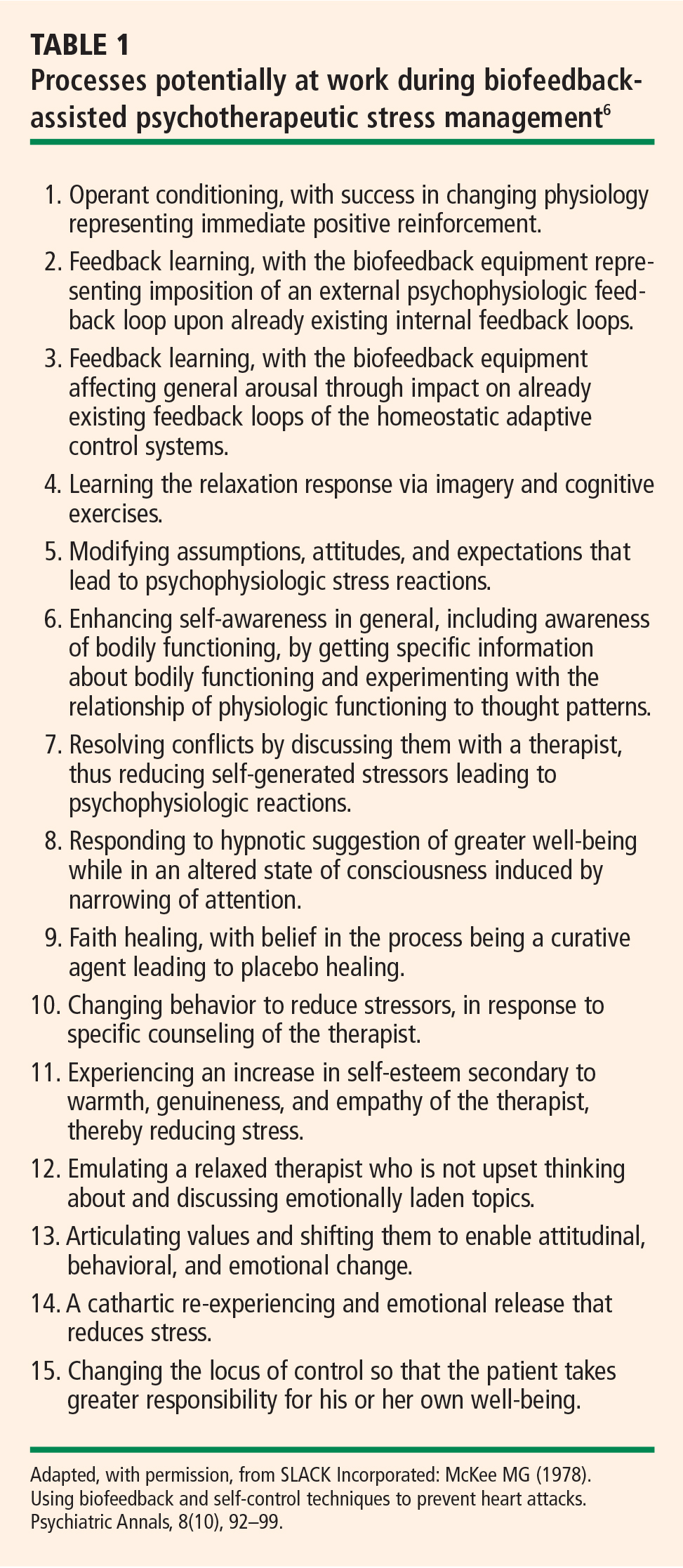
Biofeedback training in this model may involve a coach/instructor/therapist only to the extent of explaining the equipment and its use. In other words, the coach “teaches the patient how to use the mirror.” More commonly.particularly for training in lowered arousal for patients in whom stress reactivity is a significant factor in the development and maintenance of excessive (sympathetic nervous system) arousal that leads to symptoms.a skilled therapist is present. The therapist not only teaches the patient how to use information from biofeedback instruments but also guides the patient in identifying and changing cognitive, emotional, and behavioral patterns that contribute to excessive reactivity. The relationship of physiologic reactivity to the subject matter under discussion also helps diagnostically in identifying stressful areas of life, particularly in psychophysiologic responders who are repressive and denying and who are not good at identifying the stressors in their lives. The equipment becomes a mirror that lets the patient see a problem that he or she had not identified as such.5
Therapeutic/stress-management/biofeedback model
When treating patients with disordered physiology (including autonomic imbalance) in the therapeutic/stress-management/biofeedback model, it is essential to understand each patient as an individual. In this model, stress management and psychotherapeutic interventions address particular vulnerabilities that lead to excessive arousal. This approach starts with a psychophysiologic assessment in which resting levels of relevant physiologic dimensions are measured; this is followed by imposition of stressors to measure reactivity and then by a recovery period in which rate and extent of recovery are measured. An interview and psychological test help determine which cognitive, emotional and behavioral patterns contribute to vulnerability. Patients typically respond well to this approach. It is common for patients to use such descriptions as, “I break out in a cold sweat when I’m stressed,” or “I feel heartsick when I’m stressed,” which suggests that the notion of mind-body interaction resonates with patients.6
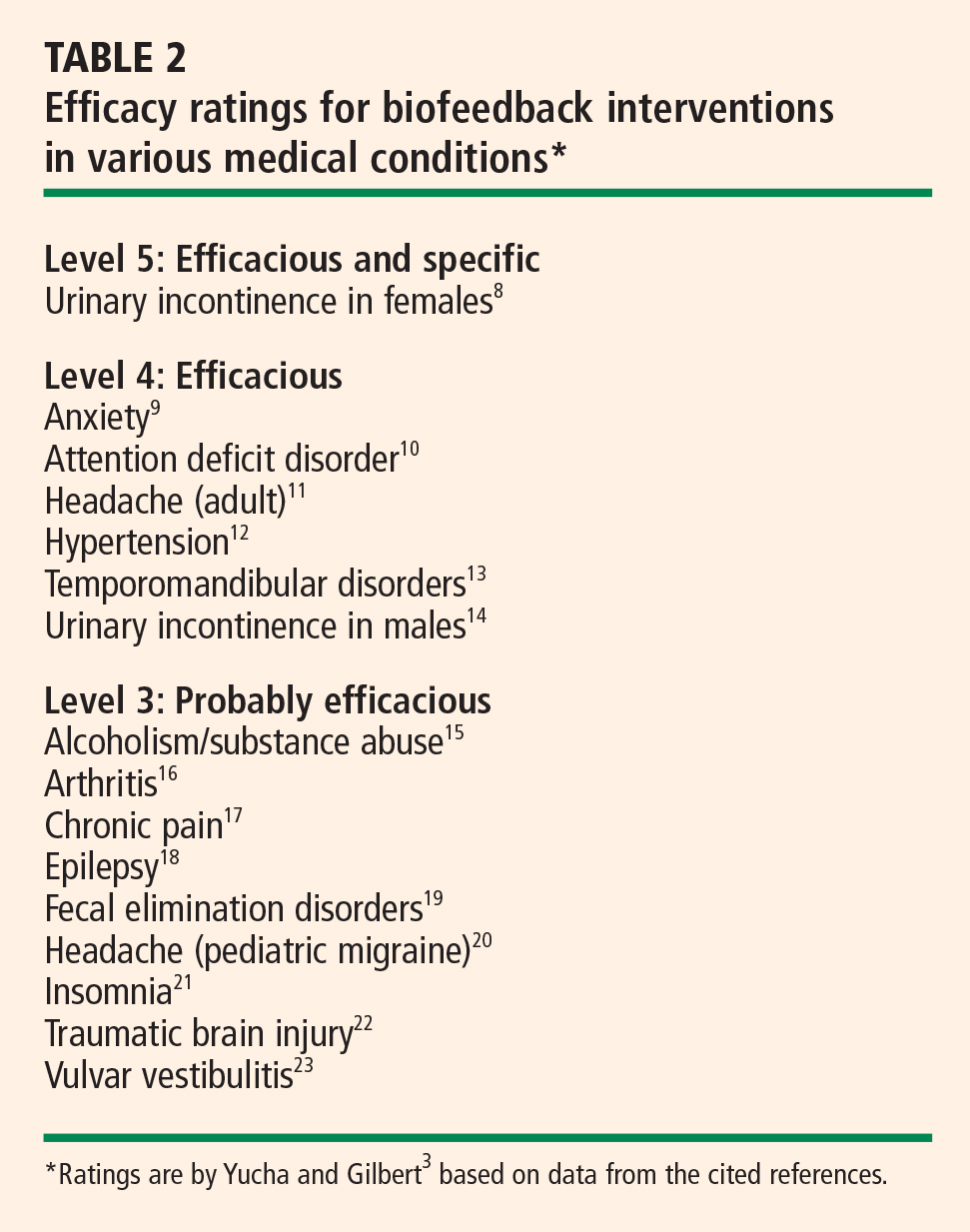
CRITERIA FOR EVALUATING EFFICACY OF BIOFEEDBACK INTERVENTIONS
Several years ago a task force of the Association for Applied Psychophysiology and Biofeedback and the Society for Neuronal Regulation published criteria for evaluating the clinical efficacy of biofeedback/psychophysiologic interventions.7 These criteria are detailed below.3,7
Level 1: Not empirically supported
This designation applies to interventions supported only by anecdotal reports and/or case studies in non–peer-reviewed venues (ie, not empirically supported).
Level 2: Possibly efficacious
This applies to interventions supported by at least one study of sufficient statistical power with well-identified outcome measures but which lacked randomized assignment to a control condition internal to the study.
Level 3: Probably efficacious
This applies to interventions supported by multiple observational studies, clinical studies, wait-list–controlled studies, and within-subject and intrasubject replication studies that demonstrate efficacy.
Level 4: Efficacious
a. In a comparison with a no-treatment control group, alternative treatment group, or sham (placebo) control using randomized assignment, the intervention is shown to be statistically significantly superior to the control condition, or the intervention is equivalent to a treatment of established efficacy in a study with sufficient power to detect moderate differences, and
b. The studies have been conducted with a population treated for a specific problem, for whom inclusion criteria are delineated in a reliable, operationally defined manner, and
c. The study used valid and clearly specified outcome measures related to the problem being treated, and
d. The data were subjected to appropriate data analysis, and
e. The diagnostic and treatment variables and procedures were clearly defined in a manner that permits replication of the study by independent researchers, and
f. The superiority or equivalence of the intervention has been shown in at least two independent research settings.
Level 5: Efficacious and specific
This designation applies when the intervention has been shown to be superior to credible sham therapy, pill therapy, or alternative bona fide treatment in at least two independent research settings.
Efficacy ratings for specific disorders
Despite high standards, biofeedback thrives
The above criteria represent high standards. Since biofeedback training is often more like physical therapy or learning a language, double-blind protocols usually are not feasible, nor is sham training. Moreover, the effectiveness of training is perhaps even more difficult to assess in daily practice, with the inevitable multiplicity of confounding variables. Nevertheless, biofeedback training for many disorders is standing the test of both time and outcomes research, and it is increasingly embraced by the public and recognized by health care insurers and professionals alike.
- Gaarder KR, Montgomery PS. Clinical biofeedback: a procedural manual. Baltimore, MD: Williams and Wilkins; 1977.
- Schwartz MS, Andrasik F. Biofeedback: a practitioner’s guide. 3rd ed. New York, NY: Guilford Press; 2003.
- Yucha C, Gilbert C. Evidence-based practice in biofeedback and neurofeedback. Wheat Ridge, CO: Association for Applied Psychophysiology and Biofeedback; 2004.
- Lehrer PM, Vaschillo E, Lu SE, et al. Heart rate variability biofeedback: effects of age on heart rate variability, baroreflex gain, and asthma. Chest 2006; 129:278–284.
- McKee MG. Contributions of psychophysiologic monitoring to diagnosis and treatment of chronic head pain: a case study. Headache Q 1991; II(4):327–330.
- McKee MG. Using biofeedback and self-control techniques to prevent heart attacks. Psychiatr Ann 1978; 8:10.
- Moss D, Gunkelman J. Task Force Report on methodology and empirically supported treatments: introduction. Appl Psychophysiol Biofeedback 2002; 27:271–272.
- Weatherall M. Biofeedback or pelvic floor muscle exercises for female genuine stress incontinence: a meta-analysis of trials identified in a systematic review. BJU Int 1999; 83:1015–1016.
- Wenck LS, Leu PW, D’Amato RC. Evaluating the efficacy of a biofeedback intervention to reduce children’s anxiety. J Clin Psychol 1996; 52:469–473.
- Kaiser DA, Othmer S. Effect of neurofeedback on variables of attention in a large multi-center trial. J Neurother 2000; 4:5–15.
- Silberstein SD, for the US Headache Consortium. Practice parameter: evidence-based guidelines for migraine headache (an evidence-based review): report of the Quality Standards Subcommittee of the American Academy of Neurology. Neurology 2000; 55:754–762.
- Yucha CB, Clark L, Smith M, Uris P, Lafleur B, Duval S. The effect of biofeedback in hypertension. Appl Nurs Res 2001; 14:29–35.
- Crider AB, Glaros AG. A meta-analysis of EMG biofeedback treatment of temporomandibular disorders. J Orofac Pain 1999; 13:29–37.
- Van Kampen M, De Weerdt W, Van Poppel H, De Ridder D, Feys H, Baert L. Effect of pelvic-floor re-education on duration and degree of incontinence after radical prostatectomy: a randomised controlled trial. Lancet 2000; 355:98–102.
- Saxby E, Peniston EG. Alpha-theta brainwave neurofeedback training: an effective treatment for male and female alcoholics with depressive symptoms. J Clin Psychol 1995; 51:685–693.
- Lavigne JV, Ross CK, Berry SL, Hayford JR, Pachman LM. Evaluation of a psychological treatment package for treating pain in juvenile rheumatoid arthritis. Arthritis Care Res 1992; 5:101–110.
- Humphreys PA, Gevirtz RN. Treatment of recurrent abdominal pain: components analysis of four treatment protocols. J Pediatr Gastroenterol Nutr 2000; 31:47–51.
- Kotchoubey B, Strehl U, Uhlmann C, et al. Modification of slow cortical potentials in patients with refractory epilepsy: a controlled outcome study. Epilepsia 2001; 42:406–416.
- Chiarioni G, Bassotti G, Stanganini S, Vantini I, Whitehead WE. Sensory retraining is key to biofeedback therapy for formed stool fecal incontinence. Am J Gastroenterol 2002; 97:109–117.
- Labbé EE. Treatment of childhood migraine with autogenic training and skin temperature biofeedback: a component analysis. Headache 1995; 35:10–13.
- Morin CM, Hauri PJ, Espie CA, Spielman AJ, Buysse DJ, Bootzin RR. Nonpharmacologic treatment of chronic insomnia. An American Academy of Sleep Medicine review. Sleep 1999; 22:1134–1156.
- Thornton K. Improvement/rehabilitation of memory functioning with neurotherapy/QEEG biofeedback. J Head Trauma Rehabil 2000; 15:1285–1296.
- Bergeron S, Binik YM, Khalifé S, et al. A randomized comparison of group cognitive–behavioral therapy, surface electromyographic biofeedback, and vestibulectomy in the treatment of dyspareunia resulting from vulvar vestibulitis. Pain 2001; 91:297–306.
Clinical biofeedback therapy is one of the many new approaches in health care aimed at helping individuals take responsibility for their well-being, including responsibility for the cognitive, emotional, and behavioral changes needed to effect healthy physiologic change. This article provides a brief survey of biofeedback therapy by defining what biofeedback involves, reviewing the various modalities that it can serve to monitor, discussing major models of biofeedback therapy, and outlining criteria for evaluating the efficacy of biofeedback interventions.
BIOFEEDBACK: BOTH PROCESS AND INSTRUMENTATION
Biofeedback refers to both a process and the instrumentation used in that process.
The process is one of learning to use physiologic information that is monitored and “fed back” through biofeedback instruments. The term dates from 1969, when it was coined to describe laboratory procedures that had been developed in the 1940s in which research subjects learned to modify heart rate, blood flow, and other physiologic functions that were not normally thought of as being subject to conscious control. Feedback itself has been present through much of human history, particularly through the use of mirrored surfaces to practice the expression of emotion.1
Biofeedback instruments monitor one or more physiologic processes, measure what is monitored and transform that measurement into auditory and/or visual signals, and present what is monitored and measured in a simple, direct, and immediate way. Biofeedback equipment typically is noninvasive. The instruments provide continuous monitoring and transformation of physiologic data into understandable feedback for the patient being monitored. Current computerized instruments can provide simultaneous displays and recording of multiple channels of physiologic information. The goal is to enable the individual being monitored to change some physiologic process, guided by the information provided by the biofeedback equipment. How many training sessions are necessary varies with the individual and the disorder, ranging from a few to 50 or more. Our experience is that the great majority of patients obtain benefit in 8 to 12 sessions.
MULTIPLE MODALITIES FOR MONITORING
Multiple modalities can be monitored via biofeedback. Surface electromyography is perhaps the most commonly used instrumentation. Other commonly used measures in a psychophysiologic/biofeedback assessment are respiration rate and depth, skin surface temperature (particularly at the fingertips), cardiovascular reactivity (particularly heart rate and blood pressure), and electrodermal response.2
Feedback of real-time physiologic data is limited only by one’s creativity and technological capabilities. Most of the early noncomputerized equipment provided feedback through the onset and offset of sounds, the changing of tones and volume, the turning on and off of lights, and digital numeric displays indicating both the direction of change and absolute values (such as digital peripheral temperature). Current computerized equipment uses such feedback features as computer games, which the patient “wins” by reaching a goal (such as a systolic blood pressure level below 130 mm Hg), mandalas that can be filled in with colors of the patient’s choosing as he or she progresses in the desired direction, and complex computer-generated figures and graphs.
Electroencephalographic biofeedback (neurofeedback) has become a separate area of study and application, with particular use in the treatment of attention deficit disorder. A baseline electroencephalogram is used in neurofeedback assessment to identify abnormal patterns, and follow-up training is provided to teach the patient to change these patterns in a healthy direction.3
More recently, heart rate variability has come into use as a measure of adaptability or autonomic balance. Soviet scientists were the first to study heart rate variability biofeedback, working with cosmonauts in measuring autonomic function. They found that the low-frequency (0.1-Hz) bands produced the highest frequency-specific oscillations in heart rate variability, and training typically proceeds in increasing amplitude of the low-frequency band (also called the baroreceptor band). Because diminished heart rate variability is a predictor of increased risk for cardiac mortality, teaching patients to increase heart rate variability made sense. The training involves instruction in breathing at an identified resonant frequency that is related to optimal low-frequency band power.4
LEARNING AND MODELS OF BIOFEEDBACK
Accurate feedback facilitates the learning of any skill, whether it be sinking a golf putt, solving an algebra problem, or controlling physiologic behavior. A man playing darts blindfolded is unlikely to achieve as good a score as he would with the blindfold off, because feedback makes a difference.5
Four conditions are important for effective learning;5 the learner must:
- Have the capacity to respond
- Be motivated to learn
- Be positively reinforced for learning
- Be given accurate information about the results of the learning effort.
Direct feedback learning model
The direct feedback learning model assumes that adding feedback to the other important conditions of learning will result in a patient gaining control of the relevant physiology being targeted. This model has been used in treating many disorders, including Raynaud phenomenon and urinary and fecal incontinence.
Biofeedback training in this model may involve a coach/instructor/therapist only to the extent of explaining the equipment and its use. In other words, the coach “teaches the patient how to use the mirror.” More commonly.particularly for training in lowered arousal for patients in whom stress reactivity is a significant factor in the development and maintenance of excessive (sympathetic nervous system) arousal that leads to symptoms.a skilled therapist is present. The therapist not only teaches the patient how to use information from biofeedback instruments but also guides the patient in identifying and changing cognitive, emotional, and behavioral patterns that contribute to excessive reactivity. The relationship of physiologic reactivity to the subject matter under discussion also helps diagnostically in identifying stressful areas of life, particularly in psychophysiologic responders who are repressive and denying and who are not good at identifying the stressors in their lives. The equipment becomes a mirror that lets the patient see a problem that he or she had not identified as such.5
Therapeutic/stress-management/biofeedback model
When treating patients with disordered physiology (including autonomic imbalance) in the therapeutic/stress-management/biofeedback model, it is essential to understand each patient as an individual. In this model, stress management and psychotherapeutic interventions address particular vulnerabilities that lead to excessive arousal. This approach starts with a psychophysiologic assessment in which resting levels of relevant physiologic dimensions are measured; this is followed by imposition of stressors to measure reactivity and then by a recovery period in which rate and extent of recovery are measured. An interview and psychological test help determine which cognitive, emotional and behavioral patterns contribute to vulnerability. Patients typically respond well to this approach. It is common for patients to use such descriptions as, “I break out in a cold sweat when I’m stressed,” or “I feel heartsick when I’m stressed,” which suggests that the notion of mind-body interaction resonates with patients.6
CRITERIA FOR EVALUATING EFFICACY OF BIOFEEDBACK INTERVENTIONS
Several years ago a task force of the Association for Applied Psychophysiology and Biofeedback and the Society for Neuronal Regulation published criteria for evaluating the clinical efficacy of biofeedback/psychophysiologic interventions.7 These criteria are detailed below.3,7
Level 1: Not empirically supported
This designation applies to interventions supported only by anecdotal reports and/or case studies in non–peer-reviewed venues (ie, not empirically supported).
Level 2: Possibly efficacious
This applies to interventions supported by at least one study of sufficient statistical power with well-identified outcome measures but which lacked randomized assignment to a control condition internal to the study.
Level 3: Probably efficacious
This applies to interventions supported by multiple observational studies, clinical studies, wait-list–controlled studies, and within-subject and intrasubject replication studies that demonstrate efficacy.
Level 4: Efficacious
a. In a comparison with a no-treatment control group, alternative treatment group, or sham (placebo) control using randomized assignment, the intervention is shown to be statistically significantly superior to the control condition, or the intervention is equivalent to a treatment of established efficacy in a study with sufficient power to detect moderate differences, and
b. The studies have been conducted with a population treated for a specific problem, for whom inclusion criteria are delineated in a reliable, operationally defined manner, and
c. The study used valid and clearly specified outcome measures related to the problem being treated, and
d. The data were subjected to appropriate data analysis, and
e. The diagnostic and treatment variables and procedures were clearly defined in a manner that permits replication of the study by independent researchers, and
f. The superiority or equivalence of the intervention has been shown in at least two independent research settings.
Level 5: Efficacious and specific
This designation applies when the intervention has been shown to be superior to credible sham therapy, pill therapy, or alternative bona fide treatment in at least two independent research settings.
Efficacy ratings for specific disorders
Despite high standards, biofeedback thrives
The above criteria represent high standards. Since biofeedback training is often more like physical therapy or learning a language, double-blind protocols usually are not feasible, nor is sham training. Moreover, the effectiveness of training is perhaps even more difficult to assess in daily practice, with the inevitable multiplicity of confounding variables. Nevertheless, biofeedback training for many disorders is standing the test of both time and outcomes research, and it is increasingly embraced by the public and recognized by health care insurers and professionals alike.
- Gaarder KR, Montgomery PS. Clinical biofeedback: a procedural manual. Baltimore, MD: Williams and Wilkins; 1977.
- Schwartz MS, Andrasik F. Biofeedback: a practitioner’s guide. 3rd ed. New York, NY: Guilford Press; 2003.
- Yucha C, Gilbert C. Evidence-based practice in biofeedback and neurofeedback. Wheat Ridge, CO: Association for Applied Psychophysiology and Biofeedback; 2004.
- Lehrer PM, Vaschillo E, Lu SE, et al. Heart rate variability biofeedback: effects of age on heart rate variability, baroreflex gain, and asthma. Chest 2006; 129:278–284.
- McKee MG. Contributions of psychophysiologic monitoring to diagnosis and treatment of chronic head pain: a case study. Headache Q 1991; II(4):327–330.
- McKee MG. Using biofeedback and self-control techniques to prevent heart attacks. Psychiatr Ann 1978; 8:10.
- Moss D, Gunkelman J. Task Force Report on methodology and empirically supported treatments: introduction. Appl Psychophysiol Biofeedback 2002; 27:271–272.
- Weatherall M. Biofeedback or pelvic floor muscle exercises for female genuine stress incontinence: a meta-analysis of trials identified in a systematic review. BJU Int 1999; 83:1015–1016.
- Wenck LS, Leu PW, D’Amato RC. Evaluating the efficacy of a biofeedback intervention to reduce children’s anxiety. J Clin Psychol 1996; 52:469–473.
- Kaiser DA, Othmer S. Effect of neurofeedback on variables of attention in a large multi-center trial. J Neurother 2000; 4:5–15.
- Silberstein SD, for the US Headache Consortium. Practice parameter: evidence-based guidelines for migraine headache (an evidence-based review): report of the Quality Standards Subcommittee of the American Academy of Neurology. Neurology 2000; 55:754–762.
- Yucha CB, Clark L, Smith M, Uris P, Lafleur B, Duval S. The effect of biofeedback in hypertension. Appl Nurs Res 2001; 14:29–35.
- Crider AB, Glaros AG. A meta-analysis of EMG biofeedback treatment of temporomandibular disorders. J Orofac Pain 1999; 13:29–37.
- Van Kampen M, De Weerdt W, Van Poppel H, De Ridder D, Feys H, Baert L. Effect of pelvic-floor re-education on duration and degree of incontinence after radical prostatectomy: a randomised controlled trial. Lancet 2000; 355:98–102.
- Saxby E, Peniston EG. Alpha-theta brainwave neurofeedback training: an effective treatment for male and female alcoholics with depressive symptoms. J Clin Psychol 1995; 51:685–693.
- Lavigne JV, Ross CK, Berry SL, Hayford JR, Pachman LM. Evaluation of a psychological treatment package for treating pain in juvenile rheumatoid arthritis. Arthritis Care Res 1992; 5:101–110.
- Humphreys PA, Gevirtz RN. Treatment of recurrent abdominal pain: components analysis of four treatment protocols. J Pediatr Gastroenterol Nutr 2000; 31:47–51.
- Kotchoubey B, Strehl U, Uhlmann C, et al. Modification of slow cortical potentials in patients with refractory epilepsy: a controlled outcome study. Epilepsia 2001; 42:406–416.
- Chiarioni G, Bassotti G, Stanganini S, Vantini I, Whitehead WE. Sensory retraining is key to biofeedback therapy for formed stool fecal incontinence. Am J Gastroenterol 2002; 97:109–117.
- Labbé EE. Treatment of childhood migraine with autogenic training and skin temperature biofeedback: a component analysis. Headache 1995; 35:10–13.
- Morin CM, Hauri PJ, Espie CA, Spielman AJ, Buysse DJ, Bootzin RR. Nonpharmacologic treatment of chronic insomnia. An American Academy of Sleep Medicine review. Sleep 1999; 22:1134–1156.
- Thornton K. Improvement/rehabilitation of memory functioning with neurotherapy/QEEG biofeedback. J Head Trauma Rehabil 2000; 15:1285–1296.
- Bergeron S, Binik YM, Khalifé S, et al. A randomized comparison of group cognitive–behavioral therapy, surface electromyographic biofeedback, and vestibulectomy in the treatment of dyspareunia resulting from vulvar vestibulitis. Pain 2001; 91:297–306.
Biofeedback therapy in cardiovascular disease: Rationale and research overview
The potential of biofeedback therapies in cardiovascular disease is only recently beginning to be explored in a systematic way. This article reviews the rationale for the use of biofeedback therapy in cardiovascular disease and briefly surveys research on the usefulness of biofeedback for several specific cardiovascular parameters and conditions.
RECOGNIZING THE POTENTIAL OF BIOFEEDBACK IN CARDIOVASCULAR DISEASE
Biofeedback is part of a group of modalities known as “self-regulation therapies,” in which a subject is taught to control the activities of his or her autonomic nervous system. The autonomic nervous system has also been called the “visceral,” “involuntary,” and “automatic” nervous system, which suggests that the physiologic processes governed by this branch of the nervous system are largely beyond conscious control. Until the 1950s, this was largely believed to be true. Physicians and scientists had been convinced that the functions regulated by the sympathetic and parasympathetic branches of the autonomic nervous system, such as digestion, blood pressure, and body temperature, were not amenable to self-regulation.
During the 1950s, however, it became clear that functions of the autonomic nervous system could be controlled by conscious thought and training. Subjects could be taught to correctly perceive and also to control heart rate, blood pressure, skin temperature, and other seemingly involuntary functions. The field of biofeedback and applied psychophysiology became possible with these discoveries and with the advent of technologies capable of measuring physiologic variables with enough sensitivity to detect small changes.
Key role of sympathetic/parasympathetic balance
In cardiovascular medicine, biofeedback has a great deal of therapeutic potential because many diseases of the heart and vasculature involve inappropriate regulation of the autonomic nervous system.
Under normal conditions, the sympathetic branch of the autonomic nervous system serves to augment cardiac function in times of stress, increasing heart rate, contractility, and blood pressure, as well as favoring clotting processes that would be mainly adaptive during the “fight or flight” response. The parasympathetic branch of the autonomic nervous system plays the opposite role during health, exerting a calming influence on cardiovascular function.
Normal cardiovascular function is regulated by a balance between sympathetic and parasympathetic inputs to the heart and blood vessels. Heart rate, for example, is governed by the parasympathetic nervous system under resting conditions, when the intrinsic firing rate of the sinus node is decreased by vagal input. Under stressful conditions, this inhibition is released and sympathetic excitation can increase the heart rate even further. In many pathological cardiac conditions, such as arrhythmias, an imbalance between the two branches of the autonomic nervous system causes at least some of the disease manifestations and often contributes to progression.
Biofeedback as a ‘physiologic beta-blocker’
Another good example is heart failure, where over-activation of the sympathetic nervous system results in many of the phenotypic changes in the myocardium and contributes to the downward spiral from compensatory cardiac hypertrophy to end-stage decompensated failure. The role of sympathetic overactivation in heart failure is clearly evident by the success of beta-adrenergic blocking agents in ameliorating symptoms and delaying disease progression. Given the role of autonomic nervous system dysregulation in cardiovascular diseases, biofeedback therapy has the potential to teach patients a skill that may allow them to decrease activation of their autonomic nervous system, theoretically acting as a “physiologic beta-blocker.”
An adjunct to stress management
The potential of biofeedback to have an impact in the arena of cardiovascular disease has not been well explored. Clinically, biofeedback is often used in the context of stress management programs, but biofeedback is not synonymous with stress management. Stress management programs most commonly involve some type of relaxation training and perhaps cognitive behavioral therapy. Biofeedback can be used to augment relaxation, helping the subject to be more aware of physiologic responses and thus be better able to elicit the relaxation response. Biofeedback can also be used to train subjects to control particular physiologic responses that contribute to symptoms or to disease progression. In cardiovascular disease, although stress management is frequently a component of cardiac rehabilitation programs, the question of whether stress management is more effective with or without biofeedback has not been systematically investigated.
PIONEERING STUDIES OF BIOFEEDBACK IN CARDIOVASCULAR DISEASE
Some of the earliest studies of physiologic regulation using biofeedback were attempted in patients with cardiovascular abnormalities. In 1971, Weiss and Engel reported success in using operant conditioning of heart rate in eight patients with premature ventricular contractions.1 All eight patients were able to achieve some degree of control, and five of the patients were able to decrease the frequency of premature beats, demonstrating increased success over a 21-month follow-up period. Interestingly, use of pharmacologic agents to understand the mechanisms of control suggested that one patient was able to decrease sympathetic control of his heart rate while another increased the parasympathetic influence.
Several years later, Pickering and Gorham reported their work with a single subject, a 31-year-old woman who had a ventricular parasystolic rhythm.2 Using a biofeedback technique, they were able to teach the woman to voluntarily control her heart rate, demonstrating that she could both increase and decrease the rate, avoiding the ranges in which the arrhythmia occurred. In the same year, Benson et al demonstrated that they could teach patients the relaxation response and decrease the incidence of premature ventricular contractions.3 Using Holter monitors for validation, these investigators showed that 4 weeks of relaxation training resulted in 8 of 11 patients being able to control their heart rates sufficiently to have therapeutic impact.
These pioneering studies were very early in the development of the field of biofeedback, but they showed what has been clearly established since—that the input of the autonomic nervous system to the heart can be regulated by biofeedback techniques.
BIOFEEDBACK STUDIES OF SPECIFIC CARDIOVASCULAR PARAMETERS AND DISEASES
A host of parameters for assessment
Many cardiovascular parameters can be used for biofeedback. Commonly these include heart rate, blood pressure, skin temperature, and, more recently, heart rate variability. In each case, the parameter is measured and displayed for the subject, and the subject is taught to make it change in a positive direction through relaxation, thought patterns, imagery, or some combination of techniques. Many times the display of the physiologic parameter and the demonstration that it can be controlled are quite surprising to the subject and lead to an enhanced desire to participate in the therapy.
Heart rate variability: A focus of recent interest
The newest parameter in use, and one that has gained considerable interest in the field of cardiovascular biofeedback, is heart rate variability.4 Heart rate variability refers to the variation within the R-R interval of the electrocardiogram during a fixed cycle. It is associated with adaptiveness of the cardiovascular system, and high variability is believed to be a sign of health. Low variability is associated with a number of disease states. Heart rate variability reflects the balance between sympathetic and parasympathetic input to the heart, and many cardiac disease states have been shown to be associated with low variability. Therapies that increase heart rate variability have been shown to improve prognosis.
On the basis of these observations, heart rate variability biofeedback is used to train patients to increase the variability in their heart rate, using feedback from equipment that records the R-R interval from the electrocardiogram or from blood pulse volume sensors. Patients learn to make the variability greater, primarily by breathing at a resonant frequency, as described by Lehrer et al.5
Several preliminary studies have been conducted with heart rate variability in cardiac patients, but much remains to be understood about its use. In 63 patients with established coronary artery disease, Del Pozo et al showed that six biofeedback sessions coupled with daily practice resulted in significantly increased heart rate variability.6 Similarly, Nolan and colleagues found that five sessions of biofeedback improved symptoms and quality of life in 46 patients with coronary artery disease.7 In 14 patients with heart failure, Luskin et al demonstrated that eight sessions of heart rate variability biofeedback produced reductions in perceived stress and improved function on the 6-minute walk test.8
It remains unclear whether heart rate variability biofeedback has more or less potential than other types of biofeedback in patients with cardiovascular disease, but these preliminary observations suggest that it may be useful in improving symptoms and quality of life.
Biofeedback in hypertension: Despite decades of study, conclusions elusive
Among diseases of the cardiovascular system, biofeedback has been used most frequently in hypertension, where it has been under investigation for more than 30 years, since the early days of biofeedback study.9 The field of biofeedback in hypertension is fraught with difficulties, rendering conclusions about its efficacy difficult.
Biofeedback has been assessed in many different types of hypertension, often within the same study. Essential hypertension and “white coat” hypertension, now known as excessive cardiovascular reactivity, have been most commonly investigated, but with no apparent consensus. The biofeedback techniques used in these studies have ranged from blood pressure biofeedback to electromyography, finger temperature, and skin conductance. More recently, heart rate variability biofeedback has also been used in this population.
In general, biofeedback has been more successful in the treatment of hypertension when respiratory training has been a component of the biofeedback. McGrady has established that certain types of patients with hypertension fare better with biofeedback than others.10 These include patients with higher baseline blood pressure, higher heart rate, cool hands, high electromyographic response, and high plasma renin activity.in short, patients who can be seen to have a high degree of sympathetic arousal.
Blood pressure can be lowered by 6 to 10 mm Hg when biofeedback is effective, which is less of an effect than that observed with most drug therapy for hypertension. Biofeedback does have the advantage, however, of improving overall cardiovascular reactivity and giving the patient a greater sense of control over his or her physical well-being, which may prove valuable in the setting of hypertension. Typically, the most effective interventions for hypertension (and perhaps for cardiovascular disease in general) are individualized for the patient and not protocol-driven. Thus, although biofeedback has potential in hypertension, its efficacy is not proven and systematic trials are lacking.
Biofeedback in heart failure: Targeting sympathetic overactivation
In patients with heart failure, the sympathetic nervous system is overactivated, as noted previously. High levels of plasma norepinephrine correlate with worse prognosis. Decreasing activation of the sympathetic nervous system improves both symptoms and prognosis, as demonstrated in patients taking beta-adrenergic blocking agents or those treated with a left ventricular assist device.
Several studies have suggested that biofeedback may be able to provide a similar reduction in sympathetic nervous system activation in patients with heart failure. Moser and colleagues showed that a single session of skin temperature biofeedback plus relaxation training increased cardiac output in patients with heart failure,11 while studies by Weiner et al,12 Bernardi et al,13 and Mangin et al14 showed that training heart failure patients to breathe more slowly increased their exercise tolerance. Although these studies are preliminary, they support the speculation that if biofeedback can decrease activation of the sympathetic nervous system in patients with heart failure, it may actually cause some degree of remodeling of the failing heart, such as that observed with beta-blockers or left ventricular assist device therapy.
BIOFEEDBACK AND STRESS MANAGEMENT: AN OPPORTUNITY FOR WIDER IMPACT
As mentioned earlier, biofeedback can serve as a component of stress management programs. Biofeedback is often a very effective adjunct to stress management because it teaches the subject to control physiologic reactions that are part of the stress response and gives the subject feedback to suggest that he or she is adequately practicing relaxation. Biofeedback-mediated stress management may actually be the most practical use of biofeedback in the setting of cardiovascular disease because it is easy to practice and can have an effect on large numbers of patients.
Mental stress has been well documented as a significant risk factor for many forms of cardiovascular disease, and stress management programs have been shown to have an impact on disease progression and symptoms. Many studies, including those reported by Sheps et al for the Psychophysiological Investigations of Myocardial Ischemia (PIMI) study,15 have shown that patients who exhibit ischemia in response to a mental stress test have increased mortality from cardiovascular disease. Jiang and colleagues,16 among others, have shown that mental stress predicts cardiac events in patients with lower ejection fractions, and Blumenthal et al17 have repeatedly demonstrated that stress management training reduces the incidence of wall motion abnormalities in patients with cardiovascular disease. Stress management is included in many cardiac rehabilitation programs, and it is likely that routine use of biofeedback as a component of stress management programs would benefit patients with cardiovascular disease, in whom reproducibly decreasing activation of the autonomic nervous system should be helpful.
According to a recent article in the Heart Advisor, 84% of physicians believe that stress is a risk for cardiovascular disease but only 35% say they feel knowledgeable about stress and a mere 5% feel that they succeed in helping stressed patients.18 Anything that could improve these numbers would be beneficial.
CONCLUSIONS
Cardiovascular conditions in which biofeedback has been shown to be helpful include arrhythmias, hypertension, Raynaud phenomenon, ischemia, infarction, and heart failure, but we have barely begun to explore the potential of biofeedback therapy. Given that many cardiovascular diseases involve inappropriate regulation of the autonomic nervous system, instruction in the use of biofeedback to control activation of the sympathetic and parasympathetic nervous systems is likely to be useful in cardiac patients. Systematic trials are needed.
- Weiss T, Engel BT. Operant conditioning of heart rate in patients with premature ventricular contractions. Psychosom Med 1971; 33:301–322.
- Pickering T, Gorham G. Learned heart-rate control by a patient with a ventricular parasystolic rhythm. Lancet 1975; 1:252–253.
- Benson H, Alexander S, Feldman CL. Decreased premature ventricular contractions through use of the relaxation response in patients with stable ischaemic heart disease. Lancet 1975; 2:380–382.
- Kranitz L, Lehrer P. Biofeedback applications in the treatment of cardiovascular diseases. Cardiol Rev 2004; 12:177–181.
- Lehrer PM, Vaschilllo E, Vaschillo B. Resonant frequency biofeedback training to increase cardiac variability: rationale and manual for training. Appl Psychophysiol Biofeedback 2000; 25:177–191.
- Del Pozo JM, Gevirtz RN, Scher B, Guarneri E. Biofeedback treatment increases heart rate variability in patients with known coronary artery disease. Am Heart J 2004; 147:E11.
- Nolan RP, Kamath MV, Floras JS, et al. Heart rate variability biofeedback as a behavioral neurocardiac intervention to enhance vagal heart rate control. Am Heart J 2005; 149:1137.
- Luskin F, Reitz M, Newell K, Quinn TG, Haskell W. A controlled pilot study of stress management training of elderly patients with congestive heart failure. Prev Cardiol 2002; 5:168–172.
- Linden W, Moseley JV. The efficacy of behavioral treatments for hypertension. Appl Psychophysiol Biofeedback 2006; 31:51–63.
- McGrady A. Good news.bad press: applied psychophysiology in cardiovascular disorders. Biofeedback Self Regul 1996; 21:335–346.
- Moser DK, Dracup K, Woo MA, Stevenson LW. Voluntary control of vascular tone by using skin-temperature biofeedback-relaxation in patients with advanced heart failure. Altern Ther Health Med 1997; 3:51–59.
- Weiner P, Waizman J, Magadle R, Berar-Yanay N, Pelled B. The effect of specific inspiratory muscle training on the sensation of dyspnea and exercise tolerance in patients with congestive heart failure. Clin Cardiol 1999; 22:727–732.
- Bernardi L, Porta C, Spicuzza L, et al. Slow breathing increases arterial baroreflex sensitivity in patients with chronic heart failure. Circulation 2002; 105:143–145.
- Mangin L, Monti A, Médigue C, et al. Altered baroreflex gain during voluntary breathing in chronic heart failure. Eur J Heart Fail 2001; 3:189–195.
- Sheps DS, McMahon RP, Becker L, et al. Mental stress-induced ischemia and all-cause mortality in patients with coronary artery disease: results from the Psychophysiological Investigations of Myocardial Ischemia study. Circulation 2002; 105:1780–1784.
- Jiang W, Babyak M, Krantz DS, et al. Mental stress–induced myocardial ischemia and cardiac events. JAMA 1996; 275:1651–1656.
- Blumenthal JA, Sherwood A, Babyak MA, et al. Effects of exercise and stress management training on markers of cardiovascular risk in patients with ischemic heart disease: a randomized controlled trial. JAMA 2005; 293:1626–1634.
- How to stop the toll of stress. Heart Advisor; March 2006:4–5.
The potential of biofeedback therapies in cardiovascular disease is only recently beginning to be explored in a systematic way. This article reviews the rationale for the use of biofeedback therapy in cardiovascular disease and briefly surveys research on the usefulness of biofeedback for several specific cardiovascular parameters and conditions.
RECOGNIZING THE POTENTIAL OF BIOFEEDBACK IN CARDIOVASCULAR DISEASE
Biofeedback is part of a group of modalities known as “self-regulation therapies,” in which a subject is taught to control the activities of his or her autonomic nervous system. The autonomic nervous system has also been called the “visceral,” “involuntary,” and “automatic” nervous system, which suggests that the physiologic processes governed by this branch of the nervous system are largely beyond conscious control. Until the 1950s, this was largely believed to be true. Physicians and scientists had been convinced that the functions regulated by the sympathetic and parasympathetic branches of the autonomic nervous system, such as digestion, blood pressure, and body temperature, were not amenable to self-regulation.
During the 1950s, however, it became clear that functions of the autonomic nervous system could be controlled by conscious thought and training. Subjects could be taught to correctly perceive and also to control heart rate, blood pressure, skin temperature, and other seemingly involuntary functions. The field of biofeedback and applied psychophysiology became possible with these discoveries and with the advent of technologies capable of measuring physiologic variables with enough sensitivity to detect small changes.
Key role of sympathetic/parasympathetic balance
In cardiovascular medicine, biofeedback has a great deal of therapeutic potential because many diseases of the heart and vasculature involve inappropriate regulation of the autonomic nervous system.
Under normal conditions, the sympathetic branch of the autonomic nervous system serves to augment cardiac function in times of stress, increasing heart rate, contractility, and blood pressure, as well as favoring clotting processes that would be mainly adaptive during the “fight or flight” response. The parasympathetic branch of the autonomic nervous system plays the opposite role during health, exerting a calming influence on cardiovascular function.
Normal cardiovascular function is regulated by a balance between sympathetic and parasympathetic inputs to the heart and blood vessels. Heart rate, for example, is governed by the parasympathetic nervous system under resting conditions, when the intrinsic firing rate of the sinus node is decreased by vagal input. Under stressful conditions, this inhibition is released and sympathetic excitation can increase the heart rate even further. In many pathological cardiac conditions, such as arrhythmias, an imbalance between the two branches of the autonomic nervous system causes at least some of the disease manifestations and often contributes to progression.
Biofeedback as a ‘physiologic beta-blocker’
Another good example is heart failure, where over-activation of the sympathetic nervous system results in many of the phenotypic changes in the myocardium and contributes to the downward spiral from compensatory cardiac hypertrophy to end-stage decompensated failure. The role of sympathetic overactivation in heart failure is clearly evident by the success of beta-adrenergic blocking agents in ameliorating symptoms and delaying disease progression. Given the role of autonomic nervous system dysregulation in cardiovascular diseases, biofeedback therapy has the potential to teach patients a skill that may allow them to decrease activation of their autonomic nervous system, theoretically acting as a “physiologic beta-blocker.”
An adjunct to stress management
The potential of biofeedback to have an impact in the arena of cardiovascular disease has not been well explored. Clinically, biofeedback is often used in the context of stress management programs, but biofeedback is not synonymous with stress management. Stress management programs most commonly involve some type of relaxation training and perhaps cognitive behavioral therapy. Biofeedback can be used to augment relaxation, helping the subject to be more aware of physiologic responses and thus be better able to elicit the relaxation response. Biofeedback can also be used to train subjects to control particular physiologic responses that contribute to symptoms or to disease progression. In cardiovascular disease, although stress management is frequently a component of cardiac rehabilitation programs, the question of whether stress management is more effective with or without biofeedback has not been systematically investigated.
PIONEERING STUDIES OF BIOFEEDBACK IN CARDIOVASCULAR DISEASE
Some of the earliest studies of physiologic regulation using biofeedback were attempted in patients with cardiovascular abnormalities. In 1971, Weiss and Engel reported success in using operant conditioning of heart rate in eight patients with premature ventricular contractions.1 All eight patients were able to achieve some degree of control, and five of the patients were able to decrease the frequency of premature beats, demonstrating increased success over a 21-month follow-up period. Interestingly, use of pharmacologic agents to understand the mechanisms of control suggested that one patient was able to decrease sympathetic control of his heart rate while another increased the parasympathetic influence.
Several years later, Pickering and Gorham reported their work with a single subject, a 31-year-old woman who had a ventricular parasystolic rhythm.2 Using a biofeedback technique, they were able to teach the woman to voluntarily control her heart rate, demonstrating that she could both increase and decrease the rate, avoiding the ranges in which the arrhythmia occurred. In the same year, Benson et al demonstrated that they could teach patients the relaxation response and decrease the incidence of premature ventricular contractions.3 Using Holter monitors for validation, these investigators showed that 4 weeks of relaxation training resulted in 8 of 11 patients being able to control their heart rates sufficiently to have therapeutic impact.
These pioneering studies were very early in the development of the field of biofeedback, but they showed what has been clearly established since—that the input of the autonomic nervous system to the heart can be regulated by biofeedback techniques.
BIOFEEDBACK STUDIES OF SPECIFIC CARDIOVASCULAR PARAMETERS AND DISEASES
A host of parameters for assessment
Many cardiovascular parameters can be used for biofeedback. Commonly these include heart rate, blood pressure, skin temperature, and, more recently, heart rate variability. In each case, the parameter is measured and displayed for the subject, and the subject is taught to make it change in a positive direction through relaxation, thought patterns, imagery, or some combination of techniques. Many times the display of the physiologic parameter and the demonstration that it can be controlled are quite surprising to the subject and lead to an enhanced desire to participate in the therapy.
Heart rate variability: A focus of recent interest
The newest parameter in use, and one that has gained considerable interest in the field of cardiovascular biofeedback, is heart rate variability.4 Heart rate variability refers to the variation within the R-R interval of the electrocardiogram during a fixed cycle. It is associated with adaptiveness of the cardiovascular system, and high variability is believed to be a sign of health. Low variability is associated with a number of disease states. Heart rate variability reflects the balance between sympathetic and parasympathetic input to the heart, and many cardiac disease states have been shown to be associated with low variability. Therapies that increase heart rate variability have been shown to improve prognosis.
On the basis of these observations, heart rate variability biofeedback is used to train patients to increase the variability in their heart rate, using feedback from equipment that records the R-R interval from the electrocardiogram or from blood pulse volume sensors. Patients learn to make the variability greater, primarily by breathing at a resonant frequency, as described by Lehrer et al.5
Several preliminary studies have been conducted with heart rate variability in cardiac patients, but much remains to be understood about its use. In 63 patients with established coronary artery disease, Del Pozo et al showed that six biofeedback sessions coupled with daily practice resulted in significantly increased heart rate variability.6 Similarly, Nolan and colleagues found that five sessions of biofeedback improved symptoms and quality of life in 46 patients with coronary artery disease.7 In 14 patients with heart failure, Luskin et al demonstrated that eight sessions of heart rate variability biofeedback produced reductions in perceived stress and improved function on the 6-minute walk test.8
It remains unclear whether heart rate variability biofeedback has more or less potential than other types of biofeedback in patients with cardiovascular disease, but these preliminary observations suggest that it may be useful in improving symptoms and quality of life.
Biofeedback in hypertension: Despite decades of study, conclusions elusive
Among diseases of the cardiovascular system, biofeedback has been used most frequently in hypertension, where it has been under investigation for more than 30 years, since the early days of biofeedback study.9 The field of biofeedback in hypertension is fraught with difficulties, rendering conclusions about its efficacy difficult.
Biofeedback has been assessed in many different types of hypertension, often within the same study. Essential hypertension and “white coat” hypertension, now known as excessive cardiovascular reactivity, have been most commonly investigated, but with no apparent consensus. The biofeedback techniques used in these studies have ranged from blood pressure biofeedback to electromyography, finger temperature, and skin conductance. More recently, heart rate variability biofeedback has also been used in this population.
In general, biofeedback has been more successful in the treatment of hypertension when respiratory training has been a component of the biofeedback. McGrady has established that certain types of patients with hypertension fare better with biofeedback than others.10 These include patients with higher baseline blood pressure, higher heart rate, cool hands, high electromyographic response, and high plasma renin activity.in short, patients who can be seen to have a high degree of sympathetic arousal.
Blood pressure can be lowered by 6 to 10 mm Hg when biofeedback is effective, which is less of an effect than that observed with most drug therapy for hypertension. Biofeedback does have the advantage, however, of improving overall cardiovascular reactivity and giving the patient a greater sense of control over his or her physical well-being, which may prove valuable in the setting of hypertension. Typically, the most effective interventions for hypertension (and perhaps for cardiovascular disease in general) are individualized for the patient and not protocol-driven. Thus, although biofeedback has potential in hypertension, its efficacy is not proven and systematic trials are lacking.
Biofeedback in heart failure: Targeting sympathetic overactivation
In patients with heart failure, the sympathetic nervous system is overactivated, as noted previously. High levels of plasma norepinephrine correlate with worse prognosis. Decreasing activation of the sympathetic nervous system improves both symptoms and prognosis, as demonstrated in patients taking beta-adrenergic blocking agents or those treated with a left ventricular assist device.
Several studies have suggested that biofeedback may be able to provide a similar reduction in sympathetic nervous system activation in patients with heart failure. Moser and colleagues showed that a single session of skin temperature biofeedback plus relaxation training increased cardiac output in patients with heart failure,11 while studies by Weiner et al,12 Bernardi et al,13 and Mangin et al14 showed that training heart failure patients to breathe more slowly increased their exercise tolerance. Although these studies are preliminary, they support the speculation that if biofeedback can decrease activation of the sympathetic nervous system in patients with heart failure, it may actually cause some degree of remodeling of the failing heart, such as that observed with beta-blockers or left ventricular assist device therapy.
BIOFEEDBACK AND STRESS MANAGEMENT: AN OPPORTUNITY FOR WIDER IMPACT
As mentioned earlier, biofeedback can serve as a component of stress management programs. Biofeedback is often a very effective adjunct to stress management because it teaches the subject to control physiologic reactions that are part of the stress response and gives the subject feedback to suggest that he or she is adequately practicing relaxation. Biofeedback-mediated stress management may actually be the most practical use of biofeedback in the setting of cardiovascular disease because it is easy to practice and can have an effect on large numbers of patients.
Mental stress has been well documented as a significant risk factor for many forms of cardiovascular disease, and stress management programs have been shown to have an impact on disease progression and symptoms. Many studies, including those reported by Sheps et al for the Psychophysiological Investigations of Myocardial Ischemia (PIMI) study,15 have shown that patients who exhibit ischemia in response to a mental stress test have increased mortality from cardiovascular disease. Jiang and colleagues,16 among others, have shown that mental stress predicts cardiac events in patients with lower ejection fractions, and Blumenthal et al17 have repeatedly demonstrated that stress management training reduces the incidence of wall motion abnormalities in patients with cardiovascular disease. Stress management is included in many cardiac rehabilitation programs, and it is likely that routine use of biofeedback as a component of stress management programs would benefit patients with cardiovascular disease, in whom reproducibly decreasing activation of the autonomic nervous system should be helpful.
According to a recent article in the Heart Advisor, 84% of physicians believe that stress is a risk for cardiovascular disease but only 35% say they feel knowledgeable about stress and a mere 5% feel that they succeed in helping stressed patients.18 Anything that could improve these numbers would be beneficial.
CONCLUSIONS
Cardiovascular conditions in which biofeedback has been shown to be helpful include arrhythmias, hypertension, Raynaud phenomenon, ischemia, infarction, and heart failure, but we have barely begun to explore the potential of biofeedback therapy. Given that many cardiovascular diseases involve inappropriate regulation of the autonomic nervous system, instruction in the use of biofeedback to control activation of the sympathetic and parasympathetic nervous systems is likely to be useful in cardiac patients. Systematic trials are needed.
- Weiss T, Engel BT. Operant conditioning of heart rate in patients with premature ventricular contractions. Psychosom Med 1971; 33:301–322.
- Pickering T, Gorham G. Learned heart-rate control by a patient with a ventricular parasystolic rhythm. Lancet 1975; 1:252–253.
- Benson H, Alexander S, Feldman CL. Decreased premature ventricular contractions through use of the relaxation response in patients with stable ischaemic heart disease. Lancet 1975; 2:380–382.
- Kranitz L, Lehrer P. Biofeedback applications in the treatment of cardiovascular diseases. Cardiol Rev 2004; 12:177–181.
- Lehrer PM, Vaschilllo E, Vaschillo B. Resonant frequency biofeedback training to increase cardiac variability: rationale and manual for training. Appl Psychophysiol Biofeedback 2000; 25:177–191.
- Del Pozo JM, Gevirtz RN, Scher B, Guarneri E. Biofeedback treatment increases heart rate variability in patients with known coronary artery disease. Am Heart J 2004; 147:E11.
- Nolan RP, Kamath MV, Floras JS, et al. Heart rate variability biofeedback as a behavioral neurocardiac intervention to enhance vagal heart rate control. Am Heart J 2005; 149:1137.
- Luskin F, Reitz M, Newell K, Quinn TG, Haskell W. A controlled pilot study of stress management training of elderly patients with congestive heart failure. Prev Cardiol 2002; 5:168–172.
- Linden W, Moseley JV. The efficacy of behavioral treatments for hypertension. Appl Psychophysiol Biofeedback 2006; 31:51–63.
- McGrady A. Good news.bad press: applied psychophysiology in cardiovascular disorders. Biofeedback Self Regul 1996; 21:335–346.
- Moser DK, Dracup K, Woo MA, Stevenson LW. Voluntary control of vascular tone by using skin-temperature biofeedback-relaxation in patients with advanced heart failure. Altern Ther Health Med 1997; 3:51–59.
- Weiner P, Waizman J, Magadle R, Berar-Yanay N, Pelled B. The effect of specific inspiratory muscle training on the sensation of dyspnea and exercise tolerance in patients with congestive heart failure. Clin Cardiol 1999; 22:727–732.
- Bernardi L, Porta C, Spicuzza L, et al. Slow breathing increases arterial baroreflex sensitivity in patients with chronic heart failure. Circulation 2002; 105:143–145.
- Mangin L, Monti A, Médigue C, et al. Altered baroreflex gain during voluntary breathing in chronic heart failure. Eur J Heart Fail 2001; 3:189–195.
- Sheps DS, McMahon RP, Becker L, et al. Mental stress-induced ischemia and all-cause mortality in patients with coronary artery disease: results from the Psychophysiological Investigations of Myocardial Ischemia study. Circulation 2002; 105:1780–1784.
- Jiang W, Babyak M, Krantz DS, et al. Mental stress–induced myocardial ischemia and cardiac events. JAMA 1996; 275:1651–1656.
- Blumenthal JA, Sherwood A, Babyak MA, et al. Effects of exercise and stress management training on markers of cardiovascular risk in patients with ischemic heart disease: a randomized controlled trial. JAMA 2005; 293:1626–1634.
- How to stop the toll of stress. Heart Advisor; March 2006:4–5.
Helping children and adults with hypnosis and biofeedback
Training in self-hypnosis, with or without biofeedback, is a valuable adjunct for children and adults with chronic illnesses or behavioral problems. After defining terms and briefly reviewing the evolution of medical hypnosis, this article provides an overview of the clinical utility and applications of self-hypnosis and various issues in its use, including patient assessment, concurrent use with biofeedback, and how health care providers can become trained in self-hypnosis instruction. Because my experience is primarily with medical hypnosis in children and adolescents, portions of this discussion will devote particular attention to the use of hypnosis in children.
DEFINITIONS
Hypnosis is a state of awareness, often but not always associated with relaxation, during which the participant can give him- or herself suggestions for desired changes to which he or she is more likely to respond than when in the usual state of awareness. Spontaneous self-hypnosis may happen while reading, listening to music, watching television, jogging, dancing, playing a musical instrument, doing tai chi, doing yoga, or performing similar activities. Terms often used to describe mind-body training include relaxation imagery, guided imagery, or visual imagery. These include the same training strategies as those used in hypnosis.
Biofeedback is a term coined in 1969 to describe procedures (developed in 1940s) for training subjects to alter physiologic responses such as brain activity, blood pressure, muscle tension, or heart rate. With biofeedback, participants are trained to improve their health and performance by using signals from their own bodies. In so doing, they strengthen awareness of the connections between their mind and body.
Cyberphysiology was defined by Dr. Earl E. Bakken at the first Archaeus Congress, held in Santa Fe, New Mexico, in 1986. “Cyber” derives from the Greek kybernan, meaning steersman or helmsman. From kybernan came the Latinate term govern, meaning “to control.” Thus, cyberphysiology means to control a physiologic response. In scientific terms, cyberphysiology is the study of how neurally mediated autonomic responses—usually viewed as automatic, reactive reflexes—can be modified by a learning process that appears to be significantly dependent on modification of mental images. Both hypnosis and biofeedback are cyberphysiologic strategies that enable the user to develop voluntary control of certain physiologic processes.
HISTORICAL BEGINNINGS OF HYPNOSIS
Franz Mesmer developed a training system that he called animal magnetism. Mesmer believed that normal body processes were disrupted when there was improper distribution of magnetism, a kind of fluid that could penetrate all matter. He described his ability to direct this magnetic fluid through his presence with the waving of a metallic rod and contact with a large wooden tub called a baquet. Mesmer was convinced that the successful therapeutic effects he observed depended on the magnetic rods he used.
When jealous and hostile colleagues challenged Mesmer’s clinical successes, King Louis XVI of France called for an investigative commission chaired by Benjamin Franklin, who was then the American ambassador to France. Other commission members included Dr. Antoine Lavoisier, the first to isolate the element of oxygen, and Dr. Antoine Guillotine, well known for developing a machine for beheading.1 After the commission conducted some clever experiments, they concluded that Mesmer’s success was related to application of the imagination. In fact, we are not far beyond that concept today, although we now have brain imaging documentation of changes in the brain associated with the practice of hypnosis.2–5
CORRECTING MISCONCEPTIONS ABOUT HYPNOSIS
Hypnosis is not sleep
Modern hypnosis is considered to have begun with Mesmer, although the term hypnosis was first used by James Braid, a Scottish ophthalmologist, in 1843. His decision to derive the word from hypnos, the Greek word for sleep, was unfortunate. Hypnosis is not sleep, but the name confuses people.
All hypnosis is self-hypnosis
Another major misconception about hypnosis is that someone—ie, the hypnotist—is in control of a person. In fact, the hypnotist is a coach or teacher who helps the patient to increase his or her self-regulation abilities.6 All hypnosis is self-hypnosis; after the initial training, the learner must reinforce the training with daily practice. Adult learners should anticipate practicing approximately 10 minutes twice daily for about 2 months in order to condition the desirable physiologic change or outcome. Children learn more easily and often can achieve desired changes over a period of a few weeks.
IMPORTANCE OF PATIENT ASSESSMENT BEFORE TEACHING SELF-HYPNOSIS
Every candidate for self-hypnosis therapy deserves a thoughtful, careful diagnostic assessment that includes appropriate laboratory procedures, radiologic procedures, or both prior to decisions about treatment. Patients are sometimes referred for specific cyberphysiologic interventions, such as hypnosis, without adequate diagnostic assessments.7 When a patient is referred for hypnosis training, the health professional who will provide the training should evaluate the extent of the previous diagnostic assessment and do more if indicated. It is also important that the health professional be knowledgeable and competent with respect to the patient’s specific problem. For example, a dentist who is board-certified in dental hypnosis should not be teaching hypnosis to children with migraine, just as a pediatrician who is board-certified in medical hypnosis should not be extracting teeth using hypnosis.
Mental imagery varies from individual to individual. Many children have visual, auditory, kinesthetic, and olfactory/taste imagery abilities and can use these easily in the process of self-hypnosis. In contrast, many adults do not generate multiple types of mental imagery, and many lack clear visual imagery. It is important that the therapist identify which types of mental imagery the patient prefers before embarking on a therapeutic approach.
CONCURRENT USE OF BIOFEEDBACK AND HYPNOSIS
Much common ground exists between hypnosis and biofeedback. Both have the potential to provide a powerful validation of mind–body links, contribute to a lowered state of sympathetic arousal, heighten awareness of internal events and sensations, facilitate imagery abilities, narrow the focus of attention, and enhance the internal locus of control.
Adding biofeedback games to self-hypnosis training can make the experience much more interesting for children. Children see evidence on the screen that, by changing their thinking, they have control over a body response such as skin temperature, electrodermal activity, or pulse rate variability. Adults also benefit from the addition of biofeedback to self-hypnosis training. A patient cannot effect a change in a biofeedback response without a change in his or her mental imagery.
A WIDE RANGE OF THERAPEUTIC APPLICATIONS
Hypnosis training is valuable as a primary intervention for prevention of juvenile migraine8,9 as well as for many performance problems (eg, fear of public speaking or playing tennis), insomnia, and many habit problems (eg, nail-biting, tics, hair-pulling). For treatment of juvenile warts, hypnosis is at least as effective as topical treatment and associated with fewer relapses.10
Hypnosis is valuable as an adjunctive intervention during painful procedures,11–13 and many adults and children use self-hypnosis to teach themselves to be comfortable through procedures without any pharmacologic treatment.14
Training in self-hypnosis is a valuable adjunct for both children and adults with chronic illnesses such as cancer, cardiac failure, asthma, hemophilia, sickle cell disease, and arthritis. Self-hypnosis helps to reduce anxiety and increase comfort, and it provides a therapeutic tool over which the patient has control. Several recent studies have demonstrated the efficacy of hypnosis in the treatment of irritable bowel syndrome.15
Hypnosis and cardiac disease
With respect to cardiac disease, training in hypnosis can help to reduce symptoms both preoperatively and postoperatively, to enhance the success of rehabilitation following myocardial infarction, and to reduce anxiety associated with chronic heart disease.16
Hypnosis also is helpful for motivating behaviors associated with prevention of cardiac disease, such as regular exercise, eating a low-fat diet, and smoking cessation. Several studies have found hypnosis to be a helpful adjunct to cognitive behavioral therapy for treatment of obesity.17 Additionally, a number of studies have demonstrated that hypnosis is useful as an initial intervention for smoking cessation,18 although only about 45% of persons who stop smoking with hypnosis continue to abstain 6 months later. In the case of both obesity and smoking cessation, hypnosis has modestly better efficacy compared with other treatments for these conditions.
TEACHING SELF-HYPNOSIS: SPECIAL CONSIDERATIONS WITH CHILDREN
Self-hypnosis has great potential in children, as children delight in recognizing their own control over problems such as bed-wetting or wheezing or test anxiety.
As noted above, success with hypnosis requires that the patient practice self-hypnosis daily. In the case of children, it is essential that the coach or teacher emphasize that the child is in control and can decide when and where to use self-hypnosis. The message should be that self-hypnosis belongs to the child and that he or she needs to practice to become more skilled (as with learning soccer or some other sport), but that no one can force him or her to practice.
The choice of strategies for teaching self-hypnosis varies depending on the child’s age and developmental stage. As children mature, their cognitive abilities change. Preschool children are concrete in their thinking, so therapists working with children of this age must select words carefully. Children between ages 2 and 5 years spend a great deal of their time in various types of behavior based on imagination and fantasy. They enjoy stories and may enter a hypnotic state as a parent or teacher reads a story to them. Unlike adults, they often prefer to practice their self-hypnosis with their eyes open. Although adolescents may enjoy learning self-hypnosis methods that are similar to those preferred by adults, immature adolescents may prefer methods that also appeal to younger children. A child with cognitive impairment can learn self-hypnosis if the therapist selects a teaching approach appropriate for the child’s actual developmental stage. Because of developmental changes, a child of 9 years is unlikely to enjoy a method he or she was taught at age 4. Therapists who work with children should be familiar with a variety of hypnosis induction strategies and be capable of creative modification to accommodate a child’s changing developmental circumstances.19,20
HYPNOSIS RESEARCH WITH CHILDREN
Most subsequent research has consisted of clinical studies documenting the efficacy of hypnosis with children in areas such as pain management, habit problems, wart reduction, and performance anxiety. A recent study completed in Cleveland, Ohio, taught stress-reduction methods, including self-hypnosis, to 8-year-old schoolchildren.30 This study concluded that a short daily stress-management intervention delivered in the classroom setting in elementary school can decrease feelings of anxiety and improve a child’s ability to relax. Many of the children in the study continued to use self-hypnosis in their daily lives after the study was completed.
A host of variables complicate research design
The variability in preferences, learning styles, and developmental stages among children complicates the design of research protocols for studying hypnosis in children. These protocols are often written to describe identical hypnotic inductions, often tape-recorded, to be used at prescribed times. Measured variables do not include whether or not a child likes the induction, listens to the tape, or focuses on entirely different mental imagery of his or her own choosing. Learning disabilities, such as auditory processing handicaps, may interfere with children’s ability to learn and remember self-hypnosis training. Furthermore, learning disabilities are often subtle and may not be recognized without detailed testing.
Each of these variables complicates efforts to perform meta-analyses of hypnosis and related interventions. Analyses of studies on the efficacy of hypnosis in children should include all strategies that induce hypnosis in children—eg, visual imagery, guided imagery, and/or progressive relaxation. Some research studies that are defined as controlled nevertheless mix different therapeutic interventions. An example would be a comparison of hypnosis with guided imagery.
The International Society of Hypnosis is currently sponsoring Cochrane reviews of hypnotherapeutic interventions, including those with children.
TRAINING IN HYPNOSIS INSTRUCTION
Health professionals who wish to teach self-hypnosis should take workshops sponsored by the American Society of Clinical Hypnosis or its component sections, or by the Society for Clinical and Experimental Hypnosis. The Society for Developmental and Behavioral Pediatrics also provides annual workshops to prepare health professionals for teaching self-hypnosis to children. Contact information for these organizations is provided in the sidebar on this page.
The basic workshops should include at least 22 hours of supervised practice of hypnosis techniques and didactic information. After completing such basic training, the professional should seek a mentor who, by phone or e-mail, can provide guidance and support. The professional who is developing skills in self-hypnosis instruction should also attend follow-up workshops, watch videotapes of other teachers, and read basic textbooks and hypnosis journals recommended by professional hypnosis societies.
Hypnosis board examinations are given in four areas: medicine, dentistry, psychology, and social work. The American Society of Clinical Hypnosis has developed a hypnosis certification program for professionals who use hypnosis in their practice and teaching.
Importantly, the professional who is developing skills in self-hypnosis instruction should learn self-hypnosis for him- or herself. Learning self-hypnosis is a valuable lifelong skill that provides many benefits.
THE FUTURE
We anticipate that appropriate and early training in self-hypnosis and biofeedback can enable children to learn to control autonomic responses relating to cardiovascular function. Preventive work by pediatric health professionals may include monitoring of autonomic responses early in life, identification of children most at risk because of autonomic lability, and interventions to reduce that risk via hypnosis and biofeedback training. We anticipate that laboratory and brain imaging studies will provide increasing documentation of the impacts of hypnotic suggestions on neural processing, and that Cochrane reviews will demonstrate increasing evidence for the clinical value of hypnosis.
- Barabasz A, Watkins JG. The history of hypnosis and its relevance to present-day psychotherapy. In: Hypnotherapeutic Techniques. 2nd ed. New York, NY: Brunner-Routledge; 2005:1–26.
- Rainville P, Duncan GH, Price DD, Carrier B, Bushnell MC. Pain affect encoded in human anterior cingulate but not somatosensory cortex. Science 1997; 277:968–971.
- Rainville P, Carrier B, Hofbauer RK, Bushnell MC, Duncan GH. Dissociation of sensory and affective dimensions of pain using hypnotic modulation. Pain 1999; 82:159–171.
- Raz A, Kirsch I, Pollard J, Nitkin-Kaner Y. Suggestion reduces the Stroop effect. Psychol Sci 2006; 17:91–95.
- Oakley DA, Deely Q, Halligan PW. Hypnotic depth and response to suggestion under standardized conditions and fMRI scanning. Int J Clin Exp Hypn 2007; 55:32–58.
- Yapko MD. The myths about hypnosis and a dose of reality. In: Trancework: An Introduction to the Practice of Clinical Hypnosis. New York, NY: Brunner-Routledge; 2003:25–55.
- Olness K, Libbey P. Unrecognized biologic bases of behavioral symptoms in patients referred for hypnotherapy. Am J Clin Hypn 1987; 30:1–8.
- Olness K, MacDonald JT, Uden DL. Comparison of self-hypnosis and propranolol in the treatment of juvenile classic migraine. Pediatrics 1987; 79:593–597.
- Olness K, Hall H, Rozniecki JJ, Schmidt W, Theoharides TC. Mast cell activation in children with migraine before and after training in self-regulation. Headache 1999; 39:101–107.
- Felt B, Hall H, Olness K, et al. Wart regression in children: comparison of relaxation-imagery to topical treatment and equal time interventions. Am J Clin Hypn 1998; 41:130–137.
- Ewin D. The effect of hypnosis and mindset on burns. Psychiatr Ann 1986; 16:115–118.
- Kuttner L. No Fears, No Tears: Children with Cancer Coping with Pain [videotape]. Vancouver, BC: Canadian Cancer Society; 1986.
- Kuttner L. No Fears, No Tears: 13 Years Later [videotape]. Vancouver, BC: Canadian Cancer Society; 1999.
- Olness KN. Perspectives from physician-patients. In: Fredericks LE, ed. The Use of Hypnosis in Surgery and Anesthesia: Psychological Preparation of the Surgical Patient. Springfield, IL: Charles C. Thomas; 2001:212–222.
- Palsson OS, Turner MJ, Johnson DA, Burnelt CK, Whitehead WE. Hypnosis treatment for severe irritable bowel syndrome: investigation of mechanism and effects on symptoms. Dig Dis Sci 2002; 47:2605–2614.
- Novoa R, Hammonds T. Clinical hypnosis for reduction of atrial fibrillation after coronary artery bypass graft surgery. Cleve Clin J Med 2008; 75(Suppl 2):S44–S47.
- Kirsch I. Hypnotic enhancement of cognitive-behavioral weight loss treatments.another meta-reanalysis. J Consult Clin Psychol 1996; 64:517–519.
- Green JP, Lynn SJ. Hypnosis and suggestion-based approaches to smoking cessation: an examination of the evidence. Int J Clin Exp Hypn 2000; 48:195–224.
- Olness K, Kohen DP. Hypnosis and Hypnotherapy with Children. 3rd ed. New York, NY: Guilford Press; 1996.
- Wester WC II, Sugarman LI, eds. Therapeutic Hypnosis with Children and Adolescents. Bethel, CT: Crown House Publishing; 2007.
- London P, Cooper LM. Norms of hypnotic susceptibility in children. Dev Psychol 1969; 1:113–124.
- Morgan AH, Hilgard JR. The Stanford Hypnotic Clinical Scale for Children. Am J Clin Hypn 1978; 21:148–169.
- Dikel W, Olness K. Self-hypnosis, biofeedback, and voluntary peripheral temperature control in children. Pediatrics 1980; 66:335–340.
- Olness KN, Conroy MM. A pilot study of voluntary control of transcutaneous PO2 by children: a brief communication. Int J Clin Exp Hypn 1985; 33:1–5.
- Hogan M, MacDonald J, Olness K. Voluntary control of auditory evoked responses by children with and without hypnosis. Am J Clin Hypn 1984; 27:91–94.
- Olness K, Culbert T, Uden D. Self-regulation of salivary immunoglobulin A by children. Pediatrics 1989; 83:66–71.
- Hall HR, Minnes L, Tosi M, Olness K. Voluntary modulation of neutrophil adhesiveness using a cyberphysiologic strategy. Int J Neurosci 1992; 63:287–297.
- Hewson-Bower B. Psychological Treatment Decreases Colds and Flu in Children by Increasing Salivary Immunoglobin A [PhD thesis]. Perth, Western Australia: Murdoch University; 1995.
- Hewson-Bower B, Drummond PD. Secretory immunoglobulin A increases during relaxation in children with and without recurrent upper respiratory tract infections. J Dev Behav Pediatr 1996; 17: 311–316.
- Bothe DA, Olness KN. The effects of a stress management technique on elementary school children [abstract]. J Dev Behav Pediatr 2006; 27:429. Abstract 5.
Training in self-hypnosis, with or without biofeedback, is a valuable adjunct for children and adults with chronic illnesses or behavioral problems. After defining terms and briefly reviewing the evolution of medical hypnosis, this article provides an overview of the clinical utility and applications of self-hypnosis and various issues in its use, including patient assessment, concurrent use with biofeedback, and how health care providers can become trained in self-hypnosis instruction. Because my experience is primarily with medical hypnosis in children and adolescents, portions of this discussion will devote particular attention to the use of hypnosis in children.
DEFINITIONS
Hypnosis is a state of awareness, often but not always associated with relaxation, during which the participant can give him- or herself suggestions for desired changes to which he or she is more likely to respond than when in the usual state of awareness. Spontaneous self-hypnosis may happen while reading, listening to music, watching television, jogging, dancing, playing a musical instrument, doing tai chi, doing yoga, or performing similar activities. Terms often used to describe mind-body training include relaxation imagery, guided imagery, or visual imagery. These include the same training strategies as those used in hypnosis.
Biofeedback is a term coined in 1969 to describe procedures (developed in 1940s) for training subjects to alter physiologic responses such as brain activity, blood pressure, muscle tension, or heart rate. With biofeedback, participants are trained to improve their health and performance by using signals from their own bodies. In so doing, they strengthen awareness of the connections between their mind and body.
Cyberphysiology was defined by Dr. Earl E. Bakken at the first Archaeus Congress, held in Santa Fe, New Mexico, in 1986. “Cyber” derives from the Greek kybernan, meaning steersman or helmsman. From kybernan came the Latinate term govern, meaning “to control.” Thus, cyberphysiology means to control a physiologic response. In scientific terms, cyberphysiology is the study of how neurally mediated autonomic responses—usually viewed as automatic, reactive reflexes—can be modified by a learning process that appears to be significantly dependent on modification of mental images. Both hypnosis and biofeedback are cyberphysiologic strategies that enable the user to develop voluntary control of certain physiologic processes.
HISTORICAL BEGINNINGS OF HYPNOSIS
Franz Mesmer developed a training system that he called animal magnetism. Mesmer believed that normal body processes were disrupted when there was improper distribution of magnetism, a kind of fluid that could penetrate all matter. He described his ability to direct this magnetic fluid through his presence with the waving of a metallic rod and contact with a large wooden tub called a baquet. Mesmer was convinced that the successful therapeutic effects he observed depended on the magnetic rods he used.
When jealous and hostile colleagues challenged Mesmer’s clinical successes, King Louis XVI of France called for an investigative commission chaired by Benjamin Franklin, who was then the American ambassador to France. Other commission members included Dr. Antoine Lavoisier, the first to isolate the element of oxygen, and Dr. Antoine Guillotine, well known for developing a machine for beheading.1 After the commission conducted some clever experiments, they concluded that Mesmer’s success was related to application of the imagination. In fact, we are not far beyond that concept today, although we now have brain imaging documentation of changes in the brain associated with the practice of hypnosis.2–5
CORRECTING MISCONCEPTIONS ABOUT HYPNOSIS
Hypnosis is not sleep
Modern hypnosis is considered to have begun with Mesmer, although the term hypnosis was first used by James Braid, a Scottish ophthalmologist, in 1843. His decision to derive the word from hypnos, the Greek word for sleep, was unfortunate. Hypnosis is not sleep, but the name confuses people.
All hypnosis is self-hypnosis
Another major misconception about hypnosis is that someone—ie, the hypnotist—is in control of a person. In fact, the hypnotist is a coach or teacher who helps the patient to increase his or her self-regulation abilities.6 All hypnosis is self-hypnosis; after the initial training, the learner must reinforce the training with daily practice. Adult learners should anticipate practicing approximately 10 minutes twice daily for about 2 months in order to condition the desirable physiologic change or outcome. Children learn more easily and often can achieve desired changes over a period of a few weeks.
IMPORTANCE OF PATIENT ASSESSMENT BEFORE TEACHING SELF-HYPNOSIS
Every candidate for self-hypnosis therapy deserves a thoughtful, careful diagnostic assessment that includes appropriate laboratory procedures, radiologic procedures, or both prior to decisions about treatment. Patients are sometimes referred for specific cyberphysiologic interventions, such as hypnosis, without adequate diagnostic assessments.7 When a patient is referred for hypnosis training, the health professional who will provide the training should evaluate the extent of the previous diagnostic assessment and do more if indicated. It is also important that the health professional be knowledgeable and competent with respect to the patient’s specific problem. For example, a dentist who is board-certified in dental hypnosis should not be teaching hypnosis to children with migraine, just as a pediatrician who is board-certified in medical hypnosis should not be extracting teeth using hypnosis.
Mental imagery varies from individual to individual. Many children have visual, auditory, kinesthetic, and olfactory/taste imagery abilities and can use these easily in the process of self-hypnosis. In contrast, many adults do not generate multiple types of mental imagery, and many lack clear visual imagery. It is important that the therapist identify which types of mental imagery the patient prefers before embarking on a therapeutic approach.
CONCURRENT USE OF BIOFEEDBACK AND HYPNOSIS
Much common ground exists between hypnosis and biofeedback. Both have the potential to provide a powerful validation of mind–body links, contribute to a lowered state of sympathetic arousal, heighten awareness of internal events and sensations, facilitate imagery abilities, narrow the focus of attention, and enhance the internal locus of control.
Adding biofeedback games to self-hypnosis training can make the experience much more interesting for children. Children see evidence on the screen that, by changing their thinking, they have control over a body response such as skin temperature, electrodermal activity, or pulse rate variability. Adults also benefit from the addition of biofeedback to self-hypnosis training. A patient cannot effect a change in a biofeedback response without a change in his or her mental imagery.
A WIDE RANGE OF THERAPEUTIC APPLICATIONS
Hypnosis training is valuable as a primary intervention for prevention of juvenile migraine8,9 as well as for many performance problems (eg, fear of public speaking or playing tennis), insomnia, and many habit problems (eg, nail-biting, tics, hair-pulling). For treatment of juvenile warts, hypnosis is at least as effective as topical treatment and associated with fewer relapses.10
Hypnosis is valuable as an adjunctive intervention during painful procedures,11–13 and many adults and children use self-hypnosis to teach themselves to be comfortable through procedures without any pharmacologic treatment.14
Training in self-hypnosis is a valuable adjunct for both children and adults with chronic illnesses such as cancer, cardiac failure, asthma, hemophilia, sickle cell disease, and arthritis. Self-hypnosis helps to reduce anxiety and increase comfort, and it provides a therapeutic tool over which the patient has control. Several recent studies have demonstrated the efficacy of hypnosis in the treatment of irritable bowel syndrome.15
Hypnosis and cardiac disease
With respect to cardiac disease, training in hypnosis can help to reduce symptoms both preoperatively and postoperatively, to enhance the success of rehabilitation following myocardial infarction, and to reduce anxiety associated with chronic heart disease.16
Hypnosis also is helpful for motivating behaviors associated with prevention of cardiac disease, such as regular exercise, eating a low-fat diet, and smoking cessation. Several studies have found hypnosis to be a helpful adjunct to cognitive behavioral therapy for treatment of obesity.17 Additionally, a number of studies have demonstrated that hypnosis is useful as an initial intervention for smoking cessation,18 although only about 45% of persons who stop smoking with hypnosis continue to abstain 6 months later. In the case of both obesity and smoking cessation, hypnosis has modestly better efficacy compared with other treatments for these conditions.
TEACHING SELF-HYPNOSIS: SPECIAL CONSIDERATIONS WITH CHILDREN
Self-hypnosis has great potential in children, as children delight in recognizing their own control over problems such as bed-wetting or wheezing or test anxiety.
As noted above, success with hypnosis requires that the patient practice self-hypnosis daily. In the case of children, it is essential that the coach or teacher emphasize that the child is in control and can decide when and where to use self-hypnosis. The message should be that self-hypnosis belongs to the child and that he or she needs to practice to become more skilled (as with learning soccer or some other sport), but that no one can force him or her to practice.
The choice of strategies for teaching self-hypnosis varies depending on the child’s age and developmental stage. As children mature, their cognitive abilities change. Preschool children are concrete in their thinking, so therapists working with children of this age must select words carefully. Children between ages 2 and 5 years spend a great deal of their time in various types of behavior based on imagination and fantasy. They enjoy stories and may enter a hypnotic state as a parent or teacher reads a story to them. Unlike adults, they often prefer to practice their self-hypnosis with their eyes open. Although adolescents may enjoy learning self-hypnosis methods that are similar to those preferred by adults, immature adolescents may prefer methods that also appeal to younger children. A child with cognitive impairment can learn self-hypnosis if the therapist selects a teaching approach appropriate for the child’s actual developmental stage. Because of developmental changes, a child of 9 years is unlikely to enjoy a method he or she was taught at age 4. Therapists who work with children should be familiar with a variety of hypnosis induction strategies and be capable of creative modification to accommodate a child’s changing developmental circumstances.19,20
HYPNOSIS RESEARCH WITH CHILDREN
Most subsequent research has consisted of clinical studies documenting the efficacy of hypnosis with children in areas such as pain management, habit problems, wart reduction, and performance anxiety. A recent study completed in Cleveland, Ohio, taught stress-reduction methods, including self-hypnosis, to 8-year-old schoolchildren.30 This study concluded that a short daily stress-management intervention delivered in the classroom setting in elementary school can decrease feelings of anxiety and improve a child’s ability to relax. Many of the children in the study continued to use self-hypnosis in their daily lives after the study was completed.
A host of variables complicate research design
The variability in preferences, learning styles, and developmental stages among children complicates the design of research protocols for studying hypnosis in children. These protocols are often written to describe identical hypnotic inductions, often tape-recorded, to be used at prescribed times. Measured variables do not include whether or not a child likes the induction, listens to the tape, or focuses on entirely different mental imagery of his or her own choosing. Learning disabilities, such as auditory processing handicaps, may interfere with children’s ability to learn and remember self-hypnosis training. Furthermore, learning disabilities are often subtle and may not be recognized without detailed testing.
Each of these variables complicates efforts to perform meta-analyses of hypnosis and related interventions. Analyses of studies on the efficacy of hypnosis in children should include all strategies that induce hypnosis in children—eg, visual imagery, guided imagery, and/or progressive relaxation. Some research studies that are defined as controlled nevertheless mix different therapeutic interventions. An example would be a comparison of hypnosis with guided imagery.
The International Society of Hypnosis is currently sponsoring Cochrane reviews of hypnotherapeutic interventions, including those with children.
TRAINING IN HYPNOSIS INSTRUCTION
Health professionals who wish to teach self-hypnosis should take workshops sponsored by the American Society of Clinical Hypnosis or its component sections, or by the Society for Clinical and Experimental Hypnosis. The Society for Developmental and Behavioral Pediatrics also provides annual workshops to prepare health professionals for teaching self-hypnosis to children. Contact information for these organizations is provided in the sidebar on this page.
The basic workshops should include at least 22 hours of supervised practice of hypnosis techniques and didactic information. After completing such basic training, the professional should seek a mentor who, by phone or e-mail, can provide guidance and support. The professional who is developing skills in self-hypnosis instruction should also attend follow-up workshops, watch videotapes of other teachers, and read basic textbooks and hypnosis journals recommended by professional hypnosis societies.
Hypnosis board examinations are given in four areas: medicine, dentistry, psychology, and social work. The American Society of Clinical Hypnosis has developed a hypnosis certification program for professionals who use hypnosis in their practice and teaching.
Importantly, the professional who is developing skills in self-hypnosis instruction should learn self-hypnosis for him- or herself. Learning self-hypnosis is a valuable lifelong skill that provides many benefits.
THE FUTURE
We anticipate that appropriate and early training in self-hypnosis and biofeedback can enable children to learn to control autonomic responses relating to cardiovascular function. Preventive work by pediatric health professionals may include monitoring of autonomic responses early in life, identification of children most at risk because of autonomic lability, and interventions to reduce that risk via hypnosis and biofeedback training. We anticipate that laboratory and brain imaging studies will provide increasing documentation of the impacts of hypnotic suggestions on neural processing, and that Cochrane reviews will demonstrate increasing evidence for the clinical value of hypnosis.
- Barabasz A, Watkins JG. The history of hypnosis and its relevance to present-day psychotherapy. In: Hypnotherapeutic Techniques. 2nd ed. New York, NY: Brunner-Routledge; 2005:1–26.
- Rainville P, Duncan GH, Price DD, Carrier B, Bushnell MC. Pain affect encoded in human anterior cingulate but not somatosensory cortex. Science 1997; 277:968–971.
- Rainville P, Carrier B, Hofbauer RK, Bushnell MC, Duncan GH. Dissociation of sensory and affective dimensions of pain using hypnotic modulation. Pain 1999; 82:159–171.
- Raz A, Kirsch I, Pollard J, Nitkin-Kaner Y. Suggestion reduces the Stroop effect. Psychol Sci 2006; 17:91–95.
- Oakley DA, Deely Q, Halligan PW. Hypnotic depth and response to suggestion under standardized conditions and fMRI scanning. Int J Clin Exp Hypn 2007; 55:32–58.
- Yapko MD. The myths about hypnosis and a dose of reality. In: Trancework: An Introduction to the Practice of Clinical Hypnosis. New York, NY: Brunner-Routledge; 2003:25–55.
- Olness K, Libbey P. Unrecognized biologic bases of behavioral symptoms in patients referred for hypnotherapy. Am J Clin Hypn 1987; 30:1–8.
- Olness K, MacDonald JT, Uden DL. Comparison of self-hypnosis and propranolol in the treatment of juvenile classic migraine. Pediatrics 1987; 79:593–597.
- Olness K, Hall H, Rozniecki JJ, Schmidt W, Theoharides TC. Mast cell activation in children with migraine before and after training in self-regulation. Headache 1999; 39:101–107.
- Felt B, Hall H, Olness K, et al. Wart regression in children: comparison of relaxation-imagery to topical treatment and equal time interventions. Am J Clin Hypn 1998; 41:130–137.
- Ewin D. The effect of hypnosis and mindset on burns. Psychiatr Ann 1986; 16:115–118.
- Kuttner L. No Fears, No Tears: Children with Cancer Coping with Pain [videotape]. Vancouver, BC: Canadian Cancer Society; 1986.
- Kuttner L. No Fears, No Tears: 13 Years Later [videotape]. Vancouver, BC: Canadian Cancer Society; 1999.
- Olness KN. Perspectives from physician-patients. In: Fredericks LE, ed. The Use of Hypnosis in Surgery and Anesthesia: Psychological Preparation of the Surgical Patient. Springfield, IL: Charles C. Thomas; 2001:212–222.
- Palsson OS, Turner MJ, Johnson DA, Burnelt CK, Whitehead WE. Hypnosis treatment for severe irritable bowel syndrome: investigation of mechanism and effects on symptoms. Dig Dis Sci 2002; 47:2605–2614.
- Novoa R, Hammonds T. Clinical hypnosis for reduction of atrial fibrillation after coronary artery bypass graft surgery. Cleve Clin J Med 2008; 75(Suppl 2):S44–S47.
- Kirsch I. Hypnotic enhancement of cognitive-behavioral weight loss treatments.another meta-reanalysis. J Consult Clin Psychol 1996; 64:517–519.
- Green JP, Lynn SJ. Hypnosis and suggestion-based approaches to smoking cessation: an examination of the evidence. Int J Clin Exp Hypn 2000; 48:195–224.
- Olness K, Kohen DP. Hypnosis and Hypnotherapy with Children. 3rd ed. New York, NY: Guilford Press; 1996.
- Wester WC II, Sugarman LI, eds. Therapeutic Hypnosis with Children and Adolescents. Bethel, CT: Crown House Publishing; 2007.
- London P, Cooper LM. Norms of hypnotic susceptibility in children. Dev Psychol 1969; 1:113–124.
- Morgan AH, Hilgard JR. The Stanford Hypnotic Clinical Scale for Children. Am J Clin Hypn 1978; 21:148–169.
- Dikel W, Olness K. Self-hypnosis, biofeedback, and voluntary peripheral temperature control in children. Pediatrics 1980; 66:335–340.
- Olness KN, Conroy MM. A pilot study of voluntary control of transcutaneous PO2 by children: a brief communication. Int J Clin Exp Hypn 1985; 33:1–5.
- Hogan M, MacDonald J, Olness K. Voluntary control of auditory evoked responses by children with and without hypnosis. Am J Clin Hypn 1984; 27:91–94.
- Olness K, Culbert T, Uden D. Self-regulation of salivary immunoglobulin A by children. Pediatrics 1989; 83:66–71.
- Hall HR, Minnes L, Tosi M, Olness K. Voluntary modulation of neutrophil adhesiveness using a cyberphysiologic strategy. Int J Neurosci 1992; 63:287–297.
- Hewson-Bower B. Psychological Treatment Decreases Colds and Flu in Children by Increasing Salivary Immunoglobin A [PhD thesis]. Perth, Western Australia: Murdoch University; 1995.
- Hewson-Bower B, Drummond PD. Secretory immunoglobulin A increases during relaxation in children with and without recurrent upper respiratory tract infections. J Dev Behav Pediatr 1996; 17: 311–316.
- Bothe DA, Olness KN. The effects of a stress management technique on elementary school children [abstract]. J Dev Behav Pediatr 2006; 27:429. Abstract 5.
Clinical hypnosis for reduction of atrial fibrillation after coronary artery bypass graft surgery
Postoperative atrial fibrillation (PAF) is the most common complication of coronary artery bypass graft surgery (CABG), affecting approximately 20% to 40% of patients undergoing this procedure.1 Occurrence of PAF has been associated with prolonged hospital and intensive care unit (ICU) stays, a decline in neurocognitive ability, an increased risk of stroke and transient ischemic attacks, increased surgical mortality, and increased resource utilization and cost.2
The role of the autonomic nervous system in atrial fibrillation (AF) has been studied extensively, but the impact of the autonomic nervous system on PAF has received little attention. Research on the mechanisms of AF has shown imbalance in the autonomic nervous system when measured using heart rate variability. These studies have demonstrated an increase in sympathetic activity approximately 20 minutes prior to the onset of AF, with a shift to parasympathetic activity directly prior to onset. A correlation between mental stress and changes in the autonomic nervous system, as assessed by heart rate variability, has also been shown.3–7
Pharmacologic interventions, including beta-blockers and amiodarone, have been proposed as preventive measures for PAF, but the incidence of PAF remains high.1 Beta-blockade may not be tolerated by patients postoperatively, and there is no consensus on dosing parameters. A meta-analysis on the use of amiodarone in the prevention of PAF was inconclusive,8 and the optimal dosing regimen and incidence of adverse events with amiodarone have not been determined.
There are a small number of studies linking clinical hypnosis to changes in the autonomic nervous system.9 Clinical hypnosis also has been associated with reductions in anxiety and depression before and after surgical procedures.10,11 If PAF is a result of transient autonomic dysfunction, then interventions that alter autonomic tone should influence the incidence and duration of PAF. We report here a retrospective analysis of the impact of clinical hypnosis on the occurrence of PAF in patients undergoing CABG.
METHODOLOGY
Fifty consecutive patients undergoing first-time CABG between October 2004 and May 2005 received preoperative hypnoidal explanation of the surgical procedure as part of their preparation for surgery. A group of 50 case-matched patients who had undergone CABG at the same center between October 2003 and May 2004 were chosen as historical controls.
The treatment group (hypnosis group) and the control group were case-matched for presence of diabetes mellitus, use of beta-adrenergic blocking agents, and use of antiarrhythmic medications. The groups were also matched for various predictors of postoperative PAF, such as age, gender, and coronary artery disease. The patients were all treated by the same surgeon (R.N.), with no significant alterations to surgical or pharmacologic protocols.
The surgeon used indirect Ericksonian techniques during the preoperative explanation of the surgery. Milton Erickson, one of the most prominent hypnotherapists in recent times, used an indirect approach to weave suggestions into the dialogue rather than giving direct commands. This approach encourages active participation and gives the patient a sense of greater control in the hospital environment. The surgeon also instructed patients in self-hypnosis using respiration and imagery.
RESULTS
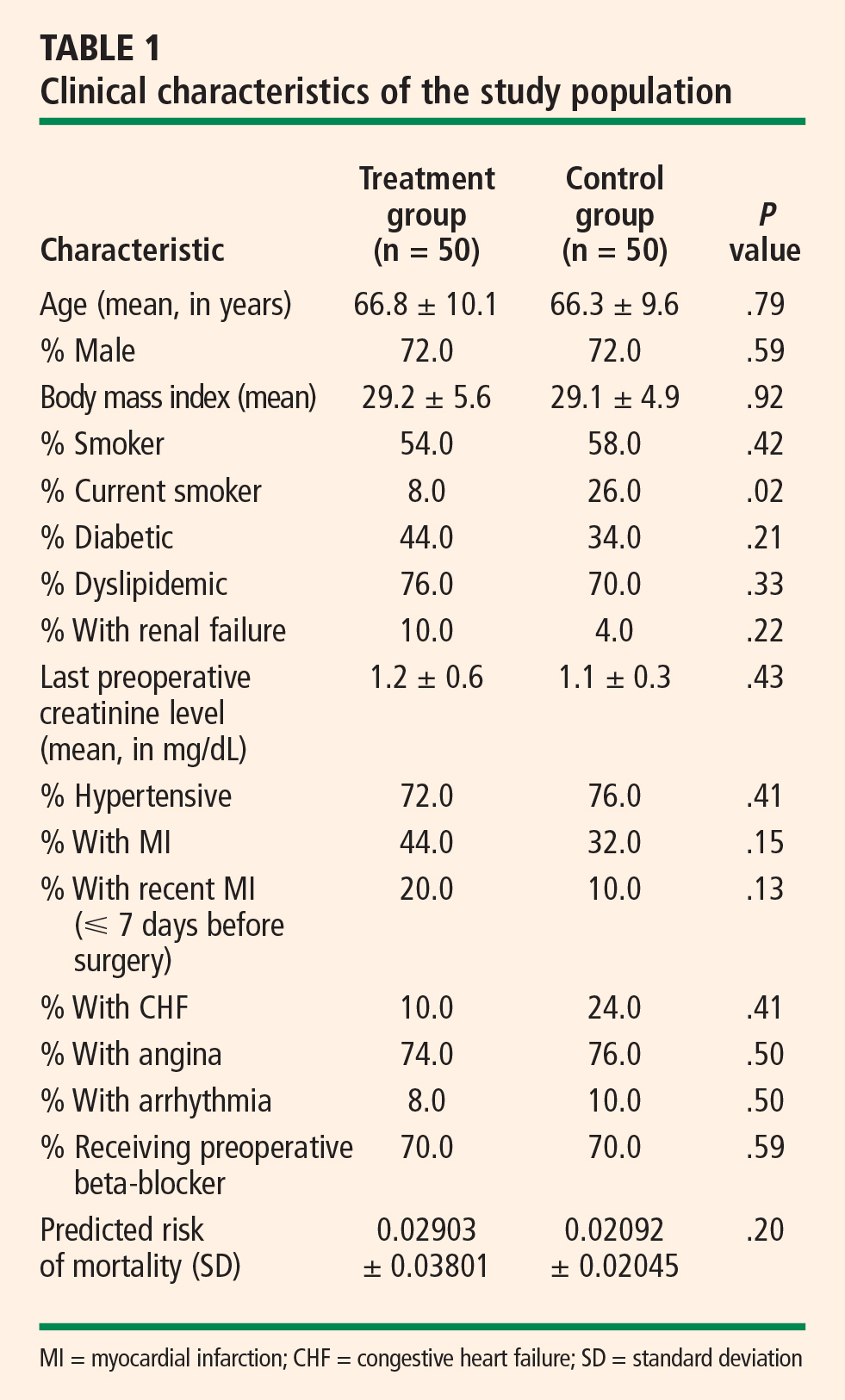
Table 2 presents outcomes in the two study groups. Patients who were treated with clinical hypnosis were less likely to experience PAF: the percentage of patients with one or more episodes of PAF was 6.0% in the treatment group versus 24.0% in the control group (P = .003). Likewise, the percentage of patients who were discharged on amiodarone was 14.0% in the treatment group versus 28.0% in the control group (P = .03).
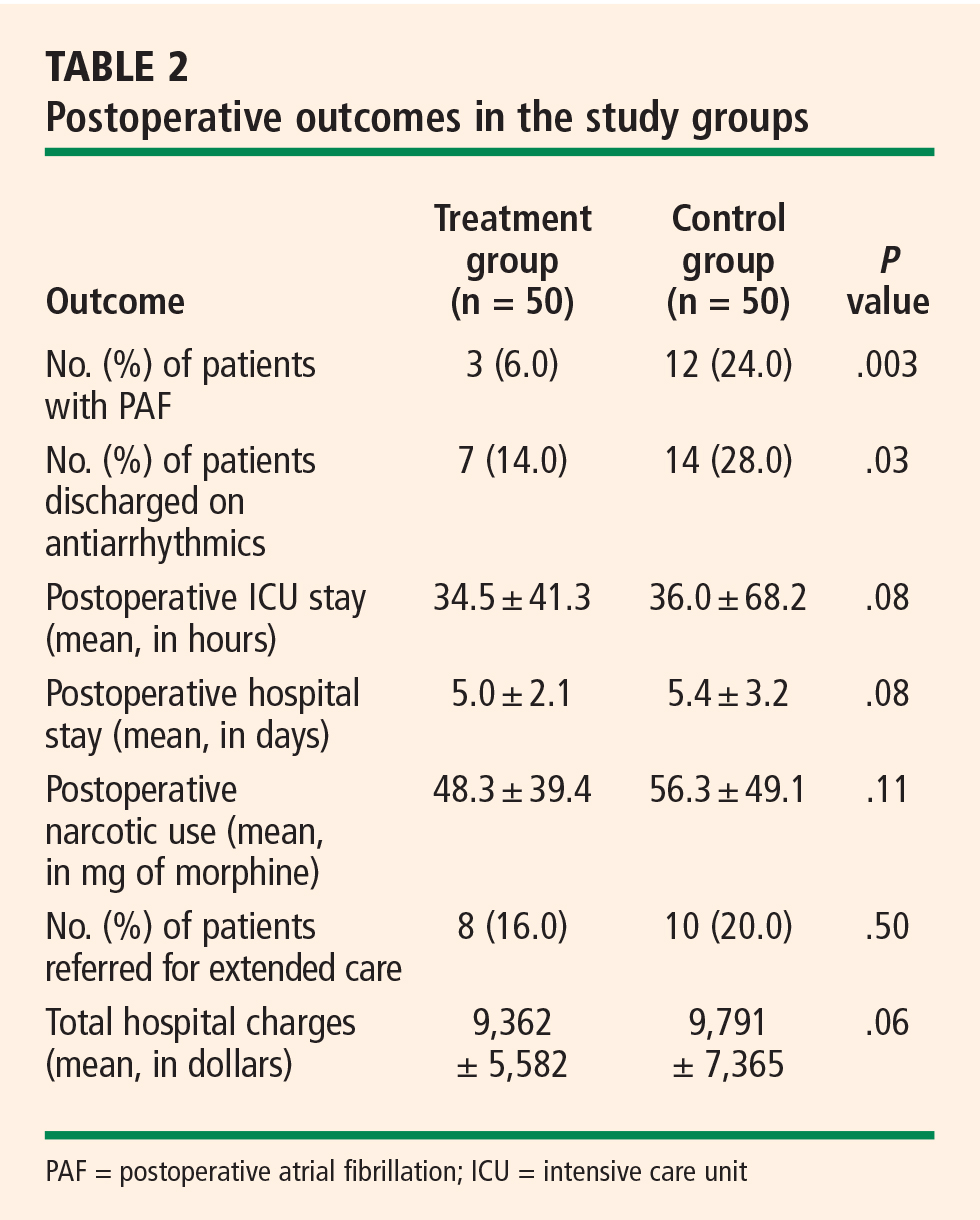
Clinical characteristics were tabulated and compared between the subjects who experienced new-onset PAF and those who did not experience PAF to determine whether there was a covariate responsible for the results observed. With the exception of age, the difference in clinical characteristics between these groups of patients was not statistically significant (using P > .10 as the threshold for significance) (Table 3).
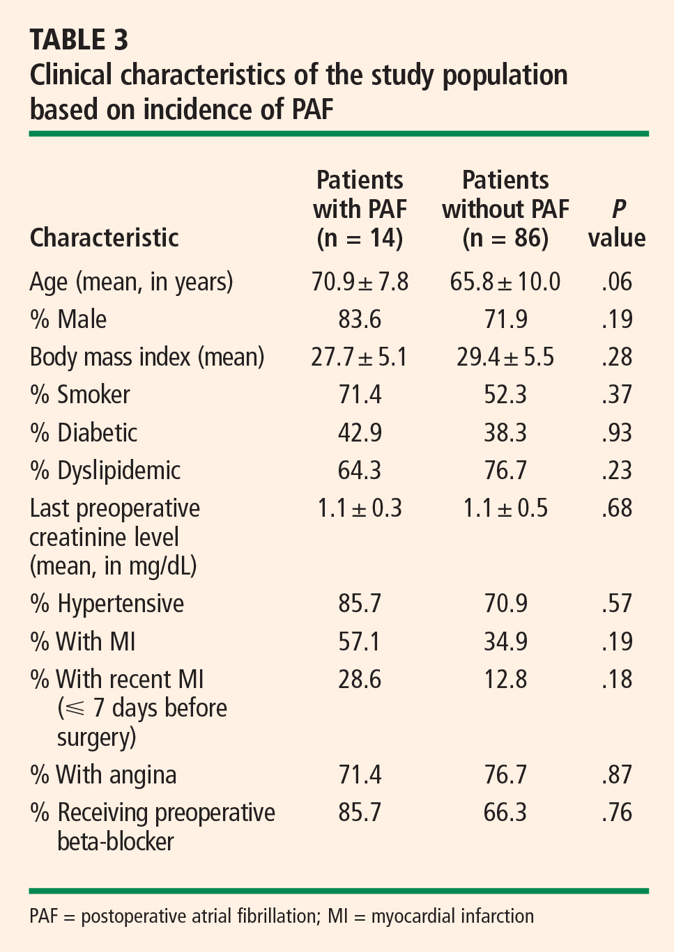
DISCUSSION
Adverse effects of PAF are well established
The onset of PAF following CABG is a common complication that has been linked to increases in morbidity and mortality, length of stay in the ICU and hospital, and total hospital charges. Villareal et al found that the odds ratio for early mortality (within 30 days) for patients who experienced PAF after CABG was 1.4 (95% confidence interval, 1.12 to 1.68; P = .002).12 In addition, patients with PAF had significantly higher rates of postoperative infections, renal failure, shock, failure of multiple organ systems, and cardiac arrest compared with those who did not have PAF.12 A literature analysis by Maisel et al found that PAF increases the likelihood that cardiac surgery patients will need to return to the operating room, be readmitted to the ICU, and require prolonged ventilation or reintubation.13 Nickerson et al showed that PAF following cardiac surgery corresponded with an increase in length of stay in both the ICU and hospital.14
Our study showed statistically significant increases in postoperative hospital charges, postoperative hospital stay, and ICU stay among patients who experienced PAF following CABG. These findings are consistent with the current literature. In a study of 720 subjects undergoing CABG, Hravnak et al reported a 1.4-day increase in length of hospital stay and a 0.3day increase in length of ICU stay among patients who had PAF compared with those who did not.15 A significant increase in postoperative hospital charges was also observed.15 Similarly, in a multicenter study of 2,417 patients undergoing isolated CABG procedures, Mathew et al observed increases in ICU stay and hospital stay among those patients who experienced PAF.2 These findings indicate that the onset of PAF after CABG is a serious complication and that further study is warranted.
Hypnosis to prevent PAF: Suggestive evidence and mechanisms
Clinical hypnosis has been shown to reduce stress and anxiety in surgical patients and can be highly individualized to address the patient’s needs during the stressful preoperative period. Saadat et al found that hypnosis administered directly before ambulatory surgery using Ericksonian techniques reduced patients’ levels of anxiety by 56% from baseline.11 In a South African study specifically in men undergoing CABG, de Klerk et al found that preoperative hypnotherapy led to reductions in both anxiety and depression at discharge that were maintained through 6-week follow-up.10
These findings, taken together with research linking clinical hypnosis to changes in the autonomic nervous system9 and the belief that PAF may result from transient autonomic dysfunction, suggest that hypnosis may reduce the incidence of PAF.
In a study of R-R interval dynamics prior to PAF in patients who had undergone CABG, Hogue et al showed that patients who experienced PAF had higher heart rates directly before PAF onset.16 Higher heart rates are associated with increased activity of the sympathetic nervous system and/or decreased activity of the parasympathetic nervous system. This finding supports our hypothesis of a relationship between PAF following CABG and excessive adrenergic activation. Chen et al have noted that catecholamine-mediated AF usually occurs in the presence of heart disease and that these types of attacks often happen during the daytime in association with physical or emotional stress.4
Bettoni and Zimmermann found that the onset of AF is preceded by a primary increase in adrenergic drive, which changes to increased vagal activity immediately before the occurrence.3 Tomita et al reported that sympathetic tone increases immediately before an occurrence of daytime AF.7 These results were supported by Lombardi et al, who detected signs of predominant sympathetic modulation and reduced vagal modulation of sinus node in AF episodes that started during the daytime.6 These episodes were characterized by atrial ectopic beats prior to onset.6
Modulations in baroreceptor reflex activity may provide further evidence of the importance of the sympathetic/parasympathetic balance in the initiation of AF. Suboptimal functioning of the baroreceptor reflex has been associated with arrhythmias and adverse cardiac events in patients and animal models. Loss of the protective effects of vagal activation has been postulated to increase vulnerability to sympathetically driven ischemia and malignant arrhythmias.17
Multiple studies have shown that the only conclusive predictor of PAF is age. Notably, heart rate variability is reduced with increased age.18 By measuring heart rate variability, Taggart et al showed that autonomic balance was improved in patients under induced stress when they were in a hypnotic state.19 Although no attempts were made to determine heart rate variability in our patient set, other studies have demonstrated an increase in heart rate variability during hypnosis. The results of our study suggest that clinical hypnosis using a personalized Ericksonian approach may have a beneficial effect on the incidence of PAF.
CONCLUSIONS
Clinical hypnosis appears to lower the incidence of PAF in patients undergoing CABG as well as to yield favorable trends toward reduced ICU and postoperative hospital stays, reduced hospital charges, and reduced use of narcotics. Although our study had a small sample size and lacked randomization, its positive results and the absence of side effects suggest that prospective randomized trials should be conducted to further delineate the role of hypnosis in the prevention of PAF. A better understanding of AF, and of the autonomic nervous system’s role in triggering and maintaining PAF, will allow more appropriate treatment of this condition.
- Halonen J, Hakala T, Auvinen T, et al. Intravenous administration of metoprolol is more effective than oral administration in the prevention of atrial fibrillation after cardiac surgery. Circulation 2006; 114(1 Suppl):I1–I4.
- Mathew JP, Parks R, Savino JS, et al. Atrial fibrillation following coronary artery bypass graft surgery: predictors, outcomes, and resource utilization. MultiCenter Study of Perioperative Ischemia Research Group. JAMA 1996; 276:300–306.
- Bettoni M, Zimmermann M. Autonomic tone variations before the onset of paroxysmal atrial fibrillation. Circulation 2002; 105:2753–2759.
- Chen J, Wasmund SL, Hamdan MH. Back to the future: the role of the autonomic nervous system in atrial fibrillation. Pacing Clin Electrophysiol 2006; 29:413–421.
- Hogue CW Jr, Creswell LL, Gutterman DD, Fleisher LA; American College of Chest Physicians. Epidemiology, mechanisms, and risks: American College of Chest Physicians guidelines for the prevention and management of postoperative atrial fibrillation after cardiac surgery. Chest 2005; 128(2 Suppl):9S–16S.
- Lombardi F, Tarricone D, Tundo F, Colombo F, Belletti S, Fiorentini C. Autonomic nervous system and paroxysmal atrial fibrillation: a study based on the analysis of RR interval changes before, during and after paroxysmal atrial fibrillation. Eur Heart J 2004; 25:1242–1248.
- Tomita T, Takei M, Saikawa Y, et al. Role of autonomic tone in the initiation and termination of paroxysmal atrial fibrillation in patients without structural heart disease. J Cardiovasc Electrophysiol 2003; 14:559–564.
- Aasbo JD, Lawrence AT, Krishnan K, Kim MH, Trohman RG. Amiodarone prophylaxis reduces major cardiovascular morbidity and length of stay after cardiac surgery: a meta-analysis. Ann Intern Med 2005; 143:327–336.
- Hippel CV, Hole G, Kaschka WP. Autonomic profile under hypnosis as assessed by heart rate variability and spectral analysis. Pharmacopsychiatry 2001; 34:111–113.
- de Klerk JE, du Plessis WF, Steyn HS, Botha M. Hypnotherapeutic ego strengthening with male South African coronary artery bypass patients. Am J Clin Hypn 2004; 47:79–92.
- Saadat H, Drummond-Lewis J, Maranets I, et al. Hypnosis reduces preoperative anxiety in adult patients. Anesth Analg 2006; 102:1394–1396.
- Villareal RP, Hariharan R, Liu BC, et al. Postoperative atrial fibrillation and mortality after coronary artery bypass surgery. J Am Coll Cardiol 2004; 43:742–748.
- Maisel WH, Rawn JD, Stevenson WG. Atrial fibrillation after cardiac surgery. Ann Intern Med 2001; 135:1061–1073.
- Nickerson NJ, Murphy SF, Dávila-Román VG, Schechtman KB, Kouchoukos NT. Obstacles to early discharge after cardiac surgery. Am J Manag Care 1999; 5:29–34.
- Hravnak M, Hoffman LA, Saul MI, et al. Resource utilization related to atrial fibrillation after coronary artery bypass grafting. Am J Crit Care 2002; 11:228–238.
- Hogue CW Jr, Domitrovich PP, Stein PK, et al. RR interval dynamics before atrial fibrillation in patients after coronary artery bypass graft surgery. Circulation 1998; 98:429–434.
- Olshansky B. Interrelationships between the autonomic nervous system and atrial fibrillation. Prog Cardiovasc Dis 2005; 48:57–78.
- Zhang J. Effect of age and sex on heart rate variability in healthy subjects. J Manipulative Physiol Ther 2007; 30:374–379.
- Taggart P, Sutton P, Redfern C, et al. The effect of mental stress on the non-dipolar components of the T wave: modulation by hypnosis. Psychosom Med 2005; 67:376–383.
Postoperative atrial fibrillation (PAF) is the most common complication of coronary artery bypass graft surgery (CABG), affecting approximately 20% to 40% of patients undergoing this procedure.1 Occurrence of PAF has been associated with prolonged hospital and intensive care unit (ICU) stays, a decline in neurocognitive ability, an increased risk of stroke and transient ischemic attacks, increased surgical mortality, and increased resource utilization and cost.2
The role of the autonomic nervous system in atrial fibrillation (AF) has been studied extensively, but the impact of the autonomic nervous system on PAF has received little attention. Research on the mechanisms of AF has shown imbalance in the autonomic nervous system when measured using heart rate variability. These studies have demonstrated an increase in sympathetic activity approximately 20 minutes prior to the onset of AF, with a shift to parasympathetic activity directly prior to onset. A correlation between mental stress and changes in the autonomic nervous system, as assessed by heart rate variability, has also been shown.3–7
Pharmacologic interventions, including beta-blockers and amiodarone, have been proposed as preventive measures for PAF, but the incidence of PAF remains high.1 Beta-blockade may not be tolerated by patients postoperatively, and there is no consensus on dosing parameters. A meta-analysis on the use of amiodarone in the prevention of PAF was inconclusive,8 and the optimal dosing regimen and incidence of adverse events with amiodarone have not been determined.
There are a small number of studies linking clinical hypnosis to changes in the autonomic nervous system.9 Clinical hypnosis also has been associated with reductions in anxiety and depression before and after surgical procedures.10,11 If PAF is a result of transient autonomic dysfunction, then interventions that alter autonomic tone should influence the incidence and duration of PAF. We report here a retrospective analysis of the impact of clinical hypnosis on the occurrence of PAF in patients undergoing CABG.
METHODOLOGY
Fifty consecutive patients undergoing first-time CABG between October 2004 and May 2005 received preoperative hypnoidal explanation of the surgical procedure as part of their preparation for surgery. A group of 50 case-matched patients who had undergone CABG at the same center between October 2003 and May 2004 were chosen as historical controls.
The treatment group (hypnosis group) and the control group were case-matched for presence of diabetes mellitus, use of beta-adrenergic blocking agents, and use of antiarrhythmic medications. The groups were also matched for various predictors of postoperative PAF, such as age, gender, and coronary artery disease. The patients were all treated by the same surgeon (R.N.), with no significant alterations to surgical or pharmacologic protocols.
The surgeon used indirect Ericksonian techniques during the preoperative explanation of the surgery. Milton Erickson, one of the most prominent hypnotherapists in recent times, used an indirect approach to weave suggestions into the dialogue rather than giving direct commands. This approach encourages active participation and gives the patient a sense of greater control in the hospital environment. The surgeon also instructed patients in self-hypnosis using respiration and imagery.
RESULTS
Table 2 presents outcomes in the two study groups. Patients who were treated with clinical hypnosis were less likely to experience PAF: the percentage of patients with one or more episodes of PAF was 6.0% in the treatment group versus 24.0% in the control group (P = .003). Likewise, the percentage of patients who were discharged on amiodarone was 14.0% in the treatment group versus 28.0% in the control group (P = .03).
Clinical characteristics were tabulated and compared between the subjects who experienced new-onset PAF and those who did not experience PAF to determine whether there was a covariate responsible for the results observed. With the exception of age, the difference in clinical characteristics between these groups of patients was not statistically significant (using P > .10 as the threshold for significance) (Table 3).
DISCUSSION
Adverse effects of PAF are well established
The onset of PAF following CABG is a common complication that has been linked to increases in morbidity and mortality, length of stay in the ICU and hospital, and total hospital charges. Villareal et al found that the odds ratio for early mortality (within 30 days) for patients who experienced PAF after CABG was 1.4 (95% confidence interval, 1.12 to 1.68; P = .002).12 In addition, patients with PAF had significantly higher rates of postoperative infections, renal failure, shock, failure of multiple organ systems, and cardiac arrest compared with those who did not have PAF.12 A literature analysis by Maisel et al found that PAF increases the likelihood that cardiac surgery patients will need to return to the operating room, be readmitted to the ICU, and require prolonged ventilation or reintubation.13 Nickerson et al showed that PAF following cardiac surgery corresponded with an increase in length of stay in both the ICU and hospital.14
Our study showed statistically significant increases in postoperative hospital charges, postoperative hospital stay, and ICU stay among patients who experienced PAF following CABG. These findings are consistent with the current literature. In a study of 720 subjects undergoing CABG, Hravnak et al reported a 1.4-day increase in length of hospital stay and a 0.3day increase in length of ICU stay among patients who had PAF compared with those who did not.15 A significant increase in postoperative hospital charges was also observed.15 Similarly, in a multicenter study of 2,417 patients undergoing isolated CABG procedures, Mathew et al observed increases in ICU stay and hospital stay among those patients who experienced PAF.2 These findings indicate that the onset of PAF after CABG is a serious complication and that further study is warranted.
Hypnosis to prevent PAF: Suggestive evidence and mechanisms
Clinical hypnosis has been shown to reduce stress and anxiety in surgical patients and can be highly individualized to address the patient’s needs during the stressful preoperative period. Saadat et al found that hypnosis administered directly before ambulatory surgery using Ericksonian techniques reduced patients’ levels of anxiety by 56% from baseline.11 In a South African study specifically in men undergoing CABG, de Klerk et al found that preoperative hypnotherapy led to reductions in both anxiety and depression at discharge that were maintained through 6-week follow-up.10
These findings, taken together with research linking clinical hypnosis to changes in the autonomic nervous system9 and the belief that PAF may result from transient autonomic dysfunction, suggest that hypnosis may reduce the incidence of PAF.
In a study of R-R interval dynamics prior to PAF in patients who had undergone CABG, Hogue et al showed that patients who experienced PAF had higher heart rates directly before PAF onset.16 Higher heart rates are associated with increased activity of the sympathetic nervous system and/or decreased activity of the parasympathetic nervous system. This finding supports our hypothesis of a relationship between PAF following CABG and excessive adrenergic activation. Chen et al have noted that catecholamine-mediated AF usually occurs in the presence of heart disease and that these types of attacks often happen during the daytime in association with physical or emotional stress.4
Bettoni and Zimmermann found that the onset of AF is preceded by a primary increase in adrenergic drive, which changes to increased vagal activity immediately before the occurrence.3 Tomita et al reported that sympathetic tone increases immediately before an occurrence of daytime AF.7 These results were supported by Lombardi et al, who detected signs of predominant sympathetic modulation and reduced vagal modulation of sinus node in AF episodes that started during the daytime.6 These episodes were characterized by atrial ectopic beats prior to onset.6
Modulations in baroreceptor reflex activity may provide further evidence of the importance of the sympathetic/parasympathetic balance in the initiation of AF. Suboptimal functioning of the baroreceptor reflex has been associated with arrhythmias and adverse cardiac events in patients and animal models. Loss of the protective effects of vagal activation has been postulated to increase vulnerability to sympathetically driven ischemia and malignant arrhythmias.17
Multiple studies have shown that the only conclusive predictor of PAF is age. Notably, heart rate variability is reduced with increased age.18 By measuring heart rate variability, Taggart et al showed that autonomic balance was improved in patients under induced stress when they were in a hypnotic state.19 Although no attempts were made to determine heart rate variability in our patient set, other studies have demonstrated an increase in heart rate variability during hypnosis. The results of our study suggest that clinical hypnosis using a personalized Ericksonian approach may have a beneficial effect on the incidence of PAF.
CONCLUSIONS
Clinical hypnosis appears to lower the incidence of PAF in patients undergoing CABG as well as to yield favorable trends toward reduced ICU and postoperative hospital stays, reduced hospital charges, and reduced use of narcotics. Although our study had a small sample size and lacked randomization, its positive results and the absence of side effects suggest that prospective randomized trials should be conducted to further delineate the role of hypnosis in the prevention of PAF. A better understanding of AF, and of the autonomic nervous system’s role in triggering and maintaining PAF, will allow more appropriate treatment of this condition.
- Halonen J, Hakala T, Auvinen T, et al. Intravenous administration of metoprolol is more effective than oral administration in the prevention of atrial fibrillation after cardiac surgery. Circulation 2006; 114(1 Suppl):I1–I4.
- Mathew JP, Parks R, Savino JS, et al. Atrial fibrillation following coronary artery bypass graft surgery: predictors, outcomes, and resource utilization. MultiCenter Study of Perioperative Ischemia Research Group. JAMA 1996; 276:300–306.
- Bettoni M, Zimmermann M. Autonomic tone variations before the onset of paroxysmal atrial fibrillation. Circulation 2002; 105:2753–2759.
- Chen J, Wasmund SL, Hamdan MH. Back to the future: the role of the autonomic nervous system in atrial fibrillation. Pacing Clin Electrophysiol 2006; 29:413–421.
- Hogue CW Jr, Creswell LL, Gutterman DD, Fleisher LA; American College of Chest Physicians. Epidemiology, mechanisms, and risks: American College of Chest Physicians guidelines for the prevention and management of postoperative atrial fibrillation after cardiac surgery. Chest 2005; 128(2 Suppl):9S–16S.
- Lombardi F, Tarricone D, Tundo F, Colombo F, Belletti S, Fiorentini C. Autonomic nervous system and paroxysmal atrial fibrillation: a study based on the analysis of RR interval changes before, during and after paroxysmal atrial fibrillation. Eur Heart J 2004; 25:1242–1248.
- Tomita T, Takei M, Saikawa Y, et al. Role of autonomic tone in the initiation and termination of paroxysmal atrial fibrillation in patients without structural heart disease. J Cardiovasc Electrophysiol 2003; 14:559–564.
- Aasbo JD, Lawrence AT, Krishnan K, Kim MH, Trohman RG. Amiodarone prophylaxis reduces major cardiovascular morbidity and length of stay after cardiac surgery: a meta-analysis. Ann Intern Med 2005; 143:327–336.
- Hippel CV, Hole G, Kaschka WP. Autonomic profile under hypnosis as assessed by heart rate variability and spectral analysis. Pharmacopsychiatry 2001; 34:111–113.
- de Klerk JE, du Plessis WF, Steyn HS, Botha M. Hypnotherapeutic ego strengthening with male South African coronary artery bypass patients. Am J Clin Hypn 2004; 47:79–92.
- Saadat H, Drummond-Lewis J, Maranets I, et al. Hypnosis reduces preoperative anxiety in adult patients. Anesth Analg 2006; 102:1394–1396.
- Villareal RP, Hariharan R, Liu BC, et al. Postoperative atrial fibrillation and mortality after coronary artery bypass surgery. J Am Coll Cardiol 2004; 43:742–748.
- Maisel WH, Rawn JD, Stevenson WG. Atrial fibrillation after cardiac surgery. Ann Intern Med 2001; 135:1061–1073.
- Nickerson NJ, Murphy SF, Dávila-Román VG, Schechtman KB, Kouchoukos NT. Obstacles to early discharge after cardiac surgery. Am J Manag Care 1999; 5:29–34.
- Hravnak M, Hoffman LA, Saul MI, et al. Resource utilization related to atrial fibrillation after coronary artery bypass grafting. Am J Crit Care 2002; 11:228–238.
- Hogue CW Jr, Domitrovich PP, Stein PK, et al. RR interval dynamics before atrial fibrillation in patients after coronary artery bypass graft surgery. Circulation 1998; 98:429–434.
- Olshansky B. Interrelationships between the autonomic nervous system and atrial fibrillation. Prog Cardiovasc Dis 2005; 48:57–78.
- Zhang J. Effect of age and sex on heart rate variability in healthy subjects. J Manipulative Physiol Ther 2007; 30:374–379.
- Taggart P, Sutton P, Redfern C, et al. The effect of mental stress on the non-dipolar components of the T wave: modulation by hypnosis. Psychosom Med 2005; 67:376–383.
Depression and coronary heart disease: Association and implications for treatment
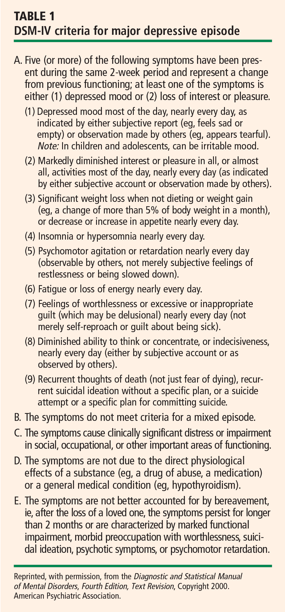
Minor depressive disorder (mDD) is not an official DSM-IV diagnosis but is used for research purposes; it is similar to MDD in duration but requires that only two to four symptoms be present.
EPIDEMIOLOGY OF DEPRESSION
Depression is a widespread and often chronic condition. Lifetime prevalence estimates for MDD are approximately 15% to 20%;2,3 1-year prevalence estimates are 5% to 10%;2,4 and point prevalence estimates range from 4% to 7%.3,5 Moreover, MDD is characterized by high rates of relapse: 22% to 50% of patients suffer recurrent episodes within 6 months after recovery.6
Women are twice as likely as men to be diagnosed with MDD, with lifetime prevalence rates of 10% to 25% in women versus 5% to 12% in men.1
Although rates of depression do not appear to increase with age, MDD often goes undertreated in older adults3 and in cardiac patients.7
DIAGNOSING AND ASSESSING DEPRESSION
The gold standard for diagnosing MDD is a clinical interview. Commonly used instruments include the Diagnostic Interview Schedule8 and the Composite International Diagnostic Interview.9 The Structured Clinical Interview for DSM-IV Axis I Disorders10 and the Schedule for Affective Disorders and Schizophrenia11 are frequently used semistructured interviews.
The most common clinical instruments for assessing the severity of depressive symptoms are the Hamilton Rating Scale for Depression (HAM-D),12 which is a clinician-rated scale, and various psychometric questionnaires, including the Beck Depression Inventory (BDI)13,14 and the Center for Epidemiological Studies Depression Scale (CES-D).15
THE DEPRESSION–HEART DISEASE LINK
Depression as a primary risk factor
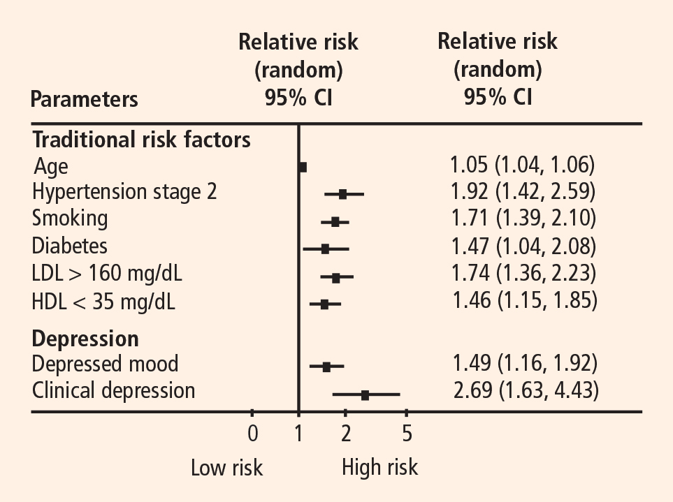
Depression as a secondary risk factor
Depression is an even stronger risk factor for cardiac events in patients with established CHD. Point estimates range from 14% to as high as 47%, with higher rates in patients with unstable angina and in patients awaiting coronary artery bypass graft (CABG) surgery; an additional 20% of patients exhibit elevated depressive symptoms or minor depression (mDD).19–25
Prospective studies have shown that depression increases the risk for death or nonfatal cardiac events approximately 2.5-fold in patients with CHD. For instance, Frasure-Smith et al followed 896 patients with a recent acute MI and found that the presence of depressive symptoms as indicated by an elevated BDI score was a significant predictor of cardiac mortality after controlling for multivariate predictors of mortality (odds ratio [OR] = 3.29 for women and 3.05 for men).26
Two recent meta-analyses confirmed the association between depression and adverse clinical outcomes in patients with CHD.27,28 For example, van Melle et al reported that post-MI depression was associated with a 2- to 2.5-fold increase in the risk of adverse health outcomes.28 In this analysis, depression’s effect on cardiac mortality and all-cause mortality was especially pronounced in older studies (before 1992) (OR = 3.2) compared with more recent studies (after 1992) (OR = 2.01).28
Duke University researchers have conducted several prospective studies in various cardiac populations.29–31 Barefoot et al assessed 1,250 patients with documented CHD using the Zung Self-Rating Depression Scale at the time of diagnostic coronary angiography and followed them for up to 19.4 years.29 Results showed that patients with moderate to severe depression were at 69% greater risk for cardiac death and 78% greater risk for all-cause death than were their nondepressed counterparts.
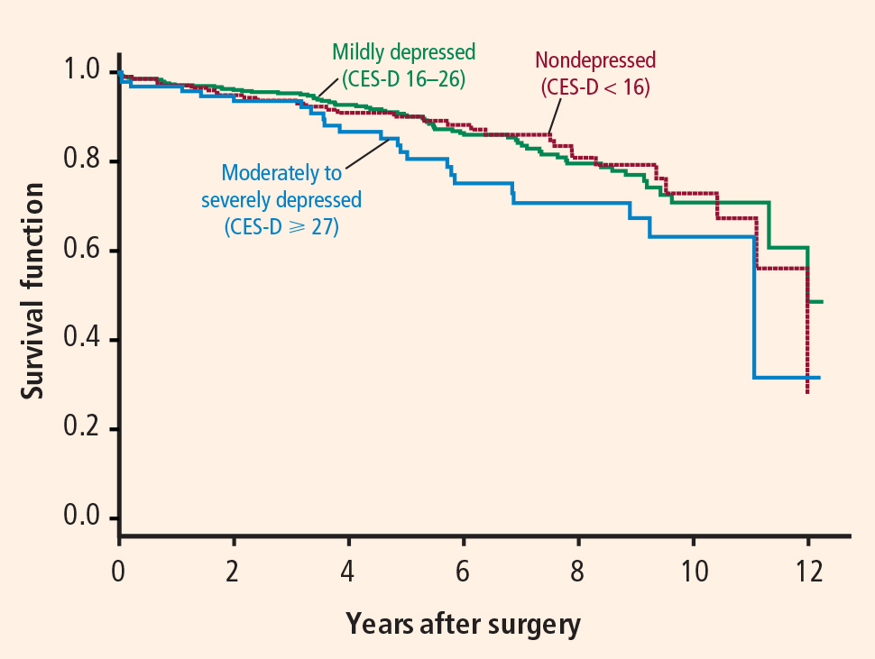
We also recently reported results from a prospective study that followed 204 patients with heart failure over a median interval of 3 years.31 Clinically significant symptoms of depression (BDI score ≥ 10) were associated with a hazard ratio of 1.56 (95% CI, 1.07 to 2.29) for the combined end point of death or cardiovascular hospitalization. These observations included adjustment for plasma NT-proBNP level, ejection fraction, and other established risk factors, suggesting that heightened risk of adverse clinical outcomes associated with depressive symptoms is not simply a reflection of the severity of heart failure.
In summary, a number of observational studies have demonstrated that depression is associated with increased risk of morbidity and mortality both in healthy populations and in a variety of populations with established cardiac disease.
BIOBEHAVIORAL MECHANISMS LINKING DEPRESSION AND CHD
A number of biobehavioral mechanisms have been hypothesized to underlie the relationship between depression and CHD. Most evidence is derived from cross-sectional studies and suggests that depression is associated with traditional risk factors for CHD, such as hypertension, diabetes, and insulin resistance,32,33 as well as changes in platelet reactivity,34 dysregulation of the autonomic nervous system35 and hypothalamic-pituitary-adrenal axis,36 and alterations in the immune response/inflammation.37 Depression is also associated with behavioral factors that are in turn associated with CHD risk, such as reduced treatment adherence,38 smoking,39 and physical inactivity.40
STUDIES OF DEPRESSION TREATMENT IN CARDIAC PATIENTS
Successful treatments for depression in patients with CHD may have the potential to improve not only quality of life but also cardiovascular and physical health. Several treatments for depression exist for use in the general population, such as antidepressant medication or psychotherapy.41 However, only three studies have tested the efficacy of these treatments in patients with CHD: SADHART, ENRICHD, and CREATE.42–44
SADHART (Sertraline Antidepressant Heart Attack Randomized Trial) was a safety and efficacy evaluation of antidepressant medication in patients with MDD and a recent MI or unstable angina.42 It showed only modest differences in reductions in depressive symptoms between sertraline recipients and placebo recipients, and it lacked statistical power to examine the impact of treatment on hard clinical end points.
ENRICHD (Enhancing Recovery in Coronary Heart Disease Patients) assessed the effect of psychosocial treatment on survival among more than than 2,400 post-MI patients.43 Although this trial found that cognitive behavior therapy resulted in significant, albeit small, improvements in depressive symptoms compared with usual care, it failed to demonstrate that treating depression and low social support was associated with increased survival.
CREATE (Canadian Cardiac Randomized Evaluation of Antidepressant and Psychotherapy Efficacy), a recent placebo-controlled trial, assessed the value of antidepressant medication and clinical management in patients with CHD.44 The study’s 284 patients, all of whom had CHD as well as MDD and a HAM-D score of 20 or greater, underwent two separate randomizations: (1) to 12 weeks of interpersonal therapy plus clinical management or 12 weeks of clinical management alone, and (2) to 12 weeks of citalopram therapy or matching placebo. There was no difference between interpersonal therapy and clinical management alone; however, citalopram was superior to placebo in reducing HAM-D scores and demonstrated better remission rates (35.9% with citalopram vs 22.5% with placebo). The same therapists who provided interpersonal therapy also performed the clinical management, so it could be argued that this was why additional interpersonal therapist time did not result in greater reductions in depressive symptoms than did clinical management alone. Furthermore, this study did not examine the effects of depression therapy on clinical outcomes.
EXERCISE AS A TREATMENT FOR DEPRESSION
There is growing evidence that exercise may be an effective treatment for depression.45 Most of the existing studies of exercise for depression have focused on aerobic exercise.
In the relatively large SMILE study (Standard Medical Intervention and Long-term Exercise),46 conducted at Duke University, 156 adult noncardiac patients with MDD were randomized to 4 months of treatment with supervised aerobic exercise, antidepressant medication (sertraline), or a combination of exercise and medication. Although antidepressant medication was associated with faster reductions in depression in the first 4 weeks of treatment among mildly depressed patients, exercise was as effective as antidepressant medication in treating depression by the end of the 16-week intervention for all participants.
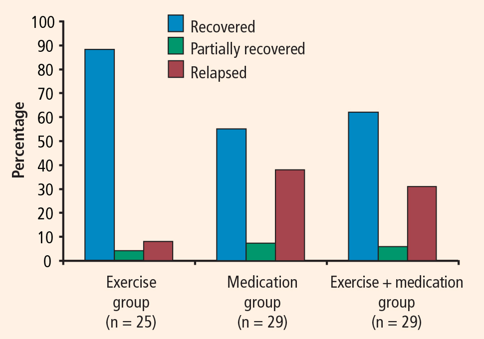
Exercise generally is considered safe for most patients with stable CHD.48 Some studies of exercise treatments for patients with CHD have tracked depressive symptoms and thus have provided insight into the potential efficacy of exercise as a treatment for depression in this population. Although most of these studies have reported significant improvements in depression after completion of an exercise program, many have had important methodologic limitations, including absence of a control group. In one of the few controlled studies in this area, Stern et al49 randomized 106 men who had a recent acute MI and elevated depression, anxiety, or low fitness to 12 weeks of exercise training, group therapy, or usual care (control). At 1-year follow-up, subjects in both the exercise and counseling groups showed improvements in depression relative to controls.
EFFECT OF EXERCISE ON CARDIOVASCULAR RISK FACTORS AND OUTCOMES
Exercise is a particularly promising intervention for depression in patients with CHD because it has well-documented cardiovascular benefits. In addition to the well-established role of exercise interventions in primary prevention, such interventions have been shown to improve outcomes for patients with CHD.50
Jolliffe et al conducted a meta-analysis comparing exercise-only interventions, comprehensive rehabilitation (including educational and behavioral components such as dietary changes and stress reduction in addition to exercise), and usual care.51 Exercise-only interventions were associated with reductions in both all-cause and cardiac mortality relative to usual care. Comprehensive rehabilitation, on the other hand, was not associated with statistically significant reductions in all-cause mortality relative to usual care, but it was associated with a decreased risk for cardiac mortality, to a slightly lesser extent than exercise-only interventions.
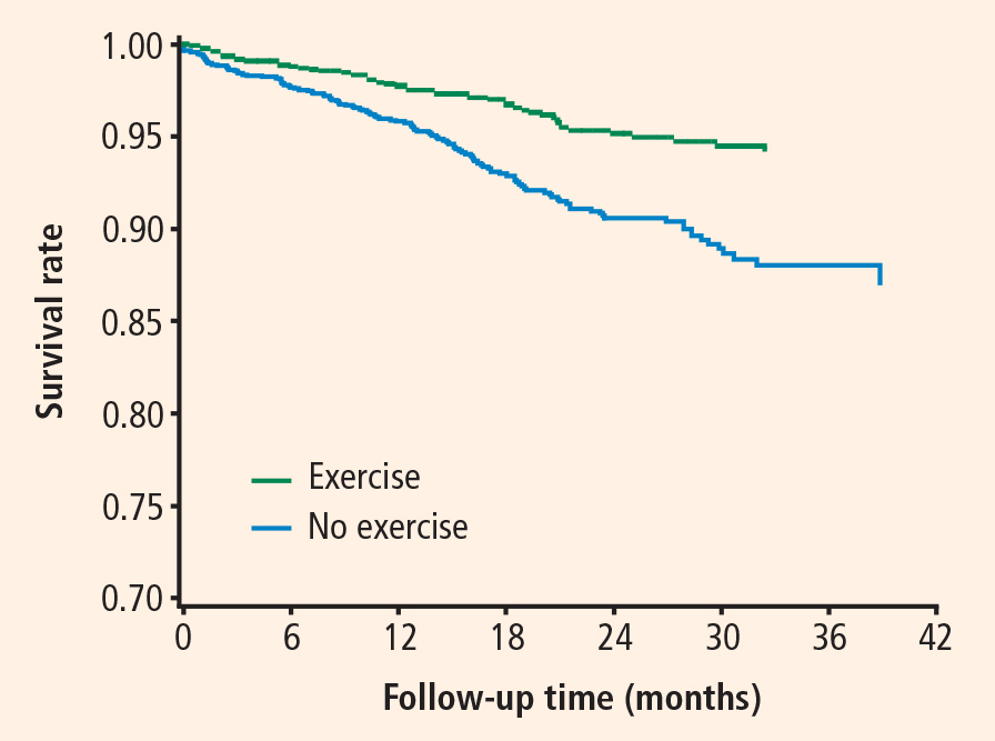
The evidence that exercise affects depression, CHD risk factors, and CHD outcomes suggests that exercise is a particularly promising intervention for depression in this population.
UPBEAT trial promises further insight
A new Duke University study known as UPBEAT (Understanding Prognostic Benefits of Exercise and Antidepressant Treatment) is randomizing 200 patients with elevated depressive symptoms to exercise, antidepressant therapy (sertraline), or placebo for 4 months.53 A variety of “biomarkers” of risk are being assessed, including measures of heart rate variability, vascular function, inflammation, and platelet aggregation. Results of this 5-year trial should be available by 2011.
CONCLUSIONS
Although depression has emerged as an important risk factor for CHD, there is no consensus on the optimal way to treat depression in patients with CHD. Interventions that are guided by an understanding of the mechanisms linking depression to CHD may prove to be most effective in improving both depression and physical health outcomes.
Exercise targets many of the mechanisms by which depression may be associated with increased risk, including autonomic nervous system activity, hypothalamic-pituitary-adrenal axis function, platelet activation, vascular function, and inflammation. Moreover, a growing body of evidence suggests that exercise is an effective treatment for depression that may be comparable in effect to antidepressant medication, at least in select subgroups (eg, patients who are receptive to exercise as a treatment for depression). The value of exercise training—not only for improving quality of life, but also for improving “biomarkers” of risk and other relevant health outcomes—is the focus of our current research efforts.
- American Psychiatric Association. Diagnostic and Statistical Manual of Mental Disorders, Text Revision. 4th ed. Washington, DC: American Psychiatric Association; 2000.
- Kessler RC, McGonagle KA, Zhao S, et al. Lifetime and 12-month prevalence of DSM-III-R psychiatric disorders in the United States. Results from the National Comorbidity Survey. Arch Gen Psychiatry 1994; 51:8–19.
- Steffens DC, Skoog I, Norton MC, et al. Prevalence of depression and its treatment in an elderly population: the Cache County study. Arch Gen Psychiatry 2000; 57:601–617.
- Regier DA, Narrow WE, Rae DS, Manderscheid RW, Locke BZ, Goodwin FK. The de facto US mental and addictive disorders service system. Epidemiologic catchment area prospective 1-year prevalence rates of disorders and services. Arch Gen Psychiatry 1993; 50:85–94.
- Ayuso-Mateos JL, Vazquez-Barquero JL, Dowrick C, et al. Depressive disorders in Europe: prevalence figures from the ODIN study. Br J Psychiatry 2001; 179:308–316.
- Belsher G, Costello CG. Relapse after recovery from unipolar depression: a critical review. Psychol Bull 1988; 104:84–96.
- Carney RM, Rich MW, Tevelde A, Saini J, Clark K, Jaffe AS. Major depressive disorder in coronary artery disease. Am J Cardiol 1987; 60:1273–1275.
- Robins LN, Helzer JE, Crougham J, Ratliff K. National Institute of Mental Health diagnostic interview schedule. Arch Gen Psychiatry 1981; 38:381–389.
- World Health Organization. Composite International Diagnostic Interview. Geneva, Switzerland: World Health Organization; 1990.
- First M, Spitzer L, Gibbon M. Structured clinical interview for axis I DSM-IV disorders. Washington, DC: American Psychiatric Press; 1995.
- Endicott J, Spitzer RL. The schedule for affective disorders and schizophrenia. Arch Gen Psychiatry 1978; 35:837–844.
- Hamilton M. A rating scale for depression. J Neurol 1960; 23:56–61.
- Beck AT, Ward CH, Mendelsohn M. An inventory for measuring depression. Arch Gen Psychiatry 1961; 4:561–571.
- Beck AT, Steer RA, Brown GK. Beck Depression Inventory Manual. 2nd ed. San Antonio, TX: The Psychological Corporation; 1996.
- Hautzinger M. The CES-D scale: a depression-rating scale for research in the general population. Diagnostica 1988; 34:167–173.
- Lett H, Blumenthal J, Babyak M, Sherwood A, Strauman T, Robins C. Depression as a risk factor for coronary artery disease: evidence, mechanisms, and treatment. Psychosom Med 2004; 66:305–315.
- Rugulies R. Depression as a predictor for coronary heart disease. A review and meta-analysis. Am J Prev Med 2002; 23:51–61.
- Rozanski A, Blumenthal JA, Davidson KW, Saab PG, Kubzansky L. The epidemiology, pathophysiology, and management of psychosocial risk factors in cardiac practice: the emerging field of behavioral cardiology. J Am Coll Cardiol 2005; 45:637–651.
- Carney RM, Rich MW, Freedland KE, et al. Major depressive disorder predicts cardiac events in patients with coronary artery disease. Psychosom Med 1988; 50:627–633.
- Schleifer SJ, Macari-Hinson MM, Coyle DA, et al. The nature and course of depression following myocardial infarction. Arch Intern Med 1989; 149:1785–1789.
- Frasure-Smith N, Lesperance F, Talajic M. Depression following myocardial infarction. Impact on 6-month survival. JAMA 1993; 270:1819–1825.
- Lesperance F, Frasure-Smith N, Juneau M, Theroux P. Depression and 1-year prognosis in unstable angina. Arch Intern Med 2000; 160:1354–1360.
- Connerney I, Shapiro PA, McLaughlin JS, Bagiella E, Sloan RP. Relation between depression after coronary artery bypass surgery and 12-month outcome: a prospective study. Lancet 2001; 358:1766–1771.
- Jiang W, Alexander J, Christopher E, et al. Relationship of depression to increased risk of mortality and rehospitalization in patients with congestive heart failure. Arch Intern Med 2001; 161:1849–1856.
- Burker EJ, Blumenthal JA, Feldman M, et al. Depression in male and female patients undergoing cardiac surgery. Br J Clin Psychol 1995; 34:119–128.
- Frasure-Smith N, Lesperance F, Juneau M, Talajic M, Bourassa MG. Gender, depression, and one-year prognosis after myocardial infarction. Psychosom Med 1999; 61:26–37.
- Barth J, Schumacher M, Hermann-Lingen C. Depression as a risk factor for mortality in patients with coronary heart disease: a meta-analysis. Psychosom Med 2004; 66:802–813.
- van Melle JP, de Jong P, Spijkerman TA, et al. Prognostic association of depression following myocardial infarction with mortality and cardiovascular events: a meta-analysis. Psychosom Med 2004; 66:814–822.
- Barefoot JC, Helms MJ, Mark DB, et al. Depression and long-term mortality risk in patients with coronary artery disease. Am J Cardiol 1996; 78:613–617.
- Blumenthal JA, Lett H, Babyak M, et al. Depression as a risk factor for mortality after coronary artery bypass surgery. Lancet 2003; 362:604–609.
- Sherwood A, Blumenthal JA, Trivedi R, et al. Relationship of depression to mortality and hospitalization in patients with heart failure. Arch Intern Med 2007; 167:367–373.
- Anderson RJ, Freedland KE, Clouse RE, Lustman PJ. The prevalence of comorbid depression in adults with diabetes: a meta-analysis. Diabetes Care 2001; 24:1069–1078.
- Thakore JH, Richards PJ, Reznek RH, Martin A, Dinan TG. Increased intra-abdominal fat deposition in patients with major depressive illness as measured by computed tomography. Biol Psychiatry 1997; 41:1140–1142.
- Musselman DL, Tomer A, Manatunga AK, et al. Exaggerated platelet reactivity in major depression. Am J Psychiatry 1996; 153:1313–1317.
- Delgado PL, Moreno FA. Role of norepinephrine in depression. J Clin Psychiatry 2000; 61:5–12.
- Akil H, Haskett RF, Young EA, et al. Multiple HPA profiles in endogenous depression: effect of age and sex on cortisol and beta-endorphin. Biol Psychiatry 1993; 33:73–85.
- Kop WJ, Gottdiener JS, Tangen CM, et al. Inflammation and coagulation factors in persons > 65 years of age with symptoms of depression but without evidence of myocardial ischemia. Am J Cardiol 2002; 89:419–424.
- Carney RM, Freedland KE, Eisen SA, Rich MW, Jaffe AS. Major depression and medication adherence in elderly patients with coronary artery disease. Health Psychol 1995; 14:88–90.
- Lehto S, Koukkunen H, Hintikka J, Viinamaki H, Laakso M, Pyorala K. Depression after coronary heart disease events. Scand Cardiovasc J 2000; 34:580–583.
- Camacho TC, Roberts RE, Lazarus NB, Kaplan GA, Cohen RD. Physical activity and depression: evidence from the Alameda County Study. Am J Epidemiol 1991; 134:220–231.
- Depression Guideline Panel. Depression in Primary Care: Volume 2. Treatment of Depression, Clinical Practice Guideline, No. 5. Rockville, MD: Agency for Health Care Policy and Research, Public Health Service, US Dept of Health and Human Services; 1993.
- Glassman AH, O’Connor CM, Califf RM, et al. Sertraline treatment of major depression in patients with acute MI or unstable angina. JAMA 2002; 288:701–709.
- Berkman LF, Blumenthal J, Burg M, et al. Effects of treating depression and low perceived social support on clinical events after myocardial infarction: the Enhancing Recovery in Coronary Heart Disease Patients (ENRICHD) randomized trial. JAMA 2003; 289:3106–3116.
- Lesperance F, Frasure-Smith N, Koszycki D, et al. Effects of citalopram and interpersonal psychotherapy on depression in patients with coronary artery disease: the Canadian Cardiac Randomized Evaluation of Antidepressant and Psychotherapy Efficacy (CREATE) trial. JAMA 2007; 297:367–379.
- Brosse AL, Sheets ES, Lett HS, Blumenthal JA. Exercise and the treatment of clinical depression in adults: recent findings and future directions. Sports Med 2002; 32:741–760.
- Blumenthal JA, Babyak MA, Moore KA, et al. Effects of exercise training on older patients with major depression. Arch Intern Med 1999; 159:2349–2356.
- Babyak M, Blumenthal JA, Herman S, et al. Exercise treatment for major depression: maintenance of therapeutic benefit at 10 months. Psychosom Med 2000; 62:633–638.
- Franklin BA, Bonzheim K, Gordon S, Timmis GC. Safety of medically supervised outpatient cardiac rehabilitation exercise therapy: a 16-year follow-up. Chest 1998; 114:902–906.
- Stern MJ, Gorman PA, Kaslow L. The group counseling v exercise therapy study. A controlled intervention with subjects following myocardial infarction. Arch Intern Med 1983; 143:1719–1725.
- Oldridge NB, Guyatt GH, Fischer ME, Rimm AA. Cardiac rehabilitation after myocardial infarction. Combined experience of randomized clinical trials. JAMA 1988; 260:945–950.
- Jolliffe JA, Rees K, Taylor RS, Thompson D, Oldridge N, Ebrahim S. Exercise-based rehabilitation for coronary heart disease. Cochrane Database Syst Rev 2001; (1):CD001800.
- Blumenthal JA, Babyak MA, Carney RM, et al. Exercise, depression, and mortality after myocardial infarction in the ENRICHD trial. Med Sci Sports Exerc 2004; 36:746–755.
- Blumenthal JA, Sherwood A, Rogers SD, et al. Understanding prognostic benefits of exercise and antidepressant therapy for persons with depression and heart disease: the UPBEAT study—rationale, design, and methodological issues. Clin Trials 2007; 4:548–559.
Minor depressive disorder (mDD) is not an official DSM-IV diagnosis but is used for research purposes; it is similar to MDD in duration but requires that only two to four symptoms be present.
EPIDEMIOLOGY OF DEPRESSION
Depression is a widespread and often chronic condition. Lifetime prevalence estimates for MDD are approximately 15% to 20%;2,3 1-year prevalence estimates are 5% to 10%;2,4 and point prevalence estimates range from 4% to 7%.3,5 Moreover, MDD is characterized by high rates of relapse: 22% to 50% of patients suffer recurrent episodes within 6 months after recovery.6
Women are twice as likely as men to be diagnosed with MDD, with lifetime prevalence rates of 10% to 25% in women versus 5% to 12% in men.1
Although rates of depression do not appear to increase with age, MDD often goes undertreated in older adults3 and in cardiac patients.7
DIAGNOSING AND ASSESSING DEPRESSION
The gold standard for diagnosing MDD is a clinical interview. Commonly used instruments include the Diagnostic Interview Schedule8 and the Composite International Diagnostic Interview.9 The Structured Clinical Interview for DSM-IV Axis I Disorders10 and the Schedule for Affective Disorders and Schizophrenia11 are frequently used semistructured interviews.
The most common clinical instruments for assessing the severity of depressive symptoms are the Hamilton Rating Scale for Depression (HAM-D),12 which is a clinician-rated scale, and various psychometric questionnaires, including the Beck Depression Inventory (BDI)13,14 and the Center for Epidemiological Studies Depression Scale (CES-D).15
THE DEPRESSION–HEART DISEASE LINK
Depression as a primary risk factor
Depression as a secondary risk factor
Depression is an even stronger risk factor for cardiac events in patients with established CHD. Point estimates range from 14% to as high as 47%, with higher rates in patients with unstable angina and in patients awaiting coronary artery bypass graft (CABG) surgery; an additional 20% of patients exhibit elevated depressive symptoms or minor depression (mDD).19–25
Prospective studies have shown that depression increases the risk for death or nonfatal cardiac events approximately 2.5-fold in patients with CHD. For instance, Frasure-Smith et al followed 896 patients with a recent acute MI and found that the presence of depressive symptoms as indicated by an elevated BDI score was a significant predictor of cardiac mortality after controlling for multivariate predictors of mortality (odds ratio [OR] = 3.29 for women and 3.05 for men).26
Two recent meta-analyses confirmed the association between depression and adverse clinical outcomes in patients with CHD.27,28 For example, van Melle et al reported that post-MI depression was associated with a 2- to 2.5-fold increase in the risk of adverse health outcomes.28 In this analysis, depression’s effect on cardiac mortality and all-cause mortality was especially pronounced in older studies (before 1992) (OR = 3.2) compared with more recent studies (after 1992) (OR = 2.01).28
Duke University researchers have conducted several prospective studies in various cardiac populations.29–31 Barefoot et al assessed 1,250 patients with documented CHD using the Zung Self-Rating Depression Scale at the time of diagnostic coronary angiography and followed them for up to 19.4 years.29 Results showed that patients with moderate to severe depression were at 69% greater risk for cardiac death and 78% greater risk for all-cause death than were their nondepressed counterparts.
We also recently reported results from a prospective study that followed 204 patients with heart failure over a median interval of 3 years.31 Clinically significant symptoms of depression (BDI score ≥ 10) were associated with a hazard ratio of 1.56 (95% CI, 1.07 to 2.29) for the combined end point of death or cardiovascular hospitalization. These observations included adjustment for plasma NT-proBNP level, ejection fraction, and other established risk factors, suggesting that heightened risk of adverse clinical outcomes associated with depressive symptoms is not simply a reflection of the severity of heart failure.
In summary, a number of observational studies have demonstrated that depression is associated with increased risk of morbidity and mortality both in healthy populations and in a variety of populations with established cardiac disease.
BIOBEHAVIORAL MECHANISMS LINKING DEPRESSION AND CHD
A number of biobehavioral mechanisms have been hypothesized to underlie the relationship between depression and CHD. Most evidence is derived from cross-sectional studies and suggests that depression is associated with traditional risk factors for CHD, such as hypertension, diabetes, and insulin resistance,32,33 as well as changes in platelet reactivity,34 dysregulation of the autonomic nervous system35 and hypothalamic-pituitary-adrenal axis,36 and alterations in the immune response/inflammation.37 Depression is also associated with behavioral factors that are in turn associated with CHD risk, such as reduced treatment adherence,38 smoking,39 and physical inactivity.40
STUDIES OF DEPRESSION TREATMENT IN CARDIAC PATIENTS
Successful treatments for depression in patients with CHD may have the potential to improve not only quality of life but also cardiovascular and physical health. Several treatments for depression exist for use in the general population, such as antidepressant medication or psychotherapy.41 However, only three studies have tested the efficacy of these treatments in patients with CHD: SADHART, ENRICHD, and CREATE.42–44
SADHART (Sertraline Antidepressant Heart Attack Randomized Trial) was a safety and efficacy evaluation of antidepressant medication in patients with MDD and a recent MI or unstable angina.42 It showed only modest differences in reductions in depressive symptoms between sertraline recipients and placebo recipients, and it lacked statistical power to examine the impact of treatment on hard clinical end points.
ENRICHD (Enhancing Recovery in Coronary Heart Disease Patients) assessed the effect of psychosocial treatment on survival among more than than 2,400 post-MI patients.43 Although this trial found that cognitive behavior therapy resulted in significant, albeit small, improvements in depressive symptoms compared with usual care, it failed to demonstrate that treating depression and low social support was associated with increased survival.
CREATE (Canadian Cardiac Randomized Evaluation of Antidepressant and Psychotherapy Efficacy), a recent placebo-controlled trial, assessed the value of antidepressant medication and clinical management in patients with CHD.44 The study’s 284 patients, all of whom had CHD as well as MDD and a HAM-D score of 20 or greater, underwent two separate randomizations: (1) to 12 weeks of interpersonal therapy plus clinical management or 12 weeks of clinical management alone, and (2) to 12 weeks of citalopram therapy or matching placebo. There was no difference between interpersonal therapy and clinical management alone; however, citalopram was superior to placebo in reducing HAM-D scores and demonstrated better remission rates (35.9% with citalopram vs 22.5% with placebo). The same therapists who provided interpersonal therapy also performed the clinical management, so it could be argued that this was why additional interpersonal therapist time did not result in greater reductions in depressive symptoms than did clinical management alone. Furthermore, this study did not examine the effects of depression therapy on clinical outcomes.
EXERCISE AS A TREATMENT FOR DEPRESSION
There is growing evidence that exercise may be an effective treatment for depression.45 Most of the existing studies of exercise for depression have focused on aerobic exercise.
In the relatively large SMILE study (Standard Medical Intervention and Long-term Exercise),46 conducted at Duke University, 156 adult noncardiac patients with MDD were randomized to 4 months of treatment with supervised aerobic exercise, antidepressant medication (sertraline), or a combination of exercise and medication. Although antidepressant medication was associated with faster reductions in depression in the first 4 weeks of treatment among mildly depressed patients, exercise was as effective as antidepressant medication in treating depression by the end of the 16-week intervention for all participants.
Exercise generally is considered safe for most patients with stable CHD.48 Some studies of exercise treatments for patients with CHD have tracked depressive symptoms and thus have provided insight into the potential efficacy of exercise as a treatment for depression in this population. Although most of these studies have reported significant improvements in depression after completion of an exercise program, many have had important methodologic limitations, including absence of a control group. In one of the few controlled studies in this area, Stern et al49 randomized 106 men who had a recent acute MI and elevated depression, anxiety, or low fitness to 12 weeks of exercise training, group therapy, or usual care (control). At 1-year follow-up, subjects in both the exercise and counseling groups showed improvements in depression relative to controls.
EFFECT OF EXERCISE ON CARDIOVASCULAR RISK FACTORS AND OUTCOMES
Exercise is a particularly promising intervention for depression in patients with CHD because it has well-documented cardiovascular benefits. In addition to the well-established role of exercise interventions in primary prevention, such interventions have been shown to improve outcomes for patients with CHD.50
Jolliffe et al conducted a meta-analysis comparing exercise-only interventions, comprehensive rehabilitation (including educational and behavioral components such as dietary changes and stress reduction in addition to exercise), and usual care.51 Exercise-only interventions were associated with reductions in both all-cause and cardiac mortality relative to usual care. Comprehensive rehabilitation, on the other hand, was not associated with statistically significant reductions in all-cause mortality relative to usual care, but it was associated with a decreased risk for cardiac mortality, to a slightly lesser extent than exercise-only interventions.
The evidence that exercise affects depression, CHD risk factors, and CHD outcomes suggests that exercise is a particularly promising intervention for depression in this population.
UPBEAT trial promises further insight
A new Duke University study known as UPBEAT (Understanding Prognostic Benefits of Exercise and Antidepressant Treatment) is randomizing 200 patients with elevated depressive symptoms to exercise, antidepressant therapy (sertraline), or placebo for 4 months.53 A variety of “biomarkers” of risk are being assessed, including measures of heart rate variability, vascular function, inflammation, and platelet aggregation. Results of this 5-year trial should be available by 2011.
CONCLUSIONS
Although depression has emerged as an important risk factor for CHD, there is no consensus on the optimal way to treat depression in patients with CHD. Interventions that are guided by an understanding of the mechanisms linking depression to CHD may prove to be most effective in improving both depression and physical health outcomes.
Exercise targets many of the mechanisms by which depression may be associated with increased risk, including autonomic nervous system activity, hypothalamic-pituitary-adrenal axis function, platelet activation, vascular function, and inflammation. Moreover, a growing body of evidence suggests that exercise is an effective treatment for depression that may be comparable in effect to antidepressant medication, at least in select subgroups (eg, patients who are receptive to exercise as a treatment for depression). The value of exercise training—not only for improving quality of life, but also for improving “biomarkers” of risk and other relevant health outcomes—is the focus of our current research efforts.
- American Psychiatric Association. Diagnostic and Statistical Manual of Mental Disorders, Text Revision. 4th ed. Washington, DC: American Psychiatric Association; 2000.
- Kessler RC, McGonagle KA, Zhao S, et al. Lifetime and 12-month prevalence of DSM-III-R psychiatric disorders in the United States. Results from the National Comorbidity Survey. Arch Gen Psychiatry 1994; 51:8–19.
- Steffens DC, Skoog I, Norton MC, et al. Prevalence of depression and its treatment in an elderly population: the Cache County study. Arch Gen Psychiatry 2000; 57:601–617.
- Regier DA, Narrow WE, Rae DS, Manderscheid RW, Locke BZ, Goodwin FK. The de facto US mental and addictive disorders service system. Epidemiologic catchment area prospective 1-year prevalence rates of disorders and services. Arch Gen Psychiatry 1993; 50:85–94.
- Ayuso-Mateos JL, Vazquez-Barquero JL, Dowrick C, et al. Depressive disorders in Europe: prevalence figures from the ODIN study. Br J Psychiatry 2001; 179:308–316.
- Belsher G, Costello CG. Relapse after recovery from unipolar depression: a critical review. Psychol Bull 1988; 104:84–96.
- Carney RM, Rich MW, Tevelde A, Saini J, Clark K, Jaffe AS. Major depressive disorder in coronary artery disease. Am J Cardiol 1987; 60:1273–1275.
- Robins LN, Helzer JE, Crougham J, Ratliff K. National Institute of Mental Health diagnostic interview schedule. Arch Gen Psychiatry 1981; 38:381–389.
- World Health Organization. Composite International Diagnostic Interview. Geneva, Switzerland: World Health Organization; 1990.
- First M, Spitzer L, Gibbon M. Structured clinical interview for axis I DSM-IV disorders. Washington, DC: American Psychiatric Press; 1995.
- Endicott J, Spitzer RL. The schedule for affective disorders and schizophrenia. Arch Gen Psychiatry 1978; 35:837–844.
- Hamilton M. A rating scale for depression. J Neurol 1960; 23:56–61.
- Beck AT, Ward CH, Mendelsohn M. An inventory for measuring depression. Arch Gen Psychiatry 1961; 4:561–571.
- Beck AT, Steer RA, Brown GK. Beck Depression Inventory Manual. 2nd ed. San Antonio, TX: The Psychological Corporation; 1996.
- Hautzinger M. The CES-D scale: a depression-rating scale for research in the general population. Diagnostica 1988; 34:167–173.
- Lett H, Blumenthal J, Babyak M, Sherwood A, Strauman T, Robins C. Depression as a risk factor for coronary artery disease: evidence, mechanisms, and treatment. Psychosom Med 2004; 66:305–315.
- Rugulies R. Depression as a predictor for coronary heart disease. A review and meta-analysis. Am J Prev Med 2002; 23:51–61.
- Rozanski A, Blumenthal JA, Davidson KW, Saab PG, Kubzansky L. The epidemiology, pathophysiology, and management of psychosocial risk factors in cardiac practice: the emerging field of behavioral cardiology. J Am Coll Cardiol 2005; 45:637–651.
- Carney RM, Rich MW, Freedland KE, et al. Major depressive disorder predicts cardiac events in patients with coronary artery disease. Psychosom Med 1988; 50:627–633.
- Schleifer SJ, Macari-Hinson MM, Coyle DA, et al. The nature and course of depression following myocardial infarction. Arch Intern Med 1989; 149:1785–1789.
- Frasure-Smith N, Lesperance F, Talajic M. Depression following myocardial infarction. Impact on 6-month survival. JAMA 1993; 270:1819–1825.
- Lesperance F, Frasure-Smith N, Juneau M, Theroux P. Depression and 1-year prognosis in unstable angina. Arch Intern Med 2000; 160:1354–1360.
- Connerney I, Shapiro PA, McLaughlin JS, Bagiella E, Sloan RP. Relation between depression after coronary artery bypass surgery and 12-month outcome: a prospective study. Lancet 2001; 358:1766–1771.
- Jiang W, Alexander J, Christopher E, et al. Relationship of depression to increased risk of mortality and rehospitalization in patients with congestive heart failure. Arch Intern Med 2001; 161:1849–1856.
- Burker EJ, Blumenthal JA, Feldman M, et al. Depression in male and female patients undergoing cardiac surgery. Br J Clin Psychol 1995; 34:119–128.
- Frasure-Smith N, Lesperance F, Juneau M, Talajic M, Bourassa MG. Gender, depression, and one-year prognosis after myocardial infarction. Psychosom Med 1999; 61:26–37.
- Barth J, Schumacher M, Hermann-Lingen C. Depression as a risk factor for mortality in patients with coronary heart disease: a meta-analysis. Psychosom Med 2004; 66:802–813.
- van Melle JP, de Jong P, Spijkerman TA, et al. Prognostic association of depression following myocardial infarction with mortality and cardiovascular events: a meta-analysis. Psychosom Med 2004; 66:814–822.
- Barefoot JC, Helms MJ, Mark DB, et al. Depression and long-term mortality risk in patients with coronary artery disease. Am J Cardiol 1996; 78:613–617.
- Blumenthal JA, Lett H, Babyak M, et al. Depression as a risk factor for mortality after coronary artery bypass surgery. Lancet 2003; 362:604–609.
- Sherwood A, Blumenthal JA, Trivedi R, et al. Relationship of depression to mortality and hospitalization in patients with heart failure. Arch Intern Med 2007; 167:367–373.
- Anderson RJ, Freedland KE, Clouse RE, Lustman PJ. The prevalence of comorbid depression in adults with diabetes: a meta-analysis. Diabetes Care 2001; 24:1069–1078.
- Thakore JH, Richards PJ, Reznek RH, Martin A, Dinan TG. Increased intra-abdominal fat deposition in patients with major depressive illness as measured by computed tomography. Biol Psychiatry 1997; 41:1140–1142.
- Musselman DL, Tomer A, Manatunga AK, et al. Exaggerated platelet reactivity in major depression. Am J Psychiatry 1996; 153:1313–1317.
- Delgado PL, Moreno FA. Role of norepinephrine in depression. J Clin Psychiatry 2000; 61:5–12.
- Akil H, Haskett RF, Young EA, et al. Multiple HPA profiles in endogenous depression: effect of age and sex on cortisol and beta-endorphin. Biol Psychiatry 1993; 33:73–85.
- Kop WJ, Gottdiener JS, Tangen CM, et al. Inflammation and coagulation factors in persons > 65 years of age with symptoms of depression but without evidence of myocardial ischemia. Am J Cardiol 2002; 89:419–424.
- Carney RM, Freedland KE, Eisen SA, Rich MW, Jaffe AS. Major depression and medication adherence in elderly patients with coronary artery disease. Health Psychol 1995; 14:88–90.
- Lehto S, Koukkunen H, Hintikka J, Viinamaki H, Laakso M, Pyorala K. Depression after coronary heart disease events. Scand Cardiovasc J 2000; 34:580–583.
- Camacho TC, Roberts RE, Lazarus NB, Kaplan GA, Cohen RD. Physical activity and depression: evidence from the Alameda County Study. Am J Epidemiol 1991; 134:220–231.
- Depression Guideline Panel. Depression in Primary Care: Volume 2. Treatment of Depression, Clinical Practice Guideline, No. 5. Rockville, MD: Agency for Health Care Policy and Research, Public Health Service, US Dept of Health and Human Services; 1993.
- Glassman AH, O’Connor CM, Califf RM, et al. Sertraline treatment of major depression in patients with acute MI or unstable angina. JAMA 2002; 288:701–709.
- Berkman LF, Blumenthal J, Burg M, et al. Effects of treating depression and low perceived social support on clinical events after myocardial infarction: the Enhancing Recovery in Coronary Heart Disease Patients (ENRICHD) randomized trial. JAMA 2003; 289:3106–3116.
- Lesperance F, Frasure-Smith N, Koszycki D, et al. Effects of citalopram and interpersonal psychotherapy on depression in patients with coronary artery disease: the Canadian Cardiac Randomized Evaluation of Antidepressant and Psychotherapy Efficacy (CREATE) trial. JAMA 2007; 297:367–379.
- Brosse AL, Sheets ES, Lett HS, Blumenthal JA. Exercise and the treatment of clinical depression in adults: recent findings and future directions. Sports Med 2002; 32:741–760.
- Blumenthal JA, Babyak MA, Moore KA, et al. Effects of exercise training on older patients with major depression. Arch Intern Med 1999; 159:2349–2356.
- Babyak M, Blumenthal JA, Herman S, et al. Exercise treatment for major depression: maintenance of therapeutic benefit at 10 months. Psychosom Med 2000; 62:633–638.
- Franklin BA, Bonzheim K, Gordon S, Timmis GC. Safety of medically supervised outpatient cardiac rehabilitation exercise therapy: a 16-year follow-up. Chest 1998; 114:902–906.
- Stern MJ, Gorman PA, Kaslow L. The group counseling v exercise therapy study. A controlled intervention with subjects following myocardial infarction. Arch Intern Med 1983; 143:1719–1725.
- Oldridge NB, Guyatt GH, Fischer ME, Rimm AA. Cardiac rehabilitation after myocardial infarction. Combined experience of randomized clinical trials. JAMA 1988; 260:945–950.
- Jolliffe JA, Rees K, Taylor RS, Thompson D, Oldridge N, Ebrahim S. Exercise-based rehabilitation for coronary heart disease. Cochrane Database Syst Rev 2001; (1):CD001800.
- Blumenthal JA, Babyak MA, Carney RM, et al. Exercise, depression, and mortality after myocardial infarction in the ENRICHD trial. Med Sci Sports Exerc 2004; 36:746–755.
- Blumenthal JA, Sherwood A, Rogers SD, et al. Understanding prognostic benefits of exercise and antidepressant therapy for persons with depression and heart disease: the UPBEAT study—rationale, design, and methodological issues. Clin Trials 2007; 4:548–559.
Cardiovascular autonomic dysfunction in patients with movement disorders
The Lewy body is the pathologic hallmark of both Parkinson disease and dementia with Lewy bodies. Lewy bodies are seen microscopically as neuronal inclusions containing alpha-synuclein and associated proteins. In contrast, glial inclusions involving alpha-synuclein are seen in multiple system atrophy. Because Lewy bodies are observed in autonomic regulatory regions of the brain, they are of interest in the study of the autonomic dysfunction that figures prominently in several parkinsonian syndromes. Cardiovascular autonomic dysfunction in parkinsonian syndromes includes orthostatic hypotension, postprandial hypotension, and supine hypertension.
This article will describe the major clinical and pathologic features of movement disorders with Lewy body pathology, the likelihood of autonomic dysregulation in these disorders, and issues involved in the treatment of autonomic dysfunction in patients with these movement disorders.
OVERVIEW OF PARKINSONIAN SYNDROMES
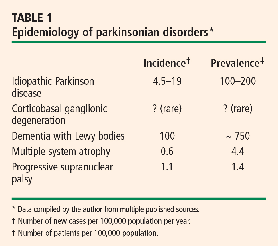
Tauopathies versus synucleinopathies
Pathologically, parkinsonian syndromes can be divided into two groups: the tauopathies and the synucleinopathies.
The tauopathies, so named because of the presence of hyperphosphylated tau protein, include progressive supranuclear palsy and corticobasal ganglionic degeneration as well as a number of other neurodegenerative conditions (ie, Pick disease, FTDP-17, primary progressive aphasia, argyrophilic grain disease) that do not cause parkinsonian features.
Synucleinopathies, the focus of this article, are disorders in which the protein alpha-synuclein accumulates in the cytoplasm. They include idiopathic Parkinson disease, multiple system atrophy, dementia with Lewy bodies, and pure autonomic failure. In multiple system atrophy, deposits of alpha-synuclein are prominent in glial cytoplasmic inclusions. In both Parkinson disease and dementia with Lewy bodies, alpha-synuclein is present in Lewy bodies. Although primary autonomic failure is not a movement disorder, its pathology is similar to that of the other synucleinopathies, with alpha-synuclein accumulation in both the central and peripheral nervous systems, as well as the presence of Lewy bodies.
IDIOPATHIC PARKINSON DISEASE
Autonomic dysfunction usually occurs late in idiopathic Parkinson disease, and its severity is less than that observed with other parkinsonian syndromes. However, the lifetime risk of significant autonomic dysfunction in patients with Parkinson disease is approximately 1 in 3.1 Almost 60% of patients with idiopathic Parkinson disease meet the criterion for a diagnosis of orthostatic hypotension—ie, a fall in systolic blood pressure of at least 20 mm Hg—and orthostatic hypotension is symptomatic in about 20% of patients.1
MULTIPLE SYSTEM ATROPHY
The nomenclature of multiple system atrophy has been evolving slowly. In 1900, Dejerine and Thomas described olivopontocerebellar atrophy, a progressive cerebellar degeneration with parkinsonism. In 1960, Shy and Drager described the Shy Drager syndrome, which has prominent autonomic features common to Parkinson disease, such as orthostatic hypotension, urinary and fecal incontinence, loss of sweating, iris atrophy, external ocular palsies, rigidity, tremor, loss of associated movement, and impotence.2
Also in 1960, Van der Eecken described striatonigral degeneration, an akinetic, rigid, parkinsonian syndrome that did not respond well to medications and was associated with autonomic dysfunction.3
In 1969, Graham and Oppenheimer realized an overlap to these syndromes and coined the term multiple system atrophy. They used it to refer to a gradually progressive idiopathic neurodegenerative process of adult onset characterized by varying proportions of cerebellar dysfunction, autonomic failure, and parkinsonism, and which is poorly responsive to levodopa therapy.4
Newer terminology is more specific for the predominant symptoms in the syndrome. A predominance of parkinsonism with this syndrome is referred to as “parkinsonian type of multiple system atrophy” (MSA-P), whereas a predominance of cerebellar signs is termed “multiple system atrophy with cerebellar-predominant symptoms” (MSA-C). The parkinsonian type is about four times as common as the cerebellar type.5 Autonomic dysfunction is common to both types, and its severity varies.
Parkinsonism is the most common symptom in multiple system atrophy, followed by autonomic failure and cerebellar signs. Approximately one fourth of patients with multiple system atrophy have all three categories of symptoms. Pyramidal signs are present in approximately 60% of patients, and help distinguish this syndrome primarily from idiopathic Parkinson disease.6
Diagnostic criteria
Autonomic dysfunction in the form of orthostatic hypotension and/or urinary incontinence is a key diagnostic criterion for multiple system atrophy.
Parkinsonism. Parkinsonian features of the syndrome are bradykinesia, rigidity, postural instability, and tremor.
Cerebellar dysfunction. Features of cerebellar dysfunction include gait ataxia, ataxic dysarthria, limb ataxia, and sustained gaze-evoked nystagmus.
Corticospinal tract dysfunction (extensor plantar response with hyperreflexia) also helps establish the diagnosis because this feature separates multiple system atrophy from idiopathic Parkinson disease as well as some of the other parkinsonian syndromes.
Diagnostic categories
The above diagnostic criteria can be combined to make a diagnosis of possible, probable, or definite multiple system atrophy.
Possible. For a diagnosis of possible multiple system atrophy, one of the above diagnostic criteria must be present along with two features from separate domains. If the case meets the criteria for parkinsonism (bradykinesia plus at least one of the other aforementioned features of parkinsonism), a poor levodopa response qualifies as a feature.
Probable. A diagnosis of probable multiple system atrophy must meet the criterion for autonomic dysfunction plus either the criterion for parkinsonism (with poor levodopa response) or the criterion for cerebellar dysfunction.
Definite. Definite multiple system atrophy requires pathological confirmation.
Extrapyramidal features in multiple system atrophy
In addition to prominent autonomic and/or cerebellar dysfunction, differences in extrapyramidal features help distinguish multiple system atrophy from idiopathic Parkinson disease. Tremor is less common in multiple system atrophy than in idiopathic Parkinson disease, and the akinetic/rigid symptoms tend to be symmetric in multiple system atrophy, rather than asymmetric as in Parkinson disease. Postural instability occurs early in multiple system atrophy but does not occur until late in idiopathic Parkinson disease. Moreover, multiple system atrophy responds poorly to levodopa and is characterized by more rapid disease progression. The presence of early autonomic and cerebellar symptoms is diagnostic for multiple system atrophy.
Pathology
The pathologic hallmark of multiple system atrophy is alpha-synuclein deposits in the glial or glial cytoplasmic inclusions (Papp-Lantos inclusions), which are diffuse through the central nervous system but are present particularly in the brainstem and spinal cord.
DEMENTIA WITH LEWY BODIES
Dementia with Lewy bodies is also known as diffuse Lewy body disease, senile dementia of the Lewy body type, Lewy body variant of Alzheimer disease, and Parkinson disease with dementia.
Dementia with Lewy bodies is descriptive for the entire series of these diseases. Pathologically, it is identical to Parkinson disease with dementia, with the only difference being an objective criterion based on the duration of dementia. Dementia less than 1 year after onset of parkinsonism is considered dementia with Lewy bodies, whereas dementia more than 1 year after the onset of parkinsonism is considered Parkinson disease with dementia. Whether these are separate disorders or two ends of a spectrum of disease is unclear.
Clinical criteria
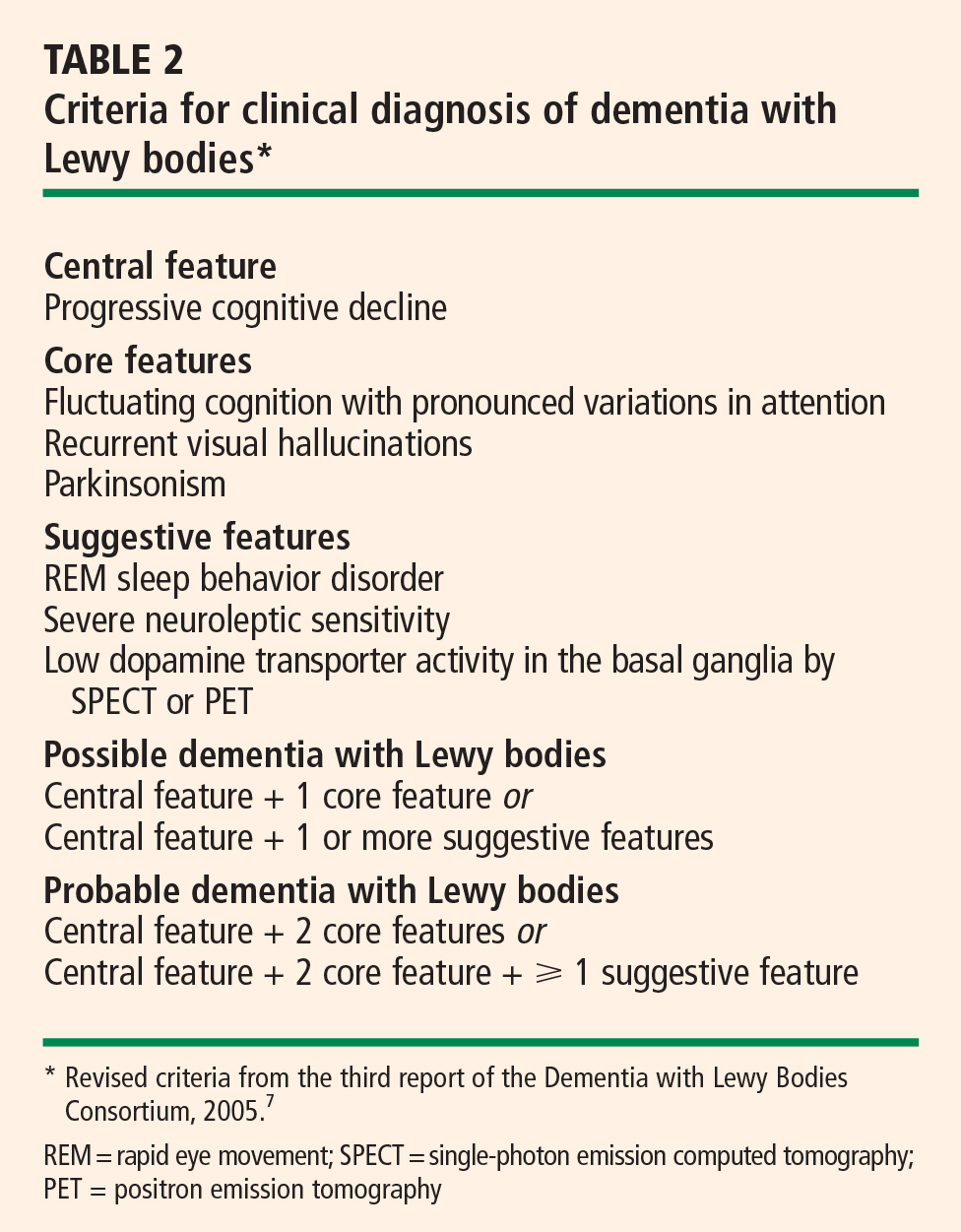
Central feature: progressive cognitive decline. Dementia with Lewy bodies is characterized by prominent progressive cognitive decline that is uncharacteristic of Parkinson disease. In particular, patients with dementia with Lewy bodies have fluctuating cognition, pronounced variations in attention, and early hallucinations when either off medications or on low doses of dopamimetic medications.
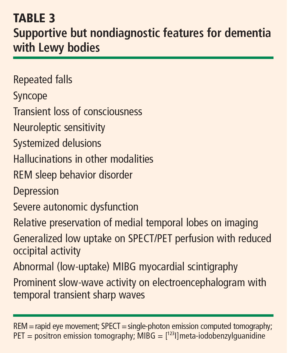
Pattern of dementia is more subcortical than cortical. The cognitive changes are different from those present in Alzheimer disease. In contrast to patients with Alzheimer disease, those with dementia with Lewy bodies have more subcortical than cortical dementia, resulting in executive dysfunction and inattention, whereas patients with Alzheimer disease have dysfunction of naming and memory.8
Pathology: diffuse distribution of Lewy bodies. As in Parkinson disease, the pathology is characterized by the appearance of Lewy bodies (positive stain for alpha-synuclein), but their distribution is more diffuse than in Parkinson disease and includes the brainstem, subcortical nuclei, limbic cortex, and neocortex, which may lead to hallucinations in affected patients.
Autonomic features are also diffuse. Autonomic features are also more common in dementia with Lewy bodies than in idiopathic Parkinson disease, which may relate to the different distribution of pathology in these diseases. Significant autonomic failure is present in 62% of patients with dementia with Lewy bodies,9 and the autonomic failure is believed to result from dysfunction of peripheral postganglionic neurons in addition to numerous cortical and brainstem Lewy bodies. Patients with dementia with Lewy bodies also have significant deposits in intermediolateral columns of the spinal cord and autonomic ganglia and sympathetic neurons.
PURE AUTONOMIC FAILURE
The pathology of pure autonomic failure is similar to that of dementia with Lewy bodies and idiopathic Parkinson disease. In contrast to these disorders, however, pure autonomic failure is characterized by a less significant presence of Lewy bodies in the cortex and brainstem, although the pathology in the spinal cord and peripheral nervous system is quite prominent.
Pure autonomic failure is a sporadic disease with onset after age 60 years. It is characterized by slowly progressive isolated impairment of the autonomic nervous system, which manifests particularly as orthostatic hypotension and also as significant bladder and sexual dysfunction. The condition is ultimately disabling as a result of the orthostatic hypotension.
CARDIOVASCULAR AUTONOMIC DYSFUNCTION
Orthostatic hypotension is the most limiting of the cardiovascular autonomic dysfunctions in the neurodegenerative disorders discussed here. Postprandial hypotension is also prevalent in these disorders, as is supine hypertension, which makes successful treatment of cardiovascular autonomic dysfunction difficult.
Orthostatic hypotension is defined as a decrease in systolic blood pressure of at least 20 mm Hg, or a decrease in diastolic blood pressure of at least 10 mm Hg, upon tilting or standing.
In contrast, in normal subjects the initial response upon standing is a pooling of 500 to 1,000 mL of blood and a reduction in venous return and cardiac output. A resultant decrease in blood pressure would occur if not for the baroreceptor reflex, which increases sympathetic tone and decreases vagal parasympathetic tone. Vasopressin is then released from the posterior pituitary, which increases peripheral vascular resistance, venous return, and cardiac output. As a result, the normal response to standing is a modest decrease in systolic blood pressure—ie, by 5 to 10 mm Hg— and an increase in diastolic blood pressure by a similar amount, as well as a compensatory increase in pulse rate of 10 to 25 beats per minute.
Approaches to therapy for orthostatic hypotension
Nonpharmacologic approaches to orthostatic hypotension include raising the head of the patient’s bed by 30 degrees, use of compression stockings, and liberalizing the use of fluids and salt.
Often, however, patients require pharmacologic therapy. Fluorohydrocortisone and midodrine are the primary drugs used for this purpose, but pyrodostigmine also has shown some efficacy in doses of 60 mg or greater in small clinical trials. Less-effective options include nonsteroidal anti-inflammatory drugs, vasopressin analogs, erythropoietin, and caffeine.
Management of postprandial hypotension
Reducing meal size while increasing the frequency of meals and adding caffeine are dietary approaches to treat postprandial hypotension. Somatostatin analogs may be helpful, although data to support their use for this indication are limited.
Treatment of supine hypertension is more difficult
The management of supine hypertension is difficult in patients with neurodegenerative disorders. Supine hypertension is defined as a blood pressure greater than 140/90 mm Hg, but the threshold for concern is uncertain. Most patients with neurodegenerative disorders are plagued more by hypotension than by hypertension, but the hypertension can be deleterious to their health, particularly when they are being treated for their hypotension during the day. Some clinicians choose to treat the hypertension if it is significant in the evening. Most important is to remove the midodrine at night and minimize the use of fluorohydrocortisone. Other options are nitrate derivatives, hydralazine, and calcium channel blockers. The proposed benefits of minoxidil and clonidine are controversial.
Autonomic complications of dopaminergic therapy
Complicating the management of autonomic dysfunction in patients with parkinsonian features is that drug therapies for Parkinson disease exacerbate orthostatic hypotension to varying degrees. Selegiline, amantadine, and dopamine agonists exacerbate orthostasis to a greater degree than levodopa does. Therefore, we are apt to start treatment with levodopa as patients develop more features of autonomic dysfunction, as well as in patients with advanced Parkinson disease or in patients who are older than 70 years of age.
Multiple system atrophy may respond only to high doses of levodopa (> 1 g), and when autonomic symptoms are prominent, patients with multiple system atrophy may not tolerate dopaminergic therapy at all. In patients with dementia with Lewy bodies, the use of dopaminergic therapies is limited not so much by autonomic dysfunction but because of hallucinations and cognitive decline.
SUMMARY
Central autonomic dysfunction predominates in patients with multiple system atrophy. Peripheral autonomic dysfunction predominates in the other parkinsonian disorders with Lewy body pathology, and this includes idiopathic Parkinson disease, dementia with Lewy bodies, and the related disorder, pure autonomic failure, in which there are no parkinsonian features.
- Magalhaes M, Wenning GK, Daniel SE, Quinn NP. Autonomic dysfunction in pathologically confirmed multiple system atrophy and idiopathic Parkinson’s disease—a retrospective comparison. Acta Neurol Scand 1995; 91:98–102.
- Shy GM, Drager GA. A neurological syndrome associated with orthostatic hypotension: a clinical-pathologic study. Arch Neurol 1960; 2:511–527.
- Van der Eecken H, Adams RD, van Bogaert L. Striopallidal-nigral degeneration. An hitherto undescribed lesion in paralysis agitans. J Neuropathol Exp Neurol 1960; 19:159–160.
- Graham JG, Oppenheimer DR. Orthostatic hypotension and nicotine sensitivity in a case of multiple system atrophy. J Neurol Neurosurg Psychiatry 1969; 32:28–34.
- Wenning GK, Geser F, Stampfer-Kountchev M, Tison F. Multiple system atrophy: an update. Mov Disord 2003; 18(Suppl 6):S34–S42.
- Hughes AJ, Daniel SE, Ben-Shlomo Y, Lees AJ. The accuracy of diagnosis of parkinsonian syndromes in a specialist movement disorder service. Brain 2002; 125:861–870.
- McKeith IG, Dickson DW, Lowe J, et al. Diagnosis and management of dementia with Lewy bodies: third report of the DLB Consortium. Neurology 2005; 65:1863–1872.
- Kraybill ML, Larson EB, Tsuang DW, et al. Cognitive differences in dementia patients with autopsy-verified AD, Lewy body pathology, or both. Neurology 2005; 64:2069–2073.
- Horimoto Y, Matsumoto M, Akatsu H, et al. Autonomic dysfunctions in dementia with Lewy bodies. J Neurol 2003; 250:530–533.
The Lewy body is the pathologic hallmark of both Parkinson disease and dementia with Lewy bodies. Lewy bodies are seen microscopically as neuronal inclusions containing alpha-synuclein and associated proteins. In contrast, glial inclusions involving alpha-synuclein are seen in multiple system atrophy. Because Lewy bodies are observed in autonomic regulatory regions of the brain, they are of interest in the study of the autonomic dysfunction that figures prominently in several parkinsonian syndromes. Cardiovascular autonomic dysfunction in parkinsonian syndromes includes orthostatic hypotension, postprandial hypotension, and supine hypertension.
This article will describe the major clinical and pathologic features of movement disorders with Lewy body pathology, the likelihood of autonomic dysregulation in these disorders, and issues involved in the treatment of autonomic dysfunction in patients with these movement disorders.
OVERVIEW OF PARKINSONIAN SYNDROMES
Tauopathies versus synucleinopathies
Pathologically, parkinsonian syndromes can be divided into two groups: the tauopathies and the synucleinopathies.
The tauopathies, so named because of the presence of hyperphosphylated tau protein, include progressive supranuclear palsy and corticobasal ganglionic degeneration as well as a number of other neurodegenerative conditions (ie, Pick disease, FTDP-17, primary progressive aphasia, argyrophilic grain disease) that do not cause parkinsonian features.
Synucleinopathies, the focus of this article, are disorders in which the protein alpha-synuclein accumulates in the cytoplasm. They include idiopathic Parkinson disease, multiple system atrophy, dementia with Lewy bodies, and pure autonomic failure. In multiple system atrophy, deposits of alpha-synuclein are prominent in glial cytoplasmic inclusions. In both Parkinson disease and dementia with Lewy bodies, alpha-synuclein is present in Lewy bodies. Although primary autonomic failure is not a movement disorder, its pathology is similar to that of the other synucleinopathies, with alpha-synuclein accumulation in both the central and peripheral nervous systems, as well as the presence of Lewy bodies.
IDIOPATHIC PARKINSON DISEASE
Autonomic dysfunction usually occurs late in idiopathic Parkinson disease, and its severity is less than that observed with other parkinsonian syndromes. However, the lifetime risk of significant autonomic dysfunction in patients with Parkinson disease is approximately 1 in 3.1 Almost 60% of patients with idiopathic Parkinson disease meet the criterion for a diagnosis of orthostatic hypotension—ie, a fall in systolic blood pressure of at least 20 mm Hg—and orthostatic hypotension is symptomatic in about 20% of patients.1
MULTIPLE SYSTEM ATROPHY
The nomenclature of multiple system atrophy has been evolving slowly. In 1900, Dejerine and Thomas described olivopontocerebellar atrophy, a progressive cerebellar degeneration with parkinsonism. In 1960, Shy and Drager described the Shy Drager syndrome, which has prominent autonomic features common to Parkinson disease, such as orthostatic hypotension, urinary and fecal incontinence, loss of sweating, iris atrophy, external ocular palsies, rigidity, tremor, loss of associated movement, and impotence.2
Also in 1960, Van der Eecken described striatonigral degeneration, an akinetic, rigid, parkinsonian syndrome that did not respond well to medications and was associated with autonomic dysfunction.3
In 1969, Graham and Oppenheimer realized an overlap to these syndromes and coined the term multiple system atrophy. They used it to refer to a gradually progressive idiopathic neurodegenerative process of adult onset characterized by varying proportions of cerebellar dysfunction, autonomic failure, and parkinsonism, and which is poorly responsive to levodopa therapy.4
Newer terminology is more specific for the predominant symptoms in the syndrome. A predominance of parkinsonism with this syndrome is referred to as “parkinsonian type of multiple system atrophy” (MSA-P), whereas a predominance of cerebellar signs is termed “multiple system atrophy with cerebellar-predominant symptoms” (MSA-C). The parkinsonian type is about four times as common as the cerebellar type.5 Autonomic dysfunction is common to both types, and its severity varies.
Parkinsonism is the most common symptom in multiple system atrophy, followed by autonomic failure and cerebellar signs. Approximately one fourth of patients with multiple system atrophy have all three categories of symptoms. Pyramidal signs are present in approximately 60% of patients, and help distinguish this syndrome primarily from idiopathic Parkinson disease.6
Diagnostic criteria
Autonomic dysfunction in the form of orthostatic hypotension and/or urinary incontinence is a key diagnostic criterion for multiple system atrophy.
Parkinsonism. Parkinsonian features of the syndrome are bradykinesia, rigidity, postural instability, and tremor.
Cerebellar dysfunction. Features of cerebellar dysfunction include gait ataxia, ataxic dysarthria, limb ataxia, and sustained gaze-evoked nystagmus.
Corticospinal tract dysfunction (extensor plantar response with hyperreflexia) also helps establish the diagnosis because this feature separates multiple system atrophy from idiopathic Parkinson disease as well as some of the other parkinsonian syndromes.
Diagnostic categories
The above diagnostic criteria can be combined to make a diagnosis of possible, probable, or definite multiple system atrophy.
Possible. For a diagnosis of possible multiple system atrophy, one of the above diagnostic criteria must be present along with two features from separate domains. If the case meets the criteria for parkinsonism (bradykinesia plus at least one of the other aforementioned features of parkinsonism), a poor levodopa response qualifies as a feature.
Probable. A diagnosis of probable multiple system atrophy must meet the criterion for autonomic dysfunction plus either the criterion for parkinsonism (with poor levodopa response) or the criterion for cerebellar dysfunction.
Definite. Definite multiple system atrophy requires pathological confirmation.
Extrapyramidal features in multiple system atrophy
In addition to prominent autonomic and/or cerebellar dysfunction, differences in extrapyramidal features help distinguish multiple system atrophy from idiopathic Parkinson disease. Tremor is less common in multiple system atrophy than in idiopathic Parkinson disease, and the akinetic/rigid symptoms tend to be symmetric in multiple system atrophy, rather than asymmetric as in Parkinson disease. Postural instability occurs early in multiple system atrophy but does not occur until late in idiopathic Parkinson disease. Moreover, multiple system atrophy responds poorly to levodopa and is characterized by more rapid disease progression. The presence of early autonomic and cerebellar symptoms is diagnostic for multiple system atrophy.
Pathology
The pathologic hallmark of multiple system atrophy is alpha-synuclein deposits in the glial or glial cytoplasmic inclusions (Papp-Lantos inclusions), which are diffuse through the central nervous system but are present particularly in the brainstem and spinal cord.
DEMENTIA WITH LEWY BODIES
Dementia with Lewy bodies is also known as diffuse Lewy body disease, senile dementia of the Lewy body type, Lewy body variant of Alzheimer disease, and Parkinson disease with dementia.
Dementia with Lewy bodies is descriptive for the entire series of these diseases. Pathologically, it is identical to Parkinson disease with dementia, with the only difference being an objective criterion based on the duration of dementia. Dementia less than 1 year after onset of parkinsonism is considered dementia with Lewy bodies, whereas dementia more than 1 year after the onset of parkinsonism is considered Parkinson disease with dementia. Whether these are separate disorders or two ends of a spectrum of disease is unclear.
Clinical criteria
Central feature: progressive cognitive decline. Dementia with Lewy bodies is characterized by prominent progressive cognitive decline that is uncharacteristic of Parkinson disease. In particular, patients with dementia with Lewy bodies have fluctuating cognition, pronounced variations in attention, and early hallucinations when either off medications or on low doses of dopamimetic medications.
Pattern of dementia is more subcortical than cortical. The cognitive changes are different from those present in Alzheimer disease. In contrast to patients with Alzheimer disease, those with dementia with Lewy bodies have more subcortical than cortical dementia, resulting in executive dysfunction and inattention, whereas patients with Alzheimer disease have dysfunction of naming and memory.8
Pathology: diffuse distribution of Lewy bodies. As in Parkinson disease, the pathology is characterized by the appearance of Lewy bodies (positive stain for alpha-synuclein), but their distribution is more diffuse than in Parkinson disease and includes the brainstem, subcortical nuclei, limbic cortex, and neocortex, which may lead to hallucinations in affected patients.
Autonomic features are also diffuse. Autonomic features are also more common in dementia with Lewy bodies than in idiopathic Parkinson disease, which may relate to the different distribution of pathology in these diseases. Significant autonomic failure is present in 62% of patients with dementia with Lewy bodies,9 and the autonomic failure is believed to result from dysfunction of peripheral postganglionic neurons in addition to numerous cortical and brainstem Lewy bodies. Patients with dementia with Lewy bodies also have significant deposits in intermediolateral columns of the spinal cord and autonomic ganglia and sympathetic neurons.
PURE AUTONOMIC FAILURE
The pathology of pure autonomic failure is similar to that of dementia with Lewy bodies and idiopathic Parkinson disease. In contrast to these disorders, however, pure autonomic failure is characterized by a less significant presence of Lewy bodies in the cortex and brainstem, although the pathology in the spinal cord and peripheral nervous system is quite prominent.
Pure autonomic failure is a sporadic disease with onset after age 60 years. It is characterized by slowly progressive isolated impairment of the autonomic nervous system, which manifests particularly as orthostatic hypotension and also as significant bladder and sexual dysfunction. The condition is ultimately disabling as a result of the orthostatic hypotension.
CARDIOVASCULAR AUTONOMIC DYSFUNCTION
Orthostatic hypotension is the most limiting of the cardiovascular autonomic dysfunctions in the neurodegenerative disorders discussed here. Postprandial hypotension is also prevalent in these disorders, as is supine hypertension, which makes successful treatment of cardiovascular autonomic dysfunction difficult.
Orthostatic hypotension is defined as a decrease in systolic blood pressure of at least 20 mm Hg, or a decrease in diastolic blood pressure of at least 10 mm Hg, upon tilting or standing.
In contrast, in normal subjects the initial response upon standing is a pooling of 500 to 1,000 mL of blood and a reduction in venous return and cardiac output. A resultant decrease in blood pressure would occur if not for the baroreceptor reflex, which increases sympathetic tone and decreases vagal parasympathetic tone. Vasopressin is then released from the posterior pituitary, which increases peripheral vascular resistance, venous return, and cardiac output. As a result, the normal response to standing is a modest decrease in systolic blood pressure—ie, by 5 to 10 mm Hg— and an increase in diastolic blood pressure by a similar amount, as well as a compensatory increase in pulse rate of 10 to 25 beats per minute.
Approaches to therapy for orthostatic hypotension
Nonpharmacologic approaches to orthostatic hypotension include raising the head of the patient’s bed by 30 degrees, use of compression stockings, and liberalizing the use of fluids and salt.
Often, however, patients require pharmacologic therapy. Fluorohydrocortisone and midodrine are the primary drugs used for this purpose, but pyrodostigmine also has shown some efficacy in doses of 60 mg or greater in small clinical trials. Less-effective options include nonsteroidal anti-inflammatory drugs, vasopressin analogs, erythropoietin, and caffeine.
Management of postprandial hypotension
Reducing meal size while increasing the frequency of meals and adding caffeine are dietary approaches to treat postprandial hypotension. Somatostatin analogs may be helpful, although data to support their use for this indication are limited.
Treatment of supine hypertension is more difficult
The management of supine hypertension is difficult in patients with neurodegenerative disorders. Supine hypertension is defined as a blood pressure greater than 140/90 mm Hg, but the threshold for concern is uncertain. Most patients with neurodegenerative disorders are plagued more by hypotension than by hypertension, but the hypertension can be deleterious to their health, particularly when they are being treated for their hypotension during the day. Some clinicians choose to treat the hypertension if it is significant in the evening. Most important is to remove the midodrine at night and minimize the use of fluorohydrocortisone. Other options are nitrate derivatives, hydralazine, and calcium channel blockers. The proposed benefits of minoxidil and clonidine are controversial.
Autonomic complications of dopaminergic therapy
Complicating the management of autonomic dysfunction in patients with parkinsonian features is that drug therapies for Parkinson disease exacerbate orthostatic hypotension to varying degrees. Selegiline, amantadine, and dopamine agonists exacerbate orthostasis to a greater degree than levodopa does. Therefore, we are apt to start treatment with levodopa as patients develop more features of autonomic dysfunction, as well as in patients with advanced Parkinson disease or in patients who are older than 70 years of age.
Multiple system atrophy may respond only to high doses of levodopa (> 1 g), and when autonomic symptoms are prominent, patients with multiple system atrophy may not tolerate dopaminergic therapy at all. In patients with dementia with Lewy bodies, the use of dopaminergic therapies is limited not so much by autonomic dysfunction but because of hallucinations and cognitive decline.
SUMMARY
Central autonomic dysfunction predominates in patients with multiple system atrophy. Peripheral autonomic dysfunction predominates in the other parkinsonian disorders with Lewy body pathology, and this includes idiopathic Parkinson disease, dementia with Lewy bodies, and the related disorder, pure autonomic failure, in which there are no parkinsonian features.
- Magalhaes M, Wenning GK, Daniel SE, Quinn NP. Autonomic dysfunction in pathologically confirmed multiple system atrophy and idiopathic Parkinson’s disease—a retrospective comparison. Acta Neurol Scand 1995; 91:98–102.
- Shy GM, Drager GA. A neurological syndrome associated with orthostatic hypotension: a clinical-pathologic study. Arch Neurol 1960; 2:511–527.
- Van der Eecken H, Adams RD, van Bogaert L. Striopallidal-nigral degeneration. An hitherto undescribed lesion in paralysis agitans. J Neuropathol Exp Neurol 1960; 19:159–160.
- Graham JG, Oppenheimer DR. Orthostatic hypotension and nicotine sensitivity in a case of multiple system atrophy. J Neurol Neurosurg Psychiatry 1969; 32:28–34.
- Wenning GK, Geser F, Stampfer-Kountchev M, Tison F. Multiple system atrophy: an update. Mov Disord 2003; 18(Suppl 6):S34–S42.
- Hughes AJ, Daniel SE, Ben-Shlomo Y, Lees AJ. The accuracy of diagnosis of parkinsonian syndromes in a specialist movement disorder service. Brain 2002; 125:861–870.
- McKeith IG, Dickson DW, Lowe J, et al. Diagnosis and management of dementia with Lewy bodies: third report of the DLB Consortium. Neurology 2005; 65:1863–1872.
- Kraybill ML, Larson EB, Tsuang DW, et al. Cognitive differences in dementia patients with autopsy-verified AD, Lewy body pathology, or both. Neurology 2005; 64:2069–2073.
- Horimoto Y, Matsumoto M, Akatsu H, et al. Autonomic dysfunctions in dementia with Lewy bodies. J Neurol 2003; 250:530–533.
Deep brain stimulation: How does it work?
Whether deep brain stimulation can dramatically help patients with Parkinson’s disease (PD) and other movement disorders is no longer questioned. Rather, how it works is not well understood: how do patients with seemingly diverse conditions show improvement with the same intervention?
Patients with advanced PD often freeze when trying to walk and have tremor, rigidity, bradykinesia, and gait and balance problems. With deep brain stimulation, a patient typically experiences a marked improvement in these motor symptoms.
Similarly, patients with hypokinetic disorders such as generalized dystonia who have extensive involuntary movements involving multiple body parts may experience a significant reduction in these movements and regain function during deep brain stimulation. In my experience, it is not unusual for patients who were not ambulatory as a result of their dystonic movements to regain function to the point where they can walk unassisted and, in some cases, participate in physical activities such as racquetball or jogging on a treadmill. One of my patients with generalized dystonia could walk no farther than several meters before deep brain stimulation but afterward was able to run on a treadmill. This patient did not gain this type of function immediately after stimulation, but after sustained efforts at programming his stimulation device over the course of 1 year he was able to travel to Europe, hike in the mountains, and jog on a treadmill.
In addition to treating movement disorders, deep brain stimulation is being used experimentally to treat patients with behavioral disorders such as depression and obsessive-compulsive disorder that are refractive to standard therapy. Broadening our understanding of the mechanisms responsible for success with deep brain stimulation is important since it may help to improve current applications and develop new ones. This article discusses our research in deep brain stimulation using microelectrode recording of structures within the basal ganglia–thalamocortical circuit in the MPTP monkey model of PD.
INSIGHTS INTO MECHANISMS OF STIMULATION PROMISE TECHNOLOGICAL REFINEMENTS
One rationale for attempting to better understand how deep brain stimulation works is that such knowledge may enable us to improve the technology to better apply the technique.
Electrode design is one important area of potential improvement. Diseases that may one day be treated with deep brain stimulation will likely require electrodes of different shapes than those used currently, to accommodate other targets in the brain. At present, a single lead shape is used to stimulate the subthalamic nucleus (STN) and the globus pallidus internus (GPi) for treating PD. Possible future targets include the globus pallidus externus (GPe), various subnuclei of the thalamus, portions of the striatum, and other subcortical and cortical structures that have different geometric configurations and physiologic characteristics. Since these structures and regions of the brain differ from one another in size and shape, it is highly likely that new electrode designs will be needed to take advantage of this geometric and physiologic variability. Future electrodes may vary in size and shape from those used currently, incorporate three-dimensional designs, and require a current source that allows the pattern of stimulation to be varied based on the physiologic changes that characterize each neurologic disorder.
Directionality may be another important feature of electrode design. With presently used electrodes, electric current spreads in all directions. To spread the current or increase the volume of tissue affected by stimulation, one must increase the voltage being passed through the lead. This results in a larger region of tissue being affected by stimulation, but the current density varies based on distance from the stimulation site, with neural tissue close to the site being affected differently from tissue that is farther away. Moreover, the current cannot be directed or aimed in one direction or the other. A split-band design could spread current in opposing directions, and a three-dimensional directional design involving several contacts could affect a volume of tissue more homogeneously.
PROGRESS IN DEFINING PD PATHOPHYSIOLOGY
As with any disease, defining the problem and understanding the underlying pathophysiology are essential first steps to finding an effective treatment for PD. In the 1930s and 1940s, numerous attempts were made to treat PD with surgical therapies. Surgical targets were chosen throughout the length of the neuraxis, including the cortex, the internal capsule, the basal ganglia, the thalamus, the cerebral peduncle, and the spinal cord itself. The underlying pathophysiology was not well understood, however, so the rationale for surgery was weak at best. For example, lesioning the cortex improved parkinsonian tremor, but it also caused paralysis and was associated with considerable morbidity.
Evidence of a common circuit
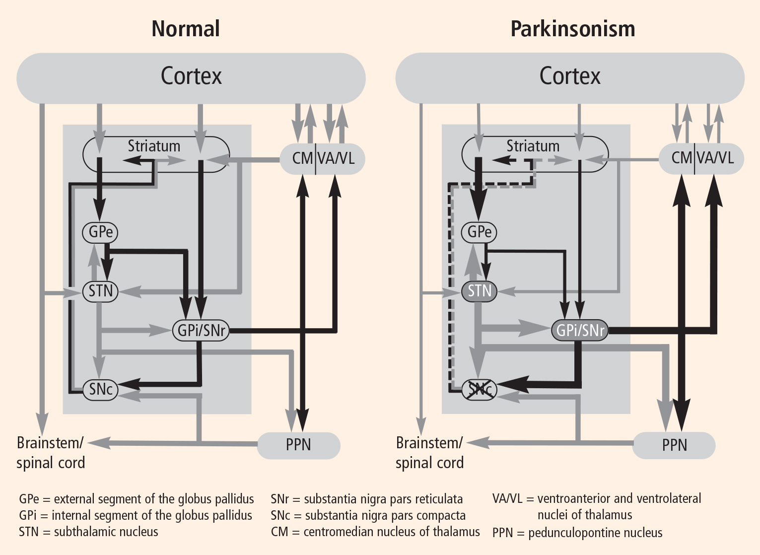
In PD, degeneration of dopamine-producing neurons in the substantia nigra pars compacta reduces dopamine levels in the striatum. In MPTP monkey models of PD there is also a loss of dopamine-producing cells in the substantia nigra pars compacta. These animals develop the cardinal motor symptoms of PD and are considered a good model of the human disorder. By recording from the basal ganglia–thalamocortical circuit in this model, we and others have observed excessive activity in the STN and GPi.1–4 In addition, cells in these regions in the monkey model were more likely to discharge in bursts compared with cells from healthy monkeys, and they showed a higher degree of synchronized oscillatory activity among neighboring neurons.5,6
Ultimate goal: The ability to individualize therapy
Understanding how such changes relate to parkinsonian symptoms will enable us to develop stimulation strategies that are focused on ameliorating the particular physiologic changes in PD. Since PD can lead to distinctly different clinical pictures, it would be ideal to be able to individualize therapy based on the particular motor symptoms each patient experiences. This may require stimulation strategies that affect either a particular region of the targeted structure or a particular physiologic change that occurs in the disease state.
THE ‘RATE HYPOTHESIS’: ALTERED CELLULAR DISCHARGE RATES CAUSE PARKINSONIAN MOTOR SYMPTOMS
A good model for PD was lacking prior to the 1980s. As a result, there was little understanding of the pathophysiologic basis for this disorder. A breakthrough in the mid-1980s revolutionized research in this field. A group of young people developed parkinsonian symptoms, and it was discovered that they had all used recreational “designer drugs” containing an impurity: the neurotoxin 1-methyl-4-phenyl-1,2,3,6tetrahydropyridine (MPTP). Now given to primates to simulate PD, MPTP causes all of the classic symptoms of PD except tremor (this may vary from species to species), including freezing, slowness, stiffness, and gait and balance problems. Like humans with PD, primates with MPTP-induced PD even develop dyskinesia after prolonged treatment with levodopa.
Experimentation with MPTP monkeys in the late 1980s led to the “rate hypothesis,” which basically states that when dopamine production is reduced from the substantia nigra compacta (as in PD), changes in striatal activity lead to suppression of GPe activity and a reduction in inhibitory output from the GPe to the STN. This decrease in inhibitory output allows the STN to be overactive, which, in conjunction with a reduction of direct striatal inhibition of the GPi, causes excessive GPi activity and a suppression of thalamic activity to the cortex (Figure 1).
When recording electrodes were placed in these structures in the monkey brain, rate changes were reported to occur in each of these structures in the parkinsonian state.1–4,7 Action potentials recorded from the GPi in MPTP-treated monkeys occurred at a much faster rate than those in healthy monkeys.
Pallidotomy revisited: Dramatic symptom improvement is possible
On the basis of the above and other studies in the MPTP monkey model of PD, investigators in the 1990s reasoned that reduced dopamine in PD led to excessive activity in portions of this circuit. While I would like to say that this led to the rationale for lesioning the STN and GPi for the treatment of PD, this approach had already been taken in the early 1930s and 1940s and continued into the 1960s; it was largely stopped with the introduction of levodopa and was restarted again after the realization that chronic levodopa therapy was associated with a variety of side effects, including the development of excessive involuntary movement and motor fluctuations.
Pallidotomy (lesioning of the pallidum), although tried as a treatment for PD in the 1930s and 1940s, had been abandoned as a result of its inconsistent benefit and lack of effect on parkinsonian tremor. It underwent a resurgence in the 1990s through the work of a group in New York8 that revived Lars Leksell’s pallidotomy approach of the 1960s9 at a time when basic science studies provided the rationale for surgical therapy to create lesions in the GPi. These basic science studies also provided critical new information about the optimal site for lesioning, which led to improved and more consistent outcomes.10–13 In the early years, lesions were created in the anterior (nonmotor) portion of the pallidum but led to inconsistent results. In the 1990s, with a better understanding of the portion of the pallidum involved in motor control, destroying brain tissue by creating a lesion in the posterolateral “motor” region of the pallidum resulted in such dramatic improvement in motor signs that waiting lists of up to 4 years were common for patients who wanted the procedure.
Although unilateral pallidotomy led to marked improvement in motor symptoms on the contralateral side, attempts at bilateral lesions to improve both sides of the body, as well as axial symptoms, were associated with marked hypophonia and, in some reports, cognitive decline. This led physicians and scientists to search for a procedure that could be performed bilaterally without the high incidence of side effects associated with lesioning procedures—and thus to the birth of deep brain stimulation.
Deep brain stimulation as lesion simulation
During the early experience with pallidotomy, the area to be lesioned would first be stimulated with the lesioning probe to observe its effects and thereby determine the precise area in which to create a lesion. At the time, no mechanism existed to leave the stimulator in place rather than create a lesion. But after the development of implantable stimulation devices, chronic stimulation could be delivered bilaterally to the pallidum and STN, resulting in a markedly improved treatment. Since side effects associated with stimulation are reversible, the ability to perform such procedures on both sides of the brain and to adjust stimulation parameters in order to optimize benefits while minimizing side effects made deep brain stimulation the procedure of choice for patients with advanced PD and led to its exploration for treatment of other neurologic disorders.
Because stimulation produced the same or similar benefit as a lesion, most physicians thought that stimulation must work in a similar manner, ie, by decreasing output from the stimulated structure. The rationale for this hypothesis received support from the “rate” model of PD, which postulated that PD motor symptoms occur as a result of overactivity in the STN and GPi. It was postulated that deep brain stimulation improved clinical symptoms by suppressing output from the stimulated structure—in other words, deep brain stimulation effectively caused a physiologic ablation.14,15
FURTHER RESEARCH GIVES RISE TO THE ‘PATTERN HYPOTHESIS’
Deep brain stimulation in the monkey model
To test the effects of deep brain stimulation, we have performed it in primates with MPTP-induced parkinsonism. Custom-made leads sized to fit a monkey brain are implanted in the same deep brain structures that are targeted when treating PD in humans. Each animal lead has four contacts 0.5 mm in size. We implant a pulse generator, connect the pulse generator to the lead, and set stimulation parameters to improve motor symptoms to mimic a human therapeutic setting as closely as possible. We then record from the basal ganglia structures before, during, and after stimulation that improves the monkey’s motor symptoms. This allows us to determine which changes in neuronal activity in the basal ganglia circuit during stimulation are associated with an improvement in motor symptoms.
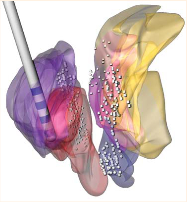
In earlier studies examining the mechanism underlying deep brain stimulation, neural activity was recorded only after stimulation, so that activity that occurred during stimulation had to be inferred from that which occurred immediately after stimulation was stopped. We developed a method to subtract artifact produced from stimulation without losing data. This method has been validated, is now used in a number of laboratories, and has revolutionized our ability to study the effect of stimulation on neuronal activity.17
A paradoxical finding
Based on the rate hypothesis, we expected that increased output from the GPi would cause parkinsonian symptoms and predicted that stimulation of the STN should suppress its output, which would suppress excitatory activity to the GPi from the STN and thereby reduce its output. Reduction of the inhibitory output from the GPi to the thalamus would, in turn, lead to a restoration of thalamocortical function and a reduction in the motor signs associated with PD. However, stimulating the STN was found to increase GPi activity.18 Despite increased rates, the incidence and intensity of symptoms were reduced. Further complicating the picture, we were contemporaneously exploring the effect of creating lesions in other parts of the basal ganglia that also led to increased rates of GPi activity, but in this case we observed that the increased rates were associated with a worsening of motor symptoms. In short, we had two laboratories working in parallel that had apparently obtained opposite results: increased GPi activity was associated with improved symptoms in one laboratory and with worse symptoms in the other.18,19
Patterns of activity are more important than rate
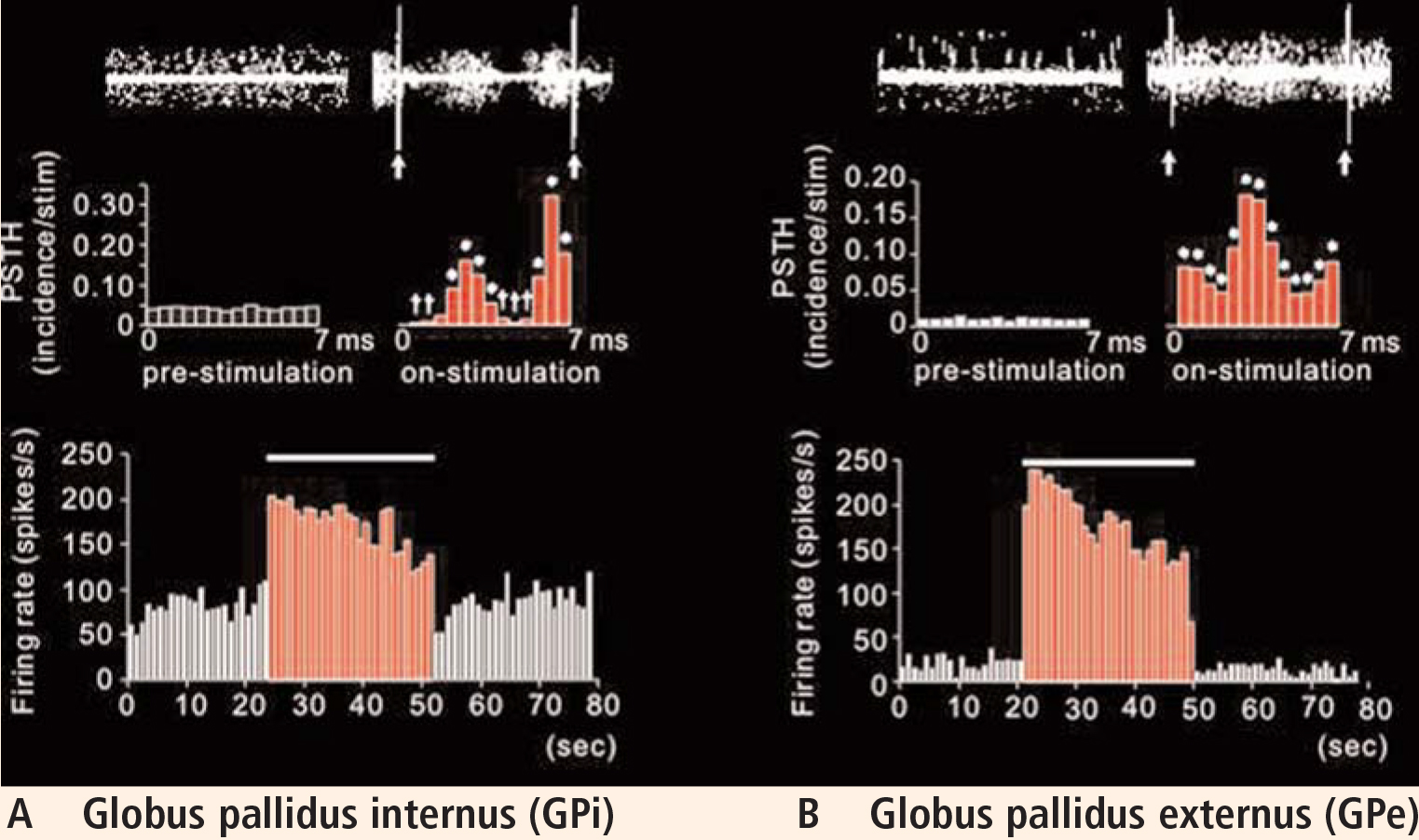
Knowledge that stimulation activated output from the stimulated region and changed the pattern of neuronal activity led us to ponder whether other targets, or even other ways to deliver stimulation, might work better to improve parkinsonian symptoms.
A focus on GPe stimulation
As a result of these observations, we reasoned that since GPe activity is also altered in PD and its rates are reduced, driving the output from this region that is inhibitory to the STN and GPi may help to reduce and regularize that activity at a point in the circuit that could provide even greater improvement in the motor symptoms associated with PD. Based on this hypothesis, we performed direct stimulation of the GPe in the MPTP monkey model of PD and evaluated its effect on motor behavior and neuronal activity in the circuit.
As an interesting sidelight, it should be noted that long before we developed this hypothesis, we had observations from a 1994 experiment (only recently published20) in which bradykinesia was improved upon acute stimulation in the GPe prior to making a lesion in the GPi. With sustained stimulation in this patient, we observed development of dyskinetic movements. Since we reasoned that lesions in this region would worsen parkinsonian symptoms—a rationale recently supported by a publication from our laboratory in 200619—and since we had no means by which to stimulate this region chronically at the time, this observation was filed away and we continued with lesioning the GPi for the treatment of these patients.
However, with the advent of chronic deep brain stimulation, we opted to reexplore this series of experiments in MPTP-treated monkeys. A lead was placed such that three of its contacts were in the GPe and one was in the GPi. Bradykinesia was assessed by determining the time it took for the monkey to retrieve raisins from a Klüver board. By inducing symptoms on one side only, we were able to use the healthy side as a control. We observed that before stimulation, retrieval took more than twice as long on the affected side. Stimulation of only 2 V had no effect, but increasing the voltage to 5.5 V significantly improved retrieval time.21
Plotting the data using post-stimulus time histograms showed that stimulation of the GPe inhibited the STN, confirming our hypothesis that stimulation activated the output from the stimulated structure (the GPe sends inhibitory projections to the STN). The responses observed were dramatic, with the majority of cells in the STN showing almost complete suppression of activity (Vitek et al, unpublished data).
In light of this observation, we expected that the rate of activity in the GPi would be reduced. Interestingly, although the rate was changed in most cells compared with control, what was most striking was the relatively stereotyped pattern of inhibition and excitation that occurred following each pulse of GPe stimulation. Although shifted in absolute frequency, the pattern that occurred was similar to that observed during STN stimulation, with alternating periods of excitation and inhibition evident in the post-stimulus time histogram.
Further evaluation of the data revealed a change in burst and oscillatory activity in the STN. Analysis of the data showed a shift in the distribution of power from low to high frequencies. Stimulation reduced activity in the low-frequency range and increased power in higher frequencies, similar to that in normal movement.
Further analysis of the spike trains revealed that entropy (a reflection of noise in the spike signal) was reduced under stimulation parameters that resulted in a reduction in symptoms. In contrast, stimulation parameters that resulted in worsening symptoms increased measures of entropy (Dorval, data submitted for publication).
PATTERN CHANGES AFFECT INFORMATION PROCESSING ACROSS THE BASAL GANGLIA–THALAMOCORTICAL NETWORK
There is a lack of consensus about the precise physiologic effect of deep brain stimulation for improving symptoms in movement disorders. Many researchers continue to believe that deep brain stimulation works through inhibition. An alternate explanation is that at effective stimulation parameters, the net effect is activation of output from the stimulated structure. Various modalities, including modeling,22,23 microdialysis,24 functional magnetic resonance imaging,25 and positron emission tomography,26,27 provide additional evidence that activation occurs during stimulation.
While one cannot discount a role for rate changes in mediating the effects of deep brain stimulation, there is now increasing evidence suggesting that pattern changes induced in the network as a result of stimulation-induced activation of output from the stimulated structure play an integral role in this process.
Research often leads to unpredictable outcomes. The prevailing hypothesis a decade ago concerning the pathophysiologic basis of PD (and still believed in many centers) was that rate is the controlling factor. But we have seen in our animal models that symptoms improve with increased rate in the GPi during stimulation in the STN. Similarly, GPi rates are abnormally low in patients with dystonia and in PD patients during dyskinesia, yet lesioning in the GPi that further reduces its output leads to improvement in these conditions. Based on these observations, it would appear that rate is unlikely to be the critical factor; we now must take into account other factors, such as pattern, oscillation, and synchronization, as well as changes in the network dynamics. Deep brain stimulation is changing the informational content of the neural network, and these changes are occurring across populations of neurons through the whole basal ganglia circuit. Knowing how these changes result in improvement in the neurologic disorder being treated will be critical to our understanding of not only how deep brain stimulation works, but how to make it work better and how to apply it effectively to other neurologic disorders.
FUTURE DIRECTIONS
Future research should focus on multiunit recording simultaneously across nodal points in the basal ganglia–thalamocortical circuit to assess population and network dynamics. This approach would provide information on the real-time effects of stimulation in the network. Until now, most studies have collected recordings from one cell at a time. This is a very labor-intensive process and limits our ability to relate what happens at one point in the circuit to what happens at another point. Multiunit recording across multiple nodes within the basal ganglia–thalamocortical circuit will help us address this question and tell us what happens across populations of neurons at multiple sites in the motor circuit and how this is changed during stimulation. Such an approach will help us to better understand the pathophysiologic basis for the development of neurologic disorders and how stimulation works to improve these disorders. This information is a critical step toward the ability to knowingly change network activity in a way that is predictable and more compatible with the normal state, as well as toward the application of this technology to other disorders.
The potential for clinical applications of deep brain stimulation is dramatic, but we must proceed with caution. Indications should be based on sound scientific rationale, and outcomes must be accurately and systematically documented. Move forward we must, but with caution—most certainly.
Acknowledgments
The author thanks Dr. Jianyu Zhang for his work in preparing Figure 2 and Drs. Svjetlana Miocinovic and Cameron McIntyre for their work in developing the software program Cicerone that was used to prepare this figure. The author also thanks Drs. Takao Hashimoto, Jianyu Zhang, and Weidong Xu for their vital contributions to our deep brain stimulation research program, without which none of this work would have been possible.
- DeLong MR. Primate models of movement disorders of basal ganglia origin. Trends Neurosci 1990; 13:281–285.
- Filion M, Tremblay L. Abnormal spontaneous activity of globus pallidus neurons in monkeys with MPTP-induced parkinsonism. Brain Res 1991; 547:142–151.
- Miller WC, DeLong MR. Altered tonic activity of neurons in the globus pallidus and subthalamic nucleus in the primate MPTP model of parkinsonism. In: Carpenter MB, Jayaraman A, eds. The Basal Ganglia II. Structure and Function: Current Concepts. New York, NY: Plenum; 1987:415–427.
- Bergman H, Wichmann T, Karmon B, DeLong MR. The primate subthalamic nucleus. II. Neuronal activity in the MPTP model of parkinsonism. J Neurophysiol 1994; 72:507–520.
- Nini A, Feingold A, Slovin H, Bergman H. Neurons in the globus pallidus do not show correlated activity in the normal monkey, but phase-locked oscillations appear in the MPTP model of parkinsonism. J Neurophysiol 1995; 74:1800–1805.
- Raz A, Vaadia E, Bergman H. Firing patterns and correlations of spontaneous discharge of pallidal neurons in the normal and the tremulous 1-methyl-4-phenyl-1,2,3,6-tetrahydropyridine vervet model of parkinsonism. J Neurosci 2000; 20:8559–8571.
- Elder C, Vitek J. The motor thalamus: alteration of neuronal activity in the parkinsonian state. In: Kultas-Ilinsky K, Ilinsky IA, eds. Basal Ganglia and Thalamus in Health and Movement Disorders. New York, NY: Kluwer Academic Plenum Publishers; 2001:257–265.
- Fazzini E, Dogali M, Sterio D, Eidelberg D, Beric A. Stereotactic pallidotomy for Parkinson’s disease: a long-term follow-up of unilateral pallidotomy. Neurology 1997; 48:1273–1277.
- Svennilson E, Torvik A, Lowe R, Leksell L. Treatment of parkinsonism by stereotactic thermolesions in the pallidal region. A clinical evaluation of 81 cases. Acta Psychiatr Scand 1960; 35:358–377.
- Vitek JL, Bakay RA, Freeman A, et al. Randomized trial of pallidotomy versus medical therapy for Parkinson’s disease. Ann Neurol 2003; 53:558–569.
- Dogali M, Fazzini E, Kolodny E, et al. Stereotactic ventral pallidotomy for Parkinson’s disease. Neurology 1995; 45:753–761.
- Lozano AM, Lang AF, Galvez-Jimenez N, et al. Effect of GPi pallidotomy on motor function in Parkinson’s disease. Lancet 1995; 346:1383–1387.
- Baron MS, Vitek JL, Bakay RA, et al. Treatment of advanced Parkinson’s disease by posterior GPi pallidotomy: 1-year results of a pilot study. Ann Neurol 1996; 40:355–366.
- Benazzouz A, Hallett M. Mechanism of action of deep brain stimulation. Neurology 2000; 55(12 Suppl 6):S13–S16.
- Dostrovsky JO, Levy R, Wu JP, Hutchison WD, Tasker RR, Lozano AM. Microstimulation-induced inhibition of neuronal firing in human globus pallidus. J Neurophysiol 2000; 84:570–574.
- Miocinovic S, Zhang J, Xu W, et al. Stereotactic neurosurgical planning, recording, and visualization for deep brain stimulation in nonhuman primates. J Neurosci Methods 2007; 162:32–41.
- Hashimoto T, Elder CM, Vitek JL. A template subtraction method for stimulus artifact removal in high-frequency deep brain stimulation. J Neurosci Methods 2002; 113:181–186.
- Hashimoto T, Elder CM, Okun MS, Patrick SK, Vitek JL. Stimulation of the subthalamic nucleus changes the firing pattern of pallidal neurons. J Neurosci 2003; 23:1916–1923.
- Zhang J, Russo GS, Mewes K, Rye DB, Vitek JL. Lesions in monkey globus pallidus externus exacerbate parkinsonian symptoms. Exp Neurol 2006; 199:446–453.
- Vitek JL, Hashimoto T, Peoples J, DeLong MR, Bakay RA. Acute stimulation in the external segment of the globus pallidus improves parkinsonian motor signs. Mov Disord 2004; 19:907–915.
- Zhang J, Russo GS, Chen X, Hashimoto T, Elder CM, Vitek JL. Deep brain stimulation of monkey globus pallidus externus in experimental parkinsonism. Abstract presented at: 33rd Annual Meeting of the Society for Neuroscience; November 8–12, 2003; New Orleans, LA.
- McIntyre CC, Grill WM. Extracellular stimulation of central neurons: influence of stimulus waveform and frequency on neuronal output. J Neurophysiol 2002; 88:1592–1604.
- McIntyre CC, Grill WM, Sherman DL, Thakor NV. Cellular effects of deep brain stimulation: model-based analysis of activation and inhibition. J Neurophysiol 2004; 91:1457–1469.
- Windels F, Bruet N, Poupard A, et al. Influence of the frequency parameter on extracellular glutamate and gamma-aminobutyric acid in substantia nigra and globus pallidus during electrical stimulation of subthalamic nucleus in rats. J Neurosci Res 2003; 72:259–267.
- Jech R, Urgosik D, Tintera J, et al. Functional magnetic resonance imaging during deep brain stimulation: a pilot study in four patients with Parkinson’s disease. Mov Disord 2001; 16:1126–1132.
- Perlmutter JS, Mink JW, Bastian AJ, et al. Blood flow responses to deep brain stimulation of thalamus. Neurology 2002; 58:1388–1394.
- Hershey T, Revilla FJ, Wernle AR, et al. Cortical and subcortical blood flow effects of subthalamic nucleus stimulation in PD. Neurology 2003; 61:816–821.
Whether deep brain stimulation can dramatically help patients with Parkinson’s disease (PD) and other movement disorders is no longer questioned. Rather, how it works is not well understood: how do patients with seemingly diverse conditions show improvement with the same intervention?
Patients with advanced PD often freeze when trying to walk and have tremor, rigidity, bradykinesia, and gait and balance problems. With deep brain stimulation, a patient typically experiences a marked improvement in these motor symptoms.
Similarly, patients with hypokinetic disorders such as generalized dystonia who have extensive involuntary movements involving multiple body parts may experience a significant reduction in these movements and regain function during deep brain stimulation. In my experience, it is not unusual for patients who were not ambulatory as a result of their dystonic movements to regain function to the point where they can walk unassisted and, in some cases, participate in physical activities such as racquetball or jogging on a treadmill. One of my patients with generalized dystonia could walk no farther than several meters before deep brain stimulation but afterward was able to run on a treadmill. This patient did not gain this type of function immediately after stimulation, but after sustained efforts at programming his stimulation device over the course of 1 year he was able to travel to Europe, hike in the mountains, and jog on a treadmill.
In addition to treating movement disorders, deep brain stimulation is being used experimentally to treat patients with behavioral disorders such as depression and obsessive-compulsive disorder that are refractive to standard therapy. Broadening our understanding of the mechanisms responsible for success with deep brain stimulation is important since it may help to improve current applications and develop new ones. This article discusses our research in deep brain stimulation using microelectrode recording of structures within the basal ganglia–thalamocortical circuit in the MPTP monkey model of PD.
INSIGHTS INTO MECHANISMS OF STIMULATION PROMISE TECHNOLOGICAL REFINEMENTS
One rationale for attempting to better understand how deep brain stimulation works is that such knowledge may enable us to improve the technology to better apply the technique.
Electrode design is one important area of potential improvement. Diseases that may one day be treated with deep brain stimulation will likely require electrodes of different shapes than those used currently, to accommodate other targets in the brain. At present, a single lead shape is used to stimulate the subthalamic nucleus (STN) and the globus pallidus internus (GPi) for treating PD. Possible future targets include the globus pallidus externus (GPe), various subnuclei of the thalamus, portions of the striatum, and other subcortical and cortical structures that have different geometric configurations and physiologic characteristics. Since these structures and regions of the brain differ from one another in size and shape, it is highly likely that new electrode designs will be needed to take advantage of this geometric and physiologic variability. Future electrodes may vary in size and shape from those used currently, incorporate three-dimensional designs, and require a current source that allows the pattern of stimulation to be varied based on the physiologic changes that characterize each neurologic disorder.
Directionality may be another important feature of electrode design. With presently used electrodes, electric current spreads in all directions. To spread the current or increase the volume of tissue affected by stimulation, one must increase the voltage being passed through the lead. This results in a larger region of tissue being affected by stimulation, but the current density varies based on distance from the stimulation site, with neural tissue close to the site being affected differently from tissue that is farther away. Moreover, the current cannot be directed or aimed in one direction or the other. A split-band design could spread current in opposing directions, and a three-dimensional directional design involving several contacts could affect a volume of tissue more homogeneously.
PROGRESS IN DEFINING PD PATHOPHYSIOLOGY
As with any disease, defining the problem and understanding the underlying pathophysiology are essential first steps to finding an effective treatment for PD. In the 1930s and 1940s, numerous attempts were made to treat PD with surgical therapies. Surgical targets were chosen throughout the length of the neuraxis, including the cortex, the internal capsule, the basal ganglia, the thalamus, the cerebral peduncle, and the spinal cord itself. The underlying pathophysiology was not well understood, however, so the rationale for surgery was weak at best. For example, lesioning the cortex improved parkinsonian tremor, but it also caused paralysis and was associated with considerable morbidity.
Evidence of a common circuit
In PD, degeneration of dopamine-producing neurons in the substantia nigra pars compacta reduces dopamine levels in the striatum. In MPTP monkey models of PD there is also a loss of dopamine-producing cells in the substantia nigra pars compacta. These animals develop the cardinal motor symptoms of PD and are considered a good model of the human disorder. By recording from the basal ganglia–thalamocortical circuit in this model, we and others have observed excessive activity in the STN and GPi.1–4 In addition, cells in these regions in the monkey model were more likely to discharge in bursts compared with cells from healthy monkeys, and they showed a higher degree of synchronized oscillatory activity among neighboring neurons.5,6
Ultimate goal: The ability to individualize therapy
Understanding how such changes relate to parkinsonian symptoms will enable us to develop stimulation strategies that are focused on ameliorating the particular physiologic changes in PD. Since PD can lead to distinctly different clinical pictures, it would be ideal to be able to individualize therapy based on the particular motor symptoms each patient experiences. This may require stimulation strategies that affect either a particular region of the targeted structure or a particular physiologic change that occurs in the disease state.
THE ‘RATE HYPOTHESIS’: ALTERED CELLULAR DISCHARGE RATES CAUSE PARKINSONIAN MOTOR SYMPTOMS
A good model for PD was lacking prior to the 1980s. As a result, there was little understanding of the pathophysiologic basis for this disorder. A breakthrough in the mid-1980s revolutionized research in this field. A group of young people developed parkinsonian symptoms, and it was discovered that they had all used recreational “designer drugs” containing an impurity: the neurotoxin 1-methyl-4-phenyl-1,2,3,6tetrahydropyridine (MPTP). Now given to primates to simulate PD, MPTP causes all of the classic symptoms of PD except tremor (this may vary from species to species), including freezing, slowness, stiffness, and gait and balance problems. Like humans with PD, primates with MPTP-induced PD even develop dyskinesia after prolonged treatment with levodopa.
Experimentation with MPTP monkeys in the late 1980s led to the “rate hypothesis,” which basically states that when dopamine production is reduced from the substantia nigra compacta (as in PD), changes in striatal activity lead to suppression of GPe activity and a reduction in inhibitory output from the GPe to the STN. This decrease in inhibitory output allows the STN to be overactive, which, in conjunction with a reduction of direct striatal inhibition of the GPi, causes excessive GPi activity and a suppression of thalamic activity to the cortex (Figure 1).
When recording electrodes were placed in these structures in the monkey brain, rate changes were reported to occur in each of these structures in the parkinsonian state.1–4,7 Action potentials recorded from the GPi in MPTP-treated monkeys occurred at a much faster rate than those in healthy monkeys.
Pallidotomy revisited: Dramatic symptom improvement is possible
On the basis of the above and other studies in the MPTP monkey model of PD, investigators in the 1990s reasoned that reduced dopamine in PD led to excessive activity in portions of this circuit. While I would like to say that this led to the rationale for lesioning the STN and GPi for the treatment of PD, this approach had already been taken in the early 1930s and 1940s and continued into the 1960s; it was largely stopped with the introduction of levodopa and was restarted again after the realization that chronic levodopa therapy was associated with a variety of side effects, including the development of excessive involuntary movement and motor fluctuations.
Pallidotomy (lesioning of the pallidum), although tried as a treatment for PD in the 1930s and 1940s, had been abandoned as a result of its inconsistent benefit and lack of effect on parkinsonian tremor. It underwent a resurgence in the 1990s through the work of a group in New York8 that revived Lars Leksell’s pallidotomy approach of the 1960s9 at a time when basic science studies provided the rationale for surgical therapy to create lesions in the GPi. These basic science studies also provided critical new information about the optimal site for lesioning, which led to improved and more consistent outcomes.10–13 In the early years, lesions were created in the anterior (nonmotor) portion of the pallidum but led to inconsistent results. In the 1990s, with a better understanding of the portion of the pallidum involved in motor control, destroying brain tissue by creating a lesion in the posterolateral “motor” region of the pallidum resulted in such dramatic improvement in motor signs that waiting lists of up to 4 years were common for patients who wanted the procedure.
Although unilateral pallidotomy led to marked improvement in motor symptoms on the contralateral side, attempts at bilateral lesions to improve both sides of the body, as well as axial symptoms, were associated with marked hypophonia and, in some reports, cognitive decline. This led physicians and scientists to search for a procedure that could be performed bilaterally without the high incidence of side effects associated with lesioning procedures—and thus to the birth of deep brain stimulation.
Deep brain stimulation as lesion simulation
During the early experience with pallidotomy, the area to be lesioned would first be stimulated with the lesioning probe to observe its effects and thereby determine the precise area in which to create a lesion. At the time, no mechanism existed to leave the stimulator in place rather than create a lesion. But after the development of implantable stimulation devices, chronic stimulation could be delivered bilaterally to the pallidum and STN, resulting in a markedly improved treatment. Since side effects associated with stimulation are reversible, the ability to perform such procedures on both sides of the brain and to adjust stimulation parameters in order to optimize benefits while minimizing side effects made deep brain stimulation the procedure of choice for patients with advanced PD and led to its exploration for treatment of other neurologic disorders.
Because stimulation produced the same or similar benefit as a lesion, most physicians thought that stimulation must work in a similar manner, ie, by decreasing output from the stimulated structure. The rationale for this hypothesis received support from the “rate” model of PD, which postulated that PD motor symptoms occur as a result of overactivity in the STN and GPi. It was postulated that deep brain stimulation improved clinical symptoms by suppressing output from the stimulated structure—in other words, deep brain stimulation effectively caused a physiologic ablation.14,15
FURTHER RESEARCH GIVES RISE TO THE ‘PATTERN HYPOTHESIS’
Deep brain stimulation in the monkey model
To test the effects of deep brain stimulation, we have performed it in primates with MPTP-induced parkinsonism. Custom-made leads sized to fit a monkey brain are implanted in the same deep brain structures that are targeted when treating PD in humans. Each animal lead has four contacts 0.5 mm in size. We implant a pulse generator, connect the pulse generator to the lead, and set stimulation parameters to improve motor symptoms to mimic a human therapeutic setting as closely as possible. We then record from the basal ganglia structures before, during, and after stimulation that improves the monkey’s motor symptoms. This allows us to determine which changes in neuronal activity in the basal ganglia circuit during stimulation are associated with an improvement in motor symptoms.
In earlier studies examining the mechanism underlying deep brain stimulation, neural activity was recorded only after stimulation, so that activity that occurred during stimulation had to be inferred from that which occurred immediately after stimulation was stopped. We developed a method to subtract artifact produced from stimulation without losing data. This method has been validated, is now used in a number of laboratories, and has revolutionized our ability to study the effect of stimulation on neuronal activity.17
A paradoxical finding
Based on the rate hypothesis, we expected that increased output from the GPi would cause parkinsonian symptoms and predicted that stimulation of the STN should suppress its output, which would suppress excitatory activity to the GPi from the STN and thereby reduce its output. Reduction of the inhibitory output from the GPi to the thalamus would, in turn, lead to a restoration of thalamocortical function and a reduction in the motor signs associated with PD. However, stimulating the STN was found to increase GPi activity.18 Despite increased rates, the incidence and intensity of symptoms were reduced. Further complicating the picture, we were contemporaneously exploring the effect of creating lesions in other parts of the basal ganglia that also led to increased rates of GPi activity, but in this case we observed that the increased rates were associated with a worsening of motor symptoms. In short, we had two laboratories working in parallel that had apparently obtained opposite results: increased GPi activity was associated with improved symptoms in one laboratory and with worse symptoms in the other.18,19
Patterns of activity are more important than rate
Knowledge that stimulation activated output from the stimulated region and changed the pattern of neuronal activity led us to ponder whether other targets, or even other ways to deliver stimulation, might work better to improve parkinsonian symptoms.
A focus on GPe stimulation
As a result of these observations, we reasoned that since GPe activity is also altered in PD and its rates are reduced, driving the output from this region that is inhibitory to the STN and GPi may help to reduce and regularize that activity at a point in the circuit that could provide even greater improvement in the motor symptoms associated with PD. Based on this hypothesis, we performed direct stimulation of the GPe in the MPTP monkey model of PD and evaluated its effect on motor behavior and neuronal activity in the circuit.
As an interesting sidelight, it should be noted that long before we developed this hypothesis, we had observations from a 1994 experiment (only recently published20) in which bradykinesia was improved upon acute stimulation in the GPe prior to making a lesion in the GPi. With sustained stimulation in this patient, we observed development of dyskinetic movements. Since we reasoned that lesions in this region would worsen parkinsonian symptoms—a rationale recently supported by a publication from our laboratory in 200619—and since we had no means by which to stimulate this region chronically at the time, this observation was filed away and we continued with lesioning the GPi for the treatment of these patients.
However, with the advent of chronic deep brain stimulation, we opted to reexplore this series of experiments in MPTP-treated monkeys. A lead was placed such that three of its contacts were in the GPe and one was in the GPi. Bradykinesia was assessed by determining the time it took for the monkey to retrieve raisins from a Klüver board. By inducing symptoms on one side only, we were able to use the healthy side as a control. We observed that before stimulation, retrieval took more than twice as long on the affected side. Stimulation of only 2 V had no effect, but increasing the voltage to 5.5 V significantly improved retrieval time.21
Plotting the data using post-stimulus time histograms showed that stimulation of the GPe inhibited the STN, confirming our hypothesis that stimulation activated the output from the stimulated structure (the GPe sends inhibitory projections to the STN). The responses observed were dramatic, with the majority of cells in the STN showing almost complete suppression of activity (Vitek et al, unpublished data).
In light of this observation, we expected that the rate of activity in the GPi would be reduced. Interestingly, although the rate was changed in most cells compared with control, what was most striking was the relatively stereotyped pattern of inhibition and excitation that occurred following each pulse of GPe stimulation. Although shifted in absolute frequency, the pattern that occurred was similar to that observed during STN stimulation, with alternating periods of excitation and inhibition evident in the post-stimulus time histogram.
Further evaluation of the data revealed a change in burst and oscillatory activity in the STN. Analysis of the data showed a shift in the distribution of power from low to high frequencies. Stimulation reduced activity in the low-frequency range and increased power in higher frequencies, similar to that in normal movement.
Further analysis of the spike trains revealed that entropy (a reflection of noise in the spike signal) was reduced under stimulation parameters that resulted in a reduction in symptoms. In contrast, stimulation parameters that resulted in worsening symptoms increased measures of entropy (Dorval, data submitted for publication).
PATTERN CHANGES AFFECT INFORMATION PROCESSING ACROSS THE BASAL GANGLIA–THALAMOCORTICAL NETWORK
There is a lack of consensus about the precise physiologic effect of deep brain stimulation for improving symptoms in movement disorders. Many researchers continue to believe that deep brain stimulation works through inhibition. An alternate explanation is that at effective stimulation parameters, the net effect is activation of output from the stimulated structure. Various modalities, including modeling,22,23 microdialysis,24 functional magnetic resonance imaging,25 and positron emission tomography,26,27 provide additional evidence that activation occurs during stimulation.
While one cannot discount a role for rate changes in mediating the effects of deep brain stimulation, there is now increasing evidence suggesting that pattern changes induced in the network as a result of stimulation-induced activation of output from the stimulated structure play an integral role in this process.
Research often leads to unpredictable outcomes. The prevailing hypothesis a decade ago concerning the pathophysiologic basis of PD (and still believed in many centers) was that rate is the controlling factor. But we have seen in our animal models that symptoms improve with increased rate in the GPi during stimulation in the STN. Similarly, GPi rates are abnormally low in patients with dystonia and in PD patients during dyskinesia, yet lesioning in the GPi that further reduces its output leads to improvement in these conditions. Based on these observations, it would appear that rate is unlikely to be the critical factor; we now must take into account other factors, such as pattern, oscillation, and synchronization, as well as changes in the network dynamics. Deep brain stimulation is changing the informational content of the neural network, and these changes are occurring across populations of neurons through the whole basal ganglia circuit. Knowing how these changes result in improvement in the neurologic disorder being treated will be critical to our understanding of not only how deep brain stimulation works, but how to make it work better and how to apply it effectively to other neurologic disorders.
FUTURE DIRECTIONS
Future research should focus on multiunit recording simultaneously across nodal points in the basal ganglia–thalamocortical circuit to assess population and network dynamics. This approach would provide information on the real-time effects of stimulation in the network. Until now, most studies have collected recordings from one cell at a time. This is a very labor-intensive process and limits our ability to relate what happens at one point in the circuit to what happens at another point. Multiunit recording across multiple nodes within the basal ganglia–thalamocortical circuit will help us address this question and tell us what happens across populations of neurons at multiple sites in the motor circuit and how this is changed during stimulation. Such an approach will help us to better understand the pathophysiologic basis for the development of neurologic disorders and how stimulation works to improve these disorders. This information is a critical step toward the ability to knowingly change network activity in a way that is predictable and more compatible with the normal state, as well as toward the application of this technology to other disorders.
The potential for clinical applications of deep brain stimulation is dramatic, but we must proceed with caution. Indications should be based on sound scientific rationale, and outcomes must be accurately and systematically documented. Move forward we must, but with caution—most certainly.
Acknowledgments
The author thanks Dr. Jianyu Zhang for his work in preparing Figure 2 and Drs. Svjetlana Miocinovic and Cameron McIntyre for their work in developing the software program Cicerone that was used to prepare this figure. The author also thanks Drs. Takao Hashimoto, Jianyu Zhang, and Weidong Xu for their vital contributions to our deep brain stimulation research program, without which none of this work would have been possible.
- DeLong MR. Primate models of movement disorders of basal ganglia origin. Trends Neurosci 1990; 13:281–285.
- Filion M, Tremblay L. Abnormal spontaneous activity of globus pallidus neurons in monkeys with MPTP-induced parkinsonism. Brain Res 1991; 547:142–151.
- Miller WC, DeLong MR. Altered tonic activity of neurons in the globus pallidus and subthalamic nucleus in the primate MPTP model of parkinsonism. In: Carpenter MB, Jayaraman A, eds. The Basal Ganglia II. Structure and Function: Current Concepts. New York, NY: Plenum; 1987:415–427.
- Bergman H, Wichmann T, Karmon B, DeLong MR. The primate subthalamic nucleus. II. Neuronal activity in the MPTP model of parkinsonism. J Neurophysiol 1994; 72:507–520.
- Nini A, Feingold A, Slovin H, Bergman H. Neurons in the globus pallidus do not show correlated activity in the normal monkey, but phase-locked oscillations appear in the MPTP model of parkinsonism. J Neurophysiol 1995; 74:1800–1805.
- Raz A, Vaadia E, Bergman H. Firing patterns and correlations of spontaneous discharge of pallidal neurons in the normal and the tremulous 1-methyl-4-phenyl-1,2,3,6-tetrahydropyridine vervet model of parkinsonism. J Neurosci 2000; 20:8559–8571.
- Elder C, Vitek J. The motor thalamus: alteration of neuronal activity in the parkinsonian state. In: Kultas-Ilinsky K, Ilinsky IA, eds. Basal Ganglia and Thalamus in Health and Movement Disorders. New York, NY: Kluwer Academic Plenum Publishers; 2001:257–265.
- Fazzini E, Dogali M, Sterio D, Eidelberg D, Beric A. Stereotactic pallidotomy for Parkinson’s disease: a long-term follow-up of unilateral pallidotomy. Neurology 1997; 48:1273–1277.
- Svennilson E, Torvik A, Lowe R, Leksell L. Treatment of parkinsonism by stereotactic thermolesions in the pallidal region. A clinical evaluation of 81 cases. Acta Psychiatr Scand 1960; 35:358–377.
- Vitek JL, Bakay RA, Freeman A, et al. Randomized trial of pallidotomy versus medical therapy for Parkinson’s disease. Ann Neurol 2003; 53:558–569.
- Dogali M, Fazzini E, Kolodny E, et al. Stereotactic ventral pallidotomy for Parkinson’s disease. Neurology 1995; 45:753–761.
- Lozano AM, Lang AF, Galvez-Jimenez N, et al. Effect of GPi pallidotomy on motor function in Parkinson’s disease. Lancet 1995; 346:1383–1387.
- Baron MS, Vitek JL, Bakay RA, et al. Treatment of advanced Parkinson’s disease by posterior GPi pallidotomy: 1-year results of a pilot study. Ann Neurol 1996; 40:355–366.
- Benazzouz A, Hallett M. Mechanism of action of deep brain stimulation. Neurology 2000; 55(12 Suppl 6):S13–S16.
- Dostrovsky JO, Levy R, Wu JP, Hutchison WD, Tasker RR, Lozano AM. Microstimulation-induced inhibition of neuronal firing in human globus pallidus. J Neurophysiol 2000; 84:570–574.
- Miocinovic S, Zhang J, Xu W, et al. Stereotactic neurosurgical planning, recording, and visualization for deep brain stimulation in nonhuman primates. J Neurosci Methods 2007; 162:32–41.
- Hashimoto T, Elder CM, Vitek JL. A template subtraction method for stimulus artifact removal in high-frequency deep brain stimulation. J Neurosci Methods 2002; 113:181–186.
- Hashimoto T, Elder CM, Okun MS, Patrick SK, Vitek JL. Stimulation of the subthalamic nucleus changes the firing pattern of pallidal neurons. J Neurosci 2003; 23:1916–1923.
- Zhang J, Russo GS, Mewes K, Rye DB, Vitek JL. Lesions in monkey globus pallidus externus exacerbate parkinsonian symptoms. Exp Neurol 2006; 199:446–453.
- Vitek JL, Hashimoto T, Peoples J, DeLong MR, Bakay RA. Acute stimulation in the external segment of the globus pallidus improves parkinsonian motor signs. Mov Disord 2004; 19:907–915.
- Zhang J, Russo GS, Chen X, Hashimoto T, Elder CM, Vitek JL. Deep brain stimulation of monkey globus pallidus externus in experimental parkinsonism. Abstract presented at: 33rd Annual Meeting of the Society for Neuroscience; November 8–12, 2003; New Orleans, LA.
- McIntyre CC, Grill WM. Extracellular stimulation of central neurons: influence of stimulus waveform and frequency on neuronal output. J Neurophysiol 2002; 88:1592–1604.
- McIntyre CC, Grill WM, Sherman DL, Thakor NV. Cellular effects of deep brain stimulation: model-based analysis of activation and inhibition. J Neurophysiol 2004; 91:1457–1469.
- Windels F, Bruet N, Poupard A, et al. Influence of the frequency parameter on extracellular glutamate and gamma-aminobutyric acid in substantia nigra and globus pallidus during electrical stimulation of subthalamic nucleus in rats. J Neurosci Res 2003; 72:259–267.
- Jech R, Urgosik D, Tintera J, et al. Functional magnetic resonance imaging during deep brain stimulation: a pilot study in four patients with Parkinson’s disease. Mov Disord 2001; 16:1126–1132.
- Perlmutter JS, Mink JW, Bastian AJ, et al. Blood flow responses to deep brain stimulation of thalamus. Neurology 2002; 58:1388–1394.
- Hershey T, Revilla FJ, Wernle AR, et al. Cortical and subcortical blood flow effects of subthalamic nucleus stimulation in PD. Neurology 2003; 61:816–821.
Sudden unexpected death in epilepsy: Impact, mechanisms, and prevention
The intimate interplay between heart and brain is well illustrated in epilepsy and may underlie the mechanism of one of its most devastating consequences: sudden unexpected death in epilepsy (SUDEP). This article will briefly describe the potential mechanisms of SUDEP, elaborate on the evidence for a likely cardiac pathophysiology, and review considerations in SUDEP prevention. We begin with a couple of brief case presentations and an epidemiologic overview to illustrate the concept and significance of SUDEP.
CASE PRESENTATIONS
A patient with near-SUDEP
The following is an actual message received by one of the authors:
Dr. Najm: A quick note regarding a 27-year-old male patient of yours with cerebral palsy and seizure disorder. Yesterday, while being transported from floor to floor, he had a cardiac arrest and was successfully resuscitated. Immediately after the code he developed seizures, which were treated with phenytoin and lorazepam. He is now in the neurointensive care unit. Thank you.
This case represents a scenario of near-SUDEP in which death was prevented by the fortuitous presence of immediate medical assistance at the time of cardiac arrest. Had this patient been home at the time of this incident, he almost certainly would have simply been found dead in his bed, like many SUDEP victims.
A typical case with multiple risk factors
A 32-year-old man underwent left temporal lobectomy at the Cleveland Clinic for treatment of medically refractory focal epilepsy. His seizure frequency improved after surgery, but he continued to have rare convulsions. Nevertheless, he discontinued all his anticonvulsant medications on his own. One year later, he was found dead on his bathroom floor. No obvious cause of death was identified.
This case illustrates several characteristics of the patient typically at risk for SUDEP: young, male, with intractable poorly controlled epilepsy, and not taking antiepileptic medications.
EPIDEMIOLOGY AND RISK FACTORS
Epilepsy affects 1% of the US population. Among those affected by epilepsy, SUDEP is a common cause of mortality. Estimates of SUDEP incidence range from 0.7 to 1.3 cases per 1,000 patient-years in large cohorts of patients with epilepsy1,2 and from 3.5 to 9.3 cases per 1,000 patient-years in anticonvulsant drug registries, medical device registries, and epilepsy surgery programs.3–5 SUDEP accounts for up to 17% of all deaths in patients with epilepsy6,7 and exceeds the expected rate of sudden death in the general population by nearly 24 times.6,8
Several potential risk factors for SUDEP have been investigated, but results from different studies are conflicting. Consistently identified risk factors include young age, early onset of seizures, refractoriness of epilepsy, the presence of generalized tonic-clonic seizures, male sex, and being in bed at the time of death. Weaker risk factors include being in the prone position at the time of death, having one or more subtherapeutic blood levels of anticonvulsant medication, having a structural brain lesion, and being asleep.9 The current consensus is that SUDEP is primarily a “seizure-related” occurrence, but the exact mechanisms underlying SUDEP are unknown.
PROPOSED MECHANISMS
Pulmonary pathophysiology
Central apnea and acute neurogenic pulmonary edema are the two major proposed pathways linking seizures to SUDEP. Evidence exists for each pathway.
Central apnea. In a prospective study of patients in an epilepsy monitoring unit, central apnea lasting at least 10 seconds was observed postictally in 40% of the recorded seizures.10 Otherwise healthy young epilepsy patients have been reported to develop central apnea immediately following complex partial seizures.11 Neurotransmitters mediating the brain’s own seizure-terminating mechanism could also be inhibiting the brainstem and causing postictal apnea.
Acute neurogenic pulmonary edema has been well described in relation to severe head injury and subarachnoid hemorrhage. Pulmonary edema is frequently found in SUDEP patients at autopsy.12 Intense generalized vasoconstriction induced by massive seizure-related sympathetic outburst can lead to increased pulmonary vascular resistance, and thereby may mediate acute pulmonary edema.
These two mechanisms—central apnea and acute neurogenic pulmonary edema—are not mutually exclusive. In the only animal model of SUDEP, one third of animals died from hypoventilation and had associated pulmonary edema at autopsy.13 Limited opportunities for realistic and practical interventions to reverse SUDEP risks related to pulmonary causes have hindered further development of these concepts.
Cardiac pathophysiology
The most significant and widely discussed cardiac mechanism of SUDEP is cardiac arrhythmia precipitated by seizure discharges acting via the autonomic nervous system.14–19
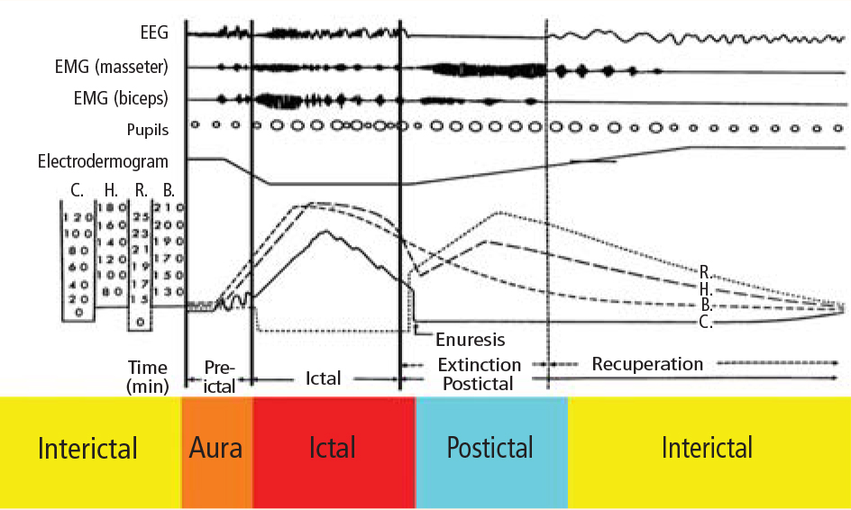
Experimental evidence. Heart rate changes, including bradycardia, tachycardia, and even asystole, have been repeatedly provoked by electrical brain stimulation of the limbic system and insular cortex.19 Some studies have suggested a lateralized influence of the insulae on cardiovascular autonomic control. In one study, intraoperative stimulation of the left posterior insula elicited a cardioinhibitory response and hypotension, whereas stimulation of the right anterior insula elicited tachycardia and hypertension.20 Such results have not always been reproducible.21–23 Other studies have suggested a localization-related influence of the limbic system on cardiovascular responses. Stimulation of the amygdala has not led to the ictal tachycardia that is commonly seen in epileptic seizures, suggesting that cortical involvement is needed for the development of tachycardia.24
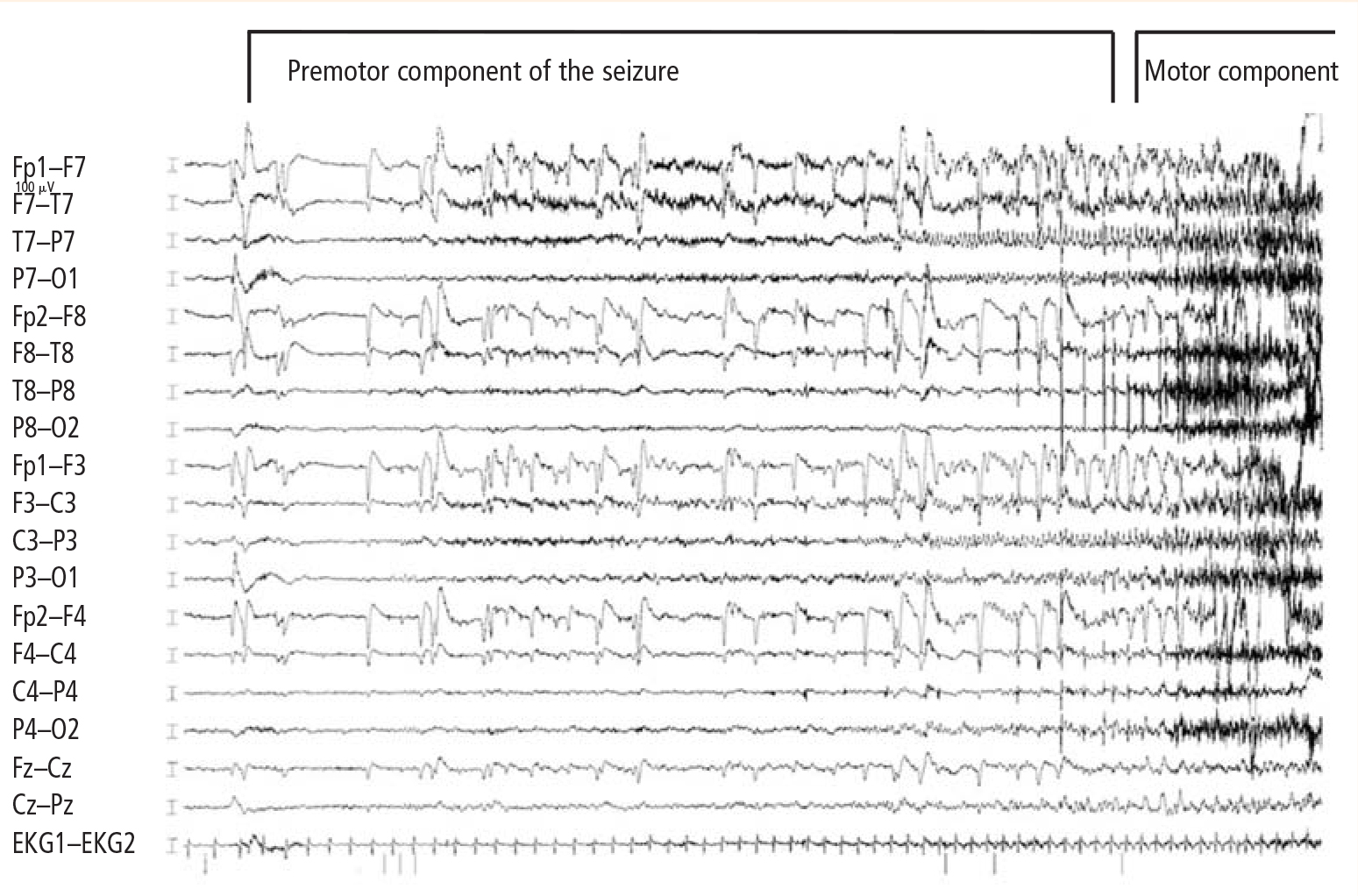
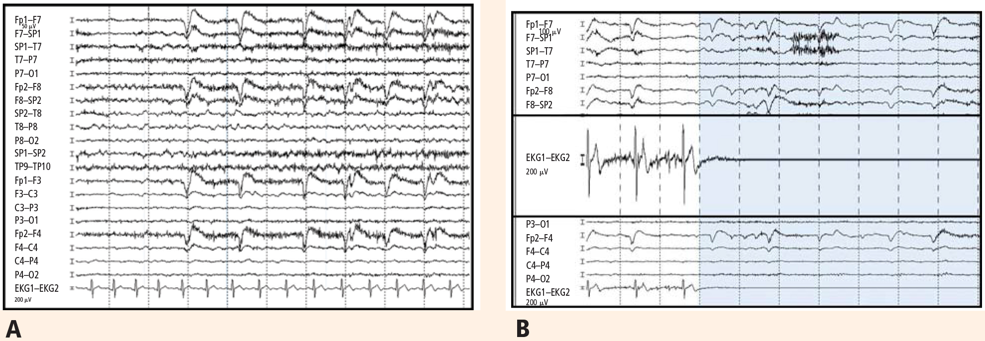
SUDEP PREVENTION
Epilepsy control is first line of defense
A careful consideration of the incidence of SUDEP in various patient populations suggests that controlling patients’ epilepsy might just be the best method of preventing SUDEP. While estimated SUDEP incidence ranges from 0.7 to 1.3 cases per 1,000 patient-years in population-based studies of patients with epilepsy,1,2 this rate escalates by nearly tenfold (3.5 to 9.3 cases per 1,000 patient-years) in cohorts with severe epilepsy, such as those derived from anticonvulsant drug registries, medical device registries, and referral centers.3–5 Therefore, medical control of seizures might reduce the incidence of SUDEP.
Epilepsy surgery cuts SUDEP risk for many patients
Studies involving epilepsy surgery programs also suggest that successful epilepsy surgery reduces the impending risks of SUDEP. In cohorts in which the estimated risk of SUDEP is almost 1% per year without surgery, SUDEP incidence was significantly lower following epilepsy surgery. In a study of 305 patients who underwent temporal lobe epilepsy surgery in the United Kingdom, the incidence of SUDEP following surgery was 2.2 cases per 1,000 person-years, and only one-third of SUDEP cases were among seizure-free patients.31 A similar incidence of 2.4 cases per 1,000 person-years was seen following epilepsy surgery in 596 Swedish patients; none of the 6 SUDEP patients in that study was seizure free.32 In a US study, no SUDEP cases occurred among 256 seizure-free patients with a follow-up of about 5 years after epilepsy surgery.33
In our own experience at the Cleveland Clinic, we have reported on outcomes among 70 patients who underwent frontal lobectomy34 and among 371 patients who underwent temporal lobectomy.35 In the frontal lobectomy study,34 2 of the 39 patients who had persistent seizures following surgery died of SUDEP during follow-up, whereas none of the 31 patients who remained seizure free were dead up to 10 years after surgery. In the temporal lobectomy report,35 2 of the 141 patients with ongoing postoperative seizures died of SUDEP, as compared with none of the 230 patients who were seizure free after a mean follow-up of 5.5 years.
Additional means of prophylaxis needed
Unfortunately, as many as 30% to 40% of patients with epilepsy continue to suffer intractable epilepsy despite all the available treatment modalities, including epilepsy surgery. For these patients, controlling seizures to reduce the risk of SUDEP is neither a possible nor a realistic means of avoiding this devastating condition, and alternative methods of prophylaxis must be sought.
CONCLUSIONS AND FUTURE RESEARCH
Patients with refractory epilepsy currently face a lifelong risk of sudden death as high as 1% per year.3 Elucidating the mechanisms of SUDEP might lead to preventive measures, which could have significant implications in reducing mortality in this patient population. Abundant evidence exists that autonomic dysfunction and cardiac arrhythmias are associated with seizures. The missing links in establishing a cardiac mechanism for SUDEP now include the following: (1) evidence of cardiac arrhythmias generally observed in seizures as a risk factor for SUDEP, (2) determination of clear electrophysiologic characteristics—from EEG and ECG standpoints—of patients at risk for SUDEP, and (3) clarification of the role of cardiac mechanisms in SUDEP and the role that cerebral influences on autonomic function might play. Early identification of patients at risk of SUDEP would offer a unique opportunity for early intervention to prevent this devastating condition.
- Nilsson L, Farahmand BY, Persson PG, Thiblin I, Tomson T. Risk factors for sudden unexpected death in epilepsy: a case-control study. Lancet 1999; 353:888–893.
- Tennis P, Cole TB, Annegers JF, Leestma JE, McNutt M, Rajput A. Cohort study of incidence of sudden unexplained death in persons with seizure disorder treated with antiepileptic drugs in Saskatchewan, Canada. Epilepsia 1995; 36:29–36.
- Dasheiff RM. Sudden unexpected death in epilepsy: a series from an epilepsy surgery program and speculation on the relationship to sudden cardiac death. J Clin Neurophysiol 1991; 8:216–222.
- Leestma JE, Annegers JF, Brodie MJ, et al. Sudden unexplained death in epilepsy: observations from a large clinical development program. Epilepsia 1997; 38:47–55.
- Sperling MR, Feldman H, Kinman J, Liporace JD, O’Connor MJ. Seizure control and mortality in epilepsy. Ann Neurol 1999; 46:45–50.
- Ficker DM. Sudden unexplained death and injury in epilepsy. Epilepsia 2000; 41(Suppl 2):S7–S12.
- Pedley TA, Hauser WA. Sudden death in epilepsy: a wake-up call for management. Lancet 2002; 359:1790–1791.
- Ficker DM, So EL, Shen WK, et al. Population-based study of the incidence of sudden unexplained death in epilepsy. Neurology 1998; 51:1270–1274.
- Monté CP, Arends JB, Tan IY, Aldenkamp AP, Limburg M, de Krom MC. Sudden unexpected death in epilepsy patients: risk factors. A systematic review. Seizure 2007; 16:1–7.
- Nashef L, Walker F, Allen P, Sander JW, Shorvon SD, Fish DR. Apnoea and bradycardia during epileptic seizures: relation to sudden death in epilepsy. J Neurol Neurosurg Psychiatry 1996; 60:297–300.
- So EL, Sam MC, Lagerlund TL. Postictal central apnea as a cause of SUDEP: evidence from near-SUDEP incident. Epilepsia 2000; 41:1494–1497.
- Terrence CF, Rao GR, Perper JA. Neurogenic pulmonary edema in unexpected, unexplained death of epileptic patients. Ann Neurol 1981; 9:458–464.
- Johnston SC, Horn JK, Valente J, Simon RP. The role of hypoventilation in a sheep model of epileptic sudden death. Ann Neurol 1995; 37:531–537.
- Blumhardt LD, Smith PE, Owen L. Electrocardiographic accompaniments of temporal lobe epileptic seizures. Lancet 1986; 1:1051–1056.
- Nei M, Ho RT, Abou-Khalil BW, et al. EEG and ECG in sudden unexplained death in epilepsy. Epilepsia 2004; 45:338–345.
- Nei M, Ho RT, Sperling MR. EKG abnormalities during partial seizures in refractory epilepsy. Epilepsia 2000; 41:542–548.
- Opherk C, Coromilas J, Hirsch LJ. Heart rate and EKG changes in 102 seizures: analysis of influencing factors. Epilepsy Res 2002; 52:117–127.
- Tigaran S, Mølgaard H, McClelland R, Dam M, Jaffe AS. Evidence of cardiac ischemia during seizures in drug refractory epilepsy patients. Neurology 2003; 60:492–495.
- Leung H, Kwan P, Elger CE. Finding the missing link between ictal bradyarrhythmia, ictal asystole, and sudden unexpected death in epilepsy. Epilepsy Behav 2006; 9:19–30.
- Yasui Y, Breder CD, Saper CB, Cechetto DF. Autonomic responses and efferent pathways from the insular cortex in the rat. J Comp Neurol 1991; 303:355–374.
- Jokeit H, Noerpel I, Herbord E, Ebner A. Heart rate does not decrease after right hemispheric amobarbital injection. Neurology 2000; 54:2347–2348.
- Zamrini EY, Meador KJ, Loring DW, et al. Unilateral cerebral inactivation produces differential left/right heart rate responses. Neurology 1990; 40:1408–1411.
- Yoon BW, Morillo CA, Cechetto DF, Hachinski V. Cerebral hemispheric lateralization in cardiac autonomic control. Arch Neurol 1997; 54:741–744.
- Keilson MJ, Hauser WA, Magrill JP. Electrocardiographic changes during electrographic seizures. Arch Neurol 1989; 46:1169–1170.
- Keilson MJ, Hauser WA, Magrill JP, Goldman M. ECG abnormalities in patients with epilepsy. Neurology 1987; 37:1624–1626.
- Galimberti CA, Marchioni E, Barzizza F, Manni R, Sartori I, Tartara A. Partial epileptic seizures of different origin variably affect cardiac rhythm. Epilepsia 1996; 37:742–747.
- Liedholm LJ, Gudjonsson O. Cardiac arrest due to partial epileptic seizures. Neurology 1992; 42:824–829.
- Leung H, Schindler K, Kwan P, Elger C. Asystole induced by electrical stimulation of the left cingulate gyrus. Epileptic Disord 2007; 9:77–81.
- Blum AS, Ives JR, Goldberger AL, et al. Oxygen desaturations triggered by partial seizures: implications for cardiopulmonary instability in epilepsy. Epilepsia 2000; 41:536–541.
- Langan Y, Nashef L, Sander JW. Case-control study of SUDEP. Neurology 2005; 64:1131–1133.
- Hennessy MJ, Langan Y, Elwes RD, Binnie CD, Polkey CE, Nashef L. A study of mortality after temporal lobe epilepsy surgery. Neurology 1999; 53:1276–1283.
- Nilsson L, Ahlbom A, Farahmand BY, Tomson T. Mortality in a population-based cohort of epilepsy surgery patients. Epilepsia 2003; 44:575–581.
- Sperling MR, Harris A, Nei M, Liporace JD, O’Connor MJ. Mortality after epilepsy surgery. Epilepsia 2005; 46(Suppl 11):49–53.
- Jeha LE, Najm I, Bingaman W, Dinner D, Widdess-Walsh P, Lüders H. Surgical outcome and prognostic factors of frontal lobe epilepsy surgery. Brain 2007; 130:574–584.
- Jeha LE, Najm IM, Bingaman WE, et al. Predictors of outcome after temporal lobectomy for the treatment of intractable epilepsy. Neurology 2006; 66:1938–1940.
The intimate interplay between heart and brain is well illustrated in epilepsy and may underlie the mechanism of one of its most devastating consequences: sudden unexpected death in epilepsy (SUDEP). This article will briefly describe the potential mechanisms of SUDEP, elaborate on the evidence for a likely cardiac pathophysiology, and review considerations in SUDEP prevention. We begin with a couple of brief case presentations and an epidemiologic overview to illustrate the concept and significance of SUDEP.
CASE PRESENTATIONS
A patient with near-SUDEP
The following is an actual message received by one of the authors:
Dr. Najm: A quick note regarding a 27-year-old male patient of yours with cerebral palsy and seizure disorder. Yesterday, while being transported from floor to floor, he had a cardiac arrest and was successfully resuscitated. Immediately after the code he developed seizures, which were treated with phenytoin and lorazepam. He is now in the neurointensive care unit. Thank you.
This case represents a scenario of near-SUDEP in which death was prevented by the fortuitous presence of immediate medical assistance at the time of cardiac arrest. Had this patient been home at the time of this incident, he almost certainly would have simply been found dead in his bed, like many SUDEP victims.
A typical case with multiple risk factors
A 32-year-old man underwent left temporal lobectomy at the Cleveland Clinic for treatment of medically refractory focal epilepsy. His seizure frequency improved after surgery, but he continued to have rare convulsions. Nevertheless, he discontinued all his anticonvulsant medications on his own. One year later, he was found dead on his bathroom floor. No obvious cause of death was identified.
This case illustrates several characteristics of the patient typically at risk for SUDEP: young, male, with intractable poorly controlled epilepsy, and not taking antiepileptic medications.
EPIDEMIOLOGY AND RISK FACTORS
Epilepsy affects 1% of the US population. Among those affected by epilepsy, SUDEP is a common cause of mortality. Estimates of SUDEP incidence range from 0.7 to 1.3 cases per 1,000 patient-years in large cohorts of patients with epilepsy1,2 and from 3.5 to 9.3 cases per 1,000 patient-years in anticonvulsant drug registries, medical device registries, and epilepsy surgery programs.3–5 SUDEP accounts for up to 17% of all deaths in patients with epilepsy6,7 and exceeds the expected rate of sudden death in the general population by nearly 24 times.6,8
Several potential risk factors for SUDEP have been investigated, but results from different studies are conflicting. Consistently identified risk factors include young age, early onset of seizures, refractoriness of epilepsy, the presence of generalized tonic-clonic seizures, male sex, and being in bed at the time of death. Weaker risk factors include being in the prone position at the time of death, having one or more subtherapeutic blood levels of anticonvulsant medication, having a structural brain lesion, and being asleep.9 The current consensus is that SUDEP is primarily a “seizure-related” occurrence, but the exact mechanisms underlying SUDEP are unknown.
PROPOSED MECHANISMS
Pulmonary pathophysiology
Central apnea and acute neurogenic pulmonary edema are the two major proposed pathways linking seizures to SUDEP. Evidence exists for each pathway.
Central apnea. In a prospective study of patients in an epilepsy monitoring unit, central apnea lasting at least 10 seconds was observed postictally in 40% of the recorded seizures.10 Otherwise healthy young epilepsy patients have been reported to develop central apnea immediately following complex partial seizures.11 Neurotransmitters mediating the brain’s own seizure-terminating mechanism could also be inhibiting the brainstem and causing postictal apnea.
Acute neurogenic pulmonary edema has been well described in relation to severe head injury and subarachnoid hemorrhage. Pulmonary edema is frequently found in SUDEP patients at autopsy.12 Intense generalized vasoconstriction induced by massive seizure-related sympathetic outburst can lead to increased pulmonary vascular resistance, and thereby may mediate acute pulmonary edema.
These two mechanisms—central apnea and acute neurogenic pulmonary edema—are not mutually exclusive. In the only animal model of SUDEP, one third of animals died from hypoventilation and had associated pulmonary edema at autopsy.13 Limited opportunities for realistic and practical interventions to reverse SUDEP risks related to pulmonary causes have hindered further development of these concepts.
Cardiac pathophysiology
The most significant and widely discussed cardiac mechanism of SUDEP is cardiac arrhythmia precipitated by seizure discharges acting via the autonomic nervous system.14–19
Experimental evidence. Heart rate changes, including bradycardia, tachycardia, and even asystole, have been repeatedly provoked by electrical brain stimulation of the limbic system and insular cortex.19 Some studies have suggested a lateralized influence of the insulae on cardiovascular autonomic control. In one study, intraoperative stimulation of the left posterior insula elicited a cardioinhibitory response and hypotension, whereas stimulation of the right anterior insula elicited tachycardia and hypertension.20 Such results have not always been reproducible.21–23 Other studies have suggested a localization-related influence of the limbic system on cardiovascular responses. Stimulation of the amygdala has not led to the ictal tachycardia that is commonly seen in epileptic seizures, suggesting that cortical involvement is needed for the development of tachycardia.24
SUDEP PREVENTION
Epilepsy control is first line of defense
A careful consideration of the incidence of SUDEP in various patient populations suggests that controlling patients’ epilepsy might just be the best method of preventing SUDEP. While estimated SUDEP incidence ranges from 0.7 to 1.3 cases per 1,000 patient-years in population-based studies of patients with epilepsy,1,2 this rate escalates by nearly tenfold (3.5 to 9.3 cases per 1,000 patient-years) in cohorts with severe epilepsy, such as those derived from anticonvulsant drug registries, medical device registries, and referral centers.3–5 Therefore, medical control of seizures might reduce the incidence of SUDEP.
Epilepsy surgery cuts SUDEP risk for many patients
Studies involving epilepsy surgery programs also suggest that successful epilepsy surgery reduces the impending risks of SUDEP. In cohorts in which the estimated risk of SUDEP is almost 1% per year without surgery, SUDEP incidence was significantly lower following epilepsy surgery. In a study of 305 patients who underwent temporal lobe epilepsy surgery in the United Kingdom, the incidence of SUDEP following surgery was 2.2 cases per 1,000 person-years, and only one-third of SUDEP cases were among seizure-free patients.31 A similar incidence of 2.4 cases per 1,000 person-years was seen following epilepsy surgery in 596 Swedish patients; none of the 6 SUDEP patients in that study was seizure free.32 In a US study, no SUDEP cases occurred among 256 seizure-free patients with a follow-up of about 5 years after epilepsy surgery.33
In our own experience at the Cleveland Clinic, we have reported on outcomes among 70 patients who underwent frontal lobectomy34 and among 371 patients who underwent temporal lobectomy.35 In the frontal lobectomy study,34 2 of the 39 patients who had persistent seizures following surgery died of SUDEP during follow-up, whereas none of the 31 patients who remained seizure free were dead up to 10 years after surgery. In the temporal lobectomy report,35 2 of the 141 patients with ongoing postoperative seizures died of SUDEP, as compared with none of the 230 patients who were seizure free after a mean follow-up of 5.5 years.
Additional means of prophylaxis needed
Unfortunately, as many as 30% to 40% of patients with epilepsy continue to suffer intractable epilepsy despite all the available treatment modalities, including epilepsy surgery. For these patients, controlling seizures to reduce the risk of SUDEP is neither a possible nor a realistic means of avoiding this devastating condition, and alternative methods of prophylaxis must be sought.
CONCLUSIONS AND FUTURE RESEARCH
Patients with refractory epilepsy currently face a lifelong risk of sudden death as high as 1% per year.3 Elucidating the mechanisms of SUDEP might lead to preventive measures, which could have significant implications in reducing mortality in this patient population. Abundant evidence exists that autonomic dysfunction and cardiac arrhythmias are associated with seizures. The missing links in establishing a cardiac mechanism for SUDEP now include the following: (1) evidence of cardiac arrhythmias generally observed in seizures as a risk factor for SUDEP, (2) determination of clear electrophysiologic characteristics—from EEG and ECG standpoints—of patients at risk for SUDEP, and (3) clarification of the role of cardiac mechanisms in SUDEP and the role that cerebral influences on autonomic function might play. Early identification of patients at risk of SUDEP would offer a unique opportunity for early intervention to prevent this devastating condition.
- Nilsson L, Farahmand BY, Persson PG, Thiblin I, Tomson T. Risk factors for sudden unexpected death in epilepsy: a case-control study. Lancet 1999; 353:888–893.
- Tennis P, Cole TB, Annegers JF, Leestma JE, McNutt M, Rajput A. Cohort study of incidence of sudden unexplained death in persons with seizure disorder treated with antiepileptic drugs in Saskatchewan, Canada. Epilepsia 1995; 36:29–36.
- Dasheiff RM. Sudden unexpected death in epilepsy: a series from an epilepsy surgery program and speculation on the relationship to sudden cardiac death. J Clin Neurophysiol 1991; 8:216–222.
- Leestma JE, Annegers JF, Brodie MJ, et al. Sudden unexplained death in epilepsy: observations from a large clinical development program. Epilepsia 1997; 38:47–55.
- Sperling MR, Feldman H, Kinman J, Liporace JD, O’Connor MJ. Seizure control and mortality in epilepsy. Ann Neurol 1999; 46:45–50.
- Ficker DM. Sudden unexplained death and injury in epilepsy. Epilepsia 2000; 41(Suppl 2):S7–S12.
- Pedley TA, Hauser WA. Sudden death in epilepsy: a wake-up call for management. Lancet 2002; 359:1790–1791.
- Ficker DM, So EL, Shen WK, et al. Population-based study of the incidence of sudden unexplained death in epilepsy. Neurology 1998; 51:1270–1274.
- Monté CP, Arends JB, Tan IY, Aldenkamp AP, Limburg M, de Krom MC. Sudden unexpected death in epilepsy patients: risk factors. A systematic review. Seizure 2007; 16:1–7.
- Nashef L, Walker F, Allen P, Sander JW, Shorvon SD, Fish DR. Apnoea and bradycardia during epileptic seizures: relation to sudden death in epilepsy. J Neurol Neurosurg Psychiatry 1996; 60:297–300.
- So EL, Sam MC, Lagerlund TL. Postictal central apnea as a cause of SUDEP: evidence from near-SUDEP incident. Epilepsia 2000; 41:1494–1497.
- Terrence CF, Rao GR, Perper JA. Neurogenic pulmonary edema in unexpected, unexplained death of epileptic patients. Ann Neurol 1981; 9:458–464.
- Johnston SC, Horn JK, Valente J, Simon RP. The role of hypoventilation in a sheep model of epileptic sudden death. Ann Neurol 1995; 37:531–537.
- Blumhardt LD, Smith PE, Owen L. Electrocardiographic accompaniments of temporal lobe epileptic seizures. Lancet 1986; 1:1051–1056.
- Nei M, Ho RT, Abou-Khalil BW, et al. EEG and ECG in sudden unexplained death in epilepsy. Epilepsia 2004; 45:338–345.
- Nei M, Ho RT, Sperling MR. EKG abnormalities during partial seizures in refractory epilepsy. Epilepsia 2000; 41:542–548.
- Opherk C, Coromilas J, Hirsch LJ. Heart rate and EKG changes in 102 seizures: analysis of influencing factors. Epilepsy Res 2002; 52:117–127.
- Tigaran S, Mølgaard H, McClelland R, Dam M, Jaffe AS. Evidence of cardiac ischemia during seizures in drug refractory epilepsy patients. Neurology 2003; 60:492–495.
- Leung H, Kwan P, Elger CE. Finding the missing link between ictal bradyarrhythmia, ictal asystole, and sudden unexpected death in epilepsy. Epilepsy Behav 2006; 9:19–30.
- Yasui Y, Breder CD, Saper CB, Cechetto DF. Autonomic responses and efferent pathways from the insular cortex in the rat. J Comp Neurol 1991; 303:355–374.
- Jokeit H, Noerpel I, Herbord E, Ebner A. Heart rate does not decrease after right hemispheric amobarbital injection. Neurology 2000; 54:2347–2348.
- Zamrini EY, Meador KJ, Loring DW, et al. Unilateral cerebral inactivation produces differential left/right heart rate responses. Neurology 1990; 40:1408–1411.
- Yoon BW, Morillo CA, Cechetto DF, Hachinski V. Cerebral hemispheric lateralization in cardiac autonomic control. Arch Neurol 1997; 54:741–744.
- Keilson MJ, Hauser WA, Magrill JP. Electrocardiographic changes during electrographic seizures. Arch Neurol 1989; 46:1169–1170.
- Keilson MJ, Hauser WA, Magrill JP, Goldman M. ECG abnormalities in patients with epilepsy. Neurology 1987; 37:1624–1626.
- Galimberti CA, Marchioni E, Barzizza F, Manni R, Sartori I, Tartara A. Partial epileptic seizures of different origin variably affect cardiac rhythm. Epilepsia 1996; 37:742–747.
- Liedholm LJ, Gudjonsson O. Cardiac arrest due to partial epileptic seizures. Neurology 1992; 42:824–829.
- Leung H, Schindler K, Kwan P, Elger C. Asystole induced by electrical stimulation of the left cingulate gyrus. Epileptic Disord 2007; 9:77–81.
- Blum AS, Ives JR, Goldberger AL, et al. Oxygen desaturations triggered by partial seizures: implications for cardiopulmonary instability in epilepsy. Epilepsia 2000; 41:536–541.
- Langan Y, Nashef L, Sander JW. Case-control study of SUDEP. Neurology 2005; 64:1131–1133.
- Hennessy MJ, Langan Y, Elwes RD, Binnie CD, Polkey CE, Nashef L. A study of mortality after temporal lobe epilepsy surgery. Neurology 1999; 53:1276–1283.
- Nilsson L, Ahlbom A, Farahmand BY, Tomson T. Mortality in a population-based cohort of epilepsy surgery patients. Epilepsia 2003; 44:575–581.
- Sperling MR, Harris A, Nei M, Liporace JD, O’Connor MJ. Mortality after epilepsy surgery. Epilepsia 2005; 46(Suppl 11):49–53.
- Jeha LE, Najm I, Bingaman W, Dinner D, Widdess-Walsh P, Lüders H. Surgical outcome and prognostic factors of frontal lobe epilepsy surgery. Brain 2007; 130:574–584.
- Jeha LE, Najm IM, Bingaman WE, et al. Predictors of outcome after temporal lobectomy for the treatment of intractable epilepsy. Neurology 2006; 66:1938–1940.
Evaluating brain function in patients with disorders of consciousness
Consciousness has long been a fascinating subject to both philosophers and scientists, yet consciousness has only recently been taken into account by neuroscientists as a topic for research. This article discusses research done over the past 10 years evaluating brain function in patients with disorders of consciousness—specifically those in a vegetative or minimally conscious state. We highlight physiologic, sensory, perceptual, cognitive, and behavioral commonalities and disparities between patients with anoxic and traumatic brain injuries, with the aim of characterizing the neurophysiologic and neuroanatomic differences between these two main causes of disorders of consciousness.
WHAT IS CONSCIOUSNESS?
Although consciousness is difficult to describe, it can be defined as a combination of wakefulness and awareness.1 As for the brain systems supporting these two aspects of consciousness, it has been suggested that the brainstem ascending reticular formation system and its thalamic projections support alertness and the sleep-wake cycle, and that conscious awareness relies on a functional thalamocortical and corticocortical system.
DISORDERS OF CONSCIOUSNESS: A VARIETY OF STATES
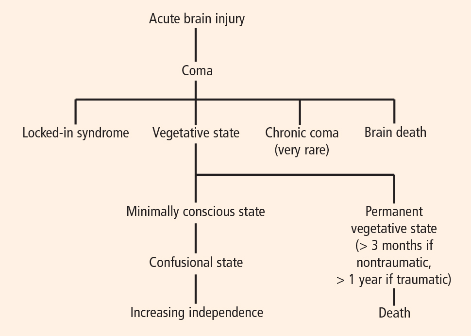
Coma: Near-complete unresponsiveness
Coma is a condition of almost complete unresponsiveness in which the patient lies with eyes closed, very limited reflexes, no cyclical wakefulness, and, above all, no signs of awareness. Coma is normally attained after an acute brain insult and may last about 2 weeks, although chronic coma cases have been described, and is usually caused by either temporary or permanent damage to the reticular system.
Vegetative state: Wakefulness without awareness
Following a coma, some patients may enter a vegetative state, which involves a complete absence of consciousness of one’s environment but with preserved sleep-wake cycles and autonomic functions. The vegetative state is easily differentiated from brain death, in which the electroencephalogram shows no brain wave or activity.4 Brain death is the irreversible end of all brain activity and should not be confused with a persistent vegetative state.
The vegetative state is a condition of wakefulness without awareness in which the patient exhibits a partially preserved sleep-wake cycle and a variable portfolio of reflexes and spontaneous nonvolitional behaviors. A patient who has been in a vegetative state for more than 1 month with no improvement is often said to be in a persistent vegetative state. The term permanent vegetative state, implying no chance of recovery, is sometimes used when the vegetative state persists for 3 months after a nontraumatic insult, such as cardiac arrest, or for 1 year after a traumatic brain injury.
Minimally conscious state: Conscious awareness is evident despite impairment
Some patients in a vegetative state may start to recover by entering a minimally conscious state, in which conscious awareness is evident despite profound physical and cognitive impairment. Although communication capabilities are absent, cognitively mediated (or voluntary) behavior occurs in the minimally conscious state, which may be inconsistent but is reproducible enough to be differentiated from reflexive behavior. For example, patients may occasionally be able to smile when asked to do so or follow an object with their eyes. In the minimally conscious state, patients show those basic behaviors seen in the vegetative state along with islands of presumably conscious processing such as inconsistent responses to simple commands and sustained visual pursuit.5 Patients in a minimally conscious state have a better prognosis than those in a persistent or permanent vegetative state.3
Locked-in syndrome: Not a true disorder of consciousness
Another pathology that is often confounded with vegetative or minimally conscious states is the locked-in syndrome, which is characterized by complete paralysis of voluntary muscles in all parts of the body except those controlling eye movements. Individuals with locked-in syndrome are conscious and can think and reason, but they are unable to speak or move. The disorder confines the patient to paralysis and a mute state. Communication may be possible with blinking eye movements.
WHAT CAUSES DISORDERS OF CONSCIOUSNESS?
Disorders of consciousness mostly stem from acute brain insults, which may be caused by hypoxicischemic neural injury or traumatic brain injury. Although traumatic brain injury is currently the most common cause of vegetative and minimally conscious states, nontraumatic causes are becoming more frequent as a result of scientific and technological developments in resuscitation. Nontraumatic causes of disorders of consciousness include stroke, cardiopulmonary arrest, and meningoencephalitis; additionally, patients in the final stage of certain neurodegenerative diseases, including Parkinson, Alzheimer, and Huntington diseases, may lapse into a minimally conscious or vegetative state.6
NEUROLOGIC FINDINGS IN COMATOSE SURVIVORS OF CARDIAC ARREST
Structural magnetic resonance imaging (MRI) of patients in a vegetative state following cardiac arrest often reveals abnormalities. Most frequently there is a white matter signal in the cerebellum, the thalamus, the frontal and parietal cortices, and the hippocampus. Widespread abnormalities may indicate little to no prospect for recovery. Pupillary light response, corneal reflexes, motor responses to pain, myoclonus status epilepticus, serum neuron-specific enolase, and somatosensory evoked potential studies can assist in predicting efficiently and accurately a poor outcome in comatose patients after cardiopulmonary resuscitation for cardiac arrest.7
DEFINITION PROBLEMS AND MISDIAGNOSIS
The diagnosis of vegetative state emerges from a negative finding—namely, the lack of behaviors that would signal conscious capabilities. Using the nonoccurrence of events as a criterion to establish a fact is inherently problematic, since the causes of a nonoccurrence are theoretically infinite. More specifically, the reasons behind the lack of evidence of voluntary movement in presumably unconscious patients can be classified in terms of malfunctioning of either sensoriperceptual, output/motor, or central processing.
Deficits in sensoriperceptual processing
A patient might have deafness that may lead to a deficit in speech comprehension, or perhaps the auditory pathway and first cortical pathways are spared but the patient is aphasic and cannot process additive events such as speech. In a cohort of 42 patients, we found 17 who lacked the fourth or fifth components of the brain auditory evoked potentials to clicks presented binaurally, signaling severe damage to the auditory pathway.8 It is useless to ask such patients to follow commands, since the sensory input is damaged and the movement (or lack of movement) has no validity for the diagnosis. A similar argument can apply for patients who may show some fixation but exhibit delayed or absent visual evoked potentials when presented with written commands.
Deficits in motor processing
The second type of lesions that may contribute to misdiagnosis in these patients are those found in the effector systems. If the motor voluntary pathways are damaged—either in the motor cortex or in the corticospinal or corticobulbar pathways—then movement might be impaired enough to prevent responses by the patient. Patients of this type are sometimes diagnosed as being in a vegetative state although they might actually have locked-in syndrome,9 with preserved cognition but an inability to initiate voluntary responses as a result of a lesion in the pontine peduncle.
Although the effector systems are difficult to test in unresponsive subjects, some strategies may be tried. Before testing for volition, it is necessary to assess all possible hand, leg, and face reflexes in order to map reflexive behavior. Commands should then specifically target those muscles that showed total or partial preservation of reflexes. To test the output pathways from the cortex to the medulla, a more specialized assessment is needed; the Impaired Consciousness Research Group at the University of Cambridge has developed a simple protocol to assess the ability of the motor cortex to elicit muscle twitches by measuring the motor evoked potentials to simple pulses of transcranial magnetic stimulation. The minimal pulse intensity is determined by electromyographic recordings when transcranial magnetic stimulation pulses are applied to the left or right motor cortices for the hands and feet. The results have shown 2 out of 34 patients to have no detectable motor evoked potentials and 5 patients to have severe delay at maximum pulse intensity [unpublished data]. These results confirm the need for a full neurologic and neurophysiologic assessment in subjects who are unresponsive or show low levels of response, both acutely and more chronically, to minimize the risk of misdiagnosis.
Deficits in central processing
The key element in the assessment of cognitive processing in patients in a vegetative or minimally conscious state is determining deficits in their capacity to process external stimuli in a conscious manner (central processing). This is by far the most difficult characteristic to be determined since the only accepted criteria for awareness are verbal report or voluntary movement, both of which are absent in the vegetative state and are inconsistent and difficult to determine behaviorally in the minimally conscious state.
CLUES TO BRAIN FUNCTION IN DISORDERS OF CONSCIOUSNESS
It is important to differentiate a patient in a persistent vegetative state from a patient in a minimally conscious state, as the latter patient has a much higher chance of a favorable outcome. Evaluation of cerebral metabolism and imaging studies can both provide clues to brain function.
Cerebral metabolism
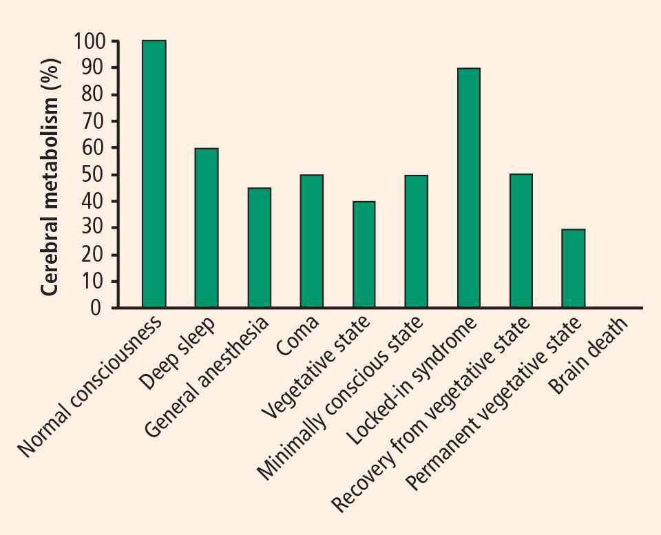
Neuroimaging studies
In the past few years, studies have found that some patients in a vegetative or minimally conscious state can activate cortical networks in response to auditory, visual, and tactile stimuli.10 A challenge in neuroscience is to devise a reliable, objective test to assess awareness without relying on explicit voluntary movements or verbal responses. Such a test would have important theoretical and practical implications. Recent evidence from functional neuroimaging and neurophysiology suggests that some patients with disorders of consciousness exhibit partially preserved conscious processing despite having no clinical or verbal output.11
During a positron emission tomography study, Menon et al showed photographs to a 26-year-old woman who was in a vegetative state 4 months after becoming comatose from an acute febrile illness.12 They found significant activation in the right fusiform gyrus and extrastriate visual association areas when the woman was shown photos of people familiar to her as compared with repixellated versions of the same photos with the faces made unrecognizable. The activation pattern she exhibited was similar to that of healthy volunteers. Interestingly, a few months after this study, the patient became increasingly responsive.
Our group conducted the first evaluation of emotion in the minimally conscious state using functional MRI (fMRI) in a 17-year-old male following a traumatic brain injury.13 The patient was able to localize noxious stimuli, exhibited spontaneous eye opening, and occasionally smiled appropriately and followed people with his eyes. Imaging was performed while he listened to two recordings—one of his mother reading a story about his life, and one of a matched control voice reading the same story. Digital subtraction imaging disclosed strong activation of two areas related to emotion, the amygdala and the bilateral insula, while the recording of the patient’s mother was played. Activation was also evident in the auditory cortex in the superior temporal lobe. The patient recovered 6 months following this study.
Classical conditioning
Classical conditioning represents an alternate approach to MRI for assessing brain function in patients with disordered consciousness.8 Trace conditioning of the eye-blink response is considered to be an objective test of awareness.14 This test involves highly specific learning, requiring an anticipatory electromyographic response to a paired stimulus (eg, a tone followed by an aversive stimulus such as an air puff to the eyes) but not to an unpaired stimulus (eg, a white noise that is not followed by an aversive stimulus). This effect increases in amplitude as the aversive stimulus approaches. Our laboratory is applying this method to study learning and memory in patients with disordered consciousness.
DETERMINING AWARENESS WITHOUT REPORT
The proposed neural correlates of consciousness do not usually take into account the levels of consciousness.15,16 In order to build the framework for a cognitive neuroscience of consciousness, we must consider the content of the consciousness experience in fully awake subjects and patients as well as the cognitive processes occurring in unconscious and conscious subjects.
Two main approaches can be used to assess conscious processing in unresponsive patients. The first is to look for neural correlates in direct intentional actions or imagined actions,11 and the second is to look for physiologic correlates of the cognitive processes required during the conscious processing of stimuli.17
Searching for neural correlates of intended actions
The first approach can have enormous impact in the diagnostic arena (as well as in the legal and ethical arenas), such as in the case reported by Owen et al in which a patient showed brain activity related to imagining actions as prompted by spoken instructions during fMRI evaluation.11 Unfortunately, cases such as these are scarce. Moreover, imagining of actions relies not only on a spared comprehension capacity and preserved memory but also on the subject’s willingness to perform the task. It would seem that only a minority of patients in a vegetative state seem to have the cognitive abilities preserved to accomplish these types of tasks.
Searching for physiologic correlates of cognitive processes
The second approach would tend to work with memory and switching attention capabilities in unresponsive patients, assuming that conscious processing does not exist without these cognitive processes. The evidence for this approach comes from electrophysiology. Cognitive evoked potentials are commonly applied to assess basic auditory or visual cortical processing, automatic attention, and focus attention.18 Both the mismatch negativity wave (a correlate of automatic attention) and the p300 (a correlate of focus attention) are sometimes present,19 specifically in patients in vegetative or minimally conscious states, and they are a good predictor of awakening in stroke, hemorrhage, and traumatic brain injury.20
In day-to-day practice in a neurology clinic or emergency room, it is more feasible to assess cognitive capabilities using event-related potentials than fMRI since they are more widely distributed, more easily validated, shorter, and statistically more powerful in single-subject analysis,21 and because they do not frequently rely on speech comprehension.
NEUROPATHOLOGY AND fMRI
The cause of the brain injury leading to a vegetative or minimally conscious state frequently determines the neuropathology.22 It has also been demonstrated that severely disabled patients (such as those emerging from a minimally conscious state) differ from vegetative state patients in terms of lesions and severity.23
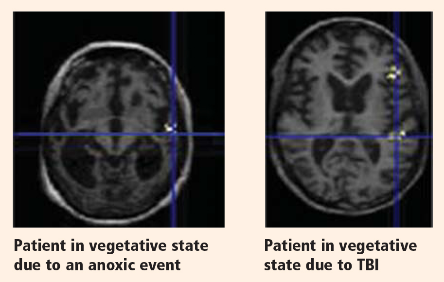
Although residual activity as seen on functional neuroimaging may be unequivocal in some cases, it may represent only fragmentary cognitive processing; it is important not to assume that normal awareness is present. Much still needs to be learned, but results from neuroimaging studies demonstrate that a small proportion of patients in a vegetative or minimally conscious state have some preserved cognitive processes. These findings have ethical and legal implications. For instance, careless bedside chatter among family members or medical personnel is inappropriate and should be avoided. Whether functional neuroimaging can effectively evaluate neuroprocessing in patients in whom cognitive output is difficult to assess remains to be determined. Such evaluation may one day help to predict prognosis. It may also someday help to facilitate communication with patients with locked-in syndrome, who are cognitively intact but are without verbal or motor output.
CONCLUSIONS
It is highly improbable to find patients with preserved cortical connectivity, since structural22 and functional19 studies have demonstrated only a small proportion of patients in a vegetative or minimally conscious state who have relatively preserved brains and cognitive processing. The more we study patients who are unresponsive or show low levels of response, the more complex cognitive processes we find in subpopulations of these patients. Language-related cortical activation is now the most common finding.13,19,24 More recently, a few researchers working with severely damaged patients have started to test paradigms with the aim of uncovering conscious processes that have no need of verbal or movement responses.
The time has come for clinicians in acute care centers to immediately follow their administration of coma scales in unresponsive patients with the use of more sophisticated methodology to assess not only reflexive and intentional behaviors but also these patients’ physiologic and cognitive characteristics. In the field of neurodegenerative disease, it took several years for clinicians to start using more sensitive cognitive tools than just the mini-mental state examination and computed tomography or three-dimensional T1-weighted structural MRI, but nowadays volumetric MRI and detailed cognitive assessments are widely used to diagnose and characterize patients with neurodegenerative disorders. The same path should be taken for patients with severe brain damage. The information yielded by such an approach may one day help to determine a diagnosis or prognosis, guide treatment, or facilitate communication in patients with pathologies of consciousness.
- Jennett B, Plum F. Persistent vegetative state after brain damage. A syndrome in search of a name. Lancet 1972; 1:734–737.
- The Multi-Society Task Force on PVS. Medical aspects of the persistent vegetative state.first of two parts. N Engl J Med 1994; 330:1499–1508.
- Giacino JT, Ashwal S, Childs N, et al. The minimally conscious state: definition and diagnostic criteria. Neurology 2002; 58:349–353.
- Wijdicks EFM. The diagnosis of brain death. N Engl J Med 2000; 344:1215–1221.
- Giacino JT, Trott CT. Rehabilitative management of patients with disorders of consciousness: grand rounds. J Head Trauma Rehabil 2004; 19:254–265.
- Bernat JL. Chronic disorders of consciouness. Lancet 2006; 367: 1181–1192.
- Wijdicks EFM, Hijdra A, Young GB, Bassetti CL, Wiebe S. Practice parameter: prediction of outcome in comatose survivors after cardiopulmonary resuscitation (an evidence-based review). Report of the Quality Standards Subcommittee of the American Academy of Neurology. Neurology 2006; 67:203–210.
- Bekinschtein TA. Cognitive Processes in the Vegetative and Minimally Conscious State [thesis]. Buenos Aires, Argentina: University of Buenos Aires; 2006.
- Onofrj M, Thomas A, Paci C, Scesi M, Tombari R. Event related potentials recorded in patients with locked-in syndrome. J Neurol Neurosurg Psychiatry 1997; 63:759–764.
- Schiff ND. Multimodal neuroimaging approaches to disorders of consciousness. J Head Trauma Rehabil 2006; 21:388–397.
- Owen AM, Coleman MR, Boly M, Davis MH, Laureys S, Pickard JD. Detecting awareness in the vegetative state. Science 2006; 313:1402.
- Menon DK, Owen AM, Williams EJ, et al. Cortical processing in persistent vegetative state. Lancet 1998; 352:200.
- Bekinschtein T, Niklison J, Sigman L, et al. Emotion processing in the minimally conscious state [letter]. J Neurol Neurosurg Psychiatry 2004; 75:788.
- Clark RE, Squire LR. Classical conditioning and brain systems: the role of awareness. Science 1998; 280:77–81.
- Koch C. The Quest for Consciousness: A Neurobiological Approach. Greenwood Village, CO: Roberts & Company Publishers; 2004.
- Dehaene S, Naccache L. Towards a cognitive neuroscience of consciousness: basic evidence and a workspace framework. Cognition 2001; 79:1–37.
- Naccache L. Is she conscious? Science 2006; 313:1395–1396.
- Picton TW, Bentin S, Berg P, et al. Guidelines for using human event-related potentials to study cognition: recording standards and publication criteria. Psychophysiology 2000; 37:127–152.
- Kotchoubey B, Lang S, Mezger G, et al. Information processing in severe disorders of consciousness: vegetative state and minimally conscious state. Clin Neurophysiol 2005; 116:2441–2453.
- Daltrozzo J, Wioland N, Mutschler V, Kotchoubey B. Predicting coma and other low responsive patients outcome using event-related brain potentials: a meta-analysis. Clin Neurophysiol 2007; 118:606–614.
- Quian Quiroga R, Garcia H. Single-trial event-related potentials with wavelet denoising. Clin Neurophysiol 2003; 114:376–390.
- Adams JH, Graham DI, Jennett B. The neuropathology of the vegetative state after an acute brain insult. Brain 2000; 123:1327–1338.
- Jennett B. Thirty years of the vegetative state: clinical, ethical and legal problems. Prog Brain Res 2005; 150:537–543.
- Schiff ND, Rodriguez-Moreno D, Kamal A, et al. fMRI reveals large-scale network activation in minimally conscious patients. Neurology 2005; 64:514–523.
Consciousness has long been a fascinating subject to both philosophers and scientists, yet consciousness has only recently been taken into account by neuroscientists as a topic for research. This article discusses research done over the past 10 years evaluating brain function in patients with disorders of consciousness—specifically those in a vegetative or minimally conscious state. We highlight physiologic, sensory, perceptual, cognitive, and behavioral commonalities and disparities between patients with anoxic and traumatic brain injuries, with the aim of characterizing the neurophysiologic and neuroanatomic differences between these two main causes of disorders of consciousness.
WHAT IS CONSCIOUSNESS?
Although consciousness is difficult to describe, it can be defined as a combination of wakefulness and awareness.1 As for the brain systems supporting these two aspects of consciousness, it has been suggested that the brainstem ascending reticular formation system and its thalamic projections support alertness and the sleep-wake cycle, and that conscious awareness relies on a functional thalamocortical and corticocortical system.
DISORDERS OF CONSCIOUSNESS: A VARIETY OF STATES
Coma: Near-complete unresponsiveness
Coma is a condition of almost complete unresponsiveness in which the patient lies with eyes closed, very limited reflexes, no cyclical wakefulness, and, above all, no signs of awareness. Coma is normally attained after an acute brain insult and may last about 2 weeks, although chronic coma cases have been described, and is usually caused by either temporary or permanent damage to the reticular system.
Vegetative state: Wakefulness without awareness
Following a coma, some patients may enter a vegetative state, which involves a complete absence of consciousness of one’s environment but with preserved sleep-wake cycles and autonomic functions. The vegetative state is easily differentiated from brain death, in which the electroencephalogram shows no brain wave or activity.4 Brain death is the irreversible end of all brain activity and should not be confused with a persistent vegetative state.
The vegetative state is a condition of wakefulness without awareness in which the patient exhibits a partially preserved sleep-wake cycle and a variable portfolio of reflexes and spontaneous nonvolitional behaviors. A patient who has been in a vegetative state for more than 1 month with no improvement is often said to be in a persistent vegetative state. The term permanent vegetative state, implying no chance of recovery, is sometimes used when the vegetative state persists for 3 months after a nontraumatic insult, such as cardiac arrest, or for 1 year after a traumatic brain injury.
Minimally conscious state: Conscious awareness is evident despite impairment
Some patients in a vegetative state may start to recover by entering a minimally conscious state, in which conscious awareness is evident despite profound physical and cognitive impairment. Although communication capabilities are absent, cognitively mediated (or voluntary) behavior occurs in the minimally conscious state, which may be inconsistent but is reproducible enough to be differentiated from reflexive behavior. For example, patients may occasionally be able to smile when asked to do so or follow an object with their eyes. In the minimally conscious state, patients show those basic behaviors seen in the vegetative state along with islands of presumably conscious processing such as inconsistent responses to simple commands and sustained visual pursuit.5 Patients in a minimally conscious state have a better prognosis than those in a persistent or permanent vegetative state.3
Locked-in syndrome: Not a true disorder of consciousness
Another pathology that is often confounded with vegetative or minimally conscious states is the locked-in syndrome, which is characterized by complete paralysis of voluntary muscles in all parts of the body except those controlling eye movements. Individuals with locked-in syndrome are conscious and can think and reason, but they are unable to speak or move. The disorder confines the patient to paralysis and a mute state. Communication may be possible with blinking eye movements.
WHAT CAUSES DISORDERS OF CONSCIOUSNESS?
Disorders of consciousness mostly stem from acute brain insults, which may be caused by hypoxicischemic neural injury or traumatic brain injury. Although traumatic brain injury is currently the most common cause of vegetative and minimally conscious states, nontraumatic causes are becoming more frequent as a result of scientific and technological developments in resuscitation. Nontraumatic causes of disorders of consciousness include stroke, cardiopulmonary arrest, and meningoencephalitis; additionally, patients in the final stage of certain neurodegenerative diseases, including Parkinson, Alzheimer, and Huntington diseases, may lapse into a minimally conscious or vegetative state.6
NEUROLOGIC FINDINGS IN COMATOSE SURVIVORS OF CARDIAC ARREST
Structural magnetic resonance imaging (MRI) of patients in a vegetative state following cardiac arrest often reveals abnormalities. Most frequently there is a white matter signal in the cerebellum, the thalamus, the frontal and parietal cortices, and the hippocampus. Widespread abnormalities may indicate little to no prospect for recovery. Pupillary light response, corneal reflexes, motor responses to pain, myoclonus status epilepticus, serum neuron-specific enolase, and somatosensory evoked potential studies can assist in predicting efficiently and accurately a poor outcome in comatose patients after cardiopulmonary resuscitation for cardiac arrest.7
DEFINITION PROBLEMS AND MISDIAGNOSIS
The diagnosis of vegetative state emerges from a negative finding—namely, the lack of behaviors that would signal conscious capabilities. Using the nonoccurrence of events as a criterion to establish a fact is inherently problematic, since the causes of a nonoccurrence are theoretically infinite. More specifically, the reasons behind the lack of evidence of voluntary movement in presumably unconscious patients can be classified in terms of malfunctioning of either sensoriperceptual, output/motor, or central processing.
Deficits in sensoriperceptual processing
A patient might have deafness that may lead to a deficit in speech comprehension, or perhaps the auditory pathway and first cortical pathways are spared but the patient is aphasic and cannot process additive events such as speech. In a cohort of 42 patients, we found 17 who lacked the fourth or fifth components of the brain auditory evoked potentials to clicks presented binaurally, signaling severe damage to the auditory pathway.8 It is useless to ask such patients to follow commands, since the sensory input is damaged and the movement (or lack of movement) has no validity for the diagnosis. A similar argument can apply for patients who may show some fixation but exhibit delayed or absent visual evoked potentials when presented with written commands.
Deficits in motor processing
The second type of lesions that may contribute to misdiagnosis in these patients are those found in the effector systems. If the motor voluntary pathways are damaged—either in the motor cortex or in the corticospinal or corticobulbar pathways—then movement might be impaired enough to prevent responses by the patient. Patients of this type are sometimes diagnosed as being in a vegetative state although they might actually have locked-in syndrome,9 with preserved cognition but an inability to initiate voluntary responses as a result of a lesion in the pontine peduncle.
Although the effector systems are difficult to test in unresponsive subjects, some strategies may be tried. Before testing for volition, it is necessary to assess all possible hand, leg, and face reflexes in order to map reflexive behavior. Commands should then specifically target those muscles that showed total or partial preservation of reflexes. To test the output pathways from the cortex to the medulla, a more specialized assessment is needed; the Impaired Consciousness Research Group at the University of Cambridge has developed a simple protocol to assess the ability of the motor cortex to elicit muscle twitches by measuring the motor evoked potentials to simple pulses of transcranial magnetic stimulation. The minimal pulse intensity is determined by electromyographic recordings when transcranial magnetic stimulation pulses are applied to the left or right motor cortices for the hands and feet. The results have shown 2 out of 34 patients to have no detectable motor evoked potentials and 5 patients to have severe delay at maximum pulse intensity [unpublished data]. These results confirm the need for a full neurologic and neurophysiologic assessment in subjects who are unresponsive or show low levels of response, both acutely and more chronically, to minimize the risk of misdiagnosis.
Deficits in central processing
The key element in the assessment of cognitive processing in patients in a vegetative or minimally conscious state is determining deficits in their capacity to process external stimuli in a conscious manner (central processing). This is by far the most difficult characteristic to be determined since the only accepted criteria for awareness are verbal report or voluntary movement, both of which are absent in the vegetative state and are inconsistent and difficult to determine behaviorally in the minimally conscious state.
CLUES TO BRAIN FUNCTION IN DISORDERS OF CONSCIOUSNESS
It is important to differentiate a patient in a persistent vegetative state from a patient in a minimally conscious state, as the latter patient has a much higher chance of a favorable outcome. Evaluation of cerebral metabolism and imaging studies can both provide clues to brain function.
Cerebral metabolism
Neuroimaging studies
In the past few years, studies have found that some patients in a vegetative or minimally conscious state can activate cortical networks in response to auditory, visual, and tactile stimuli.10 A challenge in neuroscience is to devise a reliable, objective test to assess awareness without relying on explicit voluntary movements or verbal responses. Such a test would have important theoretical and practical implications. Recent evidence from functional neuroimaging and neurophysiology suggests that some patients with disorders of consciousness exhibit partially preserved conscious processing despite having no clinical or verbal output.11
During a positron emission tomography study, Menon et al showed photographs to a 26-year-old woman who was in a vegetative state 4 months after becoming comatose from an acute febrile illness.12 They found significant activation in the right fusiform gyrus and extrastriate visual association areas when the woman was shown photos of people familiar to her as compared with repixellated versions of the same photos with the faces made unrecognizable. The activation pattern she exhibited was similar to that of healthy volunteers. Interestingly, a few months after this study, the patient became increasingly responsive.
Our group conducted the first evaluation of emotion in the minimally conscious state using functional MRI (fMRI) in a 17-year-old male following a traumatic brain injury.13 The patient was able to localize noxious stimuli, exhibited spontaneous eye opening, and occasionally smiled appropriately and followed people with his eyes. Imaging was performed while he listened to two recordings—one of his mother reading a story about his life, and one of a matched control voice reading the same story. Digital subtraction imaging disclosed strong activation of two areas related to emotion, the amygdala and the bilateral insula, while the recording of the patient’s mother was played. Activation was also evident in the auditory cortex in the superior temporal lobe. The patient recovered 6 months following this study.
Classical conditioning
Classical conditioning represents an alternate approach to MRI for assessing brain function in patients with disordered consciousness.8 Trace conditioning of the eye-blink response is considered to be an objective test of awareness.14 This test involves highly specific learning, requiring an anticipatory electromyographic response to a paired stimulus (eg, a tone followed by an aversive stimulus such as an air puff to the eyes) but not to an unpaired stimulus (eg, a white noise that is not followed by an aversive stimulus). This effect increases in amplitude as the aversive stimulus approaches. Our laboratory is applying this method to study learning and memory in patients with disordered consciousness.
DETERMINING AWARENESS WITHOUT REPORT
The proposed neural correlates of consciousness do not usually take into account the levels of consciousness.15,16 In order to build the framework for a cognitive neuroscience of consciousness, we must consider the content of the consciousness experience in fully awake subjects and patients as well as the cognitive processes occurring in unconscious and conscious subjects.
Two main approaches can be used to assess conscious processing in unresponsive patients. The first is to look for neural correlates in direct intentional actions or imagined actions,11 and the second is to look for physiologic correlates of the cognitive processes required during the conscious processing of stimuli.17
Searching for neural correlates of intended actions
The first approach can have enormous impact in the diagnostic arena (as well as in the legal and ethical arenas), such as in the case reported by Owen et al in which a patient showed brain activity related to imagining actions as prompted by spoken instructions during fMRI evaluation.11 Unfortunately, cases such as these are scarce. Moreover, imagining of actions relies not only on a spared comprehension capacity and preserved memory but also on the subject’s willingness to perform the task. It would seem that only a minority of patients in a vegetative state seem to have the cognitive abilities preserved to accomplish these types of tasks.
Searching for physiologic correlates of cognitive processes
The second approach would tend to work with memory and switching attention capabilities in unresponsive patients, assuming that conscious processing does not exist without these cognitive processes. The evidence for this approach comes from electrophysiology. Cognitive evoked potentials are commonly applied to assess basic auditory or visual cortical processing, automatic attention, and focus attention.18 Both the mismatch negativity wave (a correlate of automatic attention) and the p300 (a correlate of focus attention) are sometimes present,19 specifically in patients in vegetative or minimally conscious states, and they are a good predictor of awakening in stroke, hemorrhage, and traumatic brain injury.20
In day-to-day practice in a neurology clinic or emergency room, it is more feasible to assess cognitive capabilities using event-related potentials than fMRI since they are more widely distributed, more easily validated, shorter, and statistically more powerful in single-subject analysis,21 and because they do not frequently rely on speech comprehension.
NEUROPATHOLOGY AND fMRI
The cause of the brain injury leading to a vegetative or minimally conscious state frequently determines the neuropathology.22 It has also been demonstrated that severely disabled patients (such as those emerging from a minimally conscious state) differ from vegetative state patients in terms of lesions and severity.23
Although residual activity as seen on functional neuroimaging may be unequivocal in some cases, it may represent only fragmentary cognitive processing; it is important not to assume that normal awareness is present. Much still needs to be learned, but results from neuroimaging studies demonstrate that a small proportion of patients in a vegetative or minimally conscious state have some preserved cognitive processes. These findings have ethical and legal implications. For instance, careless bedside chatter among family members or medical personnel is inappropriate and should be avoided. Whether functional neuroimaging can effectively evaluate neuroprocessing in patients in whom cognitive output is difficult to assess remains to be determined. Such evaluation may one day help to predict prognosis. It may also someday help to facilitate communication with patients with locked-in syndrome, who are cognitively intact but are without verbal or motor output.
CONCLUSIONS
It is highly improbable to find patients with preserved cortical connectivity, since structural22 and functional19 studies have demonstrated only a small proportion of patients in a vegetative or minimally conscious state who have relatively preserved brains and cognitive processing. The more we study patients who are unresponsive or show low levels of response, the more complex cognitive processes we find in subpopulations of these patients. Language-related cortical activation is now the most common finding.13,19,24 More recently, a few researchers working with severely damaged patients have started to test paradigms with the aim of uncovering conscious processes that have no need of verbal or movement responses.
The time has come for clinicians in acute care centers to immediately follow their administration of coma scales in unresponsive patients with the use of more sophisticated methodology to assess not only reflexive and intentional behaviors but also these patients’ physiologic and cognitive characteristics. In the field of neurodegenerative disease, it took several years for clinicians to start using more sensitive cognitive tools than just the mini-mental state examination and computed tomography or three-dimensional T1-weighted structural MRI, but nowadays volumetric MRI and detailed cognitive assessments are widely used to diagnose and characterize patients with neurodegenerative disorders. The same path should be taken for patients with severe brain damage. The information yielded by such an approach may one day help to determine a diagnosis or prognosis, guide treatment, or facilitate communication in patients with pathologies of consciousness.
- Jennett B, Plum F. Persistent vegetative state after brain damage. A syndrome in search of a name. Lancet 1972; 1:734–737.
- The Multi-Society Task Force on PVS. Medical aspects of the persistent vegetative state.first of two parts. N Engl J Med 1994; 330:1499–1508.
- Giacino JT, Ashwal S, Childs N, et al. The minimally conscious state: definition and diagnostic criteria. Neurology 2002; 58:349–353.
- Wijdicks EFM. The diagnosis of brain death. N Engl J Med 2000; 344:1215–1221.
- Giacino JT, Trott CT. Rehabilitative management of patients with disorders of consciousness: grand rounds. J Head Trauma Rehabil 2004; 19:254–265.
- Bernat JL. Chronic disorders of consciouness. Lancet 2006; 367: 1181–1192.
- Wijdicks EFM, Hijdra A, Young GB, Bassetti CL, Wiebe S. Practice parameter: prediction of outcome in comatose survivors after cardiopulmonary resuscitation (an evidence-based review). Report of the Quality Standards Subcommittee of the American Academy of Neurology. Neurology 2006; 67:203–210.
- Bekinschtein TA. Cognitive Processes in the Vegetative and Minimally Conscious State [thesis]. Buenos Aires, Argentina: University of Buenos Aires; 2006.
- Onofrj M, Thomas A, Paci C, Scesi M, Tombari R. Event related potentials recorded in patients with locked-in syndrome. J Neurol Neurosurg Psychiatry 1997; 63:759–764.
- Schiff ND. Multimodal neuroimaging approaches to disorders of consciousness. J Head Trauma Rehabil 2006; 21:388–397.
- Owen AM, Coleman MR, Boly M, Davis MH, Laureys S, Pickard JD. Detecting awareness in the vegetative state. Science 2006; 313:1402.
- Menon DK, Owen AM, Williams EJ, et al. Cortical processing in persistent vegetative state. Lancet 1998; 352:200.
- Bekinschtein T, Niklison J, Sigman L, et al. Emotion processing in the minimally conscious state [letter]. J Neurol Neurosurg Psychiatry 2004; 75:788.
- Clark RE, Squire LR. Classical conditioning and brain systems: the role of awareness. Science 1998; 280:77–81.
- Koch C. The Quest for Consciousness: A Neurobiological Approach. Greenwood Village, CO: Roberts & Company Publishers; 2004.
- Dehaene S, Naccache L. Towards a cognitive neuroscience of consciousness: basic evidence and a workspace framework. Cognition 2001; 79:1–37.
- Naccache L. Is she conscious? Science 2006; 313:1395–1396.
- Picton TW, Bentin S, Berg P, et al. Guidelines for using human event-related potentials to study cognition: recording standards and publication criteria. Psychophysiology 2000; 37:127–152.
- Kotchoubey B, Lang S, Mezger G, et al. Information processing in severe disorders of consciousness: vegetative state and minimally conscious state. Clin Neurophysiol 2005; 116:2441–2453.
- Daltrozzo J, Wioland N, Mutschler V, Kotchoubey B. Predicting coma and other low responsive patients outcome using event-related brain potentials: a meta-analysis. Clin Neurophysiol 2007; 118:606–614.
- Quian Quiroga R, Garcia H. Single-trial event-related potentials with wavelet denoising. Clin Neurophysiol 2003; 114:376–390.
- Adams JH, Graham DI, Jennett B. The neuropathology of the vegetative state after an acute brain insult. Brain 2000; 123:1327–1338.
- Jennett B. Thirty years of the vegetative state: clinical, ethical and legal problems. Prog Brain Res 2005; 150:537–543.
- Schiff ND, Rodriguez-Moreno D, Kamal A, et al. fMRI reveals large-scale network activation in minimally conscious patients. Neurology 2005; 64:514–523.
Preconditioning paradigms and pathways in the brain
The brain relies upon internal defense mechanisms for protection from injurious stimuli. Preconditioning is a phenomenon whereby low doses of these noxious insults shield the brain from future insults rather than inflicting damage. Preconditioning stimuli include but are not limited to transient global and focal ischemia,1–4 cortical spreading depression,5–7 brief episodes of seizure, exposure to anesthetic inhalants,8–10 low doses of endotoxin (lipopolysaccharide [LPS]),11,12 hypothermia and hyperthermia,13,14 and 3-nitropropionic acid treatment.15,16
Depending on the specific preconditioning stimulus, a state of neuronal tolerance can be established in at least two temporal profiles: one in which the trigger induces protection within minutes (rapid or acute tolerance),17 and one in which the protected state develops after a delay of several hours to days (delayed tolerance).4 Some preconditioning paradigms induce both phases of ischemic tolerance, while others can induce only the acute phase or only the delayed phase.18–21 The acute phase is most likely due to rapid posttranslational modifications of proteins.22,23 In contrast, the delayed phase is dependent on de novo protein synthesis.24,25
Preconditioning by ischemic tolerance was first identified in the heart by Murry et al,26 and was subsequently found to occur in the brain4,27 and a variety of organs including the liver, intestine, kidney, and lung. Preconditioning stimuli can be cross-tolerant, safeguarding against other types of injury. For example, endotoxin preconditioning can protect against subsequent ischemia and vice versa. Thus, there may be some overlapping mechanisms in preconditioning, and unraveling these pathways may uncover an arsenal of neuroprotective therapeutic targets. In this review, we will compare different preconditioning paradigms and discuss potential mechanisms in initiating brain ischemic tolerance.
PARADIGMS TO ESTABLISH PRECONDITIONING
Refinement of various preconditioning models is of great clinical significance. Cardiovascular or cerebrovascular surgery has a negative impact on brain function due to stoppage of blood flow during surgery. In fact, more than 25% of patients who receive coronary artery bypass surgery suffer from temporary or permanent memory loss.28,29 As a result, it is of premier importance to develop strategies to protect the brain either prior to vascular surgeries or in patients at high risk of stroke. While it would be dangerous and impractical to precondition at-risk patients with ischemia, the identification of underlying preconditioning mechanisms may lead to safer therapeutic factors that can be administered before surgery.
Ischemia
Global ischemic preconditioning in the brain is accomplished by occlusion of the bilateral common carotid arteries. In contrast, in focal ischemic preconditioning, occlusion of one side of the middle cerebral artery is induced for about 1 to 20 minutes, depending on methods and animal species.4,30–32 Twenty-four hours after ischemic preconditioning, stroke is induced in these animals. Preconditioning-induced neuroprotection is observed not only in terms of infarct volume but also in terms of neurological scores and behavior studies.
Lipopolysaccharide
Tolerance to ischemic injury can also be induced by a small dose of LPS injected into the peritoneal cavity. Dosages vary from 0.05 to 1 mg/kg body weight in small rodents such as mice and rats.11,33–36 This dose of LPS usually does not bring abnormal signs and symptoms to the animals. The ischemic protection yields a reduction of infarct volume of approximately 30%. This tolerant state can be sustained for about 1 week, with maximum protection occurring around 2 to 3 days after injection of LPS.
Hypoxia
A relatively convenient method for preconditioning animals is hypoxic exposure. Animals are put in a chamber in which oxygen and nitrogen proportions can be controlled. Oxygen concentration usually ranges from 8% to 13% with normobaric pressure. Exposure time ranges from 1 to 6 hours. Twenty-four to 72 hours later, transient or permanent focal stroke is induced in the animals.37–40 Hypoxia-preconditioned neuroprotection usually starts at 1 to 3 days with a significant reduction of infarct size. Hypoxic preconditioning has also been demonstrated for in vitro neuron culture models using oxygen-glucose deprivation injury.41
3-Nitropropionic acid
3-Nitropropionic acid (3-NP) is an irreversible inhibitor of succinate dehydrogenase, an enzyme required for oxidative phosphorylation and adenosine triphosphate production. When applied at low doses 1 to 4 days before ischemia, 3-NP can lead to ischemic tolerance in the forebrain of gerbils and rats.16,42,43 The dose ranges from 1 to 20 mg/kg body weight.16 Such treatment significantly improves neurological behavior and increases neuronal survival in the CA1 region of hippocampus. In addition, 3-NP preconditioning induces tolerance to hypoxia in hippocampal slice preparations.15,44
Hypothermia and hyperthermia
Hypothermia is a well-characterized protective procedure used during and after cerebral surgery. It is also reported that brief hypothermic or hyperthermic exposure can also lead to ischemic tolerance. The temperatures adopted range from 25°C to 32°C13,45,46 in hypothermia and from 42°C to 43°C in hyperthermia.14
Cortical spreading depression
Cortical spreading depression is defined as the electrophysiologic phenomenon of slowly propagating transient depolarization waves across the cortex. Usually 5 M of potassium chloride is infused into the cortex, or a cotton pad soaked with the solution is put on the surface of dura mater, which results in depolarization, firing of neurons, and cortical spreading depression. Cortical spreading depression induces a prolonged phase of ischemic tolerance that lasts 1 to 7 days.5,6,47,48
Anesthetics
Exposure to volatile anesthetics such as isoflurane and halothane within pharmacologic concentration ranges also confers delayed-phase ischemic tolerance of the brain.8–10,49
MOLECULAR PRECONDITIONING PATHWAYS
Mechanistically, cellular preconditioning can be subdivided into intrinsic neuronal pathways (preventing excitotoxic damage, signaling through anti-apoptotic molecules, and treatment by neurotrophic factors) or extrinsic nonneuronal pathways (peripheral cytokine production, microglial activation, and regulation of the cerebrovascular system). Several neuroprotective molecules are expressed and signal through multiple cell types both within and peripheral to the brain, so that assigning an exact source and paradigm for preconditioning pathways has proven difficult.
NMDA receptor activation and excitotoxicity protection
In neurons, ischemic tolerance is mediated largely by the activation of the N-methyl-d-aspartate (NMDA) glutamate receptors through increases in intracellular calcium.50–52 Although glutamate receptor activation is generally believed to be responsible for much of the neuronal damage caused by excitotoxicity, it appears to also be implicated in the establishment of preconditioning. One study demonstrated that exposure of cortical cell cultures to low levels of glutamate activated NMDA receptors in preconditioning.50 In addition, preconditioning by oxygen-glucose deprivation was blocked when an NMDA antagonist was applied. NMDA receptor activation can induce a tolerant state through rapid adaptation of the voltage-dependent calcium flux. In addition, activation of NMDA receptors leads to rapid release of brain-derived neurotrophic factor, which then binds to and activates its cognate receptor, receptor tyrosine kinase B. Both NMDA and tyrosine kinase B receptors activate nuclear factor–kappa B (NFκB), a transcription factor involved in protecting neurons against insults. In sublethal ischemic preconditioning, activation of NFκB and its translocation from the cytosol to the nucleus was required for the development of late cerebral protection against severe ischemia or epilepsy.53 Other key mediators involved in synaptic NMDA receptor–dependent neuroprotection are phosphatidylinositol 3-kinase (PI3K), Akt, and glycogen synthase kinase 3-beta.54
Preconditioning with cortical spreading depression results in the downregulation of the excitatory amino acid transporters EAAT1 and EAAT2 from cerebral cortex plasma membranes.55 Although these transporters are normally involved in glutamate uptake, it has been suggested that the influx of sodium that occurs during excitotoxicity may cause their reversal and result in additional glutamate release. Downregulating these transporters may thus contribute to ischemic tolerance.
Nitric oxide
Nitric oxide (NO) may play a key role as a mediator of the neuronal ischemic preconditioning response, either in conjunction with or independent of NMDA receptor activation. Both the inhibition of nitric oxide synthase (NOS) and the scavenging of NO during preconditioning significantly attenuated the induced neuronal tolerance, and neither endothelial NOS nor neuronal NOS knockout mice showed protection from rapid ischemic preconditioning.56,57 Treatment with the inducible NOS (iNOS) inhibitor aminoguanidine abolished the induced protection. The mechanisms responsible for NO-induced tolerance are not clear. Downregulation of the glutamate transporter GLT-1 might play a role.58 A common link to NMDA receptor activation and NO is p21ras (Ras). Preconditioning induces p21ras activation in an NMDA- and NO-dependent manner and leads to the downstream activation of Raf kinase, mitogen-activated protein kinases, and extracellular regulated kinase.59 Inhibition of these kinases attenuates subsequent protection from ischemia.60,61 Pharmacologic inhibition of Ras, as well as a dominant negative Ras mutant, blocked preconditioning, whereas a constitutively active form of Ras promoted neuroprotection against lethal insults. An important consideration regarding NO is also that preconditioning by volatile anesthetics appears to involve NO pathways.9
NO and reactive oxygen species (ROS) are also implicated in regulating the peripheral cerebrovascular system. Ischemia generated by occlusion of the middle cerebral artery causes defects in cerebrovascular function for not only the infarcted area but also the surrounding ischemic region. LPS preconditioning has been reported in some cases to increase this regional cerebral blood flow both before and after ischemia.1,21,36,62–64 LPS also improves microvascular perfusion.33,64 It was recently reported that LPS-stimulated cerebral blood flow is induced through reactive oxygen and nitrogen species (ROS or NO).1 Mouse knockouts of iNOS (NO production) or of the nox2 subunit of NADPH oxidase (ROS production) eliminated the LPS-upregulated cerebrovascular activity. Furthermore, blockage of these ROS and NO pathways reduced the preconditioning effect of LPS. Therefore, LPS may play a more direct role in preventing ischemic damage by increasing blood availability to the affected brain region.
Inflammatory cytokines and the innate immune system
LPS, a component of the gram-negative bacterial cell wall, can illicit a potent innate immune response. While this systemic inflammatory response can be destructive (at doses of 5 mg/kg),65 tolerable LPS doses of 0.05 to 1 mg/kg injected intraperitoneally render the brain,11 heart,66,67 liver,68,69 kidneys,70 and pancreas71 transiently resistant to subsequent ischemic injury. This preconditioning paradigm relies on the ability of a peripheral signal to cross into multiple organ systems. LPS injected into the gut can signal through peritoneal macrophages and circulating monocytes. Toll-like receptor 4 is a pattern-recognition receptor that binds to pathogen-associated molecular patterns in LPS and initiates a signaling cascade through the NFκB pathway. This pathway culminates in the expression and secretion of several proinflammatory cytokines to fight off the infection and anti-inflammatory cytokines to control the immune response.
The major output of LPS signaling is innate production of proinflammatory cytokines to fight infection and clear cellular debris. Central cytokines, including tumor necrosis factor–alpha (TNFα), interleukin-6 (IL-6), and interleukin-1 beta (IL-1β), can be neurodestructive if administered after ischemia. TNFα administration by cerebroventricular injection after ischemia augmented the extent of injury, and blockage of TNFα signaling proved neuroprotective.11,72,73 However, in LPS preconditioning, cytokine production precedes ischemia. Intracisternal injections of TNFα before middle cerebral artery occlusion (MCAO) were protective in reducing the infarct size of pretreated mice.74 Furthermore, intracisternal injection of ceramide analog, a downstream component of the TNFα signaling pathway, was also capable of reducing the MCAO infarct area.75 Preischemic treatment with IL-6 and IL-1 also reduced neuronal damage.76,77 TNFα knockout mice eliminated the LPS protective phenotype,72 demonstrating that cytokine production is a critical feature of LPS preconditioning in ischemia. Additionally, ischemic damage in the absence of LPS preconditioning was exacerbated in TNFα receptor 1 knockout mice.78,79 Consistently, TNFα protein levels are upregulated after LPS treatment but are downregulated following LPS-preconditioned MCAO.72 A unifying theme in LPS preconditioning comprises early activation of the innate immune system with ensuing suppression in ischemia.
As a potential mechanism, the initial inflammatory response induced by LPS appears to render the innate immune system hyporesponsive to subsequent insults such as ischemia. This may occur by persistence of anti-inflammatory cytokines produced by the primary insult. These molecules are expressed in tandem with proinflammatory cytokines to control the innate immune response, but may also play a role in delayed preconditioning. For instance, intravenous or intracerebroventricular IL-10 injection can reduce the infarct size with MCAO.80 Alternatively, several proinflammatory cytokine signaling pathways may be downregulated by negative feedback inhibition.20,81 This inhibition may occur extracellularly, using soluble cytokine receptors, decoy receptors, or receptor antagonists. For example, intravenous injection of IL-1 receptor antagonist can provide neuroprotection against ischemic injury from MCAO.82,83 Cytokine feedback inhibitors that act intracellularly are also induced with the innate immune response. Intracellular inhibition may involve direct downregulation of cytokine transcription (peroxisome proliferator-activated receptor gamma [PPAR-γ]) or inhibition of intracellular signaling pathways that promote cytokine production (suppressor of cytokine signaling [SOCS] and PI3K). Antisense mRNA knockdown of SOCS-3 exacerbates ischemic injury from MCAO.84 The MCAO infarction area is increased after treatment with PPAR-γ antagonists and decreased by PPAR-γ agonists.85,86 Administration of compounds that increase PI3K signaling is also capable of reducing ischemic damage.87 Thus, several defense mechanisms designed to suppress the innate immune response may play an active role in LPS ischemic preconditioning.
Role of microglia in ischemic preconditioning
Microglia represent the resident central nervous system (CNS) component of the innate immune system. Microglia and macrophages become activated with ischemia in the infarcted and surrounded area.88 Upon activation in ischemia, microglia will become phagocytic and secrete a multitude of noxious chemokines and cytokines.89 Accordingly, anti-inflammatory antibiotics such as doxycycline and minocycline reduce microglial activation and diminish the ischemic infarction area.90 Preconditioning the brain with LPS ameliorates microglial activation, neutrophil infiltration, and circulating monocyte activation following MCAO.35 However, primary ischemic damage is not correlated with CNS infiltration of peripheral leukocytes but rather with an increase in proliferating resident microglial cells.91 Alternatively, microglia can exhibit neuroprotective properties within the brain.92 In fact, greater ischemic damage from longer periods of MCAO is correlated with fewer proliferating microglia, suggesting a protective microglial role.91 Consistently, ablation of proliferating microglia increases the infarction area following MCAO.93 Therefore, microglia can be protective in ischemia, and preconditioning with LPS may render microglia more capable of reacting to ischemic conditions.
CONCLUSIONS
Preconditioning represents an adaptive response to prime the brain for protection against future injury. Elucidation of these endogenous cell survival pathways has significant clinical implications for preventing neuronal damage in susceptible patients. For this reason, understanding the underlying mechanisms in establishing a tolerant state will be a critical step in adapting preconditioning for safe patient applications. The field of ischemic research has made great strides in deciphering causative preconditioning factors but has been hampered by the complex, multifactorial nature of preconditioning paradigms. The study of tolerance is further complicated by the fact that signaling takes place both peripheral to and within the brain in multiple cell types. Future research will require the exploration of interactions between multiple pathways and roles of individual cell types in establishing ischemic tolerance. Only with a more thorough understanding of preconditioning mechanisms can we adapt these pathways for the most efficient and protective treatments.
- Kunz A, Park L, Abe T, et al. Neurovascular protection by ischemic tolerance: role of nitric oxide and reactive oxygen species. J Neurosci 2007; 27:7083–7093.
- Simon RP, Niiro M, Gwinn R. Prior ischemic stress protects against experimental stroke. Neurosci Lett 1993; 163:135–137.
- Chen J, Simon R. Ischemic tolerance in the brain. Neurology 1997; 48:306–311.
- Kitagawa K, Matsumoto M, Tagaya M, et al. ‘Ischemic tolerance’ phenomenon found in the brain. Brain Res 1990; 528:21–24.
- Kawahara N, Ruetzler CA, Klatzo I. Protective effect of spreading depression against neuronal damage following cardiac arrest cerebral ischaemia. Neurol Res 1995; 17:9–16.
- Taga K, Patel PM, Drummond JC, Cole DJ, Kelly PJ. Transient neuronal depolarization induces tolerance to subsequent forebrain ischemia in rats. Anesthesiology 1997; 87:918–925.
- Kobayashi S, Harris VA, Welsh FA. Spreading depression induces tolerance of cortical neurons to ischemia in rat brain. J Cereb Blood Flow Metab 1995; 15:721–727.
- Baughman VL, Hoffman WE, Miletich DJ, Albrecht RF, Thomas C. Neurologic outcome in rats following incomplete cerebral ischemia during halothane, isoflurane, or N2O. Anesthesiology 1988; 69:192–198.
- Kapinya KJ, Lowl D, Futterer C, et al. Tolerance against ischemic neuronal injury can be induced by volatile anesthetics and is inducible NO synthase dependent. Stroke 2002; 33:1889–1898.
- Zheng S, Zuo Z. Isoflurane preconditioning induces neuroprotection against ischemia via activation of P38 mitogen-activated protein kinases. Mol Pharmacol 2004; 65:1172–1180.
- Tasaki K, Ruetzler CA, Ohtsuki T, et al. Lipopolysaccharide pre-treatment induces resistance against subsequent focal cerebral ischemic damage in spontaneously hypertensive rats. Brain Res 1997; 748:267–270.
- Zimmermann C, Ginis I, Furuya K, et al. Lipopolysaccharide-induced ischemic tolerance is associated with increased levels of ceramide in brain and in plasma. Brain Res 2001; 895:59–65.
- Nishio S, Yunoki M, Chen ZF, Anzivino MJ, Lee KS. Ischemic tolerance in the rat neocortex following hypothermic preconditioning. J Neurosurg 2000; 93:845–851.
- Ota A, Ikeda T, Xia XY, Xia YX, Ikenoue T. Hypoxic-ischemic tolerance induced by hyperthermic pretreatment in newborn rats. J Soc Gynecol Investig 2000; 7:102–105.
- Riepe MW, Niemi WN, Megow D, Ludolph AC, Carpenter DO. Mitochondrial oxidation in rat hippocampus can be preconditioned by selective chemical inhibition of succinic dehydrogenase. Exp Neurol 1996; 138:15–21.
- Sugino T, Nozaki K, Takagi Y, Hashimoto N. 3-Nitropropionic acid induces ischemic tolerance in gerbil hippocampus in vivo. Neurosci Lett 1999; 259:9–12.
- Perez-Pinzon MA, Xu GP, Dietrich WD, Rosenthal M, Sick TJ. Rapid preconditioning protects rats against ischemic neuronal damage after 3 but not 7 days of reperfusion following global cerebral ischemia. J Cereb Blood Flow Metab 1997; 17:175–182.
- Stagliano NE, Perez-Pinzon MA, Moskowitz MA, Huang PL. Focal ischemic preconditioning induces rapid tolerance to middle cerebral artery occlusion in mice. J Cereb Blood Flow Metab 1999; 19:757–761.
- Perez-Pinzon MA, Born JG. Rapid preconditioning neuroprotection following anoxia in hippocampal slices: role of the K+ ATP channel and protein kinase C. Neuroscience 1999; 89:453–459.
- Kariko K, Weissman D, Welsh FA. Inhibition of toll-like receptor and cytokine signaling—a unifying theme in ischemic tolerance. J Cereb Blood Flow Metab 2004; 24:1288–1304.
- Hoyte LC, Papadakis M, Barber PA, Buchan AM. Improved regional cerebral blood flow is important for the protection seen in a mouse model of late phase ischemic preconditioning. Brain Res 2006; 1121:231–237.
- Nakase H, Heimann A, Uranishi R, Riepe MW, Kempski O. Early-onset tolerance in rat global cerebral ischemia induced by a mitochondrial inhibitor. Neurosci Lett 2000; 290:105–108.
- Meller R, Cameron JA, Torrey DJ, et al. Rapid degradation of Bim by the ubiquitin-proteasome pathway mediates short-term ischemic tolerance in cultured neurons. J Biol Chem 2006; 281:7429–7436.
- Kirino T. Ischemic tolerance. J Cereb Blood Flow Metab 2002; 22:1283–1296.
- Stenzel-Poore MP, Stevens SL, Xiong Z, et al. Effect of ischaemic preconditioning on genomic response to cerebral ischaemia: similarity to neuroprotective strategies in hibernation and hypoxia-tolerant states. Lancet 2003; 362:1028–1037.
- Murry CE, Jennings RB, Reimer KA. Preconditioning with ischemia: a delay of lethal cell injury in ischemic myocardium. Circulation 1986; 74:1124–1136.
- Kitagawa K, Matsumoto M, Kuwabara K, et al. ‘Ischemic tolerance’ phenomenon detected in various brain regions. Brain Res 1991; 561:203–211.
- Roach GW, Kanchuger M, Mangano CM, et al. Adverse cerebral outcomes after coronary bypass surgery. Multicenter Study of Perioperative Ischemia Research Group and the Ischemia Research and Education Foundation Investigators. N Engl J Med 1996; 335:1857–1863.
- Newman MF, Kirchner JL, Phillips-Bute B, et al. Longitudinal assessment of neurocognitive function after coronary-artery bypass surgery. N Engl J Med 2001; 344:395–402.
- Corbett D, Crooks P. Ischemic preconditioning: a long term survival study using behavioural and histological endpoints. Brain Res 1997; 760:129–136.
- Barone FC, White RF, Spera PA, et al. Ischemic preconditioning and brain tolerance: temporal histological and functional outcomes, protein synthesis requirement, and interleukin-1 receptor antagonist and early gene expression. Stroke 1998; 29:1937–1950.
- Wu C, Zhan RZ, Qi S, et al. A forebrain ischemic preconditioning model established in C57Black/Crj6 mice. J Neurosci Methods 2001; 107:101–106.
- Dawson DA, Furuya K, Gotoh J, et al. Cerebrovascular hemodynamics and ischemic tolerance: lipopolysaccharide-induced resistance to focal cerebral ischemia is not due to changes in severity of the initial ischemic insult, but is associated with preservation of microvascular perfusion. J Cereb Blood Flow Metab 1999; 19:616–623.
- Ahmed SH, He YY, Nassief A, et al. Effects of lipopolysaccharide priming on acute ischemic brain injury. Stroke 2000; 31:193–199.
- Rosenzweig HL, Lessov NS, Henshall DC, et al. Endotoxin preconditioning prevents cellular inflammatory response during ischemic neuroprotection in mice. Stroke 2004; 35:2576–2581.
- Furuya K, Zhu L, Kawahara N, et al. Differences in infarct evolution between lipopolysaccharide-induced tolerant and nontolerant conditions to focal cerebral ischemia. J Neurosurg 2005; 103:715–723.
- Bernaudin M, Nedelec AS, Divoux D, et al. Normobaric hypoxia induces tolerance to focal permanent cerebral ischemia in association with an increased expression of hypoxia-inducible factor-1 and its target genes, erythropoietin and VEGF, in the adult mouse brain. J Cereb Blood Flow Metab 2002; 22:393–403.
- Kulinskii VI, Minakina LN, Gavrilina TV. Neuroprotective effect of hypoxic preconditioning: phenomenon and mechanisms. Bull Exp Biol Med 2002; 133:202–204.
- Miller BA, Perez RS, Shah AR, et al. Cerebral protection by hypoxic preconditioning in a murine model of focal ischemia-reperfusion. Neuroreport 2001; 12:1663–1669.
- Prass K, Scharff A, Ruscher K, et al. Hypoxia-induced stroke tolerance in the mouse is mediated by erythropoietin. Stroke 2003; 34:1981–1986.
- Liu J, Ginis I, Spatz M, Hallenbeck JM. Hypoxic preconditioning protects cultured neurons against hypoxic stress via TNF-alpha and ceramide. Am J Physiol Cell Physiol 2000; 278:C144–C153.
- Kuroiwa T, Yamada I, Endo S, Hakamata Y, Ito U. 3-Nitropropionic acid preconditioning ameliorates delayed neurological deterioration and infarction after transient focal cerebral ischemia in gerbils. Neurosci Lett 2000; 283:145–148.
- Horiguchi T, Kis B, Rajapakse N, Shimizu K, Busija DW. Opening of mitochondrial ATP-sensitive potassium channels is a trigger of 3-nitropropionic acid-induced tolerance to transient focal cerebral ischemia in rats. Stroke 2003; 34:1015–1020.
- Aketa S, Nakase H, Kamada Y, Hiramatsu K, Sakaki T. Chemical preconditioning with 3-nitropropionic acid in gerbil hippocampal slices: therapeutic window and the participation of adenosine receptor. Exp Neurol 2000; 166:385–391.
- Nishio S, Chen ZF, Yunoki M, et al. Hypothermia-induced ischemic tolerance. Ann N Y Acad Sci 1999; 890:26–41.
- Yunoki M, Nishio S, Ukita N, Anzivino MJ, Lee KS. Hypothermic preconditioning induces rapid tolerance to focal ischemic injury in the rat. Exp Neurol 2003; 181:291–300.
- Kawahara N, Ruetzler CA, Mies G, Klatzo I. Cortical spreading depression increases protein synthesis and upregulates basic fibroblast growth factor. Exp Neurol 1999; 158:27–36.
- Yanamoto H, Hashimoto N, Nagata I, Kikuchi H. Infarct tolerance against temporary focal ischemia following spreading depression in rat brain. Brain Res 1998; 784:239–249.
- McAuliffe JJ, Joseph B, Vorhees CV. Isoflurane-delayed preconditioning reduces immediate mortality and improves striatal function in adult mice after neonatal hypoxia-ischemia. Anesth Analg 2007; 104:1066–1077.
- Grabb MC, Choi DW. Ischemic tolerance in murine cortical cell culture: critical role for NMDA receptors. J Neurosci 1999; 19:1657–1662.
- Kato H, Liu Y, Araki T, Kogure K. MK-801, but not anisomycin, inhibits the induction of tolerance to ischemia in the gerbil hippocampus. Neurosci Lett 1992; 139:118–121.
- Kasischke K, Ludolph AC, Riepe MW. NMDA-antagonists reverse increased hypoxic tolerance by preceding chemical hypoxia. Neurosci Lett 1996; 214:175–178.
- Blondeau N, Widmann C, Lazdunski M, Heurteaux C. Activation of the nuclear factor-κB is a key event in brain tolerance. J Neurosci 2001; 21:4668–4677.
- Soriano FX, Papadia S, Hofmann F, et al. Preconditioning doses of NMDA promote neuroprotection by enhancing neuronal excitability. J Neurosci 2006; 26:4509–4518.
- Douen AG, Akiyama K, Hogan MJ, et al. Preconditioning with cortical spreading depression decreases intraischemic cerebral glutamate levels and down-regulates excitatory amino acid transporters EAAT1 and EAAT2 from rat cerebal cortex plasma membranes. J Neurochem 2000; 75:812–818.
- Atochin DN, Clark J, Demchenko IT, Moskowitz MA, Huang PL. Rapid cerebral ischemic preconditioning in mice deficient in endothelial and neuronal nitric oxide synthases. Stroke 2003; 34:1299–1303.
- Cho S, Park EM, Zhou P, et al. Obligatory role of inducible nitric oxide synthase in ischemic preconditioning. J Cereb Blood Flow Metab 2005; 25:493–501.
- Yamada T, Kawahara K, Kosugi T, Tanaka M. Nitric oxide produced during sublethal ischemia is crucial for the preconditioning-induced down-regulation of glutamate transporter GLT-1 in neuron/astrocyte co-cultures. Neurochem Res 2006; 31:49–56.
- Gonzalez-Zulueta M, Feldman AB, Klesse LJ, et al. Requirement for nitric oxide activation of p21ras/extracellular regulated kinase in neuronal ischemic preconditioning. Proc Natl Acad Sci U S A 2000; 97:436–441.
- Kurkinen K, Keinanen R, Li W, Koistinaho J. Preconditioning with spreading depression activates specifically protein kinase Cδ. Neuroreport 2001; 12:269–273.
- Shamloo M, Rytter A, Wieloch T. Activation of the extracellular signal-regulated protein kinase cascade in the hippocampal CA1 region in a rat model of global cerebral ischemic preconditioning. Neuroscience 1999; 93:81–88.
- Zhao L, Nowak TS Jr. CBF changes associated with focal ischemic preconditioning in the spontaneously hypertensive rat. J Cereb Blood Flow Metab 2006; 26:1128–1140.
- Nakamura H, Katsumata T, Nishiyama Y, et al. Effect of ischemic preconditioning on cerebral blood flow after subsequent lethal ischemia in gerbils. Life Sci 2006; 78:1713–1719.
- Bastide M, Gele P, Petrault O, et al. Delayed cerebrovascular protective effect of lipopolysaccharide in parallel to brain ischemic tolerance. J Cereb Blood Flow Metab 2003; 23:399–405.
- Qin L, Wu X, Block ML, et al. Systemic LPS causes chronic neuroinflammation and progressive neurodegeneration. Glia 2007; 55:453–462.
- Eising GP, Mao L, Schmid-Schonbein GW, Engler RL, Ross J. Effects of induced tolerance to bacterial lipopolysaccharide on myocardial infarct size in rats. Cardiovasc Res 1996; 31:73–81.
- Zacharowski K, Otto M, Hafner G, et al. Endotoxin induces a second window of protection in the rat heart as determined by using pnitro-blue tetrazolium staining, cardiac troponin T release, and histology. Arterioscler Thromb Vasc Biol 1999; 19:2276–2280.
- Colletti LM, Remick DG, Campbell DA Jr. LPS pretreatment protects from hepatic ischemia/reperfusion. J Surg Res 1994; 57:337–343.
- Fernandez ED, Flohe S, Siemers F, et al. Endotoxin tolerance protects against local hepatic ischemia/reperfusion injury in the rat. J Endotoxin Res 2000; 6:321–328.
- Heemann U, Szabo A, Hamar P, et al. Lipopolysaccharide pretreatment protects from renal ischemia/reperfusion injury: possible connection to an interleukin-6-dependent pathway. Am J Pathol 2000; 156:287–293.
- Obermaier R, Drognitz O, Grub A, et al. Endotoxin preconditioning in pancreatic ischemia/reperfusion injury. Pancreas 2003; 27:e51–e56.
- Rosenzweig HL, Minami M, Lessov NS, et al. Endotoxin preconditioning protects against the cytotoxic effects of TNFα after stroke: a novel role for TNFα in LPS-ischemic tolerance. J Cereb Blood Flow Metab 2007; 27:1663–1674.
- Barone FC, Arvin B, White RF, et al. Tumor necrosis factor-alpha. A mediator of focal ischemic brain injury. Stroke 1997; 28:1233–1244.
- Nawashiro H, Tasaki K, Ruetzler CA, Hallenbeck JM. TNF-alpha pretreatment induces protective effects against focal cerebral ischemia in mice. J Cereb Blood Flow Metab 1997; 17:483–490.
- Furuya K, Ginis I, Takeda H, et al. Cell permeable exogenous ceramide reduces infarct size in spontaneously hypertensive rats supporting in vitro studies that have implicated ceramide in induction of tolerance to ischemia. J Cereb Blood Flow Metab 2001; 21:226–232.
- Loddick SA, Turnbull AV, Rothwell NJ. Cerebral interleukin-6 is neuroprotective during permanent focal cerebral ischemia in the rat. J Cereb Blood Flow Metab 1998; 18:176–179.
- Ohtsuki T, Ruetzler CA, Tasaki K, Hallenbeck JM. Interleukin-1 mediates induction of tolerance to global ischemia in gerbil hippocampal CA1 neurons. J Cereb Blood Flow Metab 1996; 16:1137–1142.
- Bruce AJ, Boling W, Kindy MS, et al. Altered neuronal and microglial responses to excitotoxic and ischemic brain injury in mice lacking TNF receptors. Nat Med 1996; 2:788–794.
- Gary DS, Bruce-Keller AJ, Kindy MS, Mattson MP. Ischemic and excitotoxic brain injury is enhanced in mice lacking the p55 tumor necrosis factor receptor. J Cereb Blood Flow Metab 1998; 18:1283–1287.
- Spera PA, Ellison JA, Feuerstein GZ, Barone FC. IL-10 reduces rat brain injury following focal stroke. Neurosci Lett 1998; 251:189–192.
- Fan H, Cook JA. Molecular mechanisms of endotoxin tolerance. J Endotoxin Res 2004; 10:71–84.
- Garcia JH, Liu KF, Relton JK. Interleukin-1 receptor antagonist decreases the number of necrotic neurons in rats with middle cerebral artery occlusion. Am J Pathol 1995; 147:1477–1486.
- Relton JK, Martin D, Thompson RC, Russell DA. Peripheral administration of interleukin-1 receptor antagonist inhibits brain damage after focal cerebral ischemia in the rat. Exp Neurol 1996; 138:206–213.
- Raghavendra RV, Bowen KK, Dhodda VK, et al. Gene expression analysis of spontaneously hypertensive rat cerebral cortex following transient focal cerebral ischemia. J Neurochem 2002; 83:1072–1086.
- Victor NA, Wanderi EW, Gamboa J, et al. Altered PPARγ expression and activation after transient focal ischemia in rats. Eur J Neurosci 2006; 24:1653–1663.
- Zhao Y, Patzer A, Gohlke P, et al. The intracerebral application of the PPARγ-ligand pioglitazone confers neuroprotection against focal ischaemia in the rat brain. Eur J Neurosci 2005; 22:278–282.
- Shioda N, Ishigami T, Han F, et al. Activation of phosphatidylinositol 3-kinase/protein kinase B pathway by a vanadyl compound mediates its neuroprotective effect in mouse brain ischemia. Neuroscience 2007; 148:221–229.
- Mabuchi T, Kitagawa K, Ohtsuki T, et al. Contribution of microglia/macrophages to expansion of infarction and response of oligodendrocytes after focal cerebral ischemia in rats. Stroke 2000; 31:1735–1743.
- Lai AY, Todd KG. Microglia in cerebral ischemia: molecular actions and interactions. Can J Physiol Pharmacol 2006; 84:49–59.
- Yrjanheikki J, Keinanen R, Pellikka M, Hokfelt T, Koistinaho J. Tetracyclines inhibit microglial activation and are neuroprotective in global brain ischemia. Proc Natl Acad Sci U S A 1998; 95:15769–15774.
- Denes A, Vidyasagar R, Feng J, et al. Proliferating resident microglia after focal cerebral ischaemia in mice. J Cereb Blood Flow Metab 2007; 27:1941–1953.
- Streit WJ. Microglia as neuroprotective, immunocompetent cells of the CNS. Glia 2002; 40:133–139.
- Lalancette-Hebert M, Gowing G, Simard A, Weng YC, Kriz J. Selective ablation of proliferating microglial cells exacerbates ischemic injury in the brain. J Neurosci 2007; 27:2596–2605.
The brain relies upon internal defense mechanisms for protection from injurious stimuli. Preconditioning is a phenomenon whereby low doses of these noxious insults shield the brain from future insults rather than inflicting damage. Preconditioning stimuli include but are not limited to transient global and focal ischemia,1–4 cortical spreading depression,5–7 brief episodes of seizure, exposure to anesthetic inhalants,8–10 low doses of endotoxin (lipopolysaccharide [LPS]),11,12 hypothermia and hyperthermia,13,14 and 3-nitropropionic acid treatment.15,16
Depending on the specific preconditioning stimulus, a state of neuronal tolerance can be established in at least two temporal profiles: one in which the trigger induces protection within minutes (rapid or acute tolerance),17 and one in which the protected state develops after a delay of several hours to days (delayed tolerance).4 Some preconditioning paradigms induce both phases of ischemic tolerance, while others can induce only the acute phase or only the delayed phase.18–21 The acute phase is most likely due to rapid posttranslational modifications of proteins.22,23 In contrast, the delayed phase is dependent on de novo protein synthesis.24,25
Preconditioning by ischemic tolerance was first identified in the heart by Murry et al,26 and was subsequently found to occur in the brain4,27 and a variety of organs including the liver, intestine, kidney, and lung. Preconditioning stimuli can be cross-tolerant, safeguarding against other types of injury. For example, endotoxin preconditioning can protect against subsequent ischemia and vice versa. Thus, there may be some overlapping mechanisms in preconditioning, and unraveling these pathways may uncover an arsenal of neuroprotective therapeutic targets. In this review, we will compare different preconditioning paradigms and discuss potential mechanisms in initiating brain ischemic tolerance.
PARADIGMS TO ESTABLISH PRECONDITIONING
Refinement of various preconditioning models is of great clinical significance. Cardiovascular or cerebrovascular surgery has a negative impact on brain function due to stoppage of blood flow during surgery. In fact, more than 25% of patients who receive coronary artery bypass surgery suffer from temporary or permanent memory loss.28,29 As a result, it is of premier importance to develop strategies to protect the brain either prior to vascular surgeries or in patients at high risk of stroke. While it would be dangerous and impractical to precondition at-risk patients with ischemia, the identification of underlying preconditioning mechanisms may lead to safer therapeutic factors that can be administered before surgery.
Ischemia
Global ischemic preconditioning in the brain is accomplished by occlusion of the bilateral common carotid arteries. In contrast, in focal ischemic preconditioning, occlusion of one side of the middle cerebral artery is induced for about 1 to 20 minutes, depending on methods and animal species.4,30–32 Twenty-four hours after ischemic preconditioning, stroke is induced in these animals. Preconditioning-induced neuroprotection is observed not only in terms of infarct volume but also in terms of neurological scores and behavior studies.
Lipopolysaccharide
Tolerance to ischemic injury can also be induced by a small dose of LPS injected into the peritoneal cavity. Dosages vary from 0.05 to 1 mg/kg body weight in small rodents such as mice and rats.11,33–36 This dose of LPS usually does not bring abnormal signs and symptoms to the animals. The ischemic protection yields a reduction of infarct volume of approximately 30%. This tolerant state can be sustained for about 1 week, with maximum protection occurring around 2 to 3 days after injection of LPS.
Hypoxia
A relatively convenient method for preconditioning animals is hypoxic exposure. Animals are put in a chamber in which oxygen and nitrogen proportions can be controlled. Oxygen concentration usually ranges from 8% to 13% with normobaric pressure. Exposure time ranges from 1 to 6 hours. Twenty-four to 72 hours later, transient or permanent focal stroke is induced in the animals.37–40 Hypoxia-preconditioned neuroprotection usually starts at 1 to 3 days with a significant reduction of infarct size. Hypoxic preconditioning has also been demonstrated for in vitro neuron culture models using oxygen-glucose deprivation injury.41
3-Nitropropionic acid
3-Nitropropionic acid (3-NP) is an irreversible inhibitor of succinate dehydrogenase, an enzyme required for oxidative phosphorylation and adenosine triphosphate production. When applied at low doses 1 to 4 days before ischemia, 3-NP can lead to ischemic tolerance in the forebrain of gerbils and rats.16,42,43 The dose ranges from 1 to 20 mg/kg body weight.16 Such treatment significantly improves neurological behavior and increases neuronal survival in the CA1 region of hippocampus. In addition, 3-NP preconditioning induces tolerance to hypoxia in hippocampal slice preparations.15,44
Hypothermia and hyperthermia
Hypothermia is a well-characterized protective procedure used during and after cerebral surgery. It is also reported that brief hypothermic or hyperthermic exposure can also lead to ischemic tolerance. The temperatures adopted range from 25°C to 32°C13,45,46 in hypothermia and from 42°C to 43°C in hyperthermia.14
Cortical spreading depression
Cortical spreading depression is defined as the electrophysiologic phenomenon of slowly propagating transient depolarization waves across the cortex. Usually 5 M of potassium chloride is infused into the cortex, or a cotton pad soaked with the solution is put on the surface of dura mater, which results in depolarization, firing of neurons, and cortical spreading depression. Cortical spreading depression induces a prolonged phase of ischemic tolerance that lasts 1 to 7 days.5,6,47,48
Anesthetics
Exposure to volatile anesthetics such as isoflurane and halothane within pharmacologic concentration ranges also confers delayed-phase ischemic tolerance of the brain.8–10,49
MOLECULAR PRECONDITIONING PATHWAYS
Mechanistically, cellular preconditioning can be subdivided into intrinsic neuronal pathways (preventing excitotoxic damage, signaling through anti-apoptotic molecules, and treatment by neurotrophic factors) or extrinsic nonneuronal pathways (peripheral cytokine production, microglial activation, and regulation of the cerebrovascular system). Several neuroprotective molecules are expressed and signal through multiple cell types both within and peripheral to the brain, so that assigning an exact source and paradigm for preconditioning pathways has proven difficult.
NMDA receptor activation and excitotoxicity protection
In neurons, ischemic tolerance is mediated largely by the activation of the N-methyl-d-aspartate (NMDA) glutamate receptors through increases in intracellular calcium.50–52 Although glutamate receptor activation is generally believed to be responsible for much of the neuronal damage caused by excitotoxicity, it appears to also be implicated in the establishment of preconditioning. One study demonstrated that exposure of cortical cell cultures to low levels of glutamate activated NMDA receptors in preconditioning.50 In addition, preconditioning by oxygen-glucose deprivation was blocked when an NMDA antagonist was applied. NMDA receptor activation can induce a tolerant state through rapid adaptation of the voltage-dependent calcium flux. In addition, activation of NMDA receptors leads to rapid release of brain-derived neurotrophic factor, which then binds to and activates its cognate receptor, receptor tyrosine kinase B. Both NMDA and tyrosine kinase B receptors activate nuclear factor–kappa B (NFκB), a transcription factor involved in protecting neurons against insults. In sublethal ischemic preconditioning, activation of NFκB and its translocation from the cytosol to the nucleus was required for the development of late cerebral protection against severe ischemia or epilepsy.53 Other key mediators involved in synaptic NMDA receptor–dependent neuroprotection are phosphatidylinositol 3-kinase (PI3K), Akt, and glycogen synthase kinase 3-beta.54
Preconditioning with cortical spreading depression results in the downregulation of the excitatory amino acid transporters EAAT1 and EAAT2 from cerebral cortex plasma membranes.55 Although these transporters are normally involved in glutamate uptake, it has been suggested that the influx of sodium that occurs during excitotoxicity may cause their reversal and result in additional glutamate release. Downregulating these transporters may thus contribute to ischemic tolerance.
Nitric oxide
Nitric oxide (NO) may play a key role as a mediator of the neuronal ischemic preconditioning response, either in conjunction with or independent of NMDA receptor activation. Both the inhibition of nitric oxide synthase (NOS) and the scavenging of NO during preconditioning significantly attenuated the induced neuronal tolerance, and neither endothelial NOS nor neuronal NOS knockout mice showed protection from rapid ischemic preconditioning.56,57 Treatment with the inducible NOS (iNOS) inhibitor aminoguanidine abolished the induced protection. The mechanisms responsible for NO-induced tolerance are not clear. Downregulation of the glutamate transporter GLT-1 might play a role.58 A common link to NMDA receptor activation and NO is p21ras (Ras). Preconditioning induces p21ras activation in an NMDA- and NO-dependent manner and leads to the downstream activation of Raf kinase, mitogen-activated protein kinases, and extracellular regulated kinase.59 Inhibition of these kinases attenuates subsequent protection from ischemia.60,61 Pharmacologic inhibition of Ras, as well as a dominant negative Ras mutant, blocked preconditioning, whereas a constitutively active form of Ras promoted neuroprotection against lethal insults. An important consideration regarding NO is also that preconditioning by volatile anesthetics appears to involve NO pathways.9
NO and reactive oxygen species (ROS) are also implicated in regulating the peripheral cerebrovascular system. Ischemia generated by occlusion of the middle cerebral artery causes defects in cerebrovascular function for not only the infarcted area but also the surrounding ischemic region. LPS preconditioning has been reported in some cases to increase this regional cerebral blood flow both before and after ischemia.1,21,36,62–64 LPS also improves microvascular perfusion.33,64 It was recently reported that LPS-stimulated cerebral blood flow is induced through reactive oxygen and nitrogen species (ROS or NO).1 Mouse knockouts of iNOS (NO production) or of the nox2 subunit of NADPH oxidase (ROS production) eliminated the LPS-upregulated cerebrovascular activity. Furthermore, blockage of these ROS and NO pathways reduced the preconditioning effect of LPS. Therefore, LPS may play a more direct role in preventing ischemic damage by increasing blood availability to the affected brain region.
Inflammatory cytokines and the innate immune system
LPS, a component of the gram-negative bacterial cell wall, can illicit a potent innate immune response. While this systemic inflammatory response can be destructive (at doses of 5 mg/kg),65 tolerable LPS doses of 0.05 to 1 mg/kg injected intraperitoneally render the brain,11 heart,66,67 liver,68,69 kidneys,70 and pancreas71 transiently resistant to subsequent ischemic injury. This preconditioning paradigm relies on the ability of a peripheral signal to cross into multiple organ systems. LPS injected into the gut can signal through peritoneal macrophages and circulating monocytes. Toll-like receptor 4 is a pattern-recognition receptor that binds to pathogen-associated molecular patterns in LPS and initiates a signaling cascade through the NFκB pathway. This pathway culminates in the expression and secretion of several proinflammatory cytokines to fight off the infection and anti-inflammatory cytokines to control the immune response.
The major output of LPS signaling is innate production of proinflammatory cytokines to fight infection and clear cellular debris. Central cytokines, including tumor necrosis factor–alpha (TNFα), interleukin-6 (IL-6), and interleukin-1 beta (IL-1β), can be neurodestructive if administered after ischemia. TNFα administration by cerebroventricular injection after ischemia augmented the extent of injury, and blockage of TNFα signaling proved neuroprotective.11,72,73 However, in LPS preconditioning, cytokine production precedes ischemia. Intracisternal injections of TNFα before middle cerebral artery occlusion (MCAO) were protective in reducing the infarct size of pretreated mice.74 Furthermore, intracisternal injection of ceramide analog, a downstream component of the TNFα signaling pathway, was also capable of reducing the MCAO infarct area.75 Preischemic treatment with IL-6 and IL-1 also reduced neuronal damage.76,77 TNFα knockout mice eliminated the LPS protective phenotype,72 demonstrating that cytokine production is a critical feature of LPS preconditioning in ischemia. Additionally, ischemic damage in the absence of LPS preconditioning was exacerbated in TNFα receptor 1 knockout mice.78,79 Consistently, TNFα protein levels are upregulated after LPS treatment but are downregulated following LPS-preconditioned MCAO.72 A unifying theme in LPS preconditioning comprises early activation of the innate immune system with ensuing suppression in ischemia.
As a potential mechanism, the initial inflammatory response induced by LPS appears to render the innate immune system hyporesponsive to subsequent insults such as ischemia. This may occur by persistence of anti-inflammatory cytokines produced by the primary insult. These molecules are expressed in tandem with proinflammatory cytokines to control the innate immune response, but may also play a role in delayed preconditioning. For instance, intravenous or intracerebroventricular IL-10 injection can reduce the infarct size with MCAO.80 Alternatively, several proinflammatory cytokine signaling pathways may be downregulated by negative feedback inhibition.20,81 This inhibition may occur extracellularly, using soluble cytokine receptors, decoy receptors, or receptor antagonists. For example, intravenous injection of IL-1 receptor antagonist can provide neuroprotection against ischemic injury from MCAO.82,83 Cytokine feedback inhibitors that act intracellularly are also induced with the innate immune response. Intracellular inhibition may involve direct downregulation of cytokine transcription (peroxisome proliferator-activated receptor gamma [PPAR-γ]) or inhibition of intracellular signaling pathways that promote cytokine production (suppressor of cytokine signaling [SOCS] and PI3K). Antisense mRNA knockdown of SOCS-3 exacerbates ischemic injury from MCAO.84 The MCAO infarction area is increased after treatment with PPAR-γ antagonists and decreased by PPAR-γ agonists.85,86 Administration of compounds that increase PI3K signaling is also capable of reducing ischemic damage.87 Thus, several defense mechanisms designed to suppress the innate immune response may play an active role in LPS ischemic preconditioning.
Role of microglia in ischemic preconditioning
Microglia represent the resident central nervous system (CNS) component of the innate immune system. Microglia and macrophages become activated with ischemia in the infarcted and surrounded area.88 Upon activation in ischemia, microglia will become phagocytic and secrete a multitude of noxious chemokines and cytokines.89 Accordingly, anti-inflammatory antibiotics such as doxycycline and minocycline reduce microglial activation and diminish the ischemic infarction area.90 Preconditioning the brain with LPS ameliorates microglial activation, neutrophil infiltration, and circulating monocyte activation following MCAO.35 However, primary ischemic damage is not correlated with CNS infiltration of peripheral leukocytes but rather with an increase in proliferating resident microglial cells.91 Alternatively, microglia can exhibit neuroprotective properties within the brain.92 In fact, greater ischemic damage from longer periods of MCAO is correlated with fewer proliferating microglia, suggesting a protective microglial role.91 Consistently, ablation of proliferating microglia increases the infarction area following MCAO.93 Therefore, microglia can be protective in ischemia, and preconditioning with LPS may render microglia more capable of reacting to ischemic conditions.
CONCLUSIONS
Preconditioning represents an adaptive response to prime the brain for protection against future injury. Elucidation of these endogenous cell survival pathways has significant clinical implications for preventing neuronal damage in susceptible patients. For this reason, understanding the underlying mechanisms in establishing a tolerant state will be a critical step in adapting preconditioning for safe patient applications. The field of ischemic research has made great strides in deciphering causative preconditioning factors but has been hampered by the complex, multifactorial nature of preconditioning paradigms. The study of tolerance is further complicated by the fact that signaling takes place both peripheral to and within the brain in multiple cell types. Future research will require the exploration of interactions between multiple pathways and roles of individual cell types in establishing ischemic tolerance. Only with a more thorough understanding of preconditioning mechanisms can we adapt these pathways for the most efficient and protective treatments.
- Kunz A, Park L, Abe T, et al. Neurovascular protection by ischemic tolerance: role of nitric oxide and reactive oxygen species. J Neurosci 2007; 27:7083–7093.
- Simon RP, Niiro M, Gwinn R. Prior ischemic stress protects against experimental stroke. Neurosci Lett 1993; 163:135–137.
- Chen J, Simon R. Ischemic tolerance in the brain. Neurology 1997; 48:306–311.
- Kitagawa K, Matsumoto M, Tagaya M, et al. ‘Ischemic tolerance’ phenomenon found in the brain. Brain Res 1990; 528:21–24.
- Kawahara N, Ruetzler CA, Klatzo I. Protective effect of spreading depression against neuronal damage following cardiac arrest cerebral ischaemia. Neurol Res 1995; 17:9–16.
- Taga K, Patel PM, Drummond JC, Cole DJ, Kelly PJ. Transient neuronal depolarization induces tolerance to subsequent forebrain ischemia in rats. Anesthesiology 1997; 87:918–925.
- Kobayashi S, Harris VA, Welsh FA. Spreading depression induces tolerance of cortical neurons to ischemia in rat brain. J Cereb Blood Flow Metab 1995; 15:721–727.
- Baughman VL, Hoffman WE, Miletich DJ, Albrecht RF, Thomas C. Neurologic outcome in rats following incomplete cerebral ischemia during halothane, isoflurane, or N2O. Anesthesiology 1988; 69:192–198.
- Kapinya KJ, Lowl D, Futterer C, et al. Tolerance against ischemic neuronal injury can be induced by volatile anesthetics and is inducible NO synthase dependent. Stroke 2002; 33:1889–1898.
- Zheng S, Zuo Z. Isoflurane preconditioning induces neuroprotection against ischemia via activation of P38 mitogen-activated protein kinases. Mol Pharmacol 2004; 65:1172–1180.
- Tasaki K, Ruetzler CA, Ohtsuki T, et al. Lipopolysaccharide pre-treatment induces resistance against subsequent focal cerebral ischemic damage in spontaneously hypertensive rats. Brain Res 1997; 748:267–270.
- Zimmermann C, Ginis I, Furuya K, et al. Lipopolysaccharide-induced ischemic tolerance is associated with increased levels of ceramide in brain and in plasma. Brain Res 2001; 895:59–65.
- Nishio S, Yunoki M, Chen ZF, Anzivino MJ, Lee KS. Ischemic tolerance in the rat neocortex following hypothermic preconditioning. J Neurosurg 2000; 93:845–851.
- Ota A, Ikeda T, Xia XY, Xia YX, Ikenoue T. Hypoxic-ischemic tolerance induced by hyperthermic pretreatment in newborn rats. J Soc Gynecol Investig 2000; 7:102–105.
- Riepe MW, Niemi WN, Megow D, Ludolph AC, Carpenter DO. Mitochondrial oxidation in rat hippocampus can be preconditioned by selective chemical inhibition of succinic dehydrogenase. Exp Neurol 1996; 138:15–21.
- Sugino T, Nozaki K, Takagi Y, Hashimoto N. 3-Nitropropionic acid induces ischemic tolerance in gerbil hippocampus in vivo. Neurosci Lett 1999; 259:9–12.
- Perez-Pinzon MA, Xu GP, Dietrich WD, Rosenthal M, Sick TJ. Rapid preconditioning protects rats against ischemic neuronal damage after 3 but not 7 days of reperfusion following global cerebral ischemia. J Cereb Blood Flow Metab 1997; 17:175–182.
- Stagliano NE, Perez-Pinzon MA, Moskowitz MA, Huang PL. Focal ischemic preconditioning induces rapid tolerance to middle cerebral artery occlusion in mice. J Cereb Blood Flow Metab 1999; 19:757–761.
- Perez-Pinzon MA, Born JG. Rapid preconditioning neuroprotection following anoxia in hippocampal slices: role of the K+ ATP channel and protein kinase C. Neuroscience 1999; 89:453–459.
- Kariko K, Weissman D, Welsh FA. Inhibition of toll-like receptor and cytokine signaling—a unifying theme in ischemic tolerance. J Cereb Blood Flow Metab 2004; 24:1288–1304.
- Hoyte LC, Papadakis M, Barber PA, Buchan AM. Improved regional cerebral blood flow is important for the protection seen in a mouse model of late phase ischemic preconditioning. Brain Res 2006; 1121:231–237.
- Nakase H, Heimann A, Uranishi R, Riepe MW, Kempski O. Early-onset tolerance in rat global cerebral ischemia induced by a mitochondrial inhibitor. Neurosci Lett 2000; 290:105–108.
- Meller R, Cameron JA, Torrey DJ, et al. Rapid degradation of Bim by the ubiquitin-proteasome pathway mediates short-term ischemic tolerance in cultured neurons. J Biol Chem 2006; 281:7429–7436.
- Kirino T. Ischemic tolerance. J Cereb Blood Flow Metab 2002; 22:1283–1296.
- Stenzel-Poore MP, Stevens SL, Xiong Z, et al. Effect of ischaemic preconditioning on genomic response to cerebral ischaemia: similarity to neuroprotective strategies in hibernation and hypoxia-tolerant states. Lancet 2003; 362:1028–1037.
- Murry CE, Jennings RB, Reimer KA. Preconditioning with ischemia: a delay of lethal cell injury in ischemic myocardium. Circulation 1986; 74:1124–1136.
- Kitagawa K, Matsumoto M, Kuwabara K, et al. ‘Ischemic tolerance’ phenomenon detected in various brain regions. Brain Res 1991; 561:203–211.
- Roach GW, Kanchuger M, Mangano CM, et al. Adverse cerebral outcomes after coronary bypass surgery. Multicenter Study of Perioperative Ischemia Research Group and the Ischemia Research and Education Foundation Investigators. N Engl J Med 1996; 335:1857–1863.
- Newman MF, Kirchner JL, Phillips-Bute B, et al. Longitudinal assessment of neurocognitive function after coronary-artery bypass surgery. N Engl J Med 2001; 344:395–402.
- Corbett D, Crooks P. Ischemic preconditioning: a long term survival study using behavioural and histological endpoints. Brain Res 1997; 760:129–136.
- Barone FC, White RF, Spera PA, et al. Ischemic preconditioning and brain tolerance: temporal histological and functional outcomes, protein synthesis requirement, and interleukin-1 receptor antagonist and early gene expression. Stroke 1998; 29:1937–1950.
- Wu C, Zhan RZ, Qi S, et al. A forebrain ischemic preconditioning model established in C57Black/Crj6 mice. J Neurosci Methods 2001; 107:101–106.
- Dawson DA, Furuya K, Gotoh J, et al. Cerebrovascular hemodynamics and ischemic tolerance: lipopolysaccharide-induced resistance to focal cerebral ischemia is not due to changes in severity of the initial ischemic insult, but is associated with preservation of microvascular perfusion. J Cereb Blood Flow Metab 1999; 19:616–623.
- Ahmed SH, He YY, Nassief A, et al. Effects of lipopolysaccharide priming on acute ischemic brain injury. Stroke 2000; 31:193–199.
- Rosenzweig HL, Lessov NS, Henshall DC, et al. Endotoxin preconditioning prevents cellular inflammatory response during ischemic neuroprotection in mice. Stroke 2004; 35:2576–2581.
- Furuya K, Zhu L, Kawahara N, et al. Differences in infarct evolution between lipopolysaccharide-induced tolerant and nontolerant conditions to focal cerebral ischemia. J Neurosurg 2005; 103:715–723.
- Bernaudin M, Nedelec AS, Divoux D, et al. Normobaric hypoxia induces tolerance to focal permanent cerebral ischemia in association with an increased expression of hypoxia-inducible factor-1 and its target genes, erythropoietin and VEGF, in the adult mouse brain. J Cereb Blood Flow Metab 2002; 22:393–403.
- Kulinskii VI, Minakina LN, Gavrilina TV. Neuroprotective effect of hypoxic preconditioning: phenomenon and mechanisms. Bull Exp Biol Med 2002; 133:202–204.
- Miller BA, Perez RS, Shah AR, et al. Cerebral protection by hypoxic preconditioning in a murine model of focal ischemia-reperfusion. Neuroreport 2001; 12:1663–1669.
- Prass K, Scharff A, Ruscher K, et al. Hypoxia-induced stroke tolerance in the mouse is mediated by erythropoietin. Stroke 2003; 34:1981–1986.
- Liu J, Ginis I, Spatz M, Hallenbeck JM. Hypoxic preconditioning protects cultured neurons against hypoxic stress via TNF-alpha and ceramide. Am J Physiol Cell Physiol 2000; 278:C144–C153.
- Kuroiwa T, Yamada I, Endo S, Hakamata Y, Ito U. 3-Nitropropionic acid preconditioning ameliorates delayed neurological deterioration and infarction after transient focal cerebral ischemia in gerbils. Neurosci Lett 2000; 283:145–148.
- Horiguchi T, Kis B, Rajapakse N, Shimizu K, Busija DW. Opening of mitochondrial ATP-sensitive potassium channels is a trigger of 3-nitropropionic acid-induced tolerance to transient focal cerebral ischemia in rats. Stroke 2003; 34:1015–1020.
- Aketa S, Nakase H, Kamada Y, Hiramatsu K, Sakaki T. Chemical preconditioning with 3-nitropropionic acid in gerbil hippocampal slices: therapeutic window and the participation of adenosine receptor. Exp Neurol 2000; 166:385–391.
- Nishio S, Chen ZF, Yunoki M, et al. Hypothermia-induced ischemic tolerance. Ann N Y Acad Sci 1999; 890:26–41.
- Yunoki M, Nishio S, Ukita N, Anzivino MJ, Lee KS. Hypothermic preconditioning induces rapid tolerance to focal ischemic injury in the rat. Exp Neurol 2003; 181:291–300.
- Kawahara N, Ruetzler CA, Mies G, Klatzo I. Cortical spreading depression increases protein synthesis and upregulates basic fibroblast growth factor. Exp Neurol 1999; 158:27–36.
- Yanamoto H, Hashimoto N, Nagata I, Kikuchi H. Infarct tolerance against temporary focal ischemia following spreading depression in rat brain. Brain Res 1998; 784:239–249.
- McAuliffe JJ, Joseph B, Vorhees CV. Isoflurane-delayed preconditioning reduces immediate mortality and improves striatal function in adult mice after neonatal hypoxia-ischemia. Anesth Analg 2007; 104:1066–1077.
- Grabb MC, Choi DW. Ischemic tolerance in murine cortical cell culture: critical role for NMDA receptors. J Neurosci 1999; 19:1657–1662.
- Kato H, Liu Y, Araki T, Kogure K. MK-801, but not anisomycin, inhibits the induction of tolerance to ischemia in the gerbil hippocampus. Neurosci Lett 1992; 139:118–121.
- Kasischke K, Ludolph AC, Riepe MW. NMDA-antagonists reverse increased hypoxic tolerance by preceding chemical hypoxia. Neurosci Lett 1996; 214:175–178.
- Blondeau N, Widmann C, Lazdunski M, Heurteaux C. Activation of the nuclear factor-κB is a key event in brain tolerance. J Neurosci 2001; 21:4668–4677.
- Soriano FX, Papadia S, Hofmann F, et al. Preconditioning doses of NMDA promote neuroprotection by enhancing neuronal excitability. J Neurosci 2006; 26:4509–4518.
- Douen AG, Akiyama K, Hogan MJ, et al. Preconditioning with cortical spreading depression decreases intraischemic cerebral glutamate levels and down-regulates excitatory amino acid transporters EAAT1 and EAAT2 from rat cerebal cortex plasma membranes. J Neurochem 2000; 75:812–818.
- Atochin DN, Clark J, Demchenko IT, Moskowitz MA, Huang PL. Rapid cerebral ischemic preconditioning in mice deficient in endothelial and neuronal nitric oxide synthases. Stroke 2003; 34:1299–1303.
- Cho S, Park EM, Zhou P, et al. Obligatory role of inducible nitric oxide synthase in ischemic preconditioning. J Cereb Blood Flow Metab 2005; 25:493–501.
- Yamada T, Kawahara K, Kosugi T, Tanaka M. Nitric oxide produced during sublethal ischemia is crucial for the preconditioning-induced down-regulation of glutamate transporter GLT-1 in neuron/astrocyte co-cultures. Neurochem Res 2006; 31:49–56.
- Gonzalez-Zulueta M, Feldman AB, Klesse LJ, et al. Requirement for nitric oxide activation of p21ras/extracellular regulated kinase in neuronal ischemic preconditioning. Proc Natl Acad Sci U S A 2000; 97:436–441.
- Kurkinen K, Keinanen R, Li W, Koistinaho J. Preconditioning with spreading depression activates specifically protein kinase Cδ. Neuroreport 2001; 12:269–273.
- Shamloo M, Rytter A, Wieloch T. Activation of the extracellular signal-regulated protein kinase cascade in the hippocampal CA1 region in a rat model of global cerebral ischemic preconditioning. Neuroscience 1999; 93:81–88.
- Zhao L, Nowak TS Jr. CBF changes associated with focal ischemic preconditioning in the spontaneously hypertensive rat. J Cereb Blood Flow Metab 2006; 26:1128–1140.
- Nakamura H, Katsumata T, Nishiyama Y, et al. Effect of ischemic preconditioning on cerebral blood flow after subsequent lethal ischemia in gerbils. Life Sci 2006; 78:1713–1719.
- Bastide M, Gele P, Petrault O, et al. Delayed cerebrovascular protective effect of lipopolysaccharide in parallel to brain ischemic tolerance. J Cereb Blood Flow Metab 2003; 23:399–405.
- Qin L, Wu X, Block ML, et al. Systemic LPS causes chronic neuroinflammation and progressive neurodegeneration. Glia 2007; 55:453–462.
- Eising GP, Mao L, Schmid-Schonbein GW, Engler RL, Ross J. Effects of induced tolerance to bacterial lipopolysaccharide on myocardial infarct size in rats. Cardiovasc Res 1996; 31:73–81.
- Zacharowski K, Otto M, Hafner G, et al. Endotoxin induces a second window of protection in the rat heart as determined by using pnitro-blue tetrazolium staining, cardiac troponin T release, and histology. Arterioscler Thromb Vasc Biol 1999; 19:2276–2280.
- Colletti LM, Remick DG, Campbell DA Jr. LPS pretreatment protects from hepatic ischemia/reperfusion. J Surg Res 1994; 57:337–343.
- Fernandez ED, Flohe S, Siemers F, et al. Endotoxin tolerance protects against local hepatic ischemia/reperfusion injury in the rat. J Endotoxin Res 2000; 6:321–328.
- Heemann U, Szabo A, Hamar P, et al. Lipopolysaccharide pretreatment protects from renal ischemia/reperfusion injury: possible connection to an interleukin-6-dependent pathway. Am J Pathol 2000; 156:287–293.
- Obermaier R, Drognitz O, Grub A, et al. Endotoxin preconditioning in pancreatic ischemia/reperfusion injury. Pancreas 2003; 27:e51–e56.
- Rosenzweig HL, Minami M, Lessov NS, et al. Endotoxin preconditioning protects against the cytotoxic effects of TNFα after stroke: a novel role for TNFα in LPS-ischemic tolerance. J Cereb Blood Flow Metab 2007; 27:1663–1674.
- Barone FC, Arvin B, White RF, et al. Tumor necrosis factor-alpha. A mediator of focal ischemic brain injury. Stroke 1997; 28:1233–1244.
- Nawashiro H, Tasaki K, Ruetzler CA, Hallenbeck JM. TNF-alpha pretreatment induces protective effects against focal cerebral ischemia in mice. J Cereb Blood Flow Metab 1997; 17:483–490.
- Furuya K, Ginis I, Takeda H, et al. Cell permeable exogenous ceramide reduces infarct size in spontaneously hypertensive rats supporting in vitro studies that have implicated ceramide in induction of tolerance to ischemia. J Cereb Blood Flow Metab 2001; 21:226–232.
- Loddick SA, Turnbull AV, Rothwell NJ. Cerebral interleukin-6 is neuroprotective during permanent focal cerebral ischemia in the rat. J Cereb Blood Flow Metab 1998; 18:176–179.
- Ohtsuki T, Ruetzler CA, Tasaki K, Hallenbeck JM. Interleukin-1 mediates induction of tolerance to global ischemia in gerbil hippocampal CA1 neurons. J Cereb Blood Flow Metab 1996; 16:1137–1142.
- Bruce AJ, Boling W, Kindy MS, et al. Altered neuronal and microglial responses to excitotoxic and ischemic brain injury in mice lacking TNF receptors. Nat Med 1996; 2:788–794.
- Gary DS, Bruce-Keller AJ, Kindy MS, Mattson MP. Ischemic and excitotoxic brain injury is enhanced in mice lacking the p55 tumor necrosis factor receptor. J Cereb Blood Flow Metab 1998; 18:1283–1287.
- Spera PA, Ellison JA, Feuerstein GZ, Barone FC. IL-10 reduces rat brain injury following focal stroke. Neurosci Lett 1998; 251:189–192.
- Fan H, Cook JA. Molecular mechanisms of endotoxin tolerance. J Endotoxin Res 2004; 10:71–84.
- Garcia JH, Liu KF, Relton JK. Interleukin-1 receptor antagonist decreases the number of necrotic neurons in rats with middle cerebral artery occlusion. Am J Pathol 1995; 147:1477–1486.
- Relton JK, Martin D, Thompson RC, Russell DA. Peripheral administration of interleukin-1 receptor antagonist inhibits brain damage after focal cerebral ischemia in the rat. Exp Neurol 1996; 138:206–213.
- Raghavendra RV, Bowen KK, Dhodda VK, et al. Gene expression analysis of spontaneously hypertensive rat cerebral cortex following transient focal cerebral ischemia. J Neurochem 2002; 83:1072–1086.
- Victor NA, Wanderi EW, Gamboa J, et al. Altered PPARγ expression and activation after transient focal ischemia in rats. Eur J Neurosci 2006; 24:1653–1663.
- Zhao Y, Patzer A, Gohlke P, et al. The intracerebral application of the PPARγ-ligand pioglitazone confers neuroprotection against focal ischaemia in the rat brain. Eur J Neurosci 2005; 22:278–282.
- Shioda N, Ishigami T, Han F, et al. Activation of phosphatidylinositol 3-kinase/protein kinase B pathway by a vanadyl compound mediates its neuroprotective effect in mouse brain ischemia. Neuroscience 2007; 148:221–229.
- Mabuchi T, Kitagawa K, Ohtsuki T, et al. Contribution of microglia/macrophages to expansion of infarction and response of oligodendrocytes after focal cerebral ischemia in rats. Stroke 2000; 31:1735–1743.
- Lai AY, Todd KG. Microglia in cerebral ischemia: molecular actions and interactions. Can J Physiol Pharmacol 2006; 84:49–59.
- Yrjanheikki J, Keinanen R, Pellikka M, Hokfelt T, Koistinaho J. Tetracyclines inhibit microglial activation and are neuroprotective in global brain ischemia. Proc Natl Acad Sci U S A 1998; 95:15769–15774.
- Denes A, Vidyasagar R, Feng J, et al. Proliferating resident microglia after focal cerebral ischaemia in mice. J Cereb Blood Flow Metab 2007; 27:1941–1953.
- Streit WJ. Microglia as neuroprotective, immunocompetent cells of the CNS. Glia 2002; 40:133–139.
- Lalancette-Hebert M, Gowing G, Simard A, Weng YC, Kriz J. Selective ablation of proliferating microglial cells exacerbates ischemic injury in the brain. J Neurosci 2007; 27:2596–2605.