User login
News and Views that Matter to Physicians
Noninvasive ventilation prevents rehospitalization in COPD patients
LONDON – Patients with chronic obstructive pulmonary disease (COPD) and persistent hypercapnia were half as likely to be readmitted to hospital 1 year after an acute hypercapnic exacerbation if they had received home mechanical ventilation (HMV) in addition to home oxygen therapy (HOT) than if they had not.
The median admission-free survival time in the HOT-HMV U.K. trial was 4.3 months when HMV was used in addition to HOT, versus 1.4 months for HOT alone (unadjusted hazard ratio = 0.54, P = .007).
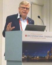
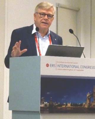
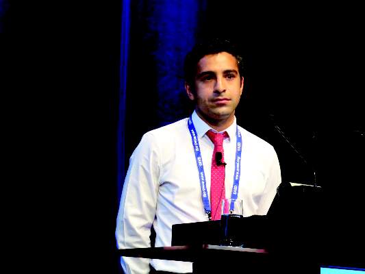
“I think what’s really important is that we now have a treatment that we know that if we direct toward [patients with persistent hypercapnia after acute hypercapnic exacerbation] that we effect a significant change in their outcomes,” said study investigator Patrick Murphy, MBBS, PhD, a consultant physician and honorary senior lecturer at the Lane Fox Respiratory Unit at Guy’s and St Thomas’ NHS Foundation Trust (London).
Speaking at the annual congress of the European Respiratory Society, he added: “We need to titrate the home ventilation to control nocturnal hypoventilation, and although I’ve not presented the data as time is short, there is no deleterious effect on quality of life.”
Nicholas Hart, MBBS, PhD, co–study investigator and clinical and academic director of Lane Fox Respiratory Unit, said in a statement issued by Philips Respironics that the results “have the ability to change the way that COPD patients are treated worldwide.”
“We’re looking forward to continuing the trial over the next 5 years to monitor survival rates, which we hope will rise, and readmission rates, which will hopefully fall,” he added.
The HOT-HMV UK Trial was conducted in 15 centers and involved patients with severe COPD who had persistent hypercapnia 2-4 weeks after experiencing an acute, life-threatening hypercapnic exacerbation requiring hospitalization. Persistent hypercapnia was defined as a pH of 7.3 or more and a PaCO2 of 7 kPa or higher. Patients had to have a 20-year or more pack year history of smoking, a forced expiratory volume in 1 second (FEV1) of 50% or less, and FEV1 to forced vital capacity (FVC) ratio of below 60%.
Dr. Murphy observed that the trial design assumed that the rate of hospital readmission at 1 year could be reduced from 55% to 25% with the use of noninvasive ventilation (NIV). The hypothesis was that HMV plus long-term HOT would increase admission-free survival compared with HOT alone.
More than 2,000 patients were initially screened for inclusion in the trial, with 116 randomized. Of the excluded patients, 1,609 did not meet inclusion criteria, 296 declined to participate, and 8 patients were not included for other reasons.
The average age of patients participating in the study was 67 years. The patients had a median body mass index of 21.6 kg/m2 and most (61%) were female. Prior long-term oxygen therapy had been used by most (80%), and 61% had three or more COPD-related hospital admissions in the last year.
Putting the primary endpoint data into perspective, Dr. Murphy said that six patients with persistent hypercapnia after treatment for an acute exacerbation needed to be treated with HMV to prevent one readmission in the following 12-month period.
Improved nocturnal hypercapnia and sleep-disordered breathing led to improved daytime hypercapnia, he observed. The change in daytime hypercapnia after 6 weeks and 3 months showed a clear statistical benefit for the combined HMV/HOT approach over HOT alone, although this lost statistical significance after 6 and 12 months’ follow-up. “That’s in part explained by the fact that the patient numbers were reduced, but also by the fact that, as part of the trial protocol, once [HOT only] patients had reached the primary outcome we allowed them to move onto HMV.”
The study was supported by Guy’s and St. Thomas’ Charity, Philips Respironics, ResMed, and the ResMed Foundation. Dr. Murphy has received hospitality for conferences and lecturing from Philips Respironics, lecturing from Fisher & Paykel Healthcare, and hospitality for conferences from ResMed.
AT THE ERS CONGRESS 2016
Key clinical point: Using home mechanical ventilation (HMV) plus home oxygen therapy (HOT) significantly improves the length of time patients stay out of the hospital.
Major finding: The median admission-free survival time was 4.3 months for HMV plus HOT versus 1.4 months for HOT alone (hazard ratio = 0.54, P = .007).
Data source: Multicenter, randomized, open-label, controlled trial of HMV plus HOT in 116 patients with chronic obstructive pulmonary disease after an acute hypercapnic exacerbation.
Disclosures: The study was supported by Guy’s and St. Thomas’ Charity, Philips Respironics, ResMed, and the ResMed Foundation. Dr. Murphy has received hospitality for conferences and lecturing from Philips Respironics, lecturing from Fisher & Paykel Healthcare, and hospitality for conferences from ResMed.
Trials confirm benefits of triple COPD therapy
LONDON – Phase III evidence confirms the multiple benefits of using a triple, fixed-dose combination (FDC) therapy over standard options in patients with severe chronic obstructive pulmonary disease (COPD), according to a presentation on two trials at the annual congress of the European Respiratory Society.
In the TRINITY trial, the combination of the inhaled corticosteroid (ICS) beclometasone diproprionate (BDP), the long-acting beta-agonist (LABA) formoterol fumarate (FF), and the long-acting muscarinic antagonist (LAMA) glycopyrronium bromide (GB) delivered via a single pressurized metered-dose inhaler (pMDI), was more effective at reducing exacerbations than was tiotropium bromide (Spiriva, Boehringer Ingelheim) monotherapy.
Results of the TRILOGY trial, which were simultaneously published in The Lancet (doi: 10.1016/S0140-6736(16)31354-X) at the time of their presentation at the ERS meeting, showed that the novel single-inhaler, triple fixed-dose combination could induce greater improvements in lung function when compared to a double fixed-dose combination of BDP and FF (Foster, Chiesi Farmaceutici SpA).
“LAMA monotherapy or ICS/LABA are standard options for treating patients with advanced COPD,” Jørgen Vestbo, MD, president of ERS and professor of respiratory medicine at the University of Manchester (England), said in an interview.
Dr. Vestbo, who was an investigator in both the TRINITY and TRILOGY trials, added that the Global Initiative for Chronic Obstructive Pulmonary Disease (GOLD) guidelines also mention that these drugs can be combined in patients who continue to experience COPD exacerbations. “But the evidence behind that is fairly weak,” he observed.
Although many patients are already being treated with triple therapy, this is via two inhalers, and “there have not been that many really good, long-term outcome studies” that have proven this approach to be the best way to manage those at risk for continued exacerbations of COPD, he said.
Drug companies are now starting to combine these three drugs into one inhaler, however, and this means that registration studies need to be done to get the products licensed, and so “there is an interest in coming up with the evidence,” Dr. Vestbo said.
“What is good about these two studies is that they are both 1-year studies and they are of sufficient size to give quite good estimates … These are studies that we should have done 5 years ago,” he said. Although the ideal is to have patients on as little therapy as possible, the results of TRINITY and TRILOGY now provide much needed evidence that it will work better than either LAMA or ICS/LABA.
The piece of evidence that is still missing is what the benefit, if any, is over a LAMA/LABA combination, a fact noted during discussion following the presentations of these data at the ERS meeting and in an editorial by Peter Calverley, MD, of the University of Liverpool (England) that accompanied the published TRILOGY findings (Lancet. 2016;388:937-8). There also is a question over whether twice daily is really better than once daily dosing, or vice versa.
“Until these next studies become available, we can be comforted by the knowledge that three therapies can be combined in a single inhaler which offers more effective therapy than at least one of the recommended treatment regimens for patients with severe COPD,” Dr. Calverley observed in reference to TRILOGY only.
Dr. Vestbo noted that at the time the TRILOGY and TRINITY studies were designed, there wasn’t the evidence from other studies such as the FLAME study (N Engl J Med. 2016. doi: 10.1056/NEJMoa1516385), showing the benefit of the LABA/LAMA combination over ICS/LABA. The TRILOGY and TRINITY studies “give that degree of evidence that was needed,” he said.
“I am not sure that the guidelines [for treating severe COPD] will change much, but at least they can say with better certainty that you can use the triple,” he added.
TRINITY – can triple better LAMA monotherapy?
The TRINITY study looked at whether patients with GOLD 3-4 COPD would be better off treated with LAMA monotherapy (tiotropium 18 mcg, one puff per day), the triple fixed-dose combination of BDP (100 mcg), FF (6 mcg), and GB (12.5 mcg) given via the novel pressurized metered-dose inhaler (two puffs twice daily), or a “free” triple combination of BDP (100 mcg) and FF (6 mcg) given via one pressurized metered-dose inhaler (two puffs daily) plus the same dose of the once-daily LAMA.
In all, 2,690 patients were randomized to these three treatments arms. The mean age of patients was 63 years and the majority (74%-77%) were men, with an average FEV1 predicted of 36% and one COPD exacerbation in the past year. Just under half of the study population were current smokers. Most (75%) had received prior treatment with an ICS/LABA combination, with about 11% receiving LAMA, and the rest either ICS/LAMA (3%), or LABA/LAMA (12%)
The annualized exacerbation rate (the primary endpoint) was 0.457 in the 1,077 patients who were treated with the triple fixed-dose combination versus 0.571 in the 1,074 patients who received tiotropium alone. The rate ratio was 0.8 indicating a 20% reduction in exacerbations was achieved (P = .003).
The annualized exacerbation rate in the 532 patients given the “free” triple combination (BDP/FF plus tiotropium) was 0.452, with a rate ratio of 0.790 (P =.010) versus those who received the LAMA as monotherapy.
There was no significant difference between the two triple combination strategies.
Presenting these data, Dr. Vestbo noted that the benefit was seen in preventing both severe and moderate COPD exacerbations. Significantly improved lung function, as measured by the change in FEV1 from baseline to week 52, was also observed to a greater degree with the triple therapy approaches than with the LAMA monotherapy.
“All three treatments were well tolerated and there were no particular safety concerns in this study,” he said.
TRILOGY – are three drugs better than two?
In contrast to the TRINITY study, the TRILOGY study looked at whether patients with severe COPD would be better off taking an ICS/LABA or the new triple fixed-dose combination pressurized metered-dose inhaler.
Just over 1,200 patients were recruited into the study, which had two co–primary endpoints: change from baseline to week 26 in predose morning FEV1 and 2-hour postdose FEV1, and the change in transition dyspnea index focal score at week 26.
Results showed that the triple fixed-dose combination improved predose FEV1 by 0.081 L and 2-hour postdose FEV1 by 0.117 L compared with the ICS/LABA combination (P less than .001 for both comparisons). Mean transition dyspnea index scores were 1.71 and 1.50, with a nonsignificant difference of 0.21.
“To be honest, I don’t think we had expected that [the triple combination] would mean much for patients, but we were hoping there would be a significant increase in lung function and a reduction of symptoms,” Dr. Vestbo said about the TRILOGY study. “What we saw was there was [symptomatic improvement] but it was not quite as impressive as we thought, but we reduced exacerbations.”
There was a significant, 23% reduction in the annualized exacerbation rate via the triple combination versus the ICS/LABA combination (0.41 vs 0.53, adjusted rate ratio 0.77, P = .005).
The triple approach was well tolerated, with no increase in adverse events versus the dual combination. The results support the idea that instead of giving patients an ICS/LABA at the start, better disease control can be achieved with a triple fixed-dose combination, Dr. Vestbo suggested.
Writing in The Lancet, Dr. Calverley noted: “The inability to meet one part of a co–primary endpoint clouds the interpretation of the other findings in the study, a familiar problem in COPD trials.” He added that there was a significant difference in the overall St George’s Respiratory Questionnaire scores favoring the triple over double therapy.
Chiesi Farmaceutici SpA funded the studies. Dr. Vestbo was an investigator for both TRINITY and TRILOGY and has received honoraria for advising and presenting from AstraZeneca, Boehringer Ingelheim, Chiesi, GlaxoSmithKline, and Novartis. Dr. Calverley has consulted for Boehringer Ingelheim, GlaxoSmithKline, and AstraZeneca.
AT THE ERS CONGRESS 2016
Key clinical point: A triple, fixed-dose combination therapy delivered by a single inhaler could be a new treatment option for severe COPD.
Major finding: Exacerbations were reduced by 20% with the triple combination versus current standards of care for COPD.
Data source: TRINITY and TRILOGY: Two 1-year, multicenter, randomized, double-blind, active controlled, parallel group, phase III studies of more than 4,000 patients with severe COPD.
Disclosures: Chiesi Farmaceutici SpA funded the studies. Dr. Vestbo was an investigator for both TRINITY and TRILOGY and has received honoraria for advising and presenting from AstraZeneca, Boehringer Ingelheim, Chiesi, GlaxoSmithKline, and Novartis. Dr. Calverley has consulted for Boehringer Ingelheim, GlaxoSmithKline, and AstraZeneca.
Insulin-dependent pilots can fly safely with glucose monitoring protocol
MUNICH – British commercial pilots with insulin-dependent diabetes are flying safely, thanks to a rigorous protocol of hourly blood glucose monitoring during every flight.
Almost every one of the 8,897 readings reported so far has been in the safe “green zone” of 5-15 mmol/L, Julia Hine, MD, said at the annual meeting of the European Association for the Study of Diabetes.
Only 3.8% fell in the cautionary “amber” range of 4-5 mmol/L or 15-19 mmol/L. Just 0.2% (22) were in the “red” range of below 4 mmol/L or above 20 mmol/L – an occurrence that demands immediate release of airplane control to the copilot.
Reassuringly, only five of those readings occurred during flight, said Dr. Hine of the Royal Surrey County Hospital, England. The rest were taken during the preflight preparation period.
She said the 4-year data confirm that the protocol works, allowing highly skilled professionals to return to their work without compromising personal safety or the safety of those entrusted to their care.
“The testing protocol can be performed safely in the cockpit, with no safety concerns,” Dr. Hine said. “It represents an advance in patient care, showing that insulin-dependent patients can perform complex occupational duties.”
The protocol was instituted in 2012, after a consortium of commercial pilots met with diabetes specialists to create a way for certified pilots to regain their class 1 licenses after a diabetes diagnosis. It’s overseen by the U.K. Civil Aviation Authority and Irish Aviation Authority. Dr. Hine said the U.K. has the largest cadre of certified pilots who have insulin-dependent diabetes in the world. Canada is the only other country with such a program, although, she said, “There are a number of European states that have expressed interest in the program.”
In the United States, a diagnosis of insulin-dependent diabetes currently excludes pilots from flying commercial aircraft. The American Diabetes Association has been lobbying for easing of this strict FAA ban since at least 2014, stating that “the FAA’s blanket ban is not medically justified.” Specifically, the ADA favors a position in which “individual assessment of people with diabetes is the appropriate approach to determining whether a person is qualified to perform certain activities,” according to a statement.
The British protocol requires stringent pre- and in-flight blood sugar monitoring. Levels must be obtained at least 1 hour before reporting for duty and at least 2 hours before commencing a flight.
Blood sugar must be regularly measured during flight at well. For pilots using insulin, levels should be drawn every 2 hours, and for those on oral medications, every 2-4 hours. A final level must be drawn 30 minutes before landing.
Additional testing is required if the pilot experiences any symptoms related to fluctuating blood sugar. All results are read out loud, reported to the copilot and into the plane’s voice data recorder, and recorded in the pilot’s flight data record.
Levels in the amber range require the pilot to take corrective action. For low blood glucose levels, the pilot must consume 10-15 grams of carbohydrate, retest after 30 minutes, and adjust insulin. For high blood glucose levels, the pilot should review insulin dosing and modify carbohydrate intake, then retest in 45 minutes.
Readings in the red range demand immediate action, which includes handing over control of the flight, repeat testing, modifying insulin and carbohydrate intake, and retesting. The pilot may only resume duty when blood glucose returns to a green level.
There is a special protocol for pilots on insulin pumps, which are not guaranteed to function if there is a sudden loss of cabin pressure.
Dr. Hine and her colleague David Russell-Jones, MD, who is the program’s medical director, presented 4 years’ worth of data on 16 of the 26 pilots who have participated in the program. All of these pilots are otherwise healthy men with well-controlled diabetes who were deemed at low risk for glycemic events. They are an average of 40 years old with an average disease duration of 8 years. Their average HbA1c was 53 mmol/l before licensure and 54.8 mmol/l afterwards. All of the pilots have normal renal function, no retinopathy, no changes in blood pressure, and good awareness of hypoglycemia.
Among them, they have accumulated 4,900 flight hours and 8,897 blood glucose readings. Most of the flights (88%) were medium- or short-haul flights of less than 6 hours.
Even though 96% of readings fell into the 5-20 mmol/l green range, the bulk of those readings were in the range of 5-9 mmol/l. There was very little hourly variation in blood glucose levels during flights up to 6 hours, showing that the protocol is effective at establishing stable glycemia.
Right now, the program is in place only for current pilots who have a new diagnosis of diabetes. But, said Dr. Russell-Jones, it may be expanded at some point to encompass people with diabetes who wish to train to become a commercial pilot.
Neither Dr. Hines nor Dr. Russell-Jones reported any financial conflicts.
On Twitter @Alz_Gal
AT EASD 2016
Key clinical point: A protocol of regular blood glucose monitoring during flight is keeping British commercial pilots in the air.
Major finding: Of 8,897 blood glucose readings, only 22 were in the dangerous “red” range that demands handing over control of the plane to the copilot.
Data source: The 16 pilots in the program have amassed 4,900 flight hours.
Disclosures: Neither Dr. Hine nor Dr. Russell-Jones had any financial disclosures.
Brain atrophy is already evident in patients with prediabetes
MUNICH – Brain changes suggestive of cerebral microvascular dysfunction are already apparent in patients with prediabetes.
The changes – increased white matter hyperintensities and decreased white matter volume – are even more pronounced in subjects with type 2 diabetes, Marnix van Agtmaal, MD, said at the annual meeting of the European Association for the Study of Diabetes. Patients with frank diabetes also showed an increase in intracranial cerebrospinal fluid – a correlate of the decrease in brain volume, said Dr. van Agtmaal of Maastricht (the Netherlands) University Medical Center.
The changes are probably caused by diabetes-related endothelial dysfunction, he said.
“The brain is highly dependent on properly functioning microcirculation. This is critical, since the brain has high energy demand and no energy reserve. In prediabetes and type 2 diabetes, microvascular endothelial dysfunction occurs. This leads to cerebral hypoperfusion, which in turns causes chronic ischemia. This contributes to small vessel disease leading to brain atrophy and, eventually, cognitive decline and dementia.”
The 2,251 subjects in the analysis were drawn from the Maastricht study, an ongoing observational study of people with type 2 diabetes.
Among the group, 350 had prediabetes, defined as impaired fasting glucose, impaired glucose tolerance, or a combination of the two. Type 2 diabetes was present in 528. The rest had healthy glucose metabolism.
As the cohort progressed from healthy glucose metabolism to prediabetes and then diabetes, they became older (aged 58 years in the healthy group vs. 62 years in the diabetes group), heavier, and displayed worsening cardiovascular risk factors, with increasing systolic blood pressure and progressively poorer lipid profiles. Kidney function was preserved in all patients, however.
The groups were not balanced in terms of sex: 56% of those with healthy glucose metabolism were women, compared with 47% of those with prediabetes and 31% of those with type 2 diabetes.
Dr. van Agtmaal and his colleagues examined white matter hyperintensities, white matter volume, gray matter volume, and intracranial CSF. They conducted three linear regression models: a crude unadjusted model, a partially adjusted model that controlled for age, sex, and intracranial volume; and a fully adjusted model that controlled for those factors, plus systolic blood pressure, lipids, smoking, kidney function, and education.
There was a clear linear association between white matter hyperintensity (WMH) volume and healthy glucose metabolism, prediabetes, and type 2 diabetes. In the crude analysis, the healthy subjects carried about 0.75 mL of WMH. Prediabetic subjects carried about 1.25 mL, and those with diabetes, about 2 mL.
In both the partially and fully adjusted models, this relationship was somewhat attenuated, but it remained significant for both prediabetes and diabetes.
The crude model also found that both diabetes groups had significantly lower white matter volume than did the healthy subjects. In healthy subjects, the mean volume was about 480 mL. This was about 467 mL in those with prediabetes and 466 mL in those with type 2 diabetes. Again, the partially and fully adjusted models slightly attenuated the relationship, but it remained significant in both disease states.
The crude model showed that gray matter was decreased in both prediabetes and type 2 diabetes. In healthy subjects, total gray matter was about 667 mL. In those with prediabetes, it was about 655 mL, and in those with type 2 diabetes, about 645 mL. However, the significant associations disappeared for both diabetes and prediabetes in both adjusted models.
Intracranial CSF was also different among the three groups in the crude model. In the healthy subjects, the total intracranial CSF volume was about 248 mL. In those with prediabetes, it was about 255 mL, and in those with type 2 diabetes, about 270 mL.
The association with prediabetes disappeared in the fully adjusted model – but for type 2 diabetes, it remained strongly significant.
Dr. van Agtmaal has not correlated the imaging findings with any cognitive testing on these subjects but said that study is coming.
“Further analysis will also look at cognitive decline and the development of dementia in the group,” he said. “We also intend to look at associations with other outcomes of cerebral dysfunction, including depression.”
Dr. van Agtmaal had no financial disclosures.
AT EASD 2016
Key clinical point: People with prediabetes and type 2 diabetes have more white matter hyperintensities and lower white matter volume than do those with healthy glucose metabolism.
Major finding: Healthy subjects carried about 0.75 mL of white matter hyperintensities, while prediabetic subjects carried about 1.25 mL, and those with diabetes, about 2.0 mL.
Data source: The subset of the Maastricht Study comprised 2,251 subjects.
Disclosures: Dr. van Agtmaal had no financial disclosures.
Hepatitis Outlook: August 2016
If you work on the front lines of medical care treating patients with hepatitis, you may not have time to review all the hepatitis research that enters the medical literature every month. Here’s a quick look at some notable news items and journal articles published over the past month, covering a variety of the major hepatitis viruses.
A study in Hepatology has provided a preclinical risk assessment paradigm with which to better understand cardiovascular drug-drug interaction risk for hepatitis C–virus infected patients treated with sofosbuvir in combination with other direct acting antivirals and the antiarrhythmic drug amiodarone.
A Japanese study found that, although levels of Wisteria floribunda agglutinin-positive Mac-2-binding protein could be a useful indicator of liver fibrosis in patients with hepatitis B or C infection, WFA+-M2BP levels in the two groups significantly differed, even in the same degree of fibrosis.
Interferon-free, guideline-tailored therapy with direct-acting antivirals is highly effective and safe for hepatitis C virus–associated mixed cryoglobulinemia patients, according to a recent study.
Another recent study found that pegylated interferon (PegIFN) intensification in hepatitis B “e” antigen (HBeAg)-positive coinfected patients did not lead to increased clearance rates of HBeAg or hepatitis B surface antigen quantification (qHBsAg), despite faster declines of antigen levels while on PegIFN.
A study in HIV Medicine found that, under real-life conditions, treatment of patients infected with hepatitis C virus and of patients coinfected with HCV/HIV with all-oral direct-acting antiviral combinations led to high and similar rates of sustained virological response 12 weeks after the end of therapy.
Hepatitis B virus coinfection was the most important risk factor for liver fibrosis and cirrhosis in HIV-infected patients, and should be diagnosed early in HIV care to optimize treatment outcomes, a recent study showed.
Immunity persisted 24 months after a single dose of inactivated hepatitis A vaccine and live attenuated hepatitis A vaccine was administered to school-age children, according to a study published in Human Vaccines & Immunotherapeutics.
A hepatitis C treatment scale-up strategy in Rhode Island could reduce cirrhosis cases and liver-related deaths by 78.9% and 72.4%, respectively, by 2030, according to a study in Epidemiology and Infection.
Viral blipping is a frequent event during nucleoside analogue treatment of patients with chronic hepatitis B virus infection, a study found, although it did not lead to any clinically significant outcomes and thus may not require more frequent blood work and patient visits in clinical practice.
A study of liver and spleen stiffness in hepatitis C virus–infected patients – with advanced liver disease and sustained virologic response after interferon-free treatment – found that improvement of liver stiffness may be due to reduced necroinflammation, and to a lesser extent regression of cirrhosis. Improvement was more pronounced between therapy baseline and end of treatment than therapy baseline and 24 weeks after end of treatment.
From 2000 to 2011, 4,346 adults who died in New York City had a report of a hepatitis B virus infection (0.7%), according to a study in Epidemiology and Infection. Of the HBV-infected decedents, 1,074 (25%) were HIV coinfected. Fifty-five percent of HBV monoinfected and 95% of HBV/HIV coinfected decedents died prematurely, the researchers found.
Prison-based hepatitis C virus treatment achieves outcomes similar to those of community-based treatment, according to a study in the Journal of Viral Hepatitis, with those not released or transferred during treatment doing particularly well.
Treatment interventions to curb the hepatitis C virus epidemic among HIV-infected men who have sex with men are effective if high-risk behavior does not increase as it has during the last decade, according to a study in Hepatology.
The results of an international quality control study underline the urgent need to improve methods used to monitor hepatitis Delta virus viremia.
An investigation of a hepatitis E virus genotype 4 outbreak in Zhejiang Province, China, found that the outbreak was most likely caused by contaminated tap water rather than food.
A German study found that short treatment with 8 weeks of sofosbuvir and ledipasvir seems highly effective and safe in well-selected hepatitis C virus mono- and HIV/HCV-coinfected patients in a real-world setting.
A study of historical events fueling the cross-continental spread of hepatitis C virus epidemics said drivers for the epidemic were the advent of intravenous medical therapies and devices, growth in the heroin trade, and population mixing during armed conflicts.
On Twitter @richpizzi
A quarter of hypertensive Medicare enrollees are nonadherent
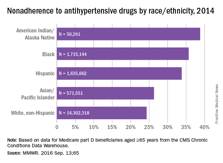
Over 26% of Medicare part D enrollees aged 65 years and over are not taking their antihypertensive drugs properly, according to a report published online Sept. 13 in MMWR.
An analysis of 2014 data showed that 4.9 million hypertensive Medicare patients were taking an incorrect dose of their medication or were not taking it at all, reported Matthew Ritchey, DPT of the Centers for Disease Control and Prevention’s division for heart disease and stroke prevention, and his associates (MMWR. 2016 Sep 13:65).
Nonadherence rates varied considerably by race and ethnicity, with American Indians/Alaska Natives the highest at 39%, followed by blacks at 36%, Hispanics at 34%, Asian/Pacific Islanders at 26%, and white non-Hispanics at 24%, the investigators noted.
The analysis included 18.5 million part D beneficiaries who filled two or more antihypertensive prescriptions in the same therapeutic class on different dates within a period of more than 90 days in 2014.
FROM MMWR
Four factors raise risk of post-TAVR endocarditis
Four factors – younger patient age, male sex, diabetes, and moderate to severe residual aortic regurgitation – are associated with a significantly increased risk of infective endocarditis after transcatheter aortic valve replacement, according to a report published online Sept. 13 in JAMA.
Until now, data pertaining to endocarditis following TAVR “have been limited to case reports and relatively small series with limited follow-up,” said Ander Regueiro, MD, of Laval University, Quebec City, and his associates.
They performed a retrospective analysis of data in a large international registry of TAVR cases to better characterize post-TAVR endocarditis.
Dr. Regueiro and his colleagues focused on 20,006 TAVR procedures done at 47 medical centers in Europe, North America, and South America during a 10-year period. The median time to symptom onset was 5.3 months after the procedure.
Infective endocarditis was definitively diagnosed in 250 of these cases. This incidence is similar to that reported for endocarditis following surgical aortic valve replacement, indicating that TAVR is no less predisposing to endocarditis despite being a less invasive approach.
The mean age of patients who developed post-TAVR endocarditis was 78.9 years, compared with 81.8 years for those who did not (HR, 0.97). The reason for this association is unclear, but it is possible that younger patients chosen for TAVR because of their prohibitive surgical risk carry a higher burden of comorbidity than do older patients. Similarly, 62% of endocarditis cases arose in men (HR, 1.69), and sex differences in comorbid conditions may explain the higher risk among men.
More patients who developed endocarditis had diabetes (41.7%), compared with those who did not develop endocarditis (30%), for an HR of 1.52. And patients who had moderate to severe residual aortic regurgitation after TAVR also were at much higher risk for endocarditis than were those who did not (HR, 2.05), the investigators noted (JAMA. 2016 Sep 13;316[10]:1083-92).
In contrast, factors that were not associated with endocarditis risk included chronic pulmonary disease, type of valve (self-expandable or balloon-expandable), and setting of the procedure (catheterization lab vs. operating room).
The bacteria that most commonly caused infective endocarditis were Enterococci species (24.6% of cases), Staphylococcus aureus (23.8%), and coagulase-negative staphylococci (16.8%). This should be taken into consideration when selecting antibiotics for prophylaxis before TAVR and when choosing empirical antibiotics for treatment while waiting for blood culture results, wrote Dr. Regueiro and his associates.
“This information may help clinicians identify patients at higher risk [for endocarditis] and aid in implementing appropriate preventive measures,” they noted.
This study was supported by a grant from the Alfonso Martin Escudero Foundation. Dr. Regueiro reported having no relevant financial disclosures; his associates reported ties to numerous industry sources.
FROM JAMA
Key clinical point: Four factors raise the risk that patients undergoing transcatheter aortic valve replacement will develop infective endocarditis.
Major finding: The mean age of patients who developed post-TAVR endocarditis was 78.9 years, compared with 81.8 years for those who didn’t (HR, 0.97).
Data source: A retrospective analysis of data in an international registry involving 20,006 patients who underwent TAVR at 47 medical centers during a 10-year period.
Disclosures: This study was supported by a grant from the Alfonso Martin Escudero Foundation. Dr. Regueiro reported having no relevant financial disclosures; his associates reported ties to numerous industry sources.
Smoking thickens LV wall, worsens function
Current smoking, as well as higher levels of cumulative cigarette exposure from past smoking, were both associated with higher left ventricular mass, a higher LV mass-to-volume ratio, and worse diastolic function in an elderly community-based population with no overt indications of coronary artery disease or heart failure, according to a report published online Sept. 13 in Circulation: Cardiovascular Imaging.
“These findings suggest that smoking is associated with subtle alterations in LV structure and function, which might help explain the higher risk of heart failure [HF] reported for smokers, independent of coronary artery disease [CAD],” said Wilson Nadruz Jr., MD, of the cardiovascular division, Brigham and Women’s Hospital, Boston, and his associates.
They analyzed links between smoking and echocardiographic features using data from the Atherosclerosis Risk in Communities (ARIC) study, an ongoing prospective observational study involving community-dwelling adults who were aged 45-64 years at baseline in 1987-1989. For their study, Dr. Nadruz and his colleagues assessed echocardiographic images taken for 4,580 ARIC participants at follow-up roughly 25 years later. None of these adults had any indication of CAD or HF; 287 (6.3%) were current smokers, 2,316 (50.5%) were former smokers, and 1,977 (43.2%) never smoked.
Compared with never smokers, current smokers showed a greater LV mass index (80.4 vs. 76.7), a greater LV mass-to-volume ratio (1.93 vs. 1.83), and a higher prevalence of LV hypertrophy (15% vs. 9%), as well as a higher prevalence of concentric LV hypertrophy and worse LV diastolic function. The same association was found between never smokers and former smokers who had higher levels of cumulative cigarette exposure, the investigators said (Circ Cardiovasc Imag. 2016 Sep 13. doi: 10.1161/circimaging.116.004950).
This association between smoking and altered LV structure and function remained robust after the data were adjusted to account for numerous cardiac risk factors such as older age, higher BMI, diabetes, hypertension, greater alcohol consumption, and higher heart rate. It also didn’t vary by patient sex, race, or income level. In contrast, there was no association between smoking and right ventricular structure or function.
“These data suggest that smoking can independently lead to thickening of the heart and worsening of heart function, which may lead to a higher risk for heart failure, even in people who don’t have heart attacks,” Dr. Nadruz said in a statement.
Looking at the results in a more positive light, senior author Scott D. Solomon, MD, professor of medicine at Harvard University, Boston, said “The good news is that former smokers had similar heart structure and function, compared with never smokers,” suggesting that “the potential effects of tobacco on the myocardium might be reversible after smoking cessation.”
FROM CIRCULATION: CARDIOVASCULAR IMAGING
Key clinical point: Current smoking was associated with higher left ventricular mass, a higher LV mass-to-volume ratio, and worse diastolic function.
Major finding: Compared with never smokers, current smokers showed a greater LV mass index (80.4 vs. 76.7), a greater LV mass-to-volume ratio (1.93 vs. 1.83), and a higher prevalence of LV hypertrophy (15% vs. 9%).
Data source: A secondary analysis of data for 4,580 elderly participants in ARIC, a large community-based cohort.
Disclosures: This study was supported by Brigham and Women’s Hospital, Boston. Dr. Nadruz and his associates reported having no relevant financial disclosures.
Four-step screen IDs silent heart attack in type 2 diabetes
MUNICH – A four-component imaging/biomarker screen was highly accurate for identifying silent myocardial infarction among asymptomatic patients with type 2 diabetes.
The screen is far more accurate than the current standards of invasive imaging or only looking for pathologic Q waves, Peter Swoboda, MD, said at the annual meeting of the European Association for the Study of Diabetes.
“By combining these four factors we came up with a tool that has a diagnostic area under the curve [AUC] of 0.85,” said Dr. Swoboda of Leeds (England) University. “This is far better than the 0.58 AUC that we have with Q waves only – a sensitivity of just 25%.”
The study was published online in June in the Journal of Clinical Endocrinology and Metabolism (JCEM 2016. doi: 10.1210/jc.2016-1318).
The screen employs noninvasive imaging and biomarkers to tap multiple clinical hallmarks of silent MI. The components are:
• electrocardiogram.
• echocardiography.
• biomarker assessment.
• cardiac magnetic resonance imaging, focusing on left ventricular ejection fraction and late gadolinium enhancement.
The study cohort comprised 100 patients with type 2 diabetes without known heart disease and with no new cardiac symptoms. They underwent cardiac MRI, a 12-lead electrocardiogram, echocardiography, and serum biomarker assessment. Late gadolinium enhancement identified evidence of silent MI in 17 patients (17%).
There were few differences in the clinical characteristics of those who had experienced MI and those who had not, Dr. Swoboda noted. There were no differences at all in diabetes-related measures, including disease duration or hemoglobin A1c levels. Blood pressures were similar. Patients with MI were significantly older (65 vs. 60 years).
In cardiac-specific measures, left ventricular ejection fraction was similar, as was left ventricular mass, end diastolic volume, and left atrial volume. There were however, other very important differences, Dr. Swoboda noted.
Imaging included a measure called “feature tracking analysis,” which measured the peak global longitudinal strain, systolic strain rate, and early and late diastolic strain rates during contraction. This analysis noted a significant difference in global longitudinal strain between the MI and non-MI groups.
Ventricular filling velocities as measured by the E/A ratio on ECG were also significantly different between the MI and non-MI groups (0.75 vs. 0.89, respectively). ECG also found pathologic Q waves in significantly more MI patients (24% vs. 7%).
Finally, the serum biomarker panel showed a very strong increase in B-type natriuretic peptide (NT-proBNP) among MI patients, compared with non-MI patients (105 vs. 52 ng/L). There were no significant differences in the other biomarkers, including C-reactive protein and high-sensitive cardiac troponin.
Dr. Swoboda and his team then compiled these findings into a composite measure, assigning them optimum cutoff measures:
• Age older than 62 years.
• E/A ratio 0.72 or lower.
• Global longitudinal strain of at least 18.4%.
• NT-proBNP more than 29 ng/L.
The system resulted in a diagnostic accuracy AUC of 0.85 – significantly better than any of the AUCs generated by the individual components. All patients who scored 0 or 1 were free of MI. Among the 28 with a score of 2, only three had experienced a silent MI. Among the 21 with a score of 3, seven had experienced a silent MI and 14 had not. Among the 16 with a score of 4, seven had experienced a silent MI and nine had not.
While Dr. Swoboda called the screening method “simple” during discussion, a colleague in the audience disagreed with that.
“A simple test is something like a blood test only, not an MRI. Not imaging. That is expensive and takes time,” said Naveed Sattar, MD, of the University of Glasgow, Scotland. “However, I do think your data add more to the evidence that BNP can be a really valuable marker of cardiovascular risk in patients with diabetes.”
Dr. Sattar recently examined the value of cardiac serum biomarkers in predicting cardiovascular disease and mortality in nearly 100,000 people without a history of heart disease. In these subjects, he wrote, “NT-proBNP assessment strongly predicted first-onset heart failure and augmented coronary heart disease and stroke prediction, suggesting that NT-proBNP concentration assessment could be used to integrate heart failure into cardiovascular disease primary prevention.”
The paper appeared online in Lancet Diabetes in September (Lancet Diab. 2016. doi: 10.1016/S2213-8587[16]30196-6).
Dr. Swoboda agreed that data continue to support the increased use of NT-proBNP as a marker of heart disease.
“I think that in the future, diabetes medicine is moving toward individualized patient care, based on individualized risk factors. The future of assessing asymptomatic cardiac patients might be a combination of BNP and MRI.”
Dr. Swoboda had no financial disclosures. Some of Dr. Sattar’s coauthors reported relationships with pharmaceutical companies.
AT EASD 2016
Key clinical point: A four-component screen accurately identified silent myocardial infarction in asymptomatic patients with type 2 diabetes
Major finding: The tool had an 82% sensitivity and 72% specificity for silent MI.
Data source: It was created in a cohort of 100 patients with type 2 diabetes and no history of heart disease.
Disclosures: Dr. Swoboda had no financial disclosures.
Low caregiver self-care linked with depression, anxiety
There is increased anxiety and depression among family caregivers who do not take care of themselves, according to a study to be presented at the 2016 ASCO Palliative Care in Oncology Symposium.
Nearly a quarter of 294 caregivers of Medicare patients with advanced cancer reported high depression scores (23%) and 34% reported borderline or high anxiety scores. Worse caregiver anxiety, depression, and mental health–related quality of life scores were significantly associated with lower scores in every self-care measure (P less than .05 for all). Lower self-care behavior scores were associated with longer durations, higher hours, and more days/week of caregiving and with fair or poor patient health.
The cross-sectional survey was conducted in community settings of eight cancer centers in Alabama, Florida, and Tennessee. The family caregivers of Medicare beneficiaries diagnosed with pancreatic, lung, brain, ovarian, head & neck, hematologic, or stage IV cancer completed measures of self-care behaviors, including health responsibility, physical activity, nutrition, spiritual growth, interpersonal relations, stress management, and sleep. Caregivers averaged 66 years and were mostly female (72.8%), white (91.2%), Protestant (76.2%), retired (54.4%), and patients’ spouse/partner (60.2%). Approximately half were rural dwellers (46.9%) and had incomes less than $50,000 (53.8%). The majority provided support 6-7 days per week (71%) for greater than 1 year (68%).
“This research serves as an important call to action for the oncology community to implement support networks and services that care for the caregiver,” ASCO representative Andrew Epstein, MD, of Memorial Sloan Kettering Cancer Center, New York, said in a written statement ahead of the symposium.
“We hope our research rallies the oncology palliative care communities to develop assessment tools and services that support caregivers,” lead author James Nicholas Dionne-Odom, PhD, RN, of the University of Alabama at Birmingham, said in the statement.
On Twitter @jessnicolecraig
FROM THE 2016 ASCO PALLIATIVE CARE IN ONCOLOGY SYMPOSIUM
Key clinical point: There is increased anxiety and depression among family caregivers who do not take care of themselves.
Major finding: Worse caregiver anxiety, depression, and mental health–related quality of life scores were significantly associated with lower scores in every self-care measure (P less than .05 for all).
Data source: A multistate and cross-sectional survey of 294 family caregivers.
Disclosures: The University of Alabama at Birmingham funded the study. One investigator reported receiving financial compensation and honoraria from Medscape, Carevive Systems, and PackHealth.
