User login
Official news magazine of the Society of Hospital Medicine
Copyright by Society of Hospital Medicine or related companies. All rights reserved. ISSN 1553-085X
nav[contains(@class, 'nav-ce-stack nav-ce-stack__large-screen')]
header[@id='header']
div[contains(@class, 'header__large-screen')]
div[contains(@class, 'read-next-article')]
div[contains(@class, 'main-prefix')]
div[contains(@class, 'nav-primary')]
nav[contains(@class, 'nav-primary')]
section[contains(@class, 'footer-nav-section-wrapper')]
footer[@id='footer']
section[contains(@class, 'nav-hidden')]
div[contains(@class, 'ce-card-content')]
nav[contains(@class, 'nav-ce-stack')]
div[contains(@class, 'view-medstat-quiz-listing-panes')]
div[contains(@class, 'pane-article-sidebar-latest-news')]
div[contains(@class, 'pane-pub-article-hospitalist')]


Creating a digital pill
Technology battles medication noncompliance
Hospitalists and other physicians have long struggled with medication noncompliance, which can lead to sicker patients and higher rates of readmittance, and costs some $100-$289 billion a year.
There is a growing field of digital devices being developed to address this problem. The Food and Drug Administration has just approved the newest one: a medication with a sensor embedded that can tell doctors if, and when, patients take their medicine, according to an article in the New York Times.1 It’s expected to become available in 2018.
The digital medication is a version of the antipsychotic Abilify. Patients who agree to take it will sign consent forms allowing their doctors (and up to four other people) to receive electronic data showing the date and time pills are ingested.
The sensor, created by Proteus Digital Health, contains copper, magnesium, and silicon, all said to be safe ingredients found in foods. The electrical signal is created when stomach fluids contact the sensor; a patch worn on the rib cage detects that signal and sends the message.
Other companies are joining the race to create digital medication technologies; these are being tested in medications for patients with conditions including heart disease, diabetes, and HIV infection. Some researchers predict the technology might have applications for monitoring the opioid intake of postsurgical patients or patients in medication clinical trials.
Reference
1. Belluck P. “First Digital Pill Approved to Worries About Biomedical ‘Big Brother.’ ” New York Times. Nov 13, 2017.
Technology battles medication noncompliance
Technology battles medication noncompliance
Hospitalists and other physicians have long struggled with medication noncompliance, which can lead to sicker patients and higher rates of readmittance, and costs some $100-$289 billion a year.
There is a growing field of digital devices being developed to address this problem. The Food and Drug Administration has just approved the newest one: a medication with a sensor embedded that can tell doctors if, and when, patients take their medicine, according to an article in the New York Times.1 It’s expected to become available in 2018.
The digital medication is a version of the antipsychotic Abilify. Patients who agree to take it will sign consent forms allowing their doctors (and up to four other people) to receive electronic data showing the date and time pills are ingested.
The sensor, created by Proteus Digital Health, contains copper, magnesium, and silicon, all said to be safe ingredients found in foods. The electrical signal is created when stomach fluids contact the sensor; a patch worn on the rib cage detects that signal and sends the message.
Other companies are joining the race to create digital medication technologies; these are being tested in medications for patients with conditions including heart disease, diabetes, and HIV infection. Some researchers predict the technology might have applications for monitoring the opioid intake of postsurgical patients or patients in medication clinical trials.
Reference
1. Belluck P. “First Digital Pill Approved to Worries About Biomedical ‘Big Brother.’ ” New York Times. Nov 13, 2017.
Galectin-3: A new post-MI prognostic biomarker?
ORLANDO – An elevated circulating galactin-3 level after an acute MI is a potent long-term predictor of both heart failure and mortality, independent of known prognostic markers, Rabea Asleh, MD, PhD, reported at the annual meeting of the American College of Cardiology.
“These findings suggest that galectin-3 measurement may have a role in the risk stratification of patients presenting with MI,” according to Dr. Asleh, an Israeli cardiologist doing a fellowship in advanced heart failure and transplant cardiology at the Mayo Clinic in Rochester, Minn.
“The changing clinical presentation of MI necessitates evolution in our approach to risk stratification,” he explained. “Over the last 2 decades we’ve observed a change in the epidemiology of MI, with more patients developing non-ST-elevation MI compared to STEMI. They present at an older age and develop heart failure with preserved ejection fraction more than heart failure with reduced ejection fraction.”
He presented a prospective population-based community cohort study of 1,401 Olmsted County, Minn., residents who had a validated MI during 2002-2012. Their mean age was 67 years, 61% were men, and 79% presented with non-STEMI. During a mean follow-up of 5.3 years, 389 of the participants developed heart failure and 512 patients died.
Galectin-3 was measured a median of 2 days post MI. The median level was 18.4 ng/mL. Patients were divided into tertiles based upon their galactin-3 measurement: Tertile 1 required a post-MI galectin-3 level below 15.2 ng/mL; tertile 2 had a level of 15.2-22.6 ng/mL; and the top tertile was for individuals with a galectin-3 above 22.6 ng/mL.
Of note, patients with a higher galectin-3 level were older, had a higher prevalence of diabetes, hypertension, hyperlipidemia, anterior MI, a higher Killip class, a higher Charlson comorbidity score, and a lower peak troponin T level. They also had a lower estimated glomerular filtration rate; indeed, the median eGFR in the top tertile for galactin-3 was 48 mL/min per 1.73 m2, compared with 68 mL/min in the lowest galectin-3 tertile. Women accounted for 27% of patients in tertile 1, 41% in tertile 2, and fully half of those in tertile 3.
In an unadjusted analysis, the risk of mortality during follow-up was sixfold greater for patients in galectin-3 tertile 3 than in tertile 1; the risk of heart failure was increased 5.5-fold.
More meaningfully, in a Cox multivariate analysis extensively adjusted for age, gender, comorbidities, malignancy, standard cardiovascular risk factors, MI characteristics, eGFR, Killip class, cardiac troponin T, and other potential confounders, patients in galectin-3 tertile 2 had a 1.6-fold increased risk of death and a 1.62-fold increased likelihood of heart failure during follow-up, compared with subjects in tertile 1, Dr. Asleh noted.
Patients in tertile 3 had a 2.4-fold increased risk of death and were at 2.1 times greater risk of heart failure than those in tertile 1. The degree of risk for heart failure associated with elevated galactin-3 was virtually identical for heart failure with preserved as compared with reduced ejection fraction, he added.
Session cochair L. Kristin Newby, MD, of Duke University, Durham, N.C., noted that the Mayo study did not adjust for brain natriuretic peptide (BNP) or N-terminal pro hormone BNP (NT-proBNP), both of which are known to be strong predictors of both heart failure and mortality after acute MI. Doesn’t their absence weaken the strength of galactin-3’s prognostic power as demonstrated in the study? she asked.
Dr. Asleh replied that those biomarkers weren’t collected in this study, which began in 2002.
“What I can tell you is, other studies show there is only a weak correlation between galactin-3 and NT-proBNP post MI. Some studies have even shown an inverse correlation,” he said. “The pathophysiological explanation is that galactin-3 is more implicated in fibrosis before the stage of development of left ventricular loading and stretching of the myocardium. So galactin-3 may be implicated in LV fibrosis leading to heart failure before the NT-proBNP comes into play.”
Also, he cited a study by other investigators conducted in patients with a left ventricular assist device for advanced heart failure. Upon device-induced left ventricular unloading the patients’ NT-proBNP levels dropped significantly while their galactin-3 remained high and unchanged. This suggests the two biomarkers are implicated in different disease pathways.
Both animal and human studies indicate galactin-3 is involved specifically in fibrosis, as opposed to, say, C-reactive protein, a well established marker of systemic inflammation, the cardiologist added.
Dr. Asleh reported having no financial conflicts of interest regarding his study, which was supported by the National Institutes of Health.
REPORTING FROM ACC 18
Key clinical point: Galectin-3 level post-MI is a potent long-term predictor of both heart failure and mortality independent of known prognostic markers.
Major finding: Post-MI patients in the top tertile of circulating galectin-3 were at an adjusted 2.4-fold increased mortality risk and a 2.05-fold greater risk of developing heart failure compared with those in the lowest tertile.
Study details: This prospective population-based cohort study included 1,401 MI patients followed for a mean of 5.3 years.
Disclosures: The National Institutes of Health supported the study. The presenter reported having no financial conflicts of interest.
A call for ‘changing the social norms’ on naloxone
SAN DIEGO – Among individuals with a history of injection drug use, more than one-third reported never or sometimes carrying naloxone, while just one in four reported carrying with it them at all times.
Those are key findings from a survey that set out to examine gaps in the naloxone cascade in a sample of people who inject drugs.
“In order to save a life, you have to have the naloxone with you at all times,” lead study author Karin E. Tobin, PhD, said in an interview at the annual meeting of the College on Problems of Drug Dependence.
While emerging research demonstrates the positive impact of opioid overdose education and community naloxone distribution programs to reduce opioid-related overdose deaths, opiate overdose continues to be a major cause of mortality, said Dr. Tobin, who is affiliated with the department of health behavior and society at Johns Hopkins University, Baltimore. “We’ve made a lot of progress in convincing people that naloxone is not addictive, and that it’s not going to cause any harm,” she said. “Now, drug users aren’t afraid to ask for it. Still, we wondered: If everyone knows about naloxone and no one is embarrassed to talk about it, why are people still dying [from opioid overdoses] in Baltimore?”
She and her associates conducted a cross-sectional survey of 353 individuals aged 18 and older in Baltimore who self-reported a lifetime history of injection drug use. The data came from a baseline survey that was conducted as part of a randomized, controlled trial testing the efficacy of a behavioral intervention focused on the Hepatitis C cascade. Individuals were asked to answer questions related to the five steps of the naloxone cascade: awareness (have you ever heard about naloxone?), access (have you ever received naloxone?), training (have you ever been trained to use naloxone?), use (have you ever used naloxone during an opiate overdose?), and possession (how often do you carry naloxone?)
More than half of the survey respondents (65%) were male; mean age was 47 years. For the previous 6 months, more than half of the sample reported the use of crack (64%), heroin (74%), and other injectable drugs (57%), while 90% reported having ever witnessed an overdose – 59% in the prior year alone. Dr. Tobin and her associates found that 90% of respondents had heard about naloxone, 69% had received it, and 60% had been trained to use it. In addition, 37% reported never carrying naloxone, 38% sometimes carried it, 33% said they had used naloxone at some point, and 25% said they always carried it with them.
On multinomial regression analysis, the researchers found that carrying naloxone often or always was significantly associated with the following variables: female sex (odds ratio, 2.77), having ever witnessed an overdose (OR, 1.84), having injected in the past 12 months (OR, 1.75), and having ever used naloxone during an overdose (OR, 4.33). The latter finding is especially important, “because it means that we just have to let people practice using it,” said Dr. Tobin, who noted that more research is needed to understand reasons why injection drug users do not always carry naloxone. “We need to start changing the social norms about carrying naloxone. You never know when it will be useful.”
The National Institute on Drug Abuse supported the study. Dr. Tobin reported having no financial disclosures.
AT CPDD 2018
Key clinical point: Increasing possession of naloxone among people who use opiates is essential.
Major finding:. Carrying naloxone often or always was significantly associated with several variables, including female sex (odds ratio, 2.77) and having ever used naloxone during an overdose (OR, 4.33).
Study details: A cross-sectional survey of 353 individuals aged 18 and older in Baltimore who self-reported a lifetime history of injection drug use.
Disclosures: The National Institute on Drug Abuse supported the study. Dr. Tobin reported having no financial disclosures.
Do free meals to physicians affect opioid prescribing?
SAN DIEGO – Physicians who receive gifts and free meals from opioid manufacturers prescribe more opioids than do their counterparts, a new study suggests.
A sampling of doctors who reported marketing payments or gifts prescribed more of the drugs the following year even as their colleagues prescribed fewer. Researchers also found signs of a dose-effect relationship between more free meals received and more opioid medications prescribed.
The findings, presented at the annual meeting of the College on Problems of Drug Dependence and recently published, do not prove a link between free meals and the massive, deadly opioid epidemic. And the purpose of pharmaceutical marketing, of course, is to persuade physicians to prescribe medications, the researchers noted. The report was published in JAMA Internal Medicine.
Still, in light of the opioid epidemic, “there’s a national effort to reduce overprescribing. Our database suggests that the pharmaceutical industry may be a counterforce,” lead author and pediatrician Scott E. Hadland, MD, MPH, of Boston University, said in an interview.
The findings suggest “it doesn’t take much money to get doctors to potentially prescribe more opioids,” he added.
According to data from the Centers for Disease Control and Prevention, a record number of people – 52,404 – died from drug overdoses in 2015, and nearly 13,000 of the deaths were attributable to prescription drugs (natural or semi-synthetic). An estimated 12.5 million people aged 12 and older in 2015, meanwhile, recently had misused prescription pain relievers.
For the new study, Dr. Hadland and his colleagues sought to understand whether opioid marketing in 2014 influenced prescribing in 2015.
The researchers retrospectively tracked 369,139 physicians in a Medicare Part D database who prescribed opioids in 2015 and found that 7% reported receiving opioid marketing – speaking fees ($6.2 million), meals ($1.8 million), travel ($731,000), consulting fees ($290,000), and education ($80,000).
Overall, received marketing.
“The effect is very subtle,” said Dr. Hadland, an addiction medicine specialist at the university. “Nine percent does not seem like a large number, but when you’re talking about hundreds of thousands of physicians, that’s a large number of opioids being prescribed.”
The study takes only Medicare Part D opioid prescriptions into account, and includes only about 42% of the active national physician workforce, he noted.
The researchers linked rising numbers of meals received in 2014 per physician – from 1 to more than 10 – to a steady increase in the number of opioid claims per physician. For example, physicians who received 1 meal made about 150 opioid claims, while those who received more than 10 made more than 700 claims.
As for physician motivations, Dr. Hadland said, he doesn’t believe “this is intentional for most physicians. If you asked the vast majority of physicians in our study, ‘Do you believe marketing is influencing your prescribing?’ most would say no.”
But the findings, he said, still raise questions.
Going forward, researchers plan to study the effect of opioid marketing on public health, he added.
Dr. Hadland reports funding from the National Institute on Drug Abuse, Society for Adolescent Health and Medicine, Thrasher Research Fund, and Academic Pediatric Association. Another author reports funding from NIDA. No additional relevant disclosures were reported.
SOURCE: Hadland SE et al. JAMA Intern Med. 2018 Jun 1;178(6):861-3.
REPORTING FROM CPDD 2018
Key clinical point: Opioid marketing appears to affect physician prescribing practices.
Major finding: Physicians who received opioid marketing payments and meals in 2014 prescribed an adjusted 9% more opioids in 2015 than did their opioid-prescribing colleagues.
Study details: Retrospective 2014-2015 analysis of 369,139 opioid-prescribing physicians in a Medicare Part D database.
Disclosures: Dr. Hadland reported funding from the National Institute on Drug Abuse, Society for Adolescent Health and Medicine, Thrasher Research Fund, and Academic Pediatric Association. Another author reported funding from NIDA. No additional relevant disclosures were reported.
Source: Hadland SE et al. JAMA Intern Med. 2018 Jun 1;178(6):861-3.
Implementing a health literacy assessment
Limited health literacy results in poor outcomes.
Hospitalists regularly treat patients with limited health literacy, and in many cases, the hospitalist may not even be aware of it. “Patients are unlikely to know or, more importantly, disclose their limited health literacy status,” according to a recent study.1 But hospitalists certainly see its effects: Limited health literacy often results in poor outcomes and high rates of readmittance.
“We know patients with limited health literacy are common and that they have poor health outcomes,” said study coauthor Robert Leverence, MD. “We also know there are ways to mitigate those outcomes. For that reason, we believe screening is important. In our study, we showed such routine screening is feasible in a large teaching hospital.”
The study describes the implementation of a hospital-wide routine health literacy assessment at an academic medical center initiated by nurses and applied to all adult inpatients. “We incorporated the health literacy screen and care plan into our electronic health record,” the authors wrote. “When a patient screens positive for limited health literacy, two automated responses are triggered: a one-time alert on chart entry for all users ... and a nursing care plan containing relevant educational recommendations.”
“To me it is a cringe-worthy event to give a 10-page AVS to a patient who can’t read,” Dr. Leverence added. “Health literacy screening allows us to tailor the discharge process to meet the needs of the individual patient. Once these patients are identified, then appropriate efforts can be efficiently deployed.”
Those efforts might include, at discharge, offering easy-to-read materials and teach-back, and having a caregiver in the room and a pharmacist performing bedside medication education.
Reference
1. Warring C, Pinkney J, Delvo-Favre E, et al. “Implementation of a Routine Health Literacy Assessment at an Academic Medical Center.” J Healthc Qual. doi: 10.1097/JHQ.0000000000000116
Limited health literacy results in poor outcomes.
Limited health literacy results in poor outcomes.
Hospitalists regularly treat patients with limited health literacy, and in many cases, the hospitalist may not even be aware of it. “Patients are unlikely to know or, more importantly, disclose their limited health literacy status,” according to a recent study.1 But hospitalists certainly see its effects: Limited health literacy often results in poor outcomes and high rates of readmittance.
“We know patients with limited health literacy are common and that they have poor health outcomes,” said study coauthor Robert Leverence, MD. “We also know there are ways to mitigate those outcomes. For that reason, we believe screening is important. In our study, we showed such routine screening is feasible in a large teaching hospital.”
The study describes the implementation of a hospital-wide routine health literacy assessment at an academic medical center initiated by nurses and applied to all adult inpatients. “We incorporated the health literacy screen and care plan into our electronic health record,” the authors wrote. “When a patient screens positive for limited health literacy, two automated responses are triggered: a one-time alert on chart entry for all users ... and a nursing care plan containing relevant educational recommendations.”
“To me it is a cringe-worthy event to give a 10-page AVS to a patient who can’t read,” Dr. Leverence added. “Health literacy screening allows us to tailor the discharge process to meet the needs of the individual patient. Once these patients are identified, then appropriate efforts can be efficiently deployed.”
Those efforts might include, at discharge, offering easy-to-read materials and teach-back, and having a caregiver in the room and a pharmacist performing bedside medication education.
Reference
1. Warring C, Pinkney J, Delvo-Favre E, et al. “Implementation of a Routine Health Literacy Assessment at an Academic Medical Center.” J Healthc Qual. doi: 10.1097/JHQ.0000000000000116
A patient portal for the inpatient experience
Hospitalists, nurses most impacted.
Hospitalists see patients at their most fragile – and, as a result, they have a unique opportunity to affect their health going forward.
“These moments can transform the way patients see their health and their behaviors, and any opportunity to position patients as empowered to influence their experience is one that can not be squandered,” said Timothy Huerta, PhD, MS, lead author on a study of patient portals and tablets during inpatient care.1 “In that context, hospitals have the opportunity to set expectations for engagement that can be influenced by technology. Patient portals, positioned within the inpatient setting, offer a platform to engage, empower, and educate.”
His experience – at the first and largest academic medical center to provide this technology across the entire hospital system – offers the first insight into the demands that such a process shift requires, he said. The researchers ran a 90-day pilot program giving tablets to 179 patients; subsequently, the health system committed to providing tablets for accessing inpatient portals in all seven of its hospitals. “Adopting this technology is not easy, and we continue to explore how we can use it more effectively. Our hope is that our experience can make the journey of others easier.”
Providing the technology is a necessary but insufficient component of implementation, he added. “This is not like the movie ‘Field of Dreams’ – if you build it they will come. It requires leaders to see the value proposition, champions throughout the organization to make a reality where the technology matters to the provision of care, and clinicians to see the tool as a means to a greater good.”
In hospitals, nursing staff and hospitalists are likely to be most impacted by the addition of these tools. “They will require choices – for example who will respond on what timeline to patient communication when using these tools – which requires collaboration across the institution.”
Reference
1. Huerta T, McAlearney AS, Rizer MK. “Introducing a Patient Portal and Electronic Tablets to Inpatient Care.” Ann Intern Med. 2017;167(11):816-7.
Hospitalists, nurses most impacted.
Hospitalists, nurses most impacted.
Hospitalists see patients at their most fragile – and, as a result, they have a unique opportunity to affect their health going forward.
“These moments can transform the way patients see their health and their behaviors, and any opportunity to position patients as empowered to influence their experience is one that can not be squandered,” said Timothy Huerta, PhD, MS, lead author on a study of patient portals and tablets during inpatient care.1 “In that context, hospitals have the opportunity to set expectations for engagement that can be influenced by technology. Patient portals, positioned within the inpatient setting, offer a platform to engage, empower, and educate.”
His experience – at the first and largest academic medical center to provide this technology across the entire hospital system – offers the first insight into the demands that such a process shift requires, he said. The researchers ran a 90-day pilot program giving tablets to 179 patients; subsequently, the health system committed to providing tablets for accessing inpatient portals in all seven of its hospitals. “Adopting this technology is not easy, and we continue to explore how we can use it more effectively. Our hope is that our experience can make the journey of others easier.”
Providing the technology is a necessary but insufficient component of implementation, he added. “This is not like the movie ‘Field of Dreams’ – if you build it they will come. It requires leaders to see the value proposition, champions throughout the organization to make a reality where the technology matters to the provision of care, and clinicians to see the tool as a means to a greater good.”
In hospitals, nursing staff and hospitalists are likely to be most impacted by the addition of these tools. “They will require choices – for example who will respond on what timeline to patient communication when using these tools – which requires collaboration across the institution.”
Reference
1. Huerta T, McAlearney AS, Rizer MK. “Introducing a Patient Portal and Electronic Tablets to Inpatient Care.” Ann Intern Med. 2017;167(11):816-7.
RBC transfusions with surgery may increase VTE risk
In patients undergoing surgery, RBC transfusions may be associated with the development of new or progressive venous thromboembolism within 30 days of the procedure, results of a recent registry study suggest.
Patients who received perioperative RBC transfusions had significantly higher odds of developing postoperative venous thromboembolism (VTE) overall, as well as higher odds specifically for deep venous thrombosis (DVT) and pulmonary embolism (PE), according to results published in JAMA Surgery.
“In a subset of patients receiving perioperative RBC transfusions, a synergistic and incremental dose-related risk for VTE development may exist,” Dr. Goel and her coauthors wrote.
The analysis was based on prospectively collected North American registry data including 750,937 patients who underwent a surgical procedure in 2014, of which 47,410 (6.3%) received one or more perioperative RBC transfusions. VTE occurred in 6,309 patients (0.8%), of which 4,336 cases were DVT (0.6%) and 2,514 were PE (0.3%).
The patients who received perioperative RBC transfusions had significantly increased odds of developing VTE in the 30-day postoperative period (adjusted odds ratio, 2.1; 95% confidence interval, 2.0-2.3) versus those who had no transfusions, according to results of a multivariable analysis adjusting for age, sex, length of hospital stay, use of mechanical ventilation, and other potentially confounding factors.
Similarly, researchers found transfused patients had higher odds of both DVT (aOR, 2.2; 95% CI, 2.1-2.4), and PE (aOR, 1.9; 95% CI, 1.7-2.1).
Odds of VTE increased significantly along with increasing number of perioperative RBC transfusions from an aOR of 2.1 for those with just one transfusion to 4.5 for those who had three or more transfusion events (P less than .001 for trend), results of a dose-response analysis showed.
The association between RBC transfusions perioperatively and VTE postoperatively remained robust after propensity score matching and was statistically significant in all surgical subspecialties, the researchers reported.
However, they also noted that these results will require validation in prospective cohort studies and randomized clinical trials. “If proven, they underscore the continued need for more stringent and optimal perioperative blood management practices in addition to rigorous VTE prophylaxis in patients undergoing surgery.”
The study was funded in part by grants from the National Institutes of Health and Cornell University. The researchers reported having no conflicts of interest.
SOURCE: Goel R et al. JAMA Surg. 2018 Jun 13. doi: 10.1001/jamasurg.2018.1565.
FROM JAMA SURGERY
Key clinical point:
Major finding: Patients who received perioperative RBC transfusions had significantly increased odds of developing VTE in the 30-day postoperative period (adjusted odds ratio, 2.1; 95% confidence interval, 2.0-2.3), compared with patients who did not receive transfusions.
Study details: An analysis of prospectively collected North American registry data including 750,937 patients who underwent a surgical procedure in 2014.
Disclosures: The study was funded in part by grants from the National Institutes of Health and Cornell University, New York. The researchers reported having no conflicts of interest.
Source: Goel R et al. JAMA Surg. 2018 Jun 13. doi: 10.1001/jamasurg.2018.1565.
Giving hospitalists a larger clinical footprint
Sneak Peek: The Hospital Leader blog
“We are playing the same sport, but a different game,” the wise, thoughtful emergency medicine attending physician once told me. “I am playing speed chess – I need to make a move quickly, or I lose – no matter what. My moves have to be right, but they don’t always necessarily need to be the optimal one. I am not always thinking five moves ahead. You guys [in internal medicine] are playing master chess. You have more time, but that means you are trying to always think about the whole game and make the best move possible.”
The pendulum has swung quickly from, “problem #7, chronic anemia: stable but I am not sure it has been worked up before, so I ordered a smear, retic count, and iron panel,” to “problem #1, acute blood loss anemia: now stable after transfusion, seems safe for discharge and GI follow-up.” (NOTE: “Acute blood loss anemia” is a phrase I learned from our “clinical documentation integrity specialist” – I think it gets me “50 CDI points” or something.)
Our job is not merely to work shifts and stabilize patients – there already is a specialty for that, and it is not the one we chose.
Clearly the correct balance is somewhere between the two extremes of “working up everything” and “deferring (nearly) everything to the outpatient setting.”
There are many forces that are contributing to current hospitalist work styles. As the work continues to become more exhaustingly intense and the average number of patients seen by a hospitalist grows impossibly upward, the duration of on-service stints has shortened.
In most settings, long gone are the days of the month-long teaching attending rotation. By day 12, I feel worn and ragged. For “nonteaching” services, hospitalists seem to increasingly treat each day as a separate shift to be covered, oftentimes handing the service back-and-forth every few days, or a week at most. With this structure, who can possibly think about the “whole patient”? Whose patient is this anyways?
Read the full post at hospitalleader.org.
Also on The Hospital Leader …
- How Hospitalists See the Forgotten Victims of Gun Violence by Vineet Arora, MD, MAPP, MHM
- Hospitals, Hospice and SNFs: The Big Deceit by Brad Flansbaum, DO, MPH, MHM
- But He’s a Good Doctor by Leslie Flores, MHA, SFHM
Sneak Peek: The Hospital Leader blog
Sneak Peek: The Hospital Leader blog
“We are playing the same sport, but a different game,” the wise, thoughtful emergency medicine attending physician once told me. “I am playing speed chess – I need to make a move quickly, or I lose – no matter what. My moves have to be right, but they don’t always necessarily need to be the optimal one. I am not always thinking five moves ahead. You guys [in internal medicine] are playing master chess. You have more time, but that means you are trying to always think about the whole game and make the best move possible.”
The pendulum has swung quickly from, “problem #7, chronic anemia: stable but I am not sure it has been worked up before, so I ordered a smear, retic count, and iron panel,” to “problem #1, acute blood loss anemia: now stable after transfusion, seems safe for discharge and GI follow-up.” (NOTE: “Acute blood loss anemia” is a phrase I learned from our “clinical documentation integrity specialist” – I think it gets me “50 CDI points” or something.)
Our job is not merely to work shifts and stabilize patients – there already is a specialty for that, and it is not the one we chose.
Clearly the correct balance is somewhere between the two extremes of “working up everything” and “deferring (nearly) everything to the outpatient setting.”
There are many forces that are contributing to current hospitalist work styles. As the work continues to become more exhaustingly intense and the average number of patients seen by a hospitalist grows impossibly upward, the duration of on-service stints has shortened.
In most settings, long gone are the days of the month-long teaching attending rotation. By day 12, I feel worn and ragged. For “nonteaching” services, hospitalists seem to increasingly treat each day as a separate shift to be covered, oftentimes handing the service back-and-forth every few days, or a week at most. With this structure, who can possibly think about the “whole patient”? Whose patient is this anyways?
Read the full post at hospitalleader.org.
Also on The Hospital Leader …
- How Hospitalists See the Forgotten Victims of Gun Violence by Vineet Arora, MD, MAPP, MHM
- Hospitals, Hospice and SNFs: The Big Deceit by Brad Flansbaum, DO, MPH, MHM
- But He’s a Good Doctor by Leslie Flores, MHA, SFHM
Diagnosing pulmonary tuberculosis in the hospital
An uncommon but serious problem in certain populations
Case
A 40-year-old Indian immigrant presented to the emergency department with hemoptysis. He had had an intermittent productive cough for the past 4 weeks with increasing fatigue and lack of appetite. He also had intermittent fever with drenching night sweats. Chest radiograph and CT scan showed a left upper lobe cavitary lesion with infiltrate. He was admitted to the hospital with concern for pneumonia and to rule out possible active pulmonary tuberculosis.
Background
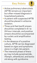
Active pulmonary tuberculosis (APTB) remains an important but often missed diagnosis in hospitalized patients in the Western world.1,2 Because of its relative rarity, the diagnosis of APTB often is delayed in the United States, which can lead hospitalized patients to nosocomial transmission, unnecessary exposures, patient harm,3 and potentially avoidable cost to the health care system.4 The diagnosis and management can be challenging involving isolation needs, sputum clearance, treatment strategy, and criteria for discharge to home.
Diagnosis
Any patient with risk factors presenting with signs and/or symptoms of APTB such as productive cough for more than 4 weeks, night sweats, weight loss, low-grade fevers, upper lobe cavitary lesions, or hemoptysis should be suspected. The diagnostic work-up for APTB should always begin with a thorough medical and social history. A chest radiograph or a CT scan should always be obtained. Risk factors for infection and for progression to active pulmonary TB are listed below.
Risk factors for TB infection:
- Close contacts of a person with APTB.
- Health care workers.
- Immigrants from high-burden countries.
- Homeless people.
- Individuals who have been incarcerated.
- International travelers.
- HIV patients.
- Intravenous drug users.
Risk factors for progression to APTB:
- HIV infection.
- Intravenous drug use.
- Silicosis.
- Younger than 5 years of age.
- Immunosuppressed.
All patients with suspected or confirmed APTB who cannot be safely discharged home (see discharge considerations below) should be kept in negative-pressure airborne isolation rooms. Isolation can be discontinued once APTB has been ruled out or the patient is determined to be noninfectious based on three consecutive negative sputum smears.
Although rapid and inexpensive, acid-fast bacilli (AFB) smear microscopy has poor sensitivity (45%-80%, with culture-confirmed APTB cases) and poor positive predictive value (50%-80%) for TB in settings in which nontuberculous mycobacteria are commonly isolated. This makes an AFB smear nondiagnostic in the early diagnosis of APTB. The burden of mycobacteria seen in the sputum smear correlates with infectivity.
To improve sensitivity of testing, it is strongly recommended that three AFB smears be completed in 8- to 24-hour intervals and positive smears be accompanied by nucleic acid amplification (NAA) testing.5 If APTB is suspected, but the patient is unable to expectorate, induced sputum samples should be obtained, and, if unable to induce sputum samples, flexible bronchoscopy sampling should be pursued especially for the high-risk populations described above.
The Centers for Disease Control and Prevention recommends that at least one sputum specimen be tested with NAA to expedite the time to diagnosis of APTB. A negative NAA does NOT rule out TB. The turnaround time for this test is about 24-48 hours. NAA has better positive predictive value (greater than 95%) with AFB smear-positive specimens in settings in which nontuberculous mycobacteria are common. The ability to confirm rapidly the presence of Mycobacterium tuberculosis is 50%-80% in AFB smear-negative, culture-positive specimens.6
In patients with clinical or radiologic suspicion of APTB who are unable to produce sputum or have negative sputum smear microscopy results, bronchoscopy is a safe and reliable method for the diagnosis of pulmonary tuberculosis. For the diagnosis of tuberculosis, bronchoalveolar lavage has a sensitivity and specificity of 60% and 100%, respectively. Adding transbronchial biopsy further increases the sensitivity to 84%, and post-bronchoscopy sputum smear microscopy increases the sensitivity to 94%.6
In 2005, the CDC released guidelines for using interferon-gamma release assays (IGRA) to test for M. tuberculosis infection.7 Both tuberculin skin testing (TST) and IGRAs assess lymphocytes’ response to M. tuberculosis. Although these tests can be supportive of a previous tuberculosis infection, they are not diagnostic tests for APTB. Neither an IGRA nor a TST can distinguish latent from active tuberculosis.
Sputum AFB culture remains the preferred method for laboratory confirmation of APTB. Once APTB is confirmed, it is essential for susceptibility testing and genotyping. However, in the absence of a positive culture, APTB can be diagnosed based on signs and symptoms alone in a high-risk patient.
Treatment
A multidisciplinary, patient-centered approach involving the patient, providers, and public health officials is required to accomplish the following treatment goals: eradicating Mycobacterium infection, eliminating the risk of transmission, avoiding the disease, and preventing drug resistance.8
Infectious disease consultation is mandatory in all HIV-positive and suspected or confirmed multidrug-resistant cases. Directly observed therapy is an essential component of APTB treatment to ensure compliance in many situations.
Admission and discharge
Admission to a hospital is not required unless a patient meets criteria for admission independent of APTB diagnosis, or proper risk stratification and assessment cannot be completed in a timely manner. A patient with suspected APTB should be placed in airborne isolation. All staff should wear N95 disposable masks or respirators while inside the patient’s room.9
Discharge considerations are listed below:
- Inform the department of health (DOH).
- Establish proper isolation precautions to minimize exposure.
- Ensure ability to stay at home until DOH and physician determines noninfectivity.
- Educate the patient about length of therapy, directly observed therapy, side effects and importance of compliance.
- Coordinate discharge with the DOH.
- Make sure proper follow-up is scheduled.
Back to the case
Our patient was placed on airborne respiratory isolation immediately upon admission and sputum was sent for AFB. Sputum smear was positive for AFB as well as a positive nucleic acid testing for Mycobacterium tuberculosis. HIV antibody testing was negative. Once the sputum AFB was determined to be positive, the department of health was informed. He was started on the intensive phase of therapy with pyrazinamide, rifampin, ethambutol, and isoniazid along with pyridoxine. He tolerated his medications well and had no immediate reactions. His family and close contacts were screened and advised to be tested.
The patient was discharged after proper follow-up with primary care doctor was scheduled. The department of health arranged for directly observed therapy. He received information about the importance of taking all of his medications and staying at home except for medical visits until the DOH had deemed him to be noninfectious.
Bottom line
APTB in the hospital is an uncommon but serious problem in certain populations. It requires a high index of suspicion and a multidisciplinary approach for effective treatment and prevention of transmission.
Dr. Mallampalli is an attending physician in hospital medicine at Geisinger in Danville, Pa., and clinical assistant professor at Temple University, Philadelphia. Dr. Velidi is an attending physician in hospital medicine at Geisinger. Dr. Courtney is associate director of the department of hospital medicine at Geisinger.
References
1. Miller AC et al. Missed opportunities to diagnose tuberculosis are common among hospitalized patients and patients seen in emergency departments. Open Forum Infectious Diseases. 2015. doi. org/10.1093/ofid/ofv171.
2. Greenaway C et al and the Canadian Collaborative Group in Nosocomial Transmission of Tuberculosis. Delay in diagnosis among hospitalized patients with active tuberculosis–predictors and outcomes. Am J Respir Crit Care Med. 2002 Apr;165:927-33.
3. Medrano BA et al. A missed tuberculosis diagnosis resulting in hospital transmission. Infect Control Hosp Epidemiol. 2014 May;35(5):534-7.
4. Kelly AM et al. Delayed tuberculosis diagnosis and costs of contact investigations for hospital exposure: New York City, 2010-2014. Am J Infect Control. 2017 May 1;45(5):483-6.
5. Lewinsohn DM et al. Official ATS/IDSA/CDC Clinical Practice Guidelines: Diagnosis of tuberculosis in adults and children. Clin Infect Dis. 2017 Jan;64:111-5.
6. Mazurek GH et al. Guidelines for using the QuantiFERON-TB Gold test for detecting Mycobacterium tuberculosis infection, United States. MMWR Recomm Rep. 2005 Dec 16;54(RR-15):49-55.
7. CDC. Trends in tuberculosis – United States, 2010. MMWR Morb Mortal Wkly Rep. 2011 Mar 25;60(11):333-7.
8. Jensen PA et al. Guidelines for preventing the transmission of Mycobacterium tuberculosis in health-care settings, 2005. MMWR Recomm Rep. 2005 Dec 30;54(RR-17):1-141.
9. Siegel JD et al and the Health Care Infection Control Practices Advisory Committee. 2007 Guideline for isolation precautions: Preventing transmission of infectious agents in health care settings. Am J Infect Control. 2007 Dec;35(10 Suppl 2):S65-164.
An uncommon but serious problem in certain populations
An uncommon but serious problem in certain populations
Case
A 40-year-old Indian immigrant presented to the emergency department with hemoptysis. He had had an intermittent productive cough for the past 4 weeks with increasing fatigue and lack of appetite. He also had intermittent fever with drenching night sweats. Chest radiograph and CT scan showed a left upper lobe cavitary lesion with infiltrate. He was admitted to the hospital with concern for pneumonia and to rule out possible active pulmonary tuberculosis.
Background
Active pulmonary tuberculosis (APTB) remains an important but often missed diagnosis in hospitalized patients in the Western world.1,2 Because of its relative rarity, the diagnosis of APTB often is delayed in the United States, which can lead hospitalized patients to nosocomial transmission, unnecessary exposures, patient harm,3 and potentially avoidable cost to the health care system.4 The diagnosis and management can be challenging involving isolation needs, sputum clearance, treatment strategy, and criteria for discharge to home.
Diagnosis
Any patient with risk factors presenting with signs and/or symptoms of APTB such as productive cough for more than 4 weeks, night sweats, weight loss, low-grade fevers, upper lobe cavitary lesions, or hemoptysis should be suspected. The diagnostic work-up for APTB should always begin with a thorough medical and social history. A chest radiograph or a CT scan should always be obtained. Risk factors for infection and for progression to active pulmonary TB are listed below.
Risk factors for TB infection:
- Close contacts of a person with APTB.
- Health care workers.
- Immigrants from high-burden countries.
- Homeless people.
- Individuals who have been incarcerated.
- International travelers.
- HIV patients.
- Intravenous drug users.
Risk factors for progression to APTB:
- HIV infection.
- Intravenous drug use.
- Silicosis.
- Younger than 5 years of age.
- Immunosuppressed.
All patients with suspected or confirmed APTB who cannot be safely discharged home (see discharge considerations below) should be kept in negative-pressure airborne isolation rooms. Isolation can be discontinued once APTB has been ruled out or the patient is determined to be noninfectious based on three consecutive negative sputum smears.
Although rapid and inexpensive, acid-fast bacilli (AFB) smear microscopy has poor sensitivity (45%-80%, with culture-confirmed APTB cases) and poor positive predictive value (50%-80%) for TB in settings in which nontuberculous mycobacteria are commonly isolated. This makes an AFB smear nondiagnostic in the early diagnosis of APTB. The burden of mycobacteria seen in the sputum smear correlates with infectivity.
To improve sensitivity of testing, it is strongly recommended that three AFB smears be completed in 8- to 24-hour intervals and positive smears be accompanied by nucleic acid amplification (NAA) testing.5 If APTB is suspected, but the patient is unable to expectorate, induced sputum samples should be obtained, and, if unable to induce sputum samples, flexible bronchoscopy sampling should be pursued especially for the high-risk populations described above.
The Centers for Disease Control and Prevention recommends that at least one sputum specimen be tested with NAA to expedite the time to diagnosis of APTB. A negative NAA does NOT rule out TB. The turnaround time for this test is about 24-48 hours. NAA has better positive predictive value (greater than 95%) with AFB smear-positive specimens in settings in which nontuberculous mycobacteria are common. The ability to confirm rapidly the presence of Mycobacterium tuberculosis is 50%-80% in AFB smear-negative, culture-positive specimens.6
In patients with clinical or radiologic suspicion of APTB who are unable to produce sputum or have negative sputum smear microscopy results, bronchoscopy is a safe and reliable method for the diagnosis of pulmonary tuberculosis. For the diagnosis of tuberculosis, bronchoalveolar lavage has a sensitivity and specificity of 60% and 100%, respectively. Adding transbronchial biopsy further increases the sensitivity to 84%, and post-bronchoscopy sputum smear microscopy increases the sensitivity to 94%.6
In 2005, the CDC released guidelines for using interferon-gamma release assays (IGRA) to test for M. tuberculosis infection.7 Both tuberculin skin testing (TST) and IGRAs assess lymphocytes’ response to M. tuberculosis. Although these tests can be supportive of a previous tuberculosis infection, they are not diagnostic tests for APTB. Neither an IGRA nor a TST can distinguish latent from active tuberculosis.
Sputum AFB culture remains the preferred method for laboratory confirmation of APTB. Once APTB is confirmed, it is essential for susceptibility testing and genotyping. However, in the absence of a positive culture, APTB can be diagnosed based on signs and symptoms alone in a high-risk patient.
Treatment
A multidisciplinary, patient-centered approach involving the patient, providers, and public health officials is required to accomplish the following treatment goals: eradicating Mycobacterium infection, eliminating the risk of transmission, avoiding the disease, and preventing drug resistance.8
Infectious disease consultation is mandatory in all HIV-positive and suspected or confirmed multidrug-resistant cases. Directly observed therapy is an essential component of APTB treatment to ensure compliance in many situations.
Admission and discharge
Admission to a hospital is not required unless a patient meets criteria for admission independent of APTB diagnosis, or proper risk stratification and assessment cannot be completed in a timely manner. A patient with suspected APTB should be placed in airborne isolation. All staff should wear N95 disposable masks or respirators while inside the patient’s room.9
Discharge considerations are listed below:
- Inform the department of health (DOH).
- Establish proper isolation precautions to minimize exposure.
- Ensure ability to stay at home until DOH and physician determines noninfectivity.
- Educate the patient about length of therapy, directly observed therapy, side effects and importance of compliance.
- Coordinate discharge with the DOH.
- Make sure proper follow-up is scheduled.
Back to the case
Our patient was placed on airborne respiratory isolation immediately upon admission and sputum was sent for AFB. Sputum smear was positive for AFB as well as a positive nucleic acid testing for Mycobacterium tuberculosis. HIV antibody testing was negative. Once the sputum AFB was determined to be positive, the department of health was informed. He was started on the intensive phase of therapy with pyrazinamide, rifampin, ethambutol, and isoniazid along with pyridoxine. He tolerated his medications well and had no immediate reactions. His family and close contacts were screened and advised to be tested.
The patient was discharged after proper follow-up with primary care doctor was scheduled. The department of health arranged for directly observed therapy. He received information about the importance of taking all of his medications and staying at home except for medical visits until the DOH had deemed him to be noninfectious.
Bottom line
APTB in the hospital is an uncommon but serious problem in certain populations. It requires a high index of suspicion and a multidisciplinary approach for effective treatment and prevention of transmission.
Dr. Mallampalli is an attending physician in hospital medicine at Geisinger in Danville, Pa., and clinical assistant professor at Temple University, Philadelphia. Dr. Velidi is an attending physician in hospital medicine at Geisinger. Dr. Courtney is associate director of the department of hospital medicine at Geisinger.
References
1. Miller AC et al. Missed opportunities to diagnose tuberculosis are common among hospitalized patients and patients seen in emergency departments. Open Forum Infectious Diseases. 2015. doi. org/10.1093/ofid/ofv171.
2. Greenaway C et al and the Canadian Collaborative Group in Nosocomial Transmission of Tuberculosis. Delay in diagnosis among hospitalized patients with active tuberculosis–predictors and outcomes. Am J Respir Crit Care Med. 2002 Apr;165:927-33.
3. Medrano BA et al. A missed tuberculosis diagnosis resulting in hospital transmission. Infect Control Hosp Epidemiol. 2014 May;35(5):534-7.
4. Kelly AM et al. Delayed tuberculosis diagnosis and costs of contact investigations for hospital exposure: New York City, 2010-2014. Am J Infect Control. 2017 May 1;45(5):483-6.
5. Lewinsohn DM et al. Official ATS/IDSA/CDC Clinical Practice Guidelines: Diagnosis of tuberculosis in adults and children. Clin Infect Dis. 2017 Jan;64:111-5.
6. Mazurek GH et al. Guidelines for using the QuantiFERON-TB Gold test for detecting Mycobacterium tuberculosis infection, United States. MMWR Recomm Rep. 2005 Dec 16;54(RR-15):49-55.
7. CDC. Trends in tuberculosis – United States, 2010. MMWR Morb Mortal Wkly Rep. 2011 Mar 25;60(11):333-7.
8. Jensen PA et al. Guidelines for preventing the transmission of Mycobacterium tuberculosis in health-care settings, 2005. MMWR Recomm Rep. 2005 Dec 30;54(RR-17):1-141.
9. Siegel JD et al and the Health Care Infection Control Practices Advisory Committee. 2007 Guideline for isolation precautions: Preventing transmission of infectious agents in health care settings. Am J Infect Control. 2007 Dec;35(10 Suppl 2):S65-164.
hs-cTnT test tied to fewer reinfarctions but no survival benefit
The introduction of the new high-sensitivity cardiac troponin T (hs-cTnT) test was linked with an 11% decrease in reinfarctions but showed no evidence of improving survival in a large longitudinal cohort study.
Over an average of 3.9 years of follow-up, the adjusted risk of all-cause mortality was identical whether patients’ initial MI was diagnosed with the new troponin test or the conventional one, reported Maria Odqvist, MD, of South Alvsborg Hospital, Boras, Sweden, and her associates. “As hs-cTn assays become widely available and clinicians gain experience interpreting the results, more work is needed to enhance clinical reasoning and implementation to improve patient outcomes,” the researchers wrote in the Journal of the American College of Cardiology.
High-sensitivity cardiac troponin T assays detect acute coronary syndrome more rapidly than their conventional predecessors. However, hs-cTnT’s higher sensitivity comes with lost specificity and hence has raised concerns about potentially wasting health care resources. Some clinicians have asked whether the new test is worthwhile and how to maximize its benefits.
The study, which included nearly 88,000 patients with initial MI from the Swedish National Patient Registry, spanned 2009-2013, when 73% of Swedish acute care hospitals transitioned from conventional troponin tests (cTn) to hs-cTnT. After adjustment for factors such as age, sex, location, chronic kidney disease, cardiovascular history, and prescriptions, Dr. Odqvist and her associates compared each test types’ risks of death, reinfarction, coronary angiography, and revascularization among patients diagnosed.
In all, 47,133 (54%) patients’ initial MI was diagnosed with cTn while 46% were diagnosed with hs-cTnT. Overall, the rate of MI rose by 5% during the 90 days after hospitals implemented hs-cTnT, compared with the 90 days before. But hospitals used varying hs-cTnT thresholds for MI, which might explain why some hospitals initially observed a marked initial decrease in MI while others saw a relatively large increase, the investigators wrote.
There were 15,766 reinfarctions over an average of 3.1 years of follow-up. Although there was no difference in all-cause mortality, risk of reinfarction was 11% lower among patients diagnosed using hs-cTnT, compared with those diagnosed by cTn.
At the hospital level, coronary angiography and revascularization became only slightly more common during the 3 months after hospitals switched to hs-cTnT, the researchers wrote. However, patients whose MIs were diagnosed with hs-cTnT were 16% more likely to undergo coronary angiography and 13% more likely to undergo revascularization within the next 30 days, compared with patients diagnosed with cTn. Patients diagnosed with hs-cTnT also were more likely to receive statins, but trends in other prescriptions did not change.
The Swedish Heart-Lung Foundation funded one investigator. Dr. Odqvist and one coinvestigator reported having no relevant conflicts of interest. Senior author Martin J. Holzmann, MD, PhD, disclosed ties to Actelion and Pfizer. Two other coinvestigators disclosed ties to Roche, Gilead, Janssen, Abbvie, CTI Bipharma, GlaxoSmithKline, Abbott Laboratories, and AstraZeneca, and Fiomi Diagnostics.
SOURCE: Odqvist M et al. J Am Coll Cardiol. 2018 Jun 12;71(23):2616-24
It’s unclear why reinfarctions were 11% lower after MI was diagnosed with high-sensitivity cardiac troponin (hs-cTn) assays instead of conventional troponin tests in this study.
Use of evidence-based medicines changed only slightly after hospitals implemented the switch. It is tempting to associate the reduction in reinfarction with increased use of angiography and revascularization, but this may be too simplistic. Intervention might have most benefited patients whom conventional troponin assay would have missed. Shifts in medication compliance or rehabilitation might also have been factors.
Used appropriately, hs-cTnT assays can more rapidly rule MI in or out, shorten emergency department stays, and cut costs. Based on the Swedish experience, the increase in MI diagnoses will be modest and manageable. Nonetheless, we will need to develop strategies to manage patients with myonecrosis from etiologies other than MI that will be detected using hs-cTn assays.
L. Kristin Newby, MD, MHS , and Angela Lowenstern, MD , of Duke University in Durham, N.C., made these comments in an accompanying editorial (J Am Coll Cardiol. 2018;71:625-7). Dr. Newby has received consulting honoraria from Roche Diagnostics, Ortho-Clinical Diagnostics, and Philips Healthcare. Dr. Lowenstern is supported by a National Institutes of Health training grant to Duke University .
The introduction of the new high-sensitivity cardiac troponin T (hs-cTnT) test was linked with an 11% decrease in reinfarctions but showed no evidence of improving survival in a large longitudinal cohort study.
Over an average of 3.9 years of follow-up, the adjusted risk of all-cause mortality was identical whether patients’ initial MI was diagnosed with the new troponin test or the conventional one, reported Maria Odqvist, MD, of South Alvsborg Hospital, Boras, Sweden, and her associates. “As hs-cTn assays become widely available and clinicians gain experience interpreting the results, more work is needed to enhance clinical reasoning and implementation to improve patient outcomes,” the researchers wrote in the Journal of the American College of Cardiology.
High-sensitivity cardiac troponin T assays detect acute coronary syndrome more rapidly than their conventional predecessors. However, hs-cTnT’s higher sensitivity comes with lost specificity and hence has raised concerns about potentially wasting health care resources. Some clinicians have asked whether the new test is worthwhile and how to maximize its benefits.
The study, which included nearly 88,000 patients with initial MI from the Swedish National Patient Registry, spanned 2009-2013, when 73% of Swedish acute care hospitals transitioned from conventional troponin tests (cTn) to hs-cTnT. After adjustment for factors such as age, sex, location, chronic kidney disease, cardiovascular history, and prescriptions, Dr. Odqvist and her associates compared each test types’ risks of death, reinfarction, coronary angiography, and revascularization among patients diagnosed.
In all, 47,133 (54%) patients’ initial MI was diagnosed with cTn while 46% were diagnosed with hs-cTnT. Overall, the rate of MI rose by 5% during the 90 days after hospitals implemented hs-cTnT, compared with the 90 days before. But hospitals used varying hs-cTnT thresholds for MI, which might explain why some hospitals initially observed a marked initial decrease in MI while others saw a relatively large increase, the investigators wrote.
There were 15,766 reinfarctions over an average of 3.1 years of follow-up. Although there was no difference in all-cause mortality, risk of reinfarction was 11% lower among patients diagnosed using hs-cTnT, compared with those diagnosed by cTn.
At the hospital level, coronary angiography and revascularization became only slightly more common during the 3 months after hospitals switched to hs-cTnT, the researchers wrote. However, patients whose MIs were diagnosed with hs-cTnT were 16% more likely to undergo coronary angiography and 13% more likely to undergo revascularization within the next 30 days, compared with patients diagnosed with cTn. Patients diagnosed with hs-cTnT also were more likely to receive statins, but trends in other prescriptions did not change.
The Swedish Heart-Lung Foundation funded one investigator. Dr. Odqvist and one coinvestigator reported having no relevant conflicts of interest. Senior author Martin J. Holzmann, MD, PhD, disclosed ties to Actelion and Pfizer. Two other coinvestigators disclosed ties to Roche, Gilead, Janssen, Abbvie, CTI Bipharma, GlaxoSmithKline, Abbott Laboratories, and AstraZeneca, and Fiomi Diagnostics.
SOURCE: Odqvist M et al. J Am Coll Cardiol. 2018 Jun 12;71(23):2616-24
FROM JACC
Key clinical point: The introduction of the new high-sensitivity cardiac troponin T test was associated with an 11% decrease in reinfarctions but showed no evidence of improving survival.
Major finding: Over an average of 3.9 years of follow-up, the adjusted risk of all-cause mortality was identical whether patients’ initial MI was diagnosed with the new troponin test or with the conventional one.
Study details: National cohort study of 87,879 patients with first myocardial infarction, 2009-2013.
Disclosures: Dr. Odqvist and one coinvestigator had no relevant disclosures. Senior author Martin J. Holzmann, MD, PhD, disclosed ties to Actelion and Pfizer. Two other coinvestigators disclosed ties to Roche, Gilead, Janssen, AbbVie, CTI Biopharma, GlaxoSmithKline, Abbott Laboratories, and AstraZeneca, and Fiomi Diagnostics.
Source: Odqvist M et al. J Am Coll Cardiol. 2018 Jun 12;71(23):2616-24.


