User login
Thymectomy improves clinical outcomes for myasthenia gravis
Thymectomy improved 3-year clinical outcomes and proved superior to medical therapy for mild to severe nonthymomatous myasthenia gravis, according to a report published online Aug. 11 in the New England Journal of Medicine.
Compared with standard prednisone therapy, thymectomy plus prednisone decreased the number and severity of symptoms, allowed the lowering of steroid doses, decreased the number and length of hospitalizations for disease exacerbations, reduced the need for immunosuppressive agents, and improved health-related quality of life in an international, randomized clinical trial, said Gil I. Wolfe, MD, of the department of neurology, State University of New York at Buffalo and his associates.
Until now, thymectomy was known to be beneficial in some cases of myasthenia gravis “but with widely varying rates of clinical improvement or remission.” And the success of immunotherapy has raised the question of whether an invasive surgery is necessary. Data from randomized, controlled studies have been sparse.
Moreover, thymectomy rarely causes adverse effects, but “the procedure can cost up to $80,000 and can be associated with operative complications that need to be weighed against benefits.” In comparison, medical therapy with glucocorticoids and other immunosuppressive agents is less invasive but is definitely associated with adverse events, including some that are life threatening, and negatively impacts quality of life, the investigators said.
To address the lack of randomized controlled trial data, they assessed 3-year outcomes in 126 patients treated at 67 medical centers in 18 countries during a 6-year period. The study participants were aged 18-65 years, had a disease duration of less than 5 years at enrollment (median duration, 1 year), and had class II (mild generalized disease) to class IV (severe generalized disease) myasthenia gravis. These patients were randomly assigned to undergo thymectomy and receive standard prednisone therapy (66 participants) or to receive standard prednisone alone (60 participants).
Thymectomy was performed using a median sternotomy “with the goal of an en bloc resection of all mediastinal tissue that could anatomically contain gross or microscopic thymus.”
At follow-up, time-weighted average scores on the Quantitative Myasthenia Gravis scale were significantly lower by 2.85 points, indicating improved clinical status, in the thymectomy group than in the control group. Time-weighted average prednisone dose also was significantly lower, at an average alternate-day dose of 44 mg in the thymectomy group and 60 mg in the control group, Dr. Wolfe and his associates said (N Engl J Med. 2016 Aug 11. doi: 10.1056/NEJMoa1602489).
On a measure of treatment-related complications, scores favored thymectomy with regard to the number of patients with symptoms, the total number of symptoms, and the distress level related to symptoms throughout the study period. Fewer patients in the thymectomy group required hospitalization for exacerbations of myasthenia gravis (9% vs. 37%), and the mean cumulative number of hospital days was lower with thymectomy (8.4 vs. 19.2).
In addition, scores on the Myasthenia Gravis Activities of Daily Living scale favored thymectomy (2.24 vs. 3.41). Fewer patients in the thymectomy group required azathioprine (17% vs. 0.48%). And the percentage of patients who reported having minimal manifestations of the disease at 3 years was significantly higher with thymectomy (67%) than with prednisone alone (47%).
This study was supported by the National Institute of Neurological Disorders and Stroke, the Muscular Dystrophy Association, and the Myasthenia Gravis Foundation of America and received no commercial support. Dr. Wolfe reported ties to Alexion Pharmaceuticals, Alpha Cancer Technologies, Argenx, Baxalta, CSL Behring, Grifols, and UCB, and his associates reported ties to numerous industry sources.
Landmark trial establishes effectiveness of thymectomy in myasthenia gravis
One of the many challenges of treating patients with myasthenia gravis (MG) is the fluctuating nature of symptoms and deficits. The neurologist or neuromuscular specialist must decide whether the disease is truly worsening, whether the patient is experiencing more pronounced symptoms from intercurrent illness or the effects of a medication known to affect the neuromuscular junction adversely, or whether the patient is concerned that there might be worsening disease when all objective measures indicate stability. These factors make treatment decisions more difficult in MG than for many other neuromuscular disorders.
Similarly, researchers considering a trial investigating treatment efficacy in MG face the complex issues of disease fluctuation in cohorts of individuals with the disease, varying levels of corticosteroid and immunosuppressant doses in different MG patients, and thorny ethical dilemmas in providing accepted therapies but not withholding effective treatments from those in need.
Dr. Wolfe and his colleagues demonstrate that they have navigated these treacherous waters. They have succeeded in completing a landmark controlled clinical trial which establishes the effectiveness of transsternal thymectomy with adjuvant corticosteroid therapy in nonthymomatous MG vs. oral prednisone without surgery. While this international 36-center trial managed to recruit 126 subjects over a 6-year period, using sound inclusion and exclusion criteria and a meticulous trial design, the number of patients is not sufficient to allow for as robust a subgroup analysis for age, gender, and a variety of clinical variables reflecting severity of disease as would have been hoped for by the MG community.
Nonetheless, this paper sets the use of thymectomy in nonthymomatous MG on firmer ground going forward. The investigators will doubtless be presenting further data from the trial, including clinical-pathologic correlates and other relevant novel observations. In addition, Wolfe et al. have opened the door for future trials of thymectomy in MG to address such issues as the benefits vs. risks of performing the operation via the traditional transsternal vs. alternative non–sternal splitting approaches.
Benn E. Smith, MD, is an associate professor of neurology at the Mayo Clinic in Scottsdale, Ariz. and is the director of the sensory laboratory there. Dr. Smith is on the Editorial Advisory Board of Clinical Neurology News.
End to an 80-year controversy
These findings from Wolfe et al. end an 80-year controversy over the effectiveness of thymectomy for patients with myasthenia gravis.
Perhaps the most important benefit for patients is that even when they require prednisone following the surgery, they can take lower doses, endure fewer glucocorticoid-related symptoms, and experience less distress from those symptoms than patients who don’t undergo thymectomy.
Unfortunately, the study results cannot offer further clarity regarding patient selection for thymectomy. The patient population in this trial was so small that subgroup analyses couldn’t allow conclusions regarding the relative effectiveness of thymectomy in men vs. women or younger vs. older patients.
Allan H. Ropper, MD, is in the department of neurology at Brigham and Women’s Hospital and Harvard Medical School, both in Boston. His financial disclosures are available at NEJM.org. Dr. Ropper made these remarks in an editorial accompanying Dr. Wolfe’s report (N Engl J Med. 2016 Aug 11. doi: 10.1056/NEJMe1607953).
Landmark trial establishes effectiveness of thymectomy in myasthenia gravis
One of the many challenges of treating patients with myasthenia gravis (MG) is the fluctuating nature of symptoms and deficits. The neurologist or neuromuscular specialist must decide whether the disease is truly worsening, whether the patient is experiencing more pronounced symptoms from intercurrent illness or the effects of a medication known to affect the neuromuscular junction adversely, or whether the patient is concerned that there might be worsening disease when all objective measures indicate stability. These factors make treatment decisions more difficult in MG than for many other neuromuscular disorders.
Similarly, researchers considering a trial investigating treatment efficacy in MG face the complex issues of disease fluctuation in cohorts of individuals with the disease, varying levels of corticosteroid and immunosuppressant doses in different MG patients, and thorny ethical dilemmas in providing accepted therapies but not withholding effective treatments from those in need.
Dr. Wolfe and his colleagues demonstrate that they have navigated these treacherous waters. They have succeeded in completing a landmark controlled clinical trial which establishes the effectiveness of transsternal thymectomy with adjuvant corticosteroid therapy in nonthymomatous MG vs. oral prednisone without surgery. While this international 36-center trial managed to recruit 126 subjects over a 6-year period, using sound inclusion and exclusion criteria and a meticulous trial design, the number of patients is not sufficient to allow for as robust a subgroup analysis for age, gender, and a variety of clinical variables reflecting severity of disease as would have been hoped for by the MG community.
Nonetheless, this paper sets the use of thymectomy in nonthymomatous MG on firmer ground going forward. The investigators will doubtless be presenting further data from the trial, including clinical-pathologic correlates and other relevant novel observations. In addition, Wolfe et al. have opened the door for future trials of thymectomy in MG to address such issues as the benefits vs. risks of performing the operation via the traditional transsternal vs. alternative non–sternal splitting approaches.
Benn E. Smith, MD, is an associate professor of neurology at the Mayo Clinic in Scottsdale, Ariz. and is the director of the sensory laboratory there. Dr. Smith is on the Editorial Advisory Board of Clinical Neurology News.
End to an 80-year controversy
These findings from Wolfe et al. end an 80-year controversy over the effectiveness of thymectomy for patients with myasthenia gravis.
Perhaps the most important benefit for patients is that even when they require prednisone following the surgery, they can take lower doses, endure fewer glucocorticoid-related symptoms, and experience less distress from those symptoms than patients who don’t undergo thymectomy.
Unfortunately, the study results cannot offer further clarity regarding patient selection for thymectomy. The patient population in this trial was so small that subgroup analyses couldn’t allow conclusions regarding the relative effectiveness of thymectomy in men vs. women or younger vs. older patients.
Allan H. Ropper, MD, is in the department of neurology at Brigham and Women’s Hospital and Harvard Medical School, both in Boston. His financial disclosures are available at NEJM.org. Dr. Ropper made these remarks in an editorial accompanying Dr. Wolfe’s report (N Engl J Med. 2016 Aug 11. doi: 10.1056/NEJMe1607953).
Landmark trial establishes effectiveness of thymectomy in myasthenia gravis
One of the many challenges of treating patients with myasthenia gravis (MG) is the fluctuating nature of symptoms and deficits. The neurologist or neuromuscular specialist must decide whether the disease is truly worsening, whether the patient is experiencing more pronounced symptoms from intercurrent illness or the effects of a medication known to affect the neuromuscular junction adversely, or whether the patient is concerned that there might be worsening disease when all objective measures indicate stability. These factors make treatment decisions more difficult in MG than for many other neuromuscular disorders.
Similarly, researchers considering a trial investigating treatment efficacy in MG face the complex issues of disease fluctuation in cohorts of individuals with the disease, varying levels of corticosteroid and immunosuppressant doses in different MG patients, and thorny ethical dilemmas in providing accepted therapies but not withholding effective treatments from those in need.
Dr. Wolfe and his colleagues demonstrate that they have navigated these treacherous waters. They have succeeded in completing a landmark controlled clinical trial which establishes the effectiveness of transsternal thymectomy with adjuvant corticosteroid therapy in nonthymomatous MG vs. oral prednisone without surgery. While this international 36-center trial managed to recruit 126 subjects over a 6-year period, using sound inclusion and exclusion criteria and a meticulous trial design, the number of patients is not sufficient to allow for as robust a subgroup analysis for age, gender, and a variety of clinical variables reflecting severity of disease as would have been hoped for by the MG community.
Nonetheless, this paper sets the use of thymectomy in nonthymomatous MG on firmer ground going forward. The investigators will doubtless be presenting further data from the trial, including clinical-pathologic correlates and other relevant novel observations. In addition, Wolfe et al. have opened the door for future trials of thymectomy in MG to address such issues as the benefits vs. risks of performing the operation via the traditional transsternal vs. alternative non–sternal splitting approaches.
Benn E. Smith, MD, is an associate professor of neurology at the Mayo Clinic in Scottsdale, Ariz. and is the director of the sensory laboratory there. Dr. Smith is on the Editorial Advisory Board of Clinical Neurology News.
End to an 80-year controversy
These findings from Wolfe et al. end an 80-year controversy over the effectiveness of thymectomy for patients with myasthenia gravis.
Perhaps the most important benefit for patients is that even when they require prednisone following the surgery, they can take lower doses, endure fewer glucocorticoid-related symptoms, and experience less distress from those symptoms than patients who don’t undergo thymectomy.
Unfortunately, the study results cannot offer further clarity regarding patient selection for thymectomy. The patient population in this trial was so small that subgroup analyses couldn’t allow conclusions regarding the relative effectiveness of thymectomy in men vs. women or younger vs. older patients.
Allan H. Ropper, MD, is in the department of neurology at Brigham and Women’s Hospital and Harvard Medical School, both in Boston. His financial disclosures are available at NEJM.org. Dr. Ropper made these remarks in an editorial accompanying Dr. Wolfe’s report (N Engl J Med. 2016 Aug 11. doi: 10.1056/NEJMe1607953).
Thymectomy improved 3-year clinical outcomes and proved superior to medical therapy for mild to severe nonthymomatous myasthenia gravis, according to a report published online Aug. 11 in the New England Journal of Medicine.
Compared with standard prednisone therapy, thymectomy plus prednisone decreased the number and severity of symptoms, allowed the lowering of steroid doses, decreased the number and length of hospitalizations for disease exacerbations, reduced the need for immunosuppressive agents, and improved health-related quality of life in an international, randomized clinical trial, said Gil I. Wolfe, MD, of the department of neurology, State University of New York at Buffalo and his associates.
Until now, thymectomy was known to be beneficial in some cases of myasthenia gravis “but with widely varying rates of clinical improvement or remission.” And the success of immunotherapy has raised the question of whether an invasive surgery is necessary. Data from randomized, controlled studies have been sparse.
Moreover, thymectomy rarely causes adverse effects, but “the procedure can cost up to $80,000 and can be associated with operative complications that need to be weighed against benefits.” In comparison, medical therapy with glucocorticoids and other immunosuppressive agents is less invasive but is definitely associated with adverse events, including some that are life threatening, and negatively impacts quality of life, the investigators said.
To address the lack of randomized controlled trial data, they assessed 3-year outcomes in 126 patients treated at 67 medical centers in 18 countries during a 6-year period. The study participants were aged 18-65 years, had a disease duration of less than 5 years at enrollment (median duration, 1 year), and had class II (mild generalized disease) to class IV (severe generalized disease) myasthenia gravis. These patients were randomly assigned to undergo thymectomy and receive standard prednisone therapy (66 participants) or to receive standard prednisone alone (60 participants).
Thymectomy was performed using a median sternotomy “with the goal of an en bloc resection of all mediastinal tissue that could anatomically contain gross or microscopic thymus.”
At follow-up, time-weighted average scores on the Quantitative Myasthenia Gravis scale were significantly lower by 2.85 points, indicating improved clinical status, in the thymectomy group than in the control group. Time-weighted average prednisone dose also was significantly lower, at an average alternate-day dose of 44 mg in the thymectomy group and 60 mg in the control group, Dr. Wolfe and his associates said (N Engl J Med. 2016 Aug 11. doi: 10.1056/NEJMoa1602489).
On a measure of treatment-related complications, scores favored thymectomy with regard to the number of patients with symptoms, the total number of symptoms, and the distress level related to symptoms throughout the study period. Fewer patients in the thymectomy group required hospitalization for exacerbations of myasthenia gravis (9% vs. 37%), and the mean cumulative number of hospital days was lower with thymectomy (8.4 vs. 19.2).
In addition, scores on the Myasthenia Gravis Activities of Daily Living scale favored thymectomy (2.24 vs. 3.41). Fewer patients in the thymectomy group required azathioprine (17% vs. 0.48%). And the percentage of patients who reported having minimal manifestations of the disease at 3 years was significantly higher with thymectomy (67%) than with prednisone alone (47%).
This study was supported by the National Institute of Neurological Disorders and Stroke, the Muscular Dystrophy Association, and the Myasthenia Gravis Foundation of America and received no commercial support. Dr. Wolfe reported ties to Alexion Pharmaceuticals, Alpha Cancer Technologies, Argenx, Baxalta, CSL Behring, Grifols, and UCB, and his associates reported ties to numerous industry sources.
Thymectomy improved 3-year clinical outcomes and proved superior to medical therapy for mild to severe nonthymomatous myasthenia gravis, according to a report published online Aug. 11 in the New England Journal of Medicine.
Compared with standard prednisone therapy, thymectomy plus prednisone decreased the number and severity of symptoms, allowed the lowering of steroid doses, decreased the number and length of hospitalizations for disease exacerbations, reduced the need for immunosuppressive agents, and improved health-related quality of life in an international, randomized clinical trial, said Gil I. Wolfe, MD, of the department of neurology, State University of New York at Buffalo and his associates.
Until now, thymectomy was known to be beneficial in some cases of myasthenia gravis “but with widely varying rates of clinical improvement or remission.” And the success of immunotherapy has raised the question of whether an invasive surgery is necessary. Data from randomized, controlled studies have been sparse.
Moreover, thymectomy rarely causes adverse effects, but “the procedure can cost up to $80,000 and can be associated with operative complications that need to be weighed against benefits.” In comparison, medical therapy with glucocorticoids and other immunosuppressive agents is less invasive but is definitely associated with adverse events, including some that are life threatening, and negatively impacts quality of life, the investigators said.
To address the lack of randomized controlled trial data, they assessed 3-year outcomes in 126 patients treated at 67 medical centers in 18 countries during a 6-year period. The study participants were aged 18-65 years, had a disease duration of less than 5 years at enrollment (median duration, 1 year), and had class II (mild generalized disease) to class IV (severe generalized disease) myasthenia gravis. These patients were randomly assigned to undergo thymectomy and receive standard prednisone therapy (66 participants) or to receive standard prednisone alone (60 participants).
Thymectomy was performed using a median sternotomy “with the goal of an en bloc resection of all mediastinal tissue that could anatomically contain gross or microscopic thymus.”
At follow-up, time-weighted average scores on the Quantitative Myasthenia Gravis scale were significantly lower by 2.85 points, indicating improved clinical status, in the thymectomy group than in the control group. Time-weighted average prednisone dose also was significantly lower, at an average alternate-day dose of 44 mg in the thymectomy group and 60 mg in the control group, Dr. Wolfe and his associates said (N Engl J Med. 2016 Aug 11. doi: 10.1056/NEJMoa1602489).
On a measure of treatment-related complications, scores favored thymectomy with regard to the number of patients with symptoms, the total number of symptoms, and the distress level related to symptoms throughout the study period. Fewer patients in the thymectomy group required hospitalization for exacerbations of myasthenia gravis (9% vs. 37%), and the mean cumulative number of hospital days was lower with thymectomy (8.4 vs. 19.2).
In addition, scores on the Myasthenia Gravis Activities of Daily Living scale favored thymectomy (2.24 vs. 3.41). Fewer patients in the thymectomy group required azathioprine (17% vs. 0.48%). And the percentage of patients who reported having minimal manifestations of the disease at 3 years was significantly higher with thymectomy (67%) than with prednisone alone (47%).
This study was supported by the National Institute of Neurological Disorders and Stroke, the Muscular Dystrophy Association, and the Myasthenia Gravis Foundation of America and received no commercial support. Dr. Wolfe reported ties to Alexion Pharmaceuticals, Alpha Cancer Technologies, Argenx, Baxalta, CSL Behring, Grifols, and UCB, and his associates reported ties to numerous industry sources.
FROM THE NEW ENGLAND JOURNAL OF MEDICINE
Key clinical point: Thymectomy improved 3-year clinical outcomes and was superior to medical therapy for mild to severe nonthymomatous myasthenia gravis.
Major finding: Scores on the Quantitative Myasthenia Gravis scale were significantly lower by 2.85 points, indicating improved clinical status, in the thymectomy group than in the control group.
Data source: An international, randomized, medication-controlled trial involving 126 patients at 67 medical centers.
Disclosures: This study was supported by the National Institute of Neurological Disorders and Stroke, the Muscular Dystrophy Association, and the Myasthenia Gravis Foundation of America and received no commercial support. Dr. Wolfe reported ties to Alexion Pharmaceuticals, Alpha Cancer Technologies, Argenx, Baxalta, CSL Behring, Grifols, and UCB, and his associates reported ties to numerous industry sources.
Making Fillers a Success Through Technique and Patient Education
What does your patient need to know at the first visit?
In my practice, we try our best to start the education process before the patient even comes in for the first visit. If a patient is going to have a filler injection, we mail out an information packet that contains information on what to expect, how to minimize bruising, what side effects (eg, bruising, swelling) may occur, and what to avoid posttreatment. By providing them with this information prior to their visit, they can better plan their treatment around social and work obligations.
We ask patients to avoid or minimize blood-thinning agents such as aspirin, ibuprofen, fish oil, or vitamin E starting 2 weeks prior to and 1 week after the procedure. We recommend that patients take a pineapple extract such as bromelain 500 mg twice daily on an empty stomach starting 1 week prior to the treatment and for up to 1 week posttreatment. We ask that patients avoid exercise for 24 hours to reduce late-onset bruising and to avoid dental work for 2 weeks posttreatment to reduce the risk for the filler becoming infected.
What are your go-to treatments?
I use a variety of fillers depending on the area I am treating (thin vs thick skin) or the amount of lift I need (ie, G')(Restylane [Galderma Laboratories, LP] has a high G', thus it gives a lot of lift) versus the amount of water absorption I am seeking to further plump an area after the filler integrates (Juvéderm [Allergan] can absorb up to 300% its weight in water) versus the filler’s cohesiveness (Belotero [Merz Aesthetics] is highly cohesive). If I am treating a thin-faced individual, I may start with a global volumizer such as poly-L-lactic acid to get a good foundation set in the temples, cheeks, and jawline, and then after a few months, I will add a hyaluronic acid filler to focal areas that still need to be lifted (eg, nasolabial folds, tear troughs).
What are the side effects?
Side effects are divided into common and rare. Common would be bruising and swelling, which are temporary and will go away in all patients. Rare but serious side effects are infection and embolization. Both can cause notable tissue loss and risk to the patient. Every practitioner needs to know how to recognize and treat these complications should they arise.
How do you keep patients compliant?
Patients who get good results will always return, which means being up front about how much filler a patient needs and how frequently he/she will need it, and also doing everything we can to reduce bruising and swelling.
Suggested Readings
- Dayan SH, Arkins JP, Brindise R. Soft tissue fillers and biofilms. Facial Plast Surg. 2011;27:23-28.
- Orsini RA; Plastic Surgery Educational Technology Assessment Committee. Bromelain. Plast Reconstr Surg. 2006;118:1640-1644.
What does your patient need to know at the first visit?
In my practice, we try our best to start the education process before the patient even comes in for the first visit. If a patient is going to have a filler injection, we mail out an information packet that contains information on what to expect, how to minimize bruising, what side effects (eg, bruising, swelling) may occur, and what to avoid posttreatment. By providing them with this information prior to their visit, they can better plan their treatment around social and work obligations.
We ask patients to avoid or minimize blood-thinning agents such as aspirin, ibuprofen, fish oil, or vitamin E starting 2 weeks prior to and 1 week after the procedure. We recommend that patients take a pineapple extract such as bromelain 500 mg twice daily on an empty stomach starting 1 week prior to the treatment and for up to 1 week posttreatment. We ask that patients avoid exercise for 24 hours to reduce late-onset bruising and to avoid dental work for 2 weeks posttreatment to reduce the risk for the filler becoming infected.
What are your go-to treatments?
I use a variety of fillers depending on the area I am treating (thin vs thick skin) or the amount of lift I need (ie, G')(Restylane [Galderma Laboratories, LP] has a high G', thus it gives a lot of lift) versus the amount of water absorption I am seeking to further plump an area after the filler integrates (Juvéderm [Allergan] can absorb up to 300% its weight in water) versus the filler’s cohesiveness (Belotero [Merz Aesthetics] is highly cohesive). If I am treating a thin-faced individual, I may start with a global volumizer such as poly-L-lactic acid to get a good foundation set in the temples, cheeks, and jawline, and then after a few months, I will add a hyaluronic acid filler to focal areas that still need to be lifted (eg, nasolabial folds, tear troughs).
What are the side effects?
Side effects are divided into common and rare. Common would be bruising and swelling, which are temporary and will go away in all patients. Rare but serious side effects are infection and embolization. Both can cause notable tissue loss and risk to the patient. Every practitioner needs to know how to recognize and treat these complications should they arise.
How do you keep patients compliant?
Patients who get good results will always return, which means being up front about how much filler a patient needs and how frequently he/she will need it, and also doing everything we can to reduce bruising and swelling.
Suggested Readings
- Dayan SH, Arkins JP, Brindise R. Soft tissue fillers and biofilms. Facial Plast Surg. 2011;27:23-28.
- Orsini RA; Plastic Surgery Educational Technology Assessment Committee. Bromelain. Plast Reconstr Surg. 2006;118:1640-1644.
What does your patient need to know at the first visit?
In my practice, we try our best to start the education process before the patient even comes in for the first visit. If a patient is going to have a filler injection, we mail out an information packet that contains information on what to expect, how to minimize bruising, what side effects (eg, bruising, swelling) may occur, and what to avoid posttreatment. By providing them with this information prior to their visit, they can better plan their treatment around social and work obligations.
We ask patients to avoid or minimize blood-thinning agents such as aspirin, ibuprofen, fish oil, or vitamin E starting 2 weeks prior to and 1 week after the procedure. We recommend that patients take a pineapple extract such as bromelain 500 mg twice daily on an empty stomach starting 1 week prior to the treatment and for up to 1 week posttreatment. We ask that patients avoid exercise for 24 hours to reduce late-onset bruising and to avoid dental work for 2 weeks posttreatment to reduce the risk for the filler becoming infected.
What are your go-to treatments?
I use a variety of fillers depending on the area I am treating (thin vs thick skin) or the amount of lift I need (ie, G')(Restylane [Galderma Laboratories, LP] has a high G', thus it gives a lot of lift) versus the amount of water absorption I am seeking to further plump an area after the filler integrates (Juvéderm [Allergan] can absorb up to 300% its weight in water) versus the filler’s cohesiveness (Belotero [Merz Aesthetics] is highly cohesive). If I am treating a thin-faced individual, I may start with a global volumizer such as poly-L-lactic acid to get a good foundation set in the temples, cheeks, and jawline, and then after a few months, I will add a hyaluronic acid filler to focal areas that still need to be lifted (eg, nasolabial folds, tear troughs).
What are the side effects?
Side effects are divided into common and rare. Common would be bruising and swelling, which are temporary and will go away in all patients. Rare but serious side effects are infection and embolization. Both can cause notable tissue loss and risk to the patient. Every practitioner needs to know how to recognize and treat these complications should they arise.
How do you keep patients compliant?
Patients who get good results will always return, which means being up front about how much filler a patient needs and how frequently he/she will need it, and also doing everything we can to reduce bruising and swelling.
Suggested Readings
- Dayan SH, Arkins JP, Brindise R. Soft tissue fillers and biofilms. Facial Plast Surg. 2011;27:23-28.
- Orsini RA; Plastic Surgery Educational Technology Assessment Committee. Bromelain. Plast Reconstr Surg. 2006;118:1640-1644.

Changes in HIV-related cancers reflect changes in HIV patient care
The incidence of Kaposi’s sarcoma among HIV patients has declined in the antiretroviral era, but the cancers are now presenting in older patients, after treatment starts, and in patients who have undetectable levels of HIV RNA.
Changes were also seen in the appearance of non-Hodgkin lymphoma, which is now occurring in patients with higher CD4 counts and lower HIV viral loads.
Although the findings of this large database study were largely driven by the fact that more patients are on antiretroviral therapy (ART) and in active clinical care, biological forces may also be at work, wrote Elizabeth L. Yanik, PhD, of the National Cancer Institute, Rockville, Md., and her associates (Am J Clin Oncol. 2016 Aug 9. doi: 10.1200/JCO.2016.67.6999).
“For example, cancers that develop in patients with HIV infection after immune recovery may manifest genetic or epigenetic changes that facilitate evasion from the immune system … [and] given that human herpesvirus-8 and Epstein-Barr virus are genetically heterogeneous, another possibility is that patients in whom Kaposi’s or non-Hodgkin lymphoma develops after immune reconstitution may be infected with more pathogenic strains.”
Dr. Yanik and her colleagues mined data from the Centers for AIDS Research Network of Integrated Clinical Systems (CNICS). The 24,901 patients have been followed from 1996-2011. Among them, 446 cases of Kaposi’s sarcoma (KS) and 258 cases of non-Hodgkin lymphoma (NHL) developed. Overall, KS and NHL incidence rates decreased 5% and 8% per year, respectively.
The proportion of KS diagnosed during routine care increased significantly, from 32% to 49%, reflecting the fact that more HIV patients continue to enter active clinical settings. The diagnostic setting of NHL did not change significantly over the study period, with 64% of cases being diagnosed in routine care in the latter years. From the beginning to the end of the study period, patient median age at diagnosis increased for both KS (from 37 to 42 years) and NHL (from 40 to 46 years).
The authors said this is a direct result of changing care patterns. “The proportion of KS cases diagnosed among patients who received ART increased not because KS incidence increased in patients who received ART but because of the growing fraction of the HIV population administered ART.”
As the study period progressed, more cases of KS appeared 6 months or longer after ART initiation, from 26% in the early years to 60% in the latter years. This change didn’t occur with NHL cases; 68% of them were diagnosed at least 6 months after ART began.
The mean CD4 count at diagnosis increased with time for both KS and NHL. During 2007-2011, 15% of KS cases and 24% of NHL cases were diagnosed at CD4 counts of 500 cells/mL or more, whereas less than half were diagnosed at CD4 counts less than 200 cells/mL, the authors observed.
Both cancers began to appear during periods of decreased viral load as the study progressed, although the decrease was only significant for NHL. However, from 2007 to 2011, 29% of KS cases and 51% of NHL cases were diagnosed when HIV RNA was suppressed to 500 copies/mL or lower.
Again, the authors related this to improved clinical care. “These clinical characteristics and the changes in the underlying HIV population are inherently related. Improvements in ART access and earlier initiation lead to earlier suppression of HIV RNA and, ultimately, higher CD4 counts,” they said.
On Twitter @Alz_Gal
The incidence of Kaposi’s sarcoma among HIV patients has declined in the antiretroviral era, but the cancers are now presenting in older patients, after treatment starts, and in patients who have undetectable levels of HIV RNA.
Changes were also seen in the appearance of non-Hodgkin lymphoma, which is now occurring in patients with higher CD4 counts and lower HIV viral loads.
Although the findings of this large database study were largely driven by the fact that more patients are on antiretroviral therapy (ART) and in active clinical care, biological forces may also be at work, wrote Elizabeth L. Yanik, PhD, of the National Cancer Institute, Rockville, Md., and her associates (Am J Clin Oncol. 2016 Aug 9. doi: 10.1200/JCO.2016.67.6999).
“For example, cancers that develop in patients with HIV infection after immune recovery may manifest genetic or epigenetic changes that facilitate evasion from the immune system … [and] given that human herpesvirus-8 and Epstein-Barr virus are genetically heterogeneous, another possibility is that patients in whom Kaposi’s or non-Hodgkin lymphoma develops after immune reconstitution may be infected with more pathogenic strains.”
Dr. Yanik and her colleagues mined data from the Centers for AIDS Research Network of Integrated Clinical Systems (CNICS). The 24,901 patients have been followed from 1996-2011. Among them, 446 cases of Kaposi’s sarcoma (KS) and 258 cases of non-Hodgkin lymphoma (NHL) developed. Overall, KS and NHL incidence rates decreased 5% and 8% per year, respectively.
The proportion of KS diagnosed during routine care increased significantly, from 32% to 49%, reflecting the fact that more HIV patients continue to enter active clinical settings. The diagnostic setting of NHL did not change significantly over the study period, with 64% of cases being diagnosed in routine care in the latter years. From the beginning to the end of the study period, patient median age at diagnosis increased for both KS (from 37 to 42 years) and NHL (from 40 to 46 years).
The authors said this is a direct result of changing care patterns. “The proportion of KS cases diagnosed among patients who received ART increased not because KS incidence increased in patients who received ART but because of the growing fraction of the HIV population administered ART.”
As the study period progressed, more cases of KS appeared 6 months or longer after ART initiation, from 26% in the early years to 60% in the latter years. This change didn’t occur with NHL cases; 68% of them were diagnosed at least 6 months after ART began.
The mean CD4 count at diagnosis increased with time for both KS and NHL. During 2007-2011, 15% of KS cases and 24% of NHL cases were diagnosed at CD4 counts of 500 cells/mL or more, whereas less than half were diagnosed at CD4 counts less than 200 cells/mL, the authors observed.
Both cancers began to appear during periods of decreased viral load as the study progressed, although the decrease was only significant for NHL. However, from 2007 to 2011, 29% of KS cases and 51% of NHL cases were diagnosed when HIV RNA was suppressed to 500 copies/mL or lower.
Again, the authors related this to improved clinical care. “These clinical characteristics and the changes in the underlying HIV population are inherently related. Improvements in ART access and earlier initiation lead to earlier suppression of HIV RNA and, ultimately, higher CD4 counts,” they said.
On Twitter @Alz_Gal
The incidence of Kaposi’s sarcoma among HIV patients has declined in the antiretroviral era, but the cancers are now presenting in older patients, after treatment starts, and in patients who have undetectable levels of HIV RNA.
Changes were also seen in the appearance of non-Hodgkin lymphoma, which is now occurring in patients with higher CD4 counts and lower HIV viral loads.
Although the findings of this large database study were largely driven by the fact that more patients are on antiretroviral therapy (ART) and in active clinical care, biological forces may also be at work, wrote Elizabeth L. Yanik, PhD, of the National Cancer Institute, Rockville, Md., and her associates (Am J Clin Oncol. 2016 Aug 9. doi: 10.1200/JCO.2016.67.6999).
“For example, cancers that develop in patients with HIV infection after immune recovery may manifest genetic or epigenetic changes that facilitate evasion from the immune system … [and] given that human herpesvirus-8 and Epstein-Barr virus are genetically heterogeneous, another possibility is that patients in whom Kaposi’s or non-Hodgkin lymphoma develops after immune reconstitution may be infected with more pathogenic strains.”
Dr. Yanik and her colleagues mined data from the Centers for AIDS Research Network of Integrated Clinical Systems (CNICS). The 24,901 patients have been followed from 1996-2011. Among them, 446 cases of Kaposi’s sarcoma (KS) and 258 cases of non-Hodgkin lymphoma (NHL) developed. Overall, KS and NHL incidence rates decreased 5% and 8% per year, respectively.
The proportion of KS diagnosed during routine care increased significantly, from 32% to 49%, reflecting the fact that more HIV patients continue to enter active clinical settings. The diagnostic setting of NHL did not change significantly over the study period, with 64% of cases being diagnosed in routine care in the latter years. From the beginning to the end of the study period, patient median age at diagnosis increased for both KS (from 37 to 42 years) and NHL (from 40 to 46 years).
The authors said this is a direct result of changing care patterns. “The proportion of KS cases diagnosed among patients who received ART increased not because KS incidence increased in patients who received ART but because of the growing fraction of the HIV population administered ART.”
As the study period progressed, more cases of KS appeared 6 months or longer after ART initiation, from 26% in the early years to 60% in the latter years. This change didn’t occur with NHL cases; 68% of them were diagnosed at least 6 months after ART began.
The mean CD4 count at diagnosis increased with time for both KS and NHL. During 2007-2011, 15% of KS cases and 24% of NHL cases were diagnosed at CD4 counts of 500 cells/mL or more, whereas less than half were diagnosed at CD4 counts less than 200 cells/mL, the authors observed.
Both cancers began to appear during periods of decreased viral load as the study progressed, although the decrease was only significant for NHL. However, from 2007 to 2011, 29% of KS cases and 51% of NHL cases were diagnosed when HIV RNA was suppressed to 500 copies/mL or lower.
Again, the authors related this to improved clinical care. “These clinical characteristics and the changes in the underlying HIV population are inherently related. Improvements in ART access and earlier initiation lead to earlier suppression of HIV RNA and, ultimately, higher CD4 counts,” they said.
On Twitter @Alz_Gal
FROM THE JOURNAL OF CLINICAL ONCOLOGY
Key clinical point: Kaposi’s sarcoma and non-Hodgkin lymphoma are appearing in older, less- immunosuppressed patients.
Major finding: From 1996-2011, median patient age at diagnosis increased for both Kaposi’s sarcoma (from 37 to 42 years) and non-Hodgkin lymphoma (from 40 to 46 years).
Data source: The database study examined 446 cases of Kaposi’s sarcoma and 258 cases of non-Hodgkin lymphoma.
Disclosures: The National Cancer Institute headed the study; Dr. Yanik had no financial disclosures.
West Nile testing underutilized in endemic areas
Testing for West Nile virus (WNV) is underutilized in areas where the disease is endemic, according to Jakapat Vanichanan, MD, and his associates.
In a sample of 751 patients admitted to Houston hospitals for meningitis or encephalitis, 390 patients experienced onset of symptoms during the WNV peak season between June and October, but only 281 of the 751 patients received WNV testing. Of the 281 patients tested for WNV, 32 were diagnosed with acute infection.
Patients tested for WNV were more likely to have acute focal neurologic deficits, be of older age, require an MRI, be prescribed antiviral therapy, have worse clinical outcomes, and experience concomitant testing for mycobacterial, fungal, or other viral infections. Of the 32 patients diagnosed with WNV, 16 were admitted with meningitis and 16 were admitted with encephalitis.
“Although supportive treatment remains the standard of care for patients with WNND [West Nile neuroinvasive diseases], performing appropriate WNV testing may yield several benefits. An accurate diagnosis more precisely defines disease burden and epidemiology, an ongoing surveillance deficiency. Moreover, identifying WNV may lead to early detection of long-term neurologic and neurocognitive sequelae after WNND and thus enable earlier intervention,” the investigators said.
Find the full study in Emerging Infectious Diseases (doi: 10.3201/eid2209.152050).
Testing for West Nile virus (WNV) is underutilized in areas where the disease is endemic, according to Jakapat Vanichanan, MD, and his associates.
In a sample of 751 patients admitted to Houston hospitals for meningitis or encephalitis, 390 patients experienced onset of symptoms during the WNV peak season between June and October, but only 281 of the 751 patients received WNV testing. Of the 281 patients tested for WNV, 32 were diagnosed with acute infection.
Patients tested for WNV were more likely to have acute focal neurologic deficits, be of older age, require an MRI, be prescribed antiviral therapy, have worse clinical outcomes, and experience concomitant testing for mycobacterial, fungal, or other viral infections. Of the 32 patients diagnosed with WNV, 16 were admitted with meningitis and 16 were admitted with encephalitis.
“Although supportive treatment remains the standard of care for patients with WNND [West Nile neuroinvasive diseases], performing appropriate WNV testing may yield several benefits. An accurate diagnosis more precisely defines disease burden and epidemiology, an ongoing surveillance deficiency. Moreover, identifying WNV may lead to early detection of long-term neurologic and neurocognitive sequelae after WNND and thus enable earlier intervention,” the investigators said.
Find the full study in Emerging Infectious Diseases (doi: 10.3201/eid2209.152050).
Testing for West Nile virus (WNV) is underutilized in areas where the disease is endemic, according to Jakapat Vanichanan, MD, and his associates.
In a sample of 751 patients admitted to Houston hospitals for meningitis or encephalitis, 390 patients experienced onset of symptoms during the WNV peak season between June and October, but only 281 of the 751 patients received WNV testing. Of the 281 patients tested for WNV, 32 were diagnosed with acute infection.
Patients tested for WNV were more likely to have acute focal neurologic deficits, be of older age, require an MRI, be prescribed antiviral therapy, have worse clinical outcomes, and experience concomitant testing for mycobacterial, fungal, or other viral infections. Of the 32 patients diagnosed with WNV, 16 were admitted with meningitis and 16 were admitted with encephalitis.
“Although supportive treatment remains the standard of care for patients with WNND [West Nile neuroinvasive diseases], performing appropriate WNV testing may yield several benefits. An accurate diagnosis more precisely defines disease burden and epidemiology, an ongoing surveillance deficiency. Moreover, identifying WNV may lead to early detection of long-term neurologic and neurocognitive sequelae after WNND and thus enable earlier intervention,” the investigators said.
Find the full study in Emerging Infectious Diseases (doi: 10.3201/eid2209.152050).
FROM EMERGING INFECTIOUS DISEASES
Study finds clues to fibrosis progression in chronic HCV infection
Fibrosis progression in hepatitis C virus–infected individuals is not linear, is associated with Clues to fibrosis progression in chronic hepatitis C infectionClues to fibrosis progression in chronic hepatitis C infection–related flares, and varies according to stage, with those who are least fibrotic tending to have the highest progression, according to a study.
Identifying which patients with hepatitis C virus (HCV) infection will likely progress to cirrhosis has historically been a challenge, creating some difficulty adhering to guidelines recommending that patients at greater risk of fibrosis be among those prioritized for treatment. Having an ability to more accurately diagnose those most at risk could help to better guide treatment prioritization and clinical management.
To that end, Marija Zeremski, PhD, and her colleagues at Weill Cornell Medical School, New York, analyzed 936 biopsies taken from 378 patients seen at a single site between 1997 and 2013. At the time of the first biopsy, nearly two-thirds of the patients were white men in their late 40s to mid-50s, with chronic HCV infection. Nearly 88% of all patients were HCV genotype 1 infected (J Infect Dis. 2016. doi: 10.1093/infdis/jiw332).
In the study, investigators found that between the first and a follow-up biopsy, 57.4% of patients progressed by at least one fibrosis stage, 16.1% of patients had more severe fibrosis progressions of at least two stages, and 10.6% developed either stage 3-4 or 4, with nearly 6% of patients progressing to cirrhosis. Fibrosis progression between the first and last biopsies was associated with less fibrosis on the first biopsy (P less than .001).
Increased necroinflammation and the presence of at least one alanine aminotransferase (ALT) flare greater than 200 U/L during follow-up was also significantly associated with fibrosis progression (odds ratio [OR], 2.64, P less than .007). HCV genotype 3–infected patients were significantly more likely to progress to cirrhosis (P less than .001).
Intrahepatic inflammation at the time of the initial biopsy was at grade 1 or lower in 36.4% of patients, while 56.9% of patients had grade 2 (moderate) inflammation. Severe inflammation (grade 3 or higher) was found in 6.7% of patients reviewed. There was no fibrosis in 11.9% of patients, stage 1 level of fibrosis in 32.3%, stage 2 in 39.4%, and stage 3 in 16.4% of patients.
Moderate to severe fibrosis, defined as equal to or greater than stage 2, was significantly associated with elevated inflammation (greater or equal to grade 2) on the initial biopsy (OR, 9.00; P less than .001). Steatosis testing was performed on 222 patients; 59% tested positive for it. This was significantly associated with stage 2 or higher fibrosis (OR, 2.39; P = .002), and grade 2 or higher levels of inflammation (OR, 4.07; P less than .001) on the initial biopsy.
The highest fibrosis progression rate occurred between stages 0 and 1; the lowest, between stages 2 and 3.
Consecutive biopsies were separated by at least 1 year; patients were either HCV treatment naive or were treatment nonresponders. There were a total of 558 consecutive biopsy pairs available to analyze stage progression. The time between the first and last biopsies was 6.5 years (plus or minus 3 years), while the mean duration between adjacent biopsies was 4.4 years (plus or minus 1.9 years). Data regarding HCV treatment between liver biopsies were available for all but 45 patients. Forty-three percent of the remaining patients did not achieve a sustained virologic response after treatment.
Patients who’d had a cirrhosis diagnosis according to the first biopsy, those for whom treatment induced viral eradication, or those who’d had liver transplantation between biopsies were all excluded from the review.
The investigators wrote that their finding about the association between genotype 3 and cirrhosis should be “interpreted cautiously” because of the low number of these patients in their study.
This article was updated August 17, 2016.
On Twitter @whitneymcknight
Fibrosis progression in hepatitis C virus–infected individuals is not linear, is associated with Clues to fibrosis progression in chronic hepatitis C infectionClues to fibrosis progression in chronic hepatitis C infection–related flares, and varies according to stage, with those who are least fibrotic tending to have the highest progression, according to a study.
Identifying which patients with hepatitis C virus (HCV) infection will likely progress to cirrhosis has historically been a challenge, creating some difficulty adhering to guidelines recommending that patients at greater risk of fibrosis be among those prioritized for treatment. Having an ability to more accurately diagnose those most at risk could help to better guide treatment prioritization and clinical management.
To that end, Marija Zeremski, PhD, and her colleagues at Weill Cornell Medical School, New York, analyzed 936 biopsies taken from 378 patients seen at a single site between 1997 and 2013. At the time of the first biopsy, nearly two-thirds of the patients were white men in their late 40s to mid-50s, with chronic HCV infection. Nearly 88% of all patients were HCV genotype 1 infected (J Infect Dis. 2016. doi: 10.1093/infdis/jiw332).
In the study, investigators found that between the first and a follow-up biopsy, 57.4% of patients progressed by at least one fibrosis stage, 16.1% of patients had more severe fibrosis progressions of at least two stages, and 10.6% developed either stage 3-4 or 4, with nearly 6% of patients progressing to cirrhosis. Fibrosis progression between the first and last biopsies was associated with less fibrosis on the first biopsy (P less than .001).
Increased necroinflammation and the presence of at least one alanine aminotransferase (ALT) flare greater than 200 U/L during follow-up was also significantly associated with fibrosis progression (odds ratio [OR], 2.64, P less than .007). HCV genotype 3–infected patients were significantly more likely to progress to cirrhosis (P less than .001).
Intrahepatic inflammation at the time of the initial biopsy was at grade 1 or lower in 36.4% of patients, while 56.9% of patients had grade 2 (moderate) inflammation. Severe inflammation (grade 3 or higher) was found in 6.7% of patients reviewed. There was no fibrosis in 11.9% of patients, stage 1 level of fibrosis in 32.3%, stage 2 in 39.4%, and stage 3 in 16.4% of patients.
Moderate to severe fibrosis, defined as equal to or greater than stage 2, was significantly associated with elevated inflammation (greater or equal to grade 2) on the initial biopsy (OR, 9.00; P less than .001). Steatosis testing was performed on 222 patients; 59% tested positive for it. This was significantly associated with stage 2 or higher fibrosis (OR, 2.39; P = .002), and grade 2 or higher levels of inflammation (OR, 4.07; P less than .001) on the initial biopsy.
The highest fibrosis progression rate occurred between stages 0 and 1; the lowest, between stages 2 and 3.
Consecutive biopsies were separated by at least 1 year; patients were either HCV treatment naive or were treatment nonresponders. There were a total of 558 consecutive biopsy pairs available to analyze stage progression. The time between the first and last biopsies was 6.5 years (plus or minus 3 years), while the mean duration between adjacent biopsies was 4.4 years (plus or minus 1.9 years). Data regarding HCV treatment between liver biopsies were available for all but 45 patients. Forty-three percent of the remaining patients did not achieve a sustained virologic response after treatment.
Patients who’d had a cirrhosis diagnosis according to the first biopsy, those for whom treatment induced viral eradication, or those who’d had liver transplantation between biopsies were all excluded from the review.
The investigators wrote that their finding about the association between genotype 3 and cirrhosis should be “interpreted cautiously” because of the low number of these patients in their study.
This article was updated August 17, 2016.
On Twitter @whitneymcknight
Fibrosis progression in hepatitis C virus–infected individuals is not linear, is associated with Clues to fibrosis progression in chronic hepatitis C infectionClues to fibrosis progression in chronic hepatitis C infection–related flares, and varies according to stage, with those who are least fibrotic tending to have the highest progression, according to a study.
Identifying which patients with hepatitis C virus (HCV) infection will likely progress to cirrhosis has historically been a challenge, creating some difficulty adhering to guidelines recommending that patients at greater risk of fibrosis be among those prioritized for treatment. Having an ability to more accurately diagnose those most at risk could help to better guide treatment prioritization and clinical management.
To that end, Marija Zeremski, PhD, and her colleagues at Weill Cornell Medical School, New York, analyzed 936 biopsies taken from 378 patients seen at a single site between 1997 and 2013. At the time of the first biopsy, nearly two-thirds of the patients were white men in their late 40s to mid-50s, with chronic HCV infection. Nearly 88% of all patients were HCV genotype 1 infected (J Infect Dis. 2016. doi: 10.1093/infdis/jiw332).
In the study, investigators found that between the first and a follow-up biopsy, 57.4% of patients progressed by at least one fibrosis stage, 16.1% of patients had more severe fibrosis progressions of at least two stages, and 10.6% developed either stage 3-4 or 4, with nearly 6% of patients progressing to cirrhosis. Fibrosis progression between the first and last biopsies was associated with less fibrosis on the first biopsy (P less than .001).
Increased necroinflammation and the presence of at least one alanine aminotransferase (ALT) flare greater than 200 U/L during follow-up was also significantly associated with fibrosis progression (odds ratio [OR], 2.64, P less than .007). HCV genotype 3–infected patients were significantly more likely to progress to cirrhosis (P less than .001).
Intrahepatic inflammation at the time of the initial biopsy was at grade 1 or lower in 36.4% of patients, while 56.9% of patients had grade 2 (moderate) inflammation. Severe inflammation (grade 3 or higher) was found in 6.7% of patients reviewed. There was no fibrosis in 11.9% of patients, stage 1 level of fibrosis in 32.3%, stage 2 in 39.4%, and stage 3 in 16.4% of patients.
Moderate to severe fibrosis, defined as equal to or greater than stage 2, was significantly associated with elevated inflammation (greater or equal to grade 2) on the initial biopsy (OR, 9.00; P less than .001). Steatosis testing was performed on 222 patients; 59% tested positive for it. This was significantly associated with stage 2 or higher fibrosis (OR, 2.39; P = .002), and grade 2 or higher levels of inflammation (OR, 4.07; P less than .001) on the initial biopsy.
The highest fibrosis progression rate occurred between stages 0 and 1; the lowest, between stages 2 and 3.
Consecutive biopsies were separated by at least 1 year; patients were either HCV treatment naive or were treatment nonresponders. There were a total of 558 consecutive biopsy pairs available to analyze stage progression. The time between the first and last biopsies was 6.5 years (plus or minus 3 years), while the mean duration between adjacent biopsies was 4.4 years (plus or minus 1.9 years). Data regarding HCV treatment between liver biopsies were available for all but 45 patients. Forty-three percent of the remaining patients did not achieve a sustained virologic response after treatment.
Patients who’d had a cirrhosis diagnosis according to the first biopsy, those for whom treatment induced viral eradication, or those who’d had liver transplantation between biopsies were all excluded from the review.
The investigators wrote that their finding about the association between genotype 3 and cirrhosis should be “interpreted cautiously” because of the low number of these patients in their study.
This article was updated August 17, 2016.
On Twitter @whitneymcknight
FROM THE JOURNAL OF INFECTIOUS DISEASES
Key clinical point: Fibrosis progression is not linear in chronic HCV infection.
Major finding: Between the first and follow-up biopsy, 57.4% of patients progressed by at least one fibrosis stage, 16.1% of patients had more severe fibrosis progressions of at least two stages, and 10.6% developed either stage 3-4 or 4, with nearly 6% of patients progressing to cirrhosis.
Data source: A review of 936 biopsies taken from 378 patients seen at a single site between 1997 and 2013.
Disclosures: Dr. Talal and Dr. Jacobson disclosed numerous industry relationships, including with Abbott, AbbVie, and Gilead. The study was supported by the Troup Fund of the Kaleida Health Foundation and the Greenberg Foundation for Medical Research.
Second malignancy risk higher in patients with fusion-negative sarcoma
Survivors of fusion-negative sarcomas have a higher risk of developing second malignant neoplasms, report Philip J. Lupo, PhD, and his coauthors from the Baylor College of Medicine in Houston.
Investigators evaluated 4,822 survivors of fusion-positive (F+) sarcomas and 3,963 survivors of fusion-negative (F–) sarcomas aged birth-39 years. Patients were diagnosed between 1992 and 2012, and were included in the Surveillance, Epidemiology, and End Results (SEER) database.
Results showed that second malignancy risk was almost two times higher in F+ sarcoma patients, compared with the reference population (standardized incidence ratio = 1.86; 95% confidence interval, 1.48-2.30), and almost three times higher in survivors of F– sarcomas (SIR = 2.89; 95% CI, 2.30-3.59).
Second malignancy types were similar between F+ and F– survivors. However, the second malignant neoplasm rate was greater among survivors of F– sarcomas (adjusted hazard ratio = 1.38; 95% CI, 1.01-1.89) when compared with survivors of F+ sarcomas, Dr. Lupo and his colleagues reported.
Read the full study in Cancer.
Survivors of fusion-negative sarcomas have a higher risk of developing second malignant neoplasms, report Philip J. Lupo, PhD, and his coauthors from the Baylor College of Medicine in Houston.
Investigators evaluated 4,822 survivors of fusion-positive (F+) sarcomas and 3,963 survivors of fusion-negative (F–) sarcomas aged birth-39 years. Patients were diagnosed between 1992 and 2012, and were included in the Surveillance, Epidemiology, and End Results (SEER) database.
Results showed that second malignancy risk was almost two times higher in F+ sarcoma patients, compared with the reference population (standardized incidence ratio = 1.86; 95% confidence interval, 1.48-2.30), and almost three times higher in survivors of F– sarcomas (SIR = 2.89; 95% CI, 2.30-3.59).
Second malignancy types were similar between F+ and F– survivors. However, the second malignant neoplasm rate was greater among survivors of F– sarcomas (adjusted hazard ratio = 1.38; 95% CI, 1.01-1.89) when compared with survivors of F+ sarcomas, Dr. Lupo and his colleagues reported.
Read the full study in Cancer.
Survivors of fusion-negative sarcomas have a higher risk of developing second malignant neoplasms, report Philip J. Lupo, PhD, and his coauthors from the Baylor College of Medicine in Houston.
Investigators evaluated 4,822 survivors of fusion-positive (F+) sarcomas and 3,963 survivors of fusion-negative (F–) sarcomas aged birth-39 years. Patients were diagnosed between 1992 and 2012, and were included in the Surveillance, Epidemiology, and End Results (SEER) database.
Results showed that second malignancy risk was almost two times higher in F+ sarcoma patients, compared with the reference population (standardized incidence ratio = 1.86; 95% confidence interval, 1.48-2.30), and almost three times higher in survivors of F– sarcomas (SIR = 2.89; 95% CI, 2.30-3.59).
Second malignancy types were similar between F+ and F– survivors. However, the second malignant neoplasm rate was greater among survivors of F– sarcomas (adjusted hazard ratio = 1.38; 95% CI, 1.01-1.89) when compared with survivors of F+ sarcomas, Dr. Lupo and his colleagues reported.
Read the full study in Cancer.
FROM CANCER
Ebola research update: July 2016
The struggle to defeat Ebola virus disease continues globally, although that effort may not always make the headlines. To catch up on what you may have missed, here are some notable news items and journal articles published over the past few weeks that are worth a second look.
A study in the Journal of Infectious Diseases showed that responding to the challenges of confronting both the Ebola and Lassa fever viruses in West Africa will require continued investments in the development of countermeasures (e.g., vaccines, therapeutic agents, and diagnostic assays), infrastructure, and human resources.

The Partnership for Research on Ebola Virus in Liberia (PREVAIL), a U.S.-Liberia joint Clinical Research Partnership, has opened PREVAIL IV, a treatment trial for men who have survived Ebola virus disease (EVD) but continue to have evidence of Ebola virus genetic material, RNA, in their semen.
A report in Morbidity and Mortality Weekly Report (MMWR) revealed that the Sierra Leone Ministry of Health and Sanitation was able to prevent the transmission of Ebola virus within and beyond a family cluster by identifying contacts of a deceased woman who tested positive for the virus, monitoring them for Ebola signs and symptoms, ensuring timely treatment for anyone with Ebola, and conducting an epidemiologic investigation to identify the source of infection.
Lumbar punctures in three Ebola patients, presenting with behavioral modifications with ideation slowing and aggressiveness, found Ebola virus in all cerebrospinal fluids. The authors said this discovery contributes to the discussion of the concept of a specific Ebola virus encephalitis.
Researchers examined antibodies from Ebola or Marburg disease survivors 1-14 years post recovery and found persistent levels of antibodies to glycoprotein, recombinant nucleoprotein, and viral protein 40. The authors said survival of infection caused by one species imparted cross-reactive antibody responses to other filoviruses.
Yisheng Biopharma Co., a company focusing on biological vaccines and pharmaceutical products, and the United States Army Medical Research Institute of Infectious Diseases announced preliminary positive animal results of an Ebola virus-like particle–based vaccine in combination with PIKA adjuvant, a Toll-like receptor 3 (TLR3) agonist.
The U.S. Centers for Disease Control and Prevention released a supplement to MMWR chronicling the major aspects of the agency’s response to the 2014-2016 Ebola epidemic in West Africa. The reports summarized the CDC’s work, primarily during the first year and a half of the epidemic.
A study in eLife employed new human and animal occurrence data to expand upon the way in which potential bat EVD reservoir species are incorporated into a zoonotic niche map. The authors stated that it represents the most up-to-date estimate of the extent of EVD zoonotic risk in Africa.
New research in PLoS Neglected Tropical Diseases spotlights several potential host species and geographical regions as high-probability targets for future surveillance of Ebola and other filoviruses.
Soligenix Inc., a biopharmaceutical company focused on developing and commercializing products to treat rare diseases, announced positive preliminary proof-of-concept results on the development of a heat stable subunit Ebola vaccine.
A systematic review published in the American Journal of Infection Control of existing research pertinent to EVD and social media found that the utility of social media research – particularly focusing on Twitter and YouTube – to public health practitioners is warranted.
Governments and global health care organizations have a moral imperative to provide the required medical care to Ebola virus disease survivors, but must also seize the opportunity to collect evidence necessary to inform future guidelines for effective and safe medical care, researchers stated in a study published in the Journal of Hospital Infection.
The pharmaceutical manufacturer Merck announced that its investigational vaccine for Ebola Zaire, V920 was granted Breakthrough Therapy Designation by the Food and Drug Administration, and that the European Medicines Agency has granted the vaccine PRIME (PRIority MEdicines) status.
A study in the Journal of Infectious Diseases did not find strong evidence supporting respiratory or fomite-associated transmission of EVD in West Africa.
Investigators demonstrated the immunogenicity and protective efficacy of FILORAB1, a recombinant, bivalent, inactivated rabies virus–based Ebola virus vaccine, in rhesus and cynomolgus monkeys.
Cellular polyamines and hypusination are required for Ebolavirus gene expression and replication, a finding with implications for development of Ebola therapeutics, according to a study published in mBio.
The QuickNavi-Ebola immunochromatography (IC) assay is expected to be a useful tool for rapid diagnosis of Ebola virus disease, according to a study in The Journal of Infectious Diseases.
On Twitter @richpizzi
The struggle to defeat Ebola virus disease continues globally, although that effort may not always make the headlines. To catch up on what you may have missed, here are some notable news items and journal articles published over the past few weeks that are worth a second look.
A study in the Journal of Infectious Diseases showed that responding to the challenges of confronting both the Ebola and Lassa fever viruses in West Africa will require continued investments in the development of countermeasures (e.g., vaccines, therapeutic agents, and diagnostic assays), infrastructure, and human resources.
The Partnership for Research on Ebola Virus in Liberia (PREVAIL), a U.S.-Liberia joint Clinical Research Partnership, has opened PREVAIL IV, a treatment trial for men who have survived Ebola virus disease (EVD) but continue to have evidence of Ebola virus genetic material, RNA, in their semen.
A report in Morbidity and Mortality Weekly Report (MMWR) revealed that the Sierra Leone Ministry of Health and Sanitation was able to prevent the transmission of Ebola virus within and beyond a family cluster by identifying contacts of a deceased woman who tested positive for the virus, monitoring them for Ebola signs and symptoms, ensuring timely treatment for anyone with Ebola, and conducting an epidemiologic investigation to identify the source of infection.
Lumbar punctures in three Ebola patients, presenting with behavioral modifications with ideation slowing and aggressiveness, found Ebola virus in all cerebrospinal fluids. The authors said this discovery contributes to the discussion of the concept of a specific Ebola virus encephalitis.
Researchers examined antibodies from Ebola or Marburg disease survivors 1-14 years post recovery and found persistent levels of antibodies to glycoprotein, recombinant nucleoprotein, and viral protein 40. The authors said survival of infection caused by one species imparted cross-reactive antibody responses to other filoviruses.
Yisheng Biopharma Co., a company focusing on biological vaccines and pharmaceutical products, and the United States Army Medical Research Institute of Infectious Diseases announced preliminary positive animal results of an Ebola virus-like particle–based vaccine in combination with PIKA adjuvant, a Toll-like receptor 3 (TLR3) agonist.
The U.S. Centers for Disease Control and Prevention released a supplement to MMWR chronicling the major aspects of the agency’s response to the 2014-2016 Ebola epidemic in West Africa. The reports summarized the CDC’s work, primarily during the first year and a half of the epidemic.
A study in eLife employed new human and animal occurrence data to expand upon the way in which potential bat EVD reservoir species are incorporated into a zoonotic niche map. The authors stated that it represents the most up-to-date estimate of the extent of EVD zoonotic risk in Africa.
New research in PLoS Neglected Tropical Diseases spotlights several potential host species and geographical regions as high-probability targets for future surveillance of Ebola and other filoviruses.
Soligenix Inc., a biopharmaceutical company focused on developing and commercializing products to treat rare diseases, announced positive preliminary proof-of-concept results on the development of a heat stable subunit Ebola vaccine.
A systematic review published in the American Journal of Infection Control of existing research pertinent to EVD and social media found that the utility of social media research – particularly focusing on Twitter and YouTube – to public health practitioners is warranted.
Governments and global health care organizations have a moral imperative to provide the required medical care to Ebola virus disease survivors, but must also seize the opportunity to collect evidence necessary to inform future guidelines for effective and safe medical care, researchers stated in a study published in the Journal of Hospital Infection.
The pharmaceutical manufacturer Merck announced that its investigational vaccine for Ebola Zaire, V920 was granted Breakthrough Therapy Designation by the Food and Drug Administration, and that the European Medicines Agency has granted the vaccine PRIME (PRIority MEdicines) status.
A study in the Journal of Infectious Diseases did not find strong evidence supporting respiratory or fomite-associated transmission of EVD in West Africa.
Investigators demonstrated the immunogenicity and protective efficacy of FILORAB1, a recombinant, bivalent, inactivated rabies virus–based Ebola virus vaccine, in rhesus and cynomolgus monkeys.
Cellular polyamines and hypusination are required for Ebolavirus gene expression and replication, a finding with implications for development of Ebola therapeutics, according to a study published in mBio.
The QuickNavi-Ebola immunochromatography (IC) assay is expected to be a useful tool for rapid diagnosis of Ebola virus disease, according to a study in The Journal of Infectious Diseases.
On Twitter @richpizzi
The struggle to defeat Ebola virus disease continues globally, although that effort may not always make the headlines. To catch up on what you may have missed, here are some notable news items and journal articles published over the past few weeks that are worth a second look.
A study in the Journal of Infectious Diseases showed that responding to the challenges of confronting both the Ebola and Lassa fever viruses in West Africa will require continued investments in the development of countermeasures (e.g., vaccines, therapeutic agents, and diagnostic assays), infrastructure, and human resources.
The Partnership for Research on Ebola Virus in Liberia (PREVAIL), a U.S.-Liberia joint Clinical Research Partnership, has opened PREVAIL IV, a treatment trial for men who have survived Ebola virus disease (EVD) but continue to have evidence of Ebola virus genetic material, RNA, in their semen.
A report in Morbidity and Mortality Weekly Report (MMWR) revealed that the Sierra Leone Ministry of Health and Sanitation was able to prevent the transmission of Ebola virus within and beyond a family cluster by identifying contacts of a deceased woman who tested positive for the virus, monitoring them for Ebola signs and symptoms, ensuring timely treatment for anyone with Ebola, and conducting an epidemiologic investigation to identify the source of infection.
Lumbar punctures in three Ebola patients, presenting with behavioral modifications with ideation slowing and aggressiveness, found Ebola virus in all cerebrospinal fluids. The authors said this discovery contributes to the discussion of the concept of a specific Ebola virus encephalitis.
Researchers examined antibodies from Ebola or Marburg disease survivors 1-14 years post recovery and found persistent levels of antibodies to glycoprotein, recombinant nucleoprotein, and viral protein 40. The authors said survival of infection caused by one species imparted cross-reactive antibody responses to other filoviruses.
Yisheng Biopharma Co., a company focusing on biological vaccines and pharmaceutical products, and the United States Army Medical Research Institute of Infectious Diseases announced preliminary positive animal results of an Ebola virus-like particle–based vaccine in combination with PIKA adjuvant, a Toll-like receptor 3 (TLR3) agonist.
The U.S. Centers for Disease Control and Prevention released a supplement to MMWR chronicling the major aspects of the agency’s response to the 2014-2016 Ebola epidemic in West Africa. The reports summarized the CDC’s work, primarily during the first year and a half of the epidemic.
A study in eLife employed new human and animal occurrence data to expand upon the way in which potential bat EVD reservoir species are incorporated into a zoonotic niche map. The authors stated that it represents the most up-to-date estimate of the extent of EVD zoonotic risk in Africa.
New research in PLoS Neglected Tropical Diseases spotlights several potential host species and geographical regions as high-probability targets for future surveillance of Ebola and other filoviruses.
Soligenix Inc., a biopharmaceutical company focused on developing and commercializing products to treat rare diseases, announced positive preliminary proof-of-concept results on the development of a heat stable subunit Ebola vaccine.
A systematic review published in the American Journal of Infection Control of existing research pertinent to EVD and social media found that the utility of social media research – particularly focusing on Twitter and YouTube – to public health practitioners is warranted.
Governments and global health care organizations have a moral imperative to provide the required medical care to Ebola virus disease survivors, but must also seize the opportunity to collect evidence necessary to inform future guidelines for effective and safe medical care, researchers stated in a study published in the Journal of Hospital Infection.
The pharmaceutical manufacturer Merck announced that its investigational vaccine for Ebola Zaire, V920 was granted Breakthrough Therapy Designation by the Food and Drug Administration, and that the European Medicines Agency has granted the vaccine PRIME (PRIority MEdicines) status.
A study in the Journal of Infectious Diseases did not find strong evidence supporting respiratory or fomite-associated transmission of EVD in West Africa.
Investigators demonstrated the immunogenicity and protective efficacy of FILORAB1, a recombinant, bivalent, inactivated rabies virus–based Ebola virus vaccine, in rhesus and cynomolgus monkeys.
Cellular polyamines and hypusination are required for Ebolavirus gene expression and replication, a finding with implications for development of Ebola therapeutics, according to a study published in mBio.
The QuickNavi-Ebola immunochromatography (IC) assay is expected to be a useful tool for rapid diagnosis of Ebola virus disease, according to a study in The Journal of Infectious Diseases.
On Twitter @richpizzi

Work Intensity and IWPUT
After reading the July 2016 column on global periods and Current Procedural Terminology (CPT) code 99024,1 you may be wondering why you get paid what you do and how the procedure and visits all link together, which is associated with work intensity.
When CPT codes are given a value, the determination of the value of the work is performed via a survey process carried out by specialties for presentation to the American Medical Association/Specialty Society Relative Value Scale Update Committee, which is used by the Centers for Medicare & Medicaid Services (CMS) to help determine relative value units (RVUs) that determine payment. The work RVU (wRVU) is typically around half of the total RVU for each CPT code. The value is based on multiple factors including the time to perform the service, the technical skill needed, the physical effort involved, mental effort and judgment, and stress under which the physician works due to the potential risks to the patient.2 A series of instruments and calculations have been used to determine a value called intraservice work per unit of time (IWPUT), which is used to examine the intraservice (skin-to-skin) work of a procedure relative to similar procedures.
Calculating the IWPUT
To determine the IWPUT value of a procedure, a formula is used to subtract all the preservice and postservice work and look at what is left based on the total RVUs for the procedure, which can be mathematically presented using the following construct: total wRVUs (the complete work you provide in performing the service) is the sum of preservice work (eg, evaluation and management [E&M] services, preparatory work [eg, scrub, dress, wait]), intraservice (skin-to-skin) work, immediate postservice work (eg, dressings, prescriptions, instructions given by the physician), and postoperative work at E&M visits (eg, hospital days, discharge day, global follow-up visits).
All of these activities defined as E&M services are simply subtracted from the total wRVU, while wRVUs for preservice and postservice work that is not linked to a CPT global period are calculated by simply subtracting the product of each specified time by their intensity (eg, day prior evaluation, same day evaluation, and immediate post have an intensity of 0.0224, while scrub/preparation has an intensity of 0.0081),3 leaving you with intraservice (skin-to-skin) work. This intraservice work is divided by the intraservice time to give you IWPUT. For more information on the concept as well as the process and controversies, an excellent review is available from the CMS.4
Understanding the IWPUT
The procedure with the highest IWPUT value in all of medicine is an emergency endotracheal intubation (CPT code 31500), which has a value of 0.4061.5 The procedure is short and intense, and if it fails, the patient is dead. All other procedures have lower IWPUT values. For example, a small malignant excision on the trunk, arms, or legs (CPT code 11600) has an IWPUT of 0.0324, while a laparoscopic cholecystectomy with exploration of the common duct (CPT code 47564) has an IWPUT of 0.0737.5 These small values have been the drivers behind much of the Relative Value Scale Update Committee’s valuation process for more than a decade. Some specialists who perform mostly 90-day global procedures wanted IWPUT to be the critical validation factor in the process, which led to problems for the first few years of this century. It may seem obvious that if there are 2 ways to fix a broken leg, the more complex one would likely have a higher IWPUT. Because IWPUT is a pure number with no values attached, this assumption would seem reasonable. If we compare a malignant excision to a benign one, we would expect higher intensity for the malignant one, as we are going deeper and have more concerns about clear margins and recurrences. Within a group of similar procedures, these pure numbers can be useful to validate a proposed value. More wRVUs in a shorter time period would result in a higher IWPUT; however, anomalies arise. There are eleven 000 global period CPT codes, ten 010 codes, and one hundred ninety 090 codes with negative IWPUTs, implying the skin-to-skin work has a value less than 0, which is an illogical conclusion. The more logical conclusion is that the codes are overloaded with preservice and postservice times. The real travesty is when one begins to compare apples to oranges—glaucoma surgery to belly surgery, endoscopy to skin surgery, or any other comparison you can come up with—taking a number that can be used to evaluate intensity between similar procedures and generalizing across all procedures, a concept that has never been validated. The wRVUs themselves define the relativity, but in many instances the IWPUT has been used in the process to justify forcing values lower based on cross-specialty comparisons, which may lead some to think we need better measures, as has been reported in the literature.6-8 Reform likely will happen, but for now we must work within the constraints of this tiny number, the IWPUT.
Obtaining the IWPUT
You are probably wondering, “How can I learn the IWPUT for the codes I use?” You probably do not want or need to other than to gain an understanding of how they have been misused. Purchase a subscription to the Resource-Based Relative Value Scale (RBRVS) DataManager Online or access the data for free through the CMS website (https://www.cms.gov/Medicare/Medicare-Fee-for-Service-Payment/PhysicianFeeSched/PFS-Federal-Regulation-Notices-Items/CMS-1654-P.html) by downloading the CY 2017 PFS Proposed Rule Addenda, which gives you total RVUs for all CPT codes, and the CY 2017 PFS Proposed Rule Physician Time, which gives you the preservice, intraservice, and postservice times for all CPT codes.
Using CPT code 11600 as an example, the total wRVU is 1.63, while preservice evaluation time is 10 minutes, intraservice time is 10 minutes, and immediate postservice time is 5 minutes. There is also 1 global follow-up visit, a CPT 99213, built in with a wRVU of 0.97 that determines the total value of the code. Using the IWPUT formula, we have the following: {1.63 – [(10 × 0.0224) + (5 × 0.0224) + 0.97]} / 10 = 0.0324.
These data also are useful if you are negotiating a contract based on RVUs, so learn a bit more about why you get paid what you are paid.
The Importance of IWPUT
Those interested in the academic discourse behind IWPUT should consult the literature,3,9 which is open source and freely available, but for now we will concentrate on why IWPUT is an important concept. As global periods are reevaluated under the Medicare Access and CHIP Reauthorization Act of 2015,10 the removal of global period visits will cause changes in the IWPUT value of codes, which will make them outliers and therefore targeted for resurvey and revaluation. The intent of the CMS is to cut reimbursement under our fee-for-service system, so there will be pain for physicians who have flourished under the current policy. To avoid inappropriate decreases in reimbursement, we should all keep accurate records of which global postoperative services are indeed provided, which leads us back to CPT code 99024. If it is not tracked, then it may not be seen as having been done. So be sure to use it.
Remember that if you do what you document, document what you do, and report medically necessary CPT codes, you should have nothing to worry about for now.
- Siegel DM. Global visits, 99024, and MACRA: 3 things you should think about and lose sleep over but probably do not. Cutis. 2016;98:43-44, 46.
- Overview of the RBRVS. American Medical Association website. http://www.ama-assn.org/ama/pub/physician-resources/solutions-managing-your-practice/coding-billing-insurance/medicare/the-resource-based-relative-value-scale/overview-of-rbrvs.page. Accessed July 14, 2016.
- Mabry CD, McCann BC, Harris JA, et al. The use of intraservice work per unit of time (IWPUT) and the building block method (BBM) for the calculation of surgical work. Ann Surg. 2005;241:929-938; discussion 938-940.
- Wynn BO, Burgette LF, Mulcahy AW, et al. Development of a Model for the Validation of Work Relative Value Units for the Medicare Physician Fee Schedule. Santa Monica, CA: RAND Corporation; 2015. http://www.rand.org/content/dam/rand/pubs/research_reports/RR600/RR662/RAND_RR662.pdf. Accessed July 18, 2016.
- RBRVS DataManager Online. American Medical Association. https://commerce.ama-assn .org/store/catalog/productDetail.jsp?product_ id=prod280002&navAction=push. Accessed July 20, 2016.
- Horner RD, Szaflarski JP, Ying J, et al. Physician work intensity among medical specialties: emerging evidence on its magnitude and composition. Med Care. 2011;49:1007-1111.
- Jacobson CJ Jr, Bolon S, Elder N, et al. Temporal and subjective work demands in office-based patient care: an exploration of the dimensions of physician work intensity. Med Care. 2011;49:52-58.
- Horner RD, Szaflarski JP, Jacobson CJ, et al. Clinical work intensity among physician specialties: how might we assess it? what do we find? Med Care. 2011;49:108-113.
- Zwolak RM, Trout HH 3rd. Vascular surgery and the Resource-based Relative Value Scale five-year review. J Vasc Surg. 1997;25:1077-1086.
- Medicare Access and CHIP Reauthorization Act of 2015, HR 2, 114th Cong, 1st Sess (2015).
After reading the July 2016 column on global periods and Current Procedural Terminology (CPT) code 99024,1 you may be wondering why you get paid what you do and how the procedure and visits all link together, which is associated with work intensity.
When CPT codes are given a value, the determination of the value of the work is performed via a survey process carried out by specialties for presentation to the American Medical Association/Specialty Society Relative Value Scale Update Committee, which is used by the Centers for Medicare & Medicaid Services (CMS) to help determine relative value units (RVUs) that determine payment. The work RVU (wRVU) is typically around half of the total RVU for each CPT code. The value is based on multiple factors including the time to perform the service, the technical skill needed, the physical effort involved, mental effort and judgment, and stress under which the physician works due to the potential risks to the patient.2 A series of instruments and calculations have been used to determine a value called intraservice work per unit of time (IWPUT), which is used to examine the intraservice (skin-to-skin) work of a procedure relative to similar procedures.
Calculating the IWPUT
To determine the IWPUT value of a procedure, a formula is used to subtract all the preservice and postservice work and look at what is left based on the total RVUs for the procedure, which can be mathematically presented using the following construct: total wRVUs (the complete work you provide in performing the service) is the sum of preservice work (eg, evaluation and management [E&M] services, preparatory work [eg, scrub, dress, wait]), intraservice (skin-to-skin) work, immediate postservice work (eg, dressings, prescriptions, instructions given by the physician), and postoperative work at E&M visits (eg, hospital days, discharge day, global follow-up visits).
All of these activities defined as E&M services are simply subtracted from the total wRVU, while wRVUs for preservice and postservice work that is not linked to a CPT global period are calculated by simply subtracting the product of each specified time by their intensity (eg, day prior evaluation, same day evaluation, and immediate post have an intensity of 0.0224, while scrub/preparation has an intensity of 0.0081),3 leaving you with intraservice (skin-to-skin) work. This intraservice work is divided by the intraservice time to give you IWPUT. For more information on the concept as well as the process and controversies, an excellent review is available from the CMS.4
Understanding the IWPUT
The procedure with the highest IWPUT value in all of medicine is an emergency endotracheal intubation (CPT code 31500), which has a value of 0.4061.5 The procedure is short and intense, and if it fails, the patient is dead. All other procedures have lower IWPUT values. For example, a small malignant excision on the trunk, arms, or legs (CPT code 11600) has an IWPUT of 0.0324, while a laparoscopic cholecystectomy with exploration of the common duct (CPT code 47564) has an IWPUT of 0.0737.5 These small values have been the drivers behind much of the Relative Value Scale Update Committee’s valuation process for more than a decade. Some specialists who perform mostly 90-day global procedures wanted IWPUT to be the critical validation factor in the process, which led to problems for the first few years of this century. It may seem obvious that if there are 2 ways to fix a broken leg, the more complex one would likely have a higher IWPUT. Because IWPUT is a pure number with no values attached, this assumption would seem reasonable. If we compare a malignant excision to a benign one, we would expect higher intensity for the malignant one, as we are going deeper and have more concerns about clear margins and recurrences. Within a group of similar procedures, these pure numbers can be useful to validate a proposed value. More wRVUs in a shorter time period would result in a higher IWPUT; however, anomalies arise. There are eleven 000 global period CPT codes, ten 010 codes, and one hundred ninety 090 codes with negative IWPUTs, implying the skin-to-skin work has a value less than 0, which is an illogical conclusion. The more logical conclusion is that the codes are overloaded with preservice and postservice times. The real travesty is when one begins to compare apples to oranges—glaucoma surgery to belly surgery, endoscopy to skin surgery, or any other comparison you can come up with—taking a number that can be used to evaluate intensity between similar procedures and generalizing across all procedures, a concept that has never been validated. The wRVUs themselves define the relativity, but in many instances the IWPUT has been used in the process to justify forcing values lower based on cross-specialty comparisons, which may lead some to think we need better measures, as has been reported in the literature.6-8 Reform likely will happen, but for now we must work within the constraints of this tiny number, the IWPUT.
Obtaining the IWPUT
You are probably wondering, “How can I learn the IWPUT for the codes I use?” You probably do not want or need to other than to gain an understanding of how they have been misused. Purchase a subscription to the Resource-Based Relative Value Scale (RBRVS) DataManager Online or access the data for free through the CMS website (https://www.cms.gov/Medicare/Medicare-Fee-for-Service-Payment/PhysicianFeeSched/PFS-Federal-Regulation-Notices-Items/CMS-1654-P.html) by downloading the CY 2017 PFS Proposed Rule Addenda, which gives you total RVUs for all CPT codes, and the CY 2017 PFS Proposed Rule Physician Time, which gives you the preservice, intraservice, and postservice times for all CPT codes.
Using CPT code 11600 as an example, the total wRVU is 1.63, while preservice evaluation time is 10 minutes, intraservice time is 10 minutes, and immediate postservice time is 5 minutes. There is also 1 global follow-up visit, a CPT 99213, built in with a wRVU of 0.97 that determines the total value of the code. Using the IWPUT formula, we have the following: {1.63 – [(10 × 0.0224) + (5 × 0.0224) + 0.97]} / 10 = 0.0324.
These data also are useful if you are negotiating a contract based on RVUs, so learn a bit more about why you get paid what you are paid.
The Importance of IWPUT
Those interested in the academic discourse behind IWPUT should consult the literature,3,9 which is open source and freely available, but for now we will concentrate on why IWPUT is an important concept. As global periods are reevaluated under the Medicare Access and CHIP Reauthorization Act of 2015,10 the removal of global period visits will cause changes in the IWPUT value of codes, which will make them outliers and therefore targeted for resurvey and revaluation. The intent of the CMS is to cut reimbursement under our fee-for-service system, so there will be pain for physicians who have flourished under the current policy. To avoid inappropriate decreases in reimbursement, we should all keep accurate records of which global postoperative services are indeed provided, which leads us back to CPT code 99024. If it is not tracked, then it may not be seen as having been done. So be sure to use it.
Remember that if you do what you document, document what you do, and report medically necessary CPT codes, you should have nothing to worry about for now.
After reading the July 2016 column on global periods and Current Procedural Terminology (CPT) code 99024,1 you may be wondering why you get paid what you do and how the procedure and visits all link together, which is associated with work intensity.
When CPT codes are given a value, the determination of the value of the work is performed via a survey process carried out by specialties for presentation to the American Medical Association/Specialty Society Relative Value Scale Update Committee, which is used by the Centers for Medicare & Medicaid Services (CMS) to help determine relative value units (RVUs) that determine payment. The work RVU (wRVU) is typically around half of the total RVU for each CPT code. The value is based on multiple factors including the time to perform the service, the technical skill needed, the physical effort involved, mental effort and judgment, and stress under which the physician works due to the potential risks to the patient.2 A series of instruments and calculations have been used to determine a value called intraservice work per unit of time (IWPUT), which is used to examine the intraservice (skin-to-skin) work of a procedure relative to similar procedures.
Calculating the IWPUT
To determine the IWPUT value of a procedure, a formula is used to subtract all the preservice and postservice work and look at what is left based on the total RVUs for the procedure, which can be mathematically presented using the following construct: total wRVUs (the complete work you provide in performing the service) is the sum of preservice work (eg, evaluation and management [E&M] services, preparatory work [eg, scrub, dress, wait]), intraservice (skin-to-skin) work, immediate postservice work (eg, dressings, prescriptions, instructions given by the physician), and postoperative work at E&M visits (eg, hospital days, discharge day, global follow-up visits).
All of these activities defined as E&M services are simply subtracted from the total wRVU, while wRVUs for preservice and postservice work that is not linked to a CPT global period are calculated by simply subtracting the product of each specified time by their intensity (eg, day prior evaluation, same day evaluation, and immediate post have an intensity of 0.0224, while scrub/preparation has an intensity of 0.0081),3 leaving you with intraservice (skin-to-skin) work. This intraservice work is divided by the intraservice time to give you IWPUT. For more information on the concept as well as the process and controversies, an excellent review is available from the CMS.4
Understanding the IWPUT
The procedure with the highest IWPUT value in all of medicine is an emergency endotracheal intubation (CPT code 31500), which has a value of 0.4061.5 The procedure is short and intense, and if it fails, the patient is dead. All other procedures have lower IWPUT values. For example, a small malignant excision on the trunk, arms, or legs (CPT code 11600) has an IWPUT of 0.0324, while a laparoscopic cholecystectomy with exploration of the common duct (CPT code 47564) has an IWPUT of 0.0737.5 These small values have been the drivers behind much of the Relative Value Scale Update Committee’s valuation process for more than a decade. Some specialists who perform mostly 90-day global procedures wanted IWPUT to be the critical validation factor in the process, which led to problems for the first few years of this century. It may seem obvious that if there are 2 ways to fix a broken leg, the more complex one would likely have a higher IWPUT. Because IWPUT is a pure number with no values attached, this assumption would seem reasonable. If we compare a malignant excision to a benign one, we would expect higher intensity for the malignant one, as we are going deeper and have more concerns about clear margins and recurrences. Within a group of similar procedures, these pure numbers can be useful to validate a proposed value. More wRVUs in a shorter time period would result in a higher IWPUT; however, anomalies arise. There are eleven 000 global period CPT codes, ten 010 codes, and one hundred ninety 090 codes with negative IWPUTs, implying the skin-to-skin work has a value less than 0, which is an illogical conclusion. The more logical conclusion is that the codes are overloaded with preservice and postservice times. The real travesty is when one begins to compare apples to oranges—glaucoma surgery to belly surgery, endoscopy to skin surgery, or any other comparison you can come up with—taking a number that can be used to evaluate intensity between similar procedures and generalizing across all procedures, a concept that has never been validated. The wRVUs themselves define the relativity, but in many instances the IWPUT has been used in the process to justify forcing values lower based on cross-specialty comparisons, which may lead some to think we need better measures, as has been reported in the literature.6-8 Reform likely will happen, but for now we must work within the constraints of this tiny number, the IWPUT.
Obtaining the IWPUT
You are probably wondering, “How can I learn the IWPUT for the codes I use?” You probably do not want or need to other than to gain an understanding of how they have been misused. Purchase a subscription to the Resource-Based Relative Value Scale (RBRVS) DataManager Online or access the data for free through the CMS website (https://www.cms.gov/Medicare/Medicare-Fee-for-Service-Payment/PhysicianFeeSched/PFS-Federal-Regulation-Notices-Items/CMS-1654-P.html) by downloading the CY 2017 PFS Proposed Rule Addenda, which gives you total RVUs for all CPT codes, and the CY 2017 PFS Proposed Rule Physician Time, which gives you the preservice, intraservice, and postservice times for all CPT codes.
Using CPT code 11600 as an example, the total wRVU is 1.63, while preservice evaluation time is 10 minutes, intraservice time is 10 minutes, and immediate postservice time is 5 minutes. There is also 1 global follow-up visit, a CPT 99213, built in with a wRVU of 0.97 that determines the total value of the code. Using the IWPUT formula, we have the following: {1.63 – [(10 × 0.0224) + (5 × 0.0224) + 0.97]} / 10 = 0.0324.
These data also are useful if you are negotiating a contract based on RVUs, so learn a bit more about why you get paid what you are paid.
The Importance of IWPUT
Those interested in the academic discourse behind IWPUT should consult the literature,3,9 which is open source and freely available, but for now we will concentrate on why IWPUT is an important concept. As global periods are reevaluated under the Medicare Access and CHIP Reauthorization Act of 2015,10 the removal of global period visits will cause changes in the IWPUT value of codes, which will make them outliers and therefore targeted for resurvey and revaluation. The intent of the CMS is to cut reimbursement under our fee-for-service system, so there will be pain for physicians who have flourished under the current policy. To avoid inappropriate decreases in reimbursement, we should all keep accurate records of which global postoperative services are indeed provided, which leads us back to CPT code 99024. If it is not tracked, then it may not be seen as having been done. So be sure to use it.
Remember that if you do what you document, document what you do, and report medically necessary CPT codes, you should have nothing to worry about for now.
- Siegel DM. Global visits, 99024, and MACRA: 3 things you should think about and lose sleep over but probably do not. Cutis. 2016;98:43-44, 46.
- Overview of the RBRVS. American Medical Association website. http://www.ama-assn.org/ama/pub/physician-resources/solutions-managing-your-practice/coding-billing-insurance/medicare/the-resource-based-relative-value-scale/overview-of-rbrvs.page. Accessed July 14, 2016.
- Mabry CD, McCann BC, Harris JA, et al. The use of intraservice work per unit of time (IWPUT) and the building block method (BBM) for the calculation of surgical work. Ann Surg. 2005;241:929-938; discussion 938-940.
- Wynn BO, Burgette LF, Mulcahy AW, et al. Development of a Model for the Validation of Work Relative Value Units for the Medicare Physician Fee Schedule. Santa Monica, CA: RAND Corporation; 2015. http://www.rand.org/content/dam/rand/pubs/research_reports/RR600/RR662/RAND_RR662.pdf. Accessed July 18, 2016.
- RBRVS DataManager Online. American Medical Association. https://commerce.ama-assn .org/store/catalog/productDetail.jsp?product_ id=prod280002&navAction=push. Accessed July 20, 2016.
- Horner RD, Szaflarski JP, Ying J, et al. Physician work intensity among medical specialties: emerging evidence on its magnitude and composition. Med Care. 2011;49:1007-1111.
- Jacobson CJ Jr, Bolon S, Elder N, et al. Temporal and subjective work demands in office-based patient care: an exploration of the dimensions of physician work intensity. Med Care. 2011;49:52-58.
- Horner RD, Szaflarski JP, Jacobson CJ, et al. Clinical work intensity among physician specialties: how might we assess it? what do we find? Med Care. 2011;49:108-113.
- Zwolak RM, Trout HH 3rd. Vascular surgery and the Resource-based Relative Value Scale five-year review. J Vasc Surg. 1997;25:1077-1086.
- Medicare Access and CHIP Reauthorization Act of 2015, HR 2, 114th Cong, 1st Sess (2015).
- Siegel DM. Global visits, 99024, and MACRA: 3 things you should think about and lose sleep over but probably do not. Cutis. 2016;98:43-44, 46.
- Overview of the RBRVS. American Medical Association website. http://www.ama-assn.org/ama/pub/physician-resources/solutions-managing-your-practice/coding-billing-insurance/medicare/the-resource-based-relative-value-scale/overview-of-rbrvs.page. Accessed July 14, 2016.
- Mabry CD, McCann BC, Harris JA, et al. The use of intraservice work per unit of time (IWPUT) and the building block method (BBM) for the calculation of surgical work. Ann Surg. 2005;241:929-938; discussion 938-940.
- Wynn BO, Burgette LF, Mulcahy AW, et al. Development of a Model for the Validation of Work Relative Value Units for the Medicare Physician Fee Schedule. Santa Monica, CA: RAND Corporation; 2015. http://www.rand.org/content/dam/rand/pubs/research_reports/RR600/RR662/RAND_RR662.pdf. Accessed July 18, 2016.
- RBRVS DataManager Online. American Medical Association. https://commerce.ama-assn .org/store/catalog/productDetail.jsp?product_ id=prod280002&navAction=push. Accessed July 20, 2016.
- Horner RD, Szaflarski JP, Ying J, et al. Physician work intensity among medical specialties: emerging evidence on its magnitude and composition. Med Care. 2011;49:1007-1111.
- Jacobson CJ Jr, Bolon S, Elder N, et al. Temporal and subjective work demands in office-based patient care: an exploration of the dimensions of physician work intensity. Med Care. 2011;49:52-58.
- Horner RD, Szaflarski JP, Jacobson CJ, et al. Clinical work intensity among physician specialties: how might we assess it? what do we find? Med Care. 2011;49:108-113.
- Zwolak RM, Trout HH 3rd. Vascular surgery and the Resource-based Relative Value Scale five-year review. J Vasc Surg. 1997;25:1077-1086.
- Medicare Access and CHIP Reauthorization Act of 2015, HR 2, 114th Cong, 1st Sess (2015).
Practice Points
- Intraservice work per unit of time (IWPUT) examines skin-to-skin work of a procedure.
- The removal of global period visits will cause changes in the IWPUT of codes.

Mayo Clinic tops hospital rankings for 2016-2017
The Mayo Clinic in Rochester, Minn., was named the top hospital in the United States, according to the U.S. News & World Report Best Hospitals ranking for 2016-2017.
After finishing second to Massachusetts General Hospital in Boston last year, the Mayo Clinic regained the top spot it earned in 2014-2015. This year, the Cleveland Clinic finished second in the U.S. News Honor Roll, with Massachusetts General third, Johns Hopkins Hospital in Baltimore fourth, and the University of California, Los Angeles, Medical Center in fifth.
For 2016-2017, there were 20 hospitals in the Honor Roll, which is reserved for those institutions that finish at or near the top among the 16 specialties included in the U.S. News rankings. This year, 153 hospitals did well enough to be nationally ranked in one or more specialties, which is up from the 144 that were ranked nationally in at least one specialty last year.
The 16 specialties used in the analysis are cancer, cardiology and heart surgery, diabetes and endocrinology, otolaryngology, gastroenterology and gastrointestinal surgery, geriatrics, gynecology, nephrology, neurology and neurosurgery, ophthalmology, orthopedics, psychiatry, pulmonology, rehabilitation, rheumatology, and urology.
This year’s ranking process initially included 4,667 nonfederal community hospitals. The research organization RTI International conducted the physician survey and produced the Best Hospitals methodology and national rankings under contract with U.S. News.
The Mayo Clinic in Rochester, Minn., was named the top hospital in the United States, according to the U.S. News & World Report Best Hospitals ranking for 2016-2017.
After finishing second to Massachusetts General Hospital in Boston last year, the Mayo Clinic regained the top spot it earned in 2014-2015. This year, the Cleveland Clinic finished second in the U.S. News Honor Roll, with Massachusetts General third, Johns Hopkins Hospital in Baltimore fourth, and the University of California, Los Angeles, Medical Center in fifth.
For 2016-2017, there were 20 hospitals in the Honor Roll, which is reserved for those institutions that finish at or near the top among the 16 specialties included in the U.S. News rankings. This year, 153 hospitals did well enough to be nationally ranked in one or more specialties, which is up from the 144 that were ranked nationally in at least one specialty last year.
The 16 specialties used in the analysis are cancer, cardiology and heart surgery, diabetes and endocrinology, otolaryngology, gastroenterology and gastrointestinal surgery, geriatrics, gynecology, nephrology, neurology and neurosurgery, ophthalmology, orthopedics, psychiatry, pulmonology, rehabilitation, rheumatology, and urology.
This year’s ranking process initially included 4,667 nonfederal community hospitals. The research organization RTI International conducted the physician survey and produced the Best Hospitals methodology and national rankings under contract with U.S. News.
The Mayo Clinic in Rochester, Minn., was named the top hospital in the United States, according to the U.S. News & World Report Best Hospitals ranking for 2016-2017.
After finishing second to Massachusetts General Hospital in Boston last year, the Mayo Clinic regained the top spot it earned in 2014-2015. This year, the Cleveland Clinic finished second in the U.S. News Honor Roll, with Massachusetts General third, Johns Hopkins Hospital in Baltimore fourth, and the University of California, Los Angeles, Medical Center in fifth.
For 2016-2017, there were 20 hospitals in the Honor Roll, which is reserved for those institutions that finish at or near the top among the 16 specialties included in the U.S. News rankings. This year, 153 hospitals did well enough to be nationally ranked in one or more specialties, which is up from the 144 that were ranked nationally in at least one specialty last year.
The 16 specialties used in the analysis are cancer, cardiology and heart surgery, diabetes and endocrinology, otolaryngology, gastroenterology and gastrointestinal surgery, geriatrics, gynecology, nephrology, neurology and neurosurgery, ophthalmology, orthopedics, psychiatry, pulmonology, rehabilitation, rheumatology, and urology.
This year’s ranking process initially included 4,667 nonfederal community hospitals. The research organization RTI International conducted the physician survey and produced the Best Hospitals methodology and national rankings under contract with U.S. News.
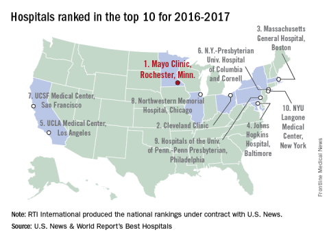
Daratumumab Effective in Combo Regimen
Adding daratumumab to bortezomib (a protease inhibitor [PI]) and dexamethasone reduces the risk of disease progression or death by 61% in patients with multiple myeloma (MM), compared with bortezomib and dexamethasone alone, according to an interim analysis of phase 3 trial results.
Related: Multiple Myeloma: Updates on Diagnosis and Management
Daratumumab is a biologic that targets CD38, a surface protein that is highly expressed across MM cells, regardless of disease stage. “CD38 is the most important tumor antigen on myeloma plasma cells,” said Antonio Palumbo, MD, University of Torino, Italy, who presented the study findings at the 2016 American Society of Clinical Oncology Annual Meeting in June. Daratumumab has more than 1 immune-mediated mechanism of action and both direct and indirect antimyeloma activity: It depletes CD38+ immunosuppressive regulatory cells and promotes T-cell expansion and activation, all while directly killing myeloma cells.
The multinational study (CASTOR) included 498 patients with MM who had received a median of 2 prior lines of therapy: bortezomib, an immunomodulatory agent, or a PI and immunomodulatory agent. Prior treatments had been unsuccessful in one-third of patients. In this study, researchers randomly assigned 251 patients to combination treatment with daratumumab.
Related: Treating Patients With Multiple Myeloma in the VA
Daratumumab significantly increased the overall response rate (83% vs 63%) and doubled rates of complete response or better (19% vs 9%). It also doubled the rates of very good partial response (59% vs 29%). The combination therapy met the primary endpoint of improved progression-free survival at a median follow-up of 7.4 months (60.7% in the combination arm, compared with 26.9%). The treatment was unblinded after meeting the primary endpoint.
The treatment benefits of the combination regimen were maintained across clinically relevant subgroups, the researchers said. The most common adverse effects were thrombocytopenia ,sensory peripheral neuropathy, anaemia, and diarrhea.
“These compelling phase 3 results demonstrate that a regimen built on daratumumab deepens clinical responses and help to underscore its potential for multiple myeloma patients who have been previously treated,” said Dr. Palumbo.
Related: A Mysterious Massive Hemorrhage
In a discussion of the study, Paul Richardson, MD, of the Dana-Farber Cancer Institute, praised the study as “outstanding” and “potentially practice changing.” However, another panel member noted that with an added cost of more than $10,000 a month, the treatment could be out of reach for many.
Adding daratumumab to bortezomib (a protease inhibitor [PI]) and dexamethasone reduces the risk of disease progression or death by 61% in patients with multiple myeloma (MM), compared with bortezomib and dexamethasone alone, according to an interim analysis of phase 3 trial results.
Related: Multiple Myeloma: Updates on Diagnosis and Management
Daratumumab is a biologic that targets CD38, a surface protein that is highly expressed across MM cells, regardless of disease stage. “CD38 is the most important tumor antigen on myeloma plasma cells,” said Antonio Palumbo, MD, University of Torino, Italy, who presented the study findings at the 2016 American Society of Clinical Oncology Annual Meeting in June. Daratumumab has more than 1 immune-mediated mechanism of action and both direct and indirect antimyeloma activity: It depletes CD38+ immunosuppressive regulatory cells and promotes T-cell expansion and activation, all while directly killing myeloma cells.
The multinational study (CASTOR) included 498 patients with MM who had received a median of 2 prior lines of therapy: bortezomib, an immunomodulatory agent, or a PI and immunomodulatory agent. Prior treatments had been unsuccessful in one-third of patients. In this study, researchers randomly assigned 251 patients to combination treatment with daratumumab.
Related: Treating Patients With Multiple Myeloma in the VA
Daratumumab significantly increased the overall response rate (83% vs 63%) and doubled rates of complete response or better (19% vs 9%). It also doubled the rates of very good partial response (59% vs 29%). The combination therapy met the primary endpoint of improved progression-free survival at a median follow-up of 7.4 months (60.7% in the combination arm, compared with 26.9%). The treatment was unblinded after meeting the primary endpoint.
The treatment benefits of the combination regimen were maintained across clinically relevant subgroups, the researchers said. The most common adverse effects were thrombocytopenia ,sensory peripheral neuropathy, anaemia, and diarrhea.
“These compelling phase 3 results demonstrate that a regimen built on daratumumab deepens clinical responses and help to underscore its potential for multiple myeloma patients who have been previously treated,” said Dr. Palumbo.
Related: A Mysterious Massive Hemorrhage
In a discussion of the study, Paul Richardson, MD, of the Dana-Farber Cancer Institute, praised the study as “outstanding” and “potentially practice changing.” However, another panel member noted that with an added cost of more than $10,000 a month, the treatment could be out of reach for many.
Adding daratumumab to bortezomib (a protease inhibitor [PI]) and dexamethasone reduces the risk of disease progression or death by 61% in patients with multiple myeloma (MM), compared with bortezomib and dexamethasone alone, according to an interim analysis of phase 3 trial results.
Related: Multiple Myeloma: Updates on Diagnosis and Management
Daratumumab is a biologic that targets CD38, a surface protein that is highly expressed across MM cells, regardless of disease stage. “CD38 is the most important tumor antigen on myeloma plasma cells,” said Antonio Palumbo, MD, University of Torino, Italy, who presented the study findings at the 2016 American Society of Clinical Oncology Annual Meeting in June. Daratumumab has more than 1 immune-mediated mechanism of action and both direct and indirect antimyeloma activity: It depletes CD38+ immunosuppressive regulatory cells and promotes T-cell expansion and activation, all while directly killing myeloma cells.
The multinational study (CASTOR) included 498 patients with MM who had received a median of 2 prior lines of therapy: bortezomib, an immunomodulatory agent, or a PI and immunomodulatory agent. Prior treatments had been unsuccessful in one-third of patients. In this study, researchers randomly assigned 251 patients to combination treatment with daratumumab.
Related: Treating Patients With Multiple Myeloma in the VA
Daratumumab significantly increased the overall response rate (83% vs 63%) and doubled rates of complete response or better (19% vs 9%). It also doubled the rates of very good partial response (59% vs 29%). The combination therapy met the primary endpoint of improved progression-free survival at a median follow-up of 7.4 months (60.7% in the combination arm, compared with 26.9%). The treatment was unblinded after meeting the primary endpoint.
The treatment benefits of the combination regimen were maintained across clinically relevant subgroups, the researchers said. The most common adverse effects were thrombocytopenia ,sensory peripheral neuropathy, anaemia, and diarrhea.
“These compelling phase 3 results demonstrate that a regimen built on daratumumab deepens clinical responses and help to underscore its potential for multiple myeloma patients who have been previously treated,” said Dr. Palumbo.
Related: A Mysterious Massive Hemorrhage
In a discussion of the study, Paul Richardson, MD, of the Dana-Farber Cancer Institute, praised the study as “outstanding” and “potentially practice changing.” However, another panel member noted that with an added cost of more than $10,000 a month, the treatment could be out of reach for many.