User login
FDA approves first naloxone nasal spray for opioid overdose
The Food and Drug Administration has approved the first nasal spray variant of the opioid-overdose drug naloxone hydrochloride.
Marketed in the United States as Narcan by Adapt Pharma, a partner of Lightlake Therapeutics, the nasal spray is known to stop or, in some cases, reverse the effects of opioid overdosing in patients. Narcan is the first naloxone hydrochloride nasal spray approved by the FDA.
“Combating the opioid abuse epidemic is a top priority for the FDA,” Dr. Stephen Ostroff, FDA acting commissioner, said in a statement released with the Nov. 18 approval announcement. “We cannot stand by while Americans are dying. While naloxone will not solve the underlying problems of the opioid epidemic, we are speeding to review new formulations that will ultimately save lives that might otherwise be lost to drug addiction and overdose.”
The nasal spray itself is available only with a prescription, and is safe for use by both adults and children, according to the FDA.
The spray delivers a dose of 4 mg naloxone in a single 0.1-mL nasal spray, which comes in a ready-to-use, needle-free device, according to Adapt Pharma. Dosage varies for each individual and should be determined by physicians.
Administration of Narcan, which is sprayed into one nostril while the patient is lying on his or her back, does not require special training and can be performed by anyone.
Narcan’s approval comes less than 4 months after the FDA granted the medication a fast-track designation and priority review status, both of which are meant to expedite the review and approval processes for drugs that “demonstrate the potential to address an unmet medical need” and “offer a significant improvement in the safety or effectiveness of the treatment, prevention, or diagnosis of a serious condition,” according to the FDA’s approval statement.
Adverse events associated with opiate withdrawal have been noted in Narcan patients. Specifically, the FDA warned that body aches, diarrhea, tachycardia, fever, piloerection, nausea, nervousness, abdominal cramps, weakness, and increased blood pressure, among other conditions, are all possible side effects of Narcan.
“Opioid overdose is responsible for the deaths of thousands of Americans in communities throughout the country, leaving a trail of devastation for friends and families,” Seamus Mulligan, Adapt Pharma’s chairman and CEO, said in a statement. “This new device makes naloxone readily available for emergency use by a friend, family member, or caregiver, as well as offering an alternative treatment option for first responders and health care providers.”
Narcan’s approval is one step of many that must be taken to adequately address and ultimately end the problem of opioid abuse in the United States, cautioned Dr. Peter Friedmann, chief research officer at Baystate Health in Springfield, Mass.
“[Narcan] is just addressing overdose; we need more and better medications for treating addiction, we need more physicians and clinicians who are skilled in using these medications,” said Dr. Friedmann. “Given the ongoing crisis of deaths from opioid overuse, this expands the options that physicians, pharmacies, and community distribution programs can use to reduce these deaths.”
Dr. Friedmann also voiced his concern regarding the pricing of Narcan. Making the drug affordable is crucial to its success at successfully treating opioid overuse, he said.
“Right now, nasal atomizers with syringes are used off label, and the prices have been going up with increasing demand,” he said. “But [Narcan] is a commercial product based around what is essentially a generic medication, so [I] hope it’s priced at a price point that’s accessible to the great majority of patients and their families who are facing addiction, many of whom don’t have huge means.”
Dr. Friedmann said that he hopes addiction medicine becomes a more attractive field for medical students and residents. It’s important for future physicians to know how to properly treat patients of addiction and administer drugs safely and effectively, he said.
“Addiction medicine is on the cusp of full recognition as a medical specialty, and we need people to go into that field to teach patients, medical students, and residents how to treat people with addiction,” Dr. Friedmann said. At the current rate, “we’re never going to have enough, so we need generalists to take this on.”
ACR: Criteria enable prompt diagnosis and treatment of CAPS
SAN FRANCISCO – Valid diagnostic criteria for cryopyrin-associated periodic syndromes are sorely needed, and a new model of CAPS diagnostic criteria enables rapid diagnosis of this rare, heterogeneous, inflammatory disease that can have devastating consequences if left undiagnosed and untreated.
“Our novel, unique approach integrated traditional methods of evidence synthesis with expert consensus, web-based decision tools, and innovative statistical methods. We hope this model can be of benefit in the diagnosis of other rare diseases,” said Dr. Jasmin B. Kümmerle-Deschner of the department of pediatrics at the University of Tübingen (Germany). She noted that the panel wanted to develop diagnostic criteria that could be used prior to genetic testing or in cases where no genetic mutation is identified.

Dr. Kümmerle-Deschner, who was on the multinational panel that developed the new diagnostic criteria, reviewed the process and presented the new criteria at the annual meeting of the American College of Rheumatology.
Cryopyrin-associated periodic syndromes (CAPS) comprise a rare, clinically heterogeneous group of devastating inflammatory diseases characterized by variable, severe system and organ inflammation resulting in permanent organ damage. The disease can manifest right after birth or later in life, and it can be associated with a mutation in the NLRP3 gene. Because the pathogenesis involves continually increased IL-1 secretion, early diagnosis and rapid initiation of IL-1 inhibition can control inflammation and prevent organ damage in children and adults with CAPS.
The lengthy process for developing the diagnostic criteria involved building an 18-member team of pediatric and adult subspecialists and experts in methods for rare disease. They generated 32 CAPS-typical items from a systematic literature review of 33 papers that described symptoms of CAPS and then reviewed the items in CAPS registries, weighting them via decision analysis software. Next, the panel refined these to 14 items plus the NLRP3 mutation. 1000Minds decision-making software ranked these 14 variables based on importance for the diagnosis of CAPS, with excellent correlation among experts.
Then a correspondence analysis determined variables consistently associated with the diagnosis of CAPS based on 284 cases and 873 controls; this process removed infrequently observed variables, such as “amyloidosis.” The remaining seven variables were found to be significantly associated with a diagnosis of CAPS (P less than .001 for all).
The final CAPS diagnosis model includes raised inflammatory markers (C-reactive protein/serum amyloid A) as a mandatory criterion, plus at least two of six different typical signs and symptoms of CAPS: urticaria-like rash, cold-stress triggered episodes, sensorineural hearing loss, musculoskeletal symptoms such as arthralgia, arthritis, and myalgia, chronic aseptic meningitis, and skeletal abnormalities such as epiphyseal overgrowth/frontal bossing. This model had a sensitivity of 81% and a specificity of 94%, performing well in all CAPS subtypes as well as in subgroups with or without evidence of germline NLRP3 mutations.
Dr. Kümmerle-Deschner opened her talk with a case description of an 81-year-old man who presented with a baffling disease. It turned out that his granddaughter was diagnosed with CAPS and 16 other family members were affected. She closed the talk by showing how this man could have been rapidly diagnosed by the new CAPS criteria. He presented with urticaria-like rash, arthralgia, arthritis, and cold-triggered episodes in the context of raised inflammatory markers.
“Early diagnosis enabling rapid treatment is possible,” she stated.
Dr. Kümmerle-Deschner disclosed financial ties with Novartis.
SAN FRANCISCO – Valid diagnostic criteria for cryopyrin-associated periodic syndromes are sorely needed, and a new model of CAPS diagnostic criteria enables rapid diagnosis of this rare, heterogeneous, inflammatory disease that can have devastating consequences if left undiagnosed and untreated.
“Our novel, unique approach integrated traditional methods of evidence synthesis with expert consensus, web-based decision tools, and innovative statistical methods. We hope this model can be of benefit in the diagnosis of other rare diseases,” said Dr. Jasmin B. Kümmerle-Deschner of the department of pediatrics at the University of Tübingen (Germany). She noted that the panel wanted to develop diagnostic criteria that could be used prior to genetic testing or in cases where no genetic mutation is identified.
Dr. Kümmerle-Deschner, who was on the multinational panel that developed the new diagnostic criteria, reviewed the process and presented the new criteria at the annual meeting of the American College of Rheumatology.
Cryopyrin-associated periodic syndromes (CAPS) comprise a rare, clinically heterogeneous group of devastating inflammatory diseases characterized by variable, severe system and organ inflammation resulting in permanent organ damage. The disease can manifest right after birth or later in life, and it can be associated with a mutation in the NLRP3 gene. Because the pathogenesis involves continually increased IL-1 secretion, early diagnosis and rapid initiation of IL-1 inhibition can control inflammation and prevent organ damage in children and adults with CAPS.
The lengthy process for developing the diagnostic criteria involved building an 18-member team of pediatric and adult subspecialists and experts in methods for rare disease. They generated 32 CAPS-typical items from a systematic literature review of 33 papers that described symptoms of CAPS and then reviewed the items in CAPS registries, weighting them via decision analysis software. Next, the panel refined these to 14 items plus the NLRP3 mutation. 1000Minds decision-making software ranked these 14 variables based on importance for the diagnosis of CAPS, with excellent correlation among experts.
Then a correspondence analysis determined variables consistently associated with the diagnosis of CAPS based on 284 cases and 873 controls; this process removed infrequently observed variables, such as “amyloidosis.” The remaining seven variables were found to be significantly associated with a diagnosis of CAPS (P less than .001 for all).
The final CAPS diagnosis model includes raised inflammatory markers (C-reactive protein/serum amyloid A) as a mandatory criterion, plus at least two of six different typical signs and symptoms of CAPS: urticaria-like rash, cold-stress triggered episodes, sensorineural hearing loss, musculoskeletal symptoms such as arthralgia, arthritis, and myalgia, chronic aseptic meningitis, and skeletal abnormalities such as epiphyseal overgrowth/frontal bossing. This model had a sensitivity of 81% and a specificity of 94%, performing well in all CAPS subtypes as well as in subgroups with or without evidence of germline NLRP3 mutations.
Dr. Kümmerle-Deschner opened her talk with a case description of an 81-year-old man who presented with a baffling disease. It turned out that his granddaughter was diagnosed with CAPS and 16 other family members were affected. She closed the talk by showing how this man could have been rapidly diagnosed by the new CAPS criteria. He presented with urticaria-like rash, arthralgia, arthritis, and cold-triggered episodes in the context of raised inflammatory markers.
“Early diagnosis enabling rapid treatment is possible,” she stated.
Dr. Kümmerle-Deschner disclosed financial ties with Novartis.
AT THE ACR ANNUAL MEETING

ICD-10 Under ACP Scrutiny
NEW YORK - While the new International Classification of Diseases, Tenth Revision, Clinical
Modification (ICD-10-CM) codes offer greater diagnostic precision, their implementation will require training of clinicians, coders, and other staff to minimize payment denials or delays from both public and private payers.
Brian Outland and colleagues from the American College of Physicians in Washington, D.C., outline some of the promises and challenges of ICD-10-CM implementation in a report online Sept. 22 in Annals of Internal Medicine.
Although completed and endorsed by the World Health Assembly in 1990, ICD-10-CM's implementation date has repeatedly been delayed, and was scheduled to take effect on Oct. 1.
The authors suggest that "the newer coding system will produce data that will indicate the clinical trajectory and other factors that will enable the data to be used in meaningful ways to better understand complications, design robust algorithms for clinical decision support, and track outcomes. Having these details built into the codes will decrease the need for health care providers to include supporting documentation with claims."
The new ICD-10-CM alphanumeric codes will contain as many as seven characters that specify categories, subcategories, laterality, severity and other features.
The use of codes that are not specific enough can result in payment denials or delays, so practices will need to keep current on payer reimbursement policies to ensure the reporting of ICD-10-CM codes that support reimbursement, the authors note.
The cost for the training of clinicians and staffs will depend on practice size, specialty, the method of training, current documentation quality, and technology readiness and availability.
Dr. Susan H. Fenton from UTHealth School of Biomedical Informatics in Houston, Texas, said by email, "One of the thoughts I cannot get away from is that the U.S. is trying to manage a 21st-century, rapidly evolving healthcare system with a 1970s technology. I can think of little else in healthcare that has remained as static since the 1970s."
"The diagnostic system added lots of codes, but the basic structure is the same," she said.
"Certainly, with more detail such as laterality, as well as first encounter, subsequent encounter, and sequelae, it will be much easier to track care for specific conditions across providers," Dr. Fenton said. "I think the issue of claims denials will have to play out over time."
Resources and tools from the Centers for Medicare & Medicaid Services (CMS) can be found online at www.roadto10.org.
The American College of Physicians also has helpful information available at www.acponline.org/ICD10.
Outland did not respond to a request for comment.
A surge in congenital syphilis reveals gaps in obstetric practice
The rate of congenital syphilis increased to 11.6 cases per 100,000 live births in 2014 in the United States—the highest rate documented since 2001, according to a new report from the Centers for Disease Control and Prevention (CDC).1 The increase in the rate of congenital syphilis reflects a rise in the rate of primary and secondary syphilis among US women, from 0.9 to 1.1 cases per 100,000 women, during the same period.1
The rate of congenital syphilis had declined between 2008 and 2012, from 10.5 cases to 8.4 cases per 100,000 live births.
Congenital syphilis occurs when an infected mother transmits the disease to her fetus during pregnancy. Among the adverse effects of congenital syphilis are deformities, stillbirth, and early infant death. “However, among mothers identified with syphilis who deliver past 20 weeks’ gestation, treatment with penicillin at least 30 days before delivery is 98% effective at preventing [congenital syphilis],” the CDC report notes.1
For the purposes of the CDC report, congenital syphilis includes “both infants and stillbirths with clinical evidence” of the disease, “as well as those infants and stillbirths born to mothers with untreated or inadequately treated syphilis, regardless of the infant’s manifestation of clinical disease.”1
CDC recommendations
The CDC notes that most of the increases in the rates of maternal and congenital syphilis likely stem from inadequate prenatal care.
“A large percentage of [congenital syphilis] cases continue to be attributable to a lack of prenatal care; even among those receiving some prenatal care, the detection and treatment of maternal syphilis is often not early enough….At particular risk are those who are uninsured or underinsured and those with substance use issues.”1
Among the recommendations of the CDC:
- Screen all pregnant women for syphilis at their first prenatal visit.
- Screen women at elevated risk for syphilis, as well as those who live in “high-morbidity geographic areas” at the beginning of the third trimester and again at delivery.
- In cases where prenatal care has been lacking, screen the woman for syphilis using a rapid plasma reagin (RPR) card and treat the patient who tests positive at the time the pregnancy is confirmed.
- Do not discharge an infant from the hospital unless the syphilis serologic status of the mother has been tested at least once during pregnancy and, preferably, again at delivery (in high-risk cases).
- Test any woman who delivers a stillborn infant for syphilis.
Reference
1. Bowen V, Su J, Torrone E, Kidd S, Weinstock H. Increase in incidence of congenital syphilis—United States, 2008–2014. MMWR. 2015;64(44):1241–1245.
The rate of congenital syphilis increased to 11.6 cases per 100,000 live births in 2014 in the United States—the highest rate documented since 2001, according to a new report from the Centers for Disease Control and Prevention (CDC).1 The increase in the rate of congenital syphilis reflects a rise in the rate of primary and secondary syphilis among US women, from 0.9 to 1.1 cases per 100,000 women, during the same period.1
The rate of congenital syphilis had declined between 2008 and 2012, from 10.5 cases to 8.4 cases per 100,000 live births.
Congenital syphilis occurs when an infected mother transmits the disease to her fetus during pregnancy. Among the adverse effects of congenital syphilis are deformities, stillbirth, and early infant death. “However, among mothers identified with syphilis who deliver past 20 weeks’ gestation, treatment with penicillin at least 30 days before delivery is 98% effective at preventing [congenital syphilis],” the CDC report notes.1
For the purposes of the CDC report, congenital syphilis includes “both infants and stillbirths with clinical evidence” of the disease, “as well as those infants and stillbirths born to mothers with untreated or inadequately treated syphilis, regardless of the infant’s manifestation of clinical disease.”1
CDC recommendations
The CDC notes that most of the increases in the rates of maternal and congenital syphilis likely stem from inadequate prenatal care.
“A large percentage of [congenital syphilis] cases continue to be attributable to a lack of prenatal care; even among those receiving some prenatal care, the detection and treatment of maternal syphilis is often not early enough….At particular risk are those who are uninsured or underinsured and those with substance use issues.”1
Among the recommendations of the CDC:
- Screen all pregnant women for syphilis at their first prenatal visit.
- Screen women at elevated risk for syphilis, as well as those who live in “high-morbidity geographic areas” at the beginning of the third trimester and again at delivery.
- In cases where prenatal care has been lacking, screen the woman for syphilis using a rapid plasma reagin (RPR) card and treat the patient who tests positive at the time the pregnancy is confirmed.
- Do not discharge an infant from the hospital unless the syphilis serologic status of the mother has been tested at least once during pregnancy and, preferably, again at delivery (in high-risk cases).
- Test any woman who delivers a stillborn infant for syphilis.
Reference
1. Bowen V, Su J, Torrone E, Kidd S, Weinstock H. Increase in incidence of congenital syphilis—United States, 2008–2014. MMWR. 2015;64(44):1241–1245.
Reference
1. Bowen V, Su J, Torrone E, Kidd S, Weinstock H. Increase in incidence of congenital syphilis—United States, 2008–2014. MMWR. 2015;64(44):1241–1245.
AK clearance rates maintained 1 year after thermally modulated PDT
Clearance rates for actinic keratoses (AKs) on the extremities were prolonged by adding thermal modulation to photodynamic therapy (PDT), a study has shown.
The single-site study of 17 adults with 10 or more AKs on their arms or legs evaluated thermally modulated PDT: 20% 5-aminolevulinic acid (ALA) applied to skin, occluded with plastic wrap and warmed with an electric heating pad to a median 41.2° C for one hour, then exposed to blue light. Treatment resulted in 90% clearance rates at 3 months and 12 months, reported Dr. Andrea Willey, a dermatologist in Sacramento, Calif., and her associates (Dermatol Surg. 2015 Nov;41[11]:1290-5).
They were inspired to conduct the study as a follow-on to a recently published pilot study of three patients, which found that clearance rates of AKs on the arm treated with thermally modulated 5-ALA blue light PDT persisted up to 14 months after treatment, when compared with the control arm, which was not heated.
In their study, there were 724 grade 1 and 2 AKs at baseline – a median of 15 per extremity; patients were followed up at 3, 6, 9, and 12 months. The total lesion count across the entire cohort was 70 at 3 months, 69 at 6 months, 64 at 9 months, and 72 at 1 year. No new AKs were seen in the treated areas during the 1-year follow-up.
There were 13 grade 3 hypertrophic AK lesions among all study participants. Although these lesions persisted, Dr. Willey and her coauthors noted that they did shrink initially after treatment.
Adverse events associated with the combination therapy were low, and included mild erythema and crusting up to 1 week post treatment, indicating the therapy was well tolerated, and patient satisfaction was high.
The results “suggest that mild skin warming may both improve efficacy and reduce variability of response to PDT in practice,” the authors wrote.
On Twitter @whitneymcknight
FROM DERMATOLOGIC SURGERY
Key clinical point: Mildly warming affected skin sites during photodynamic therapy can prolong the clearance rates for AKs on the extremities.
Major finding: AK clearance rates on the extremities remained at about 90% during the 1 year after treatment with thermally modulated PDT.
Data source: A single site, prospective 1-year study evaluating treatment with thermally modulated PDT for 17 adults with 10 or more AKs on their extremities.
Disclosures: Dr. Willey is a member of the scientific advisory board of DUSA Pharmaceuticals; she received a research grant, equipment loan, and study drug from the company. Her two coauthors had no relevant disclosures.
Myth of the Month: Does Colace work?
Myth: Docusate is a stool softener and helps with constipation.
A 60-year-old man is injured in a fall and breaks four ribs. He is in severe pain and is prescribed oxycodone and naproxen for pain. What treatment would you prescribe to help decrease problems with constipation?
A. Docusate.
B. Docusate and polyethylene glycol.
C. Psyllium.
D. Polyethylene glycol.
Constipation is extremely common, occurring in up to 20%-25% of the elderly population and 90% of patients treated with opioids. The formal definition of constipation is fewer than three bowel movements per week. Patients are concerned with other symptoms as well, including hard stool consistency and the feeling of incomplete evacuation.
An extremely commonly prescribed medication for patients with symptoms of constipation/hard stool passage is docusate (Colace). This medication is often a part of bowel programs for institutionalized/hospitalized patients, as well as being frequently prescribed when patients are treated with opiates.
Does it work?

Docusate is frequently prescribed as a “stool softener,” but does it increase water content in stool? In a randomized, controlled trial of docusate vs. psyllium, 170 adult patients with chronic constipation received either 5.1 g twice a day of psyllium or 100 mg twice a day of docusate (Aliment Pharmacol Ther. 1998 May;12[5]:491-7).
Psyllium was superior in its effect on stool frequency, stool water content, total stool output, and the combination of several objective measures of constipation. Compared with baseline, psyllium increased stool water content by 2.33%, vs .01% for docusate (P =. 007), and stool weight was increased in the group treated with psyllium, compared with docusate-treated patients (359.9 g/week vs. 271.9 g/week, respectively; P = .005). Docusate does not appear to have any effect on stool water content or amount of stool.
In a study of constipation treatment in patients receiving opioids, Dr. Yoko Tarumi and her colleagues studied 74 patients admitted to hospice units (J Pain Symptom Manage. 2013 Jan;45[1]:2-13). A total of 74 patients were randomized to receive docusate 100 mg twice a day plus senna, or placebo plus senna. Once the study was started, inclusion criteria were broadened to include hospice patients with nonmalignant disease and patients who were not on opioids.
Almost all patients in the study did receive opioids (94% of the docusate patients and 100% of placebo-treated patients). There were no significant between the groups in stool volume, frequency, consistency, or in perceived completeness of evacuation.
In a randomized, controlled study of elderly patients on a medicine ward, 34 patients were randomized to docusate or control (no laxatives)(J Chronic Dis. 1976 Jan;29[1]:59-63). There was no difference in frequency or quality of stools between groups.
A systematic review of the usefulness of docusate in chronically ill patients concluded that the widespread use of docusate for the treatment of constipation in palliative-care patients is based on inadequate experimental evidence (J Pain Symptom Manage. 2000 Feb;19[2]:130-6).
The Canadian Agency for Drugs and Technologies in Health concluded “the available evidence suggests that docusate is no more effective than placebo in the prevention or management of constipation” (Dioctyl sulfosuccinate or docusate [calcium or sodium] for the prevention or management of constipation: a review of the clinical effectiveness. Canadian Agency for Drugs and Technologies in Health; 2014 Jun 26).
Dr. Davendra Ramkumar and his colleagues published a systematic review of drug trials for the treatment of constipation in 2005 (Am J Gastroenterol. 2005 Apr;100[4]:936-71). Only polyethylene glycol and tegaserod received grade A evidence for published trials. Psyllium and lactulose received grade B evidence. Docusate received a level 3, grade C for evidence (poor quality evidence, poor evidence to support a recommendation for or against the use of the modality).
I have been surprised at how docusate has been the most commonly prescribed laxative agent. Polyethylene glycol or psyllium are better evidence-based options. Docusate is often prescribed as a stool softener, and it has even less evidence that it softens stool than its poor evidence as a laxative.
Acknowledgments
My thanks to the late Dr. David Saunders for teaching me 30 years ago that docusate was not a helpful option for the management of constipation, and to Sarah Steinkruger for doing much of the research that was used in this column.
Dr. Paauw is professor of medicine in the division of general internal medicine at the University of Washington, Seattle, and he serves as third-year medical student clerkship director at the University of Washington. Contact Dr. Paauw at [email protected].
Myth: Docusate is a stool softener and helps with constipation.
A 60-year-old man is injured in a fall and breaks four ribs. He is in severe pain and is prescribed oxycodone and naproxen for pain. What treatment would you prescribe to help decrease problems with constipation?
A. Docusate.
B. Docusate and polyethylene glycol.
C. Psyllium.
D. Polyethylene glycol.
Constipation is extremely common, occurring in up to 20%-25% of the elderly population and 90% of patients treated with opioids. The formal definition of constipation is fewer than three bowel movements per week. Patients are concerned with other symptoms as well, including hard stool consistency and the feeling of incomplete evacuation.
An extremely commonly prescribed medication for patients with symptoms of constipation/hard stool passage is docusate (Colace). This medication is often a part of bowel programs for institutionalized/hospitalized patients, as well as being frequently prescribed when patients are treated with opiates.
Does it work?
Docusate is frequently prescribed as a “stool softener,” but does it increase water content in stool? In a randomized, controlled trial of docusate vs. psyllium, 170 adult patients with chronic constipation received either 5.1 g twice a day of psyllium or 100 mg twice a day of docusate (Aliment Pharmacol Ther. 1998 May;12[5]:491-7).
Psyllium was superior in its effect on stool frequency, stool water content, total stool output, and the combination of several objective measures of constipation. Compared with baseline, psyllium increased stool water content by 2.33%, vs .01% for docusate (P =. 007), and stool weight was increased in the group treated with psyllium, compared with docusate-treated patients (359.9 g/week vs. 271.9 g/week, respectively; P = .005). Docusate does not appear to have any effect on stool water content or amount of stool.
In a study of constipation treatment in patients receiving opioids, Dr. Yoko Tarumi and her colleagues studied 74 patients admitted to hospice units (J Pain Symptom Manage. 2013 Jan;45[1]:2-13). A total of 74 patients were randomized to receive docusate 100 mg twice a day plus senna, or placebo plus senna. Once the study was started, inclusion criteria were broadened to include hospice patients with nonmalignant disease and patients who were not on opioids.
Almost all patients in the study did receive opioids (94% of the docusate patients and 100% of placebo-treated patients). There were no significant between the groups in stool volume, frequency, consistency, or in perceived completeness of evacuation.
In a randomized, controlled study of elderly patients on a medicine ward, 34 patients were randomized to docusate or control (no laxatives)(J Chronic Dis. 1976 Jan;29[1]:59-63). There was no difference in frequency or quality of stools between groups.
A systematic review of the usefulness of docusate in chronically ill patients concluded that the widespread use of docusate for the treatment of constipation in palliative-care patients is based on inadequate experimental evidence (J Pain Symptom Manage. 2000 Feb;19[2]:130-6).
The Canadian Agency for Drugs and Technologies in Health concluded “the available evidence suggests that docusate is no more effective than placebo in the prevention or management of constipation” (Dioctyl sulfosuccinate or docusate [calcium or sodium] for the prevention or management of constipation: a review of the clinical effectiveness. Canadian Agency for Drugs and Technologies in Health; 2014 Jun 26).
Dr. Davendra Ramkumar and his colleagues published a systematic review of drug trials for the treatment of constipation in 2005 (Am J Gastroenterol. 2005 Apr;100[4]:936-71). Only polyethylene glycol and tegaserod received grade A evidence for published trials. Psyllium and lactulose received grade B evidence. Docusate received a level 3, grade C for evidence (poor quality evidence, poor evidence to support a recommendation for or against the use of the modality).
I have been surprised at how docusate has been the most commonly prescribed laxative agent. Polyethylene glycol or psyllium are better evidence-based options. Docusate is often prescribed as a stool softener, and it has even less evidence that it softens stool than its poor evidence as a laxative.
Acknowledgments
My thanks to the late Dr. David Saunders for teaching me 30 years ago that docusate was not a helpful option for the management of constipation, and to Sarah Steinkruger for doing much of the research that was used in this column.
Dr. Paauw is professor of medicine in the division of general internal medicine at the University of Washington, Seattle, and he serves as third-year medical student clerkship director at the University of Washington. Contact Dr. Paauw at [email protected].

Finger-prick blood tests may need repeating
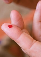
Finger-prick blood tests can deliver inaccurate results, according to a study published in the American Journal of Clinical Pathology.
Investigators found that results varied greatly when they performed multiple finger-prick tests on a single subject.
However, averaging the results of multiple droplet tests allowed the investigators to achieve results on par with venous blood tests.
They required 6 to 9 drops of blood to achieve consistent results.
“We began looking at this after we got some surprising results from our controls in an earlier study,” explained lead investigator Rebecca Richards-Kortum, PhD, of Rice University in Houston, Texas.
“Students in my lab are developing novel, low-cost platforms for anemia, platelet, and white blood cell testing in low-resource settings, and one of my students, Meaghan Bond, noticed there was wide variation in some of the benchmark tests that she was performing on hospital-grade blood analyzers.”
The benchmark controls are used to gauge the accuracy of test results from the new technology under study, so the variation among the control data was a sign that something was amiss.
What wasn’t immediately clear was whether the readings resulted from a problem with the current experiments or actual variations in the amount of hemoglobin, platelets, and white blood cells in the different drops of blood.
So Dr Richards-Kortum and Bond designed a simple protocol to test whether there was actual variation and, if so, how much.
They drew 6 successive 20 µL droplets of blood from 11 donors. As an additional test to determine whether minimum droplet size might also affect the results, the pair drew 10 successive 10 µL droplets from 7 additional donors.
All droplets were drawn from the same finger prick, and the investigators followed best practices in obtaining the droplets. The first drop was wiped away to remove contamination from disinfectants, and the finger was not squeezed or “milked,” which can lead to inaccurate results.
For experimental controls, the investigators used venipuncture to draw tubes of blood from an arm vein.
Each 20 µL droplet was analyzed with a hospital-grade blood analyzer for hemoglobin concentration, total white blood cell count, platelet count, and 3-part white blood cell differential. Each 10 µL droplet was tested for hemoglobin concentration with a popular point-of-care blood analyzer.
“A growing number of clinically important tests are performed using finger-prick blood, and this is especially true in low-resource settings,” Bond noted.
“It is important to understand how variations in finger-prick blood collection protocols can affect point-of-care test accuracy as well as how results might vary between different kinds of point-of-care tests that use finger-prick blood from the same patient.”
She and Dr Richards-Kortum found that hemoglobin content, platelet count, and white blood cell count each varied significantly from blood drop to blood drop.
“Some of the differences were surprising,” Bond said. “For example, in some donors, the hemoglobin concentration changed by more than 2 g/dL in the span of 2 successive drops of blood.”
Fortunately, the investigators found that averaging the results of the droplet tests could produce results that were on par with venous blood tests, but tests on 6 to 9 drops of blood were needed to achieve consistent results.
“Finger-prick blood tests can be accurate, and they are an important tool for healthcare providers, particularly in point-of-care and low-resource settings,” Bond said.
“Our results show that people need to take care to administer finger-prick tests in a way that produces accurate results because accuracy in these tests is increasingly important for diagnosing conditions like anemia, infections, and sickle cell anemia, malaria, HIV, and other diseases.” ![]()
Device linked to anemia in blood donors
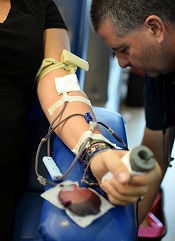
A device used to measure hemoglobin can deliver inaccurate results and may have prompted an increase in anemia among blood donors in Ireland, according to the Irish Blood Transfusion Service (IBTS).
The IBTS began using the device—called haemospect—in July 2014 but recently discovered that it cannot detect anemia accurately in all cases.
This may have resulted in anemic individuals donating blood or caused individuals to become anemic by donating.
The IBTS said this issue likely affected female donors more than males, so the organization has decided to temporarily stop taking blood donations from women who have donated in the last 18 months.
The IBTS also said it will perform a full blood count on all male and female donors going forward, until this issue has been resolved.
“Until we have a resolution to the problem that has arisen, we are asking male donors to attend our clinics and give blood,” said IBTS Medical and Scientific Director William Murphy, MD.
“We need male donors to make an extra effort to donate and maintain the blood supply. We will be double-checking all hemoglobin results on these donors.”
Discovering the problem
Haemospect is a noninvasive device that uses white light to detect hemoglobin levels. The device is made by the German company MBR Optical Systems.
IBTS learned of the potential for inaccuracies with haemospect after a blood donor contacted the organization. She had been diagnosed as severely anemic by her doctor but had given blood the week before.
“We have now discovered that the device gives inaccurate results in some individuals with anemia down to, and probably below, 8.4 g/dL,” Dr Murphy said. “As a result of the issue . . . , some women, and probably a much smaller number of men, could have been rendered iron-deficient and anemic from blood donation in the past 18 months.”
Steps to resolution
Since the issue was discovered, more than 30 women with anemia have been identified and contacted. The IBTS said it will contact all donors who are found to be anemic.
“Over the next few weeks, we will introduce new software to reanalyze all the electronic results from all donors who have been tested and accepted for donation since we introduced this device,” Dr Murphy said. “Any discrepant results will be notified to the donors involved.”
Dr Murphy also said donors who think they might be anemic or iron-deficient should visit their doctors for testing, and the IBTS will foot the bill. Concerned donors can contact the IBTS at 1850 731137.
The IBTS plans to replace haemospect with an alternative device as soon as possible. The organization has contacted the Health Products Regularity Authority about the issue with haemospect, which is used in Austria and Germany as well.
“We are determined to have this matter resolved as soon as possible,” Dr Murphy said. “We are confident that this issue . . . has not had any impact on blood received by patients.” ![]()
Group quantifies cardiotoxicity risk with HL treatment

Photo by Rhoda Baer
European researchers say they have quantified the risk of cardiovascular disease associated with treatments for Hodgkin lymphoma (HL).
The group analyzed the risks associated with specific doses of radiation and anthracycline exposure.
They believe their results, published in The Lancet Haematology, could help clinicians identify the optimal treatment regimen for each individual HL patient.
“These study results are exciting,” said Maja V. Maraldo, PhD, of Rigshospitalet in Copenhagen, Denmark.
“They should allow physicians to optimize the combination of systemic therapy and radiation and thereby balance the risks and benefits of different regimens in individual patients.”
Study details
Dr Maraldo and her colleagues analyzed data from patients who participated in 9 trials conducted by the European Organisation for Research and Treatment of Cancer (EORTC) and the Groupe d’Etude des Lymphomes de l’Adulte (GELA, now renamed LYSA) between 1964 and 2004.
In 2009 and 2010, the researchers mailed a Life Situation Questionnaire (LSQ) to the trial participants. The goal was to determine late-onset effects of HL and its treatment.
The team also reconstructed patients’ mean radiation doses to the heart and carotid arteries and the cumulative doses of anthracyclines and vinca-alkaloids they received. The incidence of cardiovascular disease was reported during follow-up and updated through the LSQ.
Patient data
The researchers were able to collect complete information on primary treatment for 6039 HL survivors. Of these patients, 2923 received the LSQ, and 1919 responded. The median follow-up was 9 years, and the patients’ median age at diagnosis was 30.
There were 1238 cardiovascular events in 703 patients, including 46 patients who died from such an event.
The events included ischemic heart disease (24%), congestive heart failure (21%), arrhythmia (17%), valvular disease (14%), disease of the arterial vessels (9%), stroke (6%), venous thromboembolism (5%), pericarditis (3%), peripheral vasculopathy (1%), other vascular events (1%), and other cardiac events (<1%).
Predictors of risk
The researchers found that the mean radiation dose to the heart, per 1 Gy increase, was a significant predictor of cardiovascular disease, with a hazard ratio of 1.015 (P=0.0014).
However, the mean radiation doses to the left internal carotid artery and the right internal carotid artery were not significant predictors of cardiovascular events (P=0.41 and 0.70, respectively).
The dose of anthracyclines, per 50 mg/m2 increase in cumulative dose, was a significant predictor of cardiovascular disease, with a hazard ratio of 1.077 (P=0.0064).
But the cumulative dose of vinblastine or vincristine was not (P=0.77 and 0.36, respectively).
The researchers said a limitation of this study is that they were not able to assess the impact of cardiovascular risk factors such as smoking, hypertension, and diabetes because that information was not consistently available.
Still, the team believes their analyses quantified the effect of radiotherapy and anthracyclines on the risk of cardiovascular disease, and the findings should aid treatment decisions in HL. ![]()
Coconut oil may prevent bloodstream infection

Coconut oil may combat infection with Candida albicans, according to preclinical research published in mSphere.
Mice on a diet that included coconut oil had significantly lower gastrointestinal colonization by C albicans than mice that were fed high-fat diets without coconut oil or mice that received a standard diet.
“We found that diet can be an effective way to reduce the amount of Candida in the mouse,” said study author Carol Kumamoto, PhD, of Tufts University School of Medicine in Boston, Massachusetts.
“The extension of this finding to the human population is something that needs to be addressed in the future.”
Previous research showed that changes to diet, including changes in the amount and type of fat, can alter gastrointestinal microbiota. And in vitro studies showed that coconut oil has antifungal properties.
So Dr Kumamoto and her colleagues studied the effect of different diets on C albicans colonization in mice.
The mice received standard diets or high-fat diets containing coconut oil, beef tallow, or soybean oil. The mice were fed these diets for 14 days prior to inoculation with C albicans and 21 days after.
At 21 days post-inoculation, gastrointestinal colonization with C albicans was significantly lower in the stomach contents of mice fed the coconut oil than mice fed the beef tallow (P<0.0001), the soybean oil (P<0.0001), or the standard diet (P<0.0001).
“When you compared a mouse on a high-fat diet that contained either beef fat or soybean oil to mice eating coconut oil, there was about a 10-fold drop in colonization,” Dr Kumamoto said.
In another experiment, the researchers switched mice receiving beef tallow to coconut oil.
“Four days after the change in diet, the colonization changed so it looked almost exactly like what you saw in a mouse who had been on coconut oil the entire time,” Dr Kumamoto said.
“There are 2 directions that we would like to take with this research now,” she added. “One of them is finding out the mechanism of how this works. That is a big question we would like to answer. The second question is whether this can have any impact on humans.”
The researchers are in discussion with Joseph Bliss, MD, PhD, of Women and Infants Hospital of Rhode Island, about launching a clinical trial testing coconut oil in hospitalized infants at high risk of developing systemic candidiasis. ![]()