User login
Increase enoxaparin doses to prevent VTEs in trauma patients
PHOENIX – Trauma patients probably need an elevated dose of enoxaparin – perhaps 40 mg twice daily – to prevent venous thromboembolisms, according to a prospective study of 85 trauma patients at the Palmetto Health Richland hospital in Columbia, S.C.
Also, antifactor 10a – a blood test often used in research to gauge how well enoxaparin (Lovenox) is thinning the blood – doesn’t work very well as an empiric measure of anticoagulation; thromboelastography (TEG) may be better, lead investigator Janise Phillips, Pharm.D., said at the Critical Care Congress, sponsored by the Society of Critical Care Medicine.

Her team tracked trauma patients who had at least three consecutive doses of enoxaparin prophylaxis for venous thromboembolism (VTE) and at least one peak antifactor 10a level drawn; enoxaparin doses were adjusted as needed to hit a weekly antifactor 10a level of 0.20-0.40 IU/mL, which is thought to be the therapeutic range for enoxaparin. Patients were in the ICU for a median of about 10 days, and in the hospital for about 2-3 weeks.
The types of trauma were not reported in the study, but the investigation confirms prior findings that critically ill trauma patients – and perhaps burn patients – need higher anticoagulant doses.
Overall, 65% (13) of patients on an initial enoxaparin regimen of 30 mg subcutaneously twice daily were below anti-factor 10a levels of 0.20-0.40 IU/mL after their first dose; 22% (8) were subtherapeutic after an initial dose of 40 mg once daily; and 21% (6) were subtherapeutic after an initial dose of 40 mg twice daily.
Antifactor 10a levels didn’t match well with clinical benefit. VTEs were diagnosed in 15% (4) of patients with an initial subtherapeutic antifactor 10a level, but 15% (4) bled on their subtherapeutic dose; 8.5% (4) of patients with an initial therapeutic level had a VTE, vs. none who were supratherapeutic after their initial dose. However, 9% (1) of supratherapeutic patients had an enoxaparin bleed.
“These were trauma patients in and out of surgery. A lot of the time, we had to stop the dose and hold it, which may” explain why subtherapeutic patients had the highest VTE risk, said Dr. Phillips, now a critical care pharmacist at the Cleveland Clinic hospital in Abu Dhabi, United Arab Emirates.
More than half of the patients were men, and being male was the only factor that seemed to increase the risk of subtherapeutic enoxaparin levels (P = .04). There was a trend for subtherapeutic levels in heavier patients – which might help explain the higher risk in men – and those with diminished kidney function. Even so, the fact that both VTEs and bleeding were most likely in underdosed patients could mean that antifactor 10a “is really not the best marker for VTE risk. At $80 a pop, it isn’t cost-effective, and [even] patients with therapeutic levels ended up with clots. TEG gives you a real time view of the coagulation status of the patient,” and may be the way to go, Dr. Phillips said.
PHOENIX – Trauma patients probably need an elevated dose of enoxaparin – perhaps 40 mg twice daily – to prevent venous thromboembolisms, according to a prospective study of 85 trauma patients at the Palmetto Health Richland hospital in Columbia, S.C.
Also, antifactor 10a – a blood test often used in research to gauge how well enoxaparin (Lovenox) is thinning the blood – doesn’t work very well as an empiric measure of anticoagulation; thromboelastography (TEG) may be better, lead investigator Janise Phillips, Pharm.D., said at the Critical Care Congress, sponsored by the Society of Critical Care Medicine.
Her team tracked trauma patients who had at least three consecutive doses of enoxaparin prophylaxis for venous thromboembolism (VTE) and at least one peak antifactor 10a level drawn; enoxaparin doses were adjusted as needed to hit a weekly antifactor 10a level of 0.20-0.40 IU/mL, which is thought to be the therapeutic range for enoxaparin. Patients were in the ICU for a median of about 10 days, and in the hospital for about 2-3 weeks.
The types of trauma were not reported in the study, but the investigation confirms prior findings that critically ill trauma patients – and perhaps burn patients – need higher anticoagulant doses.
Overall, 65% (13) of patients on an initial enoxaparin regimen of 30 mg subcutaneously twice daily were below anti-factor 10a levels of 0.20-0.40 IU/mL after their first dose; 22% (8) were subtherapeutic after an initial dose of 40 mg once daily; and 21% (6) were subtherapeutic after an initial dose of 40 mg twice daily.
Antifactor 10a levels didn’t match well with clinical benefit. VTEs were diagnosed in 15% (4) of patients with an initial subtherapeutic antifactor 10a level, but 15% (4) bled on their subtherapeutic dose; 8.5% (4) of patients with an initial therapeutic level had a VTE, vs. none who were supratherapeutic after their initial dose. However, 9% (1) of supratherapeutic patients had an enoxaparin bleed.
“These were trauma patients in and out of surgery. A lot of the time, we had to stop the dose and hold it, which may” explain why subtherapeutic patients had the highest VTE risk, said Dr. Phillips, now a critical care pharmacist at the Cleveland Clinic hospital in Abu Dhabi, United Arab Emirates.
More than half of the patients were men, and being male was the only factor that seemed to increase the risk of subtherapeutic enoxaparin levels (P = .04). There was a trend for subtherapeutic levels in heavier patients – which might help explain the higher risk in men – and those with diminished kidney function. Even so, the fact that both VTEs and bleeding were most likely in underdosed patients could mean that antifactor 10a “is really not the best marker for VTE risk. At $80 a pop, it isn’t cost-effective, and [even] patients with therapeutic levels ended up with clots. TEG gives you a real time view of the coagulation status of the patient,” and may be the way to go, Dr. Phillips said.
AT THE SCCM CRITICAL CARE CONGRESS
Key clinical point: Enoxaparin at 30 mg twice daily isn’t adequate for preventing VTEs in trauma patients.
Major finding: Overall, 65% of patients on an initial enoxaparin regimen of 30 mg subcutaneously twice daily were below anti-factor 10a levels of 0.20-0.40 IU/mL after their first dose; 22% were subtherapeutic after an initial dose of 40 mg once daily; and 21% were subtherapeutic after an initial dose of 40 mg twice daily.
Data source: Prospective study of 85 trauma patients atthe Palmetto Health Richland hospital in Columbia, S.C.
Disclosures: The lead investigator said she has no disclosures, and no outside funding was reported for the work.

IBD patients have significantly higher risk of DVT, pulmonary embolism
The risk of inflammatory bowel disease (IBD) patients developing deep vein thrombosis and pulmonary embolism was 1.98-fold and 1.80-fold higher, respectively, than those without the disease, according to research published in Thrombosis Research.
To explore the connection between deep vein thrombosis (DVT) and IBD, Dr. Wei-Sheng Chung of Taichung (Taiwan) Hospital and associates compared 11,445 IBD patients and 45,780 controls in a nationwide, population-based cohort study.
The IBD patients had a higher prevalence of comorbidities than their peers, including atrial fibrillation, hypertension, diabetes, heart failure, and cerebral vascular disease. In addition, the IBD patients who were hospitalized twice per year exhibited a significantly greater risk of developing DVT (adjusted hazard ratio, 32.9; 95% confidence interval, 20.5-52.8) and pulmonary embolism (adjusted HR, 24.2; 95% CI, 11.1-52.9) than did the comparison cohort.
Read more here: (Thromb. Res. 2015;135:492-6 [http://dx.doi.org/10.1016/j.thromres.2014.12.025]).
Group identifies new subtype of ALL

Photo by Steven Harbour
Researchers say they have discovered a new subtype of acute lymphoblastic leukemia (ALL) that is sensitive to drugs already approved to treat other hematologic malignancies.
The team uncovered cases of ALL that were dependent upon tonic pre-B-cell-receptor (BCR) signaling and therefore sensitive to drugs that inhibit tyrosine kinases downstream of the pre-BCR.
The group also developed a test that can identify patients with this subtype of ALL.
“We hope patients in this newly identified subset can be treated with these targeted drugs, . . . which are powerfully effective in the mouse experiments we have conducted on ALL,” said Markus Müschen, MD, PhD, of the University of California, San Francisco.
Dr Müschen and his colleagues described this work in Cancer Cell.
The researchers studied samples from 830 patients enrolled in 4 ongoing ALL trials and found tonic pre-BCR signaling in 112 patients (13.5%). Virtually all of the bone marrow slices from these patients showed “beautiful staining” of BCL6 expression, Dr Müschen said. (Two of the patients had weak staining.)
On the other hand, no BCL6 staining was observed in patients lacking pre-BCR signaling. These results suggest that BCL6 is a biomarker for pre-BCR signaling. And by testing patients for BCL6, we may be able to identify those who will respond to treatment with pre-BCR signaling inhibitors, the researchers said.
The team tested a range of pre-BCR signaling inhibitors in vitro. And they found a few compounds that were effective against pre-BCR+ ALL—the SYK inhibitor PRT062607, the BTK inhibitor ibrutinib, the SRC inhibitor dasatinib, and the PIK3δ inhibitor idelalisib.
Subsequent experiments revealed that dasatinib had the strongest antileukemic effect, so the researchers tested the drug in mouse models of pre-BCR+ ALL. Dasatinib significantly delayed leukemic expansion and prolonged overall survival in some mice, while completely eradicating the disease in other mice.
Dr Müschen said that dasatinib and other pre-BCR signaling inhibitors may be able to reduce the amount of conventional chemotherapy given to patients with pre-BCR+ ALL, or even replace chemotherapy altogether.
“In our experiments with mice, both combination therapy with low-dose chemotherapy and single-agent targeted therapy each worked very well,” he said. ![]()
Inhibitor outperforms BAT in myelofibrosis trial

Image by Peter Anderson
Results of a phase 3 trial suggest the JAK2/FLT3 inhibitor pacritinib may be more effective for patients with myelofibrosis (MF) than best available therapy (BAT), excluding JAK inhibitors.
Patients who received pacritinib were more likely than those who received BAT to see a reduction in spleen volume and to become transfusion-independent,
regardless of their platelet counts.
In fact, pacritinib proved particularly effective among patients with severe thrombocytopenia.
CTI Biopharma and Baxter International, Inc., the companies developing pacritinib, announced these results from the PERSIST-1 trial yesterday.
“Despite the introduction of JAK2 inhibitors as effective therapies for patients with myelofibrosis, there remains a treatment gap for patients with disease-related or treatment-emergent thrombocytopenia,” said study investigator Claire Harrison, MD, of Guy’s Hospital in London, UK.
“The currently approved drug [ruxolitinib] may require dose titration to less effective doses in this patient population, thus limiting our ability to effectively treat them. Results from the PERSIST-1 randomized trial demonstrate that pacritinib could address this unmet medical need.”
PERSIST-1 is a randomized (2:1), phase 3 trial comparing the safety and efficacy of pacritinib to BAT, other than JAK inhibitors. Investigators enrolled 327 patients with primary and secondary MF, post-polycythemia vera MF, or post-essential thrombocythemia MF.
The study’s primary endpoint was the proportion of patients achieving a 35% or greater reduction in spleen volume from baseline to week 24, as measured by MRI or CT, when compared with BAT. Once patients completed 24 weeks of treatment, or if their disease progressed, they could cross over from the BAT arm to the pacritinib arm.
Investigators found that PERSIST-1 met its primary endpoint in the intent-to-treat population. Pacritinib produced a significantly better rate of spleen volume reduction (P=0.0003) when compared to BAT.
The same was true among patients with platelet counts of less than 100,000/μL and less than 50,000/μL, both subgroups that were stratified at randomization.
The magnitude of treatment effect was consistent with previously reported phase 2 results, with the greatest reduction observed among the sickest patients (platelet counts <50,000/μL).
Fifty patients were transfusion-dependent at study entry (receiving ≥ 6 units of red blood cells over 90 days pre-entry). And, compared to BAT, pacritinib resulted in a clinically meaningful percentage of patients becoming transfusion-independent.
Ultimately, 79% of patients in the BAT arm crossed over to the pacritinib arm.
Investigators said the safety profile in the PERSIST-1 trial was consistent with prior phase 2 trials, as the most common treatment-emergent adverse events were diarrhea, nausea, and vomiting. However, the incidence of grade 3 events was lower than previously observed in phase 2 trials. And no grade 4 gastrointestinal adverse events were reported.
Three patients discontinued treatment with pacritinib, and 9 required dose reductions for diarrhea. A preliminary analysis suggested that few patients discontinued pacritinib or required a dose reduction due to treatment-related anemia or thrombocytopenia.
Additional data from ongoing analyses will be submitted for presentation at an upcoming scientific meeting, according to Baxter and CTI Biopharma. ![]()
ASH advocates use of systems-based hematologists

Photo courtesy of CDC
The American Society of Hematology (ASH) has released a report proposing a new role for hematologists specializing in non-malignant blood disorders.
ASH partnered with the healthcare consulting firm The Lewin Group to identify emerging career opportunities for health system- and hospital-based hematologists and to provide guidance on pursuing those opportunities.
The resulting report, published in Blood, outlines a few models for a systems-based clinical hematologist.
The report’s authors noted that demand for hematology expertise remains high nationwide. However, ASH and its members are concerned that changes to academic training will hinder both the recruitment of new talent to the field and the retention of seasoned experts.
The authors said that today’s hematology trainees are unlikely to receive the same non-malignant training as many “classic” hematologists trained in prior decades. And training shortfalls are further compounded by the fact that primary care physicians do not have the expertise to manage common blood disorders, which increases referrals to hematologists.
This results in higher demand for a smaller pool of hematologists entering the field with adequate training to effectively and efficiently manage non-malignant disorders.
“Given the rapid evolution and complexity of the field, the time is appropriate to identify career pathways that attract and enable physicians to practice non-malignant hematology in a sustainable manner,” said author Janis L. Abkowitz, MD, of the University of Washington in Seattle.
She and her colleagues noted that, in response to these challenges, US hematologists are defining new paths and assuming more centralized positions in large and small healthcare systems.
These systems-based hematologists are specialty-trained physicians—employed by a hospital, medical center, or health system—who optimize individual patient care as well as the overall system of healthcare delivery for patients with blood disorders.
For example, a systems-based hematologist could work closely with surgeons to minimize perioperative bleeding and could manage care pathways for patients with chronic blood diseases.
The report offered 4 examples where the involvement of a systems-based hematologist would lead to cost-effective decision-making. These were based upon interviews with 14 early adoptors of the systems-based approach to hematology.
The first example was heparin-induced thrombocytopenia (HIT). A systems-based hematologist could implement care pathways that focus on HIT by working to reduce unnecessary heparin exposure, optimizing laboratory testing for suspected HIT, and reducing unnecessary procedures in patients.
The second example was thrombotic thrombocytopenic purpura (TTP). A systems-based hematologist could optimize testing for TTP, which may reduce system-wide plasma use.
The third example was a medical director for hemostasis and thrombosis. A systems-based hematologist could foster appropriate and safe practices, including the implementation of and adherence to preventive care for thrombotic events and the optimal use of anticoagulant medications.
The fourth example was non-malignant hematology consultation in an accountable care organization (ACO) environment. The authors noted that ACOs have enabled more patients to be served by a health system, but there are fewer incentives for physicians to manage common hematology-related issues. A funded systems-based hematologist could ensure that patients have more timely access to hematology consultations.
“A systems-based hematologist position presents a unique opportunity for hematologists to design new models for care delivery and demonstrate their ability to improve clinical outcomes while maintaining or reducing costs,” Dr Abkowitz said. “Just as blood must flow throughout the body, the expertise of hematology must flow throughout the healthcare system.”
As a next step, ASH has invited its members to share practice models they have developed and examples of how they have collaborated with others to improve healthcare outcomes, reduce complications, and eliminate unnecessary spending. ![]()
New test can better predict cytokine storm, team says

Photo by Juan D. Alfonso
Scientists have developed a test that uses cells from a single donor’s blood to predict whether a new drug will cause a cytokine storm.
The group says this is an improvement over current tests, which use endothelial cells and peripheral blood mononuclear cells (PBMCs) from two separate donors and can therefore produce inaccurate results.
Furthermore, current tests cannot differentiate drugs that induce a mild cytokine storm from those that induce a severe one, but the new test can.
Jane Mitchell, PhD, of the National Heart and Lung Institute at Imperial College London in the UK, and her colleagues described the new test in The FASEB Journal.
Current tests for cytokine storm reactions use endothelial cells taken from the vessels of one donor and PBMCs from a different donor because endothelial cells are normally only grown from tissue removed in surgery or post-mortem, or from umbilical vessels after birth.
When cells from two different donors are used, one may have an immune reaction to the other. And this can result in the test falsely showing a severe immune reaction to a drug that is safe.
Dr Mitchell and her colleagues say they have solved this problem by isolating stem cells from the blood of a volunteer and using them to grow endothelial cells in a dish. The team then added PBMCs to the donor’s own endothelial cells to recreate the unique conditions found in their blood vessels.
When the scientists added the immunomodulatory drug TGN1412, the mixture of cells released a cytokine storm, as would happen inside the human body.
Responses to other drugs were consistent with those observed in humans as well. There was a modest response to alemtuzumab (Campath) and no response to the control antibodies trastuzumab (Herceptin), bevacizumab (Avastin), and ofatumumab (Arzerra).
“As biological therapies become more mainstream, it’s more likely that drugs being tested on humans for the first time will have unexpected and potentially catastrophic effects,” Dr Mitchell said.
“We’ve used adult stem cell technology to develop a laboratory test that could prevent another disaster like the TGN1412 trial [in which 6 healthy young men developed multi-organ failure]. Drug companies have the technical capacity to start using this test now, but we’re working on developing an off-the-shelf kit, which will make it easy to use on a large scale.”
The team has collaborated with the National Institute for Biological Standards and Control to validate the test and are now working with the clinical trials company Quintiles to develop the technology further. ![]()
Schizophrenia patients twice as likely to be at risk for DVT and PE
Schizophrenia patients exhibited a twofold higher adjusted risk of deep vein thrombosis and pulmonary embolism development than those without schizophrenia, according to research by Dr. Wen-Yu Hsu of China Medical University in Taiwan published in Schizophrenia Research.
The schizophrenia cohort exhibited a 2.02-fold higher adjusted hazard ratio (HR) for developing DVT and a 1.99-fold higher adjusted HR for developing PE. The population-based cohort study included 60,264 schizophrenia patients in Taiwan, compared with 60,264 control patients, obtained from the National Health Insurance Research Database in Taiwan from 1996 to 2011.
The researchers identified several lifestyle factors that could contribute to the development of DVT and PE, including a decrease in activities of daily living because of either antipsychotics or negative symptoms of schizophrenia, higher rates of smoking and metabolic syndrome among schizophrenia patients, and prolonged antipsychotic exposure. In addition, patients who are immobile could be at greater risk of developing DVT and PE. “Based on the findings of this study, receiving first-generation antipsychotics or receiving second-generation antipsychotics should be considered a contributing factor of DVT and PE development,” the researchers said.
Read more here: (Schizophr. Res. 2015;162:248-52 [doi:10.1016/j.schres.2015.01.012]).
What’s Eating You? Cutaneous Larva Migrans
Cutaneous larva migrans (CLM), also known as creeping eruption, is a pruritic serpiginous eruption caused by the migration of animal hookworm larvae through the epidermis.1,2 The most common parasites are Ancylostoma braziliense (common in dogs and cats) and Ancylostoma caninum (common in dogs).1
Disease Transmission
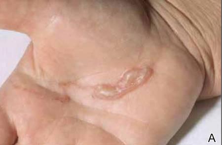
The infection is typically acquired in warm climates and tropical areas after coming in direct contact with sand or soil that is contaminated with animal feces. Therefore, the eruption most commonly occurs as a single or unilateral erythematous, pruritic, serpiginous tract on the feet, hands, or buttocks (Figure).2 The larval tract typically migrates at a rate of 1 to 2 cm per day,3 which is in contrast to the serpiginous urticarial rash of larva currens of strongyloidiasis that can travel up to 10 cm per hour.4
|
Clinical Presentation
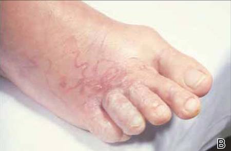
Rarely, CLM can present with bilateral lesions5; in severe cases a single patient can have hundreds of lesions. It also may present as folliculitis and urticarial papules.6 Shih et al7 reported a patient with CLM that presented as a diffuse papular urticarialike eruption following a trip to Thailand. This case may represent an underdiagnosed presentation of CLM. Patients with a history of exposure to contaminated sand or soil diffusely on the body may exhibit lesions in less classic locations, such as the trunk and upper proximal extremities.3
Cutaneous larva migrans is a self-limited eruption, as the larvae cannot complete their lifecycles in the human body and typically die within 2 to 8 weeks.2 However, rare cases lasting up to a year have been reported.3 Sarasombath and Young2 reported a case of CLM that persisted for 4 months with intermittent symptoms characterized by several weeklong intervals with no symptoms or visible rash.
Cutaneous larva migrans typically presents with isolated dermatologic symptoms. Rare cases associated with Löffler syndrome characterized by migratory pulmonary infiltrates and peripheral eosinophilia have been reported.8 Two proposed mechanisms for pulmonary involvement include direct invasion of the lungs by the helminths and a systemic immunologic process triggered by the helminths, resulting in eosinophilic pulmonary infiltration.9
Diagnosis
Cutaneous larva migrans is a clinical diagnosis and skin biopsy usually is not obtained because the larvae often are located 1 to 2 cm beyond the visible erythematous border.3,5 Rarely, the parasites are found on biopsy, revealing larvae that are 0.5-mm thick and up to 10-mm long.10 The larvae typically are confined to the deep epidermis because the parasite lacks the collagenase required to penetrate the basement membrane.2
Langley et al11 showed that confocal scanning laser microscopy can be an effective method for identifying the highly refractile oval larva that disrupt the normal honeycomb pattern of the epidermis. Performing a 4-mm punch biopsy over the identified site can allow for precise excision and treatment of the intact hookworm larvae of CLM. There also are limited reports of dermoscopy being used to facilitate diagnosis of CLM.12 Dermoscopic features of CLM include translucent, brown, structureless areas in a segmental arrangement corresponding to the larval bodies and red-dotted vessels corresponding to an empty burrow.13 However, Zalaudek et al13 concluded that the efficacy of dermoscopy in aiding in the diagnosis of CLM has not been fully established.
Treatment
Cutaneous larva migrans is a self-limited condition that often resolves within 2 to 8 weeks; however, pruritus can be intense and patients therefore are seldom willing to forego treatment. Treatment options include a single oral dose of albendazole 400 mg in adults, with increased efficacy if administered daily for 3 to 5 days (or 10–15 mg/kg, with a maximum dose of 800 mg daily in children), a single oral dose of ivermectin 12 mg in adults (or 150 µg/kg in children), or topical application of thiabendazole 10% to 15% three times daily for at least 15 days.14 Cases of CLM complicated by Löffler syndrome may require a longer treatment course, such as a 7-day course of albendazole 400 mg daily. Tan and Liu9 reported a case of CLM complicated by Löffler syndrome that was successfully treated with albendazole. In this patient, initial treatment with 2 courses of mebendazole (3 days each for a total of 6 days) resulted in improvement of cutaneous lesions but not the pulmonary infiltrate. A subsequent prolonged course of albendazole and intravenous hydrocortisone for 5 days resulted in complete resolution of the pulmonary infiltrate and peripheral eosinophilia. The authors concluded that inadequacy of treatment with mebendazole may be related to differences in the rate of absorption and efficacy when compared to albendazole.9
Conclusion
Cutaneous larva migrans is a self-limited and pruritic skin eruption that is acquired after direct inoculation with sand or soil that is contaminated with feces containing A braziliense or A caninum. Although the classic presentation is readily identifiable, there are a variety of atypical presentations that may go undiagnosed. Symptomatic relief usually can be achieved with short courses of oral or topical antihelminth medications.
1. Berlin JM, Goldberg SJ, McDonough RD, et al. JAAD grand rounds quiz. serpiginous eruption on the leg. J Am Acad Dermatol. 2010;63:921-922.
2. Sarasombath PA, Young PK. An unusual presentation of cutaneous larva migrans. Arch Dermatol. 2007;143:955.
3. Patel S, Aboutalebi S, Vindhya PL, et al. What’s eating you? extensive cutaneous larva migrans (Ancylostoma braziliense). Cutis. 2008;82:239-240.
4. Elston DM, Czarnik K, Brockett R, et al. What’s eating you? Strongyloides stercoralis. Cutis. 2003;71:22-24.
5. Duarte De Sousa ICV, De La Pascua L. Bilateral cutaneous larva migrans [poster reference number 4677]. J Am Acad Dermatol. 2012;66(4, suppl 1):AB106.
6. Caumes E, Ly F, Bricaire F. Cutaneous larva migrans with folliculitis: report of seven cases and review of the literature. Br J Dermatol. 2002;146:314-316.
7. Shih PY, Hsieh MY, Huang YH, et al. Multiple pruritic erythematous papules on the trunk after a trip to Thailand–quiz case. Arch Dermatol. 2010;146:557-562.
8. Wright DO, Gold ED. Löffler’s syndrome associated with creeping eruption (cutaneous helminthiasis): report of twenty-six cases. Arch Intern Med. 1946;78:303-312.
9. Tan SK, Liu TT. Cutaneous larva migrans complicated by Löffler’s syndrome. Arch Dermatol. 2010;146:210-212.
10. Rapini RP, ed. Practical Dermatopathology. Philadelphia, PA: Elsevier; 2005.
11. Langley R, Webb A, Haldane D, et al. Confocal microscopy of cutaneous larva migrans. J Am Acad Dermatol. 2011;64(2, suppl 1):AB100.
12. Aljasser MI, Lui H, Zeng H, et al. Dermoscopy and near-infrared fluorescence imaging of cutaneous larva migrans. Photodermatol Photoimmunol Photomed. 2013;29:337-338.
13. Zalaudek I, Giacomel J, Cabo H, et al. Entodermoscopy: a new tool for diagnosing skin infections and infestations. Dermatology. 2008;216:14-23.
14. Caumes E. Treatment of cutaneous larva migrans. Clin Infect Dis. 2000;30:811-814.
Cutaneous larva migrans (CLM), also known as creeping eruption, is a pruritic serpiginous eruption caused by the migration of animal hookworm larvae through the epidermis.1,2 The most common parasites are Ancylostoma braziliense (common in dogs and cats) and Ancylostoma caninum (common in dogs).1
Disease Transmission
The infection is typically acquired in warm climates and tropical areas after coming in direct contact with sand or soil that is contaminated with animal feces. Therefore, the eruption most commonly occurs as a single or unilateral erythematous, pruritic, serpiginous tract on the feet, hands, or buttocks (Figure).2 The larval tract typically migrates at a rate of 1 to 2 cm per day,3 which is in contrast to the serpiginous urticarial rash of larva currens of strongyloidiasis that can travel up to 10 cm per hour.4
|
|
Clinical Presentation
Rarely, CLM can present with bilateral lesions5; in severe cases a single patient can have hundreds of lesions. It also may present as folliculitis and urticarial papules.6 Shih et al7 reported a patient with CLM that presented as a diffuse papular urticarialike eruption following a trip to Thailand. This case may represent an underdiagnosed presentation of CLM. Patients with a history of exposure to contaminated sand or soil diffusely on the body may exhibit lesions in less classic locations, such as the trunk and upper proximal extremities.3
Cutaneous larva migrans is a self-limited eruption, as the larvae cannot complete their lifecycles in the human body and typically die within 2 to 8 weeks.2 However, rare cases lasting up to a year have been reported.3 Sarasombath and Young2 reported a case of CLM that persisted for 4 months with intermittent symptoms characterized by several weeklong intervals with no symptoms or visible rash.
Cutaneous larva migrans typically presents with isolated dermatologic symptoms. Rare cases associated with Löffler syndrome characterized by migratory pulmonary infiltrates and peripheral eosinophilia have been reported.8 Two proposed mechanisms for pulmonary involvement include direct invasion of the lungs by the helminths and a systemic immunologic process triggered by the helminths, resulting in eosinophilic pulmonary infiltration.9
Diagnosis
Cutaneous larva migrans is a clinical diagnosis and skin biopsy usually is not obtained because the larvae often are located 1 to 2 cm beyond the visible erythematous border.3,5 Rarely, the parasites are found on biopsy, revealing larvae that are 0.5-mm thick and up to 10-mm long.10 The larvae typically are confined to the deep epidermis because the parasite lacks the collagenase required to penetrate the basement membrane.2
Langley et al11 showed that confocal scanning laser microscopy can be an effective method for identifying the highly refractile oval larva that disrupt the normal honeycomb pattern of the epidermis. Performing a 4-mm punch biopsy over the identified site can allow for precise excision and treatment of the intact hookworm larvae of CLM. There also are limited reports of dermoscopy being used to facilitate diagnosis of CLM.12 Dermoscopic features of CLM include translucent, brown, structureless areas in a segmental arrangement corresponding to the larval bodies and red-dotted vessels corresponding to an empty burrow.13 However, Zalaudek et al13 concluded that the efficacy of dermoscopy in aiding in the diagnosis of CLM has not been fully established.
Treatment
Cutaneous larva migrans is a self-limited condition that often resolves within 2 to 8 weeks; however, pruritus can be intense and patients therefore are seldom willing to forego treatment. Treatment options include a single oral dose of albendazole 400 mg in adults, with increased efficacy if administered daily for 3 to 5 days (or 10–15 mg/kg, with a maximum dose of 800 mg daily in children), a single oral dose of ivermectin 12 mg in adults (or 150 µg/kg in children), or topical application of thiabendazole 10% to 15% three times daily for at least 15 days.14 Cases of CLM complicated by Löffler syndrome may require a longer treatment course, such as a 7-day course of albendazole 400 mg daily. Tan and Liu9 reported a case of CLM complicated by Löffler syndrome that was successfully treated with albendazole. In this patient, initial treatment with 2 courses of mebendazole (3 days each for a total of 6 days) resulted in improvement of cutaneous lesions but not the pulmonary infiltrate. A subsequent prolonged course of albendazole and intravenous hydrocortisone for 5 days resulted in complete resolution of the pulmonary infiltrate and peripheral eosinophilia. The authors concluded that inadequacy of treatment with mebendazole may be related to differences in the rate of absorption and efficacy when compared to albendazole.9
Conclusion
Cutaneous larva migrans is a self-limited and pruritic skin eruption that is acquired after direct inoculation with sand or soil that is contaminated with feces containing A braziliense or A caninum. Although the classic presentation is readily identifiable, there are a variety of atypical presentations that may go undiagnosed. Symptomatic relief usually can be achieved with short courses of oral or topical antihelminth medications.
1. Berlin JM, Goldberg SJ, McDonough RD, et al. JAAD grand rounds quiz. serpiginous eruption on the leg. J Am Acad Dermatol. 2010;63:921-922.
2. Sarasombath PA, Young PK. An unusual presentation of cutaneous larva migrans. Arch Dermatol. 2007;143:955.
3. Patel S, Aboutalebi S, Vindhya PL, et al. What’s eating you? extensive cutaneous larva migrans (Ancylostoma braziliense). Cutis. 2008;82:239-240.
4. Elston DM, Czarnik K, Brockett R, et al. What’s eating you? Strongyloides stercoralis. Cutis. 2003;71:22-24.
5. Duarte De Sousa ICV, De La Pascua L. Bilateral cutaneous larva migrans [poster reference number 4677]. J Am Acad Dermatol. 2012;66(4, suppl 1):AB106.
6. Caumes E, Ly F, Bricaire F. Cutaneous larva migrans with folliculitis: report of seven cases and review of the literature. Br J Dermatol. 2002;146:314-316.
7. Shih PY, Hsieh MY, Huang YH, et al. Multiple pruritic erythematous papules on the trunk after a trip to Thailand–quiz case. Arch Dermatol. 2010;146:557-562.
8. Wright DO, Gold ED. Löffler’s syndrome associated with creeping eruption (cutaneous helminthiasis): report of twenty-six cases. Arch Intern Med. 1946;78:303-312.
9. Tan SK, Liu TT. Cutaneous larva migrans complicated by Löffler’s syndrome. Arch Dermatol. 2010;146:210-212.
10. Rapini RP, ed. Practical Dermatopathology. Philadelphia, PA: Elsevier; 2005.
11. Langley R, Webb A, Haldane D, et al. Confocal microscopy of cutaneous larva migrans. J Am Acad Dermatol. 2011;64(2, suppl 1):AB100.
12. Aljasser MI, Lui H, Zeng H, et al. Dermoscopy and near-infrared fluorescence imaging of cutaneous larva migrans. Photodermatol Photoimmunol Photomed. 2013;29:337-338.
13. Zalaudek I, Giacomel J, Cabo H, et al. Entodermoscopy: a new tool for diagnosing skin infections and infestations. Dermatology. 2008;216:14-23.
14. Caumes E. Treatment of cutaneous larva migrans. Clin Infect Dis. 2000;30:811-814.
Practice Points
- Classic cutaneous larva migrans (CLM) presents with a unilateral, serpiginous, pruritic eruption on the hands, feet, or buttocks following direct contact with sand or soil that is contaminated with Ancylostoma braziliense or Ancylostoma caninum.
- Atypical presentations of CLM include bilateral distribution; folliculitis and urticarial plaques; prolonged cases lasting up to 1 year; and Löffler syndrome characterized by migratory pulmonary infiltrates and peripheral eosinophilia.
- Cutaneous larva migrans is self-limited, but treatment often is necessary due to intense pruritus. Treatment options include a single oral dose of albendazole or ivermectin, topical thiabendazole, and prolonged courses of oral albendazole in cases complicated by Löffler syndrome.
Tide turns in favor of multivessel PCI in STEMI
SNOWMASS, COLO. – Recent data seem to refute the 2013 American Heart Association/American College of Cardiology class III recommendation to avoid multivessel percutaneous coronary intervention at the time of primary PCI for ST-elevation MI, Dr. David R. Holmes Jr. observed at the Annual Cardiovascular Conference at Snowmass.
“The current AHA/ACC guidelines for STEMI should be and are being reevaluated regarding clarifications for the indications and timing of non–infarct artery revascularization,” according to Dr. Holmes, a cardiologist at the Mayo Clinic in Rochester, Minn., and an ACC past president.

Indeed, the ACC has already withdrawn from its ‘Choosing Wisely’ campaign its former recommendation discouraging multivessel revascularization at the time of primary PCI for STEMI. The college cited “new science showing that complete revascularization of all significant blocked arteries leads to better outcomes in some heart attack patients.”
Dr. Holmes was coauthor of a meta-analysis of 4 prospective and 14 retrospective studies involving more than 40,000 patients that concluded multivessel PCI for STEMI should be discouraged, and that significant nonculprit lesions should only be treated during staged procedures (J. Am. Coll. Cardiol. 2011;58:692-703). This meta-analysis was influential in the creation of the class III ‘don’t do it’ recommendation in the AHA/ACC guidelines. But Dr. Holmes said that in hindsight, the data included in the meta-analysis were something of a mishmash and “wound up being very hard to interpret.”
Greater clarity has been brought by two more recent randomized trials: PRAMI and CvLPRIT. Both were relatively small by cardiology standards, but they ended up showing similarly striking advantages in favor of using the STEMI hospitalization to perform preventive PCI of both the infarct-related artery and non–infarct arteries with major stenoses.
PRAMI included 465 acute STEMI patients who underwent infarct artery PCI and were then randomized to preventive PCI or infarct artery–only PCI. At a mean follow-up of 23 months, the preventive multivessel PCI group had a 65% reduction in the relative risk of the primary outcome, a composite of cardiac death, nonfatal MI, or refractory angina (N. Engl. J. Med. 2013;369:1115-23).
The yet-to-be-published CvLPRIT study was presented at the 2014 European Society of Cardiology meeting in Barcelona. The multicenter study included 296 STEMI patients with angiographically established significant multivessel disease who were randomized to primary PCI of the culprit vessel only or to complete revascularization. The primary outcome, the 12-month composite of all-cause mortality, recurrent MI, heart failure, or ischemia-driven revascularization, occurred in 10% of the complete revascularization group, compared with 21.2% of patients assigned to culprit artery–only PCI.
Also at the ESC conference, CvLPRIT investigator Dr. Anthony Gershlick of the University of Leicester (England) presented a meta-analysis combining the weighted results of PRAMI, CvLPRIT, and two earlier randomized trials: HELP AMI (Int. J. Cardiovasc. Intervent. 2004;6:128-33) and an Italian trial (Heart 2010;96:662-7). The results strongly favored multivessel PCI, with a 45% reduction in mortality and a 61% decrease in recurrent MI, compared with culprit vessel–only PCI at the time of admission for STEMI.
“Maybe there aren’t any innocent bystanders,” commented Dr. Holmes. “Maybe if you have somebody who has multivessel disease and you see something you think might be an innocent bystander but is a significant lesion, maybe it’s not so innocent. Maybe by treating them all at the time of the initial intervention the patient is going to do better.”
He reported having no financial conflicts of interest regarding his presentation.
SNOWMASS, COLO. – Recent data seem to refute the 2013 American Heart Association/American College of Cardiology class III recommendation to avoid multivessel percutaneous coronary intervention at the time of primary PCI for ST-elevation MI, Dr. David R. Holmes Jr. observed at the Annual Cardiovascular Conference at Snowmass.
“The current AHA/ACC guidelines for STEMI should be and are being reevaluated regarding clarifications for the indications and timing of non–infarct artery revascularization,” according to Dr. Holmes, a cardiologist at the Mayo Clinic in Rochester, Minn., and an ACC past president.
Indeed, the ACC has already withdrawn from its ‘Choosing Wisely’ campaign its former recommendation discouraging multivessel revascularization at the time of primary PCI for STEMI. The college cited “new science showing that complete revascularization of all significant blocked arteries leads to better outcomes in some heart attack patients.”
Dr. Holmes was coauthor of a meta-analysis of 4 prospective and 14 retrospective studies involving more than 40,000 patients that concluded multivessel PCI for STEMI should be discouraged, and that significant nonculprit lesions should only be treated during staged procedures (J. Am. Coll. Cardiol. 2011;58:692-703). This meta-analysis was influential in the creation of the class III ‘don’t do it’ recommendation in the AHA/ACC guidelines. But Dr. Holmes said that in hindsight, the data included in the meta-analysis were something of a mishmash and “wound up being very hard to interpret.”
Greater clarity has been brought by two more recent randomized trials: PRAMI and CvLPRIT. Both were relatively small by cardiology standards, but they ended up showing similarly striking advantages in favor of using the STEMI hospitalization to perform preventive PCI of both the infarct-related artery and non–infarct arteries with major stenoses.
PRAMI included 465 acute STEMI patients who underwent infarct artery PCI and were then randomized to preventive PCI or infarct artery–only PCI. At a mean follow-up of 23 months, the preventive multivessel PCI group had a 65% reduction in the relative risk of the primary outcome, a composite of cardiac death, nonfatal MI, or refractory angina (N. Engl. J. Med. 2013;369:1115-23).
The yet-to-be-published CvLPRIT study was presented at the 2014 European Society of Cardiology meeting in Barcelona. The multicenter study included 296 STEMI patients with angiographically established significant multivessel disease who were randomized to primary PCI of the culprit vessel only or to complete revascularization. The primary outcome, the 12-month composite of all-cause mortality, recurrent MI, heart failure, or ischemia-driven revascularization, occurred in 10% of the complete revascularization group, compared with 21.2% of patients assigned to culprit artery–only PCI.
Also at the ESC conference, CvLPRIT investigator Dr. Anthony Gershlick of the University of Leicester (England) presented a meta-analysis combining the weighted results of PRAMI, CvLPRIT, and two earlier randomized trials: HELP AMI (Int. J. Cardiovasc. Intervent. 2004;6:128-33) and an Italian trial (Heart 2010;96:662-7). The results strongly favored multivessel PCI, with a 45% reduction in mortality and a 61% decrease in recurrent MI, compared with culprit vessel–only PCI at the time of admission for STEMI.
“Maybe there aren’t any innocent bystanders,” commented Dr. Holmes. “Maybe if you have somebody who has multivessel disease and you see something you think might be an innocent bystander but is a significant lesion, maybe it’s not so innocent. Maybe by treating them all at the time of the initial intervention the patient is going to do better.”
He reported having no financial conflicts of interest regarding his presentation.
EXPERT ANALYSIS FROM THE CARDIOVASCULAR CONFERENCE AT SNOWMASS

Photographic AK counting isn’t ready for prime time
Can photographs be as good as clinical examination for counting actinic keratoses?
Depending on the physician, AK counts varied widely on clinical assessment as well as in photos, reported Dr. Sudipta Sinnya of the Dermatology Research Centre at the University of Queensland, Brisbane, and associates. However, based on current two-dimensional technology, clinical counting yields superior results, said the researchers, who studied the counts of five trained observers carried out in two sessions with six patients.
“As technological advancements occur, three-dimensional photography will largely supersede two-dimensional photographs in clinical practice, and more robust image-capturing techniques should improve the accuracy of photographic counting,” the researchers noted.
Read the full article from Acta Dermato-Venereologica (2015 [doi:10.2340/00015555-2040]) here.