User login
How to assess the merits of psychological and neuropsychological test evaluations
Psychological and neuropsychological test evaluations, like all consultative diagnostic services, can vary in quality and clinical utility. Many of these examinations provide valuable insights and helpful recommendations; regrettably, some assessments are only marginally beneficial and can contribute to diagnostic confusion and uncertainty.
When weighing the pros and cons of evaluations, consider these best practices.
Gold-standard tests ought to be in-cluded in the assessment. These include (but are not limited to) the Wechsler Adult Intelligence Scale-Fourth Edition (WAIS-IV); Wechsler Memory Scale-Fourth Edition (WMS-IV); Delis-Kaplan Executive Function System (D-KEFS); Wechsler Individual Achievement Test-Third Edition (WIAT-III); and the Minnesota Multiphasic Personality Inventory-2 (MMPI-2). These tests have a strong evidence base that:
• demonstrates good reliability (ie, produce consistent and accurate scores across examiners and time intervals and are relatively free of measurement error)
• demonstrates good validity (ie, have been shown to measure aspects of psychological and neuropsychological functioning that they claim to measure).
Many gold-standard tests are normed on national samples and are stratified by age, sex, ethnicity or race, educational level, and geographic region. They also include normative data based on the performance of patients who have neuropsychiatric syndromes often seen by psychiatrists in practice.1
The test battery ought to comprise cognitive and neuropsychological measures as well as affective and behavioral measures. When feasible, these tests should be supplemented by informant-based measures of neuropsychiatric functioning to obtain a comprehensive assessment of the patient’s capacities and skills.
An estimated premorbid baseline should be established. This is done by taking a relevant history and administering tests, such as the National Adult Reading Test (NART), that can be used to compare against current test performance. This testing-in-context approach helps differentiate long-term limitations in information processing, which might be attributed to a DSM-5 intellectual disability, specific learning disorder, or other neurodevelopmental disorder, from a known or suspected recent neurobehavioral change.
Tests in the assessment should tap a broad set of neurobehavioral functions. Doing so ensures that, when a patient is referred with a change in cognition or other aspects of mental status, it will be easier to determine whether clinically significant score discrepancies exist across different ability and skill domains. Such dissociations in performance can have important implications for the differential diagnosis and everyday functioning.
Tests that are sensitive to a patient’s over-reporting of symptoms should be used as part of the evaluation in cases of suspected malingering—especially subtle simulation that might elude identification with brief screening-level measures.2 These tests can include the Test of Memory Malingering (TOMM) and the Structured Interview of Reported Symptoms, 2nd edition (SIRS-2).
Test recommendations ought to be grounded in findings; practical; and relatively easy to implement. They also should be consistent with the treatment setting and the patient’s lifestyle, values, and treatment preferences.3
Disclosure
Dr. Pollak reports no financial relationships with any company whose products are mentioned in this article or with manufacturers of competing products.
1. Geisinger KF, Bracken BA, Carlson JF, et al, eds. APA handbook of testing and assessment in psychology. Washington, DC: American Psychological Association Press; 2013.
2. Brady MC, Scher LM, Newman W. “I just saw Big Bird. He was 100 feet tall!” Malingering in the emergency department. Current Psychiatry. 2013;12(10):33-38,40.
3. McHugh RK, Whitton SW, Peckham AD, et al. Patient p for psychological vs pharmacologic treatment of psychiatric disorders: a meta-analytic review. J Clin Psychiatry. 2013;74(6):595-602.
Psychological and neuropsychological test evaluations, like all consultative diagnostic services, can vary in quality and clinical utility. Many of these examinations provide valuable insights and helpful recommendations; regrettably, some assessments are only marginally beneficial and can contribute to diagnostic confusion and uncertainty.
When weighing the pros and cons of evaluations, consider these best practices.
Gold-standard tests ought to be in-cluded in the assessment. These include (but are not limited to) the Wechsler Adult Intelligence Scale-Fourth Edition (WAIS-IV); Wechsler Memory Scale-Fourth Edition (WMS-IV); Delis-Kaplan Executive Function System (D-KEFS); Wechsler Individual Achievement Test-Third Edition (WIAT-III); and the Minnesota Multiphasic Personality Inventory-2 (MMPI-2). These tests have a strong evidence base that:
• demonstrates good reliability (ie, produce consistent and accurate scores across examiners and time intervals and are relatively free of measurement error)
• demonstrates good validity (ie, have been shown to measure aspects of psychological and neuropsychological functioning that they claim to measure).
Many gold-standard tests are normed on national samples and are stratified by age, sex, ethnicity or race, educational level, and geographic region. They also include normative data based on the performance of patients who have neuropsychiatric syndromes often seen by psychiatrists in practice.1
The test battery ought to comprise cognitive and neuropsychological measures as well as affective and behavioral measures. When feasible, these tests should be supplemented by informant-based measures of neuropsychiatric functioning to obtain a comprehensive assessment of the patient’s capacities and skills.
An estimated premorbid baseline should be established. This is done by taking a relevant history and administering tests, such as the National Adult Reading Test (NART), that can be used to compare against current test performance. This testing-in-context approach helps differentiate long-term limitations in information processing, which might be attributed to a DSM-5 intellectual disability, specific learning disorder, or other neurodevelopmental disorder, from a known or suspected recent neurobehavioral change.
Tests in the assessment should tap a broad set of neurobehavioral functions. Doing so ensures that, when a patient is referred with a change in cognition or other aspects of mental status, it will be easier to determine whether clinically significant score discrepancies exist across different ability and skill domains. Such dissociations in performance can have important implications for the differential diagnosis and everyday functioning.
Tests that are sensitive to a patient’s over-reporting of symptoms should be used as part of the evaluation in cases of suspected malingering—especially subtle simulation that might elude identification with brief screening-level measures.2 These tests can include the Test of Memory Malingering (TOMM) and the Structured Interview of Reported Symptoms, 2nd edition (SIRS-2).
Test recommendations ought to be grounded in findings; practical; and relatively easy to implement. They also should be consistent with the treatment setting and the patient’s lifestyle, values, and treatment preferences.3
Disclosure
Dr. Pollak reports no financial relationships with any company whose products are mentioned in this article or with manufacturers of competing products.
1. Geisinger KF, Bracken BA, Carlson JF, et al, eds. APA handbook of testing and assessment in psychology. Washington, DC: American Psychological Association Press; 2013.
2. Brady MC, Scher LM, Newman W. “I just saw Big Bird. He was 100 feet tall!” Malingering in the emergency department. Current Psychiatry. 2013;12(10):33-38,40.
3. McHugh RK, Whitton SW, Peckham AD, et al. Patient p for psychological vs pharmacologic treatment of psychiatric disorders: a meta-analytic review. J Clin Psychiatry. 2013;74(6):595-602.
Physician groups: Fix interoperability before advancing with meaningful use
Physician groups are growing increasingly frustrated with the focus on meaningful use of electronic health records at the expense of creating an interoperable health information technology infrastructure and are calling on the Department of Health & Human Services to step up on interoperability.
In an Oct. 15 letter to HHS Secretary Sylvia Burwell, a number of groups cited the HHS Office of the National Coordinator for Health Information Technology’s finding that less than 14% of physicians are able to electronically transmit health information outside of their organization because of a lack of EHR interoperability and other issues.

“These barriers to data exchange proliferated as a result of a variety of factors [including] strict MU [meaningful use] requirements and deadlines that do not provide sufficient time to focus on achieving interoperability. This dynamic is also in part due to the strict EHR certification requirements that have forced all stakeholders involved to focus on meeting MU measures as opposed to developing more innovative technological solutions that will enhance patient care and safety while growing the marketplace.”
Groups signing the letter include the American Academy of Family Physicians, American Medical Association, Medical Group Management Association, National Rural Health Association and a number of health care systems.
The letter also notes that in addition to interoperability, usability remains an issue that causes disruption in provider workflow and diverts resources away from patient care, noting that “vendors are limited from addressing these concerns as they focus on meeting increasingly complex certification requirements.”
Among its recommendations, the groups asked for HHS to recognize that the vendor community needs time to develop, test, and implement updates to meet new criteria and should be afforded that time “before continuing on with subsequent stages of the MU program. Testing and achievement of specific performance benchmarks should occur before providers are held accountable for any MU requirements.”
The letter comes as an advisory committee to the Office of the National Coordinator (ONC) is making a same-day recommendation that it delays or staggers meaningful use stage 3 to shift focus on achieving meaningful interoperability and addressing other infrastructure issues.
In its October 2014 report to Congress, the ONC acknowledged issues related to interoperability and other issues that are presenting a barrier for health IT to achieve potential.
“Despite progress in establishing standards and services to support health information exchange and interoperability, practice patterns have not changed to the point that health care providers share patient health information electronically across organizational, vendor, and geographic boundaries,” the report states. “Patient electronic health information needs to be available for appropriate use in solving major challenges, such as providing more effective care and informing and accelerating scientific research.”
To that end, ONC released during an Oct. 15 advisory committee meeting some top-level aspects of its 10-year framework on how it will improve interoperability, which is scheduled to be formalized in March 2015.
According to draft materials, the roadmap calls for health care providers to be able to send, receive, find, and use a basic set of essential health information. By 2020, more granular information should be accessible across systems, which would lead to improved quality and reduced costs. By 2024, the interoperability vision, with systems communicating in full, will lead to a learning health system and facilitate ubiquitous precision medicine.
Separately, AMA in an Oct. 14 letter to CMS and ONC criticized the meaningful use program and offered a series of recommendations to fix it before movement to stage 3 of the program. The group wants to see more flexibility in requirements physicians need to meet requirements, expanding hardship exemptions for all stages, improving quality reporting, and addressing physician EHR usability challenges.
“Many of the MU requirements were designed to increase patient choice and quality of care,” the AMA writes. “Unfortunately, many of these requirements, especially those in the latter phases of the MU program, are having the opposite effect. Oftentimes the requirements decrease the efficiency of patient visits.”
AMA also called on CMS and ONC to “study the total cost of compliance with MU to understand the impact this program is having on practice.”
Physician groups are growing increasingly frustrated with the focus on meaningful use of electronic health records at the expense of creating an interoperable health information technology infrastructure and are calling on the Department of Health & Human Services to step up on interoperability.
In an Oct. 15 letter to HHS Secretary Sylvia Burwell, a number of groups cited the HHS Office of the National Coordinator for Health Information Technology’s finding that less than 14% of physicians are able to electronically transmit health information outside of their organization because of a lack of EHR interoperability and other issues.
“These barriers to data exchange proliferated as a result of a variety of factors [including] strict MU [meaningful use] requirements and deadlines that do not provide sufficient time to focus on achieving interoperability. This dynamic is also in part due to the strict EHR certification requirements that have forced all stakeholders involved to focus on meeting MU measures as opposed to developing more innovative technological solutions that will enhance patient care and safety while growing the marketplace.”
Groups signing the letter include the American Academy of Family Physicians, American Medical Association, Medical Group Management Association, National Rural Health Association and a number of health care systems.
The letter also notes that in addition to interoperability, usability remains an issue that causes disruption in provider workflow and diverts resources away from patient care, noting that “vendors are limited from addressing these concerns as they focus on meeting increasingly complex certification requirements.”
Among its recommendations, the groups asked for HHS to recognize that the vendor community needs time to develop, test, and implement updates to meet new criteria and should be afforded that time “before continuing on with subsequent stages of the MU program. Testing and achievement of specific performance benchmarks should occur before providers are held accountable for any MU requirements.”
The letter comes as an advisory committee to the Office of the National Coordinator (ONC) is making a same-day recommendation that it delays or staggers meaningful use stage 3 to shift focus on achieving meaningful interoperability and addressing other infrastructure issues.
In its October 2014 report to Congress, the ONC acknowledged issues related to interoperability and other issues that are presenting a barrier for health IT to achieve potential.
“Despite progress in establishing standards and services to support health information exchange and interoperability, practice patterns have not changed to the point that health care providers share patient health information electronically across organizational, vendor, and geographic boundaries,” the report states. “Patient electronic health information needs to be available for appropriate use in solving major challenges, such as providing more effective care and informing and accelerating scientific research.”
To that end, ONC released during an Oct. 15 advisory committee meeting some top-level aspects of its 10-year framework on how it will improve interoperability, which is scheduled to be formalized in March 2015.
According to draft materials, the roadmap calls for health care providers to be able to send, receive, find, and use a basic set of essential health information. By 2020, more granular information should be accessible across systems, which would lead to improved quality and reduced costs. By 2024, the interoperability vision, with systems communicating in full, will lead to a learning health system and facilitate ubiquitous precision medicine.
Separately, AMA in an Oct. 14 letter to CMS and ONC criticized the meaningful use program and offered a series of recommendations to fix it before movement to stage 3 of the program. The group wants to see more flexibility in requirements physicians need to meet requirements, expanding hardship exemptions for all stages, improving quality reporting, and addressing physician EHR usability challenges.
“Many of the MU requirements were designed to increase patient choice and quality of care,” the AMA writes. “Unfortunately, many of these requirements, especially those in the latter phases of the MU program, are having the opposite effect. Oftentimes the requirements decrease the efficiency of patient visits.”
AMA also called on CMS and ONC to “study the total cost of compliance with MU to understand the impact this program is having on practice.”
Point/Counterpoint: Endo first for the treatment of infrainguinal PAD?
The BASIL study originally published in the Lancet in 2005 (366:1925-34) and subsequently reiterated in multiple publications proposes that an endovascular approach should be utilized as the first invasive treatment modality in patients with infrainguinal peripheral arterial disease whose life expectancy is less than 2 years. By contrast, those patients expected to live beyond 2 years usually should be offered bypass surgery first, especially where a vein is available as a conduit. However, as can be seen from this month’s Point/Counterpoint by Dr. George Meier III and Dr. Michael S. Conte, the debate still rages as to the benefit of open vs. endovascular procedures for these patients. We encourage readers to voice their opinions in our “Letters to the Editor” section, as well as by participating in our web-based Quick Poll to the right of this story. - Dr. Russell Samson, Medical Editor, Vascular Specialist
POINT/COUNTERPOINT
Yes, endo is generally the way to go.
By Dr. George Meier III
Endovascular treatment of lower-extremity arterial disease has rapidly expanded, now approaching the standard for treatment of patients with lower-extremity disease. While open bypass remains a gold standard for the clinical treatment of limb-threatening ischemia, there are many limitations to the use of open surgery.
First and foremost, open surgical intervention represents pain and suffering for the patient as well as a delayed recovery, compared with endovascular treatment. All other factors being equal, most patients would prefer a less-invasive approach to minimize these factors and to maximize the speed of recovery. Open surgical interventions typically require 6 weeks or longer to get back to a functional status even remotely close to the patient’s initial level of function. With more severe tissue loss or with greater pain preoperatively, the difficulty of getting the patient back to full functional status remains problematic. In John Porter’s classic paper published in the Journal of Vascular Surgery in 1998,1 wound complications occurred in 24% of the patients with a 5-year survival rate of only 49% in this relatively young population, average age 66 years. Repeat operations to maintain graft patency, treat wound complications, or treat recurrent or contralateral ischemia were required in 54% of the patients and 23% ultimately required major limb amputation.
Of the 112 patients in this study, only 14.3% achieved the ideal surgical result of an uncomplicated operation: long-term symptom relief, maintenance of functional status, and no recurrence or repeat operations. These are sobering statistics for anyone facing open revascularization for critical limb ischemia.
The BASIL trial is often put forward as an example of the best data available currently to define patient treatments in patients with critical limb ischemia.2 Despite this, BASIL was a flawed trial from the beginning because of the difficulties of truly randomizing patients with vascular disease to open surgical treatment vs. percutaneous treatment. First, all patients had to be appropriate candidates for both open and endovascular treatment. In the real world, we readily recognize that the luxury of this choice is not available to many of our patients. A lack of conduit or increased surgical risk results in endovascular treatment being the only management option for many. Additionally, patient preference increasingly plays a role in treatment selection, obviously increasing the likelihood of less invasive percutaneous treatment. Yes, the mortality of open bypass in BASIL is reported to be in the 1%-3% range, but this population has been carefully selected based on screening and treatment of underlying cardiac disease. The true incidence of cardiovascular disease is impossible to determine since significant cardiac disease negated randomization.
Unfortunately, even with all of the advances in endovascular treatments the results of percutaneous treatment have never reached the results of open bypass. Nonetheless, while the success may not be as great the risks are not as high either. The main challenge to endovascular treatment is the durability of the intervention. While we can usually treat pre-existing disease in the lower-extremity arterial tree, maintaining patency and durability is the challenge. As my esteemed colleague has noted, failure of endovascular treatment in the BASIL trial resulted in significantly worse outcomes for open bypass in those patients. While much was made about this fact when the BASIL trial was published, endovascular treatment after open failure has even a worse outcome than did open treatment after endovascular failure. The truth of the matter is that, for obvious reasons, failure begets failure.
Generally, there are two imaging approaches to defining the extent of vascular disease: first, contrast angiography via percutaneous access; and second, CT angiography using intravenous contrast. If contrast angiography is undertaken to diagnose the extent of disease, then it is a relatively limited extrapolation to treat the patient’s disease percutaneously at the time of the diagnostic angiogram. For this reason, I discuss with all patients coming for diagnostic angiography the issue of endovascular treatment. It is rare that we make patients worse with an attempt at endovascular treatment by an experienced interventionalist. Similarly, it is rare that we alter a bypass level based on an attempt at endovascular treatment. If, in my opinion, the risk of an attempted endovascular treatment is acceptable, then this is done at the time of the diagnostic angiogram. Patients appreciate this discussion prior to proceeding with diagnostic angiography.
What about inadequate autogenous conduit? Even my counterpoint opponent has published a documented 20% risk of absent or inadequate ipsilateral greater saphenous vein.3 While he and his colleagues have documented excellent results using contralateral greater saphenous vein, there is still an inevitable morbidity and, yes, even a mortality risk associated with contralateral leg vein harvest. While in a good cardiac risk patient this may be negligible, we are again facing an ever more complex and medically ill patient population to subject to vascular treatment. It is in this setting that many vascular surgeons move to prosthetic conduits for the treatment of the patient’s vascular disease. While this may provide a short-term fix for the conduit problem, in the long term the risk of sudden, uncompensated failure of limb perfusion by prosthetic graft failure may often result in a higher risk procedure at a time when the patient may be older and more severely limited. Endovascular treatment is clearly a reasonable alternative in patients where autogenous conduit is not readily available.
While this debate will inevitably continue as long as practitioners have bias toward either open or endovascular management of vascular disease, one thing is for certain: We will continue to extend the limits of treatment to ever more ill and complex patients. While we have been very successful at performing fewer and fewer morbid interventions for limb-threatening vascular disease, these patients continue to be increasingly challenging to manage.
As the overall population continues to age, the need for less invasive treatment of limb-threatening vascular disease will continue to grow. And, yes, I agree that vascular surgeons in the role of interventionalist or surgeon are the leadership for the management of CLI in the future.
Dr. Meier is professor and chief of vascular surgery at the University of Cincinnati.
References
1. J. Vasc. Surg. 1998;27:256-63; discussion 264-6
3. J. Vasc. Surg. 2002:35:1085-92
No: A selective approach remains the key.
By Dr. MICHAEL S. CONTE
Recently the term “pandemic” has been applied to the growing global impact of peripheral artery disease (PAD), currently estimated to afflict more than 200 million individuals.1 The term “critical limb ischemia” (CLI), connoting the most advanced stage of PAD with imminent limb threat, is inadequately defined2 but likely encompasses 1%-3% of PAD. Aging of the global population and the increasing prevalence of diabetes are fueling increases in CLI and its impact on public health. While traditionally treated largely by vascular surgeons plying the open bypass trade, the ongoing development and market dispersion of catheter-based technologies for CLI has led to major secular changes.
Recent estimates suggest that upward of 5 billion dollars are spent annually on CLI in the Medicare population.3, 4 Increasing volumes and costs associated with revascularizations for CLI are a major driver, yet recent data suggest that regional spending in the United States is widely disparate and not directly associated with amputation rates.5 Thus defining effectiveness and value in CLI care has become a major challenge to the vascular community.
In current everyday practice, clinicians are faced with making treatment choices for CLI patients based on limited data and lots of anecdote. While the “open vs. endo” debate goes on, in many ways it has become less broadly relevant as the sophisticated clinician recognizes the real challenge lies in defining which approach to apply first in the right patient, at the right time. The only randomized, controlled trial (RCT) done in the field, the BASIL trial,6 is more than a decade old. However its findings remain important. For patients with “severe limb ischemia” likely to survive for at least 2 years, open bypass surgery offered better outcomes over angioplasty as an initial strategy.7 Moreover, the finding in BASIL that patients undergoing bypass after prior failed angioplasty did poorly,8 suggesting “no free lunch” for endovascular failures, has since been confirmed in other large registry studies.9
Simply stated, failure matters in CLI. And although endovascular techniques have continued to improve, the growing epidemic of restenosis shows no signs of abating.10 As in the case of percutaneous coronary intervention (PCI), we will know when endo results are meaningfully improved in PAD when the procedure volume curves actually flatten, not continue to grow geometrically.
So, in selecting the optimal strategy for CLI today, let’s focus on what we seem to know and try to apply an evidence-based mentality. We know that open bypass surgery is an effective and versatile treatment, but one that carries real morbidity (10%-20%) and some mortality (1%-3%). Among many large studies, the PREVENT III multicenter trial provides benchmark data on perioperative and 1-year outcomes.11
We know that the quality of the vein is the critical technical determinant of success, and arterial anatomy is less influential as long as there is outflow to the foot.12 We know that poor quality veins, prosthetics, and other alternatives are much inferior in CLI. And we know that there is a subset of CLI patients who are at high risk for adverse surgical outcomes.13, 14 However one defines them, up to 10% of patients in the large surgical series are in a high-risk group and may not experience meaningful benefit. For endovascular treatments, the data are less clear but certain trends have been consistent. Multilevel disease, long-segment occlusions, heavily calcified lesions, and more advanced tissue loss are negative predictors of clinical success.
Thus at first glance the weaknesses of the two strategies are largely complementary.15 When I encounter an average-risk CLI patient, with an adequate saphenous vein and more than one unfavorable endo factor, I am inclined towards bypass first.
Conversely, endo-favorable anatomy in higher risk patients is a no-brainer. Lots of people fall in the middle, and a significant minority should be considered for primary amputation. Currently my practice is roughly 50% bypass surgery-first in CLI.
Endovascular innovations have made a huge impact on vascular practice, and the leadership of many vascular surgeons (e.g., my esteemed counterpoint author) has been central to advancing the field. Better wires and catheters, retrograde approaches, and drug-eluting technologies continue to be developed at a dizzying pace.
We are all continually learning. Unfortunately, we lack good objective evidence to support most of the expanding armamentarium for CLI. However it is abundantly clear that technical (angiographic) success and clinical success are far apart, which is no surprise. What is surprising is an unsettlingly common lack of honesty about such an obvious fact. Are we all guilty of looking through rose-colored glasses?
Is it really such big news that patency actually matters for most patients with CLI? Technologies will not improve quickly enough if there is no market imperative to make them better. If we continue to buy and use things that are frequently ineffective, or don’t measure it carefully, where is the motivation?
No matter the lens through which one looks at the CLI field, it is desperate for improvement. We need much better technologies that provide longer lasting solutions for patients. We need better diagnostics to predict disease progression and responses to treatment.
We need some new medical or biological therapies that truly alleviate suffering. And we largely lack data on comparative effectiveness, and value, to support thoughtful application of our current treatment arsenal.
Most importantly what we need now is less dogma, and a lot more science. Over more than two decades, multiple RCTs comparing medical, interventional and surgical therapies for coronary artery disease have formed the basis for practice guidelines.
By comparison, our field is nearly incoherent both to vascular specialists and referring physicians. It will not be easy, but this can be done in PAD as well, and the vascular community must embrace it. Moreover it is imperative that vascular surgeons help to lead these multidisciplinary efforts, and develop evidence-based global guidelines to guide best practice in CLI.16 The recent funding of the BEST-CLI trial in the United States and the BASIL-2 trial in the United Kingdom demonstrate the importance to public health and offer great opportunities.
Until better evidence is available, a rational approach to limb salvage requires flexibility, understanding of the factors predicting success/failure for each modality, and the continued use of open bypass surgery as the initial treatment option for a significant number of patients.
And for the sake of our most vulnerable patients, we better keep training vascular surgeons to do all of it well.
Dr. Conte is professor and chief, division of vascular & endovascular surgery and the Edwin J. Wylie, M.D. Chair in Vascular Surgery at the University of California, San Francisco.
References
2. J. Vasc. Surg. 2014;59:220-34
7. J. Vasc. Surg. 2010;51:5S-17S
8. J. Vasc. Surg. 2010;51:18S-31S
9. J. Vasc. Surg. 2011;54:730-6
10. J. Amer. Heart Assoc. 2013;2:e000345
11. J. Vasc. Surg. 2006;43:742-51
12. J. Vasc. Surg. 2007;46:1180-90
13. J. Vasc. Surg. 2009;50:769-75
14. J. Vasc. Surg. 2010;52:1218-25
15. J. Vasc. Surg. 2013;57:8S-13S16. J. Vasc. Surg. 2014;59:510
Link found between sleep disorders and osteoporosis risk
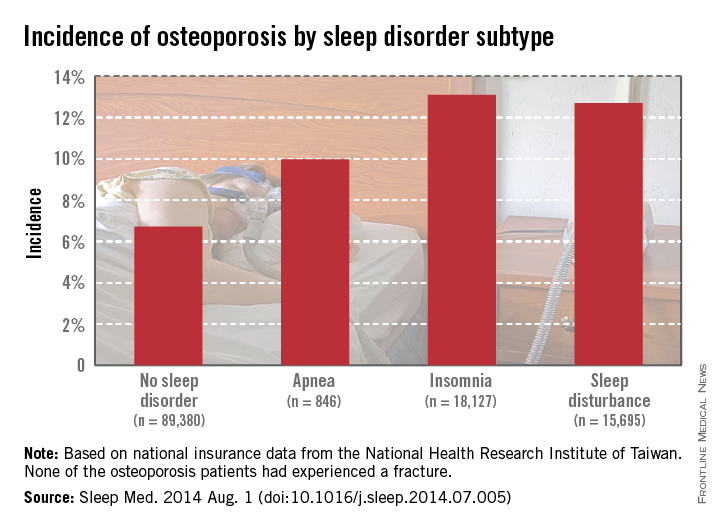
Patients with sleep disorders are much more likely to develop osteoporosis than are those without sleep disorders, according to Dr. Chia-Ming Yen and her associates.
Patients diagnosed with sleep apnea between 1998 and 2001 had an osteoporosis incidence of nearly 10% at the end of 2010, while those without sleep disorders had incidence of 6.7%. Patients with insomnia developed osteoporosis at a rate of 13.1%, and patients with other sleep disturbances had an incidence of 12.7%, Dr. Yen of the National Formosa University in Taiwan, and her associates reported (Sleep Med. 2014 Aug. 1 [doi:10.1016/j.sleep.2014.07.005]).

Women and the elderly were particularly likely to develop osteoporosis if a sleep disorder was present. Of patients aged 64 years and older who were diagnosed with osteoporosis, 36.2% also had sleep apnea, and 31.9% had another sleep disorder. Incidences of osteoporosis in women in all cases were three to five times higher than those in men, and patients with multiple comorbidities also had an increased risk of osteoporosis, the investigators reported.
The study used data collected from 1996-2010 by the National Health Research Institute of Taiwan.
Patients with sleep disorders are much more likely to develop osteoporosis than are those without sleep disorders, according to Dr. Chia-Ming Yen and her associates.
Patients diagnosed with sleep apnea between 1998 and 2001 had an osteoporosis incidence of nearly 10% at the end of 2010, while those without sleep disorders had incidence of 6.7%. Patients with insomnia developed osteoporosis at a rate of 13.1%, and patients with other sleep disturbances had an incidence of 12.7%, Dr. Yen of the National Formosa University in Taiwan, and her associates reported (Sleep Med. 2014 Aug. 1 [doi:10.1016/j.sleep.2014.07.005]).
Women and the elderly were particularly likely to develop osteoporosis if a sleep disorder was present. Of patients aged 64 years and older who were diagnosed with osteoporosis, 36.2% also had sleep apnea, and 31.9% had another sleep disorder. Incidences of osteoporosis in women in all cases were three to five times higher than those in men, and patients with multiple comorbidities also had an increased risk of osteoporosis, the investigators reported.
The study used data collected from 1996-2010 by the National Health Research Institute of Taiwan.
Enzyme ‘switch’ is key to new treatment strategy for T-ALL

Credit: Thomas Semkow
Blocking the action of an enzyme “switch” needed to activate tumor growth may be a practical strategy for treating T-cell acute lymphoblastic leukemia (T-ALL), new research suggests.
The study showed that this enzyme, JMJD3, acts as a cancer “on” switch by splitting off a chemical methyl group of another protein that is usually methylated by the tumor-suppressing enzyme PRC2.
PRC2 acts, in turn, as an “off” switch for cancer cell proliferation.
The researchers previously showed that this destabilizing and cutting loose of PRC2 leads to activation of the NOTCH1 pathway, a process common to many cancers but especially active in at least half of all people with T-ALL.
The team said the drug manufacturer GlaxoSmithKline is already developing an investigational compound called GSKJ4, whose treatment path follows the biological road map revealed in this research.
“Our investigations are showing incredible promise in fighting this disease at the transcriptional level,” said Iannis Aifantis, PhD, of NYU Langone Medical Center in New York.
“We are blocking the action of enzymes controlling the transcription of proteins involved in leukemia, rather than attempting to directly suppress cancer genes.”
Dr Aifantis and his colleagues described this approach in a letter to Nature.
The group’s findings are the culmination of several years of research to unravel precisely how PRC2 suppresses tumor growth since the team first reported the phenomenon in leukemia.
For the current study, the researchers investigated precisely how demethylation triggers the chain of events that evicts PRC2 from cells, thereby removing PRC2 suppression of NOTCH1, which directly binds to and activates cancer-causing genes.
Specifically, they focused on a protein controlled and methylated by PRC2 called H3K27, as well as two other enzymes closely tied to H3K27—JMJD3 and UTX.
The study showed that JMJD3 was highly active in both mice and human leukemia cells at all stages of tumor growth and development. By contrast, UTX was not overexpressed in leukemia, but it was highly active in noncancerous mouse and human cells.
When mice and human leukemia cells were treated with the experimental drug GSKJ4, JMJD3 activity stopped, and all cancer cells eventually died.
Subsequent experiments showed that, in leukemic JMJD3 knockout mice, NOTCH1 activity declined, while UTX activity remained the same.
The disease also progressed much faster in mice bred without UTX, while mice lived longer if they produced UTX. These findings suggest that UTX production controls several tumor-suppressing genes.
To further confirm their findings, the researchers screened more than 200 blood samples from children and adults with T-ALL, revealing several common mutations in UTX.
Dr Aifantis said plans are underway to test GSKJ4 against human leukemia cells transplanted in mice. Other experiments will use the drug in combination with standard chemotherapy in animals with leukemia.
“Our report serves as a valuable reminder of just how complex cancers like T-cell acute lymphoblastic leukemia can be,” Dr Aifantis said, “and that enzymes can play many, even opposing, roles in both tumor growth and suppression.” ![]()
Study reveals incidence of mutations linked to leukemia, lymphoma
At least 2% of people over the age of 40 and 5% over age 70 have mutations linked to leukemia and lymphoma, according to research published in Nature Medicine.
The findings, based on blood samples from nearly 3000 patients, don’t necessarily mean that people with these mutations will develop leukemia or lymphoma.
They may have a higher-than-normal risk of developing these malignancies, but more research is needed to determine the risk.
“We would not want anyone to think they should be screened for these mutations to understand their risk of leukemia or lymphoma,” said Timothy Ley, MD, of the Washington University School of Medicine in St Louis, Missouri.
“The ability to understand how mutations in these genes increase a person’s risk of blood cancers is a long way off, and genetic testing would be of no benefit at this time.”
Dr Ley and his colleagues analyzed blood samples from people enrolled in The Cancer Genome Atlas project. The patients had been diagnosed with cancer but were not known to have leukemia, lymphoma, or a blood disease.
They ranged in age from 10 to 90 at the time of diagnosis and had donated blood and tumor samples before starting cancer treatment. Therefore, any mutations the researchers identified would not have been associated with chemotherapy or radiation.
The team looked closely at 556 known cancer genes. In 341 patients ages 40 to 49, fewer than 1% had mutations in 19 leukemia- or lymphoma-related genes.
But among 475 people ages 70 to 79, more than 5% did. And more than 6% of the 132 people ages 80 to 89 had mutations in these genes.
The researchers noted that 9 of the 19 genes were mutated repeatedly, an indicator that the changes drive or initiate the expansion of blood cells.
This expansion of cells is clearly not leukemia or lymphoma, the researchers said. It may be a precursor to hematologic malignancies in a small subset of patients, but the study was not designed to predict the future risk of developing these diseases.
The researchers also said this study likely underestimates the percentage of people with mutations in leukemia and lymphoma genes because the team was only able to identify small mutations, not large structural variations or the insertions and deletions of chunks of genetic material. ![]()
Hemorrhage control system gets expanded approval

Control System
The US Food and Drug Administration has expanded the indication for the iTClamp® Hemorrhage Control System.
It is now approved to provide temporary control of severe bleeding of the neck. The product was already approved for use on the extremities, axilla, inguinal areas, and the scalp.
The iTClamp is a temporary wound closure device designed to control severe bleeding in seconds.
It seals the edges of a wound closed to create a temporary pool of blood under pressure. This forms a stable clot that mitigates further blood
loss until the wound can be surgically repaired.
Each iTClamp measures less than 2 by 2 inches and weighs less than 3 ounces. It requires only minimal training and gross motor skills to use, according to iTraumaCare, the company that makes the product.
“Addressing difficult-to-control hemorrhage in the neck has been a consistent problem with few solutions,” said Dennis Filips, MD, founder and chief medical officer of iTraumaCare.
“This expanded indication for the iTClamp will allow first responders, medical professionals, and tactical and battlefield medics to use the device in even more meaningful ways to improve patient care.” ![]()
Group creates universal platelets using iPSCs

Credit: Salk Institute
Researchers say they can use induced pluripotent stem cells (iPSCs) to produce large-scale quantities of universal donor platelets.
The team generated megakaryocytes and platelets from iPSCs under feeder-free conditions.
They were able to produce universal platelets by removing a gene essential to expression of the major histocompatibility antigens.
The resulting platelets were functional and behaved like normal human platelets.
The researchers described this method of platelet production, owned by Advanced Cell Technology, Inc., in Stem Cell Reports.
“Unlike other sources of platelets, human induced pluripotent stem cells can be propagated indefinitely, providing a potentially unlimited source of cells for therapeutic purposes,” said Robert Lanza, MD, Chief Scientific Officer at Advanced Cell Technology.
“This study shows that platelets may be produced from [iPSCs] without the need for serum and feeders and, thus, removes potential risks associated with contaminants and pathogens.”
Dr Lanza and his colleagues used a 3-step protocol to differentiate human iPSCs into megakaryocytes and functional platelets in less than 20 days. The method incorporates several discrete intermediate cells, including proprietary hemogenic endothelium-like cells.
The technique allows for long-term storage of megakaryocyte progenitors so they can be available within a few days when needed to produce large quantities of platelets for transfusion.
In addition, by knocking out the β2-microglobulin gene, the researchers were able to generate platelets that are negative for the major histocompatibility antigens.
This suggests the platelets could be transfused into almost any patient, and the method might even prevent platelet refractoriness, according to the researchers.
The team found no major differences in the iPSC platelets and normal human platelets. The iPSC platelets formed aggregates, lamellipodia, and filopodia after activation, just like normal platelets.
Also like normal platelets, the iPSC platelets circulated for at least 8 hours in macrophage-depleted NOD/SCID mice, with a time to reach maximal accumulation of 30 minutes to an hour.
In another murine experiment, iPSC platelets incorporated into a growing thrombus just like normal human platelets, with an average number of 9.0 ± 1.8 platelets per thrombus.
“The platelets generated with our technology are functional and behave like normal human platelets,” Dr Lanza said. “This technology and these results represent an important step towards generating unlimited supplies of universal donor platelets for transfusion.” ![]()
Hospitalists and Liability
In this issue of the Journal of Hospital Medicine, Schaffer and colleagues report their analysis of malpractice claims against hospitalists compared to other physician specialties.[1] In contrast to previous work examining medical liability,[2, 3] Schaffer and colleagues have been able to identify hospitalists specifically.[2, 3]
Perhaps surprisingly, their main finding was that the rate of claims against hospitalists was significantly lower than for nonhospitalist internists, emergency medicine physicians, general surgeons, and obstetriciansgynecologists. We say surprisingly, because health systems utilizing hospitalists generally include features that might increase the risk for malpractice claims.
For example, new patients are typically assigned to whichever hospitalist is up for the next admission. Research shows that strained patientphysician relationships increase the risk for malpractice claims.[4, 5] Schaffer's data suggest that lack of a preexisting relationship is a challenge, but one to which most hospitalists have grown accustomed. Hospitalists develop and hone skills that allow them to quickly establish rapport with patients and families. Moreover, though patients seldom choose their hospitalist, they often do select the hospital in which they receive their care. The research group of 1 of the authors was recently surprised to find patients had high levels of trust with their hospital physicians, despite frequently being unable to name them or identify their role.[6] We suspect patients in the study had high levels of trust with the hospital and transferred this trust to their assigned physicians as representatives of the organization. Certainly, this hypothesis needs to be tested, and in no way does it minimize the importance of a strong patient‐physician relationship.
In addition, patientphysician continuity has long been felt to be paramount to safe and effective care; however, it is difficult to achieve in hospitalist systems. Hospitalized patients experience multiple handoffs, including those at admission, for night coverage, and at the time of service change (ie, end of rotation/stint). The potential for loss of information is enormous. Though increased attention has been dedicated to handoffs among housestaff, little work has been done to describe issues related to handoffs among practicing physicians. However, some discontinuity may be advantageous. A physician newly taking over patient care from another may not be anchored to the initial diagnosis and treatment plan established by the first. This free second look may actually prevent missed/delayed diagnoses and optimize plans of care, further reducing harm from care and risk of malpractice.[7]
Hospital discharge is another highly risky time, due to issues such as tests pending at the time of discharge and the need to manage ongoing workup and treatment of unresolved issues.[8, 9] The responsibility for tying up these loose ends may be unclear as patients are transitioned from the care of hospitalists to outpatient physicians. Prior research has shown that patients are at particularly high risk for preventable adverse events after hospital discharge.[10, 11] More recently, healthcare policy has focused on measuring and incentivizing the reduction of readmissions.[12] Although only a portion of readmissions are truly preventable,[13] and many patients who suffer adverse events after discharge are not readmitted,[11] the efforts resulting from these policy initiatives may have improved the overall safety of transitions of care.
A particularly important contribution of Schaffer and colleagues' study is that it helps us identify patient safety issues related to hospital medicine. Despite intense national efforts over the past 10 to 15 years, progress has been slow in reducing the rate of adverse events among hospitalized patients.[14, 15, 16] Although adverse events and medical liability do not always correlate,[17, 18] the contributing factors identified in Schaffer and colleagues' study help direct our patient safety efforts.
Clinical judgment was the most common factor associated with hospitalist malpractice claims, with examples including failure or delay in ordering a necessary diagnostic test or specialist consultation. These results may be misinterpreted by some to suggest that ordering more tests and services may reduce risk for malpractice claims. However, there is no evidence to support the belief that these defensive medicine behaviors actually reduce risk. In fact, the opposite may be true. Research shows that abnormal tests are frequently overlooked,[9, 19] and failure to act on abnormal results is a common cause of diagnostic error.[20] Experts have called for the development of diagnosis‐related quality measures and better strategies to enhance trainees' clinical reasoning skills.[21] We suggest that future research also clarify the effect of interruptions, distractions, and workload on cognitive errors in hospital settings.
Communication failures were the second most common contributing factor. As previously mentioned, communication failures may occur between hospitalists during handoffs. We also have major opportunities to improve interprofessional teamwork, especially between physicians and nurses.[22, 23] An increasing number of hospitalist groups are collaborating with other hospital‐based professionals to implement novel strategies to improve teamwork,[24, 25] many of which were recently summarized in a review published in this journal.[26]
Documentation was the third most common contributing factor. Most malpractice claims are filed long after the alleged injury has occurred.[18] Unless the clinicians involved and the hospital in which they work are aware of an event that might result in a malpractice claim, the investigation may be severely delayed. As time goes on, professionals are less able to recall details pertinent to understanding contributing factors to an event. Thus, documentation is critical. As the saying goes, if it wasn't documented, it didn't happen. The flipside of too little documentation is, of course, too much. The increasing use of electronic health records makes it easy to copy and paste outdated information, the sloppiness of which can only hurt when attempting to defend a malpractice claim.[27]
In conclusion, despite a model with inherent features that might contribute to medical malpractice risk, hospital medicine has a claim rate lower than many other specialties. Though reassuring, hospitalists should remember that the most productive way to approach malpractice risk is reframe the problem as one that attempts to reduce risk for patients, rather than for physicians. Improving patient safety is a core value for hospital medicine. Schaffer and colleagues' study identifies factors contributing to patient safety risk in hospital medicine, allowing us to renew our efforts in focused areas.
- , , , . Liability impact of the hospitalist model of care. J Hosp Med. 2014;9(12):750–755.
- , , . Paid malpractice claims for adverse events in inpatient and outpatient settings. JAMA. 2011;305(23):2427–2431.
- , , , . Malpractice risk according to physician specialty. N Engl J Med. 2011;365(7):629–636.
- , , , . The doctor‐patient relationship and malpractice. Lessons from plaintiff depositions. Arch Intern Med. 1994;154(12):1365–1370.
- , , , , . Physician‐patient communication. The relationship with malpractice claims among primary care physicians and surgeons. JAMA. 1997;277(7):553–559.
- , , , , , . The impact of facecards on patients' knowledge, satisfaction, trust, and agreement with hospital physicians: a pilot study. J Hosp Med. 2014;9(3):137–141.
- . Does continuity of care matter? No: discontinuity can improve patient care. West J Med. 2001;175(1):5.
- , , . Tying up loose ends: discharging patients with unresolved medical issues. Arch Intern Med. 2007;167(12):1305–1311.
- , , , et al. Patient safety concerns arising from test results that return after hospital discharge. Ann Intern Med. 2005;143(2):121–128.
- , , , et al. Adverse events among medical patients after discharge from hospital. CMAJ. 2004;170(3):345–349.
- , , , , . The incidence and severity of adverse events affecting patients after discharge from the hospital. Ann Intern Med. 2003;138(3):161–167.
- U.S. Department of Health 183(7):E391–E402.
- U.S. Department of Health 363(22):2124–2134.
- , , , et al. National trends in patient safety for four common conditions, 2005–2011. N Engl J Med. 2014;370(4):341–351.
- , , , et al. Relation between malpractice claims and adverse events due to negligence. Results of the Harvard Medical Practice Study III. N Engl J Med. 1991;325(4):245–251.
- , , , et al. Claims, errors, and compensation payments in medical malpractice litigation. N Engl J Med. 2006;354(19):2024–2033.
- , , , , , . “I wish I had seen this test result earlier!”: Dissatisfaction with test result management systems in primary care. Arch Intern Med. 2004;164(20):2223–2228.
- , , , et al. Diagnostic error in medicine: analysis of 583 physician‐reported errors. Arch Intern Med. 2009;169(20):1881–1887.
- , , . Bringing diagnosis into the quality and safety equations. JAMA. 2012;308(12):1211–1212.
- , , , , , . Teamwork on inpatient medical units: assessing attitudes and barriers. Qual Saf Health Care. 2010;19(2):117–121.
- , , , et al. Patterns of nurse‐physician communication and agreement on the plan of care. Qual Saf Health Care. 2010;19(3):195–199.
- , , , et al. Effects of a multicentre teamwork and communication programme on patient outcomes: results from the Triad for Optimal Patient Safety (TOPS) project. BMJ Qual Saf. 2012;21(2):118–126.
- , , , , , . Unit‐based interprofessional leadership models in six US hospitals. J Hosp Med. 2014;9(8):545–550.
- , , , . Interdisciplinary teamwork in hospitals: A review and practical recommendations for improvement. J Hosp Med. 2012;7(1):48–54.
- , . Legal, ethical, and financial dilemmas in electronic health record adoption and use. Pediatrics. 2011;127(4):e1042–e1047.
In this issue of the Journal of Hospital Medicine, Schaffer and colleagues report their analysis of malpractice claims against hospitalists compared to other physician specialties.[1] In contrast to previous work examining medical liability,[2, 3] Schaffer and colleagues have been able to identify hospitalists specifically.[2, 3]
Perhaps surprisingly, their main finding was that the rate of claims against hospitalists was significantly lower than for nonhospitalist internists, emergency medicine physicians, general surgeons, and obstetriciansgynecologists. We say surprisingly, because health systems utilizing hospitalists generally include features that might increase the risk for malpractice claims.
For example, new patients are typically assigned to whichever hospitalist is up for the next admission. Research shows that strained patientphysician relationships increase the risk for malpractice claims.[4, 5] Schaffer's data suggest that lack of a preexisting relationship is a challenge, but one to which most hospitalists have grown accustomed. Hospitalists develop and hone skills that allow them to quickly establish rapport with patients and families. Moreover, though patients seldom choose their hospitalist, they often do select the hospital in which they receive their care. The research group of 1 of the authors was recently surprised to find patients had high levels of trust with their hospital physicians, despite frequently being unable to name them or identify their role.[6] We suspect patients in the study had high levels of trust with the hospital and transferred this trust to their assigned physicians as representatives of the organization. Certainly, this hypothesis needs to be tested, and in no way does it minimize the importance of a strong patient‐physician relationship.
In addition, patientphysician continuity has long been felt to be paramount to safe and effective care; however, it is difficult to achieve in hospitalist systems. Hospitalized patients experience multiple handoffs, including those at admission, for night coverage, and at the time of service change (ie, end of rotation/stint). The potential for loss of information is enormous. Though increased attention has been dedicated to handoffs among housestaff, little work has been done to describe issues related to handoffs among practicing physicians. However, some discontinuity may be advantageous. A physician newly taking over patient care from another may not be anchored to the initial diagnosis and treatment plan established by the first. This free second look may actually prevent missed/delayed diagnoses and optimize plans of care, further reducing harm from care and risk of malpractice.[7]
Hospital discharge is another highly risky time, due to issues such as tests pending at the time of discharge and the need to manage ongoing workup and treatment of unresolved issues.[8, 9] The responsibility for tying up these loose ends may be unclear as patients are transitioned from the care of hospitalists to outpatient physicians. Prior research has shown that patients are at particularly high risk for preventable adverse events after hospital discharge.[10, 11] More recently, healthcare policy has focused on measuring and incentivizing the reduction of readmissions.[12] Although only a portion of readmissions are truly preventable,[13] and many patients who suffer adverse events after discharge are not readmitted,[11] the efforts resulting from these policy initiatives may have improved the overall safety of transitions of care.
A particularly important contribution of Schaffer and colleagues' study is that it helps us identify patient safety issues related to hospital medicine. Despite intense national efforts over the past 10 to 15 years, progress has been slow in reducing the rate of adverse events among hospitalized patients.[14, 15, 16] Although adverse events and medical liability do not always correlate,[17, 18] the contributing factors identified in Schaffer and colleagues' study help direct our patient safety efforts.
Clinical judgment was the most common factor associated with hospitalist malpractice claims, with examples including failure or delay in ordering a necessary diagnostic test or specialist consultation. These results may be misinterpreted by some to suggest that ordering more tests and services may reduce risk for malpractice claims. However, there is no evidence to support the belief that these defensive medicine behaviors actually reduce risk. In fact, the opposite may be true. Research shows that abnormal tests are frequently overlooked,[9, 19] and failure to act on abnormal results is a common cause of diagnostic error.[20] Experts have called for the development of diagnosis‐related quality measures and better strategies to enhance trainees' clinical reasoning skills.[21] We suggest that future research also clarify the effect of interruptions, distractions, and workload on cognitive errors in hospital settings.
Communication failures were the second most common contributing factor. As previously mentioned, communication failures may occur between hospitalists during handoffs. We also have major opportunities to improve interprofessional teamwork, especially between physicians and nurses.[22, 23] An increasing number of hospitalist groups are collaborating with other hospital‐based professionals to implement novel strategies to improve teamwork,[24, 25] many of which were recently summarized in a review published in this journal.[26]
Documentation was the third most common contributing factor. Most malpractice claims are filed long after the alleged injury has occurred.[18] Unless the clinicians involved and the hospital in which they work are aware of an event that might result in a malpractice claim, the investigation may be severely delayed. As time goes on, professionals are less able to recall details pertinent to understanding contributing factors to an event. Thus, documentation is critical. As the saying goes, if it wasn't documented, it didn't happen. The flipside of too little documentation is, of course, too much. The increasing use of electronic health records makes it easy to copy and paste outdated information, the sloppiness of which can only hurt when attempting to defend a malpractice claim.[27]
In conclusion, despite a model with inherent features that might contribute to medical malpractice risk, hospital medicine has a claim rate lower than many other specialties. Though reassuring, hospitalists should remember that the most productive way to approach malpractice risk is reframe the problem as one that attempts to reduce risk for patients, rather than for physicians. Improving patient safety is a core value for hospital medicine. Schaffer and colleagues' study identifies factors contributing to patient safety risk in hospital medicine, allowing us to renew our efforts in focused areas.
- , , , . Liability impact of the hospitalist model of care. J Hosp Med. 2014;9(12):750–755.
- , , . Paid malpractice claims for adverse events in inpatient and outpatient settings. JAMA. 2011;305(23):2427–2431.
- , , , . Malpractice risk according to physician specialty. N Engl J Med. 2011;365(7):629–636.
- , , , . The doctor‐patient relationship and malpractice. Lessons from plaintiff depositions. Arch Intern Med. 1994;154(12):1365–1370.
- , , , , . Physician‐patient communication. The relationship with malpractice claims among primary care physicians and surgeons. JAMA. 1997;277(7):553–559.
- , , , , , . The impact of facecards on patients' knowledge, satisfaction, trust, and agreement with hospital physicians: a pilot study. J Hosp Med. 2014;9(3):137–141.
- . Does continuity of care matter? No: discontinuity can improve patient care. West J Med. 2001;175(1):5.
- , , . Tying up loose ends: discharging patients with unresolved medical issues. Arch Intern Med. 2007;167(12):1305–1311.
- , , , et al. Patient safety concerns arising from test results that return after hospital discharge. Ann Intern Med. 2005;143(2):121–128.
- , , , et al. Adverse events among medical patients after discharge from hospital. CMAJ. 2004;170(3):345–349.
- , , , , . The incidence and severity of adverse events affecting patients after discharge from the hospital. Ann Intern Med. 2003;138(3):161–167.
- U.S. Department of Health 183(7):E391–E402.
- U.S. Department of Health 363(22):2124–2134.
- , , , et al. National trends in patient safety for four common conditions, 2005–2011. N Engl J Med. 2014;370(4):341–351.
- , , , et al. Relation between malpractice claims and adverse events due to negligence. Results of the Harvard Medical Practice Study III. N Engl J Med. 1991;325(4):245–251.
- , , , et al. Claims, errors, and compensation payments in medical malpractice litigation. N Engl J Med. 2006;354(19):2024–2033.
- , , , , , . “I wish I had seen this test result earlier!”: Dissatisfaction with test result management systems in primary care. Arch Intern Med. 2004;164(20):2223–2228.
- , , , et al. Diagnostic error in medicine: analysis of 583 physician‐reported errors. Arch Intern Med. 2009;169(20):1881–1887.
- , , . Bringing diagnosis into the quality and safety equations. JAMA. 2012;308(12):1211–1212.
- , , , , , . Teamwork on inpatient medical units: assessing attitudes and barriers. Qual Saf Health Care. 2010;19(2):117–121.
- , , , et al. Patterns of nurse‐physician communication and agreement on the plan of care. Qual Saf Health Care. 2010;19(3):195–199.
- , , , et al. Effects of a multicentre teamwork and communication programme on patient outcomes: results from the Triad for Optimal Patient Safety (TOPS) project. BMJ Qual Saf. 2012;21(2):118–126.
- , , , , , . Unit‐based interprofessional leadership models in six US hospitals. J Hosp Med. 2014;9(8):545–550.
- , , , . Interdisciplinary teamwork in hospitals: A review and practical recommendations for improvement. J Hosp Med. 2012;7(1):48–54.
- , . Legal, ethical, and financial dilemmas in electronic health record adoption and use. Pediatrics. 2011;127(4):e1042–e1047.
ECMO in Adults
As the distribution and utilization of technology in critical care medicine expands, patients experiencing respiratory failure, heart failure, or cardiac arrest are increasingly being treated with extracorporeal membrane oxygenation (ECMO). Although not customarily responsible for managing ECMO, hospitalists need to understand the rudiments of this technology and its associated ethical issues to assure that ECMO use is consistent with patient preferences and goals of care. This review aims to help prepare hospitalists for these clinical responsibilities. Following a brief review of modern‐day ECMO, including both venoarterial extracorporeal membrane oxygenation (VA‐ECMO) and venovenous extracorporeal membrane oxygenation (VV‐ECMO), we highlight special ethical considerations that may arise with VA‐ECMO and present an ethically grounded approach to the initiation, continuation, and discontinuation of treatment.
Many of the questions regarding the use of ECMO will be familiar. Certainly, similar questions arise with other life‐sustaining therapies; however, the general hospitalist may be a bit unfamiliar with ECMO and its unique ethical challenges. For example, ECMO is only provided transiently and generally while patients are in an intensive care unit. Unlike mechanical ventilation, which may be provided long‐term via tracheostomy, there is no comparable, enduring form of ECMO. Next, patients requiring ECMO are utterly dependent on the machine for their survival. If they do not recover and are not candidates for a ventricular assist device (VAD) or transplantation, there are no other therapies to offer. In this scenario, terminal discontinuation is the only option.
Informed hospitalists, who bring to counseling sessions both an understanding of the patient and family, and technical knowledge and background information on ECMO, will be far better equipped to help patients and families facing these difficult choices. As the use of ECMO becomes more prevalent, hospitalists must be prepared to address questions related to this evolving technology.
TECHNICAL AND HISTORICAL BACKGROUND
Extracorporeal life support (ECLS) involves the use of mechanical devices when native organ function fails.[1] ECMO involves the application of ECLS to provide a replacement form of cardiac and/or pulmonary function. An illustrative figure of the ECMO circuit may be seen at The Extracorporeal Life Support Organization (ELSO) (
Encouraging outcomes of clinical trials have ushered in enthusiasm for adult ECMO in the United States.[9] For example, the Conventional Ventilation or ECMO for Severe Adult Respiratory Failure (CESAR) trial, a prospective study of adult VV‐ECMO for respiratory failure conducted in the United Kingdom from 2001 to 2006, demonstrated a measurable survival benefit. Patients with severe adult respiratory failure randomized to an ECMO center (75% received ECMO) had a 63% 6‐month survival without severe disability, versus 47% for patients managed conventionally at a tertiary care center.[10] Similarly, data from the 2009 H1N1 flu virus epidemic in Australia and New Zealand suggested a benefit when patients with acute respiratory distress syndrome, who had failed mechanical ventilation, were treated with ECMO; 76% survived, which was an improvement over previously reported mortality rates of 30% to 48%.[11]
With respect to VA‐ECMO, recent studies and case reports out of Taiwan, Germany, and France propose a survival benefit when ECMO is used in patients with cardiac failure.[12, 13, 14, 15] Patients with in‐hospital cardiac arrest refractory to cardiopulmonary resuscitation (CPR) in Taiwan had close to a 20% increase in survival to hospital discharge when treated with VA‐ECMO.[12] A retrospective study of 1764 patients who had cardiac surgery from 2002 to 2006 in Taiwan demonstrated that, of the nearly 3% who required ECMO for postoperative cardiogenic shock, 53% were successfully weaned from ECMO and had a 1‐year survival approaching 30%.[13] A 2003 to 2006 study of 5750 patients undergoing cardiac surgery in Germany found that of the 0.8% of patients requiring VA‐ECMO for refractory cardiogenic shock, 29% survived to discharge, and 22% were alive at 1 year.[14] In France, among 81 patients who received ECMO for refractory cardiogenic shock from 2002 to 2006, 42% survived to hospital discharge.[15]
The survival benefit associated with adult ECMO is thought to stem both from improvements in circuit design (advancements in the pump and oxygenator), as well as from better patient selection. Further, antithrombotic circuit tubing has allowed for lower levels of anticoagulation and less risk of fatal bleeding.[16] According to the ELSO, a group that maintains an active registry of data from medical centers providing ECMO, in 2013 there were approximately 223 ECMO centers, a significant increase from the 83 centers present in 1990; there were nearly 4400 ECMO cases (all ages) in 2013.[17]
Although the number of physicians, patients, and families who consider ECMO as a treatment option have all expanded considerably in recent years and continue to rise, the use of the technology is often discretionary, and decisions as to whether and when to initiate and discontinue ECMO are not always clear‐cut either clinically or ethically.
TREATMENT WITH ECMO
Typically ECMO is initiated not as a treatment itself, but rather as a means to support a patient with cardiopulmonary failure, in order to buy time. Time for an intervention that may serve to fix the underlying organ defect, or time to allow the organ to heal on its own. As such, ECMO is often considered either a bridge to recovery or a bridge to a definitive and longer‐term treatment option (ie, VAD, heart or lung transplantation).[16, 18] ECMO is especially valuable given that the mechanical oxygenation and perfusion provide time for additional workup and intervention, which would not otherwise be feasible for a patient suffering from acute cardiopulmonary collapse.
There are 3 possible clinical outcomes for patients treated with ECMO: (1) native cardiopulmonary recovery and successful weaning off ECMO; (2) failure to recover, with ECMO serving as a bridge to a longer‐term circulatory support device or heart or lung transplantation; or (3) death.
Presently, ECMO may only be provided in an intensive care setting and only temporarily. Patients on VV‐ECMO may be maintained on the machine for weeks to months in some cases, and may be awake, walking, and talking, potentially allowing for these individuals to directly participate in discussions about goals of care.[19, 20] In contrast, adult patients on VA‐ECMO historically have only been maintained for days to weeks on the machine, intubated and typically sedated, making their participation in goals of care discussions generally more difficult, if not impossible.[7] As collective expertise in adult VA‐ECMO grows, however, patients awaiting heart or heart/lung transplants are similarly finding support for longer periods of time, enabling wakefulness and the ability to participate in decision making. Generally speaking, if a patient on ECMO neither recovers nor is a candidate for a longer‐term support device or transplantation, the risks of thromboembolic and infectious complications from continuing the treatment will eventually outweigh any real benefit. Accordingly, ELSO recommends that ECMO should be discontinued promptly if there is no hope for healthy survival (severe brain damage, no hope of heart or lung recovery, and no hope of organ replacement by VAD or transplant).[21]
Given that approximately 32% of adults treated with ECMO for cardiac failure and 47% treated for respiratory failure will survive to hospital discharge, many patients and families will be forced to make difficult, end‐of‐life decisions with ECMO.[22] ECMO is different from other life‐sustaining therapy (LST), such as mechanical ventilation, in that it may only be provided in an intensive care setting. Furthermore, unlike patients who cannot wean from a ventilator and thus are transitioned to a tracheostomy, there is no long‐term treatment option with ECMO. Terminal discontinuation is the sole option for patients on VA‐ECMO who do not recover and are not candidates for VAD or transplantation.
The remainder of this article will examine the ethical issues that emerge with ECMO. We will focus more specifically on VA‐ECMO, although certainly issues described and the guidance offered are relevant to VV‐ECMO. VA‐ECMO presents some unique issues, however, as patients are generally (although not uniformly) intubated, sedated, and thus incapacitated and unable to participate in goals of care discussions once treatment is initiated. Thus, the hospitalist can help ensure, preemptively, that the provision of VA‐ECMO is consistent with patient preferences and goals of care. In addition, VA‐ECMO is also unique in that some patients suffering from cardiac arrest refractory to cardiopulmonary resuscitation and advanced cardiac life support may be successfully oxygenated and perfused with VA‐ECMO; thus, VA‐ECMO extends the boundaries of what we commonly consider to be the limits of cardiac resuscitation, perhaps suggesting a need to reframe do not resuscitate (DNR) discussions.
VA‐ECMO: ETHICAL CONSIDERATIONS
Ethical concerns and difficult decisions may arise at any time during treatment with VA‐ECMO. For teaching purposes, we have conceptualized the treatment trajectory as consisting of 3 phases: (1) initiation, (2) continuation, and (3) discontinuation, each with its own set of issues (Table 1). Clinically, however, each phase of treatment is intrinsically linked to the others, and in reality clinicians must look forward, anticipate upcoming decisions to the extent possible, and prepare families for what lies ahead. Before we attend to each phase, we will briefly review who makes these decisions.
| Treatment Phase | Ethical Issues | Suggested Ethical Theories |
|---|---|---|
| Initiation | Informed consent | Emergency presumption |
| Goals of Care | ||
| Proportionality | ||
| Religious or cultural objection to terminal discontinuation of life‐sustaining therapy | Preventive ethics | |
| Justice | ||
| Proportionality | ||
| Goals of care | ||
| Continuation | On‐going consent | Proportionality |
| Autonomy | ||
| Goals of care | ||
| Discontinuation | Informed consent | Goals of care |
| Autonomy | ||
| Futility disputes | Preventive Ethics | |
| Respect for persons | ||
| Mediation | ||
| Religious or cultural objection to terminal discontinuation of life‐sustaining therapy | Proportionality | |
| Goals of care |
Who Decides?
Central to contemporary Western medicine is the principle of autonomy, manifested in most medical encounters as allowing patients to decide for themselves what should be done to and for them.[23] When patients are incapacitated, however, others must decide for them. Physicians must be prepared to guide families, with limited knowledge and familiarity with VA‐ECMO, through this process, providing information so that they truly can make informed decisions.[24]
In the absence of a patient‐designated healthcare agent or proxy, we turn to the surrogate of highest priority to assist with decision making. Although this may vary by jurisdiction, the typical hierarchy for surrogate decision making is as follows from highest to lowest priority: a court‐ appointed guardian or committee, a spouse or domestic partner, an adult son or daughter (>18 years old), a parent, a sibling, and then other relatives or close friends.[25] It should be noted that all adult children, regardless of age or birth order, should have equal standing as surrogate decision makers. In addition, if the surrogate of highest priority is unavailable or unwilling to make decisions, he or she may not simply delegate decision making to another person; we instead turn to the next individual in the hierarchy presented above.
Initiation of VA‐ECMO
VA‐ECMO is often initiated in emergencies, leaving little time for customary informed consent prior to treatment. Given that the need for VA‐ECMO might be anticipated earlier in the course of illness, however, in patients with chronic heart failure, those undergoing heart surgery, or those at risk for myocardial infarction, there may be an opportunity to initiate the consent process earlier. When possible, for patients or for families/surrogates, the consent process should include a full discussion of the risks, benefits, and goals of the VA‐ECMO, to allow for consideration of both the benefits and burdens of this treatment. This process should occur in conjunction with an exploration of goals of care and current or prior expressed wishes about medical and/or end‐of‐life care. As such, the hospitalist, particularly a hospitalist who may have had a longitudinal relationship with the patient, is integral to this process.
The hospitalist can help patients to clarify goals of care and elucidate whether a trial of VA‐ECMO, should it be medically indicated, is consistent with goals and wishes. Anticipating the need for ECMO and discussing it in advance will be advantageous, regardless of the ultimate decision, for if the patient loses capacity at any point during the course of treatment, documentation from these prior discussions about goals of care and attitudes toward various treatment modalities may serve as an advance directive to guide treatment decisions. Looking forward, as the use of VA‐ECMO becomes increasingly more commonplace, discussions about advance directives may expand accordingly, routinely integrating discussions of VA‐ECMO as a vital topic for consideration and reflection.
Continuation of VA‐ECMO
Once a patient is stabilized on VA‐ECMO, an opportunity emerges to engage in more comprehensive discussions about prognosis, treatment benefit and burdens, and goals of care. If VA‐ECMO was started emergently, there may not have been an opportunity to obtain informed consent prior to treatment initiation, and this vital task must now be assumed. Regardless of the circumstances, once VA‐ECMO is underway, we recommend that physicians regularly engage in discussions of on‐going consent.
We find this term to be helpful as a reminder that, although the patient is already receiving treatment, frequent discussions regarding prognosis, burdens and benefits of treatment, and goals of care remain essential. Clinically, it is important to monitor cardiopulmonary recovery and also renal function and neurologic status. As previously discussed, VA‐ECMO will not serve to fix the underlying cardiopulmonary pathology, and in fact, complications related to VA‐ECMO may be expected to grow over time.[7] Proportionality, a careful analysis of the benefits of continuing treatment, balanced with the risks and burdens imposed, will allow for thoughtful consideration about whether continuation is in the patient's best interest and consistent with the goals of care.
Discontinuation of VA‐ECMO
Three primary clinical indications may prompt the recommendation to discontinue VA‐ECMO: (1) there may be sufficient recovery and cardiopulmonary support is no longer needed, (2) there may be insufficent recovery with plans to transition to a VAD or transplantation, (3) or there may be insufficient recovery and recommendation for terminal discontinuation.
The procedure for discontinuing VA‐ECMO may vary with the clinical circumstances and institution. To anticipate the likely outcome with VA‐ECMO removed, prior to decannulation (the removal of the ECMO cannulas), support might be weaned weaned down with echocardiography used to assess cardiac function. Should indications point to decannulation, this process may take place in the operating room or catheters may be removed at the bedside.[7] In cases of terminal discontinuation, the VA‐ECMO may be stopped (assuming the patient is adequately sedated), the patient will then be allowed to die, with the cannulas subsequently removed.
Analogous to discontinuation of other cardiac devices, such as a pacemaker or defibrillator, ceasing VA‐ECMO may result in: (1) no clinical consequences, as the patient has recovered sufficiently; (2) immediate declaration of death; or (3) the emergence of new symptoms, for example symptoms of heart failure, which may precede death.[26] So as to prospectively account for this variability, a full discussion of the rationale behind discontinuation, as well as the range of expected outcomes, should precede cessation. Similarly, clinicians should implement a plan for symptom management and palliation. In cases of expected recovery, a contingency plan should be developed in case the patient unexpectedly decompensates upon or shortly after cessation. In sum, it remains essential to understand the prospective course, as the lack of anticipatory planning may precipitate confusion, distress, and conflict for patients, family members, and the clinical team.
DNR on VA‐ECMO?
Hospitalists accustomed to writing DNR orders may be distressed to find that, in our opinion, DNR orders are not appropriate for patients who are maintained on VA‐ECMO.[9] (It should also be noted that patients on VV‐ECMO, a device that only provides pulmonary function, could suffer a cardiac arrest necessitating CPR; thus, DNR may be relevant in this clinical context.) VA‐ECMO provides more effective oxygenation and perfusion than traditional advanced cardiac life support with CPR. Thus patients on VA‐ECMO will generally not receive CPR and, consequently, there is effectively no clinical meaning to a DNR order for a patient on VA‐ECMO. That said, when discontinuing VA‐ECMO (and at times VV‐ECMO), depending on the goals of care, a DNR may be useful to prevent further aggressive treatment should the patient arrest following cessation of ECMO.
The clinician will be wise to recognize that if families request a DNR order for a patient on VA‐ECMO, they are asking for something. Although a request not to resuscitate may not make medical sense in this context, clinicians must take the time to explore what is intended by this request. For many families, DNR is a stepping stone toward de‐escalation of treatment and a first move toward withdrawal of life‐sustaining therapies.[27, 28] A nuanced understanding of what a family hopes to accomplish by the suggested order, and specifically whether and how goals of care may have changed, is vital toward the maintenance of an appropriate, timely, and evolving treatment plan.
Terminal Discontinuation of VA‐ECMO
Among clinical ethicists, some of the most distressing conversations and meetings we have had with families have emerged in the context of terminal discontinuation of VA‐ECMO. Unlike mechanical ventilation, which theoretically may be continued indefinitely via tracheostomy, VA‐ECMO is only a temporary measure and, according to ELSO, should be discontinued promptly if healthy survival is not anticipated with the possibility of stopping for futility explained to the family before ECLS is begun.[21] Given the time constraints for what may have been an emergency procedure, and given the frequent reluctance of families and surrogates to discontinue life‐sustaining therapies, how does a clinician or institution ethically enact these guidelines? With respect to practical guidance, we offer 3 suggestions for directing these conversations.
First, we suggest physicians discuss the possibility and potential rationales of terminal discontinuation early and often, ideally as part of the initial consent process. Second, informed consent conversations should address potential complications (stroke, hemorrhage, and thrombosis) and their sequelae alongside discussions with patients and surrogates about their wishes in the context of such an event. Finally, we also recommend frequently revisiting the goals of care with the surrogate throughout the course of treatment.[28] Thus, when goals of care can no longer be achieved by continuing VA‐ECMO, either: (1) because the patient has no chance for recovery; (2) because VA‐ECMO no longer serves its intended purpose; or (3) owing to harm from complications, families may be able to appreciate that continuation of the intervention has become ethically disproportionate, and ECMO is now more burdensome than beneficial. Continuous and open dialogue should build a strong foundation of trust and knowledge that allows the surrogate to understand and accept the rationale behind a recommendation to terminally discontinue treatment, should the clinical course necessitate such.[29]
CONCLUSION
With indications for and utilization of ECMO in adult patients expanding, hospitalists may be expected to encounter these technologies with greater frequency and guide patients and families with medical decision‐making. Although the ethical issues reviewed are certainly not exclusive to ECMO, specific facets of ECMO, as discussed, may precipitate unique challenges or exacerbate common ones. Hospitalists can help to uphold patient autonomy by providing information that enables patients and surrogates to actively participate in goal setting and decision‐making. As the utilization of this technology grows, further research will need to address decision‐making in the context of ECMO to ensure that the process remains optimally patient‐ and family‐centered.
Acknowledgment
Disclosures: All work was performed at Weill Cornell Medical College of Cornell University, New York Presbyterian Weill Cornell Medical Center, and University of Pennsylvania. The authors report no conflicts of interest.
- , . Current status of extracorporeal life support (ECMO) for cardiopulmonary failure. Minerva Anestesiol. 2010;76(7):534–540.
- . . The first 20 years of the heart‐lung machine. Tex Heart Inst J. 1997;24(1):1–8.
- , , , , . Venovenous extracorporeal membrane oxygenation in adults: practical aspects of circuits, cannulae, and procedures. J Cardiothorac Vasc Anesth. 2012;26(5):893–909.
- , , , et al. Extracorporeal life support for adult cardiorespiratory failure. Surgery. 1993;114(2):161–172; discussion 172–163.
- , . Extracorporeal membrane oxygenation for ARDS in adults. N Engl J Med. 2011;365(20):1905–1914.
- , , , , . Extracorporeal life support. BMJ. 2010;341:c5317.
- , , , , , . Extracorporeal membrane oxygenation for treating severe cardiac and respiratory failure in adults: part 2‐technical considerations. J Cardiothorac Vasc Anesth. 2010;24(1):164–172.
- , . Mechanical circulatory support for bridge to decision: which device and when to decide. J Card Surg. 2010;25(4):425–433.
- , , . DNR on ECMO: a paradox worth exploring. J Clin Ethics. 2013;25(1):13–19.
- , , , et al. Randomised controlled trial and parallel economic evaluation of conventional ventilatory support versus extracorporeal membrane oxygenation for severe adult respiratory failure (CESAR). Health Technol Assess. 2010;14(35):1–46.
- , , , et al. Extracorporeal membrane oxygenation for 2009 influenza A (H1N1) acute respiratory distress syndrome. JAMA. 2009;302(17):1888–1895.
- , , , et al. Cardiopulmonary resuscitation with assisted extracorporeal life‐support versus conventional cardiopulmonary resuscitation in adults with in‐hospital cardiac arrest: an observational study and propensity analysis. Lancet. 2008;372(9638):554–561.
- , , , et al. Extracorporeal membrane oxygenation for refractory cardiogenic shock after cardiac surgery: predictors of early mortality and outcome from 51 adult patients. Eur J Cardiothorac Surg. 2010;37(2):328–333.
- , , , et al. Venoarterial extracorporeal membrane oxygenation for treatment of cardiogenic shock: clinical experiences in 45 adult patients. J Thorac Cardiovasc Surg. 2008;135(2):382–388.
- , , , et al. Outcomes and long‐term quality‐of‐life of patients supported by extracorporeal membrane oxygenation for refractory cardiogenic shock. Crit Care Med. 2008;36(5):1404–1411.
- , . Extracorporeal life support as a bridge to lung transplantation. Clin Chest Med. 2011;32(2):245–251.
- Extracoporeal Life Support Organization. General Guidelines for all ECLS Cases. 2012; http://www.elsonet.org/. Accessed August 5, 2014.
- , , , et al. Impact of extracorporeal life support on outcome in patients with idiopathic pulmonary arterial hypertension awaiting lung transplantation. J Heart Lung Transplant. 2011;30(9):997–1002.
- , , , . Ambulatory extracorporeal membrane oxygenation: a new approach for bridge‐to‐lung transplantation. J Thorac Cardiovasc Surg. 2010;139(6):e137–e139.
- , , , et al. Ambulatory veno‐venous extracorporeal membrane oxygenation: innovation and pitfalls. J Thorac Cardiovasc Surg. 2011;142(4):755–761.
- Extracoporeal Life Support Organization. General Guidelines for all ECLS Cases. 2012; http://www.elsonet.org/. Accessed August 5, 2014.
- Extracorporeal Life Support Organization. ECLS Registry Report United States Summary. August 2014; http://www.elsonet.org/. Accessed August 5, 2014.
- , . Principles of Biomedical Ethics. 6th ed. New York, NY: Oxford University Press; 2009.
- , , , et al. Decision making in advanced heart failure: a scientific statement from the American Heart Association. Circulation. 2012;125(15):1928–1952.
- , , . Ethics in Clinical Practice. 2nd ed. Sudbury, MA: Jones and Bartlett; 2005.
- . Pacemaker and defibrillator deactivation in competent hospice patients: an ethical consideration. Am J Hosp Palliat Care. 2005;22(1):14–19.
- , , , . Limitation of medical care: an ethnographic analysis. J Clin Ethics. 1993;4(2):134–145.
- . A Palliative Ethic of Care: Clinical Wisdom at Life's End. Sudbury, MA: Jones and Bartlett; 2006.
- , , , , , . Extracorporeal membrane oxygenation as a bridge to chemotherapy in an orthodox Jewish patient. Oncologist. 2014;19(9):985–989.
As the distribution and utilization of technology in critical care medicine expands, patients experiencing respiratory failure, heart failure, or cardiac arrest are increasingly being treated with extracorporeal membrane oxygenation (ECMO). Although not customarily responsible for managing ECMO, hospitalists need to understand the rudiments of this technology and its associated ethical issues to assure that ECMO use is consistent with patient preferences and goals of care. This review aims to help prepare hospitalists for these clinical responsibilities. Following a brief review of modern‐day ECMO, including both venoarterial extracorporeal membrane oxygenation (VA‐ECMO) and venovenous extracorporeal membrane oxygenation (VV‐ECMO), we highlight special ethical considerations that may arise with VA‐ECMO and present an ethically grounded approach to the initiation, continuation, and discontinuation of treatment.
Many of the questions regarding the use of ECMO will be familiar. Certainly, similar questions arise with other life‐sustaining therapies; however, the general hospitalist may be a bit unfamiliar with ECMO and its unique ethical challenges. For example, ECMO is only provided transiently and generally while patients are in an intensive care unit. Unlike mechanical ventilation, which may be provided long‐term via tracheostomy, there is no comparable, enduring form of ECMO. Next, patients requiring ECMO are utterly dependent on the machine for their survival. If they do not recover and are not candidates for a ventricular assist device (VAD) or transplantation, there are no other therapies to offer. In this scenario, terminal discontinuation is the only option.
Informed hospitalists, who bring to counseling sessions both an understanding of the patient and family, and technical knowledge and background information on ECMO, will be far better equipped to help patients and families facing these difficult choices. As the use of ECMO becomes more prevalent, hospitalists must be prepared to address questions related to this evolving technology.
TECHNICAL AND HISTORICAL BACKGROUND
Extracorporeal life support (ECLS) involves the use of mechanical devices when native organ function fails.[1] ECMO involves the application of ECLS to provide a replacement form of cardiac and/or pulmonary function. An illustrative figure of the ECMO circuit may be seen at The Extracorporeal Life Support Organization (ELSO) (
Encouraging outcomes of clinical trials have ushered in enthusiasm for adult ECMO in the United States.[9] For example, the Conventional Ventilation or ECMO for Severe Adult Respiratory Failure (CESAR) trial, a prospective study of adult VV‐ECMO for respiratory failure conducted in the United Kingdom from 2001 to 2006, demonstrated a measurable survival benefit. Patients with severe adult respiratory failure randomized to an ECMO center (75% received ECMO) had a 63% 6‐month survival without severe disability, versus 47% for patients managed conventionally at a tertiary care center.[10] Similarly, data from the 2009 H1N1 flu virus epidemic in Australia and New Zealand suggested a benefit when patients with acute respiratory distress syndrome, who had failed mechanical ventilation, were treated with ECMO; 76% survived, which was an improvement over previously reported mortality rates of 30% to 48%.[11]
With respect to VA‐ECMO, recent studies and case reports out of Taiwan, Germany, and France propose a survival benefit when ECMO is used in patients with cardiac failure.[12, 13, 14, 15] Patients with in‐hospital cardiac arrest refractory to cardiopulmonary resuscitation (CPR) in Taiwan had close to a 20% increase in survival to hospital discharge when treated with VA‐ECMO.[12] A retrospective study of 1764 patients who had cardiac surgery from 2002 to 2006 in Taiwan demonstrated that, of the nearly 3% who required ECMO for postoperative cardiogenic shock, 53% were successfully weaned from ECMO and had a 1‐year survival approaching 30%.[13] A 2003 to 2006 study of 5750 patients undergoing cardiac surgery in Germany found that of the 0.8% of patients requiring VA‐ECMO for refractory cardiogenic shock, 29% survived to discharge, and 22% were alive at 1 year.[14] In France, among 81 patients who received ECMO for refractory cardiogenic shock from 2002 to 2006, 42% survived to hospital discharge.[15]
The survival benefit associated with adult ECMO is thought to stem both from improvements in circuit design (advancements in the pump and oxygenator), as well as from better patient selection. Further, antithrombotic circuit tubing has allowed for lower levels of anticoagulation and less risk of fatal bleeding.[16] According to the ELSO, a group that maintains an active registry of data from medical centers providing ECMO, in 2013 there were approximately 223 ECMO centers, a significant increase from the 83 centers present in 1990; there were nearly 4400 ECMO cases (all ages) in 2013.[17]
Although the number of physicians, patients, and families who consider ECMO as a treatment option have all expanded considerably in recent years and continue to rise, the use of the technology is often discretionary, and decisions as to whether and when to initiate and discontinue ECMO are not always clear‐cut either clinically or ethically.
TREATMENT WITH ECMO
Typically ECMO is initiated not as a treatment itself, but rather as a means to support a patient with cardiopulmonary failure, in order to buy time. Time for an intervention that may serve to fix the underlying organ defect, or time to allow the organ to heal on its own. As such, ECMO is often considered either a bridge to recovery or a bridge to a definitive and longer‐term treatment option (ie, VAD, heart or lung transplantation).[16, 18] ECMO is especially valuable given that the mechanical oxygenation and perfusion provide time for additional workup and intervention, which would not otherwise be feasible for a patient suffering from acute cardiopulmonary collapse.
There are 3 possible clinical outcomes for patients treated with ECMO: (1) native cardiopulmonary recovery and successful weaning off ECMO; (2) failure to recover, with ECMO serving as a bridge to a longer‐term circulatory support device or heart or lung transplantation; or (3) death.
Presently, ECMO may only be provided in an intensive care setting and only temporarily. Patients on VV‐ECMO may be maintained on the machine for weeks to months in some cases, and may be awake, walking, and talking, potentially allowing for these individuals to directly participate in discussions about goals of care.[19, 20] In contrast, adult patients on VA‐ECMO historically have only been maintained for days to weeks on the machine, intubated and typically sedated, making their participation in goals of care discussions generally more difficult, if not impossible.[7] As collective expertise in adult VA‐ECMO grows, however, patients awaiting heart or heart/lung transplants are similarly finding support for longer periods of time, enabling wakefulness and the ability to participate in decision making. Generally speaking, if a patient on ECMO neither recovers nor is a candidate for a longer‐term support device or transplantation, the risks of thromboembolic and infectious complications from continuing the treatment will eventually outweigh any real benefit. Accordingly, ELSO recommends that ECMO should be discontinued promptly if there is no hope for healthy survival (severe brain damage, no hope of heart or lung recovery, and no hope of organ replacement by VAD or transplant).[21]
Given that approximately 32% of adults treated with ECMO for cardiac failure and 47% treated for respiratory failure will survive to hospital discharge, many patients and families will be forced to make difficult, end‐of‐life decisions with ECMO.[22] ECMO is different from other life‐sustaining therapy (LST), such as mechanical ventilation, in that it may only be provided in an intensive care setting. Furthermore, unlike patients who cannot wean from a ventilator and thus are transitioned to a tracheostomy, there is no long‐term treatment option with ECMO. Terminal discontinuation is the sole option for patients on VA‐ECMO who do not recover and are not candidates for VAD or transplantation.
The remainder of this article will examine the ethical issues that emerge with ECMO. We will focus more specifically on VA‐ECMO, although certainly issues described and the guidance offered are relevant to VV‐ECMO. VA‐ECMO presents some unique issues, however, as patients are generally (although not uniformly) intubated, sedated, and thus incapacitated and unable to participate in goals of care discussions once treatment is initiated. Thus, the hospitalist can help ensure, preemptively, that the provision of VA‐ECMO is consistent with patient preferences and goals of care. In addition, VA‐ECMO is also unique in that some patients suffering from cardiac arrest refractory to cardiopulmonary resuscitation and advanced cardiac life support may be successfully oxygenated and perfused with VA‐ECMO; thus, VA‐ECMO extends the boundaries of what we commonly consider to be the limits of cardiac resuscitation, perhaps suggesting a need to reframe do not resuscitate (DNR) discussions.
VA‐ECMO: ETHICAL CONSIDERATIONS
Ethical concerns and difficult decisions may arise at any time during treatment with VA‐ECMO. For teaching purposes, we have conceptualized the treatment trajectory as consisting of 3 phases: (1) initiation, (2) continuation, and (3) discontinuation, each with its own set of issues (Table 1). Clinically, however, each phase of treatment is intrinsically linked to the others, and in reality clinicians must look forward, anticipate upcoming decisions to the extent possible, and prepare families for what lies ahead. Before we attend to each phase, we will briefly review who makes these decisions.
| Treatment Phase | Ethical Issues | Suggested Ethical Theories |
|---|---|---|
| Initiation | Informed consent | Emergency presumption |
| Goals of Care | ||
| Proportionality | ||
| Religious or cultural objection to terminal discontinuation of life‐sustaining therapy | Preventive ethics | |
| Justice | ||
| Proportionality | ||
| Goals of care | ||
| Continuation | On‐going consent | Proportionality |
| Autonomy | ||
| Goals of care | ||
| Discontinuation | Informed consent | Goals of care |
| Autonomy | ||
| Futility disputes | Preventive Ethics | |
| Respect for persons | ||
| Mediation | ||
| Religious or cultural objection to terminal discontinuation of life‐sustaining therapy | Proportionality | |
| Goals of care |
Who Decides?
Central to contemporary Western medicine is the principle of autonomy, manifested in most medical encounters as allowing patients to decide for themselves what should be done to and for them.[23] When patients are incapacitated, however, others must decide for them. Physicians must be prepared to guide families, with limited knowledge and familiarity with VA‐ECMO, through this process, providing information so that they truly can make informed decisions.[24]
In the absence of a patient‐designated healthcare agent or proxy, we turn to the surrogate of highest priority to assist with decision making. Although this may vary by jurisdiction, the typical hierarchy for surrogate decision making is as follows from highest to lowest priority: a court‐ appointed guardian or committee, a spouse or domestic partner, an adult son or daughter (>18 years old), a parent, a sibling, and then other relatives or close friends.[25] It should be noted that all adult children, regardless of age or birth order, should have equal standing as surrogate decision makers. In addition, if the surrogate of highest priority is unavailable or unwilling to make decisions, he or she may not simply delegate decision making to another person; we instead turn to the next individual in the hierarchy presented above.
Initiation of VA‐ECMO
VA‐ECMO is often initiated in emergencies, leaving little time for customary informed consent prior to treatment. Given that the need for VA‐ECMO might be anticipated earlier in the course of illness, however, in patients with chronic heart failure, those undergoing heart surgery, or those at risk for myocardial infarction, there may be an opportunity to initiate the consent process earlier. When possible, for patients or for families/surrogates, the consent process should include a full discussion of the risks, benefits, and goals of the VA‐ECMO, to allow for consideration of both the benefits and burdens of this treatment. This process should occur in conjunction with an exploration of goals of care and current or prior expressed wishes about medical and/or end‐of‐life care. As such, the hospitalist, particularly a hospitalist who may have had a longitudinal relationship with the patient, is integral to this process.
The hospitalist can help patients to clarify goals of care and elucidate whether a trial of VA‐ECMO, should it be medically indicated, is consistent with goals and wishes. Anticipating the need for ECMO and discussing it in advance will be advantageous, regardless of the ultimate decision, for if the patient loses capacity at any point during the course of treatment, documentation from these prior discussions about goals of care and attitudes toward various treatment modalities may serve as an advance directive to guide treatment decisions. Looking forward, as the use of VA‐ECMO becomes increasingly more commonplace, discussions about advance directives may expand accordingly, routinely integrating discussions of VA‐ECMO as a vital topic for consideration and reflection.
Continuation of VA‐ECMO
Once a patient is stabilized on VA‐ECMO, an opportunity emerges to engage in more comprehensive discussions about prognosis, treatment benefit and burdens, and goals of care. If VA‐ECMO was started emergently, there may not have been an opportunity to obtain informed consent prior to treatment initiation, and this vital task must now be assumed. Regardless of the circumstances, once VA‐ECMO is underway, we recommend that physicians regularly engage in discussions of on‐going consent.
We find this term to be helpful as a reminder that, although the patient is already receiving treatment, frequent discussions regarding prognosis, burdens and benefits of treatment, and goals of care remain essential. Clinically, it is important to monitor cardiopulmonary recovery and also renal function and neurologic status. As previously discussed, VA‐ECMO will not serve to fix the underlying cardiopulmonary pathology, and in fact, complications related to VA‐ECMO may be expected to grow over time.[7] Proportionality, a careful analysis of the benefits of continuing treatment, balanced with the risks and burdens imposed, will allow for thoughtful consideration about whether continuation is in the patient's best interest and consistent with the goals of care.
Discontinuation of VA‐ECMO
Three primary clinical indications may prompt the recommendation to discontinue VA‐ECMO: (1) there may be sufficient recovery and cardiopulmonary support is no longer needed, (2) there may be insufficent recovery with plans to transition to a VAD or transplantation, (3) or there may be insufficient recovery and recommendation for terminal discontinuation.
The procedure for discontinuing VA‐ECMO may vary with the clinical circumstances and institution. To anticipate the likely outcome with VA‐ECMO removed, prior to decannulation (the removal of the ECMO cannulas), support might be weaned weaned down with echocardiography used to assess cardiac function. Should indications point to decannulation, this process may take place in the operating room or catheters may be removed at the bedside.[7] In cases of terminal discontinuation, the VA‐ECMO may be stopped (assuming the patient is adequately sedated), the patient will then be allowed to die, with the cannulas subsequently removed.
Analogous to discontinuation of other cardiac devices, such as a pacemaker or defibrillator, ceasing VA‐ECMO may result in: (1) no clinical consequences, as the patient has recovered sufficiently; (2) immediate declaration of death; or (3) the emergence of new symptoms, for example symptoms of heart failure, which may precede death.[26] So as to prospectively account for this variability, a full discussion of the rationale behind discontinuation, as well as the range of expected outcomes, should precede cessation. Similarly, clinicians should implement a plan for symptom management and palliation. In cases of expected recovery, a contingency plan should be developed in case the patient unexpectedly decompensates upon or shortly after cessation. In sum, it remains essential to understand the prospective course, as the lack of anticipatory planning may precipitate confusion, distress, and conflict for patients, family members, and the clinical team.
DNR on VA‐ECMO?
Hospitalists accustomed to writing DNR orders may be distressed to find that, in our opinion, DNR orders are not appropriate for patients who are maintained on VA‐ECMO.[9] (It should also be noted that patients on VV‐ECMO, a device that only provides pulmonary function, could suffer a cardiac arrest necessitating CPR; thus, DNR may be relevant in this clinical context.) VA‐ECMO provides more effective oxygenation and perfusion than traditional advanced cardiac life support with CPR. Thus patients on VA‐ECMO will generally not receive CPR and, consequently, there is effectively no clinical meaning to a DNR order for a patient on VA‐ECMO. That said, when discontinuing VA‐ECMO (and at times VV‐ECMO), depending on the goals of care, a DNR may be useful to prevent further aggressive treatment should the patient arrest following cessation of ECMO.
The clinician will be wise to recognize that if families request a DNR order for a patient on VA‐ECMO, they are asking for something. Although a request not to resuscitate may not make medical sense in this context, clinicians must take the time to explore what is intended by this request. For many families, DNR is a stepping stone toward de‐escalation of treatment and a first move toward withdrawal of life‐sustaining therapies.[27, 28] A nuanced understanding of what a family hopes to accomplish by the suggested order, and specifically whether and how goals of care may have changed, is vital toward the maintenance of an appropriate, timely, and evolving treatment plan.
Terminal Discontinuation of VA‐ECMO
Among clinical ethicists, some of the most distressing conversations and meetings we have had with families have emerged in the context of terminal discontinuation of VA‐ECMO. Unlike mechanical ventilation, which theoretically may be continued indefinitely via tracheostomy, VA‐ECMO is only a temporary measure and, according to ELSO, should be discontinued promptly if healthy survival is not anticipated with the possibility of stopping for futility explained to the family before ECLS is begun.[21] Given the time constraints for what may have been an emergency procedure, and given the frequent reluctance of families and surrogates to discontinue life‐sustaining therapies, how does a clinician or institution ethically enact these guidelines? With respect to practical guidance, we offer 3 suggestions for directing these conversations.
First, we suggest physicians discuss the possibility and potential rationales of terminal discontinuation early and often, ideally as part of the initial consent process. Second, informed consent conversations should address potential complications (stroke, hemorrhage, and thrombosis) and their sequelae alongside discussions with patients and surrogates about their wishes in the context of such an event. Finally, we also recommend frequently revisiting the goals of care with the surrogate throughout the course of treatment.[28] Thus, when goals of care can no longer be achieved by continuing VA‐ECMO, either: (1) because the patient has no chance for recovery; (2) because VA‐ECMO no longer serves its intended purpose; or (3) owing to harm from complications, families may be able to appreciate that continuation of the intervention has become ethically disproportionate, and ECMO is now more burdensome than beneficial. Continuous and open dialogue should build a strong foundation of trust and knowledge that allows the surrogate to understand and accept the rationale behind a recommendation to terminally discontinue treatment, should the clinical course necessitate such.[29]
CONCLUSION
With indications for and utilization of ECMO in adult patients expanding, hospitalists may be expected to encounter these technologies with greater frequency and guide patients and families with medical decision‐making. Although the ethical issues reviewed are certainly not exclusive to ECMO, specific facets of ECMO, as discussed, may precipitate unique challenges or exacerbate common ones. Hospitalists can help to uphold patient autonomy by providing information that enables patients and surrogates to actively participate in goal setting and decision‐making. As the utilization of this technology grows, further research will need to address decision‐making in the context of ECMO to ensure that the process remains optimally patient‐ and family‐centered.
Acknowledgment
Disclosures: All work was performed at Weill Cornell Medical College of Cornell University, New York Presbyterian Weill Cornell Medical Center, and University of Pennsylvania. The authors report no conflicts of interest.
- , . Current status of extracorporeal life support (ECMO) for cardiopulmonary failure. Minerva Anestesiol. 2010;76(7):534–540.
- . . The first 20 years of the heart‐lung machine. Tex Heart Inst J. 1997;24(1):1–8.
- , , , , . Venovenous extracorporeal membrane oxygenation in adults: practical aspects of circuits, cannulae, and procedures. J Cardiothorac Vasc Anesth. 2012;26(5):893–909.
- , , , et al. Extracorporeal life support for adult cardiorespiratory failure. Surgery. 1993;114(2):161–172; discussion 172–163.
- , . Extracorporeal membrane oxygenation for ARDS in adults. N Engl J Med. 2011;365(20):1905–1914.
- , , , , . Extracorporeal life support. BMJ. 2010;341:c5317.
- , , , , , . Extracorporeal membrane oxygenation for treating severe cardiac and respiratory failure in adults: part 2‐technical considerations. J Cardiothorac Vasc Anesth. 2010;24(1):164–172.
- , . Mechanical circulatory support for bridge to decision: which device and when to decide. J Card Surg. 2010;25(4):425–433.
- , , . DNR on ECMO: a paradox worth exploring. J Clin Ethics. 2013;25(1):13–19.
- , , , et al. Randomised controlled trial and parallel economic evaluation of conventional ventilatory support versus extracorporeal membrane oxygenation for severe adult respiratory failure (CESAR). Health Technol Assess. 2010;14(35):1–46.
- , , , et al. Extracorporeal membrane oxygenation for 2009 influenza A (H1N1) acute respiratory distress syndrome. JAMA. 2009;302(17):1888–1895.
- , , , et al. Cardiopulmonary resuscitation with assisted extracorporeal life‐support versus conventional cardiopulmonary resuscitation in adults with in‐hospital cardiac arrest: an observational study and propensity analysis. Lancet. 2008;372(9638):554–561.
- , , , et al. Extracorporeal membrane oxygenation for refractory cardiogenic shock after cardiac surgery: predictors of early mortality and outcome from 51 adult patients. Eur J Cardiothorac Surg. 2010;37(2):328–333.
- , , , et al. Venoarterial extracorporeal membrane oxygenation for treatment of cardiogenic shock: clinical experiences in 45 adult patients. J Thorac Cardiovasc Surg. 2008;135(2):382–388.
- , , , et al. Outcomes and long‐term quality‐of‐life of patients supported by extracorporeal membrane oxygenation for refractory cardiogenic shock. Crit Care Med. 2008;36(5):1404–1411.
- , . Extracorporeal life support as a bridge to lung transplantation. Clin Chest Med. 2011;32(2):245–251.
- Extracoporeal Life Support Organization. General Guidelines for all ECLS Cases. 2012; http://www.elsonet.org/. Accessed August 5, 2014.
- , , , et al. Impact of extracorporeal life support on outcome in patients with idiopathic pulmonary arterial hypertension awaiting lung transplantation. J Heart Lung Transplant. 2011;30(9):997–1002.
- , , , . Ambulatory extracorporeal membrane oxygenation: a new approach for bridge‐to‐lung transplantation. J Thorac Cardiovasc Surg. 2010;139(6):e137–e139.
- , , , et al. Ambulatory veno‐venous extracorporeal membrane oxygenation: innovation and pitfalls. J Thorac Cardiovasc Surg. 2011;142(4):755–761.
- Extracoporeal Life Support Organization. General Guidelines for all ECLS Cases. 2012; http://www.elsonet.org/. Accessed August 5, 2014.
- Extracorporeal Life Support Organization. ECLS Registry Report United States Summary. August 2014; http://www.elsonet.org/. Accessed August 5, 2014.
- , . Principles of Biomedical Ethics. 6th ed. New York, NY: Oxford University Press; 2009.
- , , , et al. Decision making in advanced heart failure: a scientific statement from the American Heart Association. Circulation. 2012;125(15):1928–1952.
- , , . Ethics in Clinical Practice. 2nd ed. Sudbury, MA: Jones and Bartlett; 2005.
- . Pacemaker and defibrillator deactivation in competent hospice patients: an ethical consideration. Am J Hosp Palliat Care. 2005;22(1):14–19.
- , , , . Limitation of medical care: an ethnographic analysis. J Clin Ethics. 1993;4(2):134–145.
- . A Palliative Ethic of Care: Clinical Wisdom at Life's End. Sudbury, MA: Jones and Bartlett; 2006.
- , , , , , . Extracorporeal membrane oxygenation as a bridge to chemotherapy in an orthodox Jewish patient. Oncologist. 2014;19(9):985–989.