User login
Aspirin As Safe and Effective As LMWH for Extended Thromboprophylaxis after THA
Clinical question
Is aspirin as effective as dalteparin for extended venous thromboembolism prophylaxis in patients who have undergone total hip arthroplasty?
Bottom line
Aspirin is as effective as dalteparin for extended thromboprophylaxis in patients who had hip arthroplasty (THA) and had initially received 10 days of dalteparin prophylaxis postoperatively. Because of its relative safety, low cost, and easy administration, aspirin is an attractive alternative to low-molecular-weight heparin (LMWH) when used for this purpose. (LOE = 1b)
Reference
Study design
Randomized controlled trial (double-blinded)
Funding source
Industry
Allocation
Concealed
Setting
Inpatient (any location) with outpatient follow-up
Synopsis
Previous studies have confirmed the benefit of extended thromboprophylaxis with LMWH in patients who have undergone elective THA (EBMG Evidence Summary 3/20/2003). The cost of LMWH and the inconvenience of administering daily subcutaneous injections are high, however. In this study, investigators enrolled patients undergoing elective THA to receive extended thromboprophylaxis with either LMWH, specifically dalteparin, or aspirin. All patients received an initial 8 days to 10 days of postoperative dalteparin prophylaxis. This was followed by randomization to either dalteparin 5000 units daily or aspirin 81 mg daily for the next 28 days. To preserve masking, placebo aspirin tablets and placebo dalteparin injections were also administered. Patients with metastatic cancer or those with conditions that precluded the use of an anticoagulant or aspirin were excluded. An amendment to the initial study protocol allowed patients using long-term aspirin therapy at a dose of less than 100 mg daily to be enrolled. These patients were assigned to either dalteparin or aspirin 81 mg in addition to their usual dose of aspirin. Because of slow recruitment, study enrollment was halted prematurely after 786 patients of a targeted group of 1100 had entered. Baseline characteristics in the 2 groups were similar, with a mean age of 58 years and mean hospital length of stay of 5 days. More than 90% of the patients in the study reported adherence to all doses of the study medications. After a 90-day follow-up period, aspirin was found to be as effective as dalteparin for the prevention of symptomatic venous thromboembolism (1.3% with venous thromboembolism events in the dalteparin group, 0.3% in the aspirin group; P < .001 for noninferiority). There were no differences in clinically significant bleeding events between the 2 groups, although the trend favored aspirin (1.3% with dalteparin vs 0.5% with aspirin). In the subset of patients using long-term aspirin therapy (n = 39), one patient assigned to the aspirin group had a clinically significant, nonmajor bleeding event, but there were no venous thromboembolism events in either group.
Dr. Kulkarni is an assistant professor of hospital medicine at Northwestern University in Chicago.
Estate planning
The latest anniversary of my birth is fast approaching; but fortunately, I have learned to celebrate these annual events rather than dread them. I now understand that life gets better as we get older, on all levels – except, perhaps, the physical.
But I have also found that birthdays are a good time to pause and consider the various financial arrangements that I’ve set up over the years and to determine whether any of them need updating.
Estate plans, in particular, need regular review and revision. Nothing important has changed in your life since you drafted your will, you say? Well, chances are the laws have changed; or, other factors may have rendered your plan obsolete without your even realizing it.
I am assuming, of course, that you have in fact drafted a will. If not, do it now. Things happen; if you die without one ("intestate," in lawyers’ lingo), your heirs will be at the mercy of attorneys, bureaucrats, state and federal laws, and greed. Quarrels will ensue; decisions will be made that are almost certainly at variance with what you would have wanted; and a substantial chunk of your estate, which could have gone to loved ones or to charity, will be lost to taxes and fees. If you don’t have a will, regardless of your age or current financial status, have one written at your earliest possible convenience.
That said, let’s consider some variables that mandate your constant vigilance:
• Laws change. Trust laws, in particular, have changed a great deal in recent years, and trust strategies have changed with them. New instruments like perpetual trusts, trust protectors, directed trusts, and total return trusts may or may not work to your advantage, but you won\'t know without asking. State laws change, too, all the time.
Once a year my wife and I meet with our estate lawyer to learn about any new legislation that may have affected our plan. Last year, I learned that my irrevocable trust is no longer irrevocable; new laws now permit certain provisions to be modified.
Laws that don’t directly regulate wills and trusts can affect them as well. For instance, the ever-popular Health Insurance Portability and Accountability Act (HIPAA) affects your estate as well as your practice; under its provisions, your family cannot access your medical information or make treatment and life-support decisions without your specific permission. So if a Health Care Power of Attorney is not already part of your will, add it. And remember to modify it if your medical status, or your philosophy of life, change.
• Financial markets change. It’s not exactly a secret that asset values and interest rates are way different than they were even a few years ago. Real estate or securities bequests could now be significantly larger, or smaller. Your accountant and estate lawyer should take a look at your assets periodically and their apportionment in your will, to be sure all arrangements remain as you intend. And be sure to notify them whenever the composition of your assets changes, even if the value doesn’t. Let’s say you sell a business or property and reinvest the proceeds in something completely different; that will change how you leave that asset to your heirs, because a different set of tax laws will apply.
• Fiduciaries change. The executor of your estate and the trustee(s) of your trust(s) need periodic review. If your brother-in-law is your executor, and your sister divorces him, you may want to find a new executor. A once-vigorous trustee who is now old or sick should be replaced. Trustees are often financial institutions; if one of your corporate trustees goes belly up, or the employee you were working with retires or changes firms, you’ll need a replacement. Keep track of your fiduciaries and be prepared to make changes as needed.
• Personal circumstances change. Some changes – marriage, divorce, the death of an heir, or the birth of a new one – obviously require modifications to wills and trusts. But any significant alteration of your personal or financial circumstances probably merits at least a phone call to your financial planners. The need for changes, and your options should changes be necessary, are not always obvious.
Dr. Eastern practices dermatology and dermatologic surgery in Belleville, N.J. He is a clinical associate professor of dermatology at Seton Hall University School of Graduate Medical Education in South Orange, N.J. Dr. Eastern is a two-time past-president of the Dermatological Society of New Jersey and currently serves on its executive board. He holds teaching positions at a number of hospitals and has delivered more than 500 academic speaking presentations. He is the author of numerous articles and textbook chapters and is a long-time monthly columnist for Skin & Allergy News, a publication of IMNG Medical Media.
Assessment of physician compliance to liver function test monitoring guidance for patients treated with lapatinib
Background and objective A cumulative review of hepatobiliary abnormalities in the lapatinib clinical program resulted in inclusion of detailed instructions for liver function test (LFT) monitoring in the US prescribing information (label). We sought to determine whether or not physicians adhere to these recommended guidelines.
Methods A retrospective observational cohort study comprising 396 women with HER2 metastatic breast cancer who initiated lapatinib between March 1, 2007 and June 30, 2010. Data were captured from electronic medical records (EMR) of communitybased oncology practices. Patients were categorized by whether they initiated lapatinib before or after the label change; LFT monitoring was evaluated using a pre- versus post-label study design. We measured the proportion of patients who had LFTs within 30 days before lapatinib initiation, LFTs during each 6-week period of treatment, and lapatinib permanently withdrawn after experiencing an extreme LFT elevation.
Results Among 396 patients, 128 (32%) initiated lapatinib pre-label change, and 268 (68%) initiated post-label change. LFTs were conducted 30 days prior to lapatinib start in 82% post-label versus 63% pre-label change patients (P greater than .001). Testing during each 6-week treatment interval was higher in post-label change patients: 81% versus 68% pre-label change patients during the first 6 weeks of therapy (P equals .004), and 83% versus 62%, respectively, during weeks 18-24 (P equals .0103). Four patients experienced a severe LFT elevation: 2 pre-label patients who resumed treatment, and 2 post-label change patients with complete discontinuation.
Conclusions We demonstrated that LFT monitoring increased after the addition of detailed LFT guidance to the lapatinib label.
*Click on the link to the left for a PDF of the full article.
The demands of cancer survivorship: the who, what, when, where, why, and how
With an exponential increase in the number of cancer survivors over the past few decades, we have an opportunity and responsibility to effectively manage cancer survivors across the continuum of cancer care. The delivery of survivorship care requires realistic deliverables with defined outcomes that focus on cost, impact on disease management and prevention, and integration within a health care delivery model. Building a framework using defined time-points and definitions can be helpful. Due to the complex nature of delivering cancer survivorship care, it is necessary to establish collaborations with specialty providers including cardiologists, reproductive specialists, endocrinology, ophthalmology, allied health professionals and cancer rehab, to name a few. Strengthening relationships with primary care providers will enhance the transition from cancer care to primary care. Essential tools to help fulfill these goals and achieve national standards include using expert recommended treatment summaries and survivorship care plans. These tools support a shared care model with the goal of high quality, coordinated healthcare for the survivorship population. With limited evidence to guide the delivery of survivorship care and national standards looming, how do we meet the demands of cancer survivorship? This article explores the “the who, what, when, where, why and how?” of cancer survivorship care.
Click on the PDF icon at the top of this introduction to read the full article.
Treating the psych side
Stepping in to prevent future tragedies like the mass shooting at the D.C. Navy Yard
Words could never adequately describe the grief and astonishment most of us feel about the massacre of 12 innocent people that took place in the Navy Yard in Washington on Sept. 16. We can identify with the victims, just ordinary people going about their ordinary routines. They were just like us. This could happen anywhere, anytime.
What makes a person snap and go on a shooting rampage with the intent to slaughter innocent people, and how can these tragedies be prevented in the future? According to news reports, the father of Aaron Alexis said the shooter suffered from posttraumatic stress disorder after the 9/11 terrorist attacks. He participated in the search-and-rescue efforts after that tragic event. Subsequent to 9/11 he had his first documented violent outburst when he shot out the tires of a construction worker who he felt had disrespected him.
While it is virtually impossible to get inside the head of an individual who is capable of performing such a heinous act, truth be told, many mass murderers were once "normal people." I’ll never forget the words of wisdom I received one day on rounds during my residency when my attending reminded us then-green physicians that the No. 1 cause of death of psychiatric patients is a medical illness.
Why is that germane? Because we all treat patients who suffer from PTSD, paranoid schizophrenia, psychosis, and a host of other potentially volatile psychiatric illnesses. And, if we are honest with ourselves, most of us would have to admit that these psychiatric illnesses are relegated pretty low on our priority list when we are busy treating more acute issues, like multilobar pneumonia or acute coronary syndrome. However, when the dust clears, we have the opportunity to address their psychiatric conditions as well.
No, we are not trained to adequately treat those conditions. The medications used are beyond the scope of our training in most instances, but we can confirm with our patients, and sometimes even their family members, whether they are stable on their current regimen.
We can confirm that they have appropriate follow-up, that they can afford their medications, that they are taking their medications as prescribed, and that they are not a volcano waiting to explode.
We can get a case manager or social worker involved if there are any issues with access to psychiatric care or affording their medications. We can consult our in-house psychiatrist if we are remotely concerned about the stability of any of our patients.
Sometimes we have "problem" patients – the ones who scream, curse, and maybe even throw things. Nurses literally beg us to discharge them as soon as possible. No one wants to go into their room. And maybe, deep down inside, we secretly go above and beyond to stabilize them so they can be discharged quickly. But perhaps these are exactly the patients we need to hold onto for an extra day or two, just in case they are on the verge of a meltdown that could prove catastrophic to them and whoever just happens to be in the wrong place at the wrong time.
Dr. Hester is a hospitalist with Baltimore-Washington Medical Center who has a passion for empowering patients to partner in their health care. She is the creator of the Patient Whiz, a patient-engagement app for iOS.
Stepping in to prevent future tragedies like the mass shooting at the D.C. Navy Yard
Stepping in to prevent future tragedies like the mass shooting at the D.C. Navy Yard
Words could never adequately describe the grief and astonishment most of us feel about the massacre of 12 innocent people that took place in the Navy Yard in Washington on Sept. 16. We can identify with the victims, just ordinary people going about their ordinary routines. They were just like us. This could happen anywhere, anytime.
What makes a person snap and go on a shooting rampage with the intent to slaughter innocent people, and how can these tragedies be prevented in the future? According to news reports, the father of Aaron Alexis said the shooter suffered from posttraumatic stress disorder after the 9/11 terrorist attacks. He participated in the search-and-rescue efforts after that tragic event. Subsequent to 9/11 he had his first documented violent outburst when he shot out the tires of a construction worker who he felt had disrespected him.
While it is virtually impossible to get inside the head of an individual who is capable of performing such a heinous act, truth be told, many mass murderers were once "normal people." I’ll never forget the words of wisdom I received one day on rounds during my residency when my attending reminded us then-green physicians that the No. 1 cause of death of psychiatric patients is a medical illness.
Why is that germane? Because we all treat patients who suffer from PTSD, paranoid schizophrenia, psychosis, and a host of other potentially volatile psychiatric illnesses. And, if we are honest with ourselves, most of us would have to admit that these psychiatric illnesses are relegated pretty low on our priority list when we are busy treating more acute issues, like multilobar pneumonia or acute coronary syndrome. However, when the dust clears, we have the opportunity to address their psychiatric conditions as well.
No, we are not trained to adequately treat those conditions. The medications used are beyond the scope of our training in most instances, but we can confirm with our patients, and sometimes even their family members, whether they are stable on their current regimen.
We can confirm that they have appropriate follow-up, that they can afford their medications, that they are taking their medications as prescribed, and that they are not a volcano waiting to explode.
We can get a case manager or social worker involved if there are any issues with access to psychiatric care or affording their medications. We can consult our in-house psychiatrist if we are remotely concerned about the stability of any of our patients.
Sometimes we have "problem" patients – the ones who scream, curse, and maybe even throw things. Nurses literally beg us to discharge them as soon as possible. No one wants to go into their room. And maybe, deep down inside, we secretly go above and beyond to stabilize them so they can be discharged quickly. But perhaps these are exactly the patients we need to hold onto for an extra day or two, just in case they are on the verge of a meltdown that could prove catastrophic to them and whoever just happens to be in the wrong place at the wrong time.
Dr. Hester is a hospitalist with Baltimore-Washington Medical Center who has a passion for empowering patients to partner in their health care. She is the creator of the Patient Whiz, a patient-engagement app for iOS.
How was your night, Doc? The limits of disclosure in preop
This morning I had the usual 7:30 a.m. elective surgical case scheduled. As I usually do, I went to see my patient at around 7 a.m. to review the procedure, answer any new questions that may have come up, and mark the incision site. My patient greeted me very cheerfully with the following questions, "How was your night? Did you sleep OK? Are you feeling good this morning? No arguments at home, I hope?" I readily answered, as I almost always do, when discussing things with patients in the preop area, that I am feeling good and ready for things to go well with the case.
I have been asked such questions many times over the years. However, for some reason the interchange with my patient this morning raised additional questions for me: Is it really acceptable for patients to ask such questions? Do I have an obligation to disclose such things? Do patients really want to know the answers or are they simply making nervous conversation?
In recent years, there have been a number of articles questioning whether surgeons should be required to disclose a lack of sleep to their elective surgical patients so that the patient can make a "truly informed" decision about what the risks of their surgery are. Thus far, there have been no such requirements at any hospital in the United States that I know of. But my patient’s questions raised a number of practical issues for me with such disclosure. We had already had a conversation in my office when I originally obtained consent for the procedure. I had carefully reviewed risks, benefits, and alternatives to the operation, and I had answered a list of the patient’s questions. Since I was actually the patient’s third opinion, it was clear that the patient already had significant background knowledge about the operation. The patient had signed the consent form before leaving my office.
At some point, before, during, or after my conversation with the patient, he had decided to trust me enough to allow me to do the operation. Subsequently, while home in the days prior to the surgery date, the patient had the opportunity to change his mind, but he actually came to the hospital on the morning of surgery. He had thus actively expressed his confidence in me by showing up for surgery. In this context, I believe that the patient’s questions were really a friendly way of expressing some degree of anxiety about the operation rather than an actual second guessing of my capacity to optimally perform the surgery. In this context, I believe that it is more important that I try to alleviate the patient’s concerns than that I give an expansive discourse on whatever stressful issues may be going on in my personal life.
Of particular importance is the issue of how much I have slept. I do not believe that I should discuss any concerns I may have with lack of sleep with my patient unless I have decided that I am not the best person to perform the surgery. I do not think that I would be doing my patient a service by, for example, saying that I didn’t sleep well and studies show that I might have altered judgment and asking the patient to sign a document that I have disclosed this fact. Such a disclosure seems to be designed to protect the surgeon and the institution rather than the patient.
However, if I believe that my lack of sleep, my personal stressors, or any other distraction will significantly hinder my ability to perform the operation safely, then I should not simply disclose these issues to the patient. Rather, I should explain why I should NOT be doing the surgery and either postpone the case or find someone else to do it if the patient requests this and if it is possible. Although some commentators have suggested that a sleep-deprived surgeon is the worst person to be able to assess his or her abilities to optimally perform an operation, I am convinced that we need to depend on the surgeon to make this assessment. Three central required components of professionalism are the exercise of self-regulation, the capacity to make decisions that are altruistic, and the discipline to abide by ethical standards. The issue of self-regulation is absolutely critical to the professionalism of any surgeon. The professionalism of the surgeon is the basis for patients trusting us to operate on them and make decisions in the operating room on their behalf. To mandate a separate disclosure to the patient about the amount of sleep the surgeon got the night before, or any other distracting issue, would be to cast doubt on the professionalism of the surgeon at the very time that patients most need to trust their surgeons.
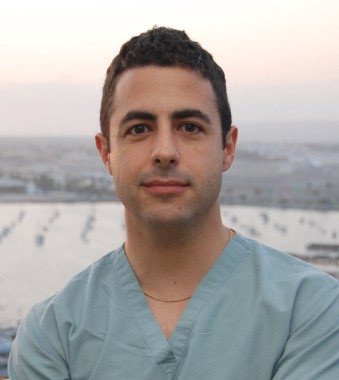
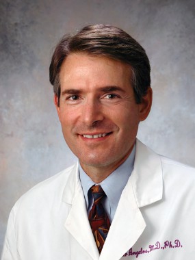
Dr. Angelos is an ACS Fellow; the Linda Kohler Anderson Professor of Surgery and Surgical Ethics; chief, endocrine surgery; and associate director of the MacLean Center for Clinical Medical Ethics at the University of Chicago.
This morning I had the usual 7:30 a.m. elective surgical case scheduled. As I usually do, I went to see my patient at around 7 a.m. to review the procedure, answer any new questions that may have come up, and mark the incision site. My patient greeted me very cheerfully with the following questions, "How was your night? Did you sleep OK? Are you feeling good this morning? No arguments at home, I hope?" I readily answered, as I almost always do, when discussing things with patients in the preop area, that I am feeling good and ready for things to go well with the case.
I have been asked such questions many times over the years. However, for some reason the interchange with my patient this morning raised additional questions for me: Is it really acceptable for patients to ask such questions? Do I have an obligation to disclose such things? Do patients really want to know the answers or are they simply making nervous conversation?
In recent years, there have been a number of articles questioning whether surgeons should be required to disclose a lack of sleep to their elective surgical patients so that the patient can make a "truly informed" decision about what the risks of their surgery are. Thus far, there have been no such requirements at any hospital in the United States that I know of. But my patient’s questions raised a number of practical issues for me with such disclosure. We had already had a conversation in my office when I originally obtained consent for the procedure. I had carefully reviewed risks, benefits, and alternatives to the operation, and I had answered a list of the patient’s questions. Since I was actually the patient’s third opinion, it was clear that the patient already had significant background knowledge about the operation. The patient had signed the consent form before leaving my office.
At some point, before, during, or after my conversation with the patient, he had decided to trust me enough to allow me to do the operation. Subsequently, while home in the days prior to the surgery date, the patient had the opportunity to change his mind, but he actually came to the hospital on the morning of surgery. He had thus actively expressed his confidence in me by showing up for surgery. In this context, I believe that the patient’s questions were really a friendly way of expressing some degree of anxiety about the operation rather than an actual second guessing of my capacity to optimally perform the surgery. In this context, I believe that it is more important that I try to alleviate the patient’s concerns than that I give an expansive discourse on whatever stressful issues may be going on in my personal life.
Of particular importance is the issue of how much I have slept. I do not believe that I should discuss any concerns I may have with lack of sleep with my patient unless I have decided that I am not the best person to perform the surgery. I do not think that I would be doing my patient a service by, for example, saying that I didn’t sleep well and studies show that I might have altered judgment and asking the patient to sign a document that I have disclosed this fact. Such a disclosure seems to be designed to protect the surgeon and the institution rather than the patient.
However, if I believe that my lack of sleep, my personal stressors, or any other distraction will significantly hinder my ability to perform the operation safely, then I should not simply disclose these issues to the patient. Rather, I should explain why I should NOT be doing the surgery and either postpone the case or find someone else to do it if the patient requests this and if it is possible. Although some commentators have suggested that a sleep-deprived surgeon is the worst person to be able to assess his or her abilities to optimally perform an operation, I am convinced that we need to depend on the surgeon to make this assessment. Three central required components of professionalism are the exercise of self-regulation, the capacity to make decisions that are altruistic, and the discipline to abide by ethical standards. The issue of self-regulation is absolutely critical to the professionalism of any surgeon. The professionalism of the surgeon is the basis for patients trusting us to operate on them and make decisions in the operating room on their behalf. To mandate a separate disclosure to the patient about the amount of sleep the surgeon got the night before, or any other distracting issue, would be to cast doubt on the professionalism of the surgeon at the very time that patients most need to trust their surgeons.
Dr. Angelos is an ACS Fellow; the Linda Kohler Anderson Professor of Surgery and Surgical Ethics; chief, endocrine surgery; and associate director of the MacLean Center for Clinical Medical Ethics at the University of Chicago.
online TK 5
Enter text here
Enter text here
Enter text here
Mulberry
Often in this column, several species within a family might be discussed in relation to a broad range of health benefits. Licorice and mushrooms are good examples. In this case, and in this column, the focus will be on several species within a family that are thought to confer the same type of dermatologic benefit. The Morus genus within the Moraceae family appears to include several species that display skin-lightening properties.
Tyrosinase is the enzyme that controls the production of melanin. Suppressing tyrosinase activity to achieve skin lightening is a well-established method in dermatologic practice. The desire for products with fewer side effects than the mainstay, hydroquinone, or natural products such as kojic acid or arbutin, has led to investigations of several species in the Moraceae family. Notably, several Moraceae trees have been found to exhibit antioxidant activity (Int. J. Mol. Sci. 2012;13:2472-80; Biol. Pharm. Bull. 2002;25:1045-8; Biosci. Biotechnol. Biochem. 2010;74:2385-95; J. Pharm. Pharmacol. 2004;56:1291-8). The focus here, though, will be on the skin-lightening activity of various parts of Morus (commonly known as mulberry) trees.
In 2013, Singh et al. assessed the effects of mulberry, kiwi, and Sophora extracts on melanogenesis and melanin transfer in human melanocytes and in cocultures with phototype-matched normal adult epidermal keratinocytes. The extracts were evaluated against isobutylmethylxanthine, hydroquinone, vitamin C, and niacinamide. The investigators found that compared with unstimulated control, mulberry, kiwi, and Sophora extracts significantly reduced melanogenesis in normal adult epidermal melanocytes and human melanoma cells. Melanin transfer also was lowered, as was filopodia expression on melanocytes. The authors concluded that the test compounds compared well with standard-bearing depigmenting agents and warrant consideration as topical agents for diminishing hyperpigmentation (Exp. Dermatol. 2013;22:67-9).
Encouraging results in melasma treatment
A randomized, single-blind, placebo-controlled trial of 50 Filipino patients (49 women, 1 man) to examine the safety and efficacy of 75% Morus alba (white mulberry) extract oil was conducted by Alvin et al. in 2011. Patients were evaluated at weeks 4 and 8. The Melasma Area and Severity Index (MASI) score, Mexameter score, and Melasma Quality of Life (MelasQOL) score were measured, with the mulberry extract group performing significantly better than the placebo group according to all metrics.
The 25 patients treated with mulberry extract showed improvement in the MASI score, from 4.076 at baseline to 2.884 at week 8 (mean difference, 1.19); the mean difference for the placebo group was 0.06. The mean Mexameter reading revealed a significant difference, with a slight increase for the mulberry group (indicating lighter pigmentation), and the placebo group scored a slightly higher value. In addition, the MelasQOL score for the mulberry group improved markedly from baseline to week 8 (58.84 to 44.16), whereas the placebo group score improved only slightly, from 57.44 at baseline to 54.28 at week 8.
Adverse events were rare, with mild itching in 4 patients reported from the mulberry group, and 12 cases of either itching or erythema reported by the placebo group.
The investigators concluded that 75% mulberry extract oil objectively diminishes the hyperpigmentation of melasma in skin types III-V, although they recommend additional research with a larger sample size and longer treatment duration and follow-up (J. Drugs Dermatol. 2011;10:1025-31).
Paper mulberry
The bark of paper mulberry (Broussonetia papyrifera, also known as Morus papyrifera) is composed of extremely strong fibers used to produce high-quality paper and cloth. In China, the leaves, stem, leaf juice, roots, fruits, and bark have all been found to impart various health benefits, with the stem and leaf juice used to treat skin disorders and insect bites (Phytother. Res. 2012;26:1-10).
In one study, a 0.4% concentration of paper mulberry extract was demonstrated to suppress tyrosinase activity by 50% compared with 5.5% hydroquinone and 10% kojic acid. Notably, paper mulberry is not considered a significant irritant even at 1% concentration (J. Drugs Dermatol. 2009;8:s5-9).
White mulberry
In 2002, Lee et al. investigated the in vitro effects of an 85% methanol extract of dried white mulberry leaves on melanin biosynthesis. They found that one of the primary bioactive constituents, mulberroside F (moracin M-6, 3’-di-O-beta-D-glucopyranoside), inhibited the tyrosinase activity that converts dopa to dopachrome in the melanin synthesis process and also suppressed the melanin formation of melan-a cells. In addition, the mulberry extract inhibited tyrosinase activity more potently than did kojic acid (Biol. Pharm. Bull. 2002;25:1045-8).
The following year, a different team found that the young twigs of white mulberry also suppressed tyrosinase activity as well as melanin production in B-16 melanoma cells. In vivo, the extracts decreased melanin synthesis in a guinea pig model without displaying toxicity (J. Cosmet. Sci. 2003;54:133-42).
In 2006, Wang et al. investigated 25 traditional Chinese herbal medicines potentially useful in dermatology, particularly for skin whitening, and found that white mulberry was one of four species to potently inhibit tyrosinase activity, and more strongly than arbutin did (J. Ethnopharmacol. 2006;106:353-9).
Chinese mulberry/shimaguwa
In 2012, Zheng et al. isolated constituents from the roots of Chinese mulberry and found that several ingredients, including oxyresveratrol, moracenin D, sanggenon T, and kuwanon O, displayed more potent tyrosinase inhibition than kojic acid did. They concluded that Chinese mulberry is a good natural source of tyrosinase inhibitors and is potentially useful in cosmetic skin-lightening products as well as in foods as antibrowning agents (Fitoterapia 2012;83:1008-13).
Conclusion
Mulberry is actively used within the dermatologic armamentarium as one of the many options for skin lightening. A significant body of evidence has emerged over the past 15 years to establish the antityrosinase activity of various mulberry species, particularly white mulberry and paper mulberry.
Dr. Baumann is chief executive officer of the Baumann Cosmetic & Research Institute in Miami Beach. She founded the cosmetic dermatology center at the University of Miami in 1997. Dr. Baumann wrote the textbook "Cosmetic Dermatology: Principles and Practice" (McGraw-Hill, 2009), and a book for consumers, "The Skin Type Solution" (Bantam, 2006). She has contributed to the Cosmeceutical Critique column in Skin & Allergy News since January 2001 and joined the editorial advisory board in 2004. Dr. Baumann has received funding for clinical grants from Allergan, Aveeno, Avon Products, Galderma, Mary Kay, Medicis Pharmaceuticals, Neutrogena, Philosophy, Stiefel, Topix Pharmaceuticals, and Unilever.
When wounds won’t heal, try these strategies
SAN FRANCISCO – When your first or second attempts to help a skin wound heal haven’t worked, ask yourself three questions, Dr. Theodora Mauro suggested:
• Have you diagnosed the wound correctly?
• What systemic conditions are keeping the wound from healing?
• Have you tailored your treatment to the wound correctly?
Most dermatologists are pretty good at diagnosing ulcers that are caused by single etiologies. What Dr. Mauro sees more commonly in her busy referral practice, however, are wounds that won’t heal because of a combination of causes – often venous insufficiency plus something else, she said at the annual meeting of the Pacific Dermatologic Association.

"That can make the diagnosis a little more confusing," said Dr. Mauro, professor of dermatology at the University of California, San Francisco and chief of the dermatology service at the San Francisco Veterans Affairs Medical Center.
Ulcer types
The most common skin ulcer in the United States is due to venous insufficiency. These patients tend to be spread among dermatologists, vascular surgeons, and podiatrists and "are not well served by being scattered among different disciplines," she said.
Arterial ulcers make up 6%-10% of skin ulcers in the medical literature, and the incidence of diabetic ulcers is increasing, she added. Pressure ulcers are becoming more common as the population ages.
Morphology and the location of ulcers usually can distinguish the different types, but don’t forget to check the patient’s pulses and sensations, Dr. Mauro said. If you can feel pedal pulses, the patient is very likely to have a normal ankle brachial index (greater than 0.8). "It’s an easy thing to do and very helpful," she said. Also, break a Q-tip cotton swab in half and poke the patient with the sharp end. "You’d be surprised at the number of people who have altered sensation" that’s contributing to the ulceration.
A common "combination" that gets missed is a nonmelanoma skin cancer and an ulcer at that site from venous insufficiency. Consider taking biopsies of nonhealing ulcers to look for skin cancer, she said. Long-standing ulcers can develop squamous cell carcinoma within them. A basal cell carcinoma can look like a healing ulcer, but not heal.
Bullous diseases also can be hidden in ulcers, particularly in the elderly. "There seems to be a two-hit thing with venous insufficiency where you have a little bit of blistering disease and you have a lot of hydrostatic pressure, and people will get their bullae on their legs long before they get it anyplace else," Dr. Mauro said. "You put compression on and they come back next week and now they have intact bullae. Think about that as another cause."
Less common ulcerative problems that can be confusing include pyoderma gangrenosum (which may be half as common as people think, studies suggest), an underlying vasculitis, or mycobacterial infections. If you see a nonhealing hyperkeratotic lesion, consider an atypical mycobacterial infection. In Dr. Mauro’s region, these usually are in patients who went mountain biking and inoculated themselves through an ankle scratch or in patients exposed to the organism through infected water during a pedicure.
Systemic conditions
Wound healing can be inhibited by things that doctors do, and by things that patients do.
Taking antimetabolite medications (such as hydroxyurea or methotrexate), prednisone, or nonsteroidal anti-inflammatory drugs can impair wound healing, as does smoking. Radiation to the site also inhibits healing. Think twice before irradiating a basal cell carcinoma on the leg of a patient with venous insufficiency. "It’s much better to either try some topical medication or just bite the bullet, do surgery, and put the Unna boot on after the surgery, because if you irradiate [the basal cell carcinoma] and it ulcerates, then it’s really hard to heal," she said.
Coexisting cancer or arterial disease can inhibit wound healing. If you check pulses and can’t feel them, send the patient for an ankle brachial index to determine if there’s arterial disease, in which case the patient should see a vascular surgeon, she said.
When you’re taking patients’ histories, ask what they’re eating so you can assess whether they’re getting enough protein, zinc, and vitamin C. "You’d be surprised at the number of nutritional deficiencies that we see" in patients with nonhealing wounds. Her clinic picks up a zinc deficiency three to four times per year and a protein deficiency approximately twice a year. "All of these are things that you need to make collagen and granulation tissue," she explained. Supplementation with Ensure can provide these nutrients, but a less-expensive option is Carnation Breakfast Essentials, at about one-fifth the cost, she added.
Tailor therapy appropriately
Dressings can make wounds worse. Most commonly, putting a hydrocolloid dressing (such as DuoDERM) on an ulcer that has a lot of drainage will macerate and enlarge the wound. Putting stiff foams or self-adherent wraps such as Coban on wounds with a lot of edema can create secondary ulcers. And avoid compression on the legs of people with arterial insufficiency, Dr. Mauro said.
Artifactual ulcers from patients scratching or picking at wounds are increasing, she said. These patients usually will not report that they’re messing with the wound, but your level of suspicion should increase if the ulcer is above the knee. Hydrocolloid dressings work pretty well for this problem. Consider sending some of these patients to an expert in the growing field of medication therapy for dysesthesia.
Ask patients about home remedies, because they will put all kinds of things on wounds to try and help them heal. Neomycin can cause contact dermatitis. Patients may apply full-strength hydrogen peroxide, thinking that they need a disinfectant, which tends to damage the epithelium and can impair wound healing. If they insist on using hydrogen peroxide, recommend a half-strength solution.
For patients with venous insufficiency, the hydrostatic pressure from sitting can be nearly as bad as from standing. Ask patients with nonhealing wounds what they do at home and at work. If they’re sitting much of that time, recommend products to alleviate the pressure. Dr. Mauro said she has virtually become a saleswoman for La-Z-Boy furniture during her medical career because La-Z-Boy products allow patients to eat, work at computers, and function in positions with less hydrostatic pressure.
When to refer
Consider referring patients with chronic wounds that have not lost at least 50% of their width and height within 6 weeks. When a wound has been present long enough, the physiology shifts from acute to chronic. "It’s as if the body decides that it’s tired of trying to heal it and its new equilibrium is just going to be, ‘I have a wound,’ " she said.
If you want to try treating chronic wounds, applying Promogran Prisma matrix wound dressing in those without good granulation tissue may stimulate granulation tissue, she said.
Debriding an ulcer is "kind of a poor man’s way of getting platelet-derived growth factor," she added. Dr. Mauro applies a compounded 30% lidocaine ("but you can use any topical lidocaine"), leaves it on for 20-25 minutes, and debrides using a curette, which is much more precise than a scalpel. "What you’re trying to do is ‘reboot the computer’ and trick the ulcer into thinking it’s an acute ulcer rather than a chronic one," she said.
Dr. Mauro said she refers patients with arterial insufficiency to vascular surgeons. She sends patients with diabetes to podiatrists, preferably before they develop ulcers. The podiatrist can regularly trim nails for patients with poor sight and loss of sensation who may cut themselves, and can design footwear to avoid pressure that could cause an ulcer. "That will keep you out of trouble" with diabetic patients, she said.
Dr. Mauro has been a consultant for Unilever, but not in a wound-healing capacity.
On Twitter @sherryboschert
SAN FRANCISCO – When your first or second attempts to help a skin wound heal haven’t worked, ask yourself three questions, Dr. Theodora Mauro suggested:
• Have you diagnosed the wound correctly?
• What systemic conditions are keeping the wound from healing?
• Have you tailored your treatment to the wound correctly?
Most dermatologists are pretty good at diagnosing ulcers that are caused by single etiologies. What Dr. Mauro sees more commonly in her busy referral practice, however, are wounds that won’t heal because of a combination of causes – often venous insufficiency plus something else, she said at the annual meeting of the Pacific Dermatologic Association.
"That can make the diagnosis a little more confusing," said Dr. Mauro, professor of dermatology at the University of California, San Francisco and chief of the dermatology service at the San Francisco Veterans Affairs Medical Center.
Ulcer types
The most common skin ulcer in the United States is due to venous insufficiency. These patients tend to be spread among dermatologists, vascular surgeons, and podiatrists and "are not well served by being scattered among different disciplines," she said.
Arterial ulcers make up 6%-10% of skin ulcers in the medical literature, and the incidence of diabetic ulcers is increasing, she added. Pressure ulcers are becoming more common as the population ages.
Morphology and the location of ulcers usually can distinguish the different types, but don’t forget to check the patient’s pulses and sensations, Dr. Mauro said. If you can feel pedal pulses, the patient is very likely to have a normal ankle brachial index (greater than 0.8). "It’s an easy thing to do and very helpful," she said. Also, break a Q-tip cotton swab in half and poke the patient with the sharp end. "You’d be surprised at the number of people who have altered sensation" that’s contributing to the ulceration.
A common "combination" that gets missed is a nonmelanoma skin cancer and an ulcer at that site from venous insufficiency. Consider taking biopsies of nonhealing ulcers to look for skin cancer, she said. Long-standing ulcers can develop squamous cell carcinoma within them. A basal cell carcinoma can look like a healing ulcer, but not heal.
Bullous diseases also can be hidden in ulcers, particularly in the elderly. "There seems to be a two-hit thing with venous insufficiency where you have a little bit of blistering disease and you have a lot of hydrostatic pressure, and people will get their bullae on their legs long before they get it anyplace else," Dr. Mauro said. "You put compression on and they come back next week and now they have intact bullae. Think about that as another cause."
Less common ulcerative problems that can be confusing include pyoderma gangrenosum (which may be half as common as people think, studies suggest), an underlying vasculitis, or mycobacterial infections. If you see a nonhealing hyperkeratotic lesion, consider an atypical mycobacterial infection. In Dr. Mauro’s region, these usually are in patients who went mountain biking and inoculated themselves through an ankle scratch or in patients exposed to the organism through infected water during a pedicure.
Systemic conditions
Wound healing can be inhibited by things that doctors do, and by things that patients do.
Taking antimetabolite medications (such as hydroxyurea or methotrexate), prednisone, or nonsteroidal anti-inflammatory drugs can impair wound healing, as does smoking. Radiation to the site also inhibits healing. Think twice before irradiating a basal cell carcinoma on the leg of a patient with venous insufficiency. "It’s much better to either try some topical medication or just bite the bullet, do surgery, and put the Unna boot on after the surgery, because if you irradiate [the basal cell carcinoma] and it ulcerates, then it’s really hard to heal," she said.
Coexisting cancer or arterial disease can inhibit wound healing. If you check pulses and can’t feel them, send the patient for an ankle brachial index to determine if there’s arterial disease, in which case the patient should see a vascular surgeon, she said.
When you’re taking patients’ histories, ask what they’re eating so you can assess whether they’re getting enough protein, zinc, and vitamin C. "You’d be surprised at the number of nutritional deficiencies that we see" in patients with nonhealing wounds. Her clinic picks up a zinc deficiency three to four times per year and a protein deficiency approximately twice a year. "All of these are things that you need to make collagen and granulation tissue," she explained. Supplementation with Ensure can provide these nutrients, but a less-expensive option is Carnation Breakfast Essentials, at about one-fifth the cost, she added.
Tailor therapy appropriately
Dressings can make wounds worse. Most commonly, putting a hydrocolloid dressing (such as DuoDERM) on an ulcer that has a lot of drainage will macerate and enlarge the wound. Putting stiff foams or self-adherent wraps such as Coban on wounds with a lot of edema can create secondary ulcers. And avoid compression on the legs of people with arterial insufficiency, Dr. Mauro said.
Artifactual ulcers from patients scratching or picking at wounds are increasing, she said. These patients usually will not report that they’re messing with the wound, but your level of suspicion should increase if the ulcer is above the knee. Hydrocolloid dressings work pretty well for this problem. Consider sending some of these patients to an expert in the growing field of medication therapy for dysesthesia.
Ask patients about home remedies, because they will put all kinds of things on wounds to try and help them heal. Neomycin can cause contact dermatitis. Patients may apply full-strength hydrogen peroxide, thinking that they need a disinfectant, which tends to damage the epithelium and can impair wound healing. If they insist on using hydrogen peroxide, recommend a half-strength solution.
For patients with venous insufficiency, the hydrostatic pressure from sitting can be nearly as bad as from standing. Ask patients with nonhealing wounds what they do at home and at work. If they’re sitting much of that time, recommend products to alleviate the pressure. Dr. Mauro said she has virtually become a saleswoman for La-Z-Boy furniture during her medical career because La-Z-Boy products allow patients to eat, work at computers, and function in positions with less hydrostatic pressure.
When to refer
Consider referring patients with chronic wounds that have not lost at least 50% of their width and height within 6 weeks. When a wound has been present long enough, the physiology shifts from acute to chronic. "It’s as if the body decides that it’s tired of trying to heal it and its new equilibrium is just going to be, ‘I have a wound,’ " she said.
If you want to try treating chronic wounds, applying Promogran Prisma matrix wound dressing in those without good granulation tissue may stimulate granulation tissue, she said.
Debriding an ulcer is "kind of a poor man’s way of getting platelet-derived growth factor," she added. Dr. Mauro applies a compounded 30% lidocaine ("but you can use any topical lidocaine"), leaves it on for 20-25 minutes, and debrides using a curette, which is much more precise than a scalpel. "What you’re trying to do is ‘reboot the computer’ and trick the ulcer into thinking it’s an acute ulcer rather than a chronic one," she said.
Dr. Mauro said she refers patients with arterial insufficiency to vascular surgeons. She sends patients with diabetes to podiatrists, preferably before they develop ulcers. The podiatrist can regularly trim nails for patients with poor sight and loss of sensation who may cut themselves, and can design footwear to avoid pressure that could cause an ulcer. "That will keep you out of trouble" with diabetic patients, she said.
Dr. Mauro has been a consultant for Unilever, but not in a wound-healing capacity.
On Twitter @sherryboschert
EXPERT ANALYSIS FROM THE ANNUAL MEETING OF THE PACIFIC DERMATOLOGIC ASSOCIATION
Apps for physicians
When it comes to health, wellness, and fitness apps, we physicians tend to think of our patients. As in, how can this app help my patient lose weight, exercise more, monitor his diabetes, or help her stay compliant with her medications?
But what about physicians and other health care workers? Can apps help us? Absolutely. According to Dr. Craig Burkhart, a dermatologist and apps expert at the University of North Carolina at Chapel Hill, many physicians aren’t taking advantage of apps that can improve efficiency. "Most physicians I encounter have $1,000-$2,000 smartphones and tablets (when you include the service plans) and only make phone calls, check e-mails, text, and listen to music on them – all things they could do with much less expensive devices."
Dr. Burkhart says that many apps "can turn your devices into tools that help organize your e-mails, organize your schedule, bring clinical information to your fingertips, aid patient-doctor communication, secure important information, speed up and improve note taking, and more."
Apps for physicians are available in categories including patient education, drug reference, medical literature, and general reference.
Check out these handy apps and see what works for you:
• Read by QxMD. Staying up to date on medical journals is a challenge. This app lets you access, organize, and share articles from your favorite medical journals. Its magazinelike interface is popular with users, and makes reading medical journals "as fun as it can be," says Dr. Burkhart.
• PDFpen. Dr. Burkhart says this app is "essential to a paperless workflow." It allows users to sign and send documents without printing actual sheets of paper, making for a greener and more efficient office.
• Visual DX. A reference tool for physicians, this app is a digital medical image library with more than 25,000 images. It allows you to visually confirm a diagnosis and to quickly search on a disease, including symptoms and patient care management. A key feature is its ability to let you create a visual differential of medication-induced diseases for more than 700 different drugs.
• 1Password. We’ve all forgotten passwords from time to time. This app allows you to make and store highly secure and complex passwords. For Dr. Burkhart, it has "replaced memory and random sheets of paper as his password repository."
• Draw MD. We know that visuals often help us to explain complex issues to patients more effectively. This app enables you to draw and modify medical images and surgical procedures in a way that is clear and understandable to patients.
• 3D4Medical. This app can enhance patient education by using 3D technology to allow navigation around the body. You can zoom, rotate, and cut images to create different perspectives.
Dr. Jeffrey Benabio is a partner physician in the department of dermatology of the Southern California Permanente Group in San Diego and a volunteer clinical assistant professor at the University of California, San Diego. He has published numerous scientific articles and is a member and fellow of the American Academy of Dermatology, and a member of the Telemedicine Association and the American Medical Association, among others. He is board certified in dermatology as well as medicine and surgery in the state of California. Dr. Benabio has a special interest in the uses of social media for education and building a dermatology practice. He is the founder of The Derm Blog, an educational website which has had over 2 million unique visitors. Dr. Benabio is also a founding member and the skin care expert for Livestrong.com, a health and wellness website of Lance Armstrong’s the Livestrong Foundation. Dr. Benabio is @Dermdoc on Twitter.