User login
Benefits of Euglycemia
Until recently it had been argued that hospitalization was not the time in the life of a patient to insist on tight glycemic control. Hyperglycemia was understood to be a consequence of medical stress.1 It was well known that infection, sepsis, or other medical stress might exacerbate hyperglycemia or promote a diabetic crisis.25 Admittedly, the severity of hyperglycemia among patients who have diabetes was thought to predict the risk for hospitalization with infection as well as the outcome of the infectious condition.68 However, until recently strict glycemic control in the hospital was not strongly advocated because hypoglycemia might occur and might be directly and uniquely traceable to actions taken in the hospital.9, 10 Furthermore, the complications of diabetes were thought to be divided into acute metabolic emergencies and chronic tissue complications such as polyneuropathy, retinopathy, nephropathy, and macrovascular disease that would have evolved over a period longer than the duration of hospitalization, and the possibility that short‐term hyperglycemia might affect outcomes was considered unproven.
The purpose of this article is to define the specific populations, disorders, and hospital settings for which there now is strong evidence that short‐term glycemic control will affect the outcome of a course of hospital treatment.
PHYSIOLOGIC LINK BETWEEN HYPERGLYCEMIA AND ADVERSE OUTCOMES
Five years ago a caregiver would not have been likely to think of glycemic control as a contributing factor when considering specific complex problems such as pump failure or arrhythmia after cardiac surgery, long‐term mortality after myocardial infarction, acute renal failure, the need for transfusion during treatment of a complicated surgical illness, or prolonged dependence on a ventilator in the surgical ICU. Although it now known that these and other complications are linked to hospital hyperglycemia, the mechanisms of harm are several steps removed from the hyperglycemia itself. The causes of these adverse outcomes are multifactorial. The causal dependence of the injury on hyperglycemia is not easy to see. In fact, it required randomized prospectively designed trials to convincingly demonstrate the contributory role of hyperglycemia to these and other adverse outcomes.
Now that this link has been convincingly demonstrated, there is intense interest in discovering the probable mechanisms by which control of hyperglycemia and specifically the use of insulin might improve outcomes.1114 Mortality, predominantly sepsis related, was the primary outcome for which the Leuven, Belgium, report of 2001 on the surgical ICU showed improvement.15 Simplistically, in seeking a mechanism of benefit with respect to sepsis, it might be argued that if gross hyperglycemia were prevented in patients with surgical wounds, improvement of host defenses against infective organisms might be expected.1622 However, additional mechanisms of protection probably should be invoked, including those by which glycemic control and specifically insulin therapy affect endothelial function and the coagulation pathway, thus improving the ability of a patient to withstand and recover from sepsis. Insulin promotes beneficial nitric oxide synthase activation (e‐NOS) in capillary endothelium.2326 In patients with prolonged critical illness, intensive insulin therapy lowers ICAM‐1 levels, reflecting reduced endothelial activation. Whereas e‐NOS exerts a beneficial endothelial effect, hepatic iNOS activation is harmful. One proposed mechanism of benefit from adequate insulin therapy is suppression of excessive hepatic iNOS‐induced release of circulating NO, which might contribute to endothelial dysfunction, organ failure, and death.27
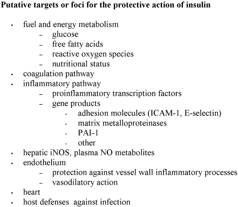
Additional proposed mechanisms of hyperglycemia‐induced harm to hospitalized patients, some potentially specifically reduced by insulin therapy, resemble those discussed in relation to macrovascular disease and include activation of inflammatory cytokines, matrix metalloproteinases, and adhesion molecules, and the adverse arrhythmogenic effects of elevated circulating nonesterified fatty acids (Figure 1).2834
CRITICAL WINDOW OF TIME
The results of several retrospectively or prospectively conducted single‐institution observational studies suggest there is a critical window of time within which clinicians must get it right, when attempting glycemic control or else jeopardize therapeutic goals, such as duration of remission in treatment of acute lymphocytic leukemia35 or avoidance of acute rejection in renal allograft surgery.36 Additionally, delayed risk of infection appears to be linked to previous glycemic control during specific early time frames surrounding surgery, renal transplantation, admission for trauma, and induction chemotherapy for leukemia (Table 1).3541 For patients who have diabetes, the potential to reduce the consequences of infections through intensified glycemic control probably begins prior to admission and in the hospital has still not been fully realized.4244
| Patients | Ascertainment of hyperglycemia | Delayed events among patients with early hyperglycemia |
|---|---|---|
| 100 postoperative uninfected diabetic patients undergoing elective surgery, monitored prospectively37 | First postoperative day | Postoperative nosocomial infection rate within 14 days was 2.7 times higher for patients having at least one BG > 220 mg/dL (33.3% vs. 11.5%). |
| 990 historical controls and 595 patients in the interventional group of postoperative cardiac diabetic patients40 | First 2 postoperative days | Incidence of deep sternal wound infection was reduced from 2.4% to 1.5% (P 0.02) after introduction of protocol to maintain mean BG 200 mg/dL. |
| 423 renal allograft recipients receiving their first cadaveric transplant36 | First 100 hours; first day. | A mean of 10.8 2.3 days after transplantation, 70% of patients developed postoperative infection, and after a mean of 7.7 2.6 days, 40% developed acute rejection. Every patient with mean BG over 200 mg/dL during the first 100 hours developed postoperative infection. On the first postoperative day the mean BG had been 248.4 mg/dL among those developing infection and 167.4 mg/dL among those without infection (P .001), and the mean BG had been 270 mmol/L among those developing rejection and 194 mmol/L among those without rejection. |
| 516 trauma patients admitted to the ICU38 | Either of first 2 hospital days | Hyperglycemia 200 mg/dL was associated with a higher infection rate (32% vs. 22%, P = .04) and with greater mortality (34% vs. 13%, P .0001) |
| 275 patients having lower‐extremity peripheral vascular surgery39 | First 48 hours | Postoperative infections within 30 days were 5.1 times more frequent in the top quartile for BG versus the lowest quartile (confidence interval 1.6‐17.1, P = .007). |
| 278 adult patients receiving induction chemotherapy for acute lymphocytic leukemia35 | First 30 days | Hyperglycemia defined as 2 BG 200 mg/dL was associated with a greater likelihood of sepsis (16.5% vs. 8.0%, P = .03) or any complicated infection (38.8% vs. 25.1%, P = .016), shorter duration of complete remission (24 vs. 52 months), and with shorter median survival (29 vs. 88 months, P = .001). |
KEY STUDIES SHOWING CLINICAL BENEFIT OF TIGHT GLYCEMIC CONTROL
A summary of several key studies that demonstrated the clinical benefit of tight glycemic control is shown in Table 2. These studies successfully separated the intensively and conventionally managed study groups according blood glucose. Although trials using glucose‐insulin‐potassium infusions (GIK) such that blood glucose was lowered have shown benefit for patients who have had myocardial infarctions4547 or cardiac surgery,48 not all GIK studies have yielded positive results. The negative results of the CREATE‐ECLA study suggest that GIK therapy per se is not beneficial unless it reduces blood glucose.49 In the setting of acute myocardial infarction, the DIGAMI 2 trial and the HI‐5 trials failed to achieve the intended separation between treatment groups.50, 51 It has been speculated that if insulin is delivered so as to not achieve normoglycemia, then hyperinsulinemia in the presence of hyperglycemia actually may be proinflammatory.52
| Population and/or setting and patients | Study design and/or intervention | Principal findings |
|---|---|---|
| Patients with diabetes mellitus and with hyperglycemia > 11 mmol/L or similar hyperglycemia without known diabetes who had had acute myocardial within the preceding 24 hours.4547 There were 620 patients, of whom 15 of the controls and 10 patients in the intensive group had no prior diagnosis of diabetes. | Prospective, randomized, controlled clinical trial. Controls received standard treatment. Treatment group received infusion of glucose‐insulin for at least 24 hours, followed by multiple injections of subcutaneous insulin for at least 3 months. | Overall mortality after 1 year was l9% in the insulin group compared to 26% among controls (P .05). The most frequent cause of death of all patients was congestive heart failure. The benefit continued for at least 3.5 years, with an absolute reduction in mortality of 11%. |
| Diabetic cardiac surgery population.40, 5557 To date, 5510 cardiac surgery patients who were either admitted or discharged with the diagnosis of diabetes have been studied. | Prospective nonrandomized study of the effects of hyperglycemia and its pharmacologic reduction with intensive intravenous insulin regimens on outcomes. | The 3‐day average of perioperative BG (3‐BG) correlated with mortality (P .0001, odds ratio 2.0 per 50 mg/dL increase in 3‐BG). Continuous intravenous insulin infusion independently reduced risk of death 60% (RR = 0.4, P .001). Length of stay in the CABG population increased by 1 day for every increase of 50 mg/dL in 3‐BG. |
| Critically ill surgical patients receiving mechanical ventilation. Overall, 13% of 1548 participants had previously diagnosed diabetes. | Prospective, randomized, controlled clinical trial. Intravenous insulin infusion targeting BG 80‐110 mg/dL was initiated in the intensive group for BG > 110 mg/dL. Intravenous insulin infusion targeting BG 180‐200 was initiated in the conventional group for BG > 215 mg/dL. A whole‐blood glucose method using a gas analyzer was employed to determine arterial blood glucose. | In the group assigned to intensive insulin therapy mortality was reduced during intensive care from 8.0% with conventional treatment to 4.6% (P .04), or a risk reduction of 42%. In the group remaining in the ICU for more than 5 days, mortality was reduced from 20.2% with conventional treatment to 10.6% with intensive insulin therapy; P = .005. The intensive group experienced reductions in overall in‐hospital mortality of 34%, in bloodstream infections of 46%, and in acute renal failure requiring dialysis or hemofiltration of 41%. Also reduced were the need for red‐cell transfusions, critical‐illness neuropathy, duration of mechanical ventilation, and length of stay in the ICU. |
| Critically ill medical patients. The intention to treat group included 1200 participants, of whom 16.9% had a history of diabetes.54 | Randomized controlled clinical trial, as above. | In the intensive group, in‐hospital mortality was not significantly reduced, and among the 433 who stayed in the ICU for less than 3 days, mortality was actually higher. However, in the intention‐to‐treat group there was reduction in newly acquired kidney injury, prolonged mechanical ventilation, and length of stay in the ICU and in the hospital. Among the 767 patients who stayed in the ICU for 3 or more days, intensive insulin therapy reduced in‐hospital mortality from 52.5% to 43.0%. |
| Patients within 24 hours of having an acute stroke and who had poststroke hyperglycemia. The results of the first 452 patients recruited to the GIST‐UK study showed that 15.3% had previously recognized diabetes.62 | Randomized controlled clinical trial. The intensive group received glucose‐potassium‐insulin (GKI) infusion, and the conventional group received saline infusion. | Although mean glucose declined in both groups, the GKI infusion safely achieved separation of groups by blood glucose. Outcomes are pending. |
The findings of the negative intensive insulin studies do not offset the evidence favoring glycemic control, derived from studies that actually achieved the lowering of blood glucose in the intensive groups, such as the successful prospectively designed trials in myocardial infarction or surgical or medical intensive care units15, 45, 47, 53, 54 and the long‐running, large, prospectively monitored Portland series utilizing insulin infusion for cardiac surgery,40, 5557 which have demonstrated the beneficial effects of euglycemia on mortality and morbidity, or the findings of Krinsley, reported elsewhere in this issue.58, 59 The well‐recognized correlation between outcomes of acute stroke and poststroke hyperglycemia has led to the design of a multicenter trial of glucose‐insulin‐potassium infusion for stroke, in which separation of groups by blood glucose has been achieved.6062 The success of GIK therapy in controlling hyperglycemia depends in part on the particular formulation of the infusion as it matches patient needs, and it is probable that insulin infusion following stroke is capable of safely achieving even tighter glycemic control than GIK.63 Intravenous insulin infusion therapy is more difficult to conduct than GIK therapy. However, because of concern about the proinflammatory and prothrombotic effects of hyperglycemia and recognition of the occasional failure of GIK infusions to control hyperglycemia and of the anti‐inflammatory, antithrombotic, and vasodilatory actions of insulin, there have been calls for additional trials of insulin infusions (as opposed to glucose‐insulin infusions) for both acute myocardial infarction and stroke.52, 64
HYPOGLYCEMIA
Serious or fatal sequelae of hypoglycemia are the principal safety risks in intensive insulin management.9, 65 Case ascertainment cannot be assured by glucose averaging methods but instead requires a method of searching for isolated episodes of hypoglycemia.10 One of the most dreaded consequences of nonfatal hypoglycemia is permanent impairment of intellectual function. Because many euglycemic medically ill patients experience alteration of sensorium while acutely ill, there is a risk that the consequences of an actual episode of severe hypoglycemia will be ascribed to other comorbidities, overlooking or misattributing the altered cognitive function that may persist at discharge to causes other than the obvious iatrogenic one. Because there is an increased risk of hypoglycemia during intensive insulin therapy, controversy has arisen over glycemic targets, especially among critically ill nonsurgical patients.54, 6669
On the other hand, the consequences of hypoglycemia during intensive intravenous insulin therapy in a surgical ICU were said to be negligible.15 In hospitalized elderly patients, hypoglycemia in association with increased mortality risk may not be an independent predictor.70 In the critical care unit, predictors of hypoglycemia are identifiable after the introduction of strict glycemic control that include not only insulin therapy but also CVVH treatment with bicarbonate substitution fluid, discontinuation of nutrition without insulin adjustment, prior diagnosis of diabetes mellitus, sepsis, and the need for inotropic or vasopressor drugs.71 A prudent policy for the future would be not only to treat hypoglycemia promptly when it does occur, with attention to subsequent monitoring to avoid relapse or recurrence, but also to actively introduce strategies for predicting the risk of hypoglycemia and preventing it, especially when high risk is identified.10 The fear of hypoglycemia should not paralyze efforts to achieve better glycemic control in hospitalized patients.
UNANSWERED QUESTIONS AND VISION FOR THE FUTURE
On general wards, 38% of admitted patients may have hyperglycemia.72 As shown in the Leuven, Belgium, study, to achieve the target BG of less than 110 mg/dL in the intensive group in the surgical critical care unit, it was necessary to administer intravenous insulin infusion to essentially all patients. In a study that utilized continuous glucose monitoring, normoglycemia in patients in the intensive care unit was achieved as little as 22% of the time.73
An actively debated subject is how best to assess hospital performance in glycemic control (glucometrics). Hospitals need to show satisfactory control of variability between patients and within the treatment course of individual patients. That is, it is not sufficient to be satisfied with reasonable results of average blood glucose, using blood glucose as the unit of observation (where n is the number of blood glucose determinations). Alternatives are to use patient day or individual patient as a unit of observation (where n is the number of patient days and the number of patients, respectively). Time‐weighting methods are cumbersome but increase the validity of the averaging method used. When the patient is the unit of observation, possible measures include a per‐patient blood glucose average, percentage of blood glucose measurements with certain ranges, time spent within certain blood glucose ranges, or area under the curve of blood glucose versus time. Caveats about glucometrics pertain to both intravenous insulin infusion and also subcutaneous insulin management.74
Although awaiting additional evidence, diabetes experts have widely accepted the proposition that hospitals should focus on prevention of hyperglycemia as an important patient safety factor.75 Although the target range for glycemic control remains controversial, many students of the subject have endorsed the recommendations mentioned in the lead article in this issue, with the understanding that these criteria were developed from the results of the Leuven, Belgium, study, in which a whole blood glucose analyzer was used for measurement, and that the method of measurement of blood glucose must be considered in interpreting applicability of the target range at individual hospitals, many of which use plasma‐correlated methods yielding higher results. However, in new settings and for medical conditions that have not yet been rigorously evaluated by clinical trials, it is an unanswered question whether intensification of glycemic control can be achieved safely outside the critical care unit and, if so, what type of insulin therapy should be used and for what conditions the benefits would outweigh the risks and justify the costs.
Most inpatient management probably will continue to be conducted using subcutaneous injection therapy,7682 designed to match carbohydrate exposure through the appropriate use of analogs or conventional insulin products. One argument for the use of insulin analogs in the hospital, using basal‐prandial‐correction therapy, is the probability of reducing hypoglycemia and getting closer to target range control among patients who are eating but who are at risk for abrupt suspension of meals. Aggressive subcutaneous management strategies are likely to be most effective when standardized protocols, order sets, and informative computerized order entry systems gain widespread hospital acceptance.
If the importance of gaining glycemic control is highly time dependent and if hyperglycemia is uncontrolled, then a strong argument can be made for routine use of intravenous insulin infusion. For appropriately selected patients, intravenous insulin infusion is cost effective,83, 84 and its use can be extended to appropriate patients outside the critical care unit.8587 Many hospitals have protocols for intravenous insulin but use them only sporadically. For patients who already are in the intensive care setting, it is imperative to develop policies that require introduction of intravenous insulin infusion at a given glycemic threshold. On general wards that lack sufficient staffing to conduct intravenous insulin therapy, it is appropriate either to transfer candidate patients to a ward that has adequate staffing when medical condition requires improved control or to develop policies and procedures that will extend the use of intravenous insulin infusion to general wards. In the future, new technologies can be envisioned that will unburden nursing staff, making intravenous insulin infusion realizable as the treatment of choice for hemodynamically stable patients in most hospital settings. These technologies will include continuous monitoring of blood glucose, dose‐defining algorithms, and the eventual development of a fully automated closed‐loop system of monitoring and delivery that might automatically match insulin delivery to carbohydrate exposure and patient insulin sensitivity.8893
- ,,.Stress‐induced hyperglycemia.Crit Care Clin.2001;17(1):107–124.
- ,,,,.Hyperosmolarity and acidosis in diabetes mellitus: a three‐year experience in Rhode Island.J Gen Intern Med.1991:495–502.
- ,,.Prognostic factors in the diabetic hyperosmolar state.J Am Geriatr Soc.1987;35:737–741.
- ,,.Predisposing factors for the diabetic hyperosmolar state.Arch Intern Med.1987;147:499–501.
- .The diabetic hyperosmolar state.Clin Geriatr Med.1990;6:797–806.
- ,,,,,.Bacteremia in adult diabetic patients.Diabetes Care.1991;14(2):89–94.
- ,,,,,.Influence of diabetes mellitus and glycaemic control on the characteristics and outcome of common infections.Diabet Med.1996:457–463.
- ,,.Diabetes and the risk of infection‐related mortality in the U.S.Diabetes Care.2001;24:1044–1049.
- ,,.Hypoglycemia in hospitalized patients.N Engl J Med.1986;315:1245–1250.
- ,,, et al.Hospital hypoglycemia: not only treatment but also prevention.Endocr Pract.2004;10(suppl 2):71–80.
- .Effect of insulin therapy on nonglycemic variables during acute illness.Endocr Pract.2004;10(suppl 2):63–70.
- .Molecular mechanisms by which metabolic control may improve outcomes.Endocr Pract.2004;10(suppl 2):57–62.
- ,,,,.Insulin infusion in acute illness.J Clin Invest.2005;115:2069–2072.
- ,,.Intensive insulin therapy in the intensive care unit: update on clinical impact and mechanisms of action.Endocr Pract.2006;12(suppl 3):14–21.
- ,,, et al.Intensive insulin therapy in critically ill patients.N Engl J Med.2001;345:1359–1367.
- ,,.Impaired granulocyte adherence. A reversible defect in host defense in patients with poorly controlled diabetes.Diabetes.1978;27:677–681.
- ,,.Impaired leukocyte function in patients with poorly controlled diabetes.Diabetes.1974;23(1):9–15.
- ,,, et al.The effect of diabetes mellitus on chemotactic and bactericidal activity of human polymorphonuclear leukocytes.Diabetes Res Clin Pract.1987;4:27–35.
- ,,,,.Polymorphonuclear leukocytes in non‐insulin‐dependent diabetes mellitus: abnormalities in metabolism and function.Ann Intern Med.1995;123:919–924.
- ,,,,.Agonist‐dependent failure of neutrophil function in diabetes correlates with extent of hyperglycemia.J Leukoc Biol.2001;70:395–404.
- ,,,,,.Insulin increases neutrophil count and phagocytic capacity after cardiac surgery.Anesth Analg.2002;94:1113–1119.
- ,,,.In vivo evidences that insulin regulates human polymorphonuclear neutrophil functions.J Leukoc Biol.2004;76:1104–1110.
- ,.Insulin‐stimulated production of nitric oxide is inhibited by wortmannin. Direct measurement in vascular endothelial cells.J Clin Invest.1996;98:894–898.
- ,,,,.Insulin‐mediated skeletal muscle vasodilation is nitric oxide dependent. A novel action of insulin to increase nitric oxide release.J Clin Invest.1994;94:1172–1179.
- ,.Effect of insulin on human aortic endothelial nitric oxide synthase.Metabolism.2000;49(2):147–50.
- ,.Nitric oxide, platelet function, myocardial infarction and reperfusion therapies.Heart Fail Rev.2003;8(1):47–54.
- ,,, et al.Intensive insulin therapy protects the endothelium of critically ill patients.J Clin Invest.2005;115:2277–2286.
- ,,, et al.Elevated circulating free fatty acid levels impair endothelium‐dependent vasodilation.J Clin Invest.1997;100:1230–1239.
- ,,, et al.Insulin inhibits intranuclear nuclear factor kappaB and stimulates IkappaB in mononuclear cells in obese subjects: evidence for an anti‐inflammatory effect?J Clin Endocrinol Metab.2001;86:3257–3265.
- ,,.The anti‐inflammatory and potential anti‐atherogenic effect of insulin: a new paradigm.Diabetologia.2002;45:924–930.
- ,,,.The potential influence of inflammation and insulin resistance on the pathogenesis and treatment of atherosclerosis‐related complications in type 2 diabetes.J Clin Endocrinol Metab.2003;88:2422–2429.
- ,,.The potential therapeutic role of insulin in acute myocardial infarction in patients admitted to intensive care and in those with unspecified hyperglycemia.Diabetes Care.2003;26:516–519.
- ,,.Insulin treatment improves the systemic inflammatory reaction to severe trauma.Ann Surg.2004;239:553–560.
- ,,, et al.Anti‐inflammatory and profibrinolytic effect of insulin in acute ST‐segment‐elevation myocardial infarction.Circulation.2004;109:849–854.
- .Relation between the duration of remission and hyperglycemia in induction chemotherapy for acute lymphocytic leukemia.Cancer.2004;100:1179–1185.
- ,,,,.Early peri‐operative glycaemic control and allograft rejection in patients with diabetes mellitus: a pilot study.Transplantation.2001;72:1321–1324.
- ,,, et al.Early postoperative glucose control predicts nosocomial infection rate in diabetic patients.J Parenter Enteral Nutr.1998;22(2):77–81.
- ,,,,.Relationship of early hyperglycemia to mortality in trauma patients.J Trauma.2004;56:1058–1062.
- ,,,,.Early post‐operative glucose levels are an independent risk factor for infection after peripheral vascular surgery. A retrospective study.Eur J Vasc Endovasc Surg.2004;5:520–525.
- ,,,.Continuous intravenous insulin infusion reduces the incidence of deep sternal wound infection in diabetic patients after cardiac surgical procedures.Ann Thorac Surg.1999;67:352–362.
- ,.Effects on outcome of in‐hospital transition from intravenous insulin infusion to subcutaneous therapy.Am J Cardiol.2006;98:557–564.
- ,,,.The impact of diabetes in patients with necrotizing soft tissue infections.Surg Infect.2005;6:427–438.
- ,,,.Infections in patients with diabetes mellitus.N Engl J Med.1999;341:1906–1912.
- ,,.Quantifying the risk of infectious diseases for people with diabetes.Diabetes Care.2003;26:510–513.
- ,,, et al.Randomized trial of insulin‐glucose infusion followed by subcutaneous insulin treatment in diabetic patients with acute myocardial infarction (DIGAMI study): effects on mortality at 1 year.J Am Coll Cardiol.1995;26:57–65.
- ,,, et al.Effects of insulin treatment on cause‐specific one‐year mortality and morbidity in diabetic patients with acute myocardial infarction.Eur Heart J.1996;17:1337–1344.
- .Prospective randomised study of intensive insulin treatment on long term survival after acute myocardial infarction in patients with diabetes mellitus.DIGAMI (Diabetes Mellitus, Insulin Glucose Infusion in Acute Myocardial Infarction) Study Group.BMJ.1997;314:1512–1515.
- ,,,,,.Tight glycemic control in diabetic coronary artery bypass graft patients improves perioperative outcomes and decreases recurrent ischemic events.Circulation.2004;109:1497–1502.
- .Effect of Glucose‐Insulin‐Potassium Infusion on Mortality in Patients With Acute ST‐Segment Elevation Myocardial Infarction: The CREATE‐ECLA Randomized Controlled Trial.JAMA.2005;293:437–446.
- ,,, et al.Intense metabolic control by means of insulin in patients with diabetes mellitus and acute myocardial infarction (DIGAMI 2): effects on mortality and morbidity.Eur Heart J.2005;26:650–661.
- ,,.The Hyperglycemia: Intensive Insulin Infusion In Infarction (HI‐5) Study: A randomized controlled trial of insulin infusion therapy for myocardial infarction.Diabetes Care.2006;29:765–770.
- .Inpatient diabetes: review of data from the cardiac care unit.Endocr Pract.2006;12(suppl 3):27–34.
- ,.Reduction of nosocomial infections in the surgical intensive‐care unit by strict glycemic control.Endocr Pract.2004;10(suppl 2):46–52.
- ,,, et al.Intensive insulin therapy in the medical ICU.N Engl J Med.2006;354:449–461.
- ,,.Glucose control lowers the risk of wound infection in diabetics after open heart operations.Ann Thorac Surg.1997;63:356–361.
- ,,, et al.Continuous insulin infusion reduces mortality in patients with diabetes undergoing coronary artery bypass grafting.J Thorac Cardiovasc Surg.2003;125:1007–1021.
- ,.Clinical effects of hyperglycemia in the cardiac surgery population: the Portland diabetic project.Endocr Pract.2006;12(suppl 3):22–26.
- .Effect of an intensive glucose management protocol on the mortality of critically ill adult patients.Mayo Clin Proc.2004;79:992–1000.
- .Decreased mortality of critically ill patients with the use of an intensive glycemic management protocol.Mayo Clin Proc.2003;78:1471–1478.
- ,,, et al.Admission glucose level and clinical outcomes in the NINDS rt‐PA Stroke. Trial.Neurology.2002;59:669–74.
- ,,,,,.Glucose potassium insulin infusions in the treatment of acute stroke patients with mild to moderate hyperglycemia: the Glucose Insulin in Stroke. Trial (GIST).Stroke.1999;30:793–799.
- ,,,.Poststroke hyperglycemia: natural history and immediate management.Stroke.2004;35(1):122–126.
- ,,,.IV insulin during acute cerebral infarction in diabetic patients.Neurology.2004;62:1441–1442.
- ,,,.Hyperglycemia, insulin, and acute ischemic stroke: a mechanistic justification for a trial of insulin infusion therapy.Stroke.2006;37(1):267–273.
- ,,.Hypoglycemia and cardiac arrest in a critically ill patient on strict glycemic control.Anesth Analg.2006;102:549–551.
- ,,.Strict glucose control in the critically ill.Br Med J.2006;332:865–866.
- .Intensive Insulin in Intensive Care.N Engl J Med.2006;354:516–518.
- ,.Counterpoint: Inpatient glucose management: a premature call to arms?Diabetes Care.2005;28:976–979.
- ,,,.Glucose control and mortality in critically ill patients.JAMA.2003;290:2041–2047.
- ,,, et al.Hypoglycemia as a predictor of mortality in hospitalized elderly patients.Arch Intern Med.2003;163:1825–1829.
- ,,, et al.Predisposing factors for hypoglycemia in the intensive care unit.Crit Care Med.2006;34:96–101.
- ,,,,,.Hyperglycemia: an independent marker of in‐hospital mortality in patients with undiagnosed diabetes.J Clin Endocrinol Metab.2002;87:978–982.
- ,,,.Intensive insulin therapy in the intensive care unit: assessment by continuous glucose monitoring.Diabetes Care.2006;29:1750–1756.
- ,,,.No patient left behind: evaluation and design of intravenous insulin infusion algorithms.Endocr Pract.2006;12(suppl 3):72–78.
- ,,, et al.Management of diabetes and hyperglycemia in hospitals.Diabetes Care.2004;27:553–591.
- ,,,.Eliminating Inpatient Sliding‐Scale Insulin: A reeducation project with medical house staff.Diabetes Care.2005;28:1008–1011.
- ,,.70/30 Insulin algorithm versus sliding scale insulin.Ann Pharmacother.2005;39:1606–1609.
- ,.Hospital management of hyperglycemia.Clin Diabetes.2004;22(2):81–88.
- ,.Subcutaneous insulin therapy in the hospital setting: issues, concerns, and implementation.Endocr Pract.2004;10(suppl 2):81–88.
- ,,,,.Hyperglycemia in the hospital.Diabetes Spectr.2005;18(1):20–27.
- .Insulin Analogues.N Engl J Med.2005;352:174–183.
- .Insulin management of diabetic patients on general medical and surgical floors.Endocr Pract.2006;12(suppl 3):86–90.
- ,,, et al.Improved perioperative glycemic control by continuous insulin infusion under supervision of an endocrinologist does not increase costs in patients with diabetes.Endocr Pract.2004;10(2):112–118.
- ,,.Effect of hyperglycemia and continuous intravenous insulin infusions on outcomes of cardiac surgical procedures: the Portland Diabetic Project.Endocr Pract.2004;10(suppl 2):21–33.
- ,,,.New insulin infusion protocol improves blood glucose control in hospitalized patients without increasing hypoglycemia.J Qual Patient Saf.2005;31:141–147.
- ,,,,.Optimizing hospital use of intravenous insulin therapy: improved management of hyperglycemia and error reduction with a new nomogram.Endocr Pract.2005;11:240–253.
- ,,,,.Implementation of a new intravenous insulin method on intermediate‐care units in hospitalized patients.Diabetes Educ.2005;31:818–823.
- ,,.Clinical results of an updated insulin infusion protocol in critically ill patients.Diabetes Spectr.2005;18(3):188–191.
- ,,.Glucommander: A computer‐directed intravenous insulin system shown to be safe, simple, and effective in 120,618 h of operation.Diabetes Care.2005;28:2418–2423.
- ,,, et al.Improving hyperglycemia management in the intensive care unit: preliminary report of a nurse‐driven quality improvement project using a redesigned insulin infusion algorithm.Diabetes Educ.2006;32:394–403.
- ,,, et al.Performance of a dose‐defining insulin infusion protocol among trauma service ICU admissions.Diabetes Technol Ther.2006;8:476–488.
- ,,, et al.A simple insulin‐nutrition protocol for tight glycemic control in critical illness: development and protocol comparison.Diabetes Technol Ther.2006;8:191–206.
- ,,, et al.Evaluation of the impact of chiropodist care in the secondary prevention of foot ulcerations in diabetic subjects.Diabetes Care.2003;26:1691–1695.
Until recently it had been argued that hospitalization was not the time in the life of a patient to insist on tight glycemic control. Hyperglycemia was understood to be a consequence of medical stress.1 It was well known that infection, sepsis, or other medical stress might exacerbate hyperglycemia or promote a diabetic crisis.25 Admittedly, the severity of hyperglycemia among patients who have diabetes was thought to predict the risk for hospitalization with infection as well as the outcome of the infectious condition.68 However, until recently strict glycemic control in the hospital was not strongly advocated because hypoglycemia might occur and might be directly and uniquely traceable to actions taken in the hospital.9, 10 Furthermore, the complications of diabetes were thought to be divided into acute metabolic emergencies and chronic tissue complications such as polyneuropathy, retinopathy, nephropathy, and macrovascular disease that would have evolved over a period longer than the duration of hospitalization, and the possibility that short‐term hyperglycemia might affect outcomes was considered unproven.
The purpose of this article is to define the specific populations, disorders, and hospital settings for which there now is strong evidence that short‐term glycemic control will affect the outcome of a course of hospital treatment.
PHYSIOLOGIC LINK BETWEEN HYPERGLYCEMIA AND ADVERSE OUTCOMES
Five years ago a caregiver would not have been likely to think of glycemic control as a contributing factor when considering specific complex problems such as pump failure or arrhythmia after cardiac surgery, long‐term mortality after myocardial infarction, acute renal failure, the need for transfusion during treatment of a complicated surgical illness, or prolonged dependence on a ventilator in the surgical ICU. Although it now known that these and other complications are linked to hospital hyperglycemia, the mechanisms of harm are several steps removed from the hyperglycemia itself. The causes of these adverse outcomes are multifactorial. The causal dependence of the injury on hyperglycemia is not easy to see. In fact, it required randomized prospectively designed trials to convincingly demonstrate the contributory role of hyperglycemia to these and other adverse outcomes.
Now that this link has been convincingly demonstrated, there is intense interest in discovering the probable mechanisms by which control of hyperglycemia and specifically the use of insulin might improve outcomes.1114 Mortality, predominantly sepsis related, was the primary outcome for which the Leuven, Belgium, report of 2001 on the surgical ICU showed improvement.15 Simplistically, in seeking a mechanism of benefit with respect to sepsis, it might be argued that if gross hyperglycemia were prevented in patients with surgical wounds, improvement of host defenses against infective organisms might be expected.1622 However, additional mechanisms of protection probably should be invoked, including those by which glycemic control and specifically insulin therapy affect endothelial function and the coagulation pathway, thus improving the ability of a patient to withstand and recover from sepsis. Insulin promotes beneficial nitric oxide synthase activation (e‐NOS) in capillary endothelium.2326 In patients with prolonged critical illness, intensive insulin therapy lowers ICAM‐1 levels, reflecting reduced endothelial activation. Whereas e‐NOS exerts a beneficial endothelial effect, hepatic iNOS activation is harmful. One proposed mechanism of benefit from adequate insulin therapy is suppression of excessive hepatic iNOS‐induced release of circulating NO, which might contribute to endothelial dysfunction, organ failure, and death.27
Additional proposed mechanisms of hyperglycemia‐induced harm to hospitalized patients, some potentially specifically reduced by insulin therapy, resemble those discussed in relation to macrovascular disease and include activation of inflammatory cytokines, matrix metalloproteinases, and adhesion molecules, and the adverse arrhythmogenic effects of elevated circulating nonesterified fatty acids (Figure 1).2834
CRITICAL WINDOW OF TIME
The results of several retrospectively or prospectively conducted single‐institution observational studies suggest there is a critical window of time within which clinicians must get it right, when attempting glycemic control or else jeopardize therapeutic goals, such as duration of remission in treatment of acute lymphocytic leukemia35 or avoidance of acute rejection in renal allograft surgery.36 Additionally, delayed risk of infection appears to be linked to previous glycemic control during specific early time frames surrounding surgery, renal transplantation, admission for trauma, and induction chemotherapy for leukemia (Table 1).3541 For patients who have diabetes, the potential to reduce the consequences of infections through intensified glycemic control probably begins prior to admission and in the hospital has still not been fully realized.4244
| Patients | Ascertainment of hyperglycemia | Delayed events among patients with early hyperglycemia |
|---|---|---|
| 100 postoperative uninfected diabetic patients undergoing elective surgery, monitored prospectively37 | First postoperative day | Postoperative nosocomial infection rate within 14 days was 2.7 times higher for patients having at least one BG > 220 mg/dL (33.3% vs. 11.5%). |
| 990 historical controls and 595 patients in the interventional group of postoperative cardiac diabetic patients40 | First 2 postoperative days | Incidence of deep sternal wound infection was reduced from 2.4% to 1.5% (P 0.02) after introduction of protocol to maintain mean BG 200 mg/dL. |
| 423 renal allograft recipients receiving their first cadaveric transplant36 | First 100 hours; first day. | A mean of 10.8 2.3 days after transplantation, 70% of patients developed postoperative infection, and after a mean of 7.7 2.6 days, 40% developed acute rejection. Every patient with mean BG over 200 mg/dL during the first 100 hours developed postoperative infection. On the first postoperative day the mean BG had been 248.4 mg/dL among those developing infection and 167.4 mg/dL among those without infection (P .001), and the mean BG had been 270 mmol/L among those developing rejection and 194 mmol/L among those without rejection. |
| 516 trauma patients admitted to the ICU38 | Either of first 2 hospital days | Hyperglycemia 200 mg/dL was associated with a higher infection rate (32% vs. 22%, P = .04) and with greater mortality (34% vs. 13%, P .0001) |
| 275 patients having lower‐extremity peripheral vascular surgery39 | First 48 hours | Postoperative infections within 30 days were 5.1 times more frequent in the top quartile for BG versus the lowest quartile (confidence interval 1.6‐17.1, P = .007). |
| 278 adult patients receiving induction chemotherapy for acute lymphocytic leukemia35 | First 30 days | Hyperglycemia defined as 2 BG 200 mg/dL was associated with a greater likelihood of sepsis (16.5% vs. 8.0%, P = .03) or any complicated infection (38.8% vs. 25.1%, P = .016), shorter duration of complete remission (24 vs. 52 months), and with shorter median survival (29 vs. 88 months, P = .001). |
KEY STUDIES SHOWING CLINICAL BENEFIT OF TIGHT GLYCEMIC CONTROL
A summary of several key studies that demonstrated the clinical benefit of tight glycemic control is shown in Table 2. These studies successfully separated the intensively and conventionally managed study groups according blood glucose. Although trials using glucose‐insulin‐potassium infusions (GIK) such that blood glucose was lowered have shown benefit for patients who have had myocardial infarctions4547 or cardiac surgery,48 not all GIK studies have yielded positive results. The negative results of the CREATE‐ECLA study suggest that GIK therapy per se is not beneficial unless it reduces blood glucose.49 In the setting of acute myocardial infarction, the DIGAMI 2 trial and the HI‐5 trials failed to achieve the intended separation between treatment groups.50, 51 It has been speculated that if insulin is delivered so as to not achieve normoglycemia, then hyperinsulinemia in the presence of hyperglycemia actually may be proinflammatory.52
| Population and/or setting and patients | Study design and/or intervention | Principal findings |
|---|---|---|
| Patients with diabetes mellitus and with hyperglycemia > 11 mmol/L or similar hyperglycemia without known diabetes who had had acute myocardial within the preceding 24 hours.4547 There were 620 patients, of whom 15 of the controls and 10 patients in the intensive group had no prior diagnosis of diabetes. | Prospective, randomized, controlled clinical trial. Controls received standard treatment. Treatment group received infusion of glucose‐insulin for at least 24 hours, followed by multiple injections of subcutaneous insulin for at least 3 months. | Overall mortality after 1 year was l9% in the insulin group compared to 26% among controls (P .05). The most frequent cause of death of all patients was congestive heart failure. The benefit continued for at least 3.5 years, with an absolute reduction in mortality of 11%. |
| Diabetic cardiac surgery population.40, 5557 To date, 5510 cardiac surgery patients who were either admitted or discharged with the diagnosis of diabetes have been studied. | Prospective nonrandomized study of the effects of hyperglycemia and its pharmacologic reduction with intensive intravenous insulin regimens on outcomes. | The 3‐day average of perioperative BG (3‐BG) correlated with mortality (P .0001, odds ratio 2.0 per 50 mg/dL increase in 3‐BG). Continuous intravenous insulin infusion independently reduced risk of death 60% (RR = 0.4, P .001). Length of stay in the CABG population increased by 1 day for every increase of 50 mg/dL in 3‐BG. |
| Critically ill surgical patients receiving mechanical ventilation. Overall, 13% of 1548 participants had previously diagnosed diabetes. | Prospective, randomized, controlled clinical trial. Intravenous insulin infusion targeting BG 80‐110 mg/dL was initiated in the intensive group for BG > 110 mg/dL. Intravenous insulin infusion targeting BG 180‐200 was initiated in the conventional group for BG > 215 mg/dL. A whole‐blood glucose method using a gas analyzer was employed to determine arterial blood glucose. | In the group assigned to intensive insulin therapy mortality was reduced during intensive care from 8.0% with conventional treatment to 4.6% (P .04), or a risk reduction of 42%. In the group remaining in the ICU for more than 5 days, mortality was reduced from 20.2% with conventional treatment to 10.6% with intensive insulin therapy; P = .005. The intensive group experienced reductions in overall in‐hospital mortality of 34%, in bloodstream infections of 46%, and in acute renal failure requiring dialysis or hemofiltration of 41%. Also reduced were the need for red‐cell transfusions, critical‐illness neuropathy, duration of mechanical ventilation, and length of stay in the ICU. |
| Critically ill medical patients. The intention to treat group included 1200 participants, of whom 16.9% had a history of diabetes.54 | Randomized controlled clinical trial, as above. | In the intensive group, in‐hospital mortality was not significantly reduced, and among the 433 who stayed in the ICU for less than 3 days, mortality was actually higher. However, in the intention‐to‐treat group there was reduction in newly acquired kidney injury, prolonged mechanical ventilation, and length of stay in the ICU and in the hospital. Among the 767 patients who stayed in the ICU for 3 or more days, intensive insulin therapy reduced in‐hospital mortality from 52.5% to 43.0%. |
| Patients within 24 hours of having an acute stroke and who had poststroke hyperglycemia. The results of the first 452 patients recruited to the GIST‐UK study showed that 15.3% had previously recognized diabetes.62 | Randomized controlled clinical trial. The intensive group received glucose‐potassium‐insulin (GKI) infusion, and the conventional group received saline infusion. | Although mean glucose declined in both groups, the GKI infusion safely achieved separation of groups by blood glucose. Outcomes are pending. |
The findings of the negative intensive insulin studies do not offset the evidence favoring glycemic control, derived from studies that actually achieved the lowering of blood glucose in the intensive groups, such as the successful prospectively designed trials in myocardial infarction or surgical or medical intensive care units15, 45, 47, 53, 54 and the long‐running, large, prospectively monitored Portland series utilizing insulin infusion for cardiac surgery,40, 5557 which have demonstrated the beneficial effects of euglycemia on mortality and morbidity, or the findings of Krinsley, reported elsewhere in this issue.58, 59 The well‐recognized correlation between outcomes of acute stroke and poststroke hyperglycemia has led to the design of a multicenter trial of glucose‐insulin‐potassium infusion for stroke, in which separation of groups by blood glucose has been achieved.6062 The success of GIK therapy in controlling hyperglycemia depends in part on the particular formulation of the infusion as it matches patient needs, and it is probable that insulin infusion following stroke is capable of safely achieving even tighter glycemic control than GIK.63 Intravenous insulin infusion therapy is more difficult to conduct than GIK therapy. However, because of concern about the proinflammatory and prothrombotic effects of hyperglycemia and recognition of the occasional failure of GIK infusions to control hyperglycemia and of the anti‐inflammatory, antithrombotic, and vasodilatory actions of insulin, there have been calls for additional trials of insulin infusions (as opposed to glucose‐insulin infusions) for both acute myocardial infarction and stroke.52, 64
HYPOGLYCEMIA
Serious or fatal sequelae of hypoglycemia are the principal safety risks in intensive insulin management.9, 65 Case ascertainment cannot be assured by glucose averaging methods but instead requires a method of searching for isolated episodes of hypoglycemia.10 One of the most dreaded consequences of nonfatal hypoglycemia is permanent impairment of intellectual function. Because many euglycemic medically ill patients experience alteration of sensorium while acutely ill, there is a risk that the consequences of an actual episode of severe hypoglycemia will be ascribed to other comorbidities, overlooking or misattributing the altered cognitive function that may persist at discharge to causes other than the obvious iatrogenic one. Because there is an increased risk of hypoglycemia during intensive insulin therapy, controversy has arisen over glycemic targets, especially among critically ill nonsurgical patients.54, 6669
On the other hand, the consequences of hypoglycemia during intensive intravenous insulin therapy in a surgical ICU were said to be negligible.15 In hospitalized elderly patients, hypoglycemia in association with increased mortality risk may not be an independent predictor.70 In the critical care unit, predictors of hypoglycemia are identifiable after the introduction of strict glycemic control that include not only insulin therapy but also CVVH treatment with bicarbonate substitution fluid, discontinuation of nutrition without insulin adjustment, prior diagnosis of diabetes mellitus, sepsis, and the need for inotropic or vasopressor drugs.71 A prudent policy for the future would be not only to treat hypoglycemia promptly when it does occur, with attention to subsequent monitoring to avoid relapse or recurrence, but also to actively introduce strategies for predicting the risk of hypoglycemia and preventing it, especially when high risk is identified.10 The fear of hypoglycemia should not paralyze efforts to achieve better glycemic control in hospitalized patients.
UNANSWERED QUESTIONS AND VISION FOR THE FUTURE
On general wards, 38% of admitted patients may have hyperglycemia.72 As shown in the Leuven, Belgium, study, to achieve the target BG of less than 110 mg/dL in the intensive group in the surgical critical care unit, it was necessary to administer intravenous insulin infusion to essentially all patients. In a study that utilized continuous glucose monitoring, normoglycemia in patients in the intensive care unit was achieved as little as 22% of the time.73
An actively debated subject is how best to assess hospital performance in glycemic control (glucometrics). Hospitals need to show satisfactory control of variability between patients and within the treatment course of individual patients. That is, it is not sufficient to be satisfied with reasonable results of average blood glucose, using blood glucose as the unit of observation (where n is the number of blood glucose determinations). Alternatives are to use patient day or individual patient as a unit of observation (where n is the number of patient days and the number of patients, respectively). Time‐weighting methods are cumbersome but increase the validity of the averaging method used. When the patient is the unit of observation, possible measures include a per‐patient blood glucose average, percentage of blood glucose measurements with certain ranges, time spent within certain blood glucose ranges, or area under the curve of blood glucose versus time. Caveats about glucometrics pertain to both intravenous insulin infusion and also subcutaneous insulin management.74
Although awaiting additional evidence, diabetes experts have widely accepted the proposition that hospitals should focus on prevention of hyperglycemia as an important patient safety factor.75 Although the target range for glycemic control remains controversial, many students of the subject have endorsed the recommendations mentioned in the lead article in this issue, with the understanding that these criteria were developed from the results of the Leuven, Belgium, study, in which a whole blood glucose analyzer was used for measurement, and that the method of measurement of blood glucose must be considered in interpreting applicability of the target range at individual hospitals, many of which use plasma‐correlated methods yielding higher results. However, in new settings and for medical conditions that have not yet been rigorously evaluated by clinical trials, it is an unanswered question whether intensification of glycemic control can be achieved safely outside the critical care unit and, if so, what type of insulin therapy should be used and for what conditions the benefits would outweigh the risks and justify the costs.
Most inpatient management probably will continue to be conducted using subcutaneous injection therapy,7682 designed to match carbohydrate exposure through the appropriate use of analogs or conventional insulin products. One argument for the use of insulin analogs in the hospital, using basal‐prandial‐correction therapy, is the probability of reducing hypoglycemia and getting closer to target range control among patients who are eating but who are at risk for abrupt suspension of meals. Aggressive subcutaneous management strategies are likely to be most effective when standardized protocols, order sets, and informative computerized order entry systems gain widespread hospital acceptance.
If the importance of gaining glycemic control is highly time dependent and if hyperglycemia is uncontrolled, then a strong argument can be made for routine use of intravenous insulin infusion. For appropriately selected patients, intravenous insulin infusion is cost effective,83, 84 and its use can be extended to appropriate patients outside the critical care unit.8587 Many hospitals have protocols for intravenous insulin but use them only sporadically. For patients who already are in the intensive care setting, it is imperative to develop policies that require introduction of intravenous insulin infusion at a given glycemic threshold. On general wards that lack sufficient staffing to conduct intravenous insulin therapy, it is appropriate either to transfer candidate patients to a ward that has adequate staffing when medical condition requires improved control or to develop policies and procedures that will extend the use of intravenous insulin infusion to general wards. In the future, new technologies can be envisioned that will unburden nursing staff, making intravenous insulin infusion realizable as the treatment of choice for hemodynamically stable patients in most hospital settings. These technologies will include continuous monitoring of blood glucose, dose‐defining algorithms, and the eventual development of a fully automated closed‐loop system of monitoring and delivery that might automatically match insulin delivery to carbohydrate exposure and patient insulin sensitivity.8893
- ,,.Stress‐induced hyperglycemia.Crit Care Clin.2001;17(1):107–124.
- ,,,,.Hyperosmolarity and acidosis in diabetes mellitus: a three‐year experience in Rhode Island.J Gen Intern Med.1991:495–502.
- ,,.Prognostic factors in the diabetic hyperosmolar state.J Am Geriatr Soc.1987;35:737–741.
- ,,.Predisposing factors for the diabetic hyperosmolar state.Arch Intern Med.1987;147:499–501.
- .The diabetic hyperosmolar state.Clin Geriatr Med.1990;6:797–806.
- ,,,,,.Bacteremia in adult diabetic patients.Diabetes Care.1991;14(2):89–94.
- ,,,,,.Influence of diabetes mellitus and glycaemic control on the characteristics and outcome of common infections.Diabet Med.1996:457–463.
- ,,.Diabetes and the risk of infection‐related mortality in the U.S.Diabetes Care.2001;24:1044–1049.
- ,,.Hypoglycemia in hospitalized patients.N Engl J Med.1986;315:1245–1250.
- ,,, et al.Hospital hypoglycemia: not only treatment but also prevention.Endocr Pract.2004;10(suppl 2):71–80.
- .Effect of insulin therapy on nonglycemic variables during acute illness.Endocr Pract.2004;10(suppl 2):63–70.
- .Molecular mechanisms by which metabolic control may improve outcomes.Endocr Pract.2004;10(suppl 2):57–62.
- ,,,,.Insulin infusion in acute illness.J Clin Invest.2005;115:2069–2072.
- ,,.Intensive insulin therapy in the intensive care unit: update on clinical impact and mechanisms of action.Endocr Pract.2006;12(suppl 3):14–21.
- ,,, et al.Intensive insulin therapy in critically ill patients.N Engl J Med.2001;345:1359–1367.
- ,,.Impaired granulocyte adherence. A reversible defect in host defense in patients with poorly controlled diabetes.Diabetes.1978;27:677–681.
- ,,.Impaired leukocyte function in patients with poorly controlled diabetes.Diabetes.1974;23(1):9–15.
- ,,, et al.The effect of diabetes mellitus on chemotactic and bactericidal activity of human polymorphonuclear leukocytes.Diabetes Res Clin Pract.1987;4:27–35.
- ,,,,.Polymorphonuclear leukocytes in non‐insulin‐dependent diabetes mellitus: abnormalities in metabolism and function.Ann Intern Med.1995;123:919–924.
- ,,,,.Agonist‐dependent failure of neutrophil function in diabetes correlates with extent of hyperglycemia.J Leukoc Biol.2001;70:395–404.
- ,,,,,.Insulin increases neutrophil count and phagocytic capacity after cardiac surgery.Anesth Analg.2002;94:1113–1119.
- ,,,.In vivo evidences that insulin regulates human polymorphonuclear neutrophil functions.J Leukoc Biol.2004;76:1104–1110.
- ,.Insulin‐stimulated production of nitric oxide is inhibited by wortmannin. Direct measurement in vascular endothelial cells.J Clin Invest.1996;98:894–898.
- ,,,,.Insulin‐mediated skeletal muscle vasodilation is nitric oxide dependent. A novel action of insulin to increase nitric oxide release.J Clin Invest.1994;94:1172–1179.
- ,.Effect of insulin on human aortic endothelial nitric oxide synthase.Metabolism.2000;49(2):147–50.
- ,.Nitric oxide, platelet function, myocardial infarction and reperfusion therapies.Heart Fail Rev.2003;8(1):47–54.
- ,,, et al.Intensive insulin therapy protects the endothelium of critically ill patients.J Clin Invest.2005;115:2277–2286.
- ,,, et al.Elevated circulating free fatty acid levels impair endothelium‐dependent vasodilation.J Clin Invest.1997;100:1230–1239.
- ,,, et al.Insulin inhibits intranuclear nuclear factor kappaB and stimulates IkappaB in mononuclear cells in obese subjects: evidence for an anti‐inflammatory effect?J Clin Endocrinol Metab.2001;86:3257–3265.
- ,,.The anti‐inflammatory and potential anti‐atherogenic effect of insulin: a new paradigm.Diabetologia.2002;45:924–930.
- ,,,.The potential influence of inflammation and insulin resistance on the pathogenesis and treatment of atherosclerosis‐related complications in type 2 diabetes.J Clin Endocrinol Metab.2003;88:2422–2429.
- ,,.The potential therapeutic role of insulin in acute myocardial infarction in patients admitted to intensive care and in those with unspecified hyperglycemia.Diabetes Care.2003;26:516–519.
- ,,.Insulin treatment improves the systemic inflammatory reaction to severe trauma.Ann Surg.2004;239:553–560.
- ,,, et al.Anti‐inflammatory and profibrinolytic effect of insulin in acute ST‐segment‐elevation myocardial infarction.Circulation.2004;109:849–854.
- .Relation between the duration of remission and hyperglycemia in induction chemotherapy for acute lymphocytic leukemia.Cancer.2004;100:1179–1185.
- ,,,,.Early peri‐operative glycaemic control and allograft rejection in patients with diabetes mellitus: a pilot study.Transplantation.2001;72:1321–1324.
- ,,, et al.Early postoperative glucose control predicts nosocomial infection rate in diabetic patients.J Parenter Enteral Nutr.1998;22(2):77–81.
- ,,,,.Relationship of early hyperglycemia to mortality in trauma patients.J Trauma.2004;56:1058–1062.
- ,,,,.Early post‐operative glucose levels are an independent risk factor for infection after peripheral vascular surgery. A retrospective study.Eur J Vasc Endovasc Surg.2004;5:520–525.
- ,,,.Continuous intravenous insulin infusion reduces the incidence of deep sternal wound infection in diabetic patients after cardiac surgical procedures.Ann Thorac Surg.1999;67:352–362.
- ,.Effects on outcome of in‐hospital transition from intravenous insulin infusion to subcutaneous therapy.Am J Cardiol.2006;98:557–564.
- ,,,.The impact of diabetes in patients with necrotizing soft tissue infections.Surg Infect.2005;6:427–438.
- ,,,.Infections in patients with diabetes mellitus.N Engl J Med.1999;341:1906–1912.
- ,,.Quantifying the risk of infectious diseases for people with diabetes.Diabetes Care.2003;26:510–513.
- ,,, et al.Randomized trial of insulin‐glucose infusion followed by subcutaneous insulin treatment in diabetic patients with acute myocardial infarction (DIGAMI study): effects on mortality at 1 year.J Am Coll Cardiol.1995;26:57–65.
- ,,, et al.Effects of insulin treatment on cause‐specific one‐year mortality and morbidity in diabetic patients with acute myocardial infarction.Eur Heart J.1996;17:1337–1344.
- .Prospective randomised study of intensive insulin treatment on long term survival after acute myocardial infarction in patients with diabetes mellitus.DIGAMI (Diabetes Mellitus, Insulin Glucose Infusion in Acute Myocardial Infarction) Study Group.BMJ.1997;314:1512–1515.
- ,,,,,.Tight glycemic control in diabetic coronary artery bypass graft patients improves perioperative outcomes and decreases recurrent ischemic events.Circulation.2004;109:1497–1502.
- .Effect of Glucose‐Insulin‐Potassium Infusion on Mortality in Patients With Acute ST‐Segment Elevation Myocardial Infarction: The CREATE‐ECLA Randomized Controlled Trial.JAMA.2005;293:437–446.
- ,,, et al.Intense metabolic control by means of insulin in patients with diabetes mellitus and acute myocardial infarction (DIGAMI 2): effects on mortality and morbidity.Eur Heart J.2005;26:650–661.
- ,,.The Hyperglycemia: Intensive Insulin Infusion In Infarction (HI‐5) Study: A randomized controlled trial of insulin infusion therapy for myocardial infarction.Diabetes Care.2006;29:765–770.
- .Inpatient diabetes: review of data from the cardiac care unit.Endocr Pract.2006;12(suppl 3):27–34.
- ,.Reduction of nosocomial infections in the surgical intensive‐care unit by strict glycemic control.Endocr Pract.2004;10(suppl 2):46–52.
- ,,, et al.Intensive insulin therapy in the medical ICU.N Engl J Med.2006;354:449–461.
- ,,.Glucose control lowers the risk of wound infection in diabetics after open heart operations.Ann Thorac Surg.1997;63:356–361.
- ,,, et al.Continuous insulin infusion reduces mortality in patients with diabetes undergoing coronary artery bypass grafting.J Thorac Cardiovasc Surg.2003;125:1007–1021.
- ,.Clinical effects of hyperglycemia in the cardiac surgery population: the Portland diabetic project.Endocr Pract.2006;12(suppl 3):22–26.
- .Effect of an intensive glucose management protocol on the mortality of critically ill adult patients.Mayo Clin Proc.2004;79:992–1000.
- .Decreased mortality of critically ill patients with the use of an intensive glycemic management protocol.Mayo Clin Proc.2003;78:1471–1478.
- ,,, et al.Admission glucose level and clinical outcomes in the NINDS rt‐PA Stroke. Trial.Neurology.2002;59:669–74.
- ,,,,,.Glucose potassium insulin infusions in the treatment of acute stroke patients with mild to moderate hyperglycemia: the Glucose Insulin in Stroke. Trial (GIST).Stroke.1999;30:793–799.
- ,,,.Poststroke hyperglycemia: natural history and immediate management.Stroke.2004;35(1):122–126.
- ,,,.IV insulin during acute cerebral infarction in diabetic patients.Neurology.2004;62:1441–1442.
- ,,,.Hyperglycemia, insulin, and acute ischemic stroke: a mechanistic justification for a trial of insulin infusion therapy.Stroke.2006;37(1):267–273.
- ,,.Hypoglycemia and cardiac arrest in a critically ill patient on strict glycemic control.Anesth Analg.2006;102:549–551.
- ,,.Strict glucose control in the critically ill.Br Med J.2006;332:865–866.
- .Intensive Insulin in Intensive Care.N Engl J Med.2006;354:516–518.
- ,.Counterpoint: Inpatient glucose management: a premature call to arms?Diabetes Care.2005;28:976–979.
- ,,,.Glucose control and mortality in critically ill patients.JAMA.2003;290:2041–2047.
- ,,, et al.Hypoglycemia as a predictor of mortality in hospitalized elderly patients.Arch Intern Med.2003;163:1825–1829.
- ,,, et al.Predisposing factors for hypoglycemia in the intensive care unit.Crit Care Med.2006;34:96–101.
- ,,,,,.Hyperglycemia: an independent marker of in‐hospital mortality in patients with undiagnosed diabetes.J Clin Endocrinol Metab.2002;87:978–982.
- ,,,.Intensive insulin therapy in the intensive care unit: assessment by continuous glucose monitoring.Diabetes Care.2006;29:1750–1756.
- ,,,.No patient left behind: evaluation and design of intravenous insulin infusion algorithms.Endocr Pract.2006;12(suppl 3):72–78.
- ,,, et al.Management of diabetes and hyperglycemia in hospitals.Diabetes Care.2004;27:553–591.
- ,,,.Eliminating Inpatient Sliding‐Scale Insulin: A reeducation project with medical house staff.Diabetes Care.2005;28:1008–1011.
- ,,.70/30 Insulin algorithm versus sliding scale insulin.Ann Pharmacother.2005;39:1606–1609.
- ,.Hospital management of hyperglycemia.Clin Diabetes.2004;22(2):81–88.
- ,.Subcutaneous insulin therapy in the hospital setting: issues, concerns, and implementation.Endocr Pract.2004;10(suppl 2):81–88.
- ,,,,.Hyperglycemia in the hospital.Diabetes Spectr.2005;18(1):20–27.
- .Insulin Analogues.N Engl J Med.2005;352:174–183.
- .Insulin management of diabetic patients on general medical and surgical floors.Endocr Pract.2006;12(suppl 3):86–90.
- ,,, et al.Improved perioperative glycemic control by continuous insulin infusion under supervision of an endocrinologist does not increase costs in patients with diabetes.Endocr Pract.2004;10(2):112–118.
- ,,.Effect of hyperglycemia and continuous intravenous insulin infusions on outcomes of cardiac surgical procedures: the Portland Diabetic Project.Endocr Pract.2004;10(suppl 2):21–33.
- ,,,.New insulin infusion protocol improves blood glucose control in hospitalized patients without increasing hypoglycemia.J Qual Patient Saf.2005;31:141–147.
- ,,,,.Optimizing hospital use of intravenous insulin therapy: improved management of hyperglycemia and error reduction with a new nomogram.Endocr Pract.2005;11:240–253.
- ,,,,.Implementation of a new intravenous insulin method on intermediate‐care units in hospitalized patients.Diabetes Educ.2005;31:818–823.
- ,,.Clinical results of an updated insulin infusion protocol in critically ill patients.Diabetes Spectr.2005;18(3):188–191.
- ,,.Glucommander: A computer‐directed intravenous insulin system shown to be safe, simple, and effective in 120,618 h of operation.Diabetes Care.2005;28:2418–2423.
- ,,, et al.Improving hyperglycemia management in the intensive care unit: preliminary report of a nurse‐driven quality improvement project using a redesigned insulin infusion algorithm.Diabetes Educ.2006;32:394–403.
- ,,, et al.Performance of a dose‐defining insulin infusion protocol among trauma service ICU admissions.Diabetes Technol Ther.2006;8:476–488.
- ,,, et al.A simple insulin‐nutrition protocol for tight glycemic control in critical illness: development and protocol comparison.Diabetes Technol Ther.2006;8:191–206.
- ,,, et al.Evaluation of the impact of chiropodist care in the secondary prevention of foot ulcerations in diabetic subjects.Diabetes Care.2003;26:1691–1695.

Charts to Screens
Federal policy makers have set 2014 as the target year for all Americans to have an electronic health record. While researchers claim that health information technology (IT) holds great promise to improve the quality and efficiency of healthcare delivery, the path to effecting the transition to computer-based documentation systems is fraught with obstacles. In addition to large initial capital investments for upgraded hardware and software, hospitals face other barriers to IT adoption. The challenges experienced by hospitals making this change include steep learning curves, workflow disruptions, and time delays.

—Richard Todd, MD
Advancements and Glitches
A 2005 American Hospital Association (AHA) survey of 900 community hospitals found a wide range of IT usage. Some hospitals have completed installation of bar coding for medication management, while a small minority are using advanced computerized physician order entry (CPOE) systems.1 Typical of many hospitals in the AHA survey, Abbott Northwestern Hospital in Minneapolis chose an incremental IT implementation approach.
Academic hospitalist Debra L. Burgy, MD, is the lead physician in Abbott Northwestern General Medicine Associates Group, affiliated with the internal medicine program at the University of Minnesota (Minneapolis), where she is also adjunct assistant professor of medicine. Hers was the first group of physicians to go live with the hospital’s electronic documentation system 16 months ago, in July of 2005.
“We went up on July 1 because we thought it might be an advantage to have a long weekend with a lower census,” she recalls. As it turned out, her group of academic hospitalists was caught short-staffed on the holiday weekend, having to adjust to their new IT roles, take care of patients, and orient the brand-new interns.
“It was kind of a sad weekend for me,” she remarks wryly.
Of the launch in July 2005, Dr. Burgy observes that the learning curve “was longer than I expected, but once you achieve it and you’re adept at most of the functions I do find [electronic documentation] better overall in many ways.”
One advantage: As an academic hospitalist, she consults with her residents and emergency department admitting physicians in real time by pulling up patients’ charts from any location.
Dr. Burgy and her colleagues still find the time required to enter the narrative part of the patient’s history of present illness difficult, as well as the discharge notes. Another bug: The system is designed to prompt the physician to complete medication reconciliation (Medication Administration Record, or MAR) at admission, transfer, and discharge. Because the medications are not organized in alphabetical order or side by side, however, the logistics of reconciling more than a few medications can be frustrating.
“Most of us end up printing out the current MAR, which seems to defeat the purpose of the computerized record,” says Dr. Burgy.
A Staged Approach

According to Mary A. Dallas, MD, chief medical information officer for Presbyterian Healthcare Services (PHS), an integrated healthcare delivery network in Albuquerque, N.M., PHS launched CPOE in the main hospital’s inpatient services area as the final step in the pharmacy automation process designed to improve patient safety and prevent medication errors.
Five years ago, the main hospital began the process of developing a closed-loop pharmacy order system. Now, with this system in place, medication orders go directly from the physician’s fingertips to a pharmacy work queue. The verified drug order is then messaged to the pharmacy robot for packaging. On the floor, nurses’ hand-held devices flash a message that the drug order is ready. Upon delivery to the floor, a nurse scans the bar code on the packaged medication, matches it to the patient’s bracelet bar code, and scans his or her badge before administering the medication. This verifies the 5 “Rs” of medication safety: right medication, right dose, right route, right patient, and right time, as well as concurrently creating the electronic MAR.
As the former medical director of the hospital’s Adult Hospitalist Service, Dr. Dallas understands the physician’s point of view. When launching the hospital’s CPOE, she was aware that, “especially in the hospitalist arena, we were adding some extra learning curve to their day.”
She also admits, “It does take longer to log onto a computer system and wait for the program to boot than it does to just scribble a medication order on paper. There’s no way to avoid that.”
As she has worked to build order sets tailored for various specialties, however, Dr. Dallas has been sensitive to challenges that can be softened. Automatic prompts at the point of order entry are carefully monitored, she points out because “surplus of medication” alert pop-ups can sometimes produce physician “alert fatigue,” and doctors may begin to ignore—rather than address—the alerts. “You have to start light and then work to get more stringent as people tolerate and get used to that system,” she says.
Getting Physician Buy-In
The launch of the CPOE system at Presbyterian Hospital in Albuquerque was the fourth such experience for Richard Todd, MD, medical director of the hospital’s Adult Hospitalist Group. He sees speed—or the lack thereof—as a major barrier to physician adoption of computerized documentation systems. He has observed that some hospitals don’t invest in the appropriate hardware required to handle such technically demanding software. As a result, a user may have to wait 25 or 30 seconds for an order entry system to boot up.
“That is an eternity in computing time,” he says, and a physician who experiences this difficulty more than twice may no longer have the patience to work with the system.

—Richard H. Bailey, MD
Physicians should be part of the IT design and selection process, Dr. Todd believes. “To get a successful adoption by physicians, the engineers need to come to the physicians’ table and not the other way around,” he says, pointing to the success of Wiz Order, Vanderbilt University School of Medicine’s order-entry system, which is part of an electronic medical record custom-built with input from doctors.
“The biggest mistake you can make is to have physicians feel that you’re forcing something down their throats that slows them down,” says Dr. Todd. “Every physician is under tremendous time pressure to get the primary job done, so if you do anything that even makes them perceive that it’s going to make them less efficient, you’re not going to get buy-in.”
S. Trent Rosenbloom, MD, MPH, trained as an internist and pediatrician and spent some time as a hospitalist early in his career. Currently, he is assistant professor in the departments of Biomedical Informatics, Internal Medicine, and Pediatrics, and in the School of Nursing at Vanderbilt University Medical Center. He and his research colleagues have investigated the factors which influence providers’ perceptions of clinical documentation tools.2
“The key issue is not so much time, but the perception of time and work flow,” he explains. “It [a computerized documentation system] could be twice as fast, but if I have to go out of my way to do it, then I might perceive it as taking more effort and more time.”
For instance, writing a drug order on paper can appear to be a faster process than finding a computer, sitting down, logging on to the system, finding the patient in the menu, opening the patient file, and then entering a drug order. Dr. Rosenbloom points out that when physicians think about these two processes, however, they may not factor in the other time factors for the paper order, such as walking to the chart, finding the chart, turning to the right page, and entering the drug order. And although computer systems are not error-free, CPOE tends to reduce transcription and other errors that in themselves can be time-consuming, if not life threatening, for the patient.
Keys to Success
Sources agreed that IT adoption by physicians increases in direct proportion to their participation in the process. “[Hospitalists and other physicians] need to make sure that their hospital includes physicians in every step of the due diligence process: looking through systems, going to the sales, actually banging on the product, and making sure that they perceive it as meeting their needs,” advises Dr. Rosenbloom.
Vendors differ in their methods for bringing client hospitals online. “A staged approach is probably best, based on what we know currently,” he suggests.
Finally, flexibility is key—for vendors and users. Dr. Rosenbloom advises teams to “expect to fail, and learn from that.” It’s important to recognize, he says, “that even if you’re putting in a computer system that has been implemented in 50% of hospitals—which hasn’t yet happened—there are idiosyncrasies and differences in your own center that will cause the implementation process to be different.”
Given hospitalists’ interest in hospital processes, leading the IT adoption effort is a natural role for hospitalist leaders, believes Richard H. Bailey, MD, medical director of Inpatient Care and Hospitalist Services at Saint Clare’s Hospital in Weston, Wis. “I was one of the most computer illiterate people I knew,” he relates. “But somehow, I got thrown into the role. We took a potential lemon, embraced it early on, and made lemonade.” TH
Gretchen Henkel also writes about benchmarking hospital medicine programs in this issue.
References
- American Hospital Association. Forward momentum: hospital use of information technology. October 2005. Available at: www.aha.org/aha/research-and-trends/AHA-policy-research/2005.html. Last accessed November 29, 2006.
- Rosenbloom ST, Crow AN, Blackford JU, et al. Cognitive factors influencing perceptions of clinical documentation tools. J Biomed Inform. 2006 Jul 8; [Epub ahead of print].
Federal policy makers have set 2014 as the target year for all Americans to have an electronic health record. While researchers claim that health information technology (IT) holds great promise to improve the quality and efficiency of healthcare delivery, the path to effecting the transition to computer-based documentation systems is fraught with obstacles. In addition to large initial capital investments for upgraded hardware and software, hospitals face other barriers to IT adoption. The challenges experienced by hospitals making this change include steep learning curves, workflow disruptions, and time delays.
—Richard Todd, MD
Advancements and Glitches
A 2005 American Hospital Association (AHA) survey of 900 community hospitals found a wide range of IT usage. Some hospitals have completed installation of bar coding for medication management, while a small minority are using advanced computerized physician order entry (CPOE) systems.1 Typical of many hospitals in the AHA survey, Abbott Northwestern Hospital in Minneapolis chose an incremental IT implementation approach.
Academic hospitalist Debra L. Burgy, MD, is the lead physician in Abbott Northwestern General Medicine Associates Group, affiliated with the internal medicine program at the University of Minnesota (Minneapolis), where she is also adjunct assistant professor of medicine. Hers was the first group of physicians to go live with the hospital’s electronic documentation system 16 months ago, in July of 2005.
“We went up on July 1 because we thought it might be an advantage to have a long weekend with a lower census,” she recalls. As it turned out, her group of academic hospitalists was caught short-staffed on the holiday weekend, having to adjust to their new IT roles, take care of patients, and orient the brand-new interns.
“It was kind of a sad weekend for me,” she remarks wryly.
Of the launch in July 2005, Dr. Burgy observes that the learning curve “was longer than I expected, but once you achieve it and you’re adept at most of the functions I do find [electronic documentation] better overall in many ways.”
One advantage: As an academic hospitalist, she consults with her residents and emergency department admitting physicians in real time by pulling up patients’ charts from any location.
Dr. Burgy and her colleagues still find the time required to enter the narrative part of the patient’s history of present illness difficult, as well as the discharge notes. Another bug: The system is designed to prompt the physician to complete medication reconciliation (Medication Administration Record, or MAR) at admission, transfer, and discharge. Because the medications are not organized in alphabetical order or side by side, however, the logistics of reconciling more than a few medications can be frustrating.
“Most of us end up printing out the current MAR, which seems to defeat the purpose of the computerized record,” says Dr. Burgy.
A Staged Approach
According to Mary A. Dallas, MD, chief medical information officer for Presbyterian Healthcare Services (PHS), an integrated healthcare delivery network in Albuquerque, N.M., PHS launched CPOE in the main hospital’s inpatient services area as the final step in the pharmacy automation process designed to improve patient safety and prevent medication errors.
Five years ago, the main hospital began the process of developing a closed-loop pharmacy order system. Now, with this system in place, medication orders go directly from the physician’s fingertips to a pharmacy work queue. The verified drug order is then messaged to the pharmacy robot for packaging. On the floor, nurses’ hand-held devices flash a message that the drug order is ready. Upon delivery to the floor, a nurse scans the bar code on the packaged medication, matches it to the patient’s bracelet bar code, and scans his or her badge before administering the medication. This verifies the 5 “Rs” of medication safety: right medication, right dose, right route, right patient, and right time, as well as concurrently creating the electronic MAR.
As the former medical director of the hospital’s Adult Hospitalist Service, Dr. Dallas understands the physician’s point of view. When launching the hospital’s CPOE, she was aware that, “especially in the hospitalist arena, we were adding some extra learning curve to their day.”
She also admits, “It does take longer to log onto a computer system and wait for the program to boot than it does to just scribble a medication order on paper. There’s no way to avoid that.”
As she has worked to build order sets tailored for various specialties, however, Dr. Dallas has been sensitive to challenges that can be softened. Automatic prompts at the point of order entry are carefully monitored, she points out because “surplus of medication” alert pop-ups can sometimes produce physician “alert fatigue,” and doctors may begin to ignore—rather than address—the alerts. “You have to start light and then work to get more stringent as people tolerate and get used to that system,” she says.
Getting Physician Buy-In
The launch of the CPOE system at Presbyterian Hospital in Albuquerque was the fourth such experience for Richard Todd, MD, medical director of the hospital’s Adult Hospitalist Group. He sees speed—or the lack thereof—as a major barrier to physician adoption of computerized documentation systems. He has observed that some hospitals don’t invest in the appropriate hardware required to handle such technically demanding software. As a result, a user may have to wait 25 or 30 seconds for an order entry system to boot up.
“That is an eternity in computing time,” he says, and a physician who experiences this difficulty more than twice may no longer have the patience to work with the system.
—Richard H. Bailey, MD
Physicians should be part of the IT design and selection process, Dr. Todd believes. “To get a successful adoption by physicians, the engineers need to come to the physicians’ table and not the other way around,” he says, pointing to the success of Wiz Order, Vanderbilt University School of Medicine’s order-entry system, which is part of an electronic medical record custom-built with input from doctors.
“The biggest mistake you can make is to have physicians feel that you’re forcing something down their throats that slows them down,” says Dr. Todd. “Every physician is under tremendous time pressure to get the primary job done, so if you do anything that even makes them perceive that it’s going to make them less efficient, you’re not going to get buy-in.”
S. Trent Rosenbloom, MD, MPH, trained as an internist and pediatrician and spent some time as a hospitalist early in his career. Currently, he is assistant professor in the departments of Biomedical Informatics, Internal Medicine, and Pediatrics, and in the School of Nursing at Vanderbilt University Medical Center. He and his research colleagues have investigated the factors which influence providers’ perceptions of clinical documentation tools.2
“The key issue is not so much time, but the perception of time and work flow,” he explains. “It [a computerized documentation system] could be twice as fast, but if I have to go out of my way to do it, then I might perceive it as taking more effort and more time.”
For instance, writing a drug order on paper can appear to be a faster process than finding a computer, sitting down, logging on to the system, finding the patient in the menu, opening the patient file, and then entering a drug order. Dr. Rosenbloom points out that when physicians think about these two processes, however, they may not factor in the other time factors for the paper order, such as walking to the chart, finding the chart, turning to the right page, and entering the drug order. And although computer systems are not error-free, CPOE tends to reduce transcription and other errors that in themselves can be time-consuming, if not life threatening, for the patient.
Keys to Success
Sources agreed that IT adoption by physicians increases in direct proportion to their participation in the process. “[Hospitalists and other physicians] need to make sure that their hospital includes physicians in every step of the due diligence process: looking through systems, going to the sales, actually banging on the product, and making sure that they perceive it as meeting their needs,” advises Dr. Rosenbloom.
Vendors differ in their methods for bringing client hospitals online. “A staged approach is probably best, based on what we know currently,” he suggests.
Finally, flexibility is key—for vendors and users. Dr. Rosenbloom advises teams to “expect to fail, and learn from that.” It’s important to recognize, he says, “that even if you’re putting in a computer system that has been implemented in 50% of hospitals—which hasn’t yet happened—there are idiosyncrasies and differences in your own center that will cause the implementation process to be different.”
Given hospitalists’ interest in hospital processes, leading the IT adoption effort is a natural role for hospitalist leaders, believes Richard H. Bailey, MD, medical director of Inpatient Care and Hospitalist Services at Saint Clare’s Hospital in Weston, Wis. “I was one of the most computer illiterate people I knew,” he relates. “But somehow, I got thrown into the role. We took a potential lemon, embraced it early on, and made lemonade.” TH
Gretchen Henkel also writes about benchmarking hospital medicine programs in this issue.
References
- American Hospital Association. Forward momentum: hospital use of information technology. October 2005. Available at: www.aha.org/aha/research-and-trends/AHA-policy-research/2005.html. Last accessed November 29, 2006.
- Rosenbloom ST, Crow AN, Blackford JU, et al. Cognitive factors influencing perceptions of clinical documentation tools. J Biomed Inform. 2006 Jul 8; [Epub ahead of print].
Skilled Labor
Emalie Gibbons Baker, CNM, arrives at St Mary’s Hospital at 7 a.m. Half an hour later she is scrubbed in, first assisting a community OB/GYN who performs a repeat cesarean delivery. By 9 a.m., the baby is safely delivered and resting with his mom, and the physician is seeing patients in her private practice a few miles from the hospital.
Now Baker cares for a nervous first-time mother in labor, sitting close to her bed and softly encouraging her through each contraction, praising her efforts when each pain subsides. She steps out to monitor an outpatient who has arrived for a labor check, performs a sterile speculum exam, and confirms the well-being of the fetus. Then reviews the signs of labor with the expectant mother, gives her a pep talk and a hug, and discharges her.
This is a typical start to a busy day for Baker, a certified nurse-midwife (CNM) laborist at St. Mary’s Hospital in Leonardtown, Md. CNM laborists like Baker work cooperatively with their collaborating physicians and midwife colleagues in the ambulatory setting and in the hospital, leaving their colleagues with time to care for high-risk women in the hospital and to hold office hours in their private practices.
Like most CNM laborists, Baker provides care during labor and the post-partum period for pregnant women and new mothers in the hospital setting. She oversees labor induction, augmentation, and pain management, including epidurals, for patients on the ward, works with the nursing staff, provides hands-on care for patients, provides first assists in cesarean deliveries, and evaluates pregnant patients who present to the emergency department (ED). When a new need arises, Baker can often provide the necessary service, and the list continues to grow as she adds circumcisions and interpretation of fetal fibronectin results to her responsibilities.

Skilled Laborers
Certified nurse-midwives are a valuable addition to the field of hospital-based healthcare. The approximately 7,000 practicing certified nurse-midwives in the United States delivered more than 310,000 babies in 2003, representing more than 10% of the vaginal deliveries in this country.
Many people are unaware that 98% of CNM-attended deliveries in the United States occur in a hospital. Certified nurse-midwives are qualified professionals who have graduated from an accredited university-based program and passed a national certification exam. Baker, like all CNMs, is a registered nurse. She earned her master’s degree in midwifery in 1994 at State University of New York Downstate University, one of 40 midwifery education programs in the United States.
Certified nurse-midwives practice in a variety of settings, including hospital and office-based practices, community health centers, and public health facilities. CNMs are licensed in all 50 states. They are reimbursed by Medicare, are Medicaid-mandated service providers, and are widely included in managed care provider listings. CNMs are experts in the management of normal birth. Studies have demonstrated that the outcomes of nurse-midwifery care are at least equivalent to those of patients managed by physicians for normal maternity care, and patients repeatedly indicate high levels of satisfaction with the care provided by nurse-midwives.
“Having [Baker] at the hospital has been a big selling point for patients. She helps by massaging the patients who are anxious or need things explained to them. I think this provides the patients with a sense of security, and they also appreciate that this is a unique and different service we provide here,” says Valinda Nwadike, MD, an OB/GYN at St. Mary’s County who previously worked with nurse-midwives in a large urban hospital in Washington, D.C. “All in all, having [Baker] on board as a CNM laborist means better patient interaction and increased quality and continuity of care. It is a very useful tool, one that improves both patient care and our quality of life as community physicians. It’s a win-win situation.”
Filling the Gap
The Southern Maryland community served by St. Mary’s Hospital is quickly growing. The hospital serves as the birth site for the nearby Patuxent Naval Air Station. The number of births at the 100-bed facility recently jumped from 600-800 births a year to more than 1,000. The four OB/GYNs serving this county of 90,000 are all in private practice. With growing businesses, these community providers found that juggling busy outpatient schedules with inpatient demands for labor support or hospital-based procedures was resulting in disruption in their clinics, lost revenue, and frustration for them and their patients alike.
Collectively, the community OB/GYNs got together and decided to hire a CNM laborist to help cover the bases. Lawrence Tilley, MD, chief of obstetrics and gynecology at the hospital, had watched the success of the hospitalist model at St. Mary’s and has a certified nurse-midwife on staff at his private office. He finds that offering nurse-midwifery services in his practice acts as a draw for patients. At the hospital, he would like to add more midwives to the staff, for 24/7 coverage.
Hundreds of miles away, in a busy urban healthcare delivery system with different needs and rhythms than those in rural Maryland, the CNM laborist model also fits the bill. At Mt. Sinai Hospital in Chicago Laborist Darryn Dunbar, CNM, attends the births of nurse-midwifery patients served by the Access Community Health Network, a large healthcare organization that manages 44 Federally Qualified Health Centers in the Chicago area. The hospital sees 4,000 births a year, of which close to 10% are attended by midwives. Dunbar is one of two CNM laborists at Mt. Sinai who care for Access midwifery patients, most of whom are on Medicaid. He works solely in the hospital, providing inpatient coverage after hours and on weekends to the clients of a seven-midwife team that, with the addition of his laborist services, is able to offer almost continuous midwifery coverage.
“The goals were to extend midwifery coverage, to increase patient satisfaction and safety by having continuity of on-site care for this group of patients, … to improve staffing ratios in labor and delivery, and [to] provide relief for the residents and house officers,” says Dunbar.
His many years of experience as a full-scope CNM in busy, urban settings with high volume and increased social and medical risk factors make him well suited to providing care in this setting. In addition, in Illinois (as of this year) Dunbar can bill directly for his services under Medicaid and receives 100% of the physician reimbursement rate. He can also serve as the billing provider when he supervises the deliveries of residents in the hospital training program.
Dunbar is a valuable member of the OB team. He receives patients who come in through the ED and helps with OB triage, first assists with cesarean deliveries on occasion, and “runs the board” when the residents are off the floor for educational obligations, are in surgery, or are busy with other patients. The nursing staff, house attendings, and residents have all come to rely on his watchful eyes and helping hands.
Nurse-midwives, according to their professional philosophy, believe the best model of healthcare for a woman and her family is one that promotes a continuous and compassionate partnership, including individualized methods of care guided by the best evidence available, therapeutic use of human presence, and skillful communication. They believe in watchful waiting and non-intervention in normal processes, the appropriate use of interventions and technology for current or potential health problems, and consultation, collaboration, and referral with other members of the healthcare team, as needed, to provide optimal healthcare.
The ability to provide this kind of care is one of the greatest strengths certified nurse-midwives bring to the communities they serve, especially in busy hospitals where the healthcare needs of women and their newborns are great and the demands on providers’ time are high.
Struggling to increase patient safety, decrease costs, and optimize productivity while maintaining good health outcomes, hospitals are increasingly turning to nurse-midwives. Meanwhile, many community OB/GYN providers are reducing their OB call due to burnout and quality of life issues, increased liability insurance premiums, and fear of litigation. Resident work-hours have decreased due to safety concerns and mandated work limits. There is a need for providers who can care for laboring women in the hospital setting, providing continuity and quality of service during their hospital admission. On-site CNM laborists fill in the gap.
Increase Safety and Quality
Having a laborist on board in the OB/GYN department of the hospital helps Yaacov Zamel, MD, a pediatric hospitalist at St. Mary’s, by allowing him to establish a working relationship with someone whose availability and practice patterns he can rely on. He also notes that this improves care for the women and babies. “The better the support for OB, the better it is for newborns. Ultimately, more patients will want to come here,” says Dr. Zamel.
Dr. Nwadike agrees that having a nurse-midwife on staff increases patient safety and the quality of care. “In our community, we needed help specifically with coverage for hospital patients and procedures, and Baker’s skills are the perfect match,” says Dr. Nwadike. “Now she is an invaluable resource and can do all of those things, as well as provide ED triage, care for unassigned patients, or manage precipitous deliveries. As a continuous presence on labor and delivery, she is a great resource for patients and can provide them with more depth, more education. There are really limitless possibilities for her role to expand.”
As experts in caring for healthy women and their newborns, with a history of achieving excellent perinatal outcomes while caring for underserved populations, certified nurse-midwives are ideal healthcare providers for women who arrive at hospitals seeking quality care. “Working as a laborist, I enjoy being able to use all of my skills. That has been very exciting,” says Baker.
At 4:30 p.m., Baker wraps up for the day. The community OB/GYN on call arrives from his office to assume care for a laboring patient on his panel. Baker updates him on the woman’s status, than wraps the patient in a warm parting hug. TH
Rima Jolivet is the senior technical advisor at the American College of Nurse-Midwives.
Emalie Gibbons Baker, CNM, arrives at St Mary’s Hospital at 7 a.m. Half an hour later she is scrubbed in, first assisting a community OB/GYN who performs a repeat cesarean delivery. By 9 a.m., the baby is safely delivered and resting with his mom, and the physician is seeing patients in her private practice a few miles from the hospital.
Now Baker cares for a nervous first-time mother in labor, sitting close to her bed and softly encouraging her through each contraction, praising her efforts when each pain subsides. She steps out to monitor an outpatient who has arrived for a labor check, performs a sterile speculum exam, and confirms the well-being of the fetus. Then reviews the signs of labor with the expectant mother, gives her a pep talk and a hug, and discharges her.
This is a typical start to a busy day for Baker, a certified nurse-midwife (CNM) laborist at St. Mary’s Hospital in Leonardtown, Md. CNM laborists like Baker work cooperatively with their collaborating physicians and midwife colleagues in the ambulatory setting and in the hospital, leaving their colleagues with time to care for high-risk women in the hospital and to hold office hours in their private practices.
Like most CNM laborists, Baker provides care during labor and the post-partum period for pregnant women and new mothers in the hospital setting. She oversees labor induction, augmentation, and pain management, including epidurals, for patients on the ward, works with the nursing staff, provides hands-on care for patients, provides first assists in cesarean deliveries, and evaluates pregnant patients who present to the emergency department (ED). When a new need arises, Baker can often provide the necessary service, and the list continues to grow as she adds circumcisions and interpretation of fetal fibronectin results to her responsibilities.
Skilled Laborers
Certified nurse-midwives are a valuable addition to the field of hospital-based healthcare. The approximately 7,000 practicing certified nurse-midwives in the United States delivered more than 310,000 babies in 2003, representing more than 10% of the vaginal deliveries in this country.
Many people are unaware that 98% of CNM-attended deliveries in the United States occur in a hospital. Certified nurse-midwives are qualified professionals who have graduated from an accredited university-based program and passed a national certification exam. Baker, like all CNMs, is a registered nurse. She earned her master’s degree in midwifery in 1994 at State University of New York Downstate University, one of 40 midwifery education programs in the United States.
Certified nurse-midwives practice in a variety of settings, including hospital and office-based practices, community health centers, and public health facilities. CNMs are licensed in all 50 states. They are reimbursed by Medicare, are Medicaid-mandated service providers, and are widely included in managed care provider listings. CNMs are experts in the management of normal birth. Studies have demonstrated that the outcomes of nurse-midwifery care are at least equivalent to those of patients managed by physicians for normal maternity care, and patients repeatedly indicate high levels of satisfaction with the care provided by nurse-midwives.
“Having [Baker] at the hospital has been a big selling point for patients. She helps by massaging the patients who are anxious or need things explained to them. I think this provides the patients with a sense of security, and they also appreciate that this is a unique and different service we provide here,” says Valinda Nwadike, MD, an OB/GYN at St. Mary’s County who previously worked with nurse-midwives in a large urban hospital in Washington, D.C. “All in all, having [Baker] on board as a CNM laborist means better patient interaction and increased quality and continuity of care. It is a very useful tool, one that improves both patient care and our quality of life as community physicians. It’s a win-win situation.”
Filling the Gap
The Southern Maryland community served by St. Mary’s Hospital is quickly growing. The hospital serves as the birth site for the nearby Patuxent Naval Air Station. The number of births at the 100-bed facility recently jumped from 600-800 births a year to more than 1,000. The four OB/GYNs serving this county of 90,000 are all in private practice. With growing businesses, these community providers found that juggling busy outpatient schedules with inpatient demands for labor support or hospital-based procedures was resulting in disruption in their clinics, lost revenue, and frustration for them and their patients alike.
Collectively, the community OB/GYNs got together and decided to hire a CNM laborist to help cover the bases. Lawrence Tilley, MD, chief of obstetrics and gynecology at the hospital, had watched the success of the hospitalist model at St. Mary’s and has a certified nurse-midwife on staff at his private office. He finds that offering nurse-midwifery services in his practice acts as a draw for patients. At the hospital, he would like to add more midwives to the staff, for 24/7 coverage.
Hundreds of miles away, in a busy urban healthcare delivery system with different needs and rhythms than those in rural Maryland, the CNM laborist model also fits the bill. At Mt. Sinai Hospital in Chicago Laborist Darryn Dunbar, CNM, attends the births of nurse-midwifery patients served by the Access Community Health Network, a large healthcare organization that manages 44 Federally Qualified Health Centers in the Chicago area. The hospital sees 4,000 births a year, of which close to 10% are attended by midwives. Dunbar is one of two CNM laborists at Mt. Sinai who care for Access midwifery patients, most of whom are on Medicaid. He works solely in the hospital, providing inpatient coverage after hours and on weekends to the clients of a seven-midwife team that, with the addition of his laborist services, is able to offer almost continuous midwifery coverage.
“The goals were to extend midwifery coverage, to increase patient satisfaction and safety by having continuity of on-site care for this group of patients, … to improve staffing ratios in labor and delivery, and [to] provide relief for the residents and house officers,” says Dunbar.
His many years of experience as a full-scope CNM in busy, urban settings with high volume and increased social and medical risk factors make him well suited to providing care in this setting. In addition, in Illinois (as of this year) Dunbar can bill directly for his services under Medicaid and receives 100% of the physician reimbursement rate. He can also serve as the billing provider when he supervises the deliveries of residents in the hospital training program.
Dunbar is a valuable member of the OB team. He receives patients who come in through the ED and helps with OB triage, first assists with cesarean deliveries on occasion, and “runs the board” when the residents are off the floor for educational obligations, are in surgery, or are busy with other patients. The nursing staff, house attendings, and residents have all come to rely on his watchful eyes and helping hands.
Nurse-midwives, according to their professional philosophy, believe the best model of healthcare for a woman and her family is one that promotes a continuous and compassionate partnership, including individualized methods of care guided by the best evidence available, therapeutic use of human presence, and skillful communication. They believe in watchful waiting and non-intervention in normal processes, the appropriate use of interventions and technology for current or potential health problems, and consultation, collaboration, and referral with other members of the healthcare team, as needed, to provide optimal healthcare.
The ability to provide this kind of care is one of the greatest strengths certified nurse-midwives bring to the communities they serve, especially in busy hospitals where the healthcare needs of women and their newborns are great and the demands on providers’ time are high.
Struggling to increase patient safety, decrease costs, and optimize productivity while maintaining good health outcomes, hospitals are increasingly turning to nurse-midwives. Meanwhile, many community OB/GYN providers are reducing their OB call due to burnout and quality of life issues, increased liability insurance premiums, and fear of litigation. Resident work-hours have decreased due to safety concerns and mandated work limits. There is a need for providers who can care for laboring women in the hospital setting, providing continuity and quality of service during their hospital admission. On-site CNM laborists fill in the gap.
Increase Safety and Quality
Having a laborist on board in the OB/GYN department of the hospital helps Yaacov Zamel, MD, a pediatric hospitalist at St. Mary’s, by allowing him to establish a working relationship with someone whose availability and practice patterns he can rely on. He also notes that this improves care for the women and babies. “The better the support for OB, the better it is for newborns. Ultimately, more patients will want to come here,” says Dr. Zamel.
Dr. Nwadike agrees that having a nurse-midwife on staff increases patient safety and the quality of care. “In our community, we needed help specifically with coverage for hospital patients and procedures, and Baker’s skills are the perfect match,” says Dr. Nwadike. “Now she is an invaluable resource and can do all of those things, as well as provide ED triage, care for unassigned patients, or manage precipitous deliveries. As a continuous presence on labor and delivery, she is a great resource for patients and can provide them with more depth, more education. There are really limitless possibilities for her role to expand.”
As experts in caring for healthy women and their newborns, with a history of achieving excellent perinatal outcomes while caring for underserved populations, certified nurse-midwives are ideal healthcare providers for women who arrive at hospitals seeking quality care. “Working as a laborist, I enjoy being able to use all of my skills. That has been very exciting,” says Baker.
At 4:30 p.m., Baker wraps up for the day. The community OB/GYN on call arrives from his office to assume care for a laboring patient on his panel. Baker updates him on the woman’s status, than wraps the patient in a warm parting hug. TH
Rima Jolivet is the senior technical advisor at the American College of Nurse-Midwives.
Hospital Advertising
By now Americans are accustomed to seeing advertisements for medical goods and services. The steady supply of direct-to-consumer TV advertisements by the pharmaceutical industry is probably the most high-profile example. But while much has been written about the negative effects of these advertisements, the impact of healthcare service advertising—by hospitals as well as by individual physicians—receives comparatively little attention and almost no debate.
While advertising by doctors and hospitals has been legal for 30 years, until recently, professional taboos discouraged the practice. Increasing economic pressures and changing cultural norms have led, however, to the demise of these informal proscriptions, and advertisements produced by hospitals and individual providers are now common.
Yet arguments against healthcare-service advertising can be made on both ethical and economic grounds. While advocates of healthcare service advertising argue that the practice is harmless, often educational, and economically essential, several recent studies of healthcare service advertising reveal that medical centers and individual physicians often create advertisements that:
- Manipulate patients’ ignorance and vulnerability; and
- Stimulate demand for unproven or ineffective therapies.1-3
These advertising practices may lead patients not only to make poor decisions about disease treatment or health maintenance, they may also encourage unnecessary risks or foster unrealistic expectations. Further, the relatively unrestrained manner in which advertising for medical services is now practiced may increase the overall cost of healthcare.
Hospital Advertising and the Ethics of Patient Decision-Making
Those who support healthcare service advertising argue that on the whole decisions regarding the purchase of medical services are not significantly different than those related to any other kind of purchase. In their opinion, buying a car and buying a cholecystectomy are—in economic terms at least—not significantly different. They argue that while consumers of healthcare—like their car-purchasing brethren—should be protected from false advertising they don’t warrant protection from more subtle or manipulative appeals.
But if the “purchasing” of medical services is unique among commercial transactions, then one could argue that consumers of healthcare are ethically entitled to special treatment. Is medicine fundamentally different? It is in both the milieu in which purchase decisions are made and the special nature of the patient-as-consumer situation.
In the majority of circumstances, the consumer of healthcare services can’t truly be informed about what he or she is buying. Assessing the efficacy and safety of medical treatments requires time, reflection, and often expertise that most patients don’t have. Even if their sponsors’ intentions are honorable it is extremely difficult for medical service advertisements to convey the complex risks-and-benefits ratios that underlie intelligent medical decision-making. Complicating matters further, indicators of quality in medicine are extremely difficult to assess for the healthcare professional—let alone the layperson. As one author has put it, “the sheer complexity of medicine, and the quality measures it has available, virtually guarantees that any statement about quality that can fit comfortably in a popular advertising format will be deceptive … .”4
Admittedly, medicine isn’t the only area in which purchasers of goods or services have limited knowledge about the items they are buying. Few people—including this author—actually understand how computers or cars work. Medicine, however, is unique in that purchasers of medical services are not only relatively uninformed, but they are also uniquely vulnerable and dependent. More often than not, patients making decisions about medical services are under severe emotional and/or physical duress. They also depend on the skills, goodwill, and conscientiousness of healthcare providers.
Yet while the vulnerable and dependent position of patients should encourage scrupulous avoidance of manipulative or emotional messages in medical service advertising, frequently just the opposite is true.1,5 In a study of advertisements produced for academic medical centers, Larson and colleagues found that more than 60% of the advertisements directly appealed to patients’ emotions. Further, the same study found that medical centers consistently promoted procedures or therapies with unproven benefits.
Hospital Advertising and Its Effect on the Cost of Healthcare Services
Hospitals, medical centers, and individual physicians currently spend millions of dollars annually advertising themselves to the public.6 The question is, “What is the return on all this money,” or (put another way) “Is all this spending worth it?” Certainly, the pervasive and increasing use of advertising by healthcare institutions indicates that the advertisers, at least, believe it is. But beyond the salutary effect advertising may have on a single institution, what is the cost of healthcare advertising for the healthcare system as a whole?
When hospital advertising first became widespread, one of the most pervasive justifications for its use was that it was not advertising at all; it was simply education.7,8 Advertising, it was argued, was a way for hospitals to educate the public on the need or availability of vital healthcare services. Defenders reasoned that it was not a matter of stimulating demand but rather of increasing utilization. While admittedly there are instances in which healthcare service advertising has increased the demand for necessary and efficacious services, it is just as likely to promote expensive, unnecessary, or inefficacious ones; for example, the aggressive advertising of whole-body computed tomographic and magnetic resonance imaging screening tests (a procedure whose benefit has never been proven and that may expose patients to invasive and costly follow-up tests).1,3,9
Admittedly, the costs of these screening tests are borne by the individual consumer, but the expensive and often unnecessary follow-up testing they may provoke are covered by all of us. Evidence also indicates that hospital advertising may (in part) be responsible for the public’s demand for costly and ineffectual treatments around the end of life, given the perception that higher technology and more advanced procedures are always better.1,9
We shouldn’t be surprised that the expansion of healthcare advertising has led to this situation. In essence, healthcare institutions that advertise without regard to the actual need for their products or services are simply behaving the same way more obviously commercial enterprises do. General Motors Corp. doesn’t need to consider the actual transportation needs of the public when it introduces a new car—only whether or not the company can sell it. By the same token, without standards for healthcare advertising that explicitly address the effect these advertisements may have on demand for unnecessary services, promotion of these often-profitable services will only continue and grow.
The Costs of Competition
Supporters of healthcare advertising also suggest that advertising is good for the healthcare consumer. They cite marketing theorists’ contentions that by providing the public with free and useful information, advertising lowers search costs—the costs associated with finding a good or service—and makes consumers more sensitive to product characteristics. The consequence, they contend, is that advertising not only ultimately lowers consumers’ cost, but it can also drive an increase in quality.10 These observations may have some merit with other sectors of the economy; they have little relevance in healthcare.
First, aggressive and well-funded advertising can easily overwhelm the disincentive of purchasing low-quality goods or services—particularly in a field like healthcare, in which quality is so difficult to measure objectively. Further, in an area like healthcare, in which there are legal restrictions on price competition and consumers typically pay through a third-party intermediary, there is little if any room for advertising to promote lower costs. The fact is that healthcare advertising is more likely to be inflationary.
When a hospital spends money to promote its new open-heart surgery program, it is most likely competing with other institutions for the same pool of patients. Because the supply of potential consumers of this service is limited, other institutions will be forced to spend more money promoting their own programs simply to maintain the market share they already have.11 As a result, advertising by one institution only increases pressure on advertising budgets across the board—a situation that inevitably leads to higher costs universally.
Advocates of healthcare advertising also argue that it can be good for the community. They argue that advertising may increase revenue for a healthcare institution, thus enabling the institution to more vigorously pursue its mission. Because the demand for legitimate healthcare services remains relatively fixed, however, the only growth healthcare advertising typically creates comes at the expense of a competitor.12 The consequence of this “zero sum game” becomes starkly apparent when one considers that hospitals and medical centers tend to compete only for the most well-insured or affluent patients. There is little healthcare advertising directed at conditions that disproportionately affect the poor or uninsured. Hospitals or medical centers with the best or most aggressive advertising campaigns tend to “cherry-pick” the highest-paying patients, leaving those patients who are less likely to pay concentrated at centers that are unable to compete. This concentration of poorly reimbursed or free care at institutions struggling to maintain financial viability can, over time, lead to lower quality and, if the institutions fail, decreased access for the most vulnerable.
Conclusion
With economic pressures and competition for healthcare expenditures growing, hospitals and individual physicians will continue to look to advertising healthcare services as a means to increase revenue. Yet patients are fundamentally different than other types of consumers. Given the typical patient’s combination of vulnerability and inequity of knowledge, it is clear that healthcare consumers deserve special protection from advertisements that play to emotions or ignorance.
Additionally, because we as a society collectively foot the bill for healthcare costs, we must think about whether we can count on individual hospitals and healthcare providers—with their own narrow financial agendas—to abstain from advertising that unnecessarily promotes increased expenditures and costs.
More studies on the direct costs of healthcare service advertising need to be done, and more light needs to be shed on the effects of the millions of dollars advertisers spend annually. Some advertising of medical services may indeed be necessary, but it cannot be allowed to threaten informed patient decision-making or the economic viability of our healthcare system. TH
Dr. Oxman is a hospitalist, a critical care and infectious diseases fellow in Boston, and a former fellow in medical ethics at Harvard.
References
- Larson RJ, Schwartz LM, Woloshin S, et al. Advertising by academic medical centers. Arch Intern Med. 2005 Mar 28;165(6):645-651.
- Finn R. Hospital marketing practices: when is it appropriate to advertise new technology? J Natl Cancer Inst. 2001 Jan;93(1):6-7.
- Illes J, Kann D, Karetsky K, et al. Advertising, patient decision making and self-referral for computed tomographic and magnetic resonance imaging. Arch Intern Med. 2004 Dec 13-27;164(22):2415-2419.
- Latham SR. Ethics in the marketing of medical services. Mt Sinai J Med. 2004 Sep;71(4):243-250.
- Greer S, Greenbaum P. Fear-based advertising and the increase in psychiatric hospitalization of adolescents. Hosp Community Psychiatry. 1992 Oct;43(10):1038-1039.
- McKneally MF. Controversies in cardiothoracic surgery: is it ethical to advertise surgical results to increase referrals? J Thorac Cardiovasc Surg. 2002 May;123(5):839-841.
- Berger JD. The ethical side of advertising. Hosp Forum. 1981 Nov-Dec;24(6):35, 38-39.
- Bonner JW III. Hospital advertising. Hosp Community Psychiatry. 1993 Apr;44(4):391-392.
- Manning S, Schneiderman LJ. Miracles or limits: what message from the medical marketplace? HEC Forum. 1996 Mar;8(2):103-108.
- Hammond KL, Jurkus AF. Healthcare professionals and the ethics of healthcare marketing. Health Mark Q. 1993;11(1-2):9-17.
- MacStravic RE. Should hospitals market? Hosp Prog. 1977 Aug;58(8):56-59, 82.
- Parrington M. The ethics and etiquette of marketing. Healthc Forum J. 1989 Jan-Feb;32(1):42.
Unforgettable
Recently The Hospitalist asked readers to share their reminiscences of patients who had left their mark in some special way on the docs’ lives. Here are those stories.
Patient al Dente
Sandi Verbin, MD, pediatric hospitalist at Temple University Children's Hospital, Philadelphia, and part-time pediatric hospitalist at Holy Redeemer Hospital, Meadowbrook, Pa.
It was midnight in the ER. My shift was ending, and I had to be up early in the morning to drive to my niece’s third birthday party about two hours away. The ER showed no signs of slowing, however, and one of the nurses approached me, asking if I could see the “quick” patient in Room 4: a child with a piece of macaroni stuck up his nose.
In over 10 years of pediatric practice, the allure of placing foreign objects in body orifices has eluded me. Why is this fun? Nevertheless, a parade of toddlers have presented themselves to me with such varied objects as crayons, tissue paper, beads, coffee beans, Play-Doh, M&Ms, rocks, and magnets in their ears and nostrils. (This doesn’t count the unfortunate innocent bystanders who presented with insects having claimed “squatter’s rights” to the patients’ auditory canals.)
Invariably, when asked, the children deny knowing how the object came to be there—at best, a lame “it just fell in” is offered. When questioned as to how the offending object came so close to the involved area, I am met with silence or a shrug. One memorable child told me he did not believe I was the doctor because I was a girl. I let it pass, considering he had put a rabbit food pellet in his ear after being dared to do so by his older brother. I felt his overall judgment was somewhat questionable.
I entered Room 4, prepared to see the usual anxious—or, as in some cases, oblivious—toddler. Instead, to my surprise, a sheepish-looking eight-year-old boy sat on the table, accompanied by his exasperated mother. The pair had been waiting for several hours for the anticipated pasta-ectomy.
Unable to stop myself, I blurted out, “What is a big boy like you doing putting macaroni up his nose? I expect to see this in little kids, but not eight-year-old boys!” The child answered that he had put the macaroni up his nose “when I was in preschool.”
“You mean five or six years ago?” I asked incredulously. When he reluctantly said yes, I explained that I would look up his nose, but that that piece of macaroni was long gone—dissolved or swallowed lo these many years ago. Sure enough, an exam revealed turbinates and mucus but no complex carbohydrates.
I explained to the mother again that there was no way the food item could have survived in the child’s nose for five years, that he would have presented with sinusitis years ago had the macaroni not been swallowed, dissolved, or sneezed out, and that there was no place else to which it could have migrated.
My best diagnosis was that the child had an unusually dry, sharp-feeling piece of mucus in his nose. The discomfort of this had caused him to admit his transgression, committed in the reckless days of toddlerhood, one which had clearly been a source of guilt for him ever since. After some saline nose drops, and no doubt due in no small part to the soul-cleansing effects of confession, the boy felt better. He and mom went home.
I left the ER to contemplate what antics the next day’s group of three-year-olds would cook up. I vowed to keep a close eye on the Jelly Beans.
Great Foster Mom
Alison Holmes, MD, MPH, pediatric hospitalist, Concord Hospital, Concord, N.H., and assistant professor of Community and Family Medicine, Dartmouth Medical School, contributed two stories.
When I was a resident, there was one chronically ill baby who was born at 34 weeks and had significant cyanotic heart disease. He would need a number of high-risk cardiac surgeries, and he also had a portion of his small intestine removed for necrotizing enterocolitis [caused by] his prematurity. After that it can be hard to absorb [nutrients] and grow. The baby had a lot of trouble with diarrhea and dehydration. We put him on the GI service, and the fluid overload from rehydration caused him to go into heart failure, and nobody could ever get the balance right.
He’d go back and forth between the GI service and the cardiology service. All the residents knew him, and he was in a horrible social situation. His mother was a drug user, and after his birth she never visited; we didn’t know anything about the father. The baby was this high-risk infant who basically had laid in the hospital with the TV on for the first five months of his life. Nobody paid attention to him, and I remember thinking, “This is horrible. He’s not going to get any love or nurturing. He’s not going to be normal, because nobody picks him up and holds him and talks to him.”
He was discharged into foster care, and I became his primary doctor. He just had the greatest foster mother in the world. She didn’t care that he had these medical problems; she was so glad to have a baby. She had been a foster parent for a while and had cared for troubled older children and had had enough of that, and she had her own 11-year-old. She was so thrilled to have this baby, and she just loved him and loved him and loved him.
I watched over the next year as he regained normal development despite all his early setbacks—both medical and social. Eventually his father did get involved; he went back into the father’s care, and the father rallied his whole family. The foster mother stays in touch with the family and is the child’s godmother.
By the time I finished residency, he was about three and a half years old, had been through three major cardiac surgeries, and was completely developmentally normal. I’ll never forget that no matter what we do medically, it’s people like that foster mother who make a difference for children.
Doing Wonders
I cared for a growth-retarded baby whose mother was in her late 30s. She was a drug user, and she had lost custody of her three prior children. Here she was with a fourth child, without any supports. She had used cocaine until about the fifth month of her pregnancy, when she decided to get some help.
I thought, “What are this baby’s chances?” But [the mom] did it. She did not go back to using drugs. She stayed clean and reconciled with the father. Life wasn’t always so kind to her. She couldn’t always hold down the same job, but at least she always had a job. She did wonders for this little girl, and she was able to turn her own life around.
Yet One More Challenge
Sandeep Sachdeva, MD, hospitalist at Swedish Medical Center; lead hospitalist, Swedish Medical Center’s Stroke Program; and clinical instructor, University of Washington, Seattle.
The patient I was most impressed by was a lady who came into the hospital about a year ago. I think she was in her mid 60s and had been blind and deaf from birth. She had obviously faced huge challenges and was living alone.
She had a very good support system: a non-governmental organization (NGO) here that provides close support for people in this type of situation. Even though she didn’t have any family support, these volunteers from the NGO would come out to make sure she was doing OK.
As I recall, she had tripped over something and had fallen and broken her hip, and she was admitted to the hospital for hip surgery. Just looking after her was a tremendous learning experience for me: She couldn't see me; she couldn’t hear me; and the only way we could communicate was through a sign language interpreter, where she had to feel the hands of the person doing sign language. She was very involved in her own care; she would ask questions, and there was no dearth of communication.
It was fascinating to deal with this lady who is—in essence—in a different universe with no sound and no sight, and who was facing the challenge of being in the hospital and recovering from her surgery. It felt very satisfying to look after her and to be inspired by her—a patient who was able to overcome yet another challenge when she is already disadvantaged. She did very well, and I believe she went home. I could still feel that she was trying to be independent and be the take-charge person that she had always been.
Patient in a Pickle
Eric Kupersmith, MD, assistant professor of medicine and director of the Hospitalist Program at Cooper Hospital, Camden, N.J. Dr. Kupersmith has plenty of patients he’ll never forget. Here are the stories of few.
There was a patient who would get admitted every month with congestive heart failure. He would develop pulmonary edema as an acute event like clockwork, and no one could figure out what was causing this. The seventh consecutive time, he was placed on my service. We eventually discovered that each month he would buy a jar of pickles and eat the whole jar. Then—at the end of the month when he finished the pickles—he would drink the entire jar full of juice.
The salt in the pickle juice caused the acute pulmonary edema, but until this piece of history was taken no one could pin down the problem. It really was a medical mystery. Figuring it out prevented his readmissions and is just the kind of thing that represents how hospitalists sometimes have to serve as diagnostic detectives.
You and Who Else?
An old woman was brought in by someone and was admitted to my service. When I entered the room, the daughter who lives [with] and cares for her said, “I know she has cancer, I know she’s demented, I think it is time to let her go. I live with her. I’m her power of attorney. There’s no document, but I’m her only daughter.”
What do I do? I figure hospice care; she’s dying in the hospital. I don’t cure her pneumonia. I let her go, and—three days later [after she had died]—I get a phone call from her two sons.
They say, “Hi Doctor, we brought our mother in. What’s the plan of care?” It turns out there was no power of attorney. The daughter did live with the patient, but apparently the daughter was mad at her two brothers, so she didn’t tell me the whole story. The sons threatened me—not with malpractice—but with going to tell the district attorney that I committed murder. And one of the sons mentioned murdering me.
Both threats resolved with tears and empathy as I met with them and acknowledged the error; after full explanations, they agreed with the final decision.
Put Bar Codes on Families, Too!
A mildly demented older man was admitted for something small. In comes another man and says, “This is my brother, and he’s ready to go.”
“Really?” I asked. “That’s your brother?”
“Yes,” he answered. “That’s my brother Jim.”
“OK, great,” I replied. And Jim takes him home.
Two hours later, the family shows up and tells me he doesn’t have a brother. It turns out that at the church the patient attends, they call each other “brother.’”
When They Know, They Know
I got called to see a patient. “What is the matter?” I asked him.
“I’m dying,” he said.
“What do you mean?” I asked. “Do you have chest pain?”
“No,” he replied.
“Are you short of breath?” I asked.
“No,” he said.
“Are you feeling a fever?” I asked.
“No! I’m dying!” he exclaimed.
I found nothing from the interview. I did a physical exam and found nothing. I called other physicians in who were seeing the patient. Everyone said, “I don’t know what he means.” The patient died within the hour.
I’ve had this happen to me four times. In three of those cases, they said, “I’m going to die today.”
In the other case, the patient said, “I don’t feel right.” When I asked him what he meant, he said, “I don’t know. I just feel weird.” And then he died that day.
Ask the Patient Why
I have a number of patients with sickle cell disease who have chronic pain syndrome. I had a female patient—about 26 years old—who basically stayed immobile for two weeks. The staff was upset with her about that.
To each other, they referred to her as noncompliant, and we wondered, “Why won’t she get up? She won’t even try to get out of bed.”
When I was assigned her case, I said to her, “Everyone says you won’t get out of bed. Will you tell me why?”
“You’re the first person to ask me that,” she said.
“Well, then,” I asked, “why?”
“I have an artificial hip,” she said, “and it is dislocated.”
When the physicians and other staff had urged her to get up, she had simply said, “My hip hurts.” It turns out that she was clinically depressed and was angry because she felt frustrated that she was always being judged.
Ultimately, she died two years later from the same problem in another hospital where she had developed DVT. In that other hospital, she’d done the same thing: She had refused to move, and the staff had told her she had to move—but no one had asked her why she hadn’t. TH
Andrea Sattinger writes frequently for The Hospitalist.
Meeting Reviews

The Mayo 3rd Annual Update in Hospital Medicine
The Third Annual Update in Hospital Medicine presented by Mayo School of Continuing Medical Education was Nov. 8-11 in Tucson, Ariz. The course directors, Ellen Willis, MD, and Adriane Budavari, MD, designed a course to cover not only general hospital medicine, but also to apply research to clinical practice, ethics, provider burnout, and medical history. The course format provided the attendees with five hours of early morning learning for four days, leaving afternoons and evenings free to enjoy Tucson.
After years of continuing medical education (CME) experience, including a stint as associate director of CME at Mayo Arizona from 1995 to 2001, Dr. Willis held the first Mayo Hospital Medicine course in Rochester, Minn., in 2004, with an attendance of 52. In 2005, the venue moved to Tucson, where attendance reached 146, climbing to 161 in 2006. The course continues to improve; we know because of attendees’ reviews.
The 2006 course began with a review of medical history followed by a discussion of disaster management and the role of the hospitalist. The course continued with review of therapeutic hypothermia in the ICU. The next topics were advance directives, ischemic stroke treatments, management of intracerebral hemorrhage, and the proposed change in definition of transient ischemic attack. The first day concluded with common ethical considerations during acute care.
Day two started with evidence-based medicine and a research-based literature review of critical care management strategies. The course continued with presentations on dementia, seizure disorders in the elderly, blistering skin disorders, and wound management principles. Palliative care was reviewed in an interactive format. The day concluded with an array of “toxic syndromes” seen in acutely ill patients, and an evidence-based review supporting more aggressive diabetes mellitus management.
The third day the course started with an exploration of alternative medicines. Then it covered avian influenza, evidence-based recommendations for radiological work-ups in commonly seen inpatient problems, delirium, and psychiatry. After that, there was an update on retrievable IVC filters and one on hospital safety. The day concluded with a panel discussion reviewing delirium case studies with attendees’ participation.
The fourth day covered diagnostic approaches to primary aldosteronism, pheochromocytoma, and also abnormal liver tests. Pulmonary literature was reviewed including the relationship between acetaminophen and respiratory disease. Discussions of common cardiac conditions and upper GI bleeding concluded the conference.
For those of you who need to store some Vitamin D before the winter, or who would like a great review of hospital medicine, the next Update in Hospital Medicine will be back in sunny Tucson Nov. 14-17, 2007.
The 7th Annual Southern Hospital Medicine Update
Ochsner Health System and the Emory University of School of Medicine (Atlanta) hosted the 7th Annual Southern Hospital Medicine Update Nov. 2-4 in New Orleans. Both institutions combined their two successful hospital medicine conferences into one regional symposium after Hurricane Katrina last year.
Steve Deitelzweig, MD, FACP, and David Lee, MD, MBA, FACP, from Ochsner partnered with Mark Williams, MD, FACP, and Dan Dressler, MD, MsC, from Emory. More than 200 healthcare professionals participated in 20 hours of continuing medical education (CME) and the rebirth of New Orleans following the devastation of Katrina last year. The update took place in the heart of the revived French Quarter at the Royal Sonesta Hotel.
The 2006 update started with a New Orleans breakfast of beignets and café au lait. Russell Holman, MD, SHM president-elect, gave a snapshot of the status of hospital medicine, followed by an inspirational talk on leadership by Ochsner CEO Patrick Quinlan.
Cardiology topics were followed by an acute coronary syndrome update, discussion of optimizing management of renal artery stenosis, and updates on congestive heart failure management. Pulmonary and critical care and perioperative management were the topics of the afternoon. Robert Centor of University of Alabama, Birmingham (UAB) worked through various cases of sodium and acid-base problems. Mike Heisler discussed the indications and various modes of mechanical ventilation. Amir Jaffer, MD, of the Cleveland Clinic concluded the day with preoperative assessment and postoperative complications. After a long day of CME guests enjoyed a wine and cheese reception in the hotel’s central courtyard, complete with live jazz music. Then it was “laissez le bon temps rouler” on Bourbon Street.
Day two commenced with vascular medicine and use of clinical case-based teaching pertaining to stroke and critical care. Steve Deitelzweig, MD, reviewed the current advances in venous thromboembolism and urged us to improve prophylaxis in the hospital. Dan Dressler, MD, presented a systematic approach to workup of syncope. The afternoon broke into two concurrent sessions.
The first session included endoscopic approaches to the management of pancreas and biliary diseases, acute gastrointestinal bleeding, and complex endocrine and rheumatology cases. The concurrent session was highlighted by the presentation by Mark Williams, MD, editor of the Journal of Hospital Medicine, on how to optimize the discharge process and Dr. Renee Meadows’ review of strategies to improve safety in the hospital. Then it was on to the French Quarter for the night.
The last half-day began with emerging infectious diseases and hospital-acquired pneumonias. The Review of Medical Literature was followed by a discussion by Jeff Wiese, MD, (Tulane, New Orleans) concerning a hypothetical case that involved the review of the most current literature in 2006. The conference concluded with the wrap-up and review of pearls by David Lee, MD, and left participants with wonderful memories of the atmosphere, foods, and music of New Orleans.
The response from the conference participants has been so positive that the conference directors decided to host the 2007 8th Annual Southern Hospital Medicine Update in New Orleans again and then move to Atlanta in 2008. The conference will expand to include pre-session procedure training, administrative courses and abstract competition next year. TH
Hospitology
Among the newest fields in medicine, the specialty “hospitology” applies the precepts of the hospitality industry to the hospital environment.
Introduction
The hospitology industry celebrated its fifth birthday this year. The term was coined by health consumer advocate Katy Ericson and was actualized by the daughter of a hotel industry mogul, Marseilles Hyatt. Like their colleagues in other new specialties, including forensic proctology and cosmetic gynecology, “hospitologists” have organized and worked hard to define their specialty and are working toward board certification.
Scope of Practice
Hospitologists have expertise in making the “visitors” feel welcome in the hospital environment. (Hospitologists prefer to refer to patients as “visitors,” though they also use the term “customer.”) As the visitor arrives from admissions, the hospitologist is waiting at bedside. The hospitologist assures the visitor that all his needs will be met in his personalized care suite.
Subsequent to the greeting, the hospitologist assists in the selection of an appropriate gown, termed “hospital attire.” There are several modish alternatives from designers such as Yves Saint Levaquin, Pierre Cardiac, and Club Medicare.
Obtaining a history and completing a physical remain a necessary part of the hospitalization experience, and hospitologists know how to establish a peaceful rapport. Previous review of material excludes the need to spend time on such distractions as past medical history or medication list, though many patients wish to discuss these at excruciating length. If this is the case, the hospitologist is ready to sit quietly while the visitor reviews any or all details of medical encounters—tangential or otherwise.
Prior to a physical exam, the patient may opt for a massage or a nap, either of which can be arranged for an extra charge.
Physical exams by hospitologists are soothing and precise, though not strictly required. Studies show that the physical exam is of low sensitivity and specificity. Nonetheless, the use of a stethoscope is encouraged to create the sense of clinical competence that visitors prefer in care providers, though having the gadget draped around one’s neck is, in most cases, sufficient.
Admission orders are a true art form. Administer adequate narcotics, benzodiazepines, and an antidepressant to visitors—whether they truly need them or not. Gently encourage smokers to quit; however, if they choose not to, then a selection of fine brands should be available for purchase and delivery to the care suite. Most modern hospitology programs offer online ordering through the in-care suite entertainment system, which features a selection of cigarettes, alcohol, and other needed substances, for a small additional fee. The concierge may be of help as well.
When it comes to diagnostic testing, there is no provider more adept at meeting a visitor’s needs than a hospitologist. Whether the visitor desires a barium enema or a PET scan, the test will be arranged immediately. Lab work may also be ordered. Though it is often difficult to obtain blood without a phlebotomy and a needle, the hospitologist will at bedside to hold the visitor’s hand, and (if need be) can offer her own blood if the visitor cannot bear the thought of “getting stuck.” There is an extra fee for this service, however.
Hospitologists command the full range of therapeutic maneuvers including heavy water hydrotherapy, splenic massage, and isotope enemas.
At time of discharge the hospitologist will have the visitor ready to go—both medically and spiritually. If he has no ride or it’s just too rainy, discharge on an alternate night is always an option. Visitors are always welcomed back, even if it’s within 30 days and with the same diagnosis.
Hospitologist Metrics
Length of stay (LOS) is an important metric for any hospitologist. LOS less than eight days may be a measure of poor performance, though we frequently see a four-day, three-night weekend admission for the busy executive.
The 30-day readmission rate is also worth following, as a happy visitor will want to return to the healing environment.
Cost per admission is of no importance; hospitologists live to serve, and finances are just a distraction from our duties. Money is the root of all evil, and hospitologists are well rooted.
Hospitologists rely heavily on EBM—experience-based medicine. Statistics can lie, but a happy smiling patient remains the proof in the hospitologist’s pudding. (Multiple flavors are available; see the menu.)
Organized Medicine and Certification
Like all good practitioners of new specialties, the hospitologists of America are well represented. The original organization was called Hospitology Organization of Haversend, Ohio (HOHO), which merged with the Hospitologist Organization of Rybeck, N.Y., (HORNY), to form the American Clinical Hospitology Organization (ACHOO), Gesundheit.
The current CEO of ACHOO Gesundheit is Moe Larryundcurly. He has represented the organization for several years and has been acknowledged by his peers to be “outstanding,” though at the time, they were all “in” and “sitting.”
The move for Bored Certification is in the air for ACHOO Gesundheit. Every hospitology program wants to have certified hospitologists. The ABIM (American Bored of Internal Medicine) and the ACP (Association of Credentialed Persons) have generally been supportive of Bored Certification, despite distraction from rival groups, such as the Socialist Generic Inpatient Medicos and other nefarious organizations.
Criteria for Bored Certification includes the following: Being bored at committee meetings, providing room and board for me when I visit, and the ability to tolerate being bored stiff, to death, and to tears.
The Future
The future is bright for hospitologists. Changes in Medicare billing, support from the hotel industry, and association with other “ologists,” such as cosmetologists and herpetologists, will only make the group stronger. Major threats to the specialty include tort law, outcomes analysis, and my brother Seymour, the crooked shyster lawyer.
Next time you go to the hospital to be “healed,” ask for a hospitologist! TH
*Hospitologist in practice
Conflict of interest statement: Dr. Newman does not own 25% of common shares of Hospitologists Incorporated (HI), although his wife does.
Jamie Newman, MD, FACP, is the physician editor of The Hospitalist, consultant, Hospital Internal Medicine, and assistant professor of internal medicine and medical history, Mayo Clinic College of Medicine, Rochester, Minn.
To the Summit
Thirty years ago I was a medical resident at Duke University (Durham, N.C.). When I entered private practice there was very little time for family, much less outside activities. Little did I know that I would become a mountain climber with a desire to scale the world’s highest mountain: Mount Everest at 29,035 feet.
As the years passed I learned that to stay healthy and to meet all the demands that life seemed to dish out, I needed some balance. So I learned to play as hard I worked. I always loved to run and hike and eventually became a competitive distance runner. I ran marathons, including New York and Boston. I’ve also competed in ultra-marathons ranging from 100- to 50,000- mile distances. I completed two of three 100-mile races, running one in 23 hours. My favorite trail ultra-marathons are in the mountains of Colorado. My first 50-mile race was in the San Juan Mountains with elevations up to 12,000 feet.
My love of the mountains and endurance sports eventually led me to rock climbing, ice climbing, and alpine mountaineering. I’ve climbed multiple alpine and ice routes in Colorado. I have also climbed Mount Rainier (Washington state), and in the St. Elias, Chugach, and Alaskan ranges in Alaska, including expeditions to Denali and Moose’s Tooth.
In 2003 I got my first taste of the Himalayas while climbing Mount Ama Dablam—22,467 feet. Ama Dablam is a neighbor of Everest’s and one of the most stunning mountains in the entire Himalayan chain. I felt at home in the Himalayas and with the Sherpa people of the region. I knew I would return and attempt Mount Everest. I eventually teamed up with a Leadville, Colo.-based group of climbers who call themselves “Team No Limits.”
In the spring of 2004 I joined an Everest Expedition to the North—or Tibetan—side of Everest. I was only permitted to climb to the North Col at 23,000 feet. I performed a full polysomnography (sleep study) at 21,000 feet, comparing the sleep of Sherpas with that of Western climbers. This was valuable experience for my scheduled summit attempt in 2006. (Interestingly, our oxygen saturations were all in the high 60s at that height, and we were asymptomatic.)
Team No Limits worked hard in their preparation for the 2006 expedition. We planned to climb from the South—or Nepal—side of Everest, choosing the Hillary or South Col route. We were extremely fortunate in that we were able to contract with the legendary Apa Sherpa to be our sirdar, or head Sherpa guide. Apa held the world’s record for the most number of successful summit attempts on Everest: 15.
Our four-member team departed for the mountain in March 2006 with hopes for a summit bid in late April or early May. The first stop was in Katmandu, Nepal, and then a nine-day trek through the Himalayas to the Everest Base Camp. Shortly after arrival at base camp one of our team members became ill with altitude illness and subsequently had to descend to a lower altitude to recover. Little did we know that this would be the second deadliest season on Everest—second only to the 1996 climbing season.
On our climb the team was struck by tragedy. Early one morning several Team No Limits members were carrying loads through the dreaded Khumbu Ice Fall when a large ice avalanche came down, killing two of our Sherpas and injuring several others. Initially we were uncertain about continuing, but eventually we decided to continue the climb.

My expedition ended shortly after the Sherpas’ deaths. One cold, windy morning while climbing to the top of the icefall, I noticed something was wrong. My breathing became very labored, and every step took great effort. When I reached the Western Cwm close to Camp One I collapsed, unable to go farther. I knew I was in grave trouble and might not make it out alive. I had pulmonary edema, a condition I knew well from my hospital experience, but in a totally different setting.
Luckily, one of our Sherpas came upon me and assisted me to Camp One. It was a long night, but I survived with the assistance of my teammates and Sherpas. I was already on nifedipine for hypertension and as a prophylactic and acetazolamide (Diamox) and was hypotensive. I took a sildenafil (Viagra; this is not at all funny if you don’t regularly use Viagra), which improved my pulmonary volume status. Sildenafil citrate inhibits cGMP specific phosphodiesterase type-5 in smooth muscle, where it is responsible for degradation of cGMP; it increases cGMP within vascular smooth muscle cells resulting in relaxation and vasodilation, leading to the vasodilation of my pulmonary vascular bed. This was the only type of bed I was thinking about.
I was able to descend to Base Camp the next day and felt better as I went to a lower altitude. But I knew my climb was over. Another team member had a suspected heart condition and was later airlifted off the mountain by helicopter.
The rest of our team was eventually successful, however. The last man standing, Doug Tumminello, a lawyer from Denver, successfully summitted the mountain on May 22. Apa Sherpa also was successful, taking his world record from 15 to 16 summits.
Before returning home I trekked to the village of Thame, Nepal, the home of the deceased Sherpas. I wanted to pay my respects to their families and friends.
Being a hospitalist suits my lifestyle very well. Now I do a good job of working hard and playing hard. The ability to play hard has allowed me to work even harder and to keep my passion for medicine alive.
Now when I have a patient in pulmonary edema—whatever the cause—I have a greater understanding of the sensation and discomfort of dyspnea and the limitations of the human body. Despite not summitting, I would not have missed the ride for the world. I have no desire to climb Everest again, but hope to continue climbing for many years to come. TH
Dr. Rigsby, Everest climber and grandfather of eight, is a 55-year-old internal medicine physician. He is a practicing hospitalist at Memorial Hospital in Chattanooga, Tenn., and a consultant for Eagle Hospitalists in Atlanta.
Thirty years ago I was a medical resident at Duke University (Durham, N.C.). When I entered private practice there was very little time for family, much less outside activities. Little did I know that I would become a mountain climber with a desire to scale the world’s highest mountain: Mount Everest at 29,035 feet.
As the years passed I learned that to stay healthy and to meet all the demands that life seemed to dish out, I needed some balance. So I learned to play as hard I worked. I always loved to run and hike and eventually became a competitive distance runner. I ran marathons, including New York and Boston. I’ve also competed in ultra-marathons ranging from 100- to 50,000- mile distances. I completed two of three 100-mile races, running one in 23 hours. My favorite trail ultra-marathons are in the mountains of Colorado. My first 50-mile race was in the San Juan Mountains with elevations up to 12,000 feet.
My love of the mountains and endurance sports eventually led me to rock climbing, ice climbing, and alpine mountaineering. I’ve climbed multiple alpine and ice routes in Colorado. I have also climbed Mount Rainier (Washington state), and in the St. Elias, Chugach, and Alaskan ranges in Alaska, including expeditions to Denali and Moose’s Tooth.
In 2003 I got my first taste of the Himalayas while climbing Mount Ama Dablam—22,467 feet. Ama Dablam is a neighbor of Everest’s and one of the most stunning mountains in the entire Himalayan chain. I felt at home in the Himalayas and with the Sherpa people of the region. I knew I would return and attempt Mount Everest. I eventually teamed up with a Leadville, Colo.-based group of climbers who call themselves “Team No Limits.”
In the spring of 2004 I joined an Everest Expedition to the North—or Tibetan—side of Everest. I was only permitted to climb to the North Col at 23,000 feet. I performed a full polysomnography (sleep study) at 21,000 feet, comparing the sleep of Sherpas with that of Western climbers. This was valuable experience for my scheduled summit attempt in 2006. (Interestingly, our oxygen saturations were all in the high 60s at that height, and we were asymptomatic.)
Team No Limits worked hard in their preparation for the 2006 expedition. We planned to climb from the South—or Nepal—side of Everest, choosing the Hillary or South Col route. We were extremely fortunate in that we were able to contract with the legendary Apa Sherpa to be our sirdar, or head Sherpa guide. Apa held the world’s record for the most number of successful summit attempts on Everest: 15.
Our four-member team departed for the mountain in March 2006 with hopes for a summit bid in late April or early May. The first stop was in Katmandu, Nepal, and then a nine-day trek through the Himalayas to the Everest Base Camp. Shortly after arrival at base camp one of our team members became ill with altitude illness and subsequently had to descend to a lower altitude to recover. Little did we know that this would be the second deadliest season on Everest—second only to the 1996 climbing season.
On our climb the team was struck by tragedy. Early one morning several Team No Limits members were carrying loads through the dreaded Khumbu Ice Fall when a large ice avalanche came down, killing two of our Sherpas and injuring several others. Initially we were uncertain about continuing, but eventually we decided to continue the climb.
My expedition ended shortly after the Sherpas’ deaths. One cold, windy morning while climbing to the top of the icefall, I noticed something was wrong. My breathing became very labored, and every step took great effort. When I reached the Western Cwm close to Camp One I collapsed, unable to go farther. I knew I was in grave trouble and might not make it out alive. I had pulmonary edema, a condition I knew well from my hospital experience, but in a totally different setting.
Luckily, one of our Sherpas came upon me and assisted me to Camp One. It was a long night, but I survived with the assistance of my teammates and Sherpas. I was already on nifedipine for hypertension and as a prophylactic and acetazolamide (Diamox) and was hypotensive. I took a sildenafil (Viagra; this is not at all funny if you don’t regularly use Viagra), which improved my pulmonary volume status. Sildenafil citrate inhibits cGMP specific phosphodiesterase type-5 in smooth muscle, where it is responsible for degradation of cGMP; it increases cGMP within vascular smooth muscle cells resulting in relaxation and vasodilation, leading to the vasodilation of my pulmonary vascular bed. This was the only type of bed I was thinking about.
I was able to descend to Base Camp the next day and felt better as I went to a lower altitude. But I knew my climb was over. Another team member had a suspected heart condition and was later airlifted off the mountain by helicopter.
The rest of our team was eventually successful, however. The last man standing, Doug Tumminello, a lawyer from Denver, successfully summitted the mountain on May 22. Apa Sherpa also was successful, taking his world record from 15 to 16 summits.
Before returning home I trekked to the village of Thame, Nepal, the home of the deceased Sherpas. I wanted to pay my respects to their families and friends.
Being a hospitalist suits my lifestyle very well. Now I do a good job of working hard and playing hard. The ability to play hard has allowed me to work even harder and to keep my passion for medicine alive.
Now when I have a patient in pulmonary edema—whatever the cause—I have a greater understanding of the sensation and discomfort of dyspnea and the limitations of the human body. Despite not summitting, I would not have missed the ride for the world. I have no desire to climb Everest again, but hope to continue climbing for many years to come. TH
Dr. Rigsby, Everest climber and grandfather of eight, is a 55-year-old internal medicine physician. He is a practicing hospitalist at Memorial Hospital in Chattanooga, Tenn., and a consultant for Eagle Hospitalists in Atlanta.
In the Literature

Treat Atrial Flutter
Da Costa A, Thévenin J, Roche F, et al. Results from the Loire-Ardèche-Drôme-Isère-Puy-de-Dôme (LADIP) trial on atrial flutter, a multicentric prospective randomized study comparing amiodarone and radiofrequency ablation after the first episode of symptomatic atrial flutter. Circulation. 2006;114:1676-1681.
Radiofrequency ablation (RFA) has high success rates in atrial flutter, and American College of Cardiology/American Hospital Association guidelines classify a first episode of well-tolerated atrial flutter as a class IIa indication for RFA treatment. The LADIP trial compared RFA with the current practice of electroosmotic flow (EOF) cardioversion plus amiodarone after a first episode of symptomatic atrial flutter.
One hundred and four consecutive patients with a documented first episode of atrial flutter were enrolled over a period of 39 months. Excluded from the study were patients under the age of 70, those who had had previous antiarrythmic treatment for atrial flutter, those who had an amiodarone contraindication, patients with New York Heart Association class IV heart failure, and those who had a history of heart block. All 52 patients in group I received RFA by a standard method. Fifty-one of the 52 patients in group II underwent intracardiac stimulation, followed, if necessary, by external or internal cardioversion. All patients in group II received amiodarone as well as vitamin K antagonists.
The patients were followed up in the outpatient department at one, three, six, 12, and 18 months after randomization and at the end of the study. At each visit, arrhythmic or cardiovascular events were recorded, and a 12-lead ECG was obtained. Patients were fitted with a Holter monitor for seven days if they had recurring palpitations or symptoms. The primary outcome studied was recurrence of symptomatic atrial flutter and occurrence of atrial fibrillation.
After a mean follow-up of 13+/-6 months, atrial flutter recurred in two of the 52 (3.8%) patients in group I and 15 of 51 (29.5%) patients in group II (P<0.0001). In group I, one patient required a second, successful ablation. All the patients who recurred in group II were successfully treated using RFA. The occurrence of significant symptomatic atrial fibrillation was 8% in both groups at the end of the first year. By the end of the study, two patients in group I and one patient in group II were in chronic atrial fibrillation. When all the episodes of atrial fibrillation were counted (including those patients whose episodes lasted <10 minutes but were documented with an event monitor), the groups did not differ significantly.
No procedure-related complications occurred in group I. In the amiodarone group, however, two patients developed hypothyroidism, one developed hyperthyroidism, and two patients had symptomatic sick sinus syndrome. There were a total of 14 deaths during the course of the study (six patients in group I and eight patients in group II); none were related to the study protocol.
This study is the largest to date showing the superiority of RFA to cardioversion plus amiodarone after the first episode of symptomatic atrial flutter. The long-term risk of subsequent atrial fibrillation was found to be similar to that of the amiodarone-treatment group. Because the mean age of patients in this study was 78, however, these findings cannot necessarily be extrapolated to younger patient populations. Further, oral amiodarone was used initially in this study. It can be argued that IV amiodarone is far more efficacious than oral forms in the acute setting. Because RFA is an invasive procedure, it is user-dependent and may be unfeasible in different care settings. Also, RFA might not be as appropriate for many symptomatic patients with atrial flutter and hemodynamic instability. Nevertheless, this study presents hospital-based physicians with an additional consideration in the acute care setting for patients with a first episode of atrial flutter.
A Transitional Care Intervention Trial
Coleman EA, Parry C, Chalmers S, et al. The care transitions intervention: results of a randomized controlled trial. Arch Intern Med. 2006;166:1822-1828.
A growing body of evidence suggests that the quality of health management decreases when patients are transitioned across sites of care—particularly when they are not adequately prepared to self-manage their chronic disease, when they receive conflicting advice from various providers, or when they do not have access to their healthcare providers. Higher rates of medication errors and lack of appropriate follow up compromise patient safety during this vulnerable period. This is a particular problem for hospitalists, who introduce an additional discontinuity into the flow of patient care. Because patients and their caregivers are the only common thread moving across various sites of care, this study targeted them for an intervention designed to improve the quality of transitional care.
The study was done in collaboration with a not-for-profit capitated system in Colorado. To be eligible for the study, patients had to be over age 65 and admitted to one of the participating hospitals. Patients had to be community dwelling with no documented dementia and had to have one of eleven diagnoses selected to reflect a higher likelihood of long-term subacute care or anticoagulation, including stroke, congestive heart failure, COPD, diabetes, hip fracture, coronary artery disease, and pulmonary embolism. The intervention group comprised 379 patients, while the control group was made up of 371 patients.
The intervention model was built on four pillars derived from prior qualitative studies about care transitions:
- Assistance with medication self-management;
- A healthcare record owned and maintained by the patient;
- Timely physician follow-up; and
- A list of red flags indicative of clinical deterioration.
Intervention-group patients had access to a personal health record that included an active problem list, medications, allergies, and a list of red flags; in addition, these patients received a series of visits and telephone calls with a “transition coach,” an advanced care nurse who encouraged self-care by patients and their caregivers, facilitated communication between providers and patients, and assisted in medication review and reconciliation.
The primary outcome measure was the rate of nonelective rehospitalization at 30, 90, and 180 days after discharge from the index hospitalization. Ninety-five percent of the intervention patients and 94.9% of the control subjects were included in the analysis. Intervention patients had lower adjusted hospital readmission rates than controls at 30 (8.3% versus 11.9%) and 90 days (16.7% versus 22.5%), P=0.048 and 0.04 respectively. The result did not achieve significance at 180 days after discharge (P=0.28). Rehospitalization for the same diagnosis as the index diagnosis within 90 and 180 days of admission was 5.3% in the intervention group versus 9.8% in the control group (P=0.04) and 8.6% in the intervention group versus 13.9% (P=0.045) in the control group, respectively, but did not meet statistical significance within 30 days of readmission.
The concepts of a transition coach and a patient-maintained record are enticing, considering the amount of time hospitalists may invest in patient education and discharge planning processes. This study is different from prior studies in that it used transition coaches instead of healthcare professionals to assume the primary role in managing the post-hospitalization course, and it provided the caregiver and patient with tools that could be applied to future care transitions. The costs of intervention in this study were found to be about $74,310 for the transition coach and other related costs, compared with a semi-annual cost savings of $147,797.
The main drawbacks of the study were that the 180-day all-cause readmission rates did not achieve statistical significance, and even though the adjusted P values for all-cause 30- and 90-day readmission rates were reported to be significant, their 95% confidence interval for the odds ratio barely meets appropriate analytical criteria (OR 0.59 [0.35-1.00] and 0.64 [0.42-0.99]). Also disappointing was the fact that there was no difference in readmission rates at 30 days for the index diagnosis. Therefore, healthcare systems would likely hesitate to implement these interventions without more definitive data showing reductions in adverse outcomes and mortality rates.
Pleural Empyema in CAP Cases
Ahmed RA, Marrie TJ, Huang JQ. Thoracic empyema in patients with community-acquired pneumonia. Am J Med. 2006 Oct;119(10):877-883.
Pleural effusions complicate up to 44% of cases of community-acquired pneumonia (CAP). Of these cases, 10% develop complicated parapneumonic effusions. In the past, pleural empyema has been associated with poor outcomes and high mortality rate. Unfortunately, most of these studies were performed before the advent of newer antimicrobial agents and more modern diagnostic and therapeutic techniques.
This prospective, population-based study included all patients older than 17 who had been admitted with a diagnosis of CAP. Most of these patients were diagnosed and managed according to a “Pneumonia Critical Pathway.” Adherence to any aspect of the pathway by the admitting physician was completely voluntary.
Of 3,675 patients enrolled in the study, 47 (1.3%) were diagnosed with empyema by the attending physician—a number which correlates with previous studies. Of these, only 24 (0.7%) were ultimately classified as “definite empyema” by one or more of the following criteria:
- Presence of microorganisms on Gram stain or culture of the pleural fluid;
- Pleural fluid with a pH <7.2 plus radiographic evidence suggesting empyema; and
- Frank pus in the pleural space at time of thoracoscopy.
The remaining 23 (0.6%) patients were classified as suspected empyema.
The study then compared the patients without empyema with patients with definite empyema. Patients with definite empyema were younger, more likely to have received antibiotics before admission, and more likely to have been admitted to the ICU. Further, these patients had a higher incidence of illicit drug use and frequently presented with a history of systemic symptoms, including fevers, chills, and pleuritic chest pain. Laboratory studies—aside from elevated WBC—were not useful in distinguishing between the two groups. Also, there were no significant features on chest radiographs to separate the two groups, although in patients with complex fluid collections, 19 of 22 patients (86%) with definite empyema had computed tomography (CT) scans suggesting the diagnosis.
Streptococcus milleri was the most common pathogen, isolated in 50% of patients with definite empyema. Patients with definite empyema were more likely to have invasive diagnostic procedures and had longer hospital stays (23.5 +/- 17 days) compared with their CAP counterparts (12.4 +/- 20.2 days, P=0.007).
Clinical and laboratory features remain nonspecific and should be used with caution when differentiating between empyema and complicated pleural effusions. Diagnostic pleural effusion aspiration is essential if infection is suspected. This study also points out the greater need of ICU support in definite empyema cases that suggest a greater severity of illness.
Interestingly, definite empyema had an in-hospital mortality rate of 4.2%, compared with 10% for CAP (P<0.05). Possible reasons for this result included the fact that 50% of the empyema cases were suspected at admission and thereby received earlier antibiotic treatment and more aggressive management than CAP cases.
Rapid Response Systems: A Call for Research
Devita MA, Bellomo R, Hillman K, et al. Findings of the first consensus conference on medical emergency teams. Crit Care Med. 2006 Sep;34(9):2463-2478.
The Institute for Healthcare Improvement has endorsed the concept of Rapid Response Teams (RRTs), and the 2005-2006 SHM survey indicated that 35% of responding hospitalist groups were involved with such systems. The field of in-house medical emergency teams suffers from a lack of quality research, however. Most of the existing data come from single-institution studies, and analysis is limited by a lack of standard definitions or processes. This consensus document addresses these issues and offers a “state of the literature” in RRTs, or—as the authors redefine them—rapid response systems, and attempts to frame the research agenda going forward.
The authors define an in-hospital medical emergency as a “mismatch between patient needs and resources available” and then proceed to outline the various types of responses that have been described, including medical emergency teams (METs), RRTs, and critical care outreach teams (CCO). According to the authors, a MET generally brings ICU capabilities, including procedures and medications, to the bedside, whereas an RRT is a “ramp-up” response, sometimes led by a nurse, that can rapidly assess and triage patients to a higher level of care. To be part of a complete RRS, any of these response options needs to have an adequate detection/triggering arm (“afferent”), a response arm (“efferent”), and administrative and QI components.
After establishing their suggestions for standardized nomenclature and the necessary components of a rapid response system (RRS), the authors review the literature and make several recommendations regarding areas for future research. In particular, they note that there is no data to demonstrate that one set of triggering criteria is superior to another to identify patients who will benefit from an RRS intervention; nor is there adequate literature on the relative effectiveness of the different types of responses. Finally, the authors make a formal recommendation that hospitals implement both afferent and efferent systems, although, interestingly, they do so based on evidence from single-center, historical-control trials and in spite of the lack of benefit seen in the only published multicenter randomized controlled trial (MERIT).
The authors also describe RRS as potentially inexpensive, but offer no data to support this claim. In fact, the prospect of dedicated 24-hour response personnel is probably more daunting for most institutions than the authors acknowledge. In any case, this is excellent reading for hospitalists, who will continue to be key players in the evolution of these systems, and the report is also accompanied by an outstanding bibliography.
Symptomatic Severe Carotid Stenosis: Endarterectomy Versus Stenting
Mas JL, Chatellier G, Beyssen B, et al. Endarterectomy versus stenting in patients with symptomatic severe carotid stenosis. N Engl J Med. 2006;355(16):1660-1671.
Two large, randomized, clinical trials have established endarterectomy as the standard treatment for severe symptomatic carotid artery stenosis. The new method of carotid stenting avoids the need for general anesthesia and may cost less than surgery, but it is unclear if stenting is as effective as or safer than endarterectomy.
The authors conducted a publicly funded, randomized controlled trial in 20 academic and 10 nonacademic centers in France to compare stenting with endarterectomy in patients with symptomatic carotid stenosis. Patients were eligible if they were 18 years of age or older, had had a hemispheric or retinal transient ischemic attack or a nondisabling stroke within 120 days of enrollment, and had a stenosis of 60% to 99% in the symptomatic carotid artery.
Patients were excluded if one of the following was present: a modified Rankin score of three or more (disabling stroke); nonatherosclerotic carotid disease; severe tandem lesions (stenosis of proximal common carotid artery or intracranial artery that was more severe than the cervical lesion); previous revascularization of the symptomatic stenosis; a history of bleeding disorder; uncontrolled hypertension or diabetes; unstable angina; contraindication to heparin, ticlopidine, or clopidogrel; life expectancy of less than two years; or percutaneous or surgical intervention within 30 days before or after the study procedure. The primary endpoint was the incidence of any stroke or death within 30 days after treatment.
The trial (EVA-3S) was stopped early, after the inclusion of 527 patients, for reasons of both safety and futility. The 30-day risk of any stroke or death was significantly higher after stenting (9.6%) than after endarterectomy (3.9%), resulting in a relative risk of 2.5 (95% CI, 1.2 to 5.1). The 30-day incidence of disabling stroke or death was 1.5% after endarterectomy (95% CI, 0.5 to 4.2) and 3.4% after stenting (95% CI, 1.7 to 6.7); the relative risk was 2.2 (95% CI, 0.7 to 7.2). At six months, the incidence of any stroke or death was 6.1% after endarterectomy and 11.7% after stenting (P=0.02). Cranial nerve injury was more common after endarterectomy than after stenting.
The practice of interventional physicians has expanded in the last few years to include placement of stents—not only in coronary arteries but also in carotid arteries and other vessels. As hospitalists, we must be aware of the latest research in this changing field to provide the best evidence-based advice to our patients.
Currently, the only use of carotid stenting that has been approved by the Food and Drug Administration (FDA) is in symptomatic patients with carotid artery stenosis of 70% or more who are at high surgical risk. This FDA approval is based on the results of the Stenting and Angioplasty with Protection in Patients at High Risk for Endarterectomy (SAPPHIRE) study, which included symptomatic patients with carotid artery stenosis exceeding 50% and asymptomatic patients, with stenosis exceeding 80%, who were at high surgical risk mainly due to severe coronary artery disease. The SAPPHIRE study showed that stenting was safer than endarterectomy mainly due to lower risk of myocardial infarction within 30 days after carotid stenting as compared with surgery. There was no significant difference in the rates of stroke or death between stenting and endarterectomy.
Why does the EVA-3S trial reported in NEJM show opposing results? The patients in the trial were different than the ones included in the SAPPHIRE study, and the periprocedural protocol was less strict. The patients in the EVA-3S trial were not at high surgical risk. Further, all patients in the EVA-3S trial had symptomatic carotid artery stenosis, whereas the majority of patients in the SAPPHIRE study were asymptomatic. Use of aspirin and clopidogrel or ticlopidine three days before carotid-artery stenting was only recommended in the EVA-3S trial but was required in the SAPPHIRE trial.
The ongoing Carotid Revascularization Endarterectomy versus Stenting Trial (CREST), funded by the National Institutes of Health, is enrolling patients with an average surgical risk similar to those in the EVA-3S study. The CREST study, which is expected to enroll 2,500 patients, may be able to provide a more definitive answer regarding the best treatment for symptomatic patients with high-grade carotid stenosis with an average surgical risk.
In the meantime, what should we recommend to our patients? For symptomatic patients with carotid artery stenosis of 70% or more, endarterectomy is superior to medical therapy alone. For asymptomatic patients with carotid artery stenosis exceeding 60%, endarterectomy is also superior to medical therapy alone, assuming a risk of perioperative stroke or death of less than 3%. Currently, the only accepted indication for stenting is in symptomatic patients with carotid artery stenosis exceeding 70% and a high surgical risk.
D-Dimer Testing to Risk Stratify VTE Patients
Palareti G, Cosmi B, Legnani C, et al. D-dimer testing to determine the duration of anticoagulation therapy. N Engl J Med. 2006;355:1780-1789.
D-dimer levels have been used to assist in diagnosing initial episodes of venous thromboembolism (VTE). Although not specific, D-dimer testing is very sensitive for VTE, giving it a high negative predictive value. Further, duplex ultrasound often remains abnormal after VTE, making the distinction between recurrent disease and old disease problematic when symptoms recur.
A recent study by Rathbun and colleagues investigated the use of D-dimer measurement in excluding recurrent VTE, finding that of former VTE patients presenting with symptoms, only 0.75% with a negative D-dimer level had recurrent VTE on ultrasound, compared to 6.0% with a positive test who had recurrent VTE. This study, conducted by Palareti and colleagues, tries to go a step further and assess whether D-dimer testing can be used to risk stratify VTE patients who are asymptomatic following treatment for an initial episode of VTE, as well as whether or not it can be used to determine the need to continue anticoagulation.
The PROLONG study was a multicenter prospective study of patients between 18 and 85 who had had their first episode of unprovoked, symptomatic VTE (including pulmonary embolism). Patients were enrolled in this study after completing treatment with vitamin K antagonists (VKA) for at least three months with a target INR (international normalized ratio) in the range of 2-3. Exclusion criteria included severe liver insufficiency, renal insufficiency with serum creatinine >2, or clear indications/contraindications for anticoagulation.
Six hundred twenty-four patients treated for VTE were enrolled in the study. All underwent compressive ultrasound in both legs to establish a baseline at the start of the study and were then instructed to stop anticoagulation. Follow-up occurred in one month, with another ultrasound to assess recurrence of VTE. Five patients were found to have VTE and were excluded. The remaining 619 patients were tested for D-dimer levels and were given thrombophilia tests. A further 11 patients were excluded due to antiphospholipid antibodies or antithrombin deficiency. Patients with factor V Leidin and G20210A mutation on the prothrombin gene were allowed to participate in the study.
Three hundred and eighty-five patients had normal D-dimer levels and were not placed on anticoagulation. The 223 patients with abnormal D-dimer levels were randomized to receive VKA (103 patients) or no treatment (120 patients). All patients were followed for minimum of 18 months. Of the 120 patients with abnormal D-dimer levels who were randomized to no treatment, 18 patients (15.0%) had recurrent VTE. Of the 103 patients with abnormal D-dimer levels who resumed anticoagulation, one had a major bleeding episode and two had recurrent VTE, for a composite result of 2.9%—a statistically significant difference (P<0.005). The group with normal D-dimer levels after initial treatment had 24 episodes of recurrent VTE (6.2%).
The study suggested that the patients with abnormal D-dimer levels who stopped anticoagulation had a statistically significant higher rate of recurrent VTE than those who continued anticoagulation. There was also a statistically significant difference in the recurrent VTE rate in the two groups who did not resume anticoagulation. Interestingly, while the absolute difference between the normal D-dimer group and the abnormal D-dimer group who resumed anticoagulation was evident (6.2% versus 2.9%), this did not reach statistical significance.
This study is promising; however, there are some caveats to take into account when trying to apply these results to current clinical practice. First, the trial was not blinded and only evaluated patients with the first unprovoked episode of VTE. It is unknown if these results will apply to secondary VTE. Older people in this study had a higher incidence of elevated D-dimer at enrollment. The authors utilized a qualitative assay for D-dimer to obtain uniform results across the multiple testing centers. Applying these results to centers that use quantitative measurements of D-dimer then becomes more difficult due to the variability inherent in the interpretation of these quantitative results. Because this study excluded patients with either severe liver disease or renal insufficiency (Cr >2.0), it remains unknown if the results are applicable to these populations.
Because D-dimer levels were only measured once at the time of the patients’ enrollment in the study, it is unknown if patients with normal levels of D-dimer might progress to abnormal D-dimer levels and, therefore, to a potentially higher risk of VTE. This question could be answered with serial testing of D-dimer levels. The study was not powered enough to detect relative risk of bleeding from anticoagulation alone. Thus, these results were taken as a composite with the VTE events.
This study argues that anticoagulation in VTE patients with abnormal D-dimer levels measured after a month of stopping a standard three-month course of anticoagulation should be continued. What is not clear is whether we should continue treating people with normal D-dimer levels. Although not statistically significant, the absolute rate of VTE of 6.2% in these patients was higher than the 2.9% rate in patients with high D-dimer levels who continued anticoagulation.
Early Administration of ACE Inhibitors in MI Patients
Borghi C, Bacchelli S, Degli Esposti D, et al. Effects of early angiotensin-converting enzyme inhibition in patients with non-ST-elevation acute anterior myocardial infarction. Am Heart J. 2006 Sep;152(3):470-477.
Angiotensin-converting enzyme inhibitors (ACEIs) have demonstrated efficacy in improving long-term survival, particularly in patients with ST-elevation MI (STEMI) with left ventricular dysfunction (LVD) and/or congestive heart failure (CHF). There is less information available from clinical trial data, however, regarding the early use of ACEIs with non-ST-elevation MI (NSTEMI) patients, who are believed to be at an overall lower risk of in-hospital morbidity and mortality than STEMI patients.
Researchers focused on the question of ACEI efficacy in NSTEMI in a post hoc analysis of the patients enrolled in the Survival of Myocardial Infarction Long-term Evaluation (SMILE) study. The original study enrolled 1,556 patients with anterior acute MI (AMI) who were admitted to 154 coronary care units in Italy. Participants were patients who presented with chest pain within 24 hours, who demonstrated electrocardiographic signs of anterior wall AMI, and who were not eligible for thrombolytic therapy or reperfusion. These patients did receive beta blockers, nitrates, analgesic agents, inotropic drugs, diuretic agents, and anticoagulation agents as deemed appropriate.
Exclusion criteria included cardiogenic shock, systolic blood pressure below 100 mm Hg, serum creatinine above 2.5 mg per deciliter, a history of CHF, prior treatment with ACEI, and contraindication to the use of ACEI. Patients were randomized to either placebo or the short-acting ACEI zofenopril, with a starting dose of 7.5 mg every 12 hours. The dose was progressively doubled until the final target dose of 30 mg twice a day was reached. Upon completion of a six-week double-blind period, the study medications were stopped, but the patients continued taking their other medications for approximately 48 additional weeks, at which time vital status was blindly obtained by questionnaire or from registry offices. The primary endpoints were the occurrence of death or CHF during the treatment period.
In this post hoc analysis, only the 526 patients with anterior MI were studied. The baseline characteristics of the placebo and zofenopril group were closely matched but were predominantly male. The primary endpoint of this analysis was the combined occurrence of death or severe CHF during the six weeks of treatment with zofenopril or placebo, both given in addition to conventional treatment. Secondary endpoints were the six-week occurrence of severe CHF, nonfatal MI or angina, and cumulative one-year mortality.
The findings of this analysis indicate a relative risk reduction (RRR) of 65% (95% CI 20%80%, 2P=0.003) of a major cardiovascular event using zofenopril in the first 6 weeks of treatment. Cumulative incidence of combined death and CHF was significantly (P=0.017) greater in the placebo group than in the group of patients given zofenopril. In addition, occurrence of severe CHF was lower in the zofenopril group (RRR 84%, 95% CI 33%97%), as was one-year mortality (RRR 43%, 95% CI 14%-57%, 2P=0.36). During the six weeks, there was a slightly lower usage of beta blockers in the zofenopril group, as well as lower usage of calcium channel blockers and diuretics in this same group at one year. Systolic blood pressure (SBP) and heart rate did not differ between the two groups.
The authors of this analysis concluded that early treatment for six weeks with zofenopril was effective in reducing death and severe CHF in non-thrombolysed anterior wall NSTEMI patients. The results were independent of SBP reduction, suggesting that zofenopril may have cardioprotective effects, preventing infarct expansion, left ventricular remodeling, and neurohormonal activation, which is involved in coronary vasoconstriction and endothelial dysfunction. Further, the relative risk reduction in composite endpoints of mortality and severe CHF exceeded that observed in the overall population in the SMILE trial (which included STEMI), drawing attention to a particular advantage of the early use of ACEI in NSTEMI patients.
Despite relevant findings, these results were derived from a post hoc analysis of the SMILE study, only including about one third of the original population. It is also a retrospective analysis, albeit recognizing the sparse availability of research in this area, thought to be related to the exclusion of such patients from most clinical trials. This analysis strongly highlights the beneficial effects of early administration ACE inhibition and should prompt prospective evaluation of these agents as first-line therapy in anterior wall NSTEMI. TH
A Case of Emphysematous Cystitis
Patient history: The patient is a 53-year-old woman, 70 days status post-allogenic stem-cell transplantation for multiple myeloma. She presents with fever, mental status changes, and abdominal distension. Abdominal films are ordered.
Salient Findings
The patient has air within the wall of the bladder (see Figure 1 below, white arrow) and within the bladder lumen, diagnostic for emphysematous cystitis. On the lateral film the intravesicular air layers with a gravity dependent air-fluid level (see Figure 2 below, black arrow). The gas in the bladder wall is seen circumferentially. Because the lucency (air) remains in the dependent portions of the bladder, it must be in the bladder wall itself.
Patient population and disease history
Fifty to 80% of patients with emphysematous cystitis are diabetic. Other predisposing factors include immunosuppression, indwelling catheters, neutropenic bladders, female gender, and bladder outlet obstruction.
Most cases are caused by E. coli; less common pathogens are Enterobacter aerogenes and Klebsiella pneumoniae.
Management
Emphysematous infections are gas-forming infections of unknown mechanism. Common symptoms of emphysematous cystitis include abdominal pain, dysuria, and pneumaturia. Most patients with emphysematous cystitis (involvement of bladder only) respond well to systemic antibiotics, good bladder drainage, and excellent glycemic control.
On occasion, patients require cystectomy if unresponsive to medical management alone. Emphysematous pyelonephritis (involvement of renal parenchymal, intrarenal collecting system, and/or perinephric tissues), on the other hand, requires antibiotics along with surgical intervention. Percutaneous catheter placement is indicated for gas in the collecting system or renal parenchyma. Further extension of the gas or abscess formation may require surgical resection and/or total nephrectomy. Patients with emphysematous pyelonephritis should have a CT scan to define the extent of involvement.
Take-home points:
- Emphysematous cystitis is most common in diabetics;
- Pathogenesis is unclear at present;
- Emphysematous cystitis typically responds to IV antibiotics; and
- Emphysematous pyelonephritis requires systemic antibiotics and surgical intervention (percutaneous catheterization, resection of involved tissue, or total nephrectomy). CT can characterize the extent of tissue involvement and guide treatment. TH
Helena Summers is a radiology resident and Erik Summers is a hospitalist at the Mayo Clinic College of Medicine, Rochester, Minn.
References
- Stamm WE; Harrison’s Textbook of Internal Medicine, 16th ed; 2005, McGraw-Hill. Chapter 269.
- Huang JJ, Tseng CC. Emphysematous pyelonephritis: clinicoradiological classification, management, prognosis, and pathogenesis. Arch Intern Med. 2000;Mar 27;160(6):797-805.
- Evanoff GV, Thompson CS, Foley R, Weinman EJ. Spectrum of gas within the kidney. Emphysematous pyelonephritis and emphysematous pyelitis. Am J Med. 1987 Jul;83(1):149-154.