User login
What effect do inhaled steroids have on delaying the progression of COPD?
The annual rate of decline in forced expiratory volume for 1 second (FEV1) has been researchers’ gold standard as an objective measure for progression of chronic obstructive pulmonary disease (COPD). Inhaled corticosteroids (ICS) do not consistently have a statistically significant impact on FEV1 decline, and thus on the progression of COPD (strength of recommendation [SOR]: B, 2 conflicting meta-analyses and numerous conflicting randomized controlled trials). In those studies that did show improvements in FEV1 decline, the change does not appear to be clinically significant (7.7 to 9.0 mL/year).
These findings do not take into account the potential impact of ICS on such patient oriented outcomes as exacerbation rates, quality of life, outpatient visits, hospitalization, and mortality.
Evidence summary
No therapies are known to improve long-term lung function in COPD; the goal of disease-moderating therapy is therefore to slow the rate of decline compared with the expected rate. All of the studies reviewed used FEV1 as an objective measure of whether ICS reduce this rate of decline in lung function.
Two recent meta-analyses evaluating medium- to high-dose ICS effects on FEV1 decline provided conflicting results. One meta-analysis evaluated 8 controlled clinical trials lasting at least 2 years (n=3715) and found that, when compared with placebo, ICS significantly reduced the rate of FEV1 decline by 7.7 mL/year (P=.02) and that high-dose ICS had a greater effect of 9.9 mL/year (P=.01).1 Another meta-analysis of 6 randomized, placebo-controlled trials with a duration of at least 2 years (n=3571) found a nonsignificant trend in favor of ICS, with a difference in FEV1 decline of 5.31 mL/year (P=.08) between the ICS and placebo groups.2
The differences observed in these 2 meta-analyses may be explained by the authors using slightly different approximations to the standard error, applying slightly different statistical analytical methods, and using different inclusion criteria for trials. However, 5 of the trials in these reviews were the same. Both meta-analyses determined only rate of lung function decline and did not evaluate clinical outcomes.
A trial not included in the previously mentioned meta-analyses evaluated post-bronchodilator FEV1 decline in 48 patients with early signs and symptoms of COPD for 2 years.3 Subjects were assigned to medium-dose fluticasone propionate or placebo. Early initiation of ICS treatment did not affect the progressive deterioration of lung function as no modifying effect on annual FEV1 decline was observed, however, the study only had power to detect a 60-mL annual drop in FEV1.
Meta-analyses and trials evaluating COPD progression have focused on a disease-oriented outcome (the rate of FEV1 decline). However, patient-oriented outcomes such as exacerbation frequency, hospitalization, health-related quality of life, and mortality might be more important measures of successful therapy. Although such patient-oriented outcomes are not the focus of this review or the included meta-analyses, a few of the small randomized controlled trials included in these meta-analyses suggest that ICS may improve such patient-oriented outcomes. Notably, exacerbation rates significantly decreased by 25% (P=.026), and health status improved (P=.0043) among patients with moderate to severe COPD who were taking fluticasone compared with those taking placebo.4 In mild to moderate COPD, patients treated with triamcinolone had fewer respiratory symptoms (P=.005), fewer visits to a physician because of respiratory illness (P=.003), and improved airway reactivity (P=.02).5 Some systematic reviews and other randomized trials suggest that ICS have significant benefit on these patient outcomes.6
Recommendations from others
Scientists from the National Heart, Lung, and Blood Institute and the World Health Organization provided an update of the Global Initiative for Chronic Obstructive Lung Disease (GOLD) in 2003.7 They reported that regular treatment with ICS does not modify the long-term decline of FEV1 in patients with COPD. However, they recommended treatment with ICS for symptomatic COPD patients with an FEV1 less than 50% of predicted (stage III: severe COPD and stage IV: very severe COPD) and repeated exacerbations (ie, 3 in the last 3 years). Guidelines from other countries also suggest that ICS do not affect the progression of COPD, but support the use of ICS for patients with severe COPD and repeated exacerbations.8-10
Smoking cessation a huge benefit to all COPD patients
Vincent Lo, MD
St. Elizabeth Family Medicine Residency Program, Utica, NY; SUNY Upstate Medical University, Syracuse
In adults aged more than 30 years old with COPD, the physiological abnormality is primarily an accelerated decline in the FEV1 from the normal rate of about 30 mL per year to nearly 60 mL per year. In patients with COPD, smoking cessation is the only proven means to slow down the progression of the disease, with up to a sustained 50% reduction in the rate of lung-function decline.
Therefore, it is imperative for family physicians to underscore the magnitude of the benefit of smoking cessation to all COPD patients and to emphasize the current evidence that inhaled corticosteroid has a limited impact in delaying the progression of the disease.
1. Sutherland ER, Allmers H, Ayas NT, Venn AJ, Martin RJ. Inhaled corticosteroids reduce the progression of airflow limitation in chronic obstructive pulmonary disease: a meta-analysis. Thorax 2003;58:937-941.
2. Highland KB, Strange C, Heffner JE. Long-term effects of inhaled corticosteroids on FEV1 in patients with chronic obstructive pulmonary disease. A meta-analysis. Ann Intern Med 2003;138:969-973.
3. van Grunsven P, Schermer T, Akkermans R, et al. Short-and long-term efficacy of fluticasone propionate in subjects with early signs and symptoms of chronic obstructive pulmonary disease. Results of the DIMCA study. Respir Med 2003;97:1303-1312.
4. Burge PS, Calverley PM, Jones PW, Spencer S, Anderson JA, Maslen TK. Randomised, double blind, placebo controlled study of fluticasone propionate in patients with moderate to severe chronic obstructive pulmonary disease: the ISOLDE trial. BMJ 2000;320:1297-1303.
5. The Lung Health Study Research Group. Effect of inhaled triamcinolone on the decline in pulmonary function in chronic obstructive pulmonary disease. N Engl J Med 2000;343:1902-1909.
6. Sin DD, McAlister FA, Man SF, Anthonisen NR. Contemporary management of chronic obstructive pulmonary disease: scientific review. JAMA 2003;290:2301-2312.
7. Global initiative for chronic obstructive lung disease. Global strategy for the diagnosis, management and prevention of chronic obstructive pulmonary disease. NHLBI/WHO workshop report. Bethesda, National Heart, Lung and Blood Institute, April 2001; update of the management sections, GOLD website (www.goldcopd.com). Accessed April 3, 2004.
8. Chronic Obstructive Pulmonary Disease. National clinical guideline on management of chronic obstructive pulmonary disease in adults in primary and secondary care. Thorax 2004;59 Suppl 1:1-232.
9. McKenzie DK, Frith PA, Burdon JG, Town GI. The COPDX Plan: Australian and New Zealand Guidelines for the management of Chronic Obstructive Pulmonary Disease 2003. Med J Aust 2003;178 Suppl:S7-S39.
10. O’Donnell DE, Aaron S, Bourbeau J, et al. Canadian Thoracic Society recommendations for management of chronic obstructive pulmonary disease—2003. Can Respir J 2003;10 Suppl A:11A-65A.
The annual rate of decline in forced expiratory volume for 1 second (FEV1) has been researchers’ gold standard as an objective measure for progression of chronic obstructive pulmonary disease (COPD). Inhaled corticosteroids (ICS) do not consistently have a statistically significant impact on FEV1 decline, and thus on the progression of COPD (strength of recommendation [SOR]: B, 2 conflicting meta-analyses and numerous conflicting randomized controlled trials). In those studies that did show improvements in FEV1 decline, the change does not appear to be clinically significant (7.7 to 9.0 mL/year).
These findings do not take into account the potential impact of ICS on such patient oriented outcomes as exacerbation rates, quality of life, outpatient visits, hospitalization, and mortality.
Evidence summary
No therapies are known to improve long-term lung function in COPD; the goal of disease-moderating therapy is therefore to slow the rate of decline compared with the expected rate. All of the studies reviewed used FEV1 as an objective measure of whether ICS reduce this rate of decline in lung function.
Two recent meta-analyses evaluating medium- to high-dose ICS effects on FEV1 decline provided conflicting results. One meta-analysis evaluated 8 controlled clinical trials lasting at least 2 years (n=3715) and found that, when compared with placebo, ICS significantly reduced the rate of FEV1 decline by 7.7 mL/year (P=.02) and that high-dose ICS had a greater effect of 9.9 mL/year (P=.01).1 Another meta-analysis of 6 randomized, placebo-controlled trials with a duration of at least 2 years (n=3571) found a nonsignificant trend in favor of ICS, with a difference in FEV1 decline of 5.31 mL/year (P=.08) between the ICS and placebo groups.2
The differences observed in these 2 meta-analyses may be explained by the authors using slightly different approximations to the standard error, applying slightly different statistical analytical methods, and using different inclusion criteria for trials. However, 5 of the trials in these reviews were the same. Both meta-analyses determined only rate of lung function decline and did not evaluate clinical outcomes.
A trial not included in the previously mentioned meta-analyses evaluated post-bronchodilator FEV1 decline in 48 patients with early signs and symptoms of COPD for 2 years.3 Subjects were assigned to medium-dose fluticasone propionate or placebo. Early initiation of ICS treatment did not affect the progressive deterioration of lung function as no modifying effect on annual FEV1 decline was observed, however, the study only had power to detect a 60-mL annual drop in FEV1.
Meta-analyses and trials evaluating COPD progression have focused on a disease-oriented outcome (the rate of FEV1 decline). However, patient-oriented outcomes such as exacerbation frequency, hospitalization, health-related quality of life, and mortality might be more important measures of successful therapy. Although such patient-oriented outcomes are not the focus of this review or the included meta-analyses, a few of the small randomized controlled trials included in these meta-analyses suggest that ICS may improve such patient-oriented outcomes. Notably, exacerbation rates significantly decreased by 25% (P=.026), and health status improved (P=.0043) among patients with moderate to severe COPD who were taking fluticasone compared with those taking placebo.4 In mild to moderate COPD, patients treated with triamcinolone had fewer respiratory symptoms (P=.005), fewer visits to a physician because of respiratory illness (P=.003), and improved airway reactivity (P=.02).5 Some systematic reviews and other randomized trials suggest that ICS have significant benefit on these patient outcomes.6
Recommendations from others
Scientists from the National Heart, Lung, and Blood Institute and the World Health Organization provided an update of the Global Initiative for Chronic Obstructive Lung Disease (GOLD) in 2003.7 They reported that regular treatment with ICS does not modify the long-term decline of FEV1 in patients with COPD. However, they recommended treatment with ICS for symptomatic COPD patients with an FEV1 less than 50% of predicted (stage III: severe COPD and stage IV: very severe COPD) and repeated exacerbations (ie, 3 in the last 3 years). Guidelines from other countries also suggest that ICS do not affect the progression of COPD, but support the use of ICS for patients with severe COPD and repeated exacerbations.8-10
Smoking cessation a huge benefit to all COPD patients
Vincent Lo, MD
St. Elizabeth Family Medicine Residency Program, Utica, NY; SUNY Upstate Medical University, Syracuse
In adults aged more than 30 years old with COPD, the physiological abnormality is primarily an accelerated decline in the FEV1 from the normal rate of about 30 mL per year to nearly 60 mL per year. In patients with COPD, smoking cessation is the only proven means to slow down the progression of the disease, with up to a sustained 50% reduction in the rate of lung-function decline.
Therefore, it is imperative for family physicians to underscore the magnitude of the benefit of smoking cessation to all COPD patients and to emphasize the current evidence that inhaled corticosteroid has a limited impact in delaying the progression of the disease.
1. Sutherland ER, Allmers H, Ayas NT, Venn AJ, Martin RJ. Inhaled corticosteroids reduce the progression of airflow limitation in chronic obstructive pulmonary disease: a meta-analysis. Thorax 2003;58:937-941.
2. Highland KB, Strange C, Heffner JE. Long-term effects of inhaled corticosteroids on FEV1 in patients with chronic obstructive pulmonary disease. A meta-analysis. Ann Intern Med 2003;138:969-973.
3. van Grunsven P, Schermer T, Akkermans R, et al. Short-and long-term efficacy of fluticasone propionate in subjects with early signs and symptoms of chronic obstructive pulmonary disease. Results of the DIMCA study. Respir Med 2003;97:1303-1312.
4. Burge PS, Calverley PM, Jones PW, Spencer S, Anderson JA, Maslen TK. Randomised, double blind, placebo controlled study of fluticasone propionate in patients with moderate to severe chronic obstructive pulmonary disease: the ISOLDE trial. BMJ 2000;320:1297-1303.
5. The Lung Health Study Research Group. Effect of inhaled triamcinolone on the decline in pulmonary function in chronic obstructive pulmonary disease. N Engl J Med 2000;343:1902-1909.
6. Sin DD, McAlister FA, Man SF, Anthonisen NR. Contemporary management of chronic obstructive pulmonary disease: scientific review. JAMA 2003;290:2301-2312.
7. Global initiative for chronic obstructive lung disease. Global strategy for the diagnosis, management and prevention of chronic obstructive pulmonary disease. NHLBI/WHO workshop report. Bethesda, National Heart, Lung and Blood Institute, April 2001; update of the management sections, GOLD website (www.goldcopd.com). Accessed April 3, 2004.
8. Chronic Obstructive Pulmonary Disease. National clinical guideline on management of chronic obstructive pulmonary disease in adults in primary and secondary care. Thorax 2004;59 Suppl 1:1-232.
9. McKenzie DK, Frith PA, Burdon JG, Town GI. The COPDX Plan: Australian and New Zealand Guidelines for the management of Chronic Obstructive Pulmonary Disease 2003. Med J Aust 2003;178 Suppl:S7-S39.
10. O’Donnell DE, Aaron S, Bourbeau J, et al. Canadian Thoracic Society recommendations for management of chronic obstructive pulmonary disease—2003. Can Respir J 2003;10 Suppl A:11A-65A.
Evidence-based answers from the Family Physicians Inquiries Network
How reliable are self-measured blood pressures taken at home?
Self-measured blood pressures (SMBP) can be precise and accurate and, thus, reliably be used as an adjunct to office blood pressure measurements in selected clinical situations (strength of recommendation [SOR]: B, extrapolation and limited trials). Clinicians using SMBP need to be aware of the difference in normal reference ranges, with pressures greater than 135/85 mm Hg considered hypertensive.
Whether hypertensive treatment should be based primarily on SMBP is unclear, and currently undergoing study. Clinicians should recommend multiple daily measurements with a validated and standardized device, preferably equipped with memory or transmission capabilities, in order to avoid patient error in transcribing and reporting values. Wrist or finger devices cannot reliably be used (SOR: B, limited comparison studies).
Evidence summary
Office blood pressure (OBP) has traditionally been used in long-term trials to describe the relationship between blood pressure and cardiovascular morbidity and mortality, as well as to establish the efficacy of antihypertensive drug therapy. A prospective randomized trial demonstrating the relationship between therapy based on SMBP to these same outcomes is in progress.1
Two large prospective cohort studies of the relationship between SMBP and morbidity and mortality made comparative baseline blood pressure measurements and followed the cohorts without suggestions or attempts to change management. The first was a rural population-based study with 1789 subjects (90% of the population) from Ohasama, Japan.2 Mean follow-up was 6.6 years with less than 1% dropout rate. The second large cohort study (SHEAF trial) included patients 60 years old with the diagnosis of hypertension.3 A total of 4939 cases were analyzed. Mean follow-up was 3.2 years with less than 1% dropout rate. Both studies show that each mm Hg increase in SMBP was a better predictor of cardiovascular events than an equivalent increase in OBP (Table 1).
Office blood pressure measurements exhibit large variability (decreased precision) and are subject to multiple biases (decreased accuracy). Self-measured blood pressures at home became common when “white-coat hypertension” was recognized to be clinically significant. It allows for a larger number of measurements for individual patients, resulting in greater precision than OBP.4 SMBP correlates better than OBP with surrogate measures of hypertensive control, such as ambulatory blood pressure measurement5 and left ventricular mass.6 Thus, SMBP might some day become the gold standard for defining hypertension in the clinical setting. Meanwhile, the correlation between OBP and SMBP can be derived via three different mathematical models using data from multiple studies. The accepted cutoff for SMBP defined hypertension is 135/85 mm Hg.7
The THOP trial8 was a single-blinded, randomized controlled trial of hypertensive treatment based on SMBP vs OBP. Four hundred patients were randomized to SMBP or OBP, with medication adjustments made by a blinded clinician. The trial design called for both treatment groups to be titrated to a diastolic blood pressure of 80 to 89 mm Hg. The follow-up was approximately 1 year. Graphical data indicate that both groups were equally effective in meeting the blood pressure goals outlined in the methods.
Other differences in outcomes were proportional to the known difference in normotensive reference ranges (eg, that OBP tend to run higher than SMBP). Patients in the SMBP group were put on less-intensive drug treatment and incurred slightly lower medical costs. SMBP patients were twice as likely to have their blood pressure medication discontinued, possibly indicating SMBP helped to identify white-coat hypertension.
TABLE 1
Increase in cardiovascular mortality for each 1 mm Hg increase in blood pressure
| Cox Proportional Relative Hazards Ratio [95 % CI] | ||||
|---|---|---|---|---|
| Home systolic BP | Home diastolic BP | Office systolic BP | Office diastolic BP | |
| Ohasama study2* | 1.021[1.001–1.041]‡ | 1.015 [0.986–1.045] | 1.005 [0.990–1.020] | 1.008 [0.984–1.033] |
| SHEAF study3† | 1.02 [1.01–1.02]‡ | 1.02 [1.01–1.03]‡ | 1.01 [1.00–1.01] | 1.00 [0.99–1.02] |
| *Results were adjusted for age, sex, smoking status, history of cardiovascular disease, and use of antihypertensive medication. | ||||
| †Increase in cardiovascular events for each 1 mm Hg increase in blood pressure. Results were adjusted for age, sex, heart rate, smoking status, history of cardiovascular events, presence of diabetes, presence of obesity, and presence of treatment for hypercholesterolemia. | ||||
| ‡Statistically significant. | ||||
Recommendations from others
In addition to diagnosing white-coat hypertension, World Health Organization/International Society of Hypertension Guidelines Committee has recommended that home blood pressure measurement is useful in the following circumstances:9
- unusual variability of blood pressure over the same or different visits
- office hypertension in subjects with low cardiovascular risk
- symptoms suggesting hypotensive episodes
- hypertension resistant to drug treatment.
Standardization and validation protocols are available from the Association for the Advancement of Medical Instrumentation,10 European Hypertension Society,11 or the British Hypertension Society (available at www.hyp.ac.uk/bhs/bp_ monitors/automatic.htm). Relatively few of the hundreds of available blood pressure measurement devices available meet these criteria. The most current Association for the Advancement of Medical Instrumentation standards are labeled as ANSI/AAMI-SP10:2002/A1:2003 standards. Table 2 lists some devices that meet the various protocols. Devices in this market change rapidly, so buyers should confirm the device they are evaluating meets current standards.
TABLE 2
Devices that meet standards for home BP measurement
| SMBP device suitable for home use | Validation protocol |
|---|---|
| A&D-767 | BHS |
| A&D-779 | International Protocol |
| A&D-787 | International Protocol |
| OMRON M5-I | International Protocol |
| OMRON 705IT | International Protocol |
| OMRON 705 CPII | International Protocol |
| OMRON MIT | BHS |
| Microlife 3BTO-A | BHS |
| Microlife 3AG1 | BHS |
| BHS: British Hypertension Society; International Protocol: European Hypertension Society | |
Self-measured BP may help us better diagnose and manage hypertension
Paul Pisarik, MD, MPH
Baylor College of Medicine, Houston, Tex
It has been shown that office blood pressure readings can give false-positive results in those who have “white coat hypertension” and give false-negative readings in those with “white coat normotension” or “masked hypertension”—patients who have normal blood pressure values in the office, but elevated blood pressure values outside the office. This is not a trivial issue. Ten to 20% of patients with normal blood pressure values in the office have elevated blood pressure values throughout the day, and evidence is beginning to mount that the cardiovascular consequences are the same for these patients as for those with sustained hypertension.1
The SHEAF trial (and other studies) have thrown another complexity into hypertension control by showing that OBP readings were inaccurate in 22% of treated hypertensive patients—13% had uncontrolled OBP with normal SMBP, and 9% had normal OBP but uncontrolled SMBP.3
Thus, SMBP is a potentially very powerful and cost-effective tool that may help us better diagnose and manage this complex disease. I have encouraged my hypertensive patients to do SMBP and, as one who has white-coat hypertension (and a strong family history of hypertension), I am diligent at taking my own SMBP on a regular basis to guard against the insidious onset of this disease.
1. Fujiwara T, Matsubara M, Ohkubo T, Imai Y. Study Design of HOMED-BP: hypertension objective treatment based on measurement by electrical devices of blood pressure. Clin Exp Hypertens 2003;25:143-144.
2. Ohkubo T, Imai Y, Tsuji I, et al. Home blood pressure measurement has a stronger predictive power for mortality than does screening blood pressure measurement: a population-based observation in Ohasama, Japan. J Hypertens 1998;16:971-975.
3. Bobrie G, Chatellier G, Genes N, et al. Cardiovascular prognosis of “masked hypertension” detected by blood pressure self-measurement in elderly treated hypertensive patients. JAMA 2004;291:1342-1349.
4. National Institutes of Health, National Heart, Lung, and Blood Institute, National High Blood Pressure Education Program. The Seventh Report of the Joint National Committee on Prevention, Detection, Evaluation, and Treatment of High Blood Pressure. Bethesda, MD: Public Health Service, NHLBI. NIH Publ. #03-5233, May 2003.
5. Brueren MM, Schouten JF, de Leeuw PW, van Montfrano GA, van Ree JW. A series of self-measurements by the patient is a reliable alternative to ambulatory blood pressure measurement. Br J Gen Pract 1998;48:1585-1589.
6. Verdecchia P. Reliability of home self-recorded arterial pressure in essential hypertension in relation to the stage of the disease. In: Ediziono GG (ed): Blood Pressure Recording in the Clinical Management of Hypertension. Rome: Pozzi; 1985;pp 40-42.
7. Thijs L, Staessen JA, Celis H, et al. Reference values for self-recorded blood pressure: a meta-analysis of summary data. Arch Intern Med 1998;158:481-488.
8. Staessen JA, Den Hond E, Celis H, et al. Antihypertensive treatment based on blood pressure measurement at home or in the physician’s office: a randomized controlled trial. JAMA 2004;291:955-964.
9. 1999 World Health Organization.International Society of Hypertension Guidelines for the Management of Hypertension. Guidelines Subcommittee. J Hypertens 1999;17:151-185.
10. White WB, Berson AS, Robbins C, et al. National standard for measurement of resting and ambulatory pressures with automated sphygmomanometers. Hypertension 1993;21:504-509.
11. O’Brien E, Waeber B, Parati G, Staessen J, Myers MG. Blood pressure measuring devices: recommendations of the European Society of Hypertension. BMJ 2001;322:531-536.
Self-measured blood pressures (SMBP) can be precise and accurate and, thus, reliably be used as an adjunct to office blood pressure measurements in selected clinical situations (strength of recommendation [SOR]: B, extrapolation and limited trials). Clinicians using SMBP need to be aware of the difference in normal reference ranges, with pressures greater than 135/85 mm Hg considered hypertensive.
Whether hypertensive treatment should be based primarily on SMBP is unclear, and currently undergoing study. Clinicians should recommend multiple daily measurements with a validated and standardized device, preferably equipped with memory or transmission capabilities, in order to avoid patient error in transcribing and reporting values. Wrist or finger devices cannot reliably be used (SOR: B, limited comparison studies).
Evidence summary
Office blood pressure (OBP) has traditionally been used in long-term trials to describe the relationship between blood pressure and cardiovascular morbidity and mortality, as well as to establish the efficacy of antihypertensive drug therapy. A prospective randomized trial demonstrating the relationship between therapy based on SMBP to these same outcomes is in progress.1
Two large prospective cohort studies of the relationship between SMBP and morbidity and mortality made comparative baseline blood pressure measurements and followed the cohorts without suggestions or attempts to change management. The first was a rural population-based study with 1789 subjects (90% of the population) from Ohasama, Japan.2 Mean follow-up was 6.6 years with less than 1% dropout rate. The second large cohort study (SHEAF trial) included patients 60 years old with the diagnosis of hypertension.3 A total of 4939 cases were analyzed. Mean follow-up was 3.2 years with less than 1% dropout rate. Both studies show that each mm Hg increase in SMBP was a better predictor of cardiovascular events than an equivalent increase in OBP (Table 1).
Office blood pressure measurements exhibit large variability (decreased precision) and are subject to multiple biases (decreased accuracy). Self-measured blood pressures at home became common when “white-coat hypertension” was recognized to be clinically significant. It allows for a larger number of measurements for individual patients, resulting in greater precision than OBP.4 SMBP correlates better than OBP with surrogate measures of hypertensive control, such as ambulatory blood pressure measurement5 and left ventricular mass.6 Thus, SMBP might some day become the gold standard for defining hypertension in the clinical setting. Meanwhile, the correlation between OBP and SMBP can be derived via three different mathematical models using data from multiple studies. The accepted cutoff for SMBP defined hypertension is 135/85 mm Hg.7
The THOP trial8 was a single-blinded, randomized controlled trial of hypertensive treatment based on SMBP vs OBP. Four hundred patients were randomized to SMBP or OBP, with medication adjustments made by a blinded clinician. The trial design called for both treatment groups to be titrated to a diastolic blood pressure of 80 to 89 mm Hg. The follow-up was approximately 1 year. Graphical data indicate that both groups were equally effective in meeting the blood pressure goals outlined in the methods.
Other differences in outcomes were proportional to the known difference in normotensive reference ranges (eg, that OBP tend to run higher than SMBP). Patients in the SMBP group were put on less-intensive drug treatment and incurred slightly lower medical costs. SMBP patients were twice as likely to have their blood pressure medication discontinued, possibly indicating SMBP helped to identify white-coat hypertension.
TABLE 1
Increase in cardiovascular mortality for each 1 mm Hg increase in blood pressure
| Cox Proportional Relative Hazards Ratio [95 % CI] | ||||
|---|---|---|---|---|
| Home systolic BP | Home diastolic BP | Office systolic BP | Office diastolic BP | |
| Ohasama study2* | 1.021[1.001–1.041]‡ | 1.015 [0.986–1.045] | 1.005 [0.990–1.020] | 1.008 [0.984–1.033] |
| SHEAF study3† | 1.02 [1.01–1.02]‡ | 1.02 [1.01–1.03]‡ | 1.01 [1.00–1.01] | 1.00 [0.99–1.02] |
| *Results were adjusted for age, sex, smoking status, history of cardiovascular disease, and use of antihypertensive medication. | ||||
| †Increase in cardiovascular events for each 1 mm Hg increase in blood pressure. Results were adjusted for age, sex, heart rate, smoking status, history of cardiovascular events, presence of diabetes, presence of obesity, and presence of treatment for hypercholesterolemia. | ||||
| ‡Statistically significant. | ||||
Recommendations from others
In addition to diagnosing white-coat hypertension, World Health Organization/International Society of Hypertension Guidelines Committee has recommended that home blood pressure measurement is useful in the following circumstances:9
- unusual variability of blood pressure over the same or different visits
- office hypertension in subjects with low cardiovascular risk
- symptoms suggesting hypotensive episodes
- hypertension resistant to drug treatment.
Standardization and validation protocols are available from the Association for the Advancement of Medical Instrumentation,10 European Hypertension Society,11 or the British Hypertension Society (available at www.hyp.ac.uk/bhs/bp_ monitors/automatic.htm). Relatively few of the hundreds of available blood pressure measurement devices available meet these criteria. The most current Association for the Advancement of Medical Instrumentation standards are labeled as ANSI/AAMI-SP10:2002/A1:2003 standards. Table 2 lists some devices that meet the various protocols. Devices in this market change rapidly, so buyers should confirm the device they are evaluating meets current standards.
TABLE 2
Devices that meet standards for home BP measurement
| SMBP device suitable for home use | Validation protocol |
|---|---|
| A&D-767 | BHS |
| A&D-779 | International Protocol |
| A&D-787 | International Protocol |
| OMRON M5-I | International Protocol |
| OMRON 705IT | International Protocol |
| OMRON 705 CPII | International Protocol |
| OMRON MIT | BHS |
| Microlife 3BTO-A | BHS |
| Microlife 3AG1 | BHS |
| BHS: British Hypertension Society; International Protocol: European Hypertension Society | |
Self-measured BP may help us better diagnose and manage hypertension
Paul Pisarik, MD, MPH
Baylor College of Medicine, Houston, Tex
It has been shown that office blood pressure readings can give false-positive results in those who have “white coat hypertension” and give false-negative readings in those with “white coat normotension” or “masked hypertension”—patients who have normal blood pressure values in the office, but elevated blood pressure values outside the office. This is not a trivial issue. Ten to 20% of patients with normal blood pressure values in the office have elevated blood pressure values throughout the day, and evidence is beginning to mount that the cardiovascular consequences are the same for these patients as for those with sustained hypertension.1
The SHEAF trial (and other studies) have thrown another complexity into hypertension control by showing that OBP readings were inaccurate in 22% of treated hypertensive patients—13% had uncontrolled OBP with normal SMBP, and 9% had normal OBP but uncontrolled SMBP.3
Thus, SMBP is a potentially very powerful and cost-effective tool that may help us better diagnose and manage this complex disease. I have encouraged my hypertensive patients to do SMBP and, as one who has white-coat hypertension (and a strong family history of hypertension), I am diligent at taking my own SMBP on a regular basis to guard against the insidious onset of this disease.
1. Fujiwara T, Matsubara M, Ohkubo T, Imai Y. Study Design of HOMED-BP: hypertension objective treatment based on measurement by electrical devices of blood pressure. Clin Exp Hypertens 2003;25:143-144.
2. Ohkubo T, Imai Y, Tsuji I, et al. Home blood pressure measurement has a stronger predictive power for mortality than does screening blood pressure measurement: a population-based observation in Ohasama, Japan. J Hypertens 1998;16:971-975.
3. Bobrie G, Chatellier G, Genes N, et al. Cardiovascular prognosis of “masked hypertension” detected by blood pressure self-measurement in elderly treated hypertensive patients. JAMA 2004;291:1342-1349.
4. National Institutes of Health, National Heart, Lung, and Blood Institute, National High Blood Pressure Education Program. The Seventh Report of the Joint National Committee on Prevention, Detection, Evaluation, and Treatment of High Blood Pressure. Bethesda, MD: Public Health Service, NHLBI. NIH Publ. #03-5233, May 2003.
5. Brueren MM, Schouten JF, de Leeuw PW, van Montfrano GA, van Ree JW. A series of self-measurements by the patient is a reliable alternative to ambulatory blood pressure measurement. Br J Gen Pract 1998;48:1585-1589.
6. Verdecchia P. Reliability of home self-recorded arterial pressure in essential hypertension in relation to the stage of the disease. In: Ediziono GG (ed): Blood Pressure Recording in the Clinical Management of Hypertension. Rome: Pozzi; 1985;pp 40-42.
7. Thijs L, Staessen JA, Celis H, et al. Reference values for self-recorded blood pressure: a meta-analysis of summary data. Arch Intern Med 1998;158:481-488.
8. Staessen JA, Den Hond E, Celis H, et al. Antihypertensive treatment based on blood pressure measurement at home or in the physician’s office: a randomized controlled trial. JAMA 2004;291:955-964.
9. 1999 World Health Organization.International Society of Hypertension Guidelines for the Management of Hypertension. Guidelines Subcommittee. J Hypertens 1999;17:151-185.
10. White WB, Berson AS, Robbins C, et al. National standard for measurement of resting and ambulatory pressures with automated sphygmomanometers. Hypertension 1993;21:504-509.
11. O’Brien E, Waeber B, Parati G, Staessen J, Myers MG. Blood pressure measuring devices: recommendations of the European Society of Hypertension. BMJ 2001;322:531-536.
Evidence-based answers from the Family Physicians Inquiries Network
Should we screen for bacterial vaginosis in those at risk for preterm labor?
Bacterial vaginosis (BV) is associated with preterm delivery (strength of recommendation [SOR]: A, meta-analysis). However, treating asymptomatic, low-risk women with BV does not always prevent preterm delivery (SOR: A, randomized controlled trials [RCTs]). There is some benefit to early screening by Gram stain using Nugent’s criteria1 (Table ) and treating BV-positive women with a history of preterm delivery, premature rupture of membranes, low birth weight infants, or spontaneous abortion. In this group, treatment has been associated with decreased rates of preterm labor, preterm prelabor rupture of membranes, and low birth weight infants (SOR: B, conflicting RCTs).
Empirically treating high-risk women without documented infection has been associated with an increase in preterm deliveries and neonatal infections (SOR: B, single RCT).
TABLE
Nugent’s Criteria
| Score | Lactobacillus morphotypes | Gardnerella and Bacteroides spp. morphotypes | Curved gram-variable rods |
|---|---|---|---|
| 0 | 4+ | 0 | 0 |
| 1 | 3+ | 1+ | 1+ or 2+ |
| 2 | 2+ | 2+ | 3+ or 4+ |
| 3 | 1+ | 3+ | |
| 4 | 0 | 4+ | |
| 1+, <1 morphotype present; 2+, 1 to 4 morphotypes present; 3+, 5 to 30 morphotypes present; 4+, >30 morphotypes present. The diagnosis of bacterial vaginosis is present with a score of 7 or greater. From Nugent 1991.1 | |||
Evidence summary
Bacterial vaginosis in early pregnancy is a risk factor for preterm delivery.2 The role of BV in preterm labor is not well understood, but it has been consistently associated with preterm labor and delivery. The detection of BV in early pregnancy seems to be a stronger risk factor for preterm delivery than BV in later pregnancy.
Studies evaluating the screening and treatment of BV in women at risk for preterm delivery have demonstrated varying results. Most treatment studies have excluded women who are in the first trimester. A meta-analysis of 7 RCTs reviewed the evidence of screening for BV in pregnancy.3 In this meta-analysis, 5 of the trials specified that women were asymptomatic, and the other 2 did not comment on whether the women were symptomatic or not. In general, there was no benefit to routine screening and treatment of BV.
However, a subgroup of high-risk women seems to benefit from screening and treatment. They defined high-risk women as those have had a preterm delivery, premature rupture of membranes, birth weight <2500 g, or spontaneous abortion. Treating BV in women with a high-risk pregnancy decreased preterm delivery (absolute risk reduction [ARR]=0.22; 90% confidence interval [CI], 0.13–0.31; number needed to treat [NNT]=4.5) regardless of antibiotic choice. However, 2 trials of high-risk women who were empirically treated for BV, but did not have BV, showed an increase in preterm delivery less than 34 weeks (number needed to harm [NNH]=11).
A new study evaluating screening for vaginal infections in pregnancy has demonstrated a reduction in preterm delivery.4 In this study, looking at a general obstetrical population in Austria, 4429 asymptomatic pregnant women between 15 and 19.6 weeks gestation had a vaginal screen for bacterial vaginosis, candidiasis and trichomoniasis. The 2048 women in the intervention group were given the results of the screen from their maternity care provider. The 2097 women in the control group and their providers did not receive the results of the vaginal screen. There were 447 women in the intervention group and 441 women in the control group with positive screens. Using the Nugent criteria, women who were diagnosed with BV received a 6-day course of intravaginal clindamycin 2% cream. Those with positive test results for Candida were treated with intravaginal clotrimazole 0.1 g; those with positive results for trichomonas received intravaginal metronidazole 500 mg for 7 days. After treatment, women with a positive test result in the intervention group had a second vaginal smear between 24 and 28 weeks. Persistent BV was treated with oral clindamycin 300 mg twice daily for 7 days. If Candida or trichomonas were noted, women were treated with the intravaginal clotrimazole or metronidazole. A statistically significant reduction was seen in preterm births in the intervention group(3.0% vs 5.3%, 95% CI, 1.2–3.6; P=.0001; number needed to screen=44).
A large study in 2000 that looked at the use of metronidazole in the treatment of asymptomatic women for BV did not demonstrate any reduction in preterm birth.5 In this study, 21,965 asymptomatic women between 8 and 22 weeks gestation were screened for BV with Gram stain using Nugent’s criteria. Then, 1953 women with BV were randomized to receive either 1 g of metronidazole orally for 2 days or placebo. Between 24 and 29 weeks, all of the women were then rescreened for BV by Gram stain. Even if the results were negative, women received another course of the metronidazole or placebo. In this study, preterm delivery rates did not improve for either low- or high-risk women. Specifically, a subgroup analysis of 213 women with previous preterm delivery did not show any benefit to treatment with metronidazole.
In 2003, a Cochrane meta-analysis of 5 studies involving 622 women with previous preterm birth showed a decrease in the risk of low birth weight infants born to women receiving antibiotics vs placebo for the treatment of BV (odds ratio [OR]=0.31; 95% CI, 0.13–0.75).6 Treatment also decreased the risk of preterm-prelabor rupture of membranes (OR=0.14; 95% CI, 0.05–0.38) compared with placebo. Unfortunately, these studies did not always specify whether women were asymptomatic for BV infection. In many of the trials, symptomatic women were excluded as they were automatically treated with antibiotics.
In 2003, 2 RCTs evaluating the early treatment of asymptomatic BV in low- and high-risk patients showed a decrease in preterm labor. The first RCT included 494 asymptomatic pregnant women who presented for prenatal care between 12 and 22 weeks gestation. If women had BV detected by Gram stain using Nugent’s criteria, they were randomized to receive either 300 mg oral clindamycin twice daily for 5 days or placebo. In the general population, treatment with clindamycin reduced the rate of late miscarriage and spontaneous preterm delivery by 10.4% (95% CI, 5.0–15.8). In women with a previous preterm delivery or a late miscarriage the proportion of preterm delivery or late miscarriage was reduced (16.6% vs 42%).7
The second RCT included 409 asymptomatic women between 13 and 20 weeks gestation with BV by Gram stain using Nugent’s criteria. Investigators randomized women to intravaginal clindamycin each night for 3 days. At a second visit, 20 to 24 days after treatment, women were retested for BV and if they were positive, they received a 7-day course of intravaginal clindamycin or placebo based on the previous randomization. In this study, the incidence of preterm birth was reduced from 10% to 4% (relative risk [RR]=0.38; 95% CI, 0.16–0.90; NNT=17). This study only included 21 women with previous preterm delivery and a subgroup analysis was not performed.8
Intravaginal clindamycin has been associated with worse pregnancy outcomes for patients who do not have bacterial vaginosis. A randomized trial of the prophylactic intravaginal clindamycin 2% cream to prevent preterm birth in high-risk women showed an increase in spontaneous preterm delivery in women who actually used all of the medication and did not have BV (NNH=12.3; P<.05).9
Recommendations from others
The US Preventive Services Task Force concludes that the evidence is insufficient to recommend for or against routinely screening for BV for high-risk pregnant women. Furthermore, they recommend against screening for average risk women.10
The Centers for Disease Control and Prevention recommends that high-risk pregnant women (eg, those women who have had a previous preterm delivery) with asymptomatic BV may be evaluated for treatment. The recommended treatment regimens are metronidazole 500 mg orally twice a day for 5 days, metronidazole gel intravaginally for 5 days, or clindamycin cream intravaginally for 7 days.11
The Cochrane Pregnancy and Childbirth Group finds no evidence supporting routine screening and treatment for asymptomatic bacterial vaginosis in pregnancy, except possibly for women with a history of preterm birth.6
The American College of Obstetrics and Gynecology summarizes no data supports screening for BV to prevent preterm birth. Their bulletin references a subgroup of women with previous preterm birth who did show benefit from treatment for BV, but the authors speculated that reanalysis with the inclusion of the largest trial to date, which did not show a benefit for this subgroup, might nullify these results.12
Until there’s more research, only screen women who are high-risk or symptomatic
Grace Alfonsi, MD
Denver Health, Denver, Colo
Although the association of BV and chorioamnionitis and preterm labor is strong, the RCTs do not show any change in outcomes by screening and treating asymptomatic BV in pregnancy except in women who already have a history of preterm labor or premature rupture of membranes. Our practice was screening and treating all pregnant women at the first prenatal visit until about 3 years ago when the RCTs failed to show an impact. The studies that brought BV to the forefront of this discussion show that the inflammatory response caused by BV start in the first trimester or before and treatment is most effective when done early. Perhaps these RCTs are not treating enough women early in pregnancy to see a difference in outcome.
The study we need to have (and which may never be done) would test the treatment of women with BV, either just before conception or early in the first trimester. I am awaiting the next round of information, but for now, I only screen women who are high risk or women who are symptomatic.
1. Nugent RP, Krohn M, Hillier S. Reliability of diagnosing bacterial vaginosis is improved by a standardized method of gram stain interpretation. J Clin Mic 1991;29:297-301.
2. Leitich H, Bodner-Adler B, Brunbauer M, Kaider A, Egarter C, Husslein P. Bacterial vaginosis as a risk factor for preterm delivery: a meta-analysis. Am J Obstet Gynecol 2003;189:139-147.
3. Guise JM, Mahon SM, Aickin M, Helfand M, Peipert JF, Westhoff C. Screening for bacterial vaginosis in pregnancy. Am J Prev Med 2001;20(3 Suppl):62-72.
4. Kiss H, Petricevic L, Husslein P. Prospective randomized controlled trial of an infection screening programme to reduce the rate of preterm delivery. BMJ 2004;329:371-374.
5. Carey JC, Klebanoff MA, Hauth JC, et al. Metronidazole to prevent preterm delivery in pregnant women with asymptomatic bacterial vaginosis. National Institute of Child Health and Human Development Network of Maternal-Fetal Medicine Units. N Engl J Med 2000;342:534-540.
6. McDonald H, Brocklehurst P, Parsons J, Vigneswaran R. Antibiotics for treating bacterial vaginosis in pregnancy (Cochrane Review). In: The Cochrane Library, Issue 3, 2004. Chichester, UK: John Wiley & Sons, Ltd.
7. Ugwumadu A, Manyonda I, Reid F, Hay P. Effect of early oral clindamycin on late miscarriage and preterm delivery in asymptomatic women with abnormal vaginal flora and bacterial vaginosis: a randomised trial. Lancet 2003;361:983-988.
8. Lamont RF, Duncan SL, Mandal D, Basset P. Intravaginal clindamycin to reduce preterm birth in women with abnormal genital tract flora. Obstet Gynecol 2003;101:516-522.
9. Vermeulen GM, Bruinse HW. Prophylactic administration of clindamycin 2% vaginal cream to reduce the incidence of spontaneous preterm birth in women with an increased recurrence risk: a randomised placebo-controlled double blind trial. Br J Obstet Gynaecol 1999;106:652-657.
10. US Preventive Services Task Force. Screening for bacterial vaginosis in pregnancy: recommendations and rationale. Am J Prev Med 2001;20(3 Suppl):59-61.
11. Sexually transmitted diseases treatment guidelines 2002. Centers for Disease Control and Prevention. Diseases characterized by vaginal discharge. MMWR Recomm Rep 2002;51(RR-6):43.-
12. ACOG Practice Bulletin. Assessment of risk factors for preterm birth. Clinical management guidelines for obstetrician-gynecologists. Number 31, October 2001. Obstet Gynecol 2001;98:709-716.
Bacterial vaginosis (BV) is associated with preterm delivery (strength of recommendation [SOR]: A, meta-analysis). However, treating asymptomatic, low-risk women with BV does not always prevent preterm delivery (SOR: A, randomized controlled trials [RCTs]). There is some benefit to early screening by Gram stain using Nugent’s criteria1 (Table ) and treating BV-positive women with a history of preterm delivery, premature rupture of membranes, low birth weight infants, or spontaneous abortion. In this group, treatment has been associated with decreased rates of preterm labor, preterm prelabor rupture of membranes, and low birth weight infants (SOR: B, conflicting RCTs).
Empirically treating high-risk women without documented infection has been associated with an increase in preterm deliveries and neonatal infections (SOR: B, single RCT).
TABLE
Nugent’s Criteria
| Score | Lactobacillus morphotypes | Gardnerella and Bacteroides spp. morphotypes | Curved gram-variable rods |
|---|---|---|---|
| 0 | 4+ | 0 | 0 |
| 1 | 3+ | 1+ | 1+ or 2+ |
| 2 | 2+ | 2+ | 3+ or 4+ |
| 3 | 1+ | 3+ | |
| 4 | 0 | 4+ | |
| 1+, <1 morphotype present; 2+, 1 to 4 morphotypes present; 3+, 5 to 30 morphotypes present; 4+, >30 morphotypes present. The diagnosis of bacterial vaginosis is present with a score of 7 or greater. From Nugent 1991.1 | |||
Evidence summary
Bacterial vaginosis in early pregnancy is a risk factor for preterm delivery.2 The role of BV in preterm labor is not well understood, but it has been consistently associated with preterm labor and delivery. The detection of BV in early pregnancy seems to be a stronger risk factor for preterm delivery than BV in later pregnancy.
Studies evaluating the screening and treatment of BV in women at risk for preterm delivery have demonstrated varying results. Most treatment studies have excluded women who are in the first trimester. A meta-analysis of 7 RCTs reviewed the evidence of screening for BV in pregnancy.3 In this meta-analysis, 5 of the trials specified that women were asymptomatic, and the other 2 did not comment on whether the women were symptomatic or not. In general, there was no benefit to routine screening and treatment of BV.
However, a subgroup of high-risk women seems to benefit from screening and treatment. They defined high-risk women as those have had a preterm delivery, premature rupture of membranes, birth weight <2500 g, or spontaneous abortion. Treating BV in women with a high-risk pregnancy decreased preterm delivery (absolute risk reduction [ARR]=0.22; 90% confidence interval [CI], 0.13–0.31; number needed to treat [NNT]=4.5) regardless of antibiotic choice. However, 2 trials of high-risk women who were empirically treated for BV, but did not have BV, showed an increase in preterm delivery less than 34 weeks (number needed to harm [NNH]=11).
A new study evaluating screening for vaginal infections in pregnancy has demonstrated a reduction in preterm delivery.4 In this study, looking at a general obstetrical population in Austria, 4429 asymptomatic pregnant women between 15 and 19.6 weeks gestation had a vaginal screen for bacterial vaginosis, candidiasis and trichomoniasis. The 2048 women in the intervention group were given the results of the screen from their maternity care provider. The 2097 women in the control group and their providers did not receive the results of the vaginal screen. There were 447 women in the intervention group and 441 women in the control group with positive screens. Using the Nugent criteria, women who were diagnosed with BV received a 6-day course of intravaginal clindamycin 2% cream. Those with positive test results for Candida were treated with intravaginal clotrimazole 0.1 g; those with positive results for trichomonas received intravaginal metronidazole 500 mg for 7 days. After treatment, women with a positive test result in the intervention group had a second vaginal smear between 24 and 28 weeks. Persistent BV was treated with oral clindamycin 300 mg twice daily for 7 days. If Candida or trichomonas were noted, women were treated with the intravaginal clotrimazole or metronidazole. A statistically significant reduction was seen in preterm births in the intervention group(3.0% vs 5.3%, 95% CI, 1.2–3.6; P=.0001; number needed to screen=44).
A large study in 2000 that looked at the use of metronidazole in the treatment of asymptomatic women for BV did not demonstrate any reduction in preterm birth.5 In this study, 21,965 asymptomatic women between 8 and 22 weeks gestation were screened for BV with Gram stain using Nugent’s criteria. Then, 1953 women with BV were randomized to receive either 1 g of metronidazole orally for 2 days or placebo. Between 24 and 29 weeks, all of the women were then rescreened for BV by Gram stain. Even if the results were negative, women received another course of the metronidazole or placebo. In this study, preterm delivery rates did not improve for either low- or high-risk women. Specifically, a subgroup analysis of 213 women with previous preterm delivery did not show any benefit to treatment with metronidazole.
In 2003, a Cochrane meta-analysis of 5 studies involving 622 women with previous preterm birth showed a decrease in the risk of low birth weight infants born to women receiving antibiotics vs placebo for the treatment of BV (odds ratio [OR]=0.31; 95% CI, 0.13–0.75).6 Treatment also decreased the risk of preterm-prelabor rupture of membranes (OR=0.14; 95% CI, 0.05–0.38) compared with placebo. Unfortunately, these studies did not always specify whether women were asymptomatic for BV infection. In many of the trials, symptomatic women were excluded as they were automatically treated with antibiotics.
In 2003, 2 RCTs evaluating the early treatment of asymptomatic BV in low- and high-risk patients showed a decrease in preterm labor. The first RCT included 494 asymptomatic pregnant women who presented for prenatal care between 12 and 22 weeks gestation. If women had BV detected by Gram stain using Nugent’s criteria, they were randomized to receive either 300 mg oral clindamycin twice daily for 5 days or placebo. In the general population, treatment with clindamycin reduced the rate of late miscarriage and spontaneous preterm delivery by 10.4% (95% CI, 5.0–15.8). In women with a previous preterm delivery or a late miscarriage the proportion of preterm delivery or late miscarriage was reduced (16.6% vs 42%).7
The second RCT included 409 asymptomatic women between 13 and 20 weeks gestation with BV by Gram stain using Nugent’s criteria. Investigators randomized women to intravaginal clindamycin each night for 3 days. At a second visit, 20 to 24 days after treatment, women were retested for BV and if they were positive, they received a 7-day course of intravaginal clindamycin or placebo based on the previous randomization. In this study, the incidence of preterm birth was reduced from 10% to 4% (relative risk [RR]=0.38; 95% CI, 0.16–0.90; NNT=17). This study only included 21 women with previous preterm delivery and a subgroup analysis was not performed.8
Intravaginal clindamycin has been associated with worse pregnancy outcomes for patients who do not have bacterial vaginosis. A randomized trial of the prophylactic intravaginal clindamycin 2% cream to prevent preterm birth in high-risk women showed an increase in spontaneous preterm delivery in women who actually used all of the medication and did not have BV (NNH=12.3; P<.05).9
Recommendations from others
The US Preventive Services Task Force concludes that the evidence is insufficient to recommend for or against routinely screening for BV for high-risk pregnant women. Furthermore, they recommend against screening for average risk women.10
The Centers for Disease Control and Prevention recommends that high-risk pregnant women (eg, those women who have had a previous preterm delivery) with asymptomatic BV may be evaluated for treatment. The recommended treatment regimens are metronidazole 500 mg orally twice a day for 5 days, metronidazole gel intravaginally for 5 days, or clindamycin cream intravaginally for 7 days.11
The Cochrane Pregnancy and Childbirth Group finds no evidence supporting routine screening and treatment for asymptomatic bacterial vaginosis in pregnancy, except possibly for women with a history of preterm birth.6
The American College of Obstetrics and Gynecology summarizes no data supports screening for BV to prevent preterm birth. Their bulletin references a subgroup of women with previous preterm birth who did show benefit from treatment for BV, but the authors speculated that reanalysis with the inclusion of the largest trial to date, which did not show a benefit for this subgroup, might nullify these results.12
Until there’s more research, only screen women who are high-risk or symptomatic
Grace Alfonsi, MD
Denver Health, Denver, Colo
Although the association of BV and chorioamnionitis and preterm labor is strong, the RCTs do not show any change in outcomes by screening and treating asymptomatic BV in pregnancy except in women who already have a history of preterm labor or premature rupture of membranes. Our practice was screening and treating all pregnant women at the first prenatal visit until about 3 years ago when the RCTs failed to show an impact. The studies that brought BV to the forefront of this discussion show that the inflammatory response caused by BV start in the first trimester or before and treatment is most effective when done early. Perhaps these RCTs are not treating enough women early in pregnancy to see a difference in outcome.
The study we need to have (and which may never be done) would test the treatment of women with BV, either just before conception or early in the first trimester. I am awaiting the next round of information, but for now, I only screen women who are high risk or women who are symptomatic.
1. Nugent RP, Krohn M, Hillier S. Reliability of diagnosing bacterial vaginosis is improved by a standardized method of gram stain interpretation. J Clin Mic 1991;29:297-301.
2. Leitich H, Bodner-Adler B, Brunbauer M, Kaider A, Egarter C, Husslein P. Bacterial vaginosis as a risk factor for preterm delivery: a meta-analysis. Am J Obstet Gynecol 2003;189:139-147.
3. Guise JM, Mahon SM, Aickin M, Helfand M, Peipert JF, Westhoff C. Screening for bacterial vaginosis in pregnancy. Am J Prev Med 2001;20(3 Suppl):62-72.
4. Kiss H, Petricevic L, Husslein P. Prospective randomized controlled trial of an infection screening programme to reduce the rate of preterm delivery. BMJ 2004;329:371-374.
5. Carey JC, Klebanoff MA, Hauth JC, et al. Metronidazole to prevent preterm delivery in pregnant women with asymptomatic bacterial vaginosis. National Institute of Child Health and Human Development Network of Maternal-Fetal Medicine Units. N Engl J Med 2000;342:534-540.
6. McDonald H, Brocklehurst P, Parsons J, Vigneswaran R. Antibiotics for treating bacterial vaginosis in pregnancy (Cochrane Review). In: The Cochrane Library, Issue 3, 2004. Chichester, UK: John Wiley & Sons, Ltd.
7. Ugwumadu A, Manyonda I, Reid F, Hay P. Effect of early oral clindamycin on late miscarriage and preterm delivery in asymptomatic women with abnormal vaginal flora and bacterial vaginosis: a randomised trial. Lancet 2003;361:983-988.
8. Lamont RF, Duncan SL, Mandal D, Basset P. Intravaginal clindamycin to reduce preterm birth in women with abnormal genital tract flora. Obstet Gynecol 2003;101:516-522.
9. Vermeulen GM, Bruinse HW. Prophylactic administration of clindamycin 2% vaginal cream to reduce the incidence of spontaneous preterm birth in women with an increased recurrence risk: a randomised placebo-controlled double blind trial. Br J Obstet Gynaecol 1999;106:652-657.
10. US Preventive Services Task Force. Screening for bacterial vaginosis in pregnancy: recommendations and rationale. Am J Prev Med 2001;20(3 Suppl):59-61.
11. Sexually transmitted diseases treatment guidelines 2002. Centers for Disease Control and Prevention. Diseases characterized by vaginal discharge. MMWR Recomm Rep 2002;51(RR-6):43.-
12. ACOG Practice Bulletin. Assessment of risk factors for preterm birth. Clinical management guidelines for obstetrician-gynecologists. Number 31, October 2001. Obstet Gynecol 2001;98:709-716.
How should we treat chronic daily headache when conservative measures fail?
For the purposes of this review, we considered conservative measures to include such therapies as nonsteroidal anti-inflammatory drugs (NSAIDs), physical therapy, and acetaminophen with codeine. Amitriptyline is the best-supported option for the treatment of chronic daily headaches for those patients who have not been treated by conservative measures (strength of recommendation [SOR]: A, based on a meta-analysis of randomized controlled trials [RCTs]).1
For patients who overuse symptomatic headache medications, medication withdrawal is effective (SOR: B,based on a systematic review of cohort and case-control studies).2 Additional therapies include other tricyclic antidepressants (TCAs), selective serotonin reuptake inhibitors (SSRIs), and prophylactic treatments for migraine (SOR: B).3
Evidence summary
Chronic daily headache is a heterogeneous primary headache disorder, often defined as a headache duration of more than 4 hours and a headache frequency of more than 15 per month; it affects less than 5% of the US population. Four headache subtypes included in the chronic daily headache definition are chronic (transformed) migraine, chronic tension-type headache, new daily persistent headache, and hemicrania continua. Each subtype may be associated with medication overuse.4
Chronic daily headache is challenging to categorize and difficult to manage, and scientific evidence to guide treatment is scant. Despite this, a few studies do offer some hopeful alternatives to those patients who have had conservative measures fail (Table).
A meta-analysis from 2001 reviewed 38 RCTs of antidepressants as prophylaxis for chronic headache. Nineteen studies investigated TCAs, 18 examined serotonin blockers, and 7 focused on SSRIs. Patients taking antidepressants were twice as likely to report headache improvement (rate ratio [RR]=2.0; 95% confidence interval [CI], 1.6–2.4), with the average amount of improvement considered to be large (standard mean difference=0.94; 95% CI, 0.65–1.2). Serotonin blockers, most of which are not available or commonly used in the US, and TCAs were all effective in decreasing the headache burden, while the results for SSRIs were less clear. Dosages of amitriptyline ranged from 10 to 150 mg daily; most of the studies used 60 to 100 mg daily.1
Medication withdrawal therapy is a treatment strategy for chronic daily headaches associated with the paradoxical induction of headaches by the frequent, long-term use of immediate relief medications such as aspirin, NSAIDs, acetaminophen, caffeine, codeine, ergotamine, and sumatriptan. A retrospective study tracked 101 men and women who underwent a controlled outpatient withdrawal of their overused medications. Headache diaries kept for 1 to 3 months reflected that 56% of the patients had at least a 50% reduction in headache days after removal of overused drugs. Twenty-two patients who had no success with withdrawal and continued to have headaches were treated with amitriptyline. Subsequently, 10 of these patients experienced a 50% reduction in headache frequency.5
A systematic review of the therapeutic approaches to medication-induced headache looked at 18 studies from 1966 to 1998. Although most were uncontrolled small trials, medically monitored withdrawal of all symptomatic headache medications is recommended by the authors. No long-term outcome comparisons between withdrawal strategies are available.2
Other therapies for treating chronic daily headache include the skeletal muscle relaxant tizanidine (Zanaflex), which was studied in an industry-sponsored, double-blind, placebo-controlled trial of 92 patients. The medication was used as prophylaxis, titrating up to a dose of 8 mg 3 times daily. The overall headache index (a measure of headache intensity, frequency, and duration) significantly decreased. The headache index decreased in the tizanidine group from 2.6 to 1.2, and in the placebo group from 2.6 to 2.1 (P =.0025). Decreases in headache frequency and headache intensity were less dramatic but still significant. This trial lasted only 12 weeks, so longer-term outcomes are not available.6
Stress management, acupuncture, botulinum toxin, behavioral therapy including relaxation therapy, biofeedback, and even Internet-based self-help have all been studied, but most of these therapies do not have significant evidence-based support.
TABLE
Treatment options for chronic daily headache
| Treatment option | Study design, no. of studies | Total no. enrolled | Outcome |
|---|---|---|---|
| Amitriptyline | Double-blind, 7 | 257 | ↓ in headache severity, frequency and/or duration |
| Fluoxetine | Double-blind, 2 | 92 | ↑ in headache-free days, mood improvement;↓ in headache severity |
| Gabapentin | Double-blind, 1 | 26 | ↓ in headache frequency |
| Botulinum toxin A | Double-blind, 1 | 16 | ↓ in headache intensity, frequency and duration |
| Tizanidine | Double-blind, 1 | 45 | ↓ in headache intensity, frequency and daily analgesic use |
| Sumatriptan | Double-blind,1 | 42 | No statistically significant change in headache intensity |
| Valproate | Open, 5 | 191 | Mixed results |
| Adapted from Redillas and Solomon 2000.3 | |||
Recommendations from others
Our literature search and review of major textbooks found no formally organized guidelines or recommendations on the treatment of chronic daily headache.
A detailed history and assessment of possible comorbid conditions is crucial
Pouran Yousefi, MD
Baylor College of Medicine, Houston, Tex
Obtaining a detailed history and the assessment of possible comorbid conditions such as psychiatric disorders, insomnia, and existing stressors is crucial to making the diagnosis of chronic daily headache and choosing therapy. A headache diary provides clinicians with helpful information such as the duration and frequency of the headaches, possible triggering factors, and the class, and numbers of analgesics used. Patients who have more than 2 episodes of migraine per week are appropriate candidates for preventive treatment.
The possibility of analgesic overuse must be considered for patients who use headache medications more than twice a week. Preventive headache medications do not work if analgesics are being overused. Once a diagnosis is made, detoxification needs to be discussed with the patient.
As a patient with chronic migraine, I have found stretching exercise, stress management, and dietary modifications very helpful. The most common foods to avoid are caffeine, chocolate, alcohol, aged or cured meat, bananas, and foods containing monosodium glutamate or tyramine.3
1. Tomkins GE, Jackson JL, O’Malley PG, Balden E, Santoro JE. Treatment of chronic headache with anti-depressants: a meta-analysis. Am J Med 2001;111:54-63.
2. Zed PJ, Loewen PS, Robinson G. Medication-induced headache: overview and systematic review of therapeutic approaches. Ann Pharmacother 1999;33:61-72.
3. Redillas C, Solomon S. Prophylactic pharmacological treatment of chronic daily headache. Headache 2000;40:83-102.
4. Levin M. Chronic daily headache and the revised international headache society classification. Curr Pain Headache Rep 2004;8:59-65.
5. Linton-Dahlöf P, Linde M, Dahlöf C. Withdrawal therapy improves chronic daily headache associated with long-term misuse of headache medication: a retrospective study. Cephalalgia 2000;20:658-662.
6. Saper JR, Lake AE, 3rd, Cantrell DT, Winner PK, White JR. Chronic daily headache prophylaxis with tizanidine: a double-blind, placebo-controlled, multicenter outcome study. Headache 2002;42:470-482.
For the purposes of this review, we considered conservative measures to include such therapies as nonsteroidal anti-inflammatory drugs (NSAIDs), physical therapy, and acetaminophen with codeine. Amitriptyline is the best-supported option for the treatment of chronic daily headaches for those patients who have not been treated by conservative measures (strength of recommendation [SOR]: A, based on a meta-analysis of randomized controlled trials [RCTs]).1
For patients who overuse symptomatic headache medications, medication withdrawal is effective (SOR: B,based on a systematic review of cohort and case-control studies).2 Additional therapies include other tricyclic antidepressants (TCAs), selective serotonin reuptake inhibitors (SSRIs), and prophylactic treatments for migraine (SOR: B).3
Evidence summary
Chronic daily headache is a heterogeneous primary headache disorder, often defined as a headache duration of more than 4 hours and a headache frequency of more than 15 per month; it affects less than 5% of the US population. Four headache subtypes included in the chronic daily headache definition are chronic (transformed) migraine, chronic tension-type headache, new daily persistent headache, and hemicrania continua. Each subtype may be associated with medication overuse.4
Chronic daily headache is challenging to categorize and difficult to manage, and scientific evidence to guide treatment is scant. Despite this, a few studies do offer some hopeful alternatives to those patients who have had conservative measures fail (Table).
A meta-analysis from 2001 reviewed 38 RCTs of antidepressants as prophylaxis for chronic headache. Nineteen studies investigated TCAs, 18 examined serotonin blockers, and 7 focused on SSRIs. Patients taking antidepressants were twice as likely to report headache improvement (rate ratio [RR]=2.0; 95% confidence interval [CI], 1.6–2.4), with the average amount of improvement considered to be large (standard mean difference=0.94; 95% CI, 0.65–1.2). Serotonin blockers, most of which are not available or commonly used in the US, and TCAs were all effective in decreasing the headache burden, while the results for SSRIs were less clear. Dosages of amitriptyline ranged from 10 to 150 mg daily; most of the studies used 60 to 100 mg daily.1
Medication withdrawal therapy is a treatment strategy for chronic daily headaches associated with the paradoxical induction of headaches by the frequent, long-term use of immediate relief medications such as aspirin, NSAIDs, acetaminophen, caffeine, codeine, ergotamine, and sumatriptan. A retrospective study tracked 101 men and women who underwent a controlled outpatient withdrawal of their overused medications. Headache diaries kept for 1 to 3 months reflected that 56% of the patients had at least a 50% reduction in headache days after removal of overused drugs. Twenty-two patients who had no success with withdrawal and continued to have headaches were treated with amitriptyline. Subsequently, 10 of these patients experienced a 50% reduction in headache frequency.5
A systematic review of the therapeutic approaches to medication-induced headache looked at 18 studies from 1966 to 1998. Although most were uncontrolled small trials, medically monitored withdrawal of all symptomatic headache medications is recommended by the authors. No long-term outcome comparisons between withdrawal strategies are available.2
Other therapies for treating chronic daily headache include the skeletal muscle relaxant tizanidine (Zanaflex), which was studied in an industry-sponsored, double-blind, placebo-controlled trial of 92 patients. The medication was used as prophylaxis, titrating up to a dose of 8 mg 3 times daily. The overall headache index (a measure of headache intensity, frequency, and duration) significantly decreased. The headache index decreased in the tizanidine group from 2.6 to 1.2, and in the placebo group from 2.6 to 2.1 (P =.0025). Decreases in headache frequency and headache intensity were less dramatic but still significant. This trial lasted only 12 weeks, so longer-term outcomes are not available.6
Stress management, acupuncture, botulinum toxin, behavioral therapy including relaxation therapy, biofeedback, and even Internet-based self-help have all been studied, but most of these therapies do not have significant evidence-based support.
TABLE
Treatment options for chronic daily headache
| Treatment option | Study design, no. of studies | Total no. enrolled | Outcome |
|---|---|---|---|
| Amitriptyline | Double-blind, 7 | 257 | ↓ in headache severity, frequency and/or duration |
| Fluoxetine | Double-blind, 2 | 92 | ↑ in headache-free days, mood improvement;↓ in headache severity |
| Gabapentin | Double-blind, 1 | 26 | ↓ in headache frequency |
| Botulinum toxin A | Double-blind, 1 | 16 | ↓ in headache intensity, frequency and duration |
| Tizanidine | Double-blind, 1 | 45 | ↓ in headache intensity, frequency and daily analgesic use |
| Sumatriptan | Double-blind,1 | 42 | No statistically significant change in headache intensity |
| Valproate | Open, 5 | 191 | Mixed results |
| Adapted from Redillas and Solomon 2000.3 | |||
Recommendations from others
Our literature search and review of major textbooks found no formally organized guidelines or recommendations on the treatment of chronic daily headache.
A detailed history and assessment of possible comorbid conditions is crucial
Pouran Yousefi, MD
Baylor College of Medicine, Houston, Tex
Obtaining a detailed history and the assessment of possible comorbid conditions such as psychiatric disorders, insomnia, and existing stressors is crucial to making the diagnosis of chronic daily headache and choosing therapy. A headache diary provides clinicians with helpful information such as the duration and frequency of the headaches, possible triggering factors, and the class, and numbers of analgesics used. Patients who have more than 2 episodes of migraine per week are appropriate candidates for preventive treatment.
The possibility of analgesic overuse must be considered for patients who use headache medications more than twice a week. Preventive headache medications do not work if analgesics are being overused. Once a diagnosis is made, detoxification needs to be discussed with the patient.
As a patient with chronic migraine, I have found stretching exercise, stress management, and dietary modifications very helpful. The most common foods to avoid are caffeine, chocolate, alcohol, aged or cured meat, bananas, and foods containing monosodium glutamate or tyramine.3
1. Tomkins GE, Jackson JL, O’Malley PG, Balden E, Santoro JE. Treatment of chronic headache with anti-depressants: a meta-analysis. Am J Med 2001;111:54-63.
2. Zed PJ, Loewen PS, Robinson G. Medication-induced headache: overview and systematic review of therapeutic approaches. Ann Pharmacother 1999;33:61-72.
3. Redillas C, Solomon S. Prophylactic pharmacological treatment of chronic daily headache. Headache 2000;40:83-102.
4. Levin M. Chronic daily headache and the revised international headache society classification. Curr Pain Headache Rep 2004;8:59-65.
5. Linton-Dahlöf P, Linde M, Dahlöf C. Withdrawal therapy improves chronic daily headache associated with long-term misuse of headache medication: a retrospective study. Cephalalgia 2000;20:658-662.
6. Saper JR, Lake AE, 3rd, Cantrell DT, Winner PK, White JR. Chronic daily headache prophylaxis with tizanidine: a double-blind, placebo-controlled, multicenter outcome study. Headache 2002;42:470-482.
Evidence-based answers from the Family Physicians Inquiries Network
What is the most effective way for relieving constipation in children aged >1 year?
A combination of laxatives, behavioral therapy, and balanced diet is the treatment of choice for pediatric constipation (strength of recommendation [SOR]: B, based on randomized, nonrandomized, controlled and uncontrolled clinical trials). Laxatives are used for disimpaction and maintenance therapy.
Trials that compare different laxatives have shown similar effectiveness, although polyethylene glycol (PEG) 3350 (MiraLax) may be better tolerated (SOR: B). The roles of dietary changes and acupuncture have been minimally studied.
Evidence summary
Constipation in toddlers is characterized by a delay or difficulty in defecation that is present for 2 or more weeks. It is a common problem, accounting for 3% of all general pediatric visits. In most children, constipation is functional without pathological cause. Treatment for pathologic constipation is not addressed here.
Laxative therapies were compared in 2 studies. An unblinded, randomized, crossover trial enrolled 37 children referred for subspecialty evaluation of functional constipation. It found that PEG 3350 and lactulose were equivalent in improving stool frequency, stool form, and ease of passage.
However, there was a significant difference in total stool transit time in subjects taking PEG compared with those taking lactulose (47.6 hours vs 55.3 hours, respectively; P=.038). In addition, twice as many parents and guardians rated PEG effective as those rating lactulose effective (84% vs 46%); and 73% of parents preferred PEG to lactulose.1
A randomized trial compared PEG with milk of magnesia in 49 children with functional constipation and encopresis. Follow-up at 1, 3, 6, and 12 months revealed similar effectiveness in increasing bowel movement frequency, decreasing soiling episodes, and decreasing abdominal pain. It also revealed that PEG was more palatable and better-tolerated than milk of magnesia (33% of children refused to take milk of magnesia, whereas none refused PEG). No side effects from PEG were reported.2
Behavioral modification has been studied for constipation-related encopresis. A randomized controlled trial of 87 children with fecal soiling compared the effect of enhanced toilet training (including behavioral therapy) with aggressive medical management that included disimpaction, enemas, and regular laxative therapy. After 12 months, the enhanced toilet training with behavioral therapy was more effective in reducing daily frequency of soiling (78% of the children had significantly decreased average daily frequency of soiling compared with 41% in the aggressive medical management group; P<.0001; absolute risk reduction=0.37; number needed to treat for 1 year=2.7).3
Dietary management centers on a balanced diet with whole grains, fruits, and vegetables. A case-control study evaluated 291 subjects with constipation and compared their diet with 1602 controls. Mean daily fiber intake was lower in the constipation group. Compared with fiber intake of more than 29 g/d, the relative risk was 8.0 for fiber intake of less than 12.4 g/d.4
A nonrandomized, controlled trial of acupuncture treatment enrolled 17 children with history of constipation for a minimum of 6 months. Bowel movement frequency improved in both males (1.5 ± 0.1/week to 4.4 ± 0.6/week; P<.01) and females (1.4 ± 0.3/week to 5.6 ± 1.1/week; P<.01) after 10 acupuncture sessions. No other bowel movement parameter was reported.5
Recommendations from others
The North American Society for Pediatric Gastroenterology and Nutrition (NASPGN) recommends parental education, initial disimpaction as needed, and maintenance therapy with a balanced diet, behavioral modification, and laxatives for all children aged >1 year. Recommended laxatives include mineral oil, magnesium hydroxide, lactulose, and sorbitol. Behavior modification includes regular toileting, unhurried time on the toilet after meals, diary of stool frequency, and a reward system.6 The American Academy of Pediatrics supports the above guidelines.
A multispecialty panel from the University of Michigan used a structured literature review as a basis for a consensus guideline. The resulting protocol was similar to the NASPGN protocol, with the addition of stool softeners as an alternative to laxatives.7
After disimpaction, try bowel training, exercise, dietary fiber, and increased fluid
Mark R. Ellis, MD, MSPH
Cox Health Systems, Springfield, MO
Successful treatment of chronic constipation in children involves skillful use of diet and lifestyle modification, medication, and behavioral interventions, especially if encopresis accompanies the constipation. After initial disimpaction with lubricant laxatives, osmotic laxatives, and enemas, I recommend a maintenance program of bowel training, exercise, dietary fiber, and increased fluid intake. Effective bowel training includes having toilet-trained children sit on the commode after breakfast and prior to afternoon outdoor play.
In keeping with Healthy People 2010 recommendations, as well as studies that link diets rich in fiber to decreased constipation, I encourage parents of toddlers and children to provide them with ample fruits, vegetables, and 6 servings of whole grains per day. I address psychosocial aspects of chronic constipation by acknowledging that family tensions may surround a child’s bowel habits, assisting parents to establish a sense of control within their child, advocating a nonpunitive approach to soiling accidents, and suggesting positive reinforcement of their child’s successes.
For medical management, I use primarily lactulose, although PEG 3350 (Miralax) has also been shown effective and safe for long-term use. I avoid mineral oil, due to the rare association with lipoid pneumonia; I also avoid sodium phosphate (Fleet) enemas in children aged <2 years, due to the associated risks of electrolyte disturbances and cardiac arrest in this population.
- Cetirizine • Zyrtec
- Clemastine • Tavist
- Doxepin, topical • Zonalon
- Fexofenadine • Allegra
- Hydroxyzine • Atarax, Vistarol
- Lansoprazole • Prevacid
- Lidocaine and prilocaine, topical • EMLA cream
- Loratadine • Claritin
- Omeprazole • Prilosec
- Pantoprazole • Protonix
- Pimecrolimus • Elidel
- Polyethylene glycol 3350 • MiraLax
- Rabeprazole • AcipHex
- Tacrolimus • Prograf
1. Gremse DA, Hixon J, Crutchfield A. A comparison of polyethylene glycol 3350 and lactulose for treatment of chronic constipation in children. Clin Pediatr (Phila) 2002;41:225-229.
2. Loening-Baucke V. Polyethylene glycol without electrolytes for children constipation and encopresis. J Pediatr Gastroenterol Nutr 2002;34:372-377.
3. Borowitz SM, Cox DJ, Sutphen JL, Kovatchev B. Treatment of childhood encopresis: a randomized trial comparing three treatment protocols. J Pediatr Gastroenterol Nutr 2002;34:378-384.
4. Roma E, Adamidis D, Nikolara R, Constantopoulos A, Messaritakis J. Diet and chronic constipation in children: the role of fiber. J Pediatr Gastroenterol Nutr 1999;28:169-174.
5. Broide E, Pintov S, Portnoy S, Barg J, Klinowski E, Scapa E. Effectiveness of acupuncture for treatment of childhood constipation. Dig Dis Sci 2001;46:1270-1275.
6. Baker SS, Liptak GS, Colleti RB, et al. Constipation in infants and children: evaluation and treatment. A medical position statement of the North American Society for Pediatric Gastroenterology and Nutrition. J Pediatr Gastroenterol Nutr 1999;29:612-626.
7. Felt B, Wise CG, Olson A, Kochhar P, Marcus S, Coran A. Guideline for the management of pediatric idiopathic constipation and soiling. Multidisciplinary team from the University of Michigan Medical Center in Ann Arbor. Arch Pediatr Adolesc Med 1999;153:380-385.
A combination of laxatives, behavioral therapy, and balanced diet is the treatment of choice for pediatric constipation (strength of recommendation [SOR]: B, based on randomized, nonrandomized, controlled and uncontrolled clinical trials). Laxatives are used for disimpaction and maintenance therapy.
Trials that compare different laxatives have shown similar effectiveness, although polyethylene glycol (PEG) 3350 (MiraLax) may be better tolerated (SOR: B). The roles of dietary changes and acupuncture have been minimally studied.
Evidence summary
Constipation in toddlers is characterized by a delay or difficulty in defecation that is present for 2 or more weeks. It is a common problem, accounting for 3% of all general pediatric visits. In most children, constipation is functional without pathological cause. Treatment for pathologic constipation is not addressed here.
Laxative therapies were compared in 2 studies. An unblinded, randomized, crossover trial enrolled 37 children referred for subspecialty evaluation of functional constipation. It found that PEG 3350 and lactulose were equivalent in improving stool frequency, stool form, and ease of passage.
However, there was a significant difference in total stool transit time in subjects taking PEG compared with those taking lactulose (47.6 hours vs 55.3 hours, respectively; P=.038). In addition, twice as many parents and guardians rated PEG effective as those rating lactulose effective (84% vs 46%); and 73% of parents preferred PEG to lactulose.1
A randomized trial compared PEG with milk of magnesia in 49 children with functional constipation and encopresis. Follow-up at 1, 3, 6, and 12 months revealed similar effectiveness in increasing bowel movement frequency, decreasing soiling episodes, and decreasing abdominal pain. It also revealed that PEG was more palatable and better-tolerated than milk of magnesia (33% of children refused to take milk of magnesia, whereas none refused PEG). No side effects from PEG were reported.2
Behavioral modification has been studied for constipation-related encopresis. A randomized controlled trial of 87 children with fecal soiling compared the effect of enhanced toilet training (including behavioral therapy) with aggressive medical management that included disimpaction, enemas, and regular laxative therapy. After 12 months, the enhanced toilet training with behavioral therapy was more effective in reducing daily frequency of soiling (78% of the children had significantly decreased average daily frequency of soiling compared with 41% in the aggressive medical management group; P<.0001; absolute risk reduction=0.37; number needed to treat for 1 year=2.7).3
Dietary management centers on a balanced diet with whole grains, fruits, and vegetables. A case-control study evaluated 291 subjects with constipation and compared their diet with 1602 controls. Mean daily fiber intake was lower in the constipation group. Compared with fiber intake of more than 29 g/d, the relative risk was 8.0 for fiber intake of less than 12.4 g/d.4
A nonrandomized, controlled trial of acupuncture treatment enrolled 17 children with history of constipation for a minimum of 6 months. Bowel movement frequency improved in both males (1.5 ± 0.1/week to 4.4 ± 0.6/week; P<.01) and females (1.4 ± 0.3/week to 5.6 ± 1.1/week; P<.01) after 10 acupuncture sessions. No other bowel movement parameter was reported.5
Recommendations from others
The North American Society for Pediatric Gastroenterology and Nutrition (NASPGN) recommends parental education, initial disimpaction as needed, and maintenance therapy with a balanced diet, behavioral modification, and laxatives for all children aged >1 year. Recommended laxatives include mineral oil, magnesium hydroxide, lactulose, and sorbitol. Behavior modification includes regular toileting, unhurried time on the toilet after meals, diary of stool frequency, and a reward system.6 The American Academy of Pediatrics supports the above guidelines.
A multispecialty panel from the University of Michigan used a structured literature review as a basis for a consensus guideline. The resulting protocol was similar to the NASPGN protocol, with the addition of stool softeners as an alternative to laxatives.7
After disimpaction, try bowel training, exercise, dietary fiber, and increased fluid
Mark R. Ellis, MD, MSPH
Cox Health Systems, Springfield, MO
Successful treatment of chronic constipation in children involves skillful use of diet and lifestyle modification, medication, and behavioral interventions, especially if encopresis accompanies the constipation. After initial disimpaction with lubricant laxatives, osmotic laxatives, and enemas, I recommend a maintenance program of bowel training, exercise, dietary fiber, and increased fluid intake. Effective bowel training includes having toilet-trained children sit on the commode after breakfast and prior to afternoon outdoor play.
In keeping with Healthy People 2010 recommendations, as well as studies that link diets rich in fiber to decreased constipation, I encourage parents of toddlers and children to provide them with ample fruits, vegetables, and 6 servings of whole grains per day. I address psychosocial aspects of chronic constipation by acknowledging that family tensions may surround a child’s bowel habits, assisting parents to establish a sense of control within their child, advocating a nonpunitive approach to soiling accidents, and suggesting positive reinforcement of their child’s successes.
For medical management, I use primarily lactulose, although PEG 3350 (Miralax) has also been shown effective and safe for long-term use. I avoid mineral oil, due to the rare association with lipoid pneumonia; I also avoid sodium phosphate (Fleet) enemas in children aged <2 years, due to the associated risks of electrolyte disturbances and cardiac arrest in this population.
- Cetirizine • Zyrtec
- Clemastine • Tavist
- Doxepin, topical • Zonalon
- Fexofenadine • Allegra
- Hydroxyzine • Atarax, Vistarol
- Lansoprazole • Prevacid
- Lidocaine and prilocaine, topical • EMLA cream
- Loratadine • Claritin
- Omeprazole • Prilosec
- Pantoprazole • Protonix
- Pimecrolimus • Elidel
- Polyethylene glycol 3350 • MiraLax
- Rabeprazole • AcipHex
- Tacrolimus • Prograf
1. Gremse DA, Hixon J, Crutchfield A. A comparison of polyethylene glycol 3350 and lactulose for treatment of chronic constipation in children. Clin Pediatr (Phila) 2002;41:225-229.
2. Loening-Baucke V. Polyethylene glycol without electrolytes for children constipation and encopresis. J Pediatr Gastroenterol Nutr 2002;34:372-377.
3. Borowitz SM, Cox DJ, Sutphen JL, Kovatchev B. Treatment of childhood encopresis: a randomized trial comparing three treatment protocols. J Pediatr Gastroenterol Nutr 2002;34:378-384.
4. Roma E, Adamidis D, Nikolara R, Constantopoulos A, Messaritakis J. Diet and chronic constipation in children: the role of fiber. J Pediatr Gastroenterol Nutr 1999;28:169-174.
5. Broide E, Pintov S, Portnoy S, Barg J, Klinowski E, Scapa E. Effectiveness of acupuncture for treatment of childhood constipation. Dig Dis Sci 2001;46:1270-1275.
6. Baker SS, Liptak GS, Colleti RB, et al. Constipation in infants and children: evaluation and treatment. A medical position statement of the North American Society for Pediatric Gastroenterology and Nutrition. J Pediatr Gastroenterol Nutr 1999;29:612-626.
7. Felt B, Wise CG, Olson A, Kochhar P, Marcus S, Coran A. Guideline for the management of pediatric idiopathic constipation and soiling. Multidisciplinary team from the University of Michigan Medical Center in Ann Arbor. Arch Pediatr Adolesc Med 1999;153:380-385.
Evidence-based answers from the Family Physicians Inquiries Network
First- or second-generation antihistamines: which are more effective at controlling pruritus?
For urticarial itch, first- and second-generation antihistamines have similar clinical benefit and are superior to placebo (strength of recommendation [SOR]: A, systematic review of randomized trials [RCT]). For itch related to atopic dermatitis, antihistamines are no better than placebo (SOR: B, small RCTs and other studies). Other categories of pruritus are best treated with non-antihistamine agents (SOR: C, based on expert opinion and disease-oriented research).
Evidence summary
Based on the advantage of nocturnal sedation of first-generation antihistamines, clinicians frequently use these agents to treat pruritus. Evidence is lacking to support this intuitive approach. Furthermore, not all pruritus can be lumped into a single category, as distinct treatment recommendations exist for different categories.
The best evidence supporting antihistamines is for the treatment of urticarial pruritus. A Medline-based review found 7 double-blind, placebo-controlled trials that compared the benefit of first- and second-generation antihistamines in 720 patients with chronic idiopathic urticaria. Hydroxyzine was used in 682 patients, while the remainder took clemastine. Second-generation agents included cetirizine, loratadine, or acrivastine. The researchers qualitatively summarized outcomes and concluded that the treatment benefits were equivalent and superior to placebo.1 The clinical practice of doubling the dose of second-generation agents for initial treatment failures was not recommended, due to absence of supporting data for this approach.
A recent review of therapies for urticarial itch concluded that second-generation antihista mines were preferred.2 However, the methodology failed to use a systematic search technique. The conclusion was based upon a single double-blind placebo-controlled study of 188 patients at least 12 years of age. They received cetirizine 10 mg daily, hydroxyzine 25 mg 3 times daily, or placebo. This study found both agents produced significant, and equivalent, pruritus reduction relative to placebo.3
In contrast to urticarial itch, pruritus from atopic dermatitis does not improve with antihistamines. An NHS Centre narrative review on relieving pruritus in atopic dermatitis concluded that there was little objective evidence to support the efficacy of first- or second-generation antihistamines; 803 participants from 16 case series and reports were included. There were no large RCTs. Results were not pooled or tested for heterogeneity, so they should be interpreted cautiously.4,5
Another systematic review focusing on pediatric patients concluded oral antihistamines are not beneficial for pruritus from atopic dermatitis. A search of Cochrane and PubMed revealed only 2 relevant RCTs involving 177 children. Cetirizine and chlorpheniramine were each compared with placebo, and no statistically significant reduction in symptoms was found.6
Vigilance must be exercised when interpreting pruritus literature. Many studies are pharmacodynamic only, omit appropriate statistical information, and measure surrogate outcomes in healthy volunteers, such as wheal and flare suppression to injected histamine. Such disease-oriented evidence has filtered into clinical recommendations.
One recent nonsystematic, narrative review of pruritic dermatoses concluded second-generation antihistamines appear to be more effective.7 This conclusion was largely based on a study of 14 young, healthy, “light-skinned” Canadian men. No placebo control was used. Seven received fexofenadine 120 mg; the other 7 took diphenhydramine 50 mg. Primary outcomes were concentrations of drug in skin punch biopsies and plasma samples, plus degree of wheal and flare suppression to histamine. In this study, fexofenadine showed statistically significant disease-oriented results.8
Recommendations from others
No evidence-based guidelines or consensus statements were found that address antihistamine preference in the treatment of pruritus. Although use of non-antihistamine agents is beyond the scope of this inquiry, an excellent topical review of pruritus was recently published that comprehensively outlined the Twycross classification system and detailed the evidence for usual and nontraditional treatments.9 Since antihistamines do not benefit atopic-related pruritus, other options include emollients, counterirritants such as menthol/camphor or capsaicin, EMLA cream, topical pramoxine, topical corticosteroids, topical doxepin, topical immunomodulators such as pimecrolimus or tacrolimus, topical aspirin, and phototherapy with psoralen ultraviolet A-range (PUVA).
Antihistamines are not likely to remedy the itch for pruritus not due to urticaria
Tim Mott, MD
FP Staff Navy Hospital, Pensacola, Fla
Pruritus is a symptom; therefore, I must ask, “what is causing it?” For pruritus not due to urticaria, antihistamines are not likely to remedy the itch. For urticarial itch, I must consider the sedative, psychomotor, and anticholinergic effects of the first-generation antihistamines. In fact, the soporific effect may be their only useful property in nonurticarial pruritus—including atopic dermatitis—where it is considered a mainstay therapy. Yet in many situations, patients with urticaria cannot risk the significant CNS side effects of first-generation agents, which are comparable to alcohol and tranquilizers. Therefore, it is reassuring that the second-generation antihistamines seem equally efficacious.
Acknowledgments
The opinions and assertions contained herein are the private views of the author and are not to be construed as official, or as reflecting the views of the US Air Force medical department or the US Air Force at large.
1. Lee EE, Maibach HI. Treatment of urticaria. An evidence-based evaluation of antihistamines. Am J Clin Dermatol 2001;2:27-32.
2. Simons FE. H1-Antihistamines: more relevant than ever in the treatment of allergic disorders. J Allergy Clin Immunol 2003;112(4 Suppl):S42-S52.
3. Breneman DL. Cetirizine versus hydroxyzine and placebo in chronic idiopathic urticaria. Ann Pharmacother 1996;30:1075-1079.
4. NHS Centre for Reviews and Dissemination. An evidence-based review of the efficacy of antihistamines in relieving pruritus in atopic dermatitis (structured abstract). Database of Abstracts of Reviews of Effectiveness 2004(2).;
5. Klein PA, Clark RA. An evidence-based review of the efficacy of antihistamines in relieving pruritis in atopic dermatitis. Arch Dermatol 1999;135:1522-1524.
6. Dimson S, Nanayakkara C. Do oral antihistamines stop the itch of atopic dermatitis? Arch Dis Child 2003;88:832-833.
7. Charlesworth EN, Beltrani VS. Pruritic dermatoses: overview of etiology and therapy. Am J Med 2002;113(Suppl 9A):25S-33S.
8. Simons FE, Silver NA, Gu X, Simons KJ. Skin concentrations of H1-receptor antagonists. J Allergy Clin Immunol 2001;107:526-530.
9. Yosipovitch G. Pruritis: an update. Curr Probl Dermatol 2003;15:143-164.
For urticarial itch, first- and second-generation antihistamines have similar clinical benefit and are superior to placebo (strength of recommendation [SOR]: A, systematic review of randomized trials [RCT]). For itch related to atopic dermatitis, antihistamines are no better than placebo (SOR: B, small RCTs and other studies). Other categories of pruritus are best treated with non-antihistamine agents (SOR: C, based on expert opinion and disease-oriented research).
Evidence summary
Based on the advantage of nocturnal sedation of first-generation antihistamines, clinicians frequently use these agents to treat pruritus. Evidence is lacking to support this intuitive approach. Furthermore, not all pruritus can be lumped into a single category, as distinct treatment recommendations exist for different categories.
The best evidence supporting antihistamines is for the treatment of urticarial pruritus. A Medline-based review found 7 double-blind, placebo-controlled trials that compared the benefit of first- and second-generation antihistamines in 720 patients with chronic idiopathic urticaria. Hydroxyzine was used in 682 patients, while the remainder took clemastine. Second-generation agents included cetirizine, loratadine, or acrivastine. The researchers qualitatively summarized outcomes and concluded that the treatment benefits were equivalent and superior to placebo.1 The clinical practice of doubling the dose of second-generation agents for initial treatment failures was not recommended, due to absence of supporting data for this approach.
A recent review of therapies for urticarial itch concluded that second-generation antihista mines were preferred.2 However, the methodology failed to use a systematic search technique. The conclusion was based upon a single double-blind placebo-controlled study of 188 patients at least 12 years of age. They received cetirizine 10 mg daily, hydroxyzine 25 mg 3 times daily, or placebo. This study found both agents produced significant, and equivalent, pruritus reduction relative to placebo.3
In contrast to urticarial itch, pruritus from atopic dermatitis does not improve with antihistamines. An NHS Centre narrative review on relieving pruritus in atopic dermatitis concluded that there was little objective evidence to support the efficacy of first- or second-generation antihistamines; 803 participants from 16 case series and reports were included. There were no large RCTs. Results were not pooled or tested for heterogeneity, so they should be interpreted cautiously.4,5
Another systematic review focusing on pediatric patients concluded oral antihistamines are not beneficial for pruritus from atopic dermatitis. A search of Cochrane and PubMed revealed only 2 relevant RCTs involving 177 children. Cetirizine and chlorpheniramine were each compared with placebo, and no statistically significant reduction in symptoms was found.6
Vigilance must be exercised when interpreting pruritus literature. Many studies are pharmacodynamic only, omit appropriate statistical information, and measure surrogate outcomes in healthy volunteers, such as wheal and flare suppression to injected histamine. Such disease-oriented evidence has filtered into clinical recommendations.
One recent nonsystematic, narrative review of pruritic dermatoses concluded second-generation antihistamines appear to be more effective.7 This conclusion was largely based on a study of 14 young, healthy, “light-skinned” Canadian men. No placebo control was used. Seven received fexofenadine 120 mg; the other 7 took diphenhydramine 50 mg. Primary outcomes were concentrations of drug in skin punch biopsies and plasma samples, plus degree of wheal and flare suppression to histamine. In this study, fexofenadine showed statistically significant disease-oriented results.8
Recommendations from others
No evidence-based guidelines or consensus statements were found that address antihistamine preference in the treatment of pruritus. Although use of non-antihistamine agents is beyond the scope of this inquiry, an excellent topical review of pruritus was recently published that comprehensively outlined the Twycross classification system and detailed the evidence for usual and nontraditional treatments.9 Since antihistamines do not benefit atopic-related pruritus, other options include emollients, counterirritants such as menthol/camphor or capsaicin, EMLA cream, topical pramoxine, topical corticosteroids, topical doxepin, topical immunomodulators such as pimecrolimus or tacrolimus, topical aspirin, and phototherapy with psoralen ultraviolet A-range (PUVA).
Antihistamines are not likely to remedy the itch for pruritus not due to urticaria
Tim Mott, MD
FP Staff Navy Hospital, Pensacola, Fla
Pruritus is a symptom; therefore, I must ask, “what is causing it?” For pruritus not due to urticaria, antihistamines are not likely to remedy the itch. For urticarial itch, I must consider the sedative, psychomotor, and anticholinergic effects of the first-generation antihistamines. In fact, the soporific effect may be their only useful property in nonurticarial pruritus—including atopic dermatitis—where it is considered a mainstay therapy. Yet in many situations, patients with urticaria cannot risk the significant CNS side effects of first-generation agents, which are comparable to alcohol and tranquilizers. Therefore, it is reassuring that the second-generation antihistamines seem equally efficacious.
Acknowledgments
The opinions and assertions contained herein are the private views of the author and are not to be construed as official, or as reflecting the views of the US Air Force medical department or the US Air Force at large.
1. Lee EE, Maibach HI. Treatment of urticaria. An evidence-based evaluation of antihistamines. Am J Clin Dermatol 2001;2:27-32.
2. Simons FE. H1-Antihistamines: more relevant than ever in the treatment of allergic disorders. J Allergy Clin Immunol 2003;112(4 Suppl):S42-S52.
3. Breneman DL. Cetirizine versus hydroxyzine and placebo in chronic idiopathic urticaria. Ann Pharmacother 1996;30:1075-1079.
4. NHS Centre for Reviews and Dissemination. An evidence-based review of the efficacy of antihistamines in relieving pruritus in atopic dermatitis (structured abstract). Database of Abstracts of Reviews of Effectiveness 2004(2).;
5. Klein PA, Clark RA. An evidence-based review of the efficacy of antihistamines in relieving pruritis in atopic dermatitis. Arch Dermatol 1999;135:1522-1524.
6. Dimson S, Nanayakkara C. Do oral antihistamines stop the itch of atopic dermatitis? Arch Dis Child 2003;88:832-833.
7. Charlesworth EN, Beltrani VS. Pruritic dermatoses: overview of etiology and therapy. Am J Med 2002;113(Suppl 9A):25S-33S.
8. Simons FE, Silver NA, Gu X, Simons KJ. Skin concentrations of H1-receptor antagonists. J Allergy Clin Immunol 2001;107:526-530.
9. Yosipovitch G. Pruritis: an update. Curr Probl Dermatol 2003;15:143-164.
Evidence-based answers from the Family Physicians Inquiries Network
Is the long-term use of proton pump inhibitors safe?
Long-term use of proton pump inhibitors (PPIs) appears safe, resulting in no clinically relevant adverse effects (strength of recommendation: B, based on nonsystematic reviews, cohort studies, or low-quality randomized controlled trials). No evidence clearly links PPIs to gastric cancer or carcinoid, enteric infections, or significant nutrient malabsorption.
Evidence summary
The long-term safety of PPIs is not completely known. There are 5 PPIs on the US market. Clinical experience with these medications ranges from 3 to 20 years. All of the identified studies addressing long-term use have follow-up of 10 years or less (Table). Studies of longer duration are warranted. We reviewed the possible adverse effects of these medications.
Gastric carcinoid. PPIs cause predictable and sustained hypergastrinemia in response to acid suppression. In rats, this causes enterochro-maffin-like cell (ECL) hyperplasia and carcinoid tumors, raising a safety concern in humans. In a nonsystematic review of 11 studies of 1800 patients who used PPIs from 6 months to 8 years, there were no neoplastic ECL changes or carcinoid tumors.1 Three other nonsystematic reviews support these findings.2-4 In a randomized controlled trial comparing efficacy and safety of rabeprazole with omeprazole for gastro-esophageal disease, 123 (51%) out of 243 patients completed 5 years of the study; no patients had neoplastic ECL changes.5
Atrophic gastritis and gastric cancer. Atrophic gastritis with intestinal metaplasia is associated with gastric adenocarcinoma. Because PPIs can theoretically cause atrophic gastritis, there is a concern that this could lead to gastric cancer. The evidence regarding atrophic gastritis is contradictory. A nonsystematic review identified 1 cohort study and 1 randomized controlled trial of patients taking omeprazole from 1 to 4 years, which showed no association between PPI use and atrophic gastritis.1 The same review reported that another cohort study of patients using omeprazole for 1 year showed an increase in atrophic gastritis. None of the studies reviewed showed an association between omeprazole use and intestinal metaplasia or its progression to gastric adenocarcinoma.1 Three other nonsystematic reviews support these findings.2,3,5 The available evidence indicates that PPI use is not clearly associated with atrophic gastritis, or with progression from gastritis to metaplasia or cancer.
Enteric infections. Because hypochlorhydria is associated with bacterial enteric infections, bacterial enteritis is a theoretical risk of long-term PPI use. A large case-control study of 54,461 patients using omeprazole for 1 year showed no association with such infections.6
Mineral malabsorption. Dietary calcium, phosphorus, magnesium, zinc, and iron depend on gastric acid for absorption. Two separate non-systematic reviews showed no problems with malabsorption of these micronutrients.1,3
B12 malabsorption. Two nonsystematic reviews showed a decrease in vitamin B12 absorption among patients on high-dose (up to 80 mg of omeprazole daily), long-term PPI therapy (eg, patients with Zollinger-Ellison syndrome).1,2 This has not been demonstrated for patients taking more typical doses of omeprazole. The clinical significance of this is unknown; however, the authors of these reviews suggested monitoring B12 levels of patients on long-term, high-dose PPI therapy.
TABLE
Potential proton pump inhibitor safety concerns
| Safety concern | PPI studied | Duration of studies | Evidence |
|---|---|---|---|
| Gastric carcinoids | Omeprazole, lansoprazole, pantoprazole, rabeprazole | 1–8 years | No increased risk1-5 |
| Gastric metaplasia/adenocarcinoma | Omeprazole | 1–5 years | No increased risk1-3,5 |
| Enteric infections | Omeprazole | 1 year | No increased risk6 |
| Mineral malabsorption | Omeprazole | 6 months–2 years | No increased risk1,3 |
| B12 malabsorption | Omeprazole | 10 years | Decreased B12 levels with high-dose therapy1,2 |
Recommendations from others
A Federal Drug Commission report indicates that labeling PPIs for cancer risk is not warranted.7 The American College of Gastroenterology and the University of Michigan Health System guidelines for treatment of gastroesophageal disease recommend long-term PPI therapy as an option without any warning against their use.8,9
No evidence of long-term adverse health effects from PPIs, but cost still a problem
Richard A. Guthmann, MD
Illinois Masonic Family Practice Residency, University of Illinois at Chicago
Proton pump inhibitors work. They effectively treat the symptoms and reduce the complication involved with peptic ulcer disease. The lack of evidence suggesting any long-term adverse health effects, even if not definitive, is very encouraging, but the cost of these medicines remains a problem. Both patients and third-party payers continue to object to their cost, and for this reason, as well as longer safety track records, less expensive medicines such as H2 blockers and over-the-counter antacids should be tried for longer-term treatment.
1. Laine L, Ahnen D, McClain C, Solcia E, Walsh JH. Review article: potential gastrointestinal effects of long-term acid suppression with proton pump inhibitors. Aliment Pharmacol Ther 2000;14:651-668.
2. Garnett WR. Considerations for long-term use of protonpump inhibitors. Am J Health Syst Pharm 1998;55:2268-2279.
3. Freston JW. Long-term acid control and proton pump inhibitors: interactions and safety issues in perspective. Am J Gastroenterol 1997;92(4 Suppl):51S-57S.
4. Freston JW, Rose PA, Heller CA, Haber M, Jennings D. Safety profile of Lansoprazole: the US clinical trial experience. Drug Saf 1999;20:195-205.
5. Thjodleifsson B, Rindi G, Fiocca R, et al. A randomized double-blind trial of the efficacy and safety of 10 or 20 mg rabeprazole compared with 20 mg omeprazole in the maintenance of gastro-oesophageal reflux disease over 5 years. Aliment Pharmacol Ther 2003;17:343-351.
6. Garcia Rodriguez LA, Ruigomez A. Gastric acid, acid-sup-pressing drugs, and bacterial gastroenteritis: how much of a risk? Epidemiology 1997;8:571-574.
7. Proton pump inhibitor relabeling for cancer risk not warranted; long-term studies recommended. FDC Rep 1996;58(Nov 11)T&G:1-2.
8. Management of gastroesophageal reflux disease (GERD). Ann Arbor, Mich: University of Michigan Health System; last updated 2002 March. Available at: cme.med.umich.edu/iCME/gerd/default.asp. Accessed on March 16, 2004.
9. DeVault KR, Castell DO. Updated guidelines for the diagnosis and treatment of gastroesophageal reflux disease. The Practice Parameters Committee of the American College of Gastroenterology. Am J Gastroenterol 1999;94:1434-1442.
Long-term use of proton pump inhibitors (PPIs) appears safe, resulting in no clinically relevant adverse effects (strength of recommendation: B, based on nonsystematic reviews, cohort studies, or low-quality randomized controlled trials). No evidence clearly links PPIs to gastric cancer or carcinoid, enteric infections, or significant nutrient malabsorption.
Evidence summary
The long-term safety of PPIs is not completely known. There are 5 PPIs on the US market. Clinical experience with these medications ranges from 3 to 20 years. All of the identified studies addressing long-term use have follow-up of 10 years or less (Table). Studies of longer duration are warranted. We reviewed the possible adverse effects of these medications.
Gastric carcinoid. PPIs cause predictable and sustained hypergastrinemia in response to acid suppression. In rats, this causes enterochro-maffin-like cell (ECL) hyperplasia and carcinoid tumors, raising a safety concern in humans. In a nonsystematic review of 11 studies of 1800 patients who used PPIs from 6 months to 8 years, there were no neoplastic ECL changes or carcinoid tumors.1 Three other nonsystematic reviews support these findings.2-4 In a randomized controlled trial comparing efficacy and safety of rabeprazole with omeprazole for gastro-esophageal disease, 123 (51%) out of 243 patients completed 5 years of the study; no patients had neoplastic ECL changes.5
Atrophic gastritis and gastric cancer. Atrophic gastritis with intestinal metaplasia is associated with gastric adenocarcinoma. Because PPIs can theoretically cause atrophic gastritis, there is a concern that this could lead to gastric cancer. The evidence regarding atrophic gastritis is contradictory. A nonsystematic review identified 1 cohort study and 1 randomized controlled trial of patients taking omeprazole from 1 to 4 years, which showed no association between PPI use and atrophic gastritis.1 The same review reported that another cohort study of patients using omeprazole for 1 year showed an increase in atrophic gastritis. None of the studies reviewed showed an association between omeprazole use and intestinal metaplasia or its progression to gastric adenocarcinoma.1 Three other nonsystematic reviews support these findings.2,3,5 The available evidence indicates that PPI use is not clearly associated with atrophic gastritis, or with progression from gastritis to metaplasia or cancer.
Enteric infections. Because hypochlorhydria is associated with bacterial enteric infections, bacterial enteritis is a theoretical risk of long-term PPI use. A large case-control study of 54,461 patients using omeprazole for 1 year showed no association with such infections.6
Mineral malabsorption. Dietary calcium, phosphorus, magnesium, zinc, and iron depend on gastric acid for absorption. Two separate non-systematic reviews showed no problems with malabsorption of these micronutrients.1,3
B12 malabsorption. Two nonsystematic reviews showed a decrease in vitamin B12 absorption among patients on high-dose (up to 80 mg of omeprazole daily), long-term PPI therapy (eg, patients with Zollinger-Ellison syndrome).1,2 This has not been demonstrated for patients taking more typical doses of omeprazole. The clinical significance of this is unknown; however, the authors of these reviews suggested monitoring B12 levels of patients on long-term, high-dose PPI therapy.
TABLE
Potential proton pump inhibitor safety concerns
| Safety concern | PPI studied | Duration of studies | Evidence |
|---|---|---|---|
| Gastric carcinoids | Omeprazole, lansoprazole, pantoprazole, rabeprazole | 1–8 years | No increased risk1-5 |
| Gastric metaplasia/adenocarcinoma | Omeprazole | 1–5 years | No increased risk1-3,5 |
| Enteric infections | Omeprazole | 1 year | No increased risk6 |
| Mineral malabsorption | Omeprazole | 6 months–2 years | No increased risk1,3 |
| B12 malabsorption | Omeprazole | 10 years | Decreased B12 levels with high-dose therapy1,2 |
Recommendations from others
A Federal Drug Commission report indicates that labeling PPIs for cancer risk is not warranted.7 The American College of Gastroenterology and the University of Michigan Health System guidelines for treatment of gastroesophageal disease recommend long-term PPI therapy as an option without any warning against their use.8,9
No evidence of long-term adverse health effects from PPIs, but cost still a problem
Richard A. Guthmann, MD
Illinois Masonic Family Practice Residency, University of Illinois at Chicago
Proton pump inhibitors work. They effectively treat the symptoms and reduce the complication involved with peptic ulcer disease. The lack of evidence suggesting any long-term adverse health effects, even if not definitive, is very encouraging, but the cost of these medicines remains a problem. Both patients and third-party payers continue to object to their cost, and for this reason, as well as longer safety track records, less expensive medicines such as H2 blockers and over-the-counter antacids should be tried for longer-term treatment.
1. Laine L, Ahnen D, McClain C, Solcia E, Walsh JH. Review article: potential gastrointestinal effects of long-term acid suppression with proton pump inhibitors. Aliment Pharmacol Ther 2000;14:651-668.
2. Garnett WR. Considerations for long-term use of protonpump inhibitors. Am J Health Syst Pharm 1998;55:2268-2279.
3. Freston JW. Long-term acid control and proton pump inhibitors: interactions and safety issues in perspective. Am J Gastroenterol 1997;92(4 Suppl):51S-57S.
4. Freston JW, Rose PA, Heller CA, Haber M, Jennings D. Safety profile of Lansoprazole: the US clinical trial experience. Drug Saf 1999;20:195-205.
5. Thjodleifsson B, Rindi G, Fiocca R, et al. A randomized double-blind trial of the efficacy and safety of 10 or 20 mg rabeprazole compared with 20 mg omeprazole in the maintenance of gastro-oesophageal reflux disease over 5 years. Aliment Pharmacol Ther 2003;17:343-351.
6. Garcia Rodriguez LA, Ruigomez A. Gastric acid, acid-sup-pressing drugs, and bacterial gastroenteritis: how much of a risk? Epidemiology 1997;8:571-574.
7. Proton pump inhibitor relabeling for cancer risk not warranted; long-term studies recommended. FDC Rep 1996;58(Nov 11)T&G:1-2.
8. Management of gastroesophageal reflux disease (GERD). Ann Arbor, Mich: University of Michigan Health System; last updated 2002 March. Available at: cme.med.umich.edu/iCME/gerd/default.asp. Accessed on March 16, 2004.
9. DeVault KR, Castell DO. Updated guidelines for the diagnosis and treatment of gastroesophageal reflux disease. The Practice Parameters Committee of the American College of Gastroenterology. Am J Gastroenterol 1999;94:1434-1442.
Evidence-based answers from the Family Physicians Inquiries Network
What is the most effective diagnostic evaluation of streptococcal pharyngitis?
Standardized clinical decision rules, such as the Centor criteria, can identify patients with low likelihood of group A beta-hemolytic streptococ-cal (GABHS) pharyngitis who require no further evaluation or antibiotics (strength of recommendation [SOR]: A, based on validated cohort studies). For patients at intermediate and higher risk by clinical prediction rules, a positive rapid anti-gen detection (RAD) test is highly specific for GABHS (SOR: A, based on systematic reviews of diagnostic trials).
A negative RAD test result, using the best technique, approaches the sensitivity of throat culture (SOR: B, based on retrospective cohort studies). In children and populations with an increased prevalence of GABHS and GABHS complications, adding a backup throat culture reduces the risk of missing GABHS due to false-negative RAD results (SOR: C, based on expert opinion).
Evidence summary
In the US, GABHS is the cause of acute pharyn-gitis in 5% to 10% of adults and 15% to 30% of children. It is the only commonly occurring cause of pharyngitis with an indication for antibiotic therapy.1 The main benefit of antibiotic treatment in adults is earlier symptom relief—1 fewer day of fever and pain if antibiotics are begun within 3 days of onset.
Antibiotic treatment also reduces the incidence of acute rheumatic fever, which complicates 1 case per 100,000 in most of the US and Europe (relative risk reduction [RRR]=0.28).2 The risk of acute rheumatic fever is higher in some populaHawaiians (13–45 per 100,000).3 Treatment may also reduce suppurative complications (peritonsil-tions, particularly Native Americans and lar or retropharyngeal abscess), which occur in 1 case out of 1000.2,4
A systematic review of the diagnosis of GABHS evaluated the accuracy of history and physical exam elements.5 Clinical prediction rules based on selected symptoms and signs can identify patients at low risk for GABHS. The 4 Centor criteria (history of fever, anterior cervical adenopathy, tonsillar exudates, absence of cough) are well validated in adult populations ( Table 1 ), while other clinical prediction rules (such as McIssac) are validated in populations with children and adults ( Table 2 ). The number of criteria present determines the likelihood ratio (LR), with which to calculate the posttest probability of GABHS.
The usefulness of clinical prediction rules depends on knowing how prevalent GABHS is among cases of pharyngitis in a particular community. In a typical US adult population, GABHS comprises 5% to 10% of cases. The presence of only 1 Centor criterion would reduce the probability of GABHS pharyngitis to 2% to 3%, while meeting all 4 criteria would raise the probability to 25% to 40%, an intermediate value ( Table 1 ). If the prevalence of GABHS pharyngitis were 50%, as in some Native communities in Alaska, meeting all 4 criteria would predict an 86% probability of pharyngitis due to GABHS. Performing additional testing for patients with intermediate or high probability based on clinical prediction rules reduces the likelihood of unnecessary antibiotic treatment.1
A systematic review6 of RAD testing demonstrates that the newer techniques (optical immunoassay, chemiluminescent DNA probes) have a sensitivity of 80% to 90%, which compares closely with that of throat culture (90%–95%). Both have a specificity greater than 95%, so false-positive test results are uncommon (LR+ =16–19). Treatment based on a positive RAD test would result in few unnecessary antibiotic prescriptions.1
A retrospective outcome study4 reviewed the frequency of suppurative complications of GABHS among 30,036 patients with pharyngitis diagnosed with either RAD testing or throat culture. Patients included adults and children in a primary care setting. Complication rates were identical. A prospective study of 465 suburban outpatients with pharyngitis assessed the accuracy of RAD diagnosis using throat culture as a reference. The RAD accuracy was 93% for pediatric patients and 97% for adults.5 In another retrospective review of RAD testing, investigators performed 11,427 RAD tests over 3 years in a private pediatric group. There were 8385 negative tests, among which follow-up cultures detected 200 (2.4%) that were positive for GABHS. In the second half of the study, a newer RAD test produced a false-negative rate of 1.4%.7 Because of the possibility of higher false-negative RAD test rates in some settings, unless the physician has ascertained that RAD testing is comparable to throat culture in their own setting, expert opinion recommends confirming a negative RAD test in children or adolescents with a throat culture.1 Patients at higher risk of GABHS or GABHS complications may also warrant throat culture back up of RAD testing.1
TABLE 1
Centor clinical prediction rules for diagnosis of GABHS (for adults)
| One point for each: History of fever, anterior cervical adenopathy, tonsillar exudates, absence of cough | |||||
| Points | LR+ | Pretest prevalence of GABHS (%) | |||
| 5 | 10 | 25 | 50 | ||
| Post-test probability of GABHS (%) | |||||
| 0 | 0.16 | 1 | 2 | 5 | 14 |
| 1 | 0.3 | 2 | 3 | 9 | 23 |
| 2 | 0.75 | 4 | 8 | 20 | 43 |
| 3 | 2.1 | 10 | 19 | 41 | 68 |
| 4 | 6.3 | 25 | 41 | 68 | 86 |
| GABHS, group A beta-hemolytic streptococcus; LR+, positive likelihood ratio. | |||||
| Adapted from data in Ebell et al 2000.5 | |||||
TABLE 2
McIssac clinical prediction rules for diagnosis of GABHS (for adults and children)
| One point for each: History of fever (or measured temperature >38°C), absence of cough, tender anterior cervical adenopathy, tonsillar swelling or exudates, age <15. Subtract 1 point if age 45 or more | |||||
| Points | LR+ | Pretest prevalence of GABHS (%) | |||
| 5 | 10 | 25 | 50 | ||
| Post-test probability of GABHS (%) | |||||
| –1 or 0 | 0.05 | <1 | 1 | 2 | 5 |
| 1 | 0.52 | 3 | 5 | 15 | 33 |
| 2 | 0.95 | 5 | 10 | 24 | 47 |
| 3 | 2.5 | 12 | 22 | 45 | 56 |
| 4 or 5 | 4.9 | 20 | 35 | 62 | 71 |
| GABHS, group A beta-hemolytic streptococcus; LR+, positive likelihood ratio. | |||||
| Adapted from data in Ebell et al 2000.5 | |||||
Recommendations from others
The Infectious Diseases Society of America recommends that if the physician is unable to exclude the diagnosis of GABHS on epidemiological or clinical grounds, either RAD testing or throat culture should be done. A positive result warrants treatment for patients with signs and symptoms of acute pharyngitis. A negative RAD result for a child or adolescent should be confirmed by throat culture unless the physician has ascertained that the sensitivity of RAD testing and throat culture are comparable in his or her practice setting.1
The American Academy of Pediatrics also recommends laboratory confirmation of GABHS pharyngitis in children with throat culture or RAD testing. If a patient suspected clinically of GABHS has a negative RAD test, a throat culture should be done. Since some experts believe RAD tests using optical immunoassay are sufficiently sensitive to be used without throat culture backup, physicians who wish to use them should validate them by comparison to throat culture in their practice.8
The RAD test helps to avoid overprescribing antibiotics
Peter Danis, MD
St. John’s Mercy Medical Center, St. Louis, Mo
The patient with a sore throat presents a diagnostic dilemma at 8:00 in the evening or on a Sunday morning. Patients (or parents) want something done, and frequently request antibiotics. Most of the time, they appreciate accurate information on the likelihood of a sore throat being a “strep throat” and the benefit or lack of benefit of antibiotics. The “in-between” cases are the toughest to manage, and the RAD test gives us the additional information needed to avoid overprescribing antibiotics. Empathetic reassurance and symptomatic treatment still suffice in most cases.
1. Bisno AL, Gerber MA, Gwaltney JM, Kaplan EL, Schwartz RH. Practice guidelines for the diagnosis and management of group A streptococcal pharyngitis. Clin Infect Dis 2002;35:113-125.
2. Cooper RJ, Hoffman JR, Bartlett JG, et al. Special report: CDC principles of judicious antibiotics use. Ann Emerg Med 2001;37:711-719.
3. Needham CA, McPherson KA, Webb KH. Streptococcal pharyngitis: impact of a high-sensitivity antigen test on physician outcome. J Clin Microbiol 1998;36:3468-3473.
4. Webb KH, Needham CA, Kurtz SR. Use of a high-sensitivity rapid strep test without culture confirmation of negative results: 2 years’ experience. J Fam Pract 2000;49:34-43.
5. Ebell MH, Smith MA, Barry HC, Ives K, Carey M. The rational clinical examination. Does this patient have strep throat? JAMA 2000;284:2912-2918.
6. Stewart MH, Siff JE, Cydulka RK. Evaluation of the patient with sore throat, earache, and sinusitis: an evidence-based approach. Emerg Med Clin North Am 1999;17:153-187.
7. Mayes T, Pichichero ME. Are follow-up throat cultures necessary when rapid antigen detection test are negative for group A streptococci?. Clin Pediatr 2001;40:191-195.
8. American Academy of Pediatrics. Group A streptococcal infections. In: Pickering LK, ed. Red Book: 2003 Report of the committee on infectious diseases. 26th ed. Elk Grove Village, Ill: American Academy of Pediatrics; 2003;573-584.
Standardized clinical decision rules, such as the Centor criteria, can identify patients with low likelihood of group A beta-hemolytic streptococ-cal (GABHS) pharyngitis who require no further evaluation or antibiotics (strength of recommendation [SOR]: A, based on validated cohort studies). For patients at intermediate and higher risk by clinical prediction rules, a positive rapid anti-gen detection (RAD) test is highly specific for GABHS (SOR: A, based on systematic reviews of diagnostic trials).
A negative RAD test result, using the best technique, approaches the sensitivity of throat culture (SOR: B, based on retrospective cohort studies). In children and populations with an increased prevalence of GABHS and GABHS complications, adding a backup throat culture reduces the risk of missing GABHS due to false-negative RAD results (SOR: C, based on expert opinion).
Evidence summary
In the US, GABHS is the cause of acute pharyn-gitis in 5% to 10% of adults and 15% to 30% of children. It is the only commonly occurring cause of pharyngitis with an indication for antibiotic therapy.1 The main benefit of antibiotic treatment in adults is earlier symptom relief—1 fewer day of fever and pain if antibiotics are begun within 3 days of onset.
Antibiotic treatment also reduces the incidence of acute rheumatic fever, which complicates 1 case per 100,000 in most of the US and Europe (relative risk reduction [RRR]=0.28).2 The risk of acute rheumatic fever is higher in some populaHawaiians (13–45 per 100,000).3 Treatment may also reduce suppurative complications (peritonsil-tions, particularly Native Americans and lar or retropharyngeal abscess), which occur in 1 case out of 1000.2,4
A systematic review of the diagnosis of GABHS evaluated the accuracy of history and physical exam elements.5 Clinical prediction rules based on selected symptoms and signs can identify patients at low risk for GABHS. The 4 Centor criteria (history of fever, anterior cervical adenopathy, tonsillar exudates, absence of cough) are well validated in adult populations ( Table 1 ), while other clinical prediction rules (such as McIssac) are validated in populations with children and adults ( Table 2 ). The number of criteria present determines the likelihood ratio (LR), with which to calculate the posttest probability of GABHS.
The usefulness of clinical prediction rules depends on knowing how prevalent GABHS is among cases of pharyngitis in a particular community. In a typical US adult population, GABHS comprises 5% to 10% of cases. The presence of only 1 Centor criterion would reduce the probability of GABHS pharyngitis to 2% to 3%, while meeting all 4 criteria would raise the probability to 25% to 40%, an intermediate value ( Table 1 ). If the prevalence of GABHS pharyngitis were 50%, as in some Native communities in Alaska, meeting all 4 criteria would predict an 86% probability of pharyngitis due to GABHS. Performing additional testing for patients with intermediate or high probability based on clinical prediction rules reduces the likelihood of unnecessary antibiotic treatment.1
A systematic review6 of RAD testing demonstrates that the newer techniques (optical immunoassay, chemiluminescent DNA probes) have a sensitivity of 80% to 90%, which compares closely with that of throat culture (90%–95%). Both have a specificity greater than 95%, so false-positive test results are uncommon (LR+ =16–19). Treatment based on a positive RAD test would result in few unnecessary antibiotic prescriptions.1
A retrospective outcome study4 reviewed the frequency of suppurative complications of GABHS among 30,036 patients with pharyngitis diagnosed with either RAD testing or throat culture. Patients included adults and children in a primary care setting. Complication rates were identical. A prospective study of 465 suburban outpatients with pharyngitis assessed the accuracy of RAD diagnosis using throat culture as a reference. The RAD accuracy was 93% for pediatric patients and 97% for adults.5 In another retrospective review of RAD testing, investigators performed 11,427 RAD tests over 3 years in a private pediatric group. There were 8385 negative tests, among which follow-up cultures detected 200 (2.4%) that were positive for GABHS. In the second half of the study, a newer RAD test produced a false-negative rate of 1.4%.7 Because of the possibility of higher false-negative RAD test rates in some settings, unless the physician has ascertained that RAD testing is comparable to throat culture in their own setting, expert opinion recommends confirming a negative RAD test in children or adolescents with a throat culture.1 Patients at higher risk of GABHS or GABHS complications may also warrant throat culture back up of RAD testing.1
TABLE 1
Centor clinical prediction rules for diagnosis of GABHS (for adults)
| One point for each: History of fever, anterior cervical adenopathy, tonsillar exudates, absence of cough | |||||
| Points | LR+ | Pretest prevalence of GABHS (%) | |||
| 5 | 10 | 25 | 50 | ||
| Post-test probability of GABHS (%) | |||||
| 0 | 0.16 | 1 | 2 | 5 | 14 |
| 1 | 0.3 | 2 | 3 | 9 | 23 |
| 2 | 0.75 | 4 | 8 | 20 | 43 |
| 3 | 2.1 | 10 | 19 | 41 | 68 |
| 4 | 6.3 | 25 | 41 | 68 | 86 |
| GABHS, group A beta-hemolytic streptococcus; LR+, positive likelihood ratio. | |||||
| Adapted from data in Ebell et al 2000.5 | |||||
TABLE 2
McIssac clinical prediction rules for diagnosis of GABHS (for adults and children)
| One point for each: History of fever (or measured temperature >38°C), absence of cough, tender anterior cervical adenopathy, tonsillar swelling or exudates, age <15. Subtract 1 point if age 45 or more | |||||
| Points | LR+ | Pretest prevalence of GABHS (%) | |||
| 5 | 10 | 25 | 50 | ||
| Post-test probability of GABHS (%) | |||||
| –1 or 0 | 0.05 | <1 | 1 | 2 | 5 |
| 1 | 0.52 | 3 | 5 | 15 | 33 |
| 2 | 0.95 | 5 | 10 | 24 | 47 |
| 3 | 2.5 | 12 | 22 | 45 | 56 |
| 4 or 5 | 4.9 | 20 | 35 | 62 | 71 |
| GABHS, group A beta-hemolytic streptococcus; LR+, positive likelihood ratio. | |||||
| Adapted from data in Ebell et al 2000.5 | |||||
Recommendations from others
The Infectious Diseases Society of America recommends that if the physician is unable to exclude the diagnosis of GABHS on epidemiological or clinical grounds, either RAD testing or throat culture should be done. A positive result warrants treatment for patients with signs and symptoms of acute pharyngitis. A negative RAD result for a child or adolescent should be confirmed by throat culture unless the physician has ascertained that the sensitivity of RAD testing and throat culture are comparable in his or her practice setting.1
The American Academy of Pediatrics also recommends laboratory confirmation of GABHS pharyngitis in children with throat culture or RAD testing. If a patient suspected clinically of GABHS has a negative RAD test, a throat culture should be done. Since some experts believe RAD tests using optical immunoassay are sufficiently sensitive to be used without throat culture backup, physicians who wish to use them should validate them by comparison to throat culture in their practice.8
The RAD test helps to avoid overprescribing antibiotics
Peter Danis, MD
St. John’s Mercy Medical Center, St. Louis, Mo
The patient with a sore throat presents a diagnostic dilemma at 8:00 in the evening or on a Sunday morning. Patients (or parents) want something done, and frequently request antibiotics. Most of the time, they appreciate accurate information on the likelihood of a sore throat being a “strep throat” and the benefit or lack of benefit of antibiotics. The “in-between” cases are the toughest to manage, and the RAD test gives us the additional information needed to avoid overprescribing antibiotics. Empathetic reassurance and symptomatic treatment still suffice in most cases.
1. Bisno AL, Gerber MA, Gwaltney JM, Kaplan EL, Schwartz RH. Practice guidelines for the diagnosis and management of group A streptococcal pharyngitis. Clin Infect Dis 2002;35:113-125.
2. Cooper RJ, Hoffman JR, Bartlett JG, et al. Special report: CDC principles of judicious antibiotics use. Ann Emerg Med 2001;37:711-719.
3. Needham CA, McPherson KA, Webb KH. Streptococcal pharyngitis: impact of a high-sensitivity antigen test on physician outcome. J Clin Microbiol 1998;36:3468-3473.
4. Webb KH, Needham CA, Kurtz SR. Use of a high-sensitivity rapid strep test without culture confirmation of negative results: 2 years’ experience. J Fam Pract 2000;49:34-43.
5. Ebell MH, Smith MA, Barry HC, Ives K, Carey M. The rational clinical examination. Does this patient have strep throat? JAMA 2000;284:2912-2918.
6. Stewart MH, Siff JE, Cydulka RK. Evaluation of the patient with sore throat, earache, and sinusitis: an evidence-based approach. Emerg Med Clin North Am 1999;17:153-187.
7. Mayes T, Pichichero ME. Are follow-up throat cultures necessary when rapid antigen detection test are negative for group A streptococci?. Clin Pediatr 2001;40:191-195.
8. American Academy of Pediatrics. Group A streptococcal infections. In: Pickering LK, ed. Red Book: 2003 Report of the committee on infectious diseases. 26th ed. Elk Grove Village, Ill: American Academy of Pediatrics; 2003;573-584.
Evidence-based answers from the Family Physicians Inquiries Network
Do routine eye exams reduce occurrence of blindness from type 2 diabetes?
Screening eye exams for patients with type 2 diabetes can detect retinopathy early enough so treatment can prevent vision loss. Patients without diabetic retinopathy who are systematically screened by mydriatic retinal photography have a 95% probability of remaining free of sight-threatening retinopathy over the next 5 years. If background or preproliferative retinopathy is found at screening (Figure), the 95% probability interval for remaining free of sight-threatening retinopathy is reduced to 12 and 4 months, respectively (strength of recommendation [SOR]: B, based on 1 prospective cohort study).
A reliably sensitive screening exam requires mydriatic retinal photography augmented by ophthalmoscopy when photographs are inconclusive (SOR: A, based on a systematic review). For patients with diabetes not differentiated by type, photocoagu-lation significantly decreases visual deterioration and reduces the chances of blindness (SOR: A, based on randomized controlled trials [RCT]).
FIGURE
Nonproliferative and proliferative diabetic retinopathy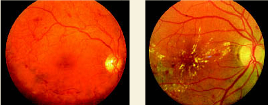
Background (nonproliferative) diabetic retinopathyis characterized by macular retinal exudates, edematous retinal thickening, dot and blot retinal hemorrhages, and retinal capillary microaneurysms.
Proliferative diabetic retinopathy. Note the network of abnormal preretinal new blood vessels just super-otemporal to the macula and the linear accumulations of blood in the preretinal vitreous below the macula.
Evidence summary
The Liverpool Diabetic Eye Study1 prospectively evaluated the risk of vision-threatening retinopathy in a cohort that included all patients with diabetes mellitus who were registered with a general practitioner and were not under the care of an ophthalmologist. A subgroup of 4770 patients with type 2 diabetes who did not have sightthreatening retinopathy at baseline underwent at least 1 additional screen. Screening included non-stereoscopic 3-field (45° or 50° field) mydriatic photography. Median follow-up was 3.5 years (range, 1–8.5 years).
The patients were divided into cohorts based on level of demonstrated retinopathy. The mean screening interval for a 95% probability of remaining free of sight-threatening retinopathy was calculated for each grade of baseline retinopathy. Screening patients with no retinopathy every 5 years provided a 95% probability of remaining free of sight-threatening retinopathy. Patients with background retinopathy must be screened annually to achieve the same result, and patients with mild preproliferative retinopathy need to be screened every 4 months ( Table ).
A systematic review2 of multiple small English-language studies evaluating screening and monitoring of diabetic retinopathy found consistent results. Screening by direct or indirect ophthalmoscopy alone detected 65% of patients with sight-threatening retinopathy. Screening by mydriatic retinal photography, augmented by ophthal-moscopy when the photographs were inconclusive, detected 88% to 100% of such cases.
An RCT of 1700 patients with diabetes and retinopathy evaluated preservation of vision with photocoagulation.3 Patients were not differentiated by type of diabetes. Each patient had initial and follow-up stereoscopic fundus photography. One eye was selected at random to receive treatment and the other remained untreated to serve as a control. Because of the magnitude of difference in vision between the eyes, the study was halted at 2 years to permit photocoagulation of the untreated eyes. Patients whose eyes had new vessels on or near the disk lost vision (defined as visual acuity less than 5/200) more often in untreated eyes (18.3% cumulative rate at 2 years) compared with treated eyes (6.4%; number needed to treat [NNT]=8.4).
Another RCT4 of patients with diabetes showed that photocoagulation maintained vision in diabetic retinopathy if the disease was not too advanced. Ninety-nine patients, also not differentiated by type of diabetes, were each treated in 1 eye chosen at random with a xenon-arc photocoagulator. Patients underwent follow-up treatments to the treated eye by clinical indication. The untreated eyes were observed as controls. Blindness occurred significantly less often in the treated eyes (19% total after 5 to 7 years) than in the control eyes (39%; NNT=5 to prevent 1 blind eye). Patients without proliferative retinopathy at onset experienced the most dramatic slowing of deterioration; photocoagulation was more useful in maintaining than in improving vision.
TABLE
Screening frequency for sight-threatening retinopathy
| Stage of retinal disease | Screening frequency* |
|---|---|
| None | 5 years |
| Background retinopathy | 1 year |
| Mild preproliferative retinopathy | 4 months |
| *For 95% chance of remaining free of sight-threatening retinopathy. | |
Recommendations from others
The American Diabetes Association 2003 Clinical Practice Recommendations5 state that patients with type 2 diabetes should have an initial dilated and comprehensive eye examination by an ophthalmologist or optometrist shortly after diagnosis of diabetes. An ophthalmologist or optometrist who is knowledgeable and experienced in diagnosing diabetic retinopathy and is aware of its management should repeat subsequent examinations for both type 1 and type 2 diabetic patients annually. Examinations are required more frequently if retinopathy is progressing.
Screen for retinal complications early, regularly, by an experienced eye doctor
Tsveti Markova, MD
Department of Family Medicine, Wayne State University, Detroit, Mich
Family physicians play a central role in the diabetes care team. They must not only achieve good blood sugar control, but also monitor for complications and coordinate their treatment. Educating patients and reaching common ground with them is essential for success. Evidence suggests that screening for eye complications reduces blindness. Patients with early retinopathy changes are usually asymptomatic; therefore it takes a committed and educated patient to comply with screening recommendations. Also, many patients have the misconception that a visual acuity exam by an optometrist is sufficient for their diabetes eye screening. Family physicians must convey the importance of screening for retinal complications early, regularly and by an optometrist or ophthalmologist experienced in management of diabetes.
1. Younis N, Broadbent DM, Vora JP, Harding SP. Incidence of sight-threatening retinopathy in patients with type 2 diabetes in the Liverpool Diabetic Eye Study: a cohort study. Lancet 2003;361:195-200.
2. Hutchinson A, McIntosh A, Peters J, et al. Effectiveness of screening and monitoring tests for diabetic retinopa-thy—a systematic review. Diabet Med 2000;17:495-506.
3. The Diabetic Retinopathy Study Research Group. Preliminary report on effects of photocoagulation therapy. Am J Ophthalmol 1976;81:383-396.
4. British Multicentre Study Group. Photocoagulation from diabetic maculopathy: a randomized controlled clinical trial using the xenon arc. Diabetes 1983;32:1010-1016.
5. Fong DS, Aiello L, Gardner TW, et al. American Diabetes Association. Diabetic retinopathy. Diabetes Care 2004;26 Suppl 1:S84-S87.
Screening eye exams for patients with type 2 diabetes can detect retinopathy early enough so treatment can prevent vision loss. Patients without diabetic retinopathy who are systematically screened by mydriatic retinal photography have a 95% probability of remaining free of sight-threatening retinopathy over the next 5 years. If background or preproliferative retinopathy is found at screening (Figure), the 95% probability interval for remaining free of sight-threatening retinopathy is reduced to 12 and 4 months, respectively (strength of recommendation [SOR]: B, based on 1 prospective cohort study).
A reliably sensitive screening exam requires mydriatic retinal photography augmented by ophthalmoscopy when photographs are inconclusive (SOR: A, based on a systematic review). For patients with diabetes not differentiated by type, photocoagu-lation significantly decreases visual deterioration and reduces the chances of blindness (SOR: A, based on randomized controlled trials [RCT]).
FIGURE
Nonproliferative and proliferative diabetic retinopathy
Background (nonproliferative) diabetic retinopathyis characterized by macular retinal exudates, edematous retinal thickening, dot and blot retinal hemorrhages, and retinal capillary microaneurysms.
Proliferative diabetic retinopathy. Note the network of abnormal preretinal new blood vessels just super-otemporal to the macula and the linear accumulations of blood in the preretinal vitreous below the macula.
Evidence summary
The Liverpool Diabetic Eye Study1 prospectively evaluated the risk of vision-threatening retinopathy in a cohort that included all patients with diabetes mellitus who were registered with a general practitioner and were not under the care of an ophthalmologist. A subgroup of 4770 patients with type 2 diabetes who did not have sightthreatening retinopathy at baseline underwent at least 1 additional screen. Screening included non-stereoscopic 3-field (45° or 50° field) mydriatic photography. Median follow-up was 3.5 years (range, 1–8.5 years).
The patients were divided into cohorts based on level of demonstrated retinopathy. The mean screening interval for a 95% probability of remaining free of sight-threatening retinopathy was calculated for each grade of baseline retinopathy. Screening patients with no retinopathy every 5 years provided a 95% probability of remaining free of sight-threatening retinopathy. Patients with background retinopathy must be screened annually to achieve the same result, and patients with mild preproliferative retinopathy need to be screened every 4 months ( Table ).
A systematic review2 of multiple small English-language studies evaluating screening and monitoring of diabetic retinopathy found consistent results. Screening by direct or indirect ophthalmoscopy alone detected 65% of patients with sight-threatening retinopathy. Screening by mydriatic retinal photography, augmented by ophthal-moscopy when the photographs were inconclusive, detected 88% to 100% of such cases.
An RCT of 1700 patients with diabetes and retinopathy evaluated preservation of vision with photocoagulation.3 Patients were not differentiated by type of diabetes. Each patient had initial and follow-up stereoscopic fundus photography. One eye was selected at random to receive treatment and the other remained untreated to serve as a control. Because of the magnitude of difference in vision between the eyes, the study was halted at 2 years to permit photocoagulation of the untreated eyes. Patients whose eyes had new vessels on or near the disk lost vision (defined as visual acuity less than 5/200) more often in untreated eyes (18.3% cumulative rate at 2 years) compared with treated eyes (6.4%; number needed to treat [NNT]=8.4).
Another RCT4 of patients with diabetes showed that photocoagulation maintained vision in diabetic retinopathy if the disease was not too advanced. Ninety-nine patients, also not differentiated by type of diabetes, were each treated in 1 eye chosen at random with a xenon-arc photocoagulator. Patients underwent follow-up treatments to the treated eye by clinical indication. The untreated eyes were observed as controls. Blindness occurred significantly less often in the treated eyes (19% total after 5 to 7 years) than in the control eyes (39%; NNT=5 to prevent 1 blind eye). Patients without proliferative retinopathy at onset experienced the most dramatic slowing of deterioration; photocoagulation was more useful in maintaining than in improving vision.
TABLE
Screening frequency for sight-threatening retinopathy
| Stage of retinal disease | Screening frequency* |
|---|---|
| None | 5 years |
| Background retinopathy | 1 year |
| Mild preproliferative retinopathy | 4 months |
| *For 95% chance of remaining free of sight-threatening retinopathy. | |
Recommendations from others
The American Diabetes Association 2003 Clinical Practice Recommendations5 state that patients with type 2 diabetes should have an initial dilated and comprehensive eye examination by an ophthalmologist or optometrist shortly after diagnosis of diabetes. An ophthalmologist or optometrist who is knowledgeable and experienced in diagnosing diabetic retinopathy and is aware of its management should repeat subsequent examinations for both type 1 and type 2 diabetic patients annually. Examinations are required more frequently if retinopathy is progressing.
Screen for retinal complications early, regularly, by an experienced eye doctor
Tsveti Markova, MD
Department of Family Medicine, Wayne State University, Detroit, Mich
Family physicians play a central role in the diabetes care team. They must not only achieve good blood sugar control, but also monitor for complications and coordinate their treatment. Educating patients and reaching common ground with them is essential for success. Evidence suggests that screening for eye complications reduces blindness. Patients with early retinopathy changes are usually asymptomatic; therefore it takes a committed and educated patient to comply with screening recommendations. Also, many patients have the misconception that a visual acuity exam by an optometrist is sufficient for their diabetes eye screening. Family physicians must convey the importance of screening for retinal complications early, regularly and by an optometrist or ophthalmologist experienced in management of diabetes.
1. Younis N, Broadbent DM, Vora JP, Harding SP. Incidence of sight-threatening retinopathy in patients with type 2 diabetes in the Liverpool Diabetic Eye Study: a cohort study. Lancet 2003;361:195-200.
2. Hutchinson A, McIntosh A, Peters J, et al. Effectiveness of screening and monitoring tests for diabetic retinopa-thy—a systematic review. Diabet Med 2000;17:495-506.
3. The Diabetic Retinopathy Study Research Group. Preliminary report on effects of photocoagulation therapy. Am J Ophthalmol 1976;81:383-396.
4. British Multicentre Study Group. Photocoagulation from diabetic maculopathy: a randomized controlled clinical trial using the xenon arc. Diabetes 1983;32:1010-1016.
5. Fong DS, Aiello L, Gardner TW, et al. American Diabetes Association. Diabetic retinopathy. Diabetes Care 2004;26 Suppl 1:S84-S87.
Evidence-based answers from the Family Physicians Inquiries Network
Do inhaled beta-agonists control cough in URIs or acute bronchitis?
Patients who receive inhaled beta-agonists for cough due to acute upper respiratory infections (URI) are just as likely to report a productive cough at 7 days compared with patients treated with placebo (strength of recommendation [SOR]: A, based on a systematic review).
One trial, however, showed a reduction in overall cough at 7 days (number needed to treat [NNT]=3, SOR: B, a small randomized controlled trial), and another trial found a reduction in overall symptom score in smokers and those with wheezing on initial exam (SOR: B, based on a small randomized controlled trial).
Evidence summary
No studies of inhaled beta-agonists have been conducted with patients who have an explicit diagnosis of acute cough due to URI. While some clinicians feel a distinction between URI and acute bronchitis should be made, there is significant overlap between these diagnoses in clinical practice, as well as in the available studies.
A systematic review looking at beta-agonists for acute bronchitis included the clinical diagnoses of both acute bronchitis and acute cough because a standard definition of bronchitis is lacking.1 Only two trials in this review examined inhaled beta-agonists. When results from these trials were combined for the outcome of productive cough at 7 days, inhaled beta-agonists showed no benefit. However, the authors note that details of the individual trials may help to clarify the effect of inhaled beta-agonists.
One trial, a randomized controlled trial of adult patients with acute bronchitis in 2 community-based family practices, compared 23 patients receiving albuterolin a multidose inhaler (MDI) with 23 patients receiving placebo inhaler.2 Patients were also randomized to receive erythromycin or placebo tablets. Patients with pneumonia or a history of asthma or chronic obstructive pulmonary disease (COPD) were excluded. At 7 days, 61% of patients in the albuterol group reported cough compared with 91% in the control group (P=.02, NNT=3). No statistically significant difference was seen in productive cough or night cough. Smokers responded to inhaled albuterol similarly to nonsmokers. Erythromycin had no effect on cough and side effects were similar among all groups.
The other trial was a randomized controlled trial of 80 adults with cough due to acute respiratory infection; it compared fenoterol aerosol 4 times daily with placebo.3 Inhaled fenoterol is not available in the US but is similar to albuterol. This study showed no difference in cough at 7 days (relative risk [RR]=0.83; 95% confidence interval [CI], 0.52–1.30). In a subgroup analysis, however, smokers and those wheezing on initial exam had lower overall symptom scores when treated with fenoterol.
Recommendations from others
We were unable to find any guidelines on the use of albuterol via MDI for cough from bronchitis or URIs.
Inhaled beta-agonists may aid symptoms; other outcomes may not be improved
Even without a history of lung disease, patients presenting with cough due to acute respiratory illness and with evidence of airflow obstruction (wheezing) appear to receive symptom relief from inhaled beta-agonists. Smokers may be another subgroup who benefit from treatment. However, important patient-oriented outcomes (such as reduced need for over-the-counter medicines, general well being, and return to work) do not improve. If using inhaled albuterol to treat acute cough in practice, one must also consider the financial costs and adverse effects associated with treatment.
1. Smucny J, Flynn C, Becker L, Glazier R. Beta2-agonists for acute bronchitis (Cochrane Review). In: The Cochrane Library, Issue 2, 2004. Chichester, UK: John Wiley & Sons, Ltd.
2. Hueston WJ. Albuterol delivered by metered-dose inhaler to treat acute bronchitis. J Fam Pract 1994;39:437-440.
3. Melbye H, Aasebo U, Straume B. Symptomatic effect of inhaled fenoterol in acute bronchitis: a placebo controlled double-blind study. Fam Pract 1991;8:216-222.
Patients who receive inhaled beta-agonists for cough due to acute upper respiratory infections (URI) are just as likely to report a productive cough at 7 days compared with patients treated with placebo (strength of recommendation [SOR]: A, based on a systematic review).
One trial, however, showed a reduction in overall cough at 7 days (number needed to treat [NNT]=3, SOR: B, a small randomized controlled trial), and another trial found a reduction in overall symptom score in smokers and those with wheezing on initial exam (SOR: B, based on a small randomized controlled trial).
Evidence summary
No studies of inhaled beta-agonists have been conducted with patients who have an explicit diagnosis of acute cough due to URI. While some clinicians feel a distinction between URI and acute bronchitis should be made, there is significant overlap between these diagnoses in clinical practice, as well as in the available studies.
A systematic review looking at beta-agonists for acute bronchitis included the clinical diagnoses of both acute bronchitis and acute cough because a standard definition of bronchitis is lacking.1 Only two trials in this review examined inhaled beta-agonists. When results from these trials were combined for the outcome of productive cough at 7 days, inhaled beta-agonists showed no benefit. However, the authors note that details of the individual trials may help to clarify the effect of inhaled beta-agonists.
One trial, a randomized controlled trial of adult patients with acute bronchitis in 2 community-based family practices, compared 23 patients receiving albuterolin a multidose inhaler (MDI) with 23 patients receiving placebo inhaler.2 Patients were also randomized to receive erythromycin or placebo tablets. Patients with pneumonia or a history of asthma or chronic obstructive pulmonary disease (COPD) were excluded. At 7 days, 61% of patients in the albuterol group reported cough compared with 91% in the control group (P=.02, NNT=3). No statistically significant difference was seen in productive cough or night cough. Smokers responded to inhaled albuterol similarly to nonsmokers. Erythromycin had no effect on cough and side effects were similar among all groups.
The other trial was a randomized controlled trial of 80 adults with cough due to acute respiratory infection; it compared fenoterol aerosol 4 times daily with placebo.3 Inhaled fenoterol is not available in the US but is similar to albuterol. This study showed no difference in cough at 7 days (relative risk [RR]=0.83; 95% confidence interval [CI], 0.52–1.30). In a subgroup analysis, however, smokers and those wheezing on initial exam had lower overall symptom scores when treated with fenoterol.
Recommendations from others
We were unable to find any guidelines on the use of albuterol via MDI for cough from bronchitis or URIs.
Inhaled beta-agonists may aid symptoms; other outcomes may not be improved
Even without a history of lung disease, patients presenting with cough due to acute respiratory illness and with evidence of airflow obstruction (wheezing) appear to receive symptom relief from inhaled beta-agonists. Smokers may be another subgroup who benefit from treatment. However, important patient-oriented outcomes (such as reduced need for over-the-counter medicines, general well being, and return to work) do not improve. If using inhaled albuterol to treat acute cough in practice, one must also consider the financial costs and adverse effects associated with treatment.
1. Smucny J, Flynn C, Becker L, Glazier R. Beta2-agonists for acute bronchitis (Cochrane Review). In: The Cochrane Library, Issue 2, 2004. Chichester, UK: John Wiley & Sons, Ltd.
2. Hueston WJ. Albuterol delivered by metered-dose inhaler to treat acute bronchitis. J Fam Pract 1994;39:437-440.
3. Melbye H, Aasebo U, Straume B. Symptomatic effect of inhaled fenoterol in acute bronchitis: a placebo controlled double-blind study. Fam Pract 1991;8:216-222.
Evidence-based answers from the Family Physicians Inquiries Network