User login
Itchy rash on hands
Based on the gastrointestinal issues, weight loss, and skin findings, the family physician suspected dermatitis herpetiformis and performed a punch biopsy of an intact vesicle. Because of the weight loss he also ordered a complete blood count, thyroid stimulating hormone blood test, chemistry panel, and an anti-gliadin antibody level. The biopsy results and positive anti-gliadin antibody results confirmed the physician’s suspicions for dermatitis herpetiformis and gluten-induced enteropathy.
Dermatitis herpetiformis is a chronic recurrent symmetric vesicular eruption that is usually associated with gluten-induced enteropathy. It most commonly occurs in the 20- to 40-year-old age group. Men are affected more often than women.
The disease is related to gluten and other diet-related antigens that cause the development of circulating immune complexes and their subsequent deposition in the skin. The term herpetiformis refers to the grouped vesicles that appear on extensor aspects of the extremities and trunk and is not a viral infection or related to the herpes viruses. The disease is characterized by the deposition of IgA along the tips of the dermal papillae. The majority of patients will also have blunting and flattening of jejunal villi, which leads to diarrhea even to the point of steatorrhea and malabsorption.
With a gluten-free diet, 80% of patients will show improvement in the skin lesions. The degree of benefit is dependent upon the strictness of the diet. A gluten-free diet may help the enteropathy and decrease the subsequent development of small bowel lymphoma. Dapsone at an initial dose of 100 to 200 mg daily with gradual reduction to a 25 to 50 mg maintenance level may be needed in addition to the gluten-free diet.
Photos and text for Photo Rounds Friday courtesy of Richard P. Usatine, MD. This case was adapted from: Hara, J. Other bullous diseases. In: Usatine R, Smith M, Mayeaux EJ, et al, eds. The Color Atlas of Family Medicine. New York, NY: McGraw-Hill; 2009:799-806.
To learn more about The Color Atlas of Family Medicine, see:
• http://www.amazon.com/Color-Atlas-Family-Medicine/dp/0071474641
The Color Atlas of Family Medicine is also available as an app for mobile devices. See
Erosions from blistering
|
|
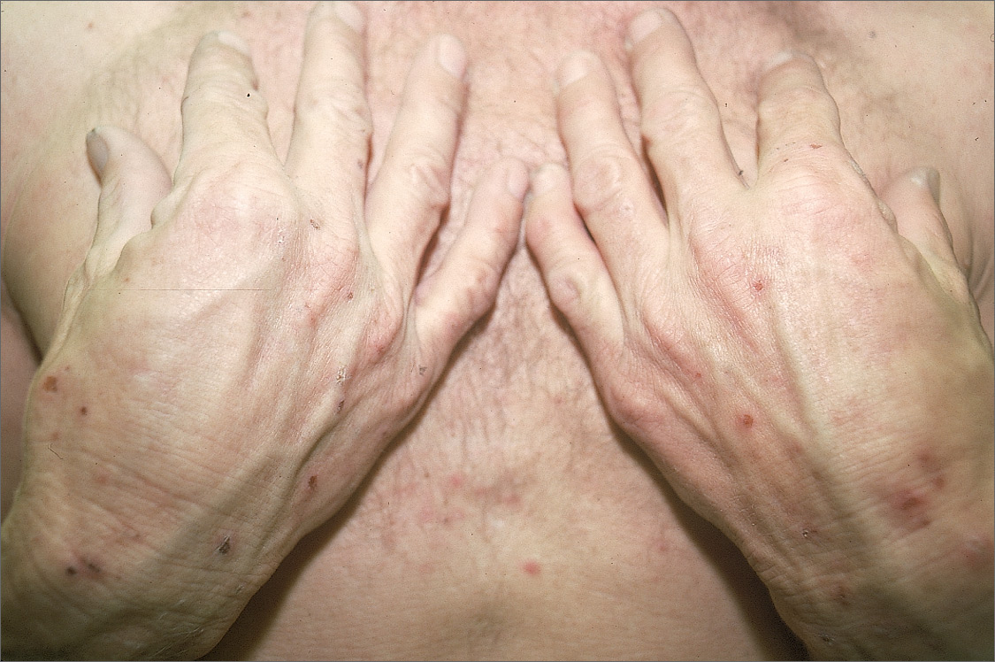
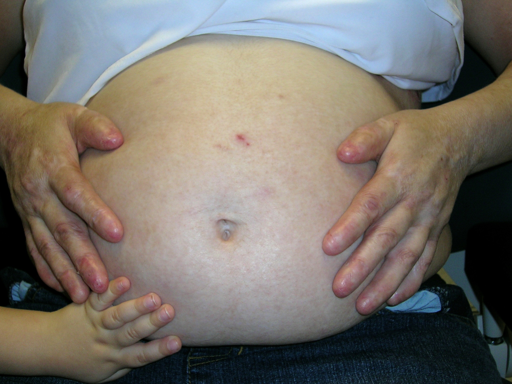
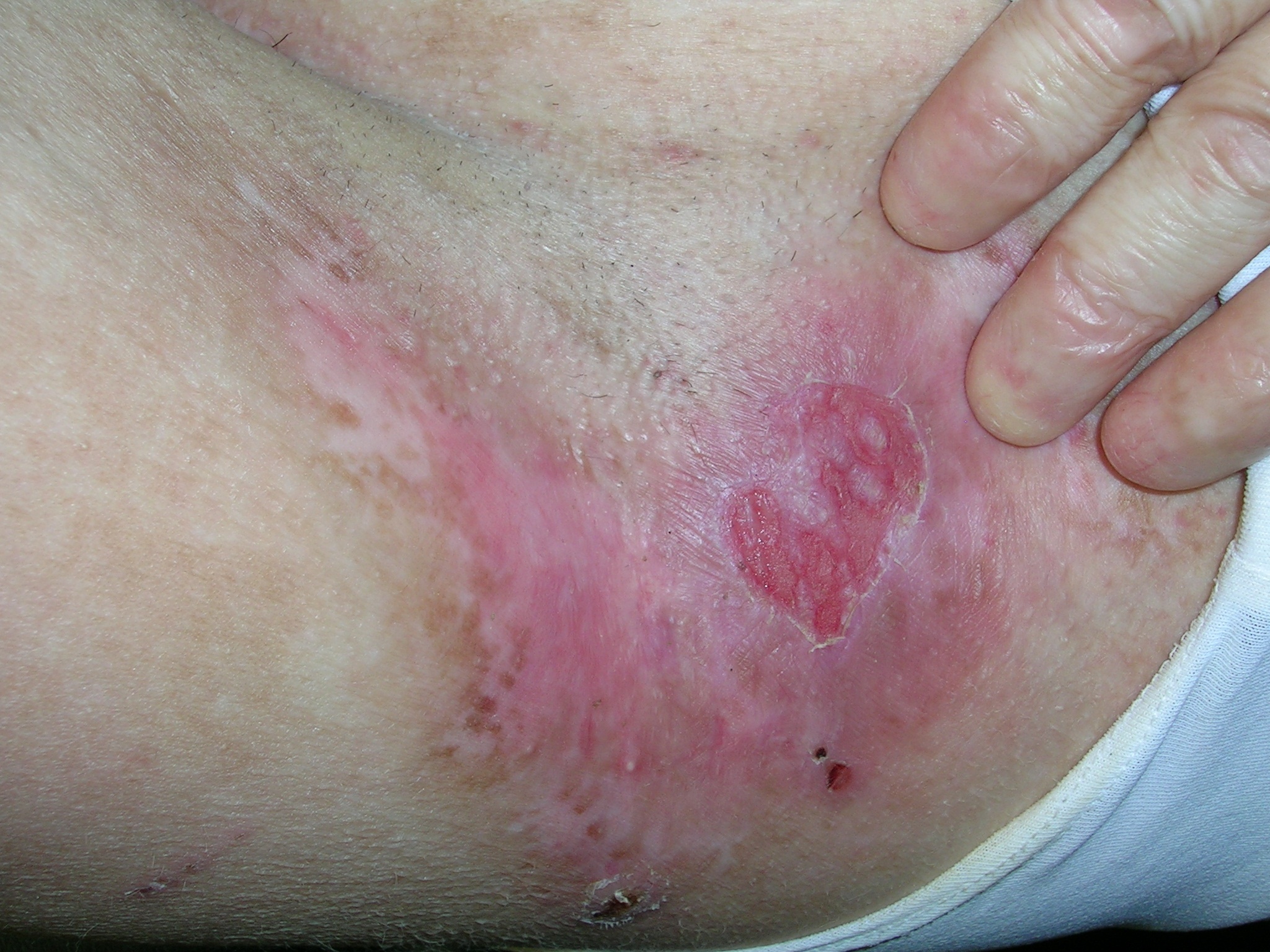
This patient had recessive dystrophic epidermolysis bullosa. (Note the erosion in the axilla [FIGURE 1] and the normal hand of the daughter on the mom’s abdomen [FIGURE 2].) Dystrophic epidermolysis bullosa belongs to a family of inherited diseases characterized by skin fragility and blister formation caused my minor skin trauma. There are autosomal recessive and autosomal dominant types. The severity of this disease may vary widely. Onset is in childhood; in later years, severe dystrophic deformities of hands and feet are characteristic. Malignant degeneration is common—especially squamous cell carcinoma in sun exposed areas.
Dystrophic epidermolysis bullosa has vesiculobullous skin separation occurring at the sub-basal lamina level. Repeated blistering of the hands can lead to fusion of the fingers and the so-called mitten deformity.
Skin biopsies can differentiate between the different forms of epidermolysis bullosa: simplex, junctional, and dystrophic. Management involves preventing trauma, careful wound care, and treating complicating infections. Other supportive measures such as pain management and nutritional support are often necessary.
In this case, the family physician prescribed a mid-potency topical steroid ointment to treat the pain and blistering in her axilla and set up a one-month follow-up appointment.
Photos and text for Photo Rounds Friday courtesy of Richard P. Usatine, MD. This case was adapted from: Hara, J. Other bullous diseases. In: Usatine R, Smith M, Mayeaux EJ, et al, eds. The Color Atlas of Family Medicine. New York, NY: McGraw-Hill; 2009:799-806.
To learn more about The Color Atlas of Family Medicine, see:
• http://www.amazon.com/Color-Atlas-Family-Medicine/dp/0071474641
The Color Atlas of Family Medicine is also available as an app for mobile devices. See
|
|
|
|
This patient had recessive dystrophic epidermolysis bullosa. (Note the erosion in the axilla [FIGURE 1] and the normal hand of the daughter on the mom’s abdomen [FIGURE 2].) Dystrophic epidermolysis bullosa belongs to a family of inherited diseases characterized by skin fragility and blister formation caused my minor skin trauma. There are autosomal recessive and autosomal dominant types. The severity of this disease may vary widely. Onset is in childhood; in later years, severe dystrophic deformities of hands and feet are characteristic. Malignant degeneration is common—especially squamous cell carcinoma in sun exposed areas.
Dystrophic epidermolysis bullosa has vesiculobullous skin separation occurring at the sub-basal lamina level. Repeated blistering of the hands can lead to fusion of the fingers and the so-called mitten deformity.
Skin biopsies can differentiate between the different forms of epidermolysis bullosa: simplex, junctional, and dystrophic. Management involves preventing trauma, careful wound care, and treating complicating infections. Other supportive measures such as pain management and nutritional support are often necessary.
In this case, the family physician prescribed a mid-potency topical steroid ointment to treat the pain and blistering in her axilla and set up a one-month follow-up appointment.
Photos and text for Photo Rounds Friday courtesy of Richard P. Usatine, MD. This case was adapted from: Hara, J. Other bullous diseases. In: Usatine R, Smith M, Mayeaux EJ, et al, eds. The Color Atlas of Family Medicine. New York, NY: McGraw-Hill; 2009:799-806.
To learn more about The Color Atlas of Family Medicine, see:
• http://www.amazon.com/Color-Atlas-Family-Medicine/dp/0071474641
The Color Atlas of Family Medicine is also available as an app for mobile devices. See
Blisters on hands
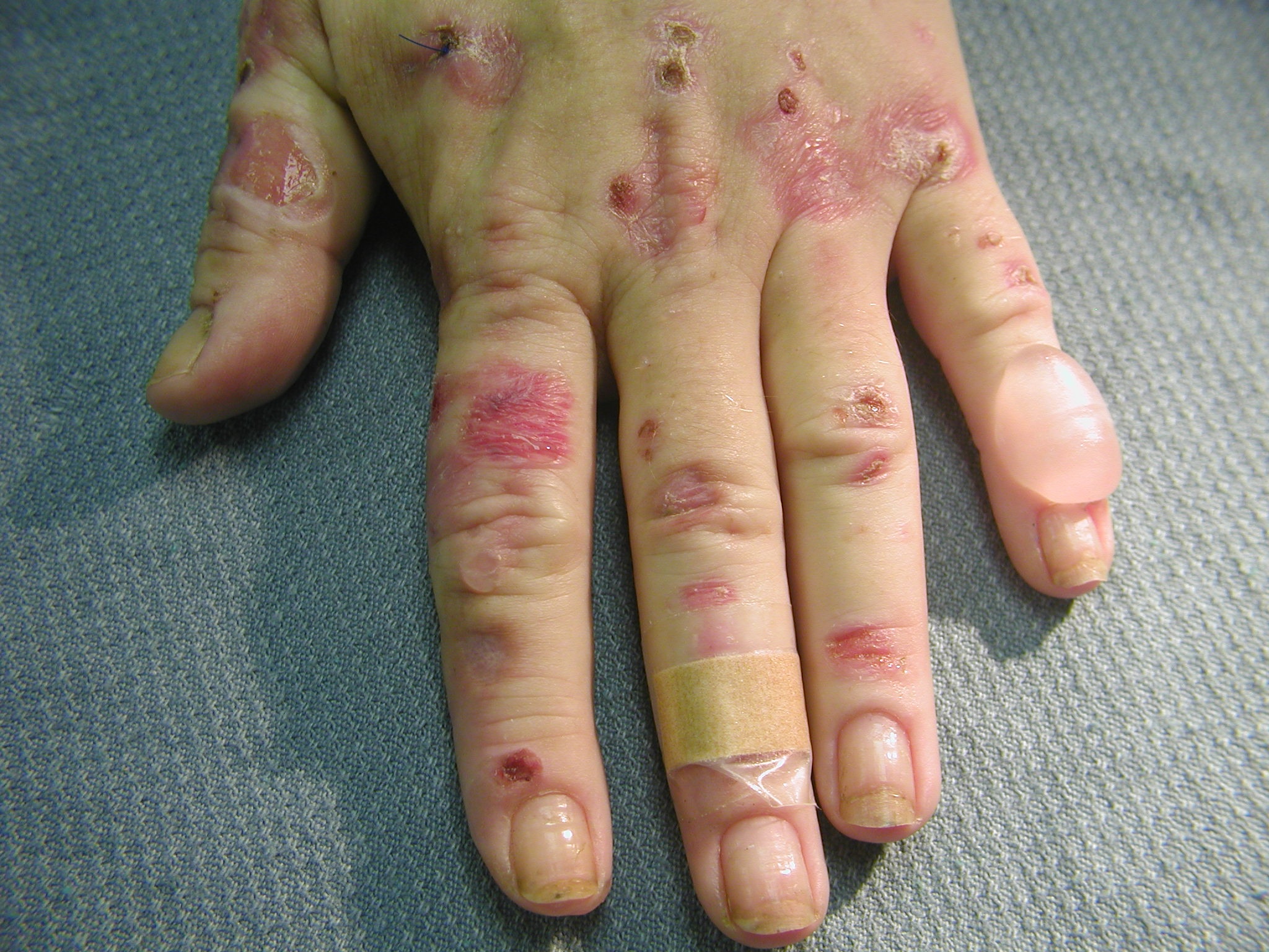
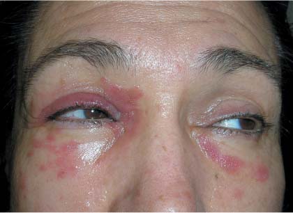
A urine sample fluoresced orange-red under a Woods lamp, prompting the FP to suspect porphyria cutanea tarda. A 24-hour urine test for porphyrins confirmed his suspicions.
Porphyria cutanea tarda occurs mostly in middle-aged adults (typically 30-50 years of age) and is rare in children. Risk factors include hepatitis C, oral contraceptive use, and prostate cancer treatment with estrogen therapy. Alcohol, pesticides, and chloroquine have been implicated as chemicals that induce porphyria cutanea tarda; it is equally common in both men and women.
The porphyrias are a family of illnesses caused by various derangements in the metabolism of porphyrin, the chemical backbone of hemoglobin. Whereas the other porphyrias are associated with systemic manifestations, porphyria cutanea tarda has no extracutaneous manifestations. The classic presentation is that of blistering (vesicles and tense bullae) on photosensitive “fragile skin.” As the blisters heal, the skin takes on an atrophic appearance. Hypertrichosis (especially on the cheeks and temples) is also common and may be the presenting feature. Classically the dorsa of the hands are affected.
If the onset is associated with alcohol ingestion, estrogen therapy, or exposure to pesticides, reducing exposure is warranted. Phlebotomy of 500 ml of blood weekly until the hemoglobin is decreased to 10 g is associated with biochemical and clinical remission within a year. Avoidance of excess sunlight exposure (to avoid hypersensitivity) is important. Avoidance of trauma and careful wound care are also necessary.
Text for Photo Rounds Friday courtesy of Richard P. Usatine, MD. Photo courtesy of Lewis Rose, MD. This case was adapted from: Hara, J. Other bullous diseases. In: Usatine R, Smith M, Mayeaux EJ, et al, eds. The Color Atlas of Family Medicine. New York, NY: McGraw-Hill; 2009:799-806.
To learn more about The Color Atlas of Family Medicine, see:
• http://www.amazon.com/Color-Atlas-Family-Medicine/dp/0071474641
The Color Atlas of Family Medicine is also available as an app for mobile devices. See
Dermatomal rash on a 6-year-old boy
THE MOTHER OF A 6-YEAR-OLD BOY brought her son into our clinic because he had recently developed abdominal pain and an itchy rash. She reported that he had a decreased appetite and was irritable. Four days earlier, the boy had complained of “stinging” pain in his right lower abdomen after playing in the yard. The next day, he developed an erythematous raised rash, which his mother thought was poison ivy. Within 48 hours, the rash developed vesicles and the mother began to suspect that her son had shingles. The child had no history of chickenpox, but had received a single dose of varicella vaccine as an infant.
The patient and his mother denied any recent fever, nausea, vomiting, respiratory symptoms, or other rashes. They reported that no family members were similarly affected. The child was otherwise healthy and not taking any medications. His exam was unremarkable except for the rash (FIGURE 1), which included healing vesicles and was limited to the T7 dermatome on the right side of his trunk.
FIGURE 1
A 6-year-old boy with a pruritic, 4-day-old vesicular rash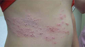
WHAT IS YOUR DIAGNOSIS?
HOW WOULD YOU TREAT THIS PATIENT?
Diagnosis: Herpes zoster
The patient had a classic case of herpes zoster (HZ), caused by a reactivation of the varicella-zoster virus (VZV), which also causes chickenpox. HZ is characterized by a painful vesicular rash distributed in a dermatomal pattern, as opposed to the chickenpox rash, which is generalized and more likely to be associated with systemic symptoms. Both rashes develop new lesions over time, producing vesicles in a variety of stages and sizes. The vesicles rupture and crust over as the patient recovers.1
Unintended results of immunization. The varicella vaccine—a live attenuated vaccine prepared from the Oka/Merck strain of VZV—may produce symptomatic infections.2 Breakthrough chickenpox with wild-type varicella is also possible after immunization, although such infections are typically mild and uncomplicated.3 To learn more, see “Varicella vaccine: Adverse effects, contraindications”.
HZ occurs infrequently in healthy children after natural infection and after immunization.2 In children with leukemia, who are more likely to develop zoster, the incidence of HZ after immunization is about 3 times lower than after natural infection, according to research data supplied by Florence Synn, MD, of Merck & Co, Inc., on May 7, 2008.
In the late 1900s, chickenpox affected about 3.5 million people annually—mostly children. Each year varicella caused 3837 to 6458 hospitalizations and an average of 96 deaths.7 These complications spurred the development of the vaccine,8 which became commercially available in 1995.5
The varicella vaccine has been shown to decrease the incidence of infection by 83% compared with historical controls, to decrease household attack rate by 81% to 90%, and to provide 96% protection when compared to placebo.3 Questions persist about its long-term effect on the complications of chickenpox.3
The vast majority of reported adverse affects (AEs) have been benign and self-limited. According to a 10-year safety review performed after some 55 million doses of the vaccine had been administered, the more serious AEs included 6 cases of herpes zoster (HZ)-related meningitis, 30 additional neurologic syndromes, and 7 patients with disseminated Oka varicella zoster virus infection. Most of these serious AEs involved immunocompromised patients.5 Of 403 samples tested, only 97 of the AEs were identified as Oka-type virus by polymerase chain reaction testing, including 57 of the 697 reports of HZ. Only 3 confirmed cases of secondary transmission of the Oka virus were reported.5
Varivax is contraindicated in immunocompromised and pregnant patients, and vaccinated individuals should avoid close contact with susceptible high-risk individuals for up to 6 weeks after immunization.3 However, studies in vaccinated patients who later developed leukemia provide some reassurance about inadvertent exposure.
Although the varicella vaccine is generally safe and efficacious, physicians should review the immunization status of all household members and discuss contact precautions with patients and their families before administering any live vaccine.
Contact dermatitis, herpes simplex comprise the differential
The differential diagnosis includes contact dermatitis, which is often associated with exposure to poison ivy. The vesicles of contact dermatitis follow the pattern of exposure, often forming a linear pattern, and are not distributed along a dermatome.4
Herpes simplex, caused by HSV-1 or HSV-2, may present with a vesicular rash similar to HZ. The rash does not follow the dermatomal distribution and the vesicles are more uniform than is seen in HZ. The rash may periodically recur, which confirms the diagnosis.1
When lab testing is helpful
Generally speaking, diagnosis of HZ does not require lab work, but tests may be helpful with atypical or complicated presentations. Direct fluorescent antibody testing of scrapings or viral specimens can demonstrate specific antigens for a relatively quick and inexpensive confirmation of VZV.
Polymerase chain reaction (PCR) testing is superior to culture for definitive identification and can distinguish between wild-type and the Oka (vaccine)-type varicella.5 Serology is available, but is used more to identify those at risk for infection than to assist with diagnosis.
With our patient, the diagnosis of HZ was evident by the history and physical exam. Testing was performed with the assistance of our microbiology lab to aid in postmarketing surveillance of the varicella vaccine. Samples, obtained from aspiration of vesicular fluid and swabs of unroofed vesicles, were placed in viral culture media and sent to the Centers for Disease Control and Prevention for PCR. The results were genotype specific for vaccine type VZV.
Is treatment necessary?
Most children with uncomplicated HZ do not require treatment. Antiviral medications, ideally given within the first 72 hours, are indicated for patients with moderate to severe pain, an extensive rash, or a rash involving the face, and for patients ages 50 years or older6 (strength of recommendation [SOR]: A). Acyclovir is the treatment of choice for children.2
Antivirals have been shown to lessen the acute pain of HZ, reduce the number of lesions, speed healing, and limit the duration of viral shedding.6 Most studies also show a decreased incidence of the persistent severe pain syndrome known as postherpetic neuralgia, which is more common in older individuals.6
Our young patient gets better, but Dad starts to itch
Our 6-year-old patient was treated symptomatically with calamine lotion and acetaminophen-codeine at night. He returned to school once his lesions crusted over. As the boy’s rash resolved, his father developed a fever and pruritic vesicular rash (FIGURE 2). The father’s varicella lesions also contained the Oka-type VZV. He, too, recovered without complications.
FIGURE 2
Father developed chickenpox as the son’s herpes zoster rash resolved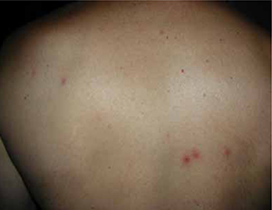
Acknowledgement
The authors thank Edward J. Mayeaux Jr, MD, DABFP, FAAFP, for his guidance in the preparation of this article.
CORRESPONDENCE
Jan Hood, MD, DABFP, Louisiana State University Health Sciences Center, 1501 Kings Highway, Shreveport, LA 71130; [email protected]
1. Habif TP. Warts, herpes simplex, and other viral infections. In: Clinical Dermatology: A Color Guide to Diagnosis and Therapy. 5th ed. Hanover, NH: Mosby/Elsevier; 2010:454–490.
2. Leung AK, Robson WL, Leong AG. Herpes zoster in childhood. J Pediatr Health Care. 2006;20:300-303.
3. Varivax [package insert] Whitehouse Station, NJ: Merck & Co, Inc; 2011.
4. Habif TP. Contact dermatitis and patch testing. In: Clinical Dermatology: A Color Guide to Diagnosis and Therapy. 5th ed. Hanover, NH: Mosby/Elsevier; 2010:130–153.
5. Galea SA, Sweet A, Beninger P, et al. The safety profile of varicella vaccine: a 10-year review. J Infect Dis. 2008;197(suppl 2):S165-S169.
6. Dworkin RH, Johnson RW, Breuer J, et al. Recommendations for the management of herpes zoster. Clin Infect Dis. 2007;44(suppl 1):S1-S26.
7. Preblud SR. Varicella: complications and costs. Pediatrics. 1986;78:728-735.
THE MOTHER OF A 6-YEAR-OLD BOY brought her son into our clinic because he had recently developed abdominal pain and an itchy rash. She reported that he had a decreased appetite and was irritable. Four days earlier, the boy had complained of “stinging” pain in his right lower abdomen after playing in the yard. The next day, he developed an erythematous raised rash, which his mother thought was poison ivy. Within 48 hours, the rash developed vesicles and the mother began to suspect that her son had shingles. The child had no history of chickenpox, but had received a single dose of varicella vaccine as an infant.
The patient and his mother denied any recent fever, nausea, vomiting, respiratory symptoms, or other rashes. They reported that no family members were similarly affected. The child was otherwise healthy and not taking any medications. His exam was unremarkable except for the rash (FIGURE 1), which included healing vesicles and was limited to the T7 dermatome on the right side of his trunk.
FIGURE 1
A 6-year-old boy with a pruritic, 4-day-old vesicular rash
WHAT IS YOUR DIAGNOSIS?
HOW WOULD YOU TREAT THIS PATIENT?
Diagnosis: Herpes zoster
The patient had a classic case of herpes zoster (HZ), caused by a reactivation of the varicella-zoster virus (VZV), which also causes chickenpox. HZ is characterized by a painful vesicular rash distributed in a dermatomal pattern, as opposed to the chickenpox rash, which is generalized and more likely to be associated with systemic symptoms. Both rashes develop new lesions over time, producing vesicles in a variety of stages and sizes. The vesicles rupture and crust over as the patient recovers.1
Unintended results of immunization. The varicella vaccine—a live attenuated vaccine prepared from the Oka/Merck strain of VZV—may produce symptomatic infections.2 Breakthrough chickenpox with wild-type varicella is also possible after immunization, although such infections are typically mild and uncomplicated.3 To learn more, see “Varicella vaccine: Adverse effects, contraindications”.
HZ occurs infrequently in healthy children after natural infection and after immunization.2 In children with leukemia, who are more likely to develop zoster, the incidence of HZ after immunization is about 3 times lower than after natural infection, according to research data supplied by Florence Synn, MD, of Merck & Co, Inc., on May 7, 2008.
In the late 1900s, chickenpox affected about 3.5 million people annually—mostly children. Each year varicella caused 3837 to 6458 hospitalizations and an average of 96 deaths.7 These complications spurred the development of the vaccine,8 which became commercially available in 1995.5
The varicella vaccine has been shown to decrease the incidence of infection by 83% compared with historical controls, to decrease household attack rate by 81% to 90%, and to provide 96% protection when compared to placebo.3 Questions persist about its long-term effect on the complications of chickenpox.3
The vast majority of reported adverse affects (AEs) have been benign and self-limited. According to a 10-year safety review performed after some 55 million doses of the vaccine had been administered, the more serious AEs included 6 cases of herpes zoster (HZ)-related meningitis, 30 additional neurologic syndromes, and 7 patients with disseminated Oka varicella zoster virus infection. Most of these serious AEs involved immunocompromised patients.5 Of 403 samples tested, only 97 of the AEs were identified as Oka-type virus by polymerase chain reaction testing, including 57 of the 697 reports of HZ. Only 3 confirmed cases of secondary transmission of the Oka virus were reported.5
Varivax is contraindicated in immunocompromised and pregnant patients, and vaccinated individuals should avoid close contact with susceptible high-risk individuals for up to 6 weeks after immunization.3 However, studies in vaccinated patients who later developed leukemia provide some reassurance about inadvertent exposure.
Although the varicella vaccine is generally safe and efficacious, physicians should review the immunization status of all household members and discuss contact precautions with patients and their families before administering any live vaccine.
Contact dermatitis, herpes simplex comprise the differential
The differential diagnosis includes contact dermatitis, which is often associated with exposure to poison ivy. The vesicles of contact dermatitis follow the pattern of exposure, often forming a linear pattern, and are not distributed along a dermatome.4
Herpes simplex, caused by HSV-1 or HSV-2, may present with a vesicular rash similar to HZ. The rash does not follow the dermatomal distribution and the vesicles are more uniform than is seen in HZ. The rash may periodically recur, which confirms the diagnosis.1
When lab testing is helpful
Generally speaking, diagnosis of HZ does not require lab work, but tests may be helpful with atypical or complicated presentations. Direct fluorescent antibody testing of scrapings or viral specimens can demonstrate specific antigens for a relatively quick and inexpensive confirmation of VZV.
Polymerase chain reaction (PCR) testing is superior to culture for definitive identification and can distinguish between wild-type and the Oka (vaccine)-type varicella.5 Serology is available, but is used more to identify those at risk for infection than to assist with diagnosis.
With our patient, the diagnosis of HZ was evident by the history and physical exam. Testing was performed with the assistance of our microbiology lab to aid in postmarketing surveillance of the varicella vaccine. Samples, obtained from aspiration of vesicular fluid and swabs of unroofed vesicles, were placed in viral culture media and sent to the Centers for Disease Control and Prevention for PCR. The results were genotype specific for vaccine type VZV.
Is treatment necessary?
Most children with uncomplicated HZ do not require treatment. Antiviral medications, ideally given within the first 72 hours, are indicated for patients with moderate to severe pain, an extensive rash, or a rash involving the face, and for patients ages 50 years or older6 (strength of recommendation [SOR]: A). Acyclovir is the treatment of choice for children.2
Antivirals have been shown to lessen the acute pain of HZ, reduce the number of lesions, speed healing, and limit the duration of viral shedding.6 Most studies also show a decreased incidence of the persistent severe pain syndrome known as postherpetic neuralgia, which is more common in older individuals.6
Our young patient gets better, but Dad starts to itch
Our 6-year-old patient was treated symptomatically with calamine lotion and acetaminophen-codeine at night. He returned to school once his lesions crusted over. As the boy’s rash resolved, his father developed a fever and pruritic vesicular rash (FIGURE 2). The father’s varicella lesions also contained the Oka-type VZV. He, too, recovered without complications.
FIGURE 2
Father developed chickenpox as the son’s herpes zoster rash resolved
Acknowledgement
The authors thank Edward J. Mayeaux Jr, MD, DABFP, FAAFP, for his guidance in the preparation of this article.
CORRESPONDENCE
Jan Hood, MD, DABFP, Louisiana State University Health Sciences Center, 1501 Kings Highway, Shreveport, LA 71130; [email protected]
1. Habif TP. Warts, herpes simplex, and other viral infections. In: Clinical Dermatology: A Color Guide to Diagnosis and Therapy. 5th ed. Hanover, NH: Mosby/Elsevier; 2010:454–490.
2. Leung AK, Robson WL, Leong AG. Herpes zoster in childhood. J Pediatr Health Care. 2006;20:300-303.
3. Varivax [package insert] Whitehouse Station, NJ: Merck & Co, Inc; 2011.
4. Habif TP. Contact dermatitis and patch testing. In: Clinical Dermatology: A Color Guide to Diagnosis and Therapy. 5th ed. Hanover, NH: Mosby/Elsevier; 2010:130–153.
5. Galea SA, Sweet A, Beninger P, et al. The safety profile of varicella vaccine: a 10-year review. J Infect Dis. 2008;197(suppl 2):S165-S169.
6. Dworkin RH, Johnson RW, Breuer J, et al. Recommendations for the management of herpes zoster. Clin Infect Dis. 2007;44(suppl 1):S1-S26.
7. Preblud SR. Varicella: complications and costs. Pediatrics. 1986;78:728-735.
Painful body lesions
The diagnosis
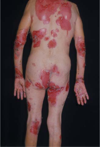
Two stat shave biopsies—one from the edge of an erosion for hematoxylin and eosion staining, and the other of perilesional skin for direct immunofluorescence—revealed a diagnosis of pemphigus foliaceous.
The large areas of skin erosion impaired the skin barrier function and made this patient similar to a burn patient. The patient was at risk of dehydration and superinfection. As a result, IV fluids were started in the hospital and intake and output were measured. The dermatologist prescribed oral prednisone and topical triamcinolone ointment twice daily for the open erosions.
The patient survived under the care of the dermatologist.
Text for Photo Rounds Friday courtesy of Richard P. Usatine, MD. Photos courtesy of Eric Kraus, MD. This case was adapted from: Mittal S. Pemphigus. In: Usatine R, Smith M, Mayeaux EJ, et al, eds. The Color Atlas of Family Medicine. New York, NY: McGraw-Hill; 2009:794-798.
To learn more about The Color Atlas of Family Medicine, see:
• http://www.amazon.com/Color-Atlas-Family-Medicine/dp/0071474641
The Color Atlas of Family Medicine is also available as an app for mobile devices. See
The diagnosis
Two stat shave biopsies—one from the edge of an erosion for hematoxylin and eosion staining, and the other of perilesional skin for direct immunofluorescence—revealed a diagnosis of pemphigus foliaceous.
The large areas of skin erosion impaired the skin barrier function and made this patient similar to a burn patient. The patient was at risk of dehydration and superinfection. As a result, IV fluids were started in the hospital and intake and output were measured. The dermatologist prescribed oral prednisone and topical triamcinolone ointment twice daily for the open erosions.
The patient survived under the care of the dermatologist.
Text for Photo Rounds Friday courtesy of Richard P. Usatine, MD. Photos courtesy of Eric Kraus, MD. This case was adapted from: Mittal S. Pemphigus. In: Usatine R, Smith M, Mayeaux EJ, et al, eds. The Color Atlas of Family Medicine. New York, NY: McGraw-Hill; 2009:794-798.
To learn more about The Color Atlas of Family Medicine, see:
• http://www.amazon.com/Color-Atlas-Family-Medicine/dp/0071474641
The Color Atlas of Family Medicine is also available as an app for mobile devices. See
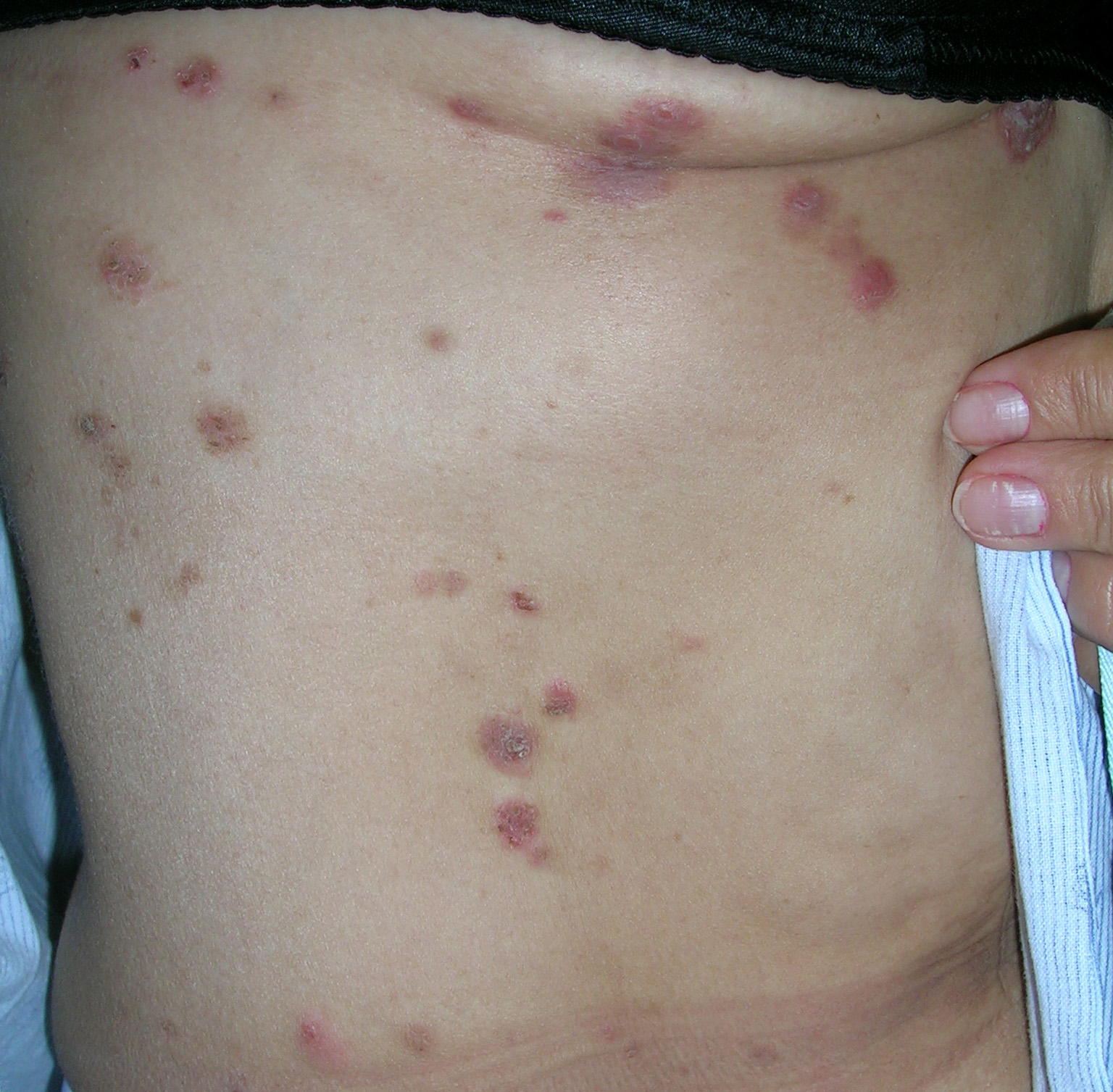
Rash in groin
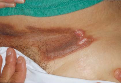
A punch biopsy confirmed the suspicion of a dermatologist that the family physician (FP) had consulted; this was a case of pemphigus vegetans. Pemphigus vegetans, a variant of pemphigus vulgaris, is associated with vegetating proliferation of the epidermis. Pemphigus vegetans is usually seen in intertriginous areas like the axilla, groin, and genital region.
Once the biopsy was read as pemphigus vegetans, the dermatologist took over management of the patient. Management is very similar to that of pemphigus vulgaris, but the lack of oral involvement makes this condition easier to treat. In this case, the patient was prescribed prednisone 60 mg a day and topical clobetasol ointment twice daily to the affected area.
Text for Photo Rounds Friday courtesy of Richard P. Usatine, MD. Photos courtesy of Eric Kraus, MD. This case was adapted from: Mittal S. Pemphigus. In: Usatine R, Smith M, Mayeaux EJ, et al, eds. The Color Atlas of Family Medicine. New York, NY: McGraw-Hill; 2009:794-798.
To learn more about The Color Atlas of Family Medicine, see:
• http://www.amazon.com/Color-Atlas-Family-Medicine/dp/0071474641
The Color Atlas of Family Medicine is also available as an app for mobile devices. See
Crusting facial lesions
|
|
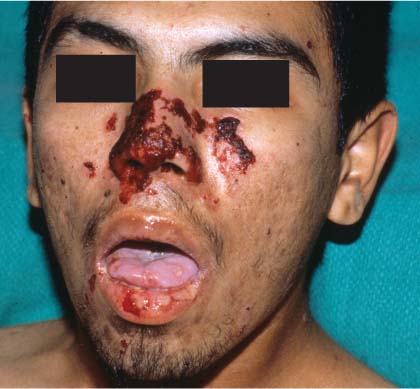
A rush biopsy ordered after the family physician (FP) consulted with a dermatologist confirmed a diagnosis of pemphigus vulgaris.
Pemphigus vulgaris should be considered in any bullous disease involving the oral mucosa. As this is a life-threatening illness, treatment should begin with oral prednisone at the same time that biopsies are performed.
Pemphigus vulgaris presents with flaccid bullae, which rupture easily, creating erosions. Since bullae are short-lived, erosions are the more common presenting physical finding. Lesions are typically tender and heal with post-inflammatory hyperpigmentation, which resolves without scarring. A Nikolsky sign may be present, but is not diagnostic. This is elicited by applying lateral pressure to a bulla or adjacent normal skin, resulting in a separation of the epidermis from deeper layers.
The most common mucosal site is the oral mucosa. Mucosal lesions may be followed by skin lesions on the scalp, face, and upper torso. Pemphigus vulgaris should be suspected if an oral ulcer persists beyond a month.
Two skin biopsies are preferred, but even a single biopsy can be used to make the diagnosis. The first biopsy is sent in formalin for routine histopathology. This biopsy should be of the freshest lesion with an intact bulla if possible. Perform a shave of the edge of the bulla to include the epidermis. The second is taken from perilesional normal skin, and is sent on a gauze pad soaked in normal saline or Michel’s solution for direct immunofluorescence.
The skin lesions are treated with topical high-potency steroids, such as clobetasol ointment. The facial lesions are treated with topical triamcinolone ointment. Oral prednisone is needed in relatively high doses.
Oral hygiene is crucial. Mouthwashes such as chlorhexidine 0.2% or 1:4 hydrogen peroxide may be used. Topical anesthetics may be used for pain. Prednisone should be started at 60 mg a day and titrated as needed. The dose may be increased by 50% every 1 to 2 weeks until disease activity is controlled. However, some experts will not go beyond 1 mg/kg/day. Once remission is induced, the dose is tapered by 25% every 1 to 2 weeks to the lowest dose needed to suppress recurrence of new lesions.
High dose and prolonged treatment with steroids can have serious side effects. Therefore, it is advisable to start adjuvant steroid-sparing therapy within 2 to 4 weeks of treatment. Adjuvant agents have a lag period of 4 to 6 weeks before they become effective, so starting them sooner allows for an earlier steroid taper. Adjuvant agents include azathioprine, cyclophosphamide, methotrexate, and mycophenolate. They may be used alone to maintain remission after steroid withdrawal.
Photos and text for Photo Rounds Friday courtesy of Richard P. Usatine, MD. This case was adapted from: Mittal S. Pemphigus. In: Usatine R, Smith M, Mayeaux EJ, et al, eds. The Color Atlas of Family Medicine. New York, NY: McGraw-Hill; 2009:794-798.
To learn more about The Color Atlas of Family Medicine, see:
• http://www.amazon.com/Color-Atlas-Family-Medicine/dp/0071474641
The Color Atlas of Family Medicine is also available as an app for mobile devices. See
|
|
|
|
A rush biopsy ordered after the family physician (FP) consulted with a dermatologist confirmed a diagnosis of pemphigus vulgaris.
Pemphigus vulgaris should be considered in any bullous disease involving the oral mucosa. As this is a life-threatening illness, treatment should begin with oral prednisone at the same time that biopsies are performed.
Pemphigus vulgaris presents with flaccid bullae, which rupture easily, creating erosions. Since bullae are short-lived, erosions are the more common presenting physical finding. Lesions are typically tender and heal with post-inflammatory hyperpigmentation, which resolves without scarring. A Nikolsky sign may be present, but is not diagnostic. This is elicited by applying lateral pressure to a bulla or adjacent normal skin, resulting in a separation of the epidermis from deeper layers.
The most common mucosal site is the oral mucosa. Mucosal lesions may be followed by skin lesions on the scalp, face, and upper torso. Pemphigus vulgaris should be suspected if an oral ulcer persists beyond a month.
Two skin biopsies are preferred, but even a single biopsy can be used to make the diagnosis. The first biopsy is sent in formalin for routine histopathology. This biopsy should be of the freshest lesion with an intact bulla if possible. Perform a shave of the edge of the bulla to include the epidermis. The second is taken from perilesional normal skin, and is sent on a gauze pad soaked in normal saline or Michel’s solution for direct immunofluorescence.
The skin lesions are treated with topical high-potency steroids, such as clobetasol ointment. The facial lesions are treated with topical triamcinolone ointment. Oral prednisone is needed in relatively high doses.
Oral hygiene is crucial. Mouthwashes such as chlorhexidine 0.2% or 1:4 hydrogen peroxide may be used. Topical anesthetics may be used for pain. Prednisone should be started at 60 mg a day and titrated as needed. The dose may be increased by 50% every 1 to 2 weeks until disease activity is controlled. However, some experts will not go beyond 1 mg/kg/day. Once remission is induced, the dose is tapered by 25% every 1 to 2 weeks to the lowest dose needed to suppress recurrence of new lesions.
High dose and prolonged treatment with steroids can have serious side effects. Therefore, it is advisable to start adjuvant steroid-sparing therapy within 2 to 4 weeks of treatment. Adjuvant agents have a lag period of 4 to 6 weeks before they become effective, so starting them sooner allows for an earlier steroid taper. Adjuvant agents include azathioprine, cyclophosphamide, methotrexate, and mycophenolate. They may be used alone to maintain remission after steroid withdrawal.
Photos and text for Photo Rounds Friday courtesy of Richard P. Usatine, MD. This case was adapted from: Mittal S. Pemphigus. In: Usatine R, Smith M, Mayeaux EJ, et al, eds. The Color Atlas of Family Medicine. New York, NY: McGraw-Hill; 2009:794-798.
To learn more about The Color Atlas of Family Medicine, see:
• http://www.amazon.com/Color-Atlas-Family-Medicine/dp/0071474641
The Color Atlas of Family Medicine is also available as an app for mobile devices. See
Red sores
| 
|
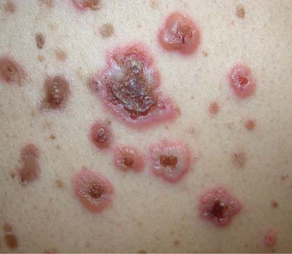
The family physician called his dermatology consultant and they agreed to start the patient on oral prednisone and to perform a punch biopsy, which revealed the diagnosis: pemphigus foliaceous.
Pemphigus foliaceous causes multiple red, scaling, crusted, and pruritic lesions that look like “corn flakes.” Shallow erosions arise when crusts are removed; blisters are rare, as the disease is superficial.
Pemphigus foliaceous initially affects the face (FIGURE 1) and scalp and may progress to involve the chest (FIGURE 2) and back. Skin biopsy is essential for an accurate diagnosis. The depth of acantholysis and site of deposition of antibody complexes help differentiate pemphigus from other bullous diseases. Two biopsies are preferred but even a single biopsy can be used to make the diagnosis. The first biopsy should be sent in formalin for routine histopathology. This biopsy should be of the freshest lesion, with an intact bulla, if possible. The second biopsy should be taken from perilesional normal skin, and should be sent on a gauze pad soaked in normal saline or Michel’s solution for direct immunofluorescence (DIF). Routine histopathology demonstrates suprabasal acantholysis and DIF shows antibody deposition in the intercellular spaces of the epidermis.
Treatment of pemphigus should be undertaken in consultation with a dermatologist. In this case, the skin lesions were treated with topical high-potency steroids (clobetasol ointment). The facial lesions were treated with topical triamcinolone ointment.
Oral prednisone should be prescribed as soon as the diagnosis is suspected. The first priority of treatment should be disease control and remission, followed by disease suppression.
Photos and text for Photo Rounds Friday courtesy of Richard P. Usatine, MD. This case was adapted from: Mittal S. Pemphigus. In: Usatine R, Smith M, Mayeaux EJ, et al, eds. The Color Atlas of Family Medicine. New York, NY: McGraw-Hill; 2009:794-798.
To learn more about The Color Atlas of Family Medicine, see:
• http://www.amazon.com/Color-Atlas-Family-Medicine/dp/0071474641
The Color Atlas of Family Medicine is also available as an app for mobile devices. See
|
|
|
The family physician called his dermatology consultant and they agreed to start the patient on oral prednisone and to perform a punch biopsy, which revealed the diagnosis: pemphigus foliaceous.
Pemphigus foliaceous causes multiple red, scaling, crusted, and pruritic lesions that look like “corn flakes.” Shallow erosions arise when crusts are removed; blisters are rare, as the disease is superficial.
Pemphigus foliaceous initially affects the face (FIGURE 1) and scalp and may progress to involve the chest (FIGURE 2) and back. Skin biopsy is essential for an accurate diagnosis. The depth of acantholysis and site of deposition of antibody complexes help differentiate pemphigus from other bullous diseases. Two biopsies are preferred but even a single biopsy can be used to make the diagnosis. The first biopsy should be sent in formalin for routine histopathology. This biopsy should be of the freshest lesion, with an intact bulla, if possible. The second biopsy should be taken from perilesional normal skin, and should be sent on a gauze pad soaked in normal saline or Michel’s solution for direct immunofluorescence (DIF). Routine histopathology demonstrates suprabasal acantholysis and DIF shows antibody deposition in the intercellular spaces of the epidermis.
Treatment of pemphigus should be undertaken in consultation with a dermatologist. In this case, the skin lesions were treated with topical high-potency steroids (clobetasol ointment). The facial lesions were treated with topical triamcinolone ointment.
Oral prednisone should be prescribed as soon as the diagnosis is suspected. The first priority of treatment should be disease control and remission, followed by disease suppression.
Photos and text for Photo Rounds Friday courtesy of Richard P. Usatine, MD. This case was adapted from: Mittal S. Pemphigus. In: Usatine R, Smith M, Mayeaux EJ, et al, eds. The Color Atlas of Family Medicine. New York, NY: McGraw-Hill; 2009:794-798.
To learn more about The Color Atlas of Family Medicine, see:
• http://www.amazon.com/Color-Atlas-Family-Medicine/dp/0071474641
The Color Atlas of Family Medicine is also available as an app for mobile devices. See
Chronic lesions on legs
A 62-YEAR-OLD MAN came into our facility for a skin exam at the urging of his wife. They were both concerned about a rash on his legs that was asymptomatic and had gradually developed over the past few years.
The patient, who worked as a laborer, was not taking any prescription medications, vitamins, or herbal products. He didn’t smoke, drink alcohol, or use recreational drugs and denied having any fevers, chills, arthralgias, or night sweats. He indicated that his father had a basal cell carcinoma.
On physical exam, we noted some seborrheic keratoses and mild acne rosacea in addition to the rash on his legs. The rash itself consisted of nonpalpable, nonblanchable macules on the lower shins, ankles, and dorsal feet with sparing of the soles (FIGURE).
FIGURE
Nonpalpable rash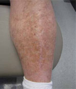
The rash on this 62-year-old man’s legs developed over several years. The nonblanchable macules on the lower shins, ankles, and dorsal feet spared the soles.
WHAT IS YOUR DIAGNOSIS?
HOW WOULD YOU TREAT THIS PATIENT?
Diagnosis: Schamberg’s disease
Schamberg’s disease is a form of progressive pigmented purpuric dermatosis. This type of nonpalpable purpura consists of reddish-brown macules that are usually located on the lower extremities and ankles, and spare the soles of the feet. These petechiae appear due to capillary leakage and breakdown of blood near the skin surface, leaving behind hemosiderin deposits.
Clinically, the lesions are asymptomatic and may persist for months or years. They are characteristically referred to as punctate "cayenne pepper" spots, and can vary in size and shape. Occasionally, mild erythema and scaling can cause slight itching.
Although these lesions have a vasculitic appearance, there is no hematologic or other internal disease association. Schamberg’s purpura may resemble stasis dermatitis, because both processes include inflammation, dilation of capillaries, extravasation of erythrocytes, and hemosiderin deposits. However, lesions due to venous insufficiency extend deep into the dermis and have more pronounced epidermal changes and dermal fibrosis.
An unknown cause. Although no one knows what causes Schamberg’s disease, a cellular immune reaction may be at work. Early endothelial expression of certain intracellular adhesion receptors has shown a common pericapillary infiltrate pattern in cases of Schamberg’s disease.1 Recent viral infection and allergic reactions to medications, such as aspirin, have also been associated with Schamberg’s disease.2
One of several pigmented purpuric dermatoses
Schamberg’s disease fits under the inclusive term of pigmented purpuric dermatoses (PPD), as a macular purpuric reddish-brown dermatosis. Other subtypes of PPD can be distinguished from Schamberg’s disease clinically. These subtypes include lichen aureus, which appears as golden patches; Majocchi’s purpura, which forms an annular pattern with telangiectasias; Gougerot-Blum purpura, which is associated with lichenoid dermatitis; and Doucas and Kapentanakis purpura, which is an eczematous variant. Other forms of purpura can be distinguished from Schamberg’s disease histologically (TABLE).3
Platelet abnormalities can also produce red, flat, nonblanchable petechiae, and may occur in patients who have taken certain prescribed drugs, over-the-counter medications, or herbal remedies; those who have received recent vaccinations; and in those who have (or have had) severe viral infections, beta-hemolytic streptococcal infections, leukemia, lupus erythematosus, or idiopathic thrombocytopenic purpura.
Scurvy can also cause perifollicular petechiae that are distributed primarily on the lower extremities and are symmetrical. Gums may be swollen or hemorrhagic, and patients will have a history of vitamin C deficiency, myalgia, and fatigue. Children may have bone tenderness, epistaxis, and hematuria. A diagnosis can be made by looking for low ascorbic acid in the serum. Prompt correction with vitamin C supplementation is also diagnostic.
Dysproteinemia can cause crops of petechiae and occasionally ecchymoses. Lesions appear on the lower extremities and sometimes on the ears and the tip of the nose. Cryoglobulinemia is associated with Raynaud’s phenomenon, cold urticaria, dizziness, and epistaxis; it is also associated with hepatitis C infection in >90% of cases.4 Macroglobulinemia is associated with dilated vessels and hemorrhage in the optic fundi, mental confusion, anemia, weight loss, hepatosplenomegaly, and lymphadenopathy. Hyperglobulinemic purpura may be associated with arthritis, xerostomia, anemia, and hepatosplenomegaly. These various dysproteinemias can be diagnosed by finding abnormal proteins through serum electrophoresis. Skin biopsy of the petechiae reveals thrombi in dermal vessels, which may be associated with leukocytoclastic vasculitis.4
Many viral infections may result in a petechial rash, including measles, rubella, hepatitis, cytomegalovirus, Coxsackievirus, and respiratory syncytial virus. Petechial rashes associated with eczema or seborrheic dermatitis-like lesions can also be seen in children with Langerhans cell histiocytosis
TABLE
Histologic spectrum of purpura3
| Histologic pattern | Diseases |
|---|---|
| Erythrocyte extravasation without perivascular inflammation or fibrin deposition | Thrombocytopenic purpura, senile purpura, steroid purpura, scurvy |
| Erythrocyte extravasation with lymphocytic perivascular infiltrate; no fibrin deposition | Lichen aureus; Schamberg’s, Majocchi’s, Gougerot-Blum, and Doucas and Kapentanakis purpuras |
| Erythrocyte extravasation with vascular damage, neutrophilic infiltrate, and fibrin deposition | Leukocytoclastic vasculitis, mixed cryoglobulinemia |
Biopsy confirms diagnosis
Platelet and clotting studies are usually normal with Schamberg’s purpura. The definitive diagnosis can be confirmed through skin biopsy, which shows capillaritis of dermal vessels. Perivascular inflammatory infiltrates with extravasations of blood cells and hemosiderin-laden macrophages are seen on histologic evaluation.
Treat mild itching with corticosteroids
These lesions pose mostly a cosmetic problem, but mild itching and scaling occasionally occur. These symptoms can be treated with topical corticosteroid therapy.
Treatment with an 8-week course of pentoxifylline (Trental)—400 mg daily—has shown resolution of lesions in some patients, but no benefit in others.5,6 In some patient’s, psoralen plus ultraviolet light therapy has been shown to provide modest improvement.7 Treatment with aminaphtone 75 mg bid for 4 weeks has recently shown improvements in patients with longstanding Schamberg’s purpura.8 A review of medications may point to a possible etiology for some patients.9
Although lesions can persist for years, the lesions may eventually clear and patients should be assured that there is no definitive underlying systemic disease associated with Schamberg’s disease.10
Educating our patient
Our patient required no treatment, because his rash was asymptomatic. We told him that the pigmentation can last for years, but could be covered with cosmetics. We also reassured him that no underlying systemic disease had caused his rash.
CORRESPONDENCE Jay Shubrook, DO, Castrop Center, O’Bleness Health System, 75 Hospital Drive, Athens, OH 45701; [email protected]
1. Ghersetich I, Lotti T, Bacci S, et al. Cell infiltrate in progressive pigmented purpura (Schamberg’s disease): immunophenotype, adhesion receptors, and intercellular relationships. Int J Dermatol. 1995;34:846-850.
2. Abeck D, Gross GE, Kuwert C, et al. Acetaminophen-induced progressive pigmentary purpura (Schamberg’s disease). J Am Acad Dermatol. 1992;27:123-124.
3. Elder D, Elenitsas R, Jaworsky C, et al. Lever’s Histopathology of the Skin. 8th ed. Philadelphia, Pa: Lippincott-Raven Publishers; 1997.
4. James WD, Berger TG, Elston DM. Andrews’ Diseases of the Skin: Clinical Dermatology. 10th ed. New York, NY: Elsevier; 2006.
5. Gandhi V, Singal A, Sachdeva B, et al. Treatment of Schamberg’s disease with pentoxifylline–therapeutic trial. Indian J Dermatol Venereol Leprol. 2003;69:25-26.
6. Kano Y, Hirayama K, Orihara M, et al. Successful treatment of Schamberg’s disease with pentoxifylline. J Am Acad Dermatol. 1997;36:827-830.
7. Seckin D, Yazici Z, Senol A, et al. A case of Schamberg’s disease responding dramatically to PUVA treatment. Photodermatol Photoimmunol Photomed. 2008;24:95-96.
8. de Godoy JM, Batigalia F. Aminaphtone in the control of Schamberg’s disease. Thromb J. 2009;7:8.-
9. Ratnam KV, Su WP, Peters MS. Purpura simplex (inflammatory purpura without vasculitis): a clinicopathologic study of 174 cases. J Am Acad Dermatol. 1991;25:642-647.
10. Habif TP. Clinical Dermatology: A Color Guide to Diagnosis and Therapy. 4th ed. Philadelphia, Pa: Mosby, Inc; 2003.
A 62-YEAR-OLD MAN came into our facility for a skin exam at the urging of his wife. They were both concerned about a rash on his legs that was asymptomatic and had gradually developed over the past few years.
The patient, who worked as a laborer, was not taking any prescription medications, vitamins, or herbal products. He didn’t smoke, drink alcohol, or use recreational drugs and denied having any fevers, chills, arthralgias, or night sweats. He indicated that his father had a basal cell carcinoma.
On physical exam, we noted some seborrheic keratoses and mild acne rosacea in addition to the rash on his legs. The rash itself consisted of nonpalpable, nonblanchable macules on the lower shins, ankles, and dorsal feet with sparing of the soles (FIGURE).
FIGURE
Nonpalpable rash
The rash on this 62-year-old man’s legs developed over several years. The nonblanchable macules on the lower shins, ankles, and dorsal feet spared the soles.
WHAT IS YOUR DIAGNOSIS?
HOW WOULD YOU TREAT THIS PATIENT?
Diagnosis: Schamberg’s disease
Schamberg’s disease is a form of progressive pigmented purpuric dermatosis. This type of nonpalpable purpura consists of reddish-brown macules that are usually located on the lower extremities and ankles, and spare the soles of the feet. These petechiae appear due to capillary leakage and breakdown of blood near the skin surface, leaving behind hemosiderin deposits.
Clinically, the lesions are asymptomatic and may persist for months or years. They are characteristically referred to as punctate "cayenne pepper" spots, and can vary in size and shape. Occasionally, mild erythema and scaling can cause slight itching.
Although these lesions have a vasculitic appearance, there is no hematologic or other internal disease association. Schamberg’s purpura may resemble stasis dermatitis, because both processes include inflammation, dilation of capillaries, extravasation of erythrocytes, and hemosiderin deposits. However, lesions due to venous insufficiency extend deep into the dermis and have more pronounced epidermal changes and dermal fibrosis.
An unknown cause. Although no one knows what causes Schamberg’s disease, a cellular immune reaction may be at work. Early endothelial expression of certain intracellular adhesion receptors has shown a common pericapillary infiltrate pattern in cases of Schamberg’s disease.1 Recent viral infection and allergic reactions to medications, such as aspirin, have also been associated with Schamberg’s disease.2
One of several pigmented purpuric dermatoses
Schamberg’s disease fits under the inclusive term of pigmented purpuric dermatoses (PPD), as a macular purpuric reddish-brown dermatosis. Other subtypes of PPD can be distinguished from Schamberg’s disease clinically. These subtypes include lichen aureus, which appears as golden patches; Majocchi’s purpura, which forms an annular pattern with telangiectasias; Gougerot-Blum purpura, which is associated with lichenoid dermatitis; and Doucas and Kapentanakis purpura, which is an eczematous variant. Other forms of purpura can be distinguished from Schamberg’s disease histologically (TABLE).3
Platelet abnormalities can also produce red, flat, nonblanchable petechiae, and may occur in patients who have taken certain prescribed drugs, over-the-counter medications, or herbal remedies; those who have received recent vaccinations; and in those who have (or have had) severe viral infections, beta-hemolytic streptococcal infections, leukemia, lupus erythematosus, or idiopathic thrombocytopenic purpura.
Scurvy can also cause perifollicular petechiae that are distributed primarily on the lower extremities and are symmetrical. Gums may be swollen or hemorrhagic, and patients will have a history of vitamin C deficiency, myalgia, and fatigue. Children may have bone tenderness, epistaxis, and hematuria. A diagnosis can be made by looking for low ascorbic acid in the serum. Prompt correction with vitamin C supplementation is also diagnostic.
Dysproteinemia can cause crops of petechiae and occasionally ecchymoses. Lesions appear on the lower extremities and sometimes on the ears and the tip of the nose. Cryoglobulinemia is associated with Raynaud’s phenomenon, cold urticaria, dizziness, and epistaxis; it is also associated with hepatitis C infection in >90% of cases.4 Macroglobulinemia is associated with dilated vessels and hemorrhage in the optic fundi, mental confusion, anemia, weight loss, hepatosplenomegaly, and lymphadenopathy. Hyperglobulinemic purpura may be associated with arthritis, xerostomia, anemia, and hepatosplenomegaly. These various dysproteinemias can be diagnosed by finding abnormal proteins through serum electrophoresis. Skin biopsy of the petechiae reveals thrombi in dermal vessels, which may be associated with leukocytoclastic vasculitis.4
Many viral infections may result in a petechial rash, including measles, rubella, hepatitis, cytomegalovirus, Coxsackievirus, and respiratory syncytial virus. Petechial rashes associated with eczema or seborrheic dermatitis-like lesions can also be seen in children with Langerhans cell histiocytosis
TABLE
Histologic spectrum of purpura3
| Histologic pattern | Diseases |
|---|---|
| Erythrocyte extravasation without perivascular inflammation or fibrin deposition | Thrombocytopenic purpura, senile purpura, steroid purpura, scurvy |
| Erythrocyte extravasation with lymphocytic perivascular infiltrate; no fibrin deposition | Lichen aureus; Schamberg’s, Majocchi’s, Gougerot-Blum, and Doucas and Kapentanakis purpuras |
| Erythrocyte extravasation with vascular damage, neutrophilic infiltrate, and fibrin deposition | Leukocytoclastic vasculitis, mixed cryoglobulinemia |
Biopsy confirms diagnosis
Platelet and clotting studies are usually normal with Schamberg’s purpura. The definitive diagnosis can be confirmed through skin biopsy, which shows capillaritis of dermal vessels. Perivascular inflammatory infiltrates with extravasations of blood cells and hemosiderin-laden macrophages are seen on histologic evaluation.
Treat mild itching with corticosteroids
These lesions pose mostly a cosmetic problem, but mild itching and scaling occasionally occur. These symptoms can be treated with topical corticosteroid therapy.
Treatment with an 8-week course of pentoxifylline (Trental)—400 mg daily—has shown resolution of lesions in some patients, but no benefit in others.5,6 In some patient’s, psoralen plus ultraviolet light therapy has been shown to provide modest improvement.7 Treatment with aminaphtone 75 mg bid for 4 weeks has recently shown improvements in patients with longstanding Schamberg’s purpura.8 A review of medications may point to a possible etiology for some patients.9
Although lesions can persist for years, the lesions may eventually clear and patients should be assured that there is no definitive underlying systemic disease associated with Schamberg’s disease.10
Educating our patient
Our patient required no treatment, because his rash was asymptomatic. We told him that the pigmentation can last for years, but could be covered with cosmetics. We also reassured him that no underlying systemic disease had caused his rash.
CORRESPONDENCE Jay Shubrook, DO, Castrop Center, O’Bleness Health System, 75 Hospital Drive, Athens, OH 45701; [email protected]
1. Ghersetich I, Lotti T, Bacci S, et al. Cell infiltrate in progressive pigmented purpura (Schamberg’s disease): immunophenotype, adhesion receptors, and intercellular relationships. Int J Dermatol. 1995;34:846-850.
2. Abeck D, Gross GE, Kuwert C, et al. Acetaminophen-induced progressive pigmentary purpura (Schamberg’s disease). J Am Acad Dermatol. 1992;27:123-124.
3. Elder D, Elenitsas R, Jaworsky C, et al. Lever’s Histopathology of the Skin. 8th ed. Philadelphia, Pa: Lippincott-Raven Publishers; 1997.
4. James WD, Berger TG, Elston DM. Andrews’ Diseases of the Skin: Clinical Dermatology. 10th ed. New York, NY: Elsevier; 2006.
5. Gandhi V, Singal A, Sachdeva B, et al. Treatment of Schamberg’s disease with pentoxifylline–therapeutic trial. Indian J Dermatol Venereol Leprol. 2003;69:25-26.
6. Kano Y, Hirayama K, Orihara M, et al. Successful treatment of Schamberg’s disease with pentoxifylline. J Am Acad Dermatol. 1997;36:827-830.
7. Seckin D, Yazici Z, Senol A, et al. A case of Schamberg’s disease responding dramatically to PUVA treatment. Photodermatol Photoimmunol Photomed. 2008;24:95-96.
8. de Godoy JM, Batigalia F. Aminaphtone in the control of Schamberg’s disease. Thromb J. 2009;7:8.-
9. Ratnam KV, Su WP, Peters MS. Purpura simplex (inflammatory purpura without vasculitis): a clinicopathologic study of 174 cases. J Am Acad Dermatol. 1991;25:642-647.
10. Habif TP. Clinical Dermatology: A Color Guide to Diagnosis and Therapy. 4th ed. Philadelphia, Pa: Mosby, Inc; 2003.
Blisters on ankle
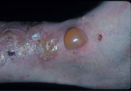
The family physician suspected bullous pemphigoid (BP) based on the collection of tense bullae on the leg; the diagnosis was confirmed with a punch biopsy at the edge of the intact bulla.
BP is a chronic autoimmune disorder of the skin with an average age of onset of 65 years. The generalized bullous form is the most common. One atypical aspect of this case was the localized nature of the bullae. Tense bullae occur on both erythematous and normal appearing skin surfaces, which usually heal without scarring. Drug-induced BP has been reported with drugs containing sulfhydryl groups, including penicillamine, furosemide, captopril, and sulfasalazine.
Biopsy is required to establish the diagnosis. A 4-mm punch biopsy from the edge of an early blister including part of the normal-appearing skin shows a subepidermal blister and an eosinophil-rich mixed dermal inflammatory infiltrate. (A scoop shave biopsy under a small intact blister is also a good method of making a diagnosis of BP or other bullous diseases.)
One can start with a standard biopsy and perform another biopsy for direct immunofluorescence (DIF) if needed. This can be done with a second 4-mm punch biopsy or a shave from perilesional skin. This biopsy for DIF should be transported in Michel’s solution. DIF demonstrates linear IgG and complement C3 deposits at the dermal–epidermal junction.
The family physician started the patient on prednisone 60 mg daily on the day of the biopsy and referred him to a dermatologist for long-term management. The patient began to heal dramatically within days.
Photo courtesy of Eric Kraus, MD. Text for Photo Rounds Friday courtesy of Richard P. Usatine, MD. This case was adapted from: Mohmand A. Bullous pemphigoid. In: Usatine R, Smith M, Mayeaux EJ, et al, eds. The Color Atlas of Family Medicine. New York, NY: McGraw-Hill; 2009:790-796.
To learn more about The Color Atlas of Family Medicine, see:
• http://www.amazon.com/Color-Atlas-Family-Medicine/dp/0071474641
The Color Atlas of Family Medicine is also available as an app for mobile devices. See