User login
A Perfect Storm: Interventions – Closing the survival gap
Editor’s Note: This is the fourth installment of a five-part monthly series that will discuss the biologic, genomic, and health system factors that contribute to the racial survival disparity in breast cancer. The series was adapted from an article that originally appeared in CA: A Cancer Journal for Clinicians,1 a journal of the American Cancer Society. Eliminating racial disparities in cancer mortality through effective interventions has become an increasingly important imperative of federal, state, and community health care programs. This month’s column reviews interventions to close this survival gap.
Insurance
It has been posited that interventions aimed at providing insurance coverage to minority patients will be able to reduce racial health care disparities.2 Studies have indicated that women without insurance present with more-advanced disease,3,4 and are more likely to receive nonstandard treatment.5 However, outside of cancer care, a large study of Medicaid expansion in Oregon demonstrated that Medicaid coverage alone generated no significant improvement in measured physical health outcomes in the first 2 years.6 Thus, coverage alone does not ensure that patients will be able to navigate the health care system and that quality care will be provided.
In breast cancer, Hoffman et al.7 evaluated the effect of race and health insurance on diagnostic time, which was defined as the number of days from suspicious finding to diagnostic resolution (either no evidence of malignancy on diagnostic mammogram or definitive diagnosis by biopsy) in a large, urban setting. The authors’ hypothesis was that every insured patient would receive the same timely diagnosis as any other patient with equivalent insurance, regardless of race or ethnicity. The study found that non-Hispanic whites with government insurance had significantly shorter diagnostic times than did non-Hispanic African Americans with government insurance: The average diagnostic times were 12 and 39 days, respectively. In addition, privately insured non-Hispanic whites also had significantly shorter diagnostic times than did privately insured non-Hispanic African Americans (16 vs. 27 days). In addition, Short et al.8 demonstrated that when the health plan status was held constant in a retrospective study of 476 white patients and 99 African American patients with newly diagnosed breast cancer, African American patients had a higher mortality rate (8.1% vs. 3.6%) and were diagnosed at a later stage. Accordingly, interventions must go beyond just providing health insurance to minorities in order to have a significant impact on the mortality gap.
Patient education and physician communication

An underlying cause frequently cited for the delayed diagnosis and treatment of African American patients with breast cancer is a lack of patient education and physician communication. These elements are essential components of quality care. In a qualitative study of low-income, ethnically diverse women older than 40 years, Allen et al.9 identified salient themes differentiating women who received timely follow-up from those who did not. For the women who delayed follow-up, prominent themes were dissatisfaction with the communication of results, disrespect on the part of providers and clinical staff, logistical barriers to accessing services, anxiety and fear about a possible cancer diagnosis, and a lack of information about breast cancer screening and symptoms.
In another study, Masi and Gehlert10 employed focus group interviews of African American adults to characterize their perceptions of breast cancer treatment. The analysis revealed a core set of themes, including mistrust of the medical establishment and concern about the effect of racism on treatment quality; the researchers concluded that “in the eyes of many study participants, the issues of trust, race, and quality of care were closely intertwined.”10 Thus, this knot that is created by underlying issues of trust can be untied only by interventions that address improved physician communication and patient education.
Janz et al.11 examined racial differences in the adequacy of information and support for women with breast cancer. The researchers used survey data from a population of 1,766 women diagnosed with nonmetastatic breast cancer and reported to the Los Angeles County Surveillance, Epidemiology, and End Results (SEER) registry. The study found that across treatment- and survivorship-related issues, African American women desired more information than white women did. One of the explanations for the unmet information needs posited by the authors is a failure to provide culturally appropriate information related to health issues. This breakdown in patient education and communication was demonstrated by Hawley et al. to hold across providers and locations.12
Hawley et al.12 evaluated the association between minority patients’ knowledge of breast cancer treatment risks and benefits and provider characteristics and treatment locations. The provider characteristics included surgeon-level independent variables, such as breast cancer procedure volume and demographics (years in practice and sex). The treatment location variable was categorized into one of three groups: National Cancer Institute–designated cancer center, American College of Surgeons cancer program, or no specific cancer program. Provider characteristics and treatment locations are factors previously identified as being associated with high-quality care.
The study employed a multivariable regression to identify associations between patient, surgeon, and treatment-setting factors and accurate knowledge of the survival benefit and recurrence risk related to mastectomy and breast-conserving surgery with radiation. The authors found that minority women were significantly less likely to have adequate knowledge and more likely to be uncertain about recurrence risk than were white patients. In the multivariate logistic regression model, neither provider characteristics nor treatment setting attenuated observed racial disparities in knowledge. Quality health care depends on the ability to make an informed treatment decision. As the authors concluded, this study underscores the need for providers to communicate information effectively to all patients, and effective communication relies on the cultural competency of providers.13 Without effective, culturally competent communication, there are treatment delays and omissions that result in poor-quality cancer care. Currently, the research has established that these communication deficits are found across providers and treatment center types.
Patient Navigation
Patient navigation has been championed as a method of improving care in breast cancer by enhancing patient communication and education, and removing barriers to timely care. Patient navigation empowers patients to become knowledgeable about their own health and supports them through the course of care.14 Patient navigation programs have been developed to address the patient communication breakdowns and underuse and misuse of treatment among vulnerable populations, which were detailed earlier in this series and are thought to contribute to the racial mortality gap.15
A benefit of patient navigation has been suggested in studies evaluating the time to diagnosis and follow-up from an abnormal screening. Markossian et al.16 evaluated the efficacy of a Chicago-based cancer patient navigation program developed to reduce the time from abnormal screening to definitive diagnostic testing. The majority of patients in this study were Hispanic (66%) and African American (32%). Compared with controls without navigation, the breast navigation group had a shorter time to diagnostic resolution. Hoffman et al.17 evaluated patient navigation in the District of Columbia to determine its ability to reduce the breast cancer diagnostic time (number of days from abnormal screening to a definitive diagnosis). African American women comprised 48% of the study population. The investigators found that women in the navigation group reached their diagnostic resolution significantly faster than did other women. Among women with breast cancer, there was a nearly fourfold reduction in time to diagnostic resolution for women in the navigation group versus women without a navigator.

In a national multicenter study, Ko et al.14 were the first to evaluate whether patient navigation can improve the quality of breast cancer care. The authors hypothesized that breast cancer patients assigned a navigator would be more likely to receive recommended standard treatment than were those without a navigator. Three separate quality measures of breast cancer care, including initiation of antiestrogen therapy, radiation therapy, and chemotherapy, were evaluated. Study participants were racially and ethnically diverse, with a plurality being African American (37.5%). The study produced mixed results: Patients in the navigation group had a statistically higher likelihood of receiving antiestrogen therapy than were non–navigated controls, but navigation patients eligible for radiation therapy were no more likely to receive it than were controls. The initiation of chemotherapy could not be accurately assessed because of a limited sample size. The study concluded that navigation alone does not remove all of the barriers to quality care for breast cancer patients, and barriers are diverse and potentially specific to the modality of treatment.
A study by Tejeda et al.18 used a systematic framework to characterize barriers faced by minority patients with breast and cervical cancer. The investigators categorized barriers as intrapersonal (defined by characteristics of the individual, such as knowledge, belief, attitudes, and transportation and financial barriers), interpersonal (defined by processes that involve other people, such as social support systems, child care, and employment issues), or institutional (defined by characteristics and policies of organizations). The authors found that although navigators were able to easily resolve intrapersonal barriers, ongoing navigation was needed to address institutional barriers. Thus, patient navigation in a vacuum does not work, and it is only in examining the entire health care system that changes can be implemented to eliminate barriers to quality care and close the racial mortality chasm.
System Change
To this effect, Clarke et al.19 performed a review of the disparities intervention literature to understand which interventions are being evaluated to improve minority health. The authors found that the majority of such interventions are focused on changing the patient rather than the system that serves her, with the most common strategy being education and training (37% of strategies studied). Interventions aimed at health care system improvements were surprisingly few, with the responsibility for change resting with the patient rather than the care delivery system. Interventions incorporating community involvement were also severely lacking and reflected only 6.5% of the reviewed intervention tactics. The majority of interventions failed to involve major stakeholders, including providers, health care institutions, community organizers, and policy makers, and accordingly, were unlikely to succeed in creating meaningful change.
In breast cancer, there have been examples of successful system-based approaches to reducing the racial mortality disparity. At New York area hospitals, Bickell et al.20 implemented a tracking and feedback registry to close the referral loop between surgeons and oncologists to decrease the underuse of valuable adjuvant treatments.
The intervention targeted important quality issues in both communication (the breakdown in dialogue among providers of different specialties and between providers and patients) and the underuse of adjuvant treatment in minorities. The approach was designed to address failures in the health care system through the involvement of leadership from pathology, surgery, and oncology. The intervention also incorporated technology, with tracking software prompting contact with patients who had failed to follow up. Among African American and Hispanic women, there were statistically significant decreases in the underuse of radiotherapy (23% before the intervention vs. 10% after the intervention), chemotherapy (26% vs. 6%), and hormonal therapy (27% vs. 11%). After the intervention, minority race was no longer a risk factor for low rates of oncology consultation or underuse of adjuvant therapy. Interestingly, four of the six hospitals involved in this study had a patient navigation system in place; however, as discussed, the navigation system alone was not enough to address the system failures that led to disparities in care.
Ansell et al.21 also described a system-based approach to reducing the breast cancer mortality disparity in Chicago. The Metropolitan Chicago Breast Cancer Task Force comprised 102 individuals and 74 Chicago area organizations to address the growing disparity in breast cancer mortality between African American and white patients. The task force identified a number of themes underlying the disparity gap, including a need for breast cancer education and outreach programs for African American women, a broken mammography process leading to quality differences between African American and white patients, and a number of barriers to diagnosis and treatment, including fear, a lack of primary care, the burden of insurance copays/deductibles, and the noncompletion of treatment for social and economic reasons. After identifying these underlying causes, the task force proposed that addressing one aspect of the health care system would not correct the problem, but rather quality improvement initiatives would have to occur across the continuum of care for breast cancer.
In Delaware, such a broad system-based intervention was implemented to eliminate health disparities in colorectal cancer.22 Delaware created a comprehensive statewide colorectal screening and treatment program, combining many of the interventions discussed previously, including insurance coverage, patient education and communication, and patient navigation, to address the entire health care system and its treatment of African Americans with colorectal cancer. The state also partnered with underserved community organizations to tailor programs locally and create targeted marketing campaigns.
The results of this system-based approach were impressive, with screening rates among African American increasing from 48% to 74% and equaling the rate among whites. In addition, among African American patients, the percentage diagnosed at advanced and regional stages declined from 79% to 40%, and the percentage diagnosed at a local stage increased from 16% to 50%. Most importantly, the mortality rate declined by 42% for African Americans, resulting in a rate almost equal to that among whites. Significantly, this program was also found to be economically viable, because the cost savings from reduced cancer incidence and the stage shift to cancers requiring less-aggressive treatment offset the program cost. As the authors concluded, this model of a comprehensive, system-wide approach to the racial mortality difference would lend itself to other cancers, and more research is needed to assess and build such an approach to breast cancer.
As discussed in the aforementioned studies, multifaceted interventions that address all stakeholders are needed to close the racial survival disparity in breast cancer. In the final installment of this series, we will address how the changing care models ushered in by the Patient Protection and Affordable Care Act have the potential to advance this agenda of creating an intervention that works across the breast cancer care continuum to reduce disparities.
Other installments of this column can be found in the Related Content box.
1. Daly B, Olopade OI. A perfect storm: How tumor biology, genomics, and health care delivery patterns collide to create a racial survival disparity in breast cancer and proposed interventions for change. CA Cancer J Clin. 2015 May-Jun;65(3):221-38.
2. Lillie-Blanton M, Hoffman C. The role of health insurance coverage in reducing racial/ethnic disparities in health care. Health Aff (Millwood). 2005 Mar-Apr;24(2):398-408.
3. Ayanian JZ, Kohler BA, Abe T, Epstein AM. The relation between health insurance coverage and clinical outcomes among women with breast cancer. N Engl J Med. 1993 Jul 29;329(5):326-31.
4. Coburn N, Fulton J, Pearlman DN, Law C, DiPaolo B, Cady B. Treatment variation by insurance status for breast cancer patients. Breast J. 2008 Mar-Apr;14(2):128-34.
5. Voti L, Richardson LC, Reis I, Fleming LE, Mackinnon J, Coebergh JW. The effect of race/ethnicity and insurance in the administration of standard therapy for local breast cancer in Florida. Breast Cancer Res Treat. 2006 Jan;95(1):89-95.
6. Baicker K, Taubman SL, Allen HL, et al. The Oregon experiment – effects of Medicaid on clinical outcomes. N Engl J Med. 2013 May 2;368(18):1713-22.
7. Hoffman HJ, LaVerda NL, Levine PH, et al. Having health insurance does not eliminate race/ethnicity-associated delays in breast cancer diagnosis in the District of Columbia. Cancer. 2011 Aug 15;117(16):3824-32.
8. Short LJ, Fisher MD, Wahl PM, et al. Disparities in medical care among commercially insured patients with newly diagnosed breast cancer: opportunities for intervention. Cancer. 2010 Jan 1;116(1):193-202.
9. Allen JD, Shelton RC, Harden E, Goldman RE. Follow-up of abnormal screening mammograms among low-income ethnically diverse women: findings from a qualitative study. Patient Educ Couns. 2008 Aug;72(2):283-92.
10. Masi CM, Gehlert S. Perceptions of breast cancer treatment among African-American women and men: implications for interventions. J Gen Intern Med. 2009 Mar;24(3):408-14.
11. Janz NK, Mujahid MS, Hawley ST, Griggs JJ, Hamilton AS, Katz SJ. Racial/ethnic differences in adequacy of information and support for women with breast cancer. Cancer. 2008 Sep 1;113(5):1058-67.
12. Hawley ST, Fagerlin A, Janz NK, Katz SJ. Racial/ethnic disparities in knowledge about risks and benefits of breast cancer treatment: does it matter where you go? Health Serv Res. 2008 Aug;43(4):1366-87.
13. Lannin DR, Mathews HF, Mitchell J, Swanson MS. Impacting cultural attitudes in African-American women to decrease breast cancer mortality. Am J Surg. 2002 Nov;184(5):418-23.
14. Ko NY, Darnell JS, Calhoun E, et al. Can patient navigation improve receipt of recommended breast cancer care? Evidence from the National Patient Navigation Research Program. J Clin Oncol. 2014 Sep 1;32(25):2758-64.
15. Vargas RB, Ryan GW, Jackson CA, Rodriguez R, Freeman HP. Characteristics of the original patient navigation programs to reduce disparities in the diagnosis and treatment of breast cancer. Cancer. 2008 Jul 15;113(2):426-33.
16. Markossian TW, Darnell JS, Calhoun EA. Follow-up and timeliness after an abnormal cancer screening among underserved, urban women in a patient navigation program. Cancer Epidemiol Biomarkers Prev. 2012 Oct;21(10):1691-700.
17. Hoffman HJ, LaVerda NL, Young HA, et al. Patient navigation significantly reduces delays in breast cancer diagnosis in the District of Columbia. Cancer Epidemiol Biomarkers Prev. 2012 Oct;21(10):1655-63.
18. Tejeda S, Darnell JS, Cho YI, Stolley MR, Markossian TW, Calhoun EA. Patient barriers to follow-up care for breast and cervical cancer abnormalities. J Womens Health (Larchmt). 2013 Jun;22(6):507-17.
19. Clarke AR, Goddu AP, Nocon RS, et al. Thirty years of disparities intervention research: what are we doing to close racial and ethnic gaps in health care? Med Care. 2013 Nov;51(11):1020-26.
20. Bickell NA, Shastri K, Fei K, et al. A tracking and feedback registry to reduce racial disparities in breast cancer care. J Natl Cancer Inst. 2008 Dec 3;100(23):1717-23.
21. Ansell D, Grabler P, Whitman S, et al. A community effort to reduce the black/white breast cancer mortality disparity in Chicago. Cancer Causes Control. 2009 Nov;20(9):1681-88.
22. Grubbs SS, Polite BN, Carney J, Jr., et al. Eliminating racial disparities in colorectal cancer in the real world: it took a village. J Clin Oncol. 2013 Jun 1;31(16):1928-30.


Olufunmilayo Olopade, MD, FACP, OON, is the Walter L. Palmer Distinguished Service Professor of Medicine and Human Genetics, and director, Center for Global Health at the University of Chicago. She is adopting emerging high throughput genomic and informatics strategies to identify genetic and nongenetic risk factors for breast cancer in order to implement precision health care in diverse populations. This innovative approach has the potential to improve the quality of care and reduce costs while saving more lives.
Disclosures: Dr. Olopade serves on the Medical Advisory Board for CancerIQ. Dr. Daly serves as a director of Quadrant Holdings Corporation and receives compensation from this entity. Frontline Medical Communications is a subsidiary of Quadrant Holdings Corporation.
Published in conjunction with Susan G. Komen®.
Editor’s Note: This is the fourth installment of a five-part monthly series that will discuss the biologic, genomic, and health system factors that contribute to the racial survival disparity in breast cancer. The series was adapted from an article that originally appeared in CA: A Cancer Journal for Clinicians,1 a journal of the American Cancer Society. Eliminating racial disparities in cancer mortality through effective interventions has become an increasingly important imperative of federal, state, and community health care programs. This month’s column reviews interventions to close this survival gap.
Insurance
It has been posited that interventions aimed at providing insurance coverage to minority patients will be able to reduce racial health care disparities.2 Studies have indicated that women without insurance present with more-advanced disease,3,4 and are more likely to receive nonstandard treatment.5 However, outside of cancer care, a large study of Medicaid expansion in Oregon demonstrated that Medicaid coverage alone generated no significant improvement in measured physical health outcomes in the first 2 years.6 Thus, coverage alone does not ensure that patients will be able to navigate the health care system and that quality care will be provided.
In breast cancer, Hoffman et al.7 evaluated the effect of race and health insurance on diagnostic time, which was defined as the number of days from suspicious finding to diagnostic resolution (either no evidence of malignancy on diagnostic mammogram or definitive diagnosis by biopsy) in a large, urban setting. The authors’ hypothesis was that every insured patient would receive the same timely diagnosis as any other patient with equivalent insurance, regardless of race or ethnicity. The study found that non-Hispanic whites with government insurance had significantly shorter diagnostic times than did non-Hispanic African Americans with government insurance: The average diagnostic times were 12 and 39 days, respectively. In addition, privately insured non-Hispanic whites also had significantly shorter diagnostic times than did privately insured non-Hispanic African Americans (16 vs. 27 days). In addition, Short et al.8 demonstrated that when the health plan status was held constant in a retrospective study of 476 white patients and 99 African American patients with newly diagnosed breast cancer, African American patients had a higher mortality rate (8.1% vs. 3.6%) and were diagnosed at a later stage. Accordingly, interventions must go beyond just providing health insurance to minorities in order to have a significant impact on the mortality gap.
Patient education and physician communication
An underlying cause frequently cited for the delayed diagnosis and treatment of African American patients with breast cancer is a lack of patient education and physician communication. These elements are essential components of quality care. In a qualitative study of low-income, ethnically diverse women older than 40 years, Allen et al.9 identified salient themes differentiating women who received timely follow-up from those who did not. For the women who delayed follow-up, prominent themes were dissatisfaction with the communication of results, disrespect on the part of providers and clinical staff, logistical barriers to accessing services, anxiety and fear about a possible cancer diagnosis, and a lack of information about breast cancer screening and symptoms.
In another study, Masi and Gehlert10 employed focus group interviews of African American adults to characterize their perceptions of breast cancer treatment. The analysis revealed a core set of themes, including mistrust of the medical establishment and concern about the effect of racism on treatment quality; the researchers concluded that “in the eyes of many study participants, the issues of trust, race, and quality of care were closely intertwined.”10 Thus, this knot that is created by underlying issues of trust can be untied only by interventions that address improved physician communication and patient education.
Janz et al.11 examined racial differences in the adequacy of information and support for women with breast cancer. The researchers used survey data from a population of 1,766 women diagnosed with nonmetastatic breast cancer and reported to the Los Angeles County Surveillance, Epidemiology, and End Results (SEER) registry. The study found that across treatment- and survivorship-related issues, African American women desired more information than white women did. One of the explanations for the unmet information needs posited by the authors is a failure to provide culturally appropriate information related to health issues. This breakdown in patient education and communication was demonstrated by Hawley et al. to hold across providers and locations.12
Hawley et al.12 evaluated the association between minority patients’ knowledge of breast cancer treatment risks and benefits and provider characteristics and treatment locations. The provider characteristics included surgeon-level independent variables, such as breast cancer procedure volume and demographics (years in practice and sex). The treatment location variable was categorized into one of three groups: National Cancer Institute–designated cancer center, American College of Surgeons cancer program, or no specific cancer program. Provider characteristics and treatment locations are factors previously identified as being associated with high-quality care.
The study employed a multivariable regression to identify associations between patient, surgeon, and treatment-setting factors and accurate knowledge of the survival benefit and recurrence risk related to mastectomy and breast-conserving surgery with radiation. The authors found that minority women were significantly less likely to have adequate knowledge and more likely to be uncertain about recurrence risk than were white patients. In the multivariate logistic regression model, neither provider characteristics nor treatment setting attenuated observed racial disparities in knowledge. Quality health care depends on the ability to make an informed treatment decision. As the authors concluded, this study underscores the need for providers to communicate information effectively to all patients, and effective communication relies on the cultural competency of providers.13 Without effective, culturally competent communication, there are treatment delays and omissions that result in poor-quality cancer care. Currently, the research has established that these communication deficits are found across providers and treatment center types.
Patient Navigation
Patient navigation has been championed as a method of improving care in breast cancer by enhancing patient communication and education, and removing barriers to timely care. Patient navigation empowers patients to become knowledgeable about their own health and supports them through the course of care.14 Patient navigation programs have been developed to address the patient communication breakdowns and underuse and misuse of treatment among vulnerable populations, which were detailed earlier in this series and are thought to contribute to the racial mortality gap.15
A benefit of patient navigation has been suggested in studies evaluating the time to diagnosis and follow-up from an abnormal screening. Markossian et al.16 evaluated the efficacy of a Chicago-based cancer patient navigation program developed to reduce the time from abnormal screening to definitive diagnostic testing. The majority of patients in this study were Hispanic (66%) and African American (32%). Compared with controls without navigation, the breast navigation group had a shorter time to diagnostic resolution. Hoffman et al.17 evaluated patient navigation in the District of Columbia to determine its ability to reduce the breast cancer diagnostic time (number of days from abnormal screening to a definitive diagnosis). African American women comprised 48% of the study population. The investigators found that women in the navigation group reached their diagnostic resolution significantly faster than did other women. Among women with breast cancer, there was a nearly fourfold reduction in time to diagnostic resolution for women in the navigation group versus women without a navigator.
In a national multicenter study, Ko et al.14 were the first to evaluate whether patient navigation can improve the quality of breast cancer care. The authors hypothesized that breast cancer patients assigned a navigator would be more likely to receive recommended standard treatment than were those without a navigator. Three separate quality measures of breast cancer care, including initiation of antiestrogen therapy, radiation therapy, and chemotherapy, were evaluated. Study participants were racially and ethnically diverse, with a plurality being African American (37.5%). The study produced mixed results: Patients in the navigation group had a statistically higher likelihood of receiving antiestrogen therapy than were non–navigated controls, but navigation patients eligible for radiation therapy were no more likely to receive it than were controls. The initiation of chemotherapy could not be accurately assessed because of a limited sample size. The study concluded that navigation alone does not remove all of the barriers to quality care for breast cancer patients, and barriers are diverse and potentially specific to the modality of treatment.
A study by Tejeda et al.18 used a systematic framework to characterize barriers faced by minority patients with breast and cervical cancer. The investigators categorized barriers as intrapersonal (defined by characteristics of the individual, such as knowledge, belief, attitudes, and transportation and financial barriers), interpersonal (defined by processes that involve other people, such as social support systems, child care, and employment issues), or institutional (defined by characteristics and policies of organizations). The authors found that although navigators were able to easily resolve intrapersonal barriers, ongoing navigation was needed to address institutional barriers. Thus, patient navigation in a vacuum does not work, and it is only in examining the entire health care system that changes can be implemented to eliminate barriers to quality care and close the racial mortality chasm.
System Change
To this effect, Clarke et al.19 performed a review of the disparities intervention literature to understand which interventions are being evaluated to improve minority health. The authors found that the majority of such interventions are focused on changing the patient rather than the system that serves her, with the most common strategy being education and training (37% of strategies studied). Interventions aimed at health care system improvements were surprisingly few, with the responsibility for change resting with the patient rather than the care delivery system. Interventions incorporating community involvement were also severely lacking and reflected only 6.5% of the reviewed intervention tactics. The majority of interventions failed to involve major stakeholders, including providers, health care institutions, community organizers, and policy makers, and accordingly, were unlikely to succeed in creating meaningful change.
In breast cancer, there have been examples of successful system-based approaches to reducing the racial mortality disparity. At New York area hospitals, Bickell et al.20 implemented a tracking and feedback registry to close the referral loop between surgeons and oncologists to decrease the underuse of valuable adjuvant treatments.
The intervention targeted important quality issues in both communication (the breakdown in dialogue among providers of different specialties and between providers and patients) and the underuse of adjuvant treatment in minorities. The approach was designed to address failures in the health care system through the involvement of leadership from pathology, surgery, and oncology. The intervention also incorporated technology, with tracking software prompting contact with patients who had failed to follow up. Among African American and Hispanic women, there were statistically significant decreases in the underuse of radiotherapy (23% before the intervention vs. 10% after the intervention), chemotherapy (26% vs. 6%), and hormonal therapy (27% vs. 11%). After the intervention, minority race was no longer a risk factor for low rates of oncology consultation or underuse of adjuvant therapy. Interestingly, four of the six hospitals involved in this study had a patient navigation system in place; however, as discussed, the navigation system alone was not enough to address the system failures that led to disparities in care.
Ansell et al.21 also described a system-based approach to reducing the breast cancer mortality disparity in Chicago. The Metropolitan Chicago Breast Cancer Task Force comprised 102 individuals and 74 Chicago area organizations to address the growing disparity in breast cancer mortality between African American and white patients. The task force identified a number of themes underlying the disparity gap, including a need for breast cancer education and outreach programs for African American women, a broken mammography process leading to quality differences between African American and white patients, and a number of barriers to diagnosis and treatment, including fear, a lack of primary care, the burden of insurance copays/deductibles, and the noncompletion of treatment for social and economic reasons. After identifying these underlying causes, the task force proposed that addressing one aspect of the health care system would not correct the problem, but rather quality improvement initiatives would have to occur across the continuum of care for breast cancer.
In Delaware, such a broad system-based intervention was implemented to eliminate health disparities in colorectal cancer.22 Delaware created a comprehensive statewide colorectal screening and treatment program, combining many of the interventions discussed previously, including insurance coverage, patient education and communication, and patient navigation, to address the entire health care system and its treatment of African Americans with colorectal cancer. The state also partnered with underserved community organizations to tailor programs locally and create targeted marketing campaigns.
The results of this system-based approach were impressive, with screening rates among African American increasing from 48% to 74% and equaling the rate among whites. In addition, among African American patients, the percentage diagnosed at advanced and regional stages declined from 79% to 40%, and the percentage diagnosed at a local stage increased from 16% to 50%. Most importantly, the mortality rate declined by 42% for African Americans, resulting in a rate almost equal to that among whites. Significantly, this program was also found to be economically viable, because the cost savings from reduced cancer incidence and the stage shift to cancers requiring less-aggressive treatment offset the program cost. As the authors concluded, this model of a comprehensive, system-wide approach to the racial mortality difference would lend itself to other cancers, and more research is needed to assess and build such an approach to breast cancer.
As discussed in the aforementioned studies, multifaceted interventions that address all stakeholders are needed to close the racial survival disparity in breast cancer. In the final installment of this series, we will address how the changing care models ushered in by the Patient Protection and Affordable Care Act have the potential to advance this agenda of creating an intervention that works across the breast cancer care continuum to reduce disparities.
Other installments of this column can be found in the Related Content box.
1. Daly B, Olopade OI. A perfect storm: How tumor biology, genomics, and health care delivery patterns collide to create a racial survival disparity in breast cancer and proposed interventions for change. CA Cancer J Clin. 2015 May-Jun;65(3):221-38.
2. Lillie-Blanton M, Hoffman C. The role of health insurance coverage in reducing racial/ethnic disparities in health care. Health Aff (Millwood). 2005 Mar-Apr;24(2):398-408.
3. Ayanian JZ, Kohler BA, Abe T, Epstein AM. The relation between health insurance coverage and clinical outcomes among women with breast cancer. N Engl J Med. 1993 Jul 29;329(5):326-31.
4. Coburn N, Fulton J, Pearlman DN, Law C, DiPaolo B, Cady B. Treatment variation by insurance status for breast cancer patients. Breast J. 2008 Mar-Apr;14(2):128-34.
5. Voti L, Richardson LC, Reis I, Fleming LE, Mackinnon J, Coebergh JW. The effect of race/ethnicity and insurance in the administration of standard therapy for local breast cancer in Florida. Breast Cancer Res Treat. 2006 Jan;95(1):89-95.
6. Baicker K, Taubman SL, Allen HL, et al. The Oregon experiment – effects of Medicaid on clinical outcomes. N Engl J Med. 2013 May 2;368(18):1713-22.
7. Hoffman HJ, LaVerda NL, Levine PH, et al. Having health insurance does not eliminate race/ethnicity-associated delays in breast cancer diagnosis in the District of Columbia. Cancer. 2011 Aug 15;117(16):3824-32.
8. Short LJ, Fisher MD, Wahl PM, et al. Disparities in medical care among commercially insured patients with newly diagnosed breast cancer: opportunities for intervention. Cancer. 2010 Jan 1;116(1):193-202.
9. Allen JD, Shelton RC, Harden E, Goldman RE. Follow-up of abnormal screening mammograms among low-income ethnically diverse women: findings from a qualitative study. Patient Educ Couns. 2008 Aug;72(2):283-92.
10. Masi CM, Gehlert S. Perceptions of breast cancer treatment among African-American women and men: implications for interventions. J Gen Intern Med. 2009 Mar;24(3):408-14.
11. Janz NK, Mujahid MS, Hawley ST, Griggs JJ, Hamilton AS, Katz SJ. Racial/ethnic differences in adequacy of information and support for women with breast cancer. Cancer. 2008 Sep 1;113(5):1058-67.
12. Hawley ST, Fagerlin A, Janz NK, Katz SJ. Racial/ethnic disparities in knowledge about risks and benefits of breast cancer treatment: does it matter where you go? Health Serv Res. 2008 Aug;43(4):1366-87.
13. Lannin DR, Mathews HF, Mitchell J, Swanson MS. Impacting cultural attitudes in African-American women to decrease breast cancer mortality. Am J Surg. 2002 Nov;184(5):418-23.
14. Ko NY, Darnell JS, Calhoun E, et al. Can patient navigation improve receipt of recommended breast cancer care? Evidence from the National Patient Navigation Research Program. J Clin Oncol. 2014 Sep 1;32(25):2758-64.
15. Vargas RB, Ryan GW, Jackson CA, Rodriguez R, Freeman HP. Characteristics of the original patient navigation programs to reduce disparities in the diagnosis and treatment of breast cancer. Cancer. 2008 Jul 15;113(2):426-33.
16. Markossian TW, Darnell JS, Calhoun EA. Follow-up and timeliness after an abnormal cancer screening among underserved, urban women in a patient navigation program. Cancer Epidemiol Biomarkers Prev. 2012 Oct;21(10):1691-700.
17. Hoffman HJ, LaVerda NL, Young HA, et al. Patient navigation significantly reduces delays in breast cancer diagnosis in the District of Columbia. Cancer Epidemiol Biomarkers Prev. 2012 Oct;21(10):1655-63.
18. Tejeda S, Darnell JS, Cho YI, Stolley MR, Markossian TW, Calhoun EA. Patient barriers to follow-up care for breast and cervical cancer abnormalities. J Womens Health (Larchmt). 2013 Jun;22(6):507-17.
19. Clarke AR, Goddu AP, Nocon RS, et al. Thirty years of disparities intervention research: what are we doing to close racial and ethnic gaps in health care? Med Care. 2013 Nov;51(11):1020-26.
20. Bickell NA, Shastri K, Fei K, et al. A tracking and feedback registry to reduce racial disparities in breast cancer care. J Natl Cancer Inst. 2008 Dec 3;100(23):1717-23.
21. Ansell D, Grabler P, Whitman S, et al. A community effort to reduce the black/white breast cancer mortality disparity in Chicago. Cancer Causes Control. 2009 Nov;20(9):1681-88.
22. Grubbs SS, Polite BN, Carney J, Jr., et al. Eliminating racial disparities in colorectal cancer in the real world: it took a village. J Clin Oncol. 2013 Jun 1;31(16):1928-30.
Olufunmilayo Olopade, MD, FACP, OON, is the Walter L. Palmer Distinguished Service Professor of Medicine and Human Genetics, and director, Center for Global Health at the University of Chicago. She is adopting emerging high throughput genomic and informatics strategies to identify genetic and nongenetic risk factors for breast cancer in order to implement precision health care in diverse populations. This innovative approach has the potential to improve the quality of care and reduce costs while saving more lives.
Disclosures: Dr. Olopade serves on the Medical Advisory Board for CancerIQ. Dr. Daly serves as a director of Quadrant Holdings Corporation and receives compensation from this entity. Frontline Medical Communications is a subsidiary of Quadrant Holdings Corporation.
Published in conjunction with Susan G. Komen®.
Editor’s Note: This is the fourth installment of a five-part monthly series that will discuss the biologic, genomic, and health system factors that contribute to the racial survival disparity in breast cancer. The series was adapted from an article that originally appeared in CA: A Cancer Journal for Clinicians,1 a journal of the American Cancer Society. Eliminating racial disparities in cancer mortality through effective interventions has become an increasingly important imperative of federal, state, and community health care programs. This month’s column reviews interventions to close this survival gap.
Insurance
It has been posited that interventions aimed at providing insurance coverage to minority patients will be able to reduce racial health care disparities.2 Studies have indicated that women without insurance present with more-advanced disease,3,4 and are more likely to receive nonstandard treatment.5 However, outside of cancer care, a large study of Medicaid expansion in Oregon demonstrated that Medicaid coverage alone generated no significant improvement in measured physical health outcomes in the first 2 years.6 Thus, coverage alone does not ensure that patients will be able to navigate the health care system and that quality care will be provided.
In breast cancer, Hoffman et al.7 evaluated the effect of race and health insurance on diagnostic time, which was defined as the number of days from suspicious finding to diagnostic resolution (either no evidence of malignancy on diagnostic mammogram or definitive diagnosis by biopsy) in a large, urban setting. The authors’ hypothesis was that every insured patient would receive the same timely diagnosis as any other patient with equivalent insurance, regardless of race or ethnicity. The study found that non-Hispanic whites with government insurance had significantly shorter diagnostic times than did non-Hispanic African Americans with government insurance: The average diagnostic times were 12 and 39 days, respectively. In addition, privately insured non-Hispanic whites also had significantly shorter diagnostic times than did privately insured non-Hispanic African Americans (16 vs. 27 days). In addition, Short et al.8 demonstrated that when the health plan status was held constant in a retrospective study of 476 white patients and 99 African American patients with newly diagnosed breast cancer, African American patients had a higher mortality rate (8.1% vs. 3.6%) and were diagnosed at a later stage. Accordingly, interventions must go beyond just providing health insurance to minorities in order to have a significant impact on the mortality gap.
Patient education and physician communication
An underlying cause frequently cited for the delayed diagnosis and treatment of African American patients with breast cancer is a lack of patient education and physician communication. These elements are essential components of quality care. In a qualitative study of low-income, ethnically diverse women older than 40 years, Allen et al.9 identified salient themes differentiating women who received timely follow-up from those who did not. For the women who delayed follow-up, prominent themes were dissatisfaction with the communication of results, disrespect on the part of providers and clinical staff, logistical barriers to accessing services, anxiety and fear about a possible cancer diagnosis, and a lack of information about breast cancer screening and symptoms.
In another study, Masi and Gehlert10 employed focus group interviews of African American adults to characterize their perceptions of breast cancer treatment. The analysis revealed a core set of themes, including mistrust of the medical establishment and concern about the effect of racism on treatment quality; the researchers concluded that “in the eyes of many study participants, the issues of trust, race, and quality of care were closely intertwined.”10 Thus, this knot that is created by underlying issues of trust can be untied only by interventions that address improved physician communication and patient education.
Janz et al.11 examined racial differences in the adequacy of information and support for women with breast cancer. The researchers used survey data from a population of 1,766 women diagnosed with nonmetastatic breast cancer and reported to the Los Angeles County Surveillance, Epidemiology, and End Results (SEER) registry. The study found that across treatment- and survivorship-related issues, African American women desired more information than white women did. One of the explanations for the unmet information needs posited by the authors is a failure to provide culturally appropriate information related to health issues. This breakdown in patient education and communication was demonstrated by Hawley et al. to hold across providers and locations.12
Hawley et al.12 evaluated the association between minority patients’ knowledge of breast cancer treatment risks and benefits and provider characteristics and treatment locations. The provider characteristics included surgeon-level independent variables, such as breast cancer procedure volume and demographics (years in practice and sex). The treatment location variable was categorized into one of three groups: National Cancer Institute–designated cancer center, American College of Surgeons cancer program, or no specific cancer program. Provider characteristics and treatment locations are factors previously identified as being associated with high-quality care.
The study employed a multivariable regression to identify associations between patient, surgeon, and treatment-setting factors and accurate knowledge of the survival benefit and recurrence risk related to mastectomy and breast-conserving surgery with radiation. The authors found that minority women were significantly less likely to have adequate knowledge and more likely to be uncertain about recurrence risk than were white patients. In the multivariate logistic regression model, neither provider characteristics nor treatment setting attenuated observed racial disparities in knowledge. Quality health care depends on the ability to make an informed treatment decision. As the authors concluded, this study underscores the need for providers to communicate information effectively to all patients, and effective communication relies on the cultural competency of providers.13 Without effective, culturally competent communication, there are treatment delays and omissions that result in poor-quality cancer care. Currently, the research has established that these communication deficits are found across providers and treatment center types.
Patient Navigation
Patient navigation has been championed as a method of improving care in breast cancer by enhancing patient communication and education, and removing barriers to timely care. Patient navigation empowers patients to become knowledgeable about their own health and supports them through the course of care.14 Patient navigation programs have been developed to address the patient communication breakdowns and underuse and misuse of treatment among vulnerable populations, which were detailed earlier in this series and are thought to contribute to the racial mortality gap.15
A benefit of patient navigation has been suggested in studies evaluating the time to diagnosis and follow-up from an abnormal screening. Markossian et al.16 evaluated the efficacy of a Chicago-based cancer patient navigation program developed to reduce the time from abnormal screening to definitive diagnostic testing. The majority of patients in this study were Hispanic (66%) and African American (32%). Compared with controls without navigation, the breast navigation group had a shorter time to diagnostic resolution. Hoffman et al.17 evaluated patient navigation in the District of Columbia to determine its ability to reduce the breast cancer diagnostic time (number of days from abnormal screening to a definitive diagnosis). African American women comprised 48% of the study population. The investigators found that women in the navigation group reached their diagnostic resolution significantly faster than did other women. Among women with breast cancer, there was a nearly fourfold reduction in time to diagnostic resolution for women in the navigation group versus women without a navigator.
In a national multicenter study, Ko et al.14 were the first to evaluate whether patient navigation can improve the quality of breast cancer care. The authors hypothesized that breast cancer patients assigned a navigator would be more likely to receive recommended standard treatment than were those without a navigator. Three separate quality measures of breast cancer care, including initiation of antiestrogen therapy, radiation therapy, and chemotherapy, were evaluated. Study participants were racially and ethnically diverse, with a plurality being African American (37.5%). The study produced mixed results: Patients in the navigation group had a statistically higher likelihood of receiving antiestrogen therapy than were non–navigated controls, but navigation patients eligible for radiation therapy were no more likely to receive it than were controls. The initiation of chemotherapy could not be accurately assessed because of a limited sample size. The study concluded that navigation alone does not remove all of the barriers to quality care for breast cancer patients, and barriers are diverse and potentially specific to the modality of treatment.
A study by Tejeda et al.18 used a systematic framework to characterize barriers faced by minority patients with breast and cervical cancer. The investigators categorized barriers as intrapersonal (defined by characteristics of the individual, such as knowledge, belief, attitudes, and transportation and financial barriers), interpersonal (defined by processes that involve other people, such as social support systems, child care, and employment issues), or institutional (defined by characteristics and policies of organizations). The authors found that although navigators were able to easily resolve intrapersonal barriers, ongoing navigation was needed to address institutional barriers. Thus, patient navigation in a vacuum does not work, and it is only in examining the entire health care system that changes can be implemented to eliminate barriers to quality care and close the racial mortality chasm.
System Change
To this effect, Clarke et al.19 performed a review of the disparities intervention literature to understand which interventions are being evaluated to improve minority health. The authors found that the majority of such interventions are focused on changing the patient rather than the system that serves her, with the most common strategy being education and training (37% of strategies studied). Interventions aimed at health care system improvements were surprisingly few, with the responsibility for change resting with the patient rather than the care delivery system. Interventions incorporating community involvement were also severely lacking and reflected only 6.5% of the reviewed intervention tactics. The majority of interventions failed to involve major stakeholders, including providers, health care institutions, community organizers, and policy makers, and accordingly, were unlikely to succeed in creating meaningful change.
In breast cancer, there have been examples of successful system-based approaches to reducing the racial mortality disparity. At New York area hospitals, Bickell et al.20 implemented a tracking and feedback registry to close the referral loop between surgeons and oncologists to decrease the underuse of valuable adjuvant treatments.
The intervention targeted important quality issues in both communication (the breakdown in dialogue among providers of different specialties and between providers and patients) and the underuse of adjuvant treatment in minorities. The approach was designed to address failures in the health care system through the involvement of leadership from pathology, surgery, and oncology. The intervention also incorporated technology, with tracking software prompting contact with patients who had failed to follow up. Among African American and Hispanic women, there were statistically significant decreases in the underuse of radiotherapy (23% before the intervention vs. 10% after the intervention), chemotherapy (26% vs. 6%), and hormonal therapy (27% vs. 11%). After the intervention, minority race was no longer a risk factor for low rates of oncology consultation or underuse of adjuvant therapy. Interestingly, four of the six hospitals involved in this study had a patient navigation system in place; however, as discussed, the navigation system alone was not enough to address the system failures that led to disparities in care.
Ansell et al.21 also described a system-based approach to reducing the breast cancer mortality disparity in Chicago. The Metropolitan Chicago Breast Cancer Task Force comprised 102 individuals and 74 Chicago area organizations to address the growing disparity in breast cancer mortality between African American and white patients. The task force identified a number of themes underlying the disparity gap, including a need for breast cancer education and outreach programs for African American women, a broken mammography process leading to quality differences between African American and white patients, and a number of barriers to diagnosis and treatment, including fear, a lack of primary care, the burden of insurance copays/deductibles, and the noncompletion of treatment for social and economic reasons. After identifying these underlying causes, the task force proposed that addressing one aspect of the health care system would not correct the problem, but rather quality improvement initiatives would have to occur across the continuum of care for breast cancer.
In Delaware, such a broad system-based intervention was implemented to eliminate health disparities in colorectal cancer.22 Delaware created a comprehensive statewide colorectal screening and treatment program, combining many of the interventions discussed previously, including insurance coverage, patient education and communication, and patient navigation, to address the entire health care system and its treatment of African Americans with colorectal cancer. The state also partnered with underserved community organizations to tailor programs locally and create targeted marketing campaigns.
The results of this system-based approach were impressive, with screening rates among African American increasing from 48% to 74% and equaling the rate among whites. In addition, among African American patients, the percentage diagnosed at advanced and regional stages declined from 79% to 40%, and the percentage diagnosed at a local stage increased from 16% to 50%. Most importantly, the mortality rate declined by 42% for African Americans, resulting in a rate almost equal to that among whites. Significantly, this program was also found to be economically viable, because the cost savings from reduced cancer incidence and the stage shift to cancers requiring less-aggressive treatment offset the program cost. As the authors concluded, this model of a comprehensive, system-wide approach to the racial mortality difference would lend itself to other cancers, and more research is needed to assess and build such an approach to breast cancer.
As discussed in the aforementioned studies, multifaceted interventions that address all stakeholders are needed to close the racial survival disparity in breast cancer. In the final installment of this series, we will address how the changing care models ushered in by the Patient Protection and Affordable Care Act have the potential to advance this agenda of creating an intervention that works across the breast cancer care continuum to reduce disparities.
Other installments of this column can be found in the Related Content box.
1. Daly B, Olopade OI. A perfect storm: How tumor biology, genomics, and health care delivery patterns collide to create a racial survival disparity in breast cancer and proposed interventions for change. CA Cancer J Clin. 2015 May-Jun;65(3):221-38.
2. Lillie-Blanton M, Hoffman C. The role of health insurance coverage in reducing racial/ethnic disparities in health care. Health Aff (Millwood). 2005 Mar-Apr;24(2):398-408.
3. Ayanian JZ, Kohler BA, Abe T, Epstein AM. The relation between health insurance coverage and clinical outcomes among women with breast cancer. N Engl J Med. 1993 Jul 29;329(5):326-31.
4. Coburn N, Fulton J, Pearlman DN, Law C, DiPaolo B, Cady B. Treatment variation by insurance status for breast cancer patients. Breast J. 2008 Mar-Apr;14(2):128-34.
5. Voti L, Richardson LC, Reis I, Fleming LE, Mackinnon J, Coebergh JW. The effect of race/ethnicity and insurance in the administration of standard therapy for local breast cancer in Florida. Breast Cancer Res Treat. 2006 Jan;95(1):89-95.
6. Baicker K, Taubman SL, Allen HL, et al. The Oregon experiment – effects of Medicaid on clinical outcomes. N Engl J Med. 2013 May 2;368(18):1713-22.
7. Hoffman HJ, LaVerda NL, Levine PH, et al. Having health insurance does not eliminate race/ethnicity-associated delays in breast cancer diagnosis in the District of Columbia. Cancer. 2011 Aug 15;117(16):3824-32.
8. Short LJ, Fisher MD, Wahl PM, et al. Disparities in medical care among commercially insured patients with newly diagnosed breast cancer: opportunities for intervention. Cancer. 2010 Jan 1;116(1):193-202.
9. Allen JD, Shelton RC, Harden E, Goldman RE. Follow-up of abnormal screening mammograms among low-income ethnically diverse women: findings from a qualitative study. Patient Educ Couns. 2008 Aug;72(2):283-92.
10. Masi CM, Gehlert S. Perceptions of breast cancer treatment among African-American women and men: implications for interventions. J Gen Intern Med. 2009 Mar;24(3):408-14.
11. Janz NK, Mujahid MS, Hawley ST, Griggs JJ, Hamilton AS, Katz SJ. Racial/ethnic differences in adequacy of information and support for women with breast cancer. Cancer. 2008 Sep 1;113(5):1058-67.
12. Hawley ST, Fagerlin A, Janz NK, Katz SJ. Racial/ethnic disparities in knowledge about risks and benefits of breast cancer treatment: does it matter where you go? Health Serv Res. 2008 Aug;43(4):1366-87.
13. Lannin DR, Mathews HF, Mitchell J, Swanson MS. Impacting cultural attitudes in African-American women to decrease breast cancer mortality. Am J Surg. 2002 Nov;184(5):418-23.
14. Ko NY, Darnell JS, Calhoun E, et al. Can patient navigation improve receipt of recommended breast cancer care? Evidence from the National Patient Navigation Research Program. J Clin Oncol. 2014 Sep 1;32(25):2758-64.
15. Vargas RB, Ryan GW, Jackson CA, Rodriguez R, Freeman HP. Characteristics of the original patient navigation programs to reduce disparities in the diagnosis and treatment of breast cancer. Cancer. 2008 Jul 15;113(2):426-33.
16. Markossian TW, Darnell JS, Calhoun EA. Follow-up and timeliness after an abnormal cancer screening among underserved, urban women in a patient navigation program. Cancer Epidemiol Biomarkers Prev. 2012 Oct;21(10):1691-700.
17. Hoffman HJ, LaVerda NL, Young HA, et al. Patient navigation significantly reduces delays in breast cancer diagnosis in the District of Columbia. Cancer Epidemiol Biomarkers Prev. 2012 Oct;21(10):1655-63.
18. Tejeda S, Darnell JS, Cho YI, Stolley MR, Markossian TW, Calhoun EA. Patient barriers to follow-up care for breast and cervical cancer abnormalities. J Womens Health (Larchmt). 2013 Jun;22(6):507-17.
19. Clarke AR, Goddu AP, Nocon RS, et al. Thirty years of disparities intervention research: what are we doing to close racial and ethnic gaps in health care? Med Care. 2013 Nov;51(11):1020-26.
20. Bickell NA, Shastri K, Fei K, et al. A tracking and feedback registry to reduce racial disparities in breast cancer care. J Natl Cancer Inst. 2008 Dec 3;100(23):1717-23.
21. Ansell D, Grabler P, Whitman S, et al. A community effort to reduce the black/white breast cancer mortality disparity in Chicago. Cancer Causes Control. 2009 Nov;20(9):1681-88.
22. Grubbs SS, Polite BN, Carney J, Jr., et al. Eliminating racial disparities in colorectal cancer in the real world: it took a village. J Clin Oncol. 2013 Jun 1;31(16):1928-30.
Olufunmilayo Olopade, MD, FACP, OON, is the Walter L. Palmer Distinguished Service Professor of Medicine and Human Genetics, and director, Center for Global Health at the University of Chicago. She is adopting emerging high throughput genomic and informatics strategies to identify genetic and nongenetic risk factors for breast cancer in order to implement precision health care in diverse populations. This innovative approach has the potential to improve the quality of care and reduce costs while saving more lives.
Disclosures: Dr. Olopade serves on the Medical Advisory Board for CancerIQ. Dr. Daly serves as a director of Quadrant Holdings Corporation and receives compensation from this entity. Frontline Medical Communications is a subsidiary of Quadrant Holdings Corporation.
Published in conjunction with Susan G. Komen®.

Adjuvant endocrine therapy for premenopausal breast cancer patients should be individualized
Oncologists should take an individualized approach when making decisions about adjuvant endocrine therapies for premenopausal hormone receptor–positive, HER2-negative early breast cancer, suggests an analysis of a pair of randomized phase III trials published online in the Journal of Clinical Oncology.
Investigators led by Meredith M. Regan, Sc.D., of Dana-Farber Cancer Institute in Boston, analyzed data from the TEXT (Tamoxifen and Exemestane Trial) and SOFT (Suppression of Ovarian Function Trial) trials of adjuvant endocrine therapies, comprising a total of nearly 5,000 women.
Results suggested that the absolute improvement in the 5-year breast cancer–free interval rate with exemestane plus ovarian function suppression (OFS) versus tamoxifen with or without OFS ranged from less than 1% in women with a lowest recurrence risk based on clinicopathologic factors to 10%-15% in women with a highest risk.
“TEXT and SOFT demonstrated that premenopausal women with hormone receptor–positive disease benefit, on average, from exemestane plus OFS versus tamoxifen with or without OFS. However, individualized treatment decisions should weigh the benefits against the adverse effects and costs of these therapy options,” the investigators wrote.
“In the absence of predictive biomarkers, consideration of a patient’s prognosis, as illustrated by STEPP [Subpopulation Treatment Effect Pattern Plot] analysis of a composite measure of recurrence risk in the TEXT and SOFT populations, is integral to this decision making,” they added.
In the SOFT trial, women were randomized to 5 years of tamoxifen alone (as an active comparator), tamoxifen plus OFS, or exemestane (Aromasin) plus OFS. In the TEXT trial, women were randomized to 5 years of exemestane plus OFS or of tamoxifen plus OFS.
Dr. Regan and colleagues based their analyses on a total of 4,891 women. They assessed each patient’s composite recurrence risk from a Cox model that included a set of conventional clinicopathologic factors: age, nodal status, tumor size and grade, and estrogen receptor, progesterone receptor, and Ki-67 expression levels. And they used STEPP methodology to assess the impact of endocrine therapy across groups having different risk.
The median duration of follow-up was 5.6 years in the SOFT trial and 6 years in the TEXT trial. Results showed that the 5-year breast cancer–free interval rate was 90.8% for the study cohort as a whole. But it ranged considerably from 98.6% for patients with composite risk in the lowest quartile to 77.5% for patients with composite risk in the highest quartiles, the investigators reported (J Clin Oncol. 2016. doi: 10.1200/JCO.2015.64.3171).
In the SOFT population, patients who remained premenopausal after neoadjuvant or adjuvant chemotherapy had an absolute improvement of 5% or more in the 5-year breast cancer–free interval rate with exemestane plus OFS, compared with tamoxifen plus OFS or tamoxifen alone. The difference was 10%-15% for the subset at intermediate to high risk for recurrence.
In addition, a benefit of tamoxifen plus OFS over tamoxifen alone was evident in patients having the highest composite risk.
Among patients who were not given chemotherapy, who on average had the lowest composite recurrence risk, the 5-year breast cancer–free interval rate was excellent regardless of the endocrine therapy received.
In the TEXT trial population, the benefit of exemestane plus OFS over tamoxifen plus OFS in 5-year breast cancer–free interval rate ranged from 5% to 15%. Again, the patients who were not given chemotherapy, who had the lowest composite recurrence risk, fared well regardless of which endocrine therapy they received.
These findings should help guide clinical decisions in premenopausal women with hormone receptor–positive, HER2-negative breast cancer, both at the extremes of risk and in the scenario of intermediate risk, where factors such as patient preference, tolerance, and cost play a greater role, according to the investigators.
“Further follow-up of TEXT and SOFT patients is essential to guide patient care,” they concluded.
Oncologists should take an individualized approach when making decisions about adjuvant endocrine therapies for premenopausal hormone receptor–positive, HER2-negative early breast cancer, suggests an analysis of a pair of randomized phase III trials published online in the Journal of Clinical Oncology.
Investigators led by Meredith M. Regan, Sc.D., of Dana-Farber Cancer Institute in Boston, analyzed data from the TEXT (Tamoxifen and Exemestane Trial) and SOFT (Suppression of Ovarian Function Trial) trials of adjuvant endocrine therapies, comprising a total of nearly 5,000 women.
Results suggested that the absolute improvement in the 5-year breast cancer–free interval rate with exemestane plus ovarian function suppression (OFS) versus tamoxifen with or without OFS ranged from less than 1% in women with a lowest recurrence risk based on clinicopathologic factors to 10%-15% in women with a highest risk.
“TEXT and SOFT demonstrated that premenopausal women with hormone receptor–positive disease benefit, on average, from exemestane plus OFS versus tamoxifen with or without OFS. However, individualized treatment decisions should weigh the benefits against the adverse effects and costs of these therapy options,” the investigators wrote.
“In the absence of predictive biomarkers, consideration of a patient’s prognosis, as illustrated by STEPP [Subpopulation Treatment Effect Pattern Plot] analysis of a composite measure of recurrence risk in the TEXT and SOFT populations, is integral to this decision making,” they added.
In the SOFT trial, women were randomized to 5 years of tamoxifen alone (as an active comparator), tamoxifen plus OFS, or exemestane (Aromasin) plus OFS. In the TEXT trial, women were randomized to 5 years of exemestane plus OFS or of tamoxifen plus OFS.
Dr. Regan and colleagues based their analyses on a total of 4,891 women. They assessed each patient’s composite recurrence risk from a Cox model that included a set of conventional clinicopathologic factors: age, nodal status, tumor size and grade, and estrogen receptor, progesterone receptor, and Ki-67 expression levels. And they used STEPP methodology to assess the impact of endocrine therapy across groups having different risk.
The median duration of follow-up was 5.6 years in the SOFT trial and 6 years in the TEXT trial. Results showed that the 5-year breast cancer–free interval rate was 90.8% for the study cohort as a whole. But it ranged considerably from 98.6% for patients with composite risk in the lowest quartile to 77.5% for patients with composite risk in the highest quartiles, the investigators reported (J Clin Oncol. 2016. doi: 10.1200/JCO.2015.64.3171).
In the SOFT population, patients who remained premenopausal after neoadjuvant or adjuvant chemotherapy had an absolute improvement of 5% or more in the 5-year breast cancer–free interval rate with exemestane plus OFS, compared with tamoxifen plus OFS or tamoxifen alone. The difference was 10%-15% for the subset at intermediate to high risk for recurrence.
In addition, a benefit of tamoxifen plus OFS over tamoxifen alone was evident in patients having the highest composite risk.
Among patients who were not given chemotherapy, who on average had the lowest composite recurrence risk, the 5-year breast cancer–free interval rate was excellent regardless of the endocrine therapy received.
In the TEXT trial population, the benefit of exemestane plus OFS over tamoxifen plus OFS in 5-year breast cancer–free interval rate ranged from 5% to 15%. Again, the patients who were not given chemotherapy, who had the lowest composite recurrence risk, fared well regardless of which endocrine therapy they received.
These findings should help guide clinical decisions in premenopausal women with hormone receptor–positive, HER2-negative breast cancer, both at the extremes of risk and in the scenario of intermediate risk, where factors such as patient preference, tolerance, and cost play a greater role, according to the investigators.
“Further follow-up of TEXT and SOFT patients is essential to guide patient care,” they concluded.
Oncologists should take an individualized approach when making decisions about adjuvant endocrine therapies for premenopausal hormone receptor–positive, HER2-negative early breast cancer, suggests an analysis of a pair of randomized phase III trials published online in the Journal of Clinical Oncology.
Investigators led by Meredith M. Regan, Sc.D., of Dana-Farber Cancer Institute in Boston, analyzed data from the TEXT (Tamoxifen and Exemestane Trial) and SOFT (Suppression of Ovarian Function Trial) trials of adjuvant endocrine therapies, comprising a total of nearly 5,000 women.
Results suggested that the absolute improvement in the 5-year breast cancer–free interval rate with exemestane plus ovarian function suppression (OFS) versus tamoxifen with or without OFS ranged from less than 1% in women with a lowest recurrence risk based on clinicopathologic factors to 10%-15% in women with a highest risk.
“TEXT and SOFT demonstrated that premenopausal women with hormone receptor–positive disease benefit, on average, from exemestane plus OFS versus tamoxifen with or without OFS. However, individualized treatment decisions should weigh the benefits against the adverse effects and costs of these therapy options,” the investigators wrote.
“In the absence of predictive biomarkers, consideration of a patient’s prognosis, as illustrated by STEPP [Subpopulation Treatment Effect Pattern Plot] analysis of a composite measure of recurrence risk in the TEXT and SOFT populations, is integral to this decision making,” they added.
In the SOFT trial, women were randomized to 5 years of tamoxifen alone (as an active comparator), tamoxifen plus OFS, or exemestane (Aromasin) plus OFS. In the TEXT trial, women were randomized to 5 years of exemestane plus OFS or of tamoxifen plus OFS.
Dr. Regan and colleagues based their analyses on a total of 4,891 women. They assessed each patient’s composite recurrence risk from a Cox model that included a set of conventional clinicopathologic factors: age, nodal status, tumor size and grade, and estrogen receptor, progesterone receptor, and Ki-67 expression levels. And they used STEPP methodology to assess the impact of endocrine therapy across groups having different risk.
The median duration of follow-up was 5.6 years in the SOFT trial and 6 years in the TEXT trial. Results showed that the 5-year breast cancer–free interval rate was 90.8% for the study cohort as a whole. But it ranged considerably from 98.6% for patients with composite risk in the lowest quartile to 77.5% for patients with composite risk in the highest quartiles, the investigators reported (J Clin Oncol. 2016. doi: 10.1200/JCO.2015.64.3171).
In the SOFT population, patients who remained premenopausal after neoadjuvant or adjuvant chemotherapy had an absolute improvement of 5% or more in the 5-year breast cancer–free interval rate with exemestane plus OFS, compared with tamoxifen plus OFS or tamoxifen alone. The difference was 10%-15% for the subset at intermediate to high risk for recurrence.
In addition, a benefit of tamoxifen plus OFS over tamoxifen alone was evident in patients having the highest composite risk.
Among patients who were not given chemotherapy, who on average had the lowest composite recurrence risk, the 5-year breast cancer–free interval rate was excellent regardless of the endocrine therapy received.
In the TEXT trial population, the benefit of exemestane plus OFS over tamoxifen plus OFS in 5-year breast cancer–free interval rate ranged from 5% to 15%. Again, the patients who were not given chemotherapy, who had the lowest composite recurrence risk, fared well regardless of which endocrine therapy they received.
These findings should help guide clinical decisions in premenopausal women with hormone receptor–positive, HER2-negative breast cancer, both at the extremes of risk and in the scenario of intermediate risk, where factors such as patient preference, tolerance, and cost play a greater role, according to the investigators.
“Further follow-up of TEXT and SOFT patients is essential to guide patient care,” they concluded.
FROM THE JOURNAL OF CLINICAL ONCOLOGY
Key clinical point: Decisions about adjuvant endocrine therapies in premenopausal women with HR–positive, HER2-negative early breast cancer should be individualized.
Major finding: The absolute improvement in 5-year breast cancer–free interval rate with exemestane plus OFS versus tamoxifen with or without OFS ranged from less than 1% for those with lowest recurrence risk to 15% for those with highest recurrence risk.
Data source: An analysis of data from 4,891 women treated in the TEXT and SOFT trials of adjuvant endocrine therapies for premenopausal HR–positive, HER2-negative breast cancer.
Disclosures: Dr. Reagan disclosed that her institution receives research funding from Veridex, OncoGenex, Pfizer, Ipsen, Novartis, Merck, Ferring Pharmaceuticals, Celgene, and AstraZeneca.
Breast cancer treatment linked to mild systolic dysfunction
AMSTERDAM – Breast cancer patients who underwent chemotherapy or radiotherapy had about a two-fold increased prevalence of mild systolic cardiac dysfunction a median of 10 years after treatment, compared with age-matched controls in a study that included a total of 700 people.
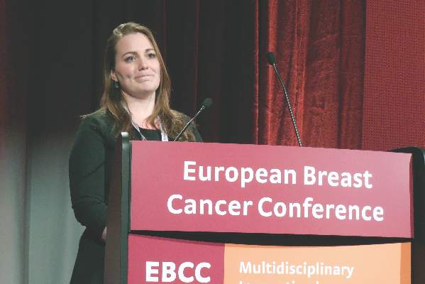
But even longer follow-up of treated breast cancer patients is needed to determine whether the excess mild cardiac dysfunction seen in this analysis eventually progresses to more severe cardiac impairment, Liselotte M. Boerman said at the European Breast Cancer Conference.
Data from the Breast Cancer Long-term Outcome of Cardiac Dysfunction (BLOC) study showed that 175 breast cancer patients who received chemotherapy (and may have also received radiotherapy) had a 2.5-fold higher prevalence of a left ventricular ejection fraction (LVEF) below 54% (95% confidence interval, 1.2-5.4) when measured by echocardiography a median of 10 years after treatment, compared with an equal number of age-matched individuals from the general population.
A separate group of 175 patients treated with radiotherapy only and evaluated by echocardiography a median of 10 years later had a 2.3-fold increased prevalence (1.1-4.7) of a LVEF below 54% when compared with an equal number of age-matched individuals, said Ms. Boerman, an epidemiology researcher at the University of Groningen (the Netherlands).
This degree of left-ventricular dysfunction was found in 15% of the chemotherapy patients and 6% of their controls, and in 16% of the radiotherapy patients and 8% of their controls.
However, the treated breast cancer patients had no long-term increase in their prevalence of more significant systolic cardiac dysfunction, defined as a LVEF of less than 45%, compared with the controls, and the overall rate of systolic dysfunction of this severity was low, affecting fewer than 1% of patients.
Also, the chemotherapy and radiotherapy patients showed no significant increase in the prevalence of diastolic cardiac dysfunction, defined as delayed cardiac relaxation beyond the age-appropriate range. Treated patients did show, after 10 years, a suggestion of an increased prevalence of diagnosed cardiovascular disease, which was 2.3-fold higher (1.0-4.9) in the chemotherapy-receiving patients, compared with their controls; and 70% higher (0.9-3.4) among the patients treated with radiotherapy, compared with their controls, Ms. Boerman said.
The study used data collected from breast cancer patients younger than 80 years old treated after 1980 and controls seen by general practice Dutch physicians. The chemotherapy patients were diagnosed at an average age of 49 years old (range 26-66 years old). About 78% had received treatment with an anthracycline agent and 7% had received trastuzumab (Herceptin). Radiotherapy had also been administered to 70%, while 62% had also received hormonal therapy, and 7% either had a recurrence or developed a tumor in their contralateral breast.
None of the radiotherapy-only patients had received chemotherapy, but 21% had also received hormonal therapy. Their average age at diagnosis was 54 years old (range 32-79 years old), and 10% had a recurrence or a contralateral tumor.

Follow-up echocardiography occurred 5-34 years after the index treatment, at a median age of 60 years old. Echocardiography follow-up occurred in 70% of the chemotherapy breast cancer patients contacted, and in 63% of those who received radiotherapy only. Among controls, about half of those selected by age matching, and contacted, agreed to participate.
Rates of cardiovascular-disease risk factors – dyslipidemia, hypertension, and diabetes – were at roughly similar levels in the cases and controls at the time of breast cancer diagnosis. But the rate of current smoking at the time of diagnosis appeared higher in the cases (30% among those who received chemotherapy and 33% among those on radiotherapy), compared with their respective control groups (22% and 30%). Ms. Boerman said that a multivariate analysis had not yet been run on the data but should occur soon.
“The prevalence of cardiac dysfunction was higher [in treated patients] than I would have expected, but there is potential bias as only 70% of invited patients actually participated,” commented Dr. Robert Mansel, a professor at the Institute of Cancer & Genetics at Cardiff University (Wales). He also noted the very low rate of patients who developed severe cardiac dysfunction.
Ms. Boerman and Dr. Mansel reported having no financial disclosures.
On Twitter @mitchelzoler
AMSTERDAM – Breast cancer patients who underwent chemotherapy or radiotherapy had about a two-fold increased prevalence of mild systolic cardiac dysfunction a median of 10 years after treatment, compared with age-matched controls in a study that included a total of 700 people.
But even longer follow-up of treated breast cancer patients is needed to determine whether the excess mild cardiac dysfunction seen in this analysis eventually progresses to more severe cardiac impairment, Liselotte M. Boerman said at the European Breast Cancer Conference.
Data from the Breast Cancer Long-term Outcome of Cardiac Dysfunction (BLOC) study showed that 175 breast cancer patients who received chemotherapy (and may have also received radiotherapy) had a 2.5-fold higher prevalence of a left ventricular ejection fraction (LVEF) below 54% (95% confidence interval, 1.2-5.4) when measured by echocardiography a median of 10 years after treatment, compared with an equal number of age-matched individuals from the general population.
A separate group of 175 patients treated with radiotherapy only and evaluated by echocardiography a median of 10 years later had a 2.3-fold increased prevalence (1.1-4.7) of a LVEF below 54% when compared with an equal number of age-matched individuals, said Ms. Boerman, an epidemiology researcher at the University of Groningen (the Netherlands).
This degree of left-ventricular dysfunction was found in 15% of the chemotherapy patients and 6% of their controls, and in 16% of the radiotherapy patients and 8% of their controls.
However, the treated breast cancer patients had no long-term increase in their prevalence of more significant systolic cardiac dysfunction, defined as a LVEF of less than 45%, compared with the controls, and the overall rate of systolic dysfunction of this severity was low, affecting fewer than 1% of patients.
Also, the chemotherapy and radiotherapy patients showed no significant increase in the prevalence of diastolic cardiac dysfunction, defined as delayed cardiac relaxation beyond the age-appropriate range. Treated patients did show, after 10 years, a suggestion of an increased prevalence of diagnosed cardiovascular disease, which was 2.3-fold higher (1.0-4.9) in the chemotherapy-receiving patients, compared with their controls; and 70% higher (0.9-3.4) among the patients treated with radiotherapy, compared with their controls, Ms. Boerman said.
The study used data collected from breast cancer patients younger than 80 years old treated after 1980 and controls seen by general practice Dutch physicians. The chemotherapy patients were diagnosed at an average age of 49 years old (range 26-66 years old). About 78% had received treatment with an anthracycline agent and 7% had received trastuzumab (Herceptin). Radiotherapy had also been administered to 70%, while 62% had also received hormonal therapy, and 7% either had a recurrence or developed a tumor in their contralateral breast.
None of the radiotherapy-only patients had received chemotherapy, but 21% had also received hormonal therapy. Their average age at diagnosis was 54 years old (range 32-79 years old), and 10% had a recurrence or a contralateral tumor.
Follow-up echocardiography occurred 5-34 years after the index treatment, at a median age of 60 years old. Echocardiography follow-up occurred in 70% of the chemotherapy breast cancer patients contacted, and in 63% of those who received radiotherapy only. Among controls, about half of those selected by age matching, and contacted, agreed to participate.
Rates of cardiovascular-disease risk factors – dyslipidemia, hypertension, and diabetes – were at roughly similar levels in the cases and controls at the time of breast cancer diagnosis. But the rate of current smoking at the time of diagnosis appeared higher in the cases (30% among those who received chemotherapy and 33% among those on radiotherapy), compared with their respective control groups (22% and 30%). Ms. Boerman said that a multivariate analysis had not yet been run on the data but should occur soon.
“The prevalence of cardiac dysfunction was higher [in treated patients] than I would have expected, but there is potential bias as only 70% of invited patients actually participated,” commented Dr. Robert Mansel, a professor at the Institute of Cancer & Genetics at Cardiff University (Wales). He also noted the very low rate of patients who developed severe cardiac dysfunction.
Ms. Boerman and Dr. Mansel reported having no financial disclosures.
On Twitter @mitchelzoler
AMSTERDAM – Breast cancer patients who underwent chemotherapy or radiotherapy had about a two-fold increased prevalence of mild systolic cardiac dysfunction a median of 10 years after treatment, compared with age-matched controls in a study that included a total of 700 people.
But even longer follow-up of treated breast cancer patients is needed to determine whether the excess mild cardiac dysfunction seen in this analysis eventually progresses to more severe cardiac impairment, Liselotte M. Boerman said at the European Breast Cancer Conference.
Data from the Breast Cancer Long-term Outcome of Cardiac Dysfunction (BLOC) study showed that 175 breast cancer patients who received chemotherapy (and may have also received radiotherapy) had a 2.5-fold higher prevalence of a left ventricular ejection fraction (LVEF) below 54% (95% confidence interval, 1.2-5.4) when measured by echocardiography a median of 10 years after treatment, compared with an equal number of age-matched individuals from the general population.
A separate group of 175 patients treated with radiotherapy only and evaluated by echocardiography a median of 10 years later had a 2.3-fold increased prevalence (1.1-4.7) of a LVEF below 54% when compared with an equal number of age-matched individuals, said Ms. Boerman, an epidemiology researcher at the University of Groningen (the Netherlands).
This degree of left-ventricular dysfunction was found in 15% of the chemotherapy patients and 6% of their controls, and in 16% of the radiotherapy patients and 8% of their controls.
However, the treated breast cancer patients had no long-term increase in their prevalence of more significant systolic cardiac dysfunction, defined as a LVEF of less than 45%, compared with the controls, and the overall rate of systolic dysfunction of this severity was low, affecting fewer than 1% of patients.
Also, the chemotherapy and radiotherapy patients showed no significant increase in the prevalence of diastolic cardiac dysfunction, defined as delayed cardiac relaxation beyond the age-appropriate range. Treated patients did show, after 10 years, a suggestion of an increased prevalence of diagnosed cardiovascular disease, which was 2.3-fold higher (1.0-4.9) in the chemotherapy-receiving patients, compared with their controls; and 70% higher (0.9-3.4) among the patients treated with radiotherapy, compared with their controls, Ms. Boerman said.
The study used data collected from breast cancer patients younger than 80 years old treated after 1980 and controls seen by general practice Dutch physicians. The chemotherapy patients were diagnosed at an average age of 49 years old (range 26-66 years old). About 78% had received treatment with an anthracycline agent and 7% had received trastuzumab (Herceptin). Radiotherapy had also been administered to 70%, while 62% had also received hormonal therapy, and 7% either had a recurrence or developed a tumor in their contralateral breast.
None of the radiotherapy-only patients had received chemotherapy, but 21% had also received hormonal therapy. Their average age at diagnosis was 54 years old (range 32-79 years old), and 10% had a recurrence or a contralateral tumor.
Follow-up echocardiography occurred 5-34 years after the index treatment, at a median age of 60 years old. Echocardiography follow-up occurred in 70% of the chemotherapy breast cancer patients contacted, and in 63% of those who received radiotherapy only. Among controls, about half of those selected by age matching, and contacted, agreed to participate.
Rates of cardiovascular-disease risk factors – dyslipidemia, hypertension, and diabetes – were at roughly similar levels in the cases and controls at the time of breast cancer diagnosis. But the rate of current smoking at the time of diagnosis appeared higher in the cases (30% among those who received chemotherapy and 33% among those on radiotherapy), compared with their respective control groups (22% and 30%). Ms. Boerman said that a multivariate analysis had not yet been run on the data but should occur soon.
“The prevalence of cardiac dysfunction was higher [in treated patients] than I would have expected, but there is potential bias as only 70% of invited patients actually participated,” commented Dr. Robert Mansel, a professor at the Institute of Cancer & Genetics at Cardiff University (Wales). He also noted the very low rate of patients who developed severe cardiac dysfunction.
Ms. Boerman and Dr. Mansel reported having no financial disclosures.
On Twitter @mitchelzoler
AT EBCC10
Key clinical point: Breast cancer patients treated with chemotherapy or radiotherapy showed a doubled rate of mild left-ventricular dysfunction, compared with matched controls 10 years after treatment.
Major finding: Mildly reduced left-ventricular function occurred in 15% of post-chemotherapy patients, compared with 6% of controls.
Data source: Echocardiography examinations conducted on 350 Dutch breast cancer patients and an equal number of age-matched controls.
Disclosures: Ms. Boerman and Dr. Mansel reported having no financial disclosures.
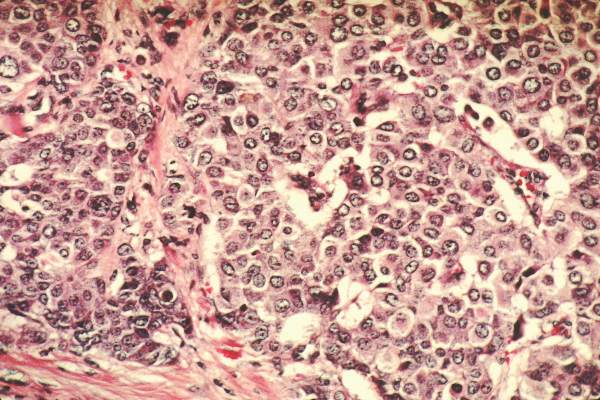
Phone monitoring program helps cut chemotherapy symptom severity
AMSTERDAM – A telephone-based system to monitor chemotherapy-induced symptoms when patients are home and facilitate interventions as needed led to significant cuts in symptom burden in a single-center, randomized trial with 152 breast cancer patients.
“Symptom care at home gives patients symptom care when and where they need it,” Kathi H. Mooney, Ph.D., said at the European Breast Cancer Conference. “Rarely is symptom monitoring extended to when patients are home, but that is when symptoms are most problematic for patients.”

The 30-day program was linked with significant cuts in the number of days with any of seven chemotherapy-induced symptoms. In addition, overall days with any severe symptom fell by 48% (P = .006) and the number of days with any moderate symptoms fell by 38% (P = .011), reported Dr. Mooney, professor of nursing at the University of Utah, Salt Lake City.
She and her associates designed a telephone-based system to address the usual reticence that chemotherapy patients have to report their symptoms. Prior study results had documented that patients with moderate or severe symptoms initiated calls for about 5% of episodes, even when explicitly told to report symptoms. This led the Utah researchers to develop an automated, interactive system that phoned patients daily.
The enrolled patients spanned the full spectrum of breast cancer stages; the average age was 53 years. All patients received a daily, automated call that prompted them to rate each of 11 chemotherapy symptoms on a scale of 0-10.
The calls also provided automated, self-management coaching for problematic symptoms. The investigational Symptom Care at Home intervention involved a nurse practitioner getting back to patients who reported poorly-controlled symptoms. These follow-up calls averaged just under 7 minutes in length, and on average each patient received 11 calls during the 30 days of the intervention.
After 30 days, the 83 patients in the Symptom Care at Home program had statistically-significant reductions in days with moderate or worse episodes for 7 of the 11 symptoms tallied: numbness or tingling, anxiety, nausea, pain, depressed mood, sore mouth, and fatigue. The most robust effects were a 72% drop in days with moderate or worse numbness or tingling, a 66% reduction in anxiety days, and a 61% cut in nausea days.

“I like the intervention used in this study. I think it shows what can happen when you collect symptom information in a way that is patient friendly,” said Dr. Robert Mansel, professor at the Institute of Cancer & Genetics at Cardiff University, South Wales. “I was delighted to see that this intervention really made a difference. The findings show that patients often suffer a lot more from treatment than they are usually prepared to tell us.”
In addition to the efficacy of this intervention, “patients told us that they liked having someone to talk with about their symptoms,” Dr. Mooney said in an interview. “Patients are concerned about their symptoms.”
One aspect of the Symptom Care at Home program that has not yet been analyzed is its cost efficacy. The University of Utah’s Huntsman Cancer Institute, where the program was developed, is awaiting results from a cost-benefit analysis before deciding whether to make the telephone system part of routine practice, she said.
Dr. Mooney and Dr. Mansel reported having no financial disclosures.
On Twitter @mitchelzoler
AMSTERDAM – A telephone-based system to monitor chemotherapy-induced symptoms when patients are home and facilitate interventions as needed led to significant cuts in symptom burden in a single-center, randomized trial with 152 breast cancer patients.
“Symptom care at home gives patients symptom care when and where they need it,” Kathi H. Mooney, Ph.D., said at the European Breast Cancer Conference. “Rarely is symptom monitoring extended to when patients are home, but that is when symptoms are most problematic for patients.”
The 30-day program was linked with significant cuts in the number of days with any of seven chemotherapy-induced symptoms. In addition, overall days with any severe symptom fell by 48% (P = .006) and the number of days with any moderate symptoms fell by 38% (P = .011), reported Dr. Mooney, professor of nursing at the University of Utah, Salt Lake City.
She and her associates designed a telephone-based system to address the usual reticence that chemotherapy patients have to report their symptoms. Prior study results had documented that patients with moderate or severe symptoms initiated calls for about 5% of episodes, even when explicitly told to report symptoms. This led the Utah researchers to develop an automated, interactive system that phoned patients daily.
The enrolled patients spanned the full spectrum of breast cancer stages; the average age was 53 years. All patients received a daily, automated call that prompted them to rate each of 11 chemotherapy symptoms on a scale of 0-10.
The calls also provided automated, self-management coaching for problematic symptoms. The investigational Symptom Care at Home intervention involved a nurse practitioner getting back to patients who reported poorly-controlled symptoms. These follow-up calls averaged just under 7 minutes in length, and on average each patient received 11 calls during the 30 days of the intervention.
After 30 days, the 83 patients in the Symptom Care at Home program had statistically-significant reductions in days with moderate or worse episodes for 7 of the 11 symptoms tallied: numbness or tingling, anxiety, nausea, pain, depressed mood, sore mouth, and fatigue. The most robust effects were a 72% drop in days with moderate or worse numbness or tingling, a 66% reduction in anxiety days, and a 61% cut in nausea days.
“I like the intervention used in this study. I think it shows what can happen when you collect symptom information in a way that is patient friendly,” said Dr. Robert Mansel, professor at the Institute of Cancer & Genetics at Cardiff University, South Wales. “I was delighted to see that this intervention really made a difference. The findings show that patients often suffer a lot more from treatment than they are usually prepared to tell us.”
In addition to the efficacy of this intervention, “patients told us that they liked having someone to talk with about their symptoms,” Dr. Mooney said in an interview. “Patients are concerned about their symptoms.”
One aspect of the Symptom Care at Home program that has not yet been analyzed is its cost efficacy. The University of Utah’s Huntsman Cancer Institute, where the program was developed, is awaiting results from a cost-benefit analysis before deciding whether to make the telephone system part of routine practice, she said.
Dr. Mooney and Dr. Mansel reported having no financial disclosures.
On Twitter @mitchelzoler
AMSTERDAM – A telephone-based system to monitor chemotherapy-induced symptoms when patients are home and facilitate interventions as needed led to significant cuts in symptom burden in a single-center, randomized trial with 152 breast cancer patients.
“Symptom care at home gives patients symptom care when and where they need it,” Kathi H. Mooney, Ph.D., said at the European Breast Cancer Conference. “Rarely is symptom monitoring extended to when patients are home, but that is when symptoms are most problematic for patients.”
The 30-day program was linked with significant cuts in the number of days with any of seven chemotherapy-induced symptoms. In addition, overall days with any severe symptom fell by 48% (P = .006) and the number of days with any moderate symptoms fell by 38% (P = .011), reported Dr. Mooney, professor of nursing at the University of Utah, Salt Lake City.
She and her associates designed a telephone-based system to address the usual reticence that chemotherapy patients have to report their symptoms. Prior study results had documented that patients with moderate or severe symptoms initiated calls for about 5% of episodes, even when explicitly told to report symptoms. This led the Utah researchers to develop an automated, interactive system that phoned patients daily.
The enrolled patients spanned the full spectrum of breast cancer stages; the average age was 53 years. All patients received a daily, automated call that prompted them to rate each of 11 chemotherapy symptoms on a scale of 0-10.
The calls also provided automated, self-management coaching for problematic symptoms. The investigational Symptom Care at Home intervention involved a nurse practitioner getting back to patients who reported poorly-controlled symptoms. These follow-up calls averaged just under 7 minutes in length, and on average each patient received 11 calls during the 30 days of the intervention.
After 30 days, the 83 patients in the Symptom Care at Home program had statistically-significant reductions in days with moderate or worse episodes for 7 of the 11 symptoms tallied: numbness or tingling, anxiety, nausea, pain, depressed mood, sore mouth, and fatigue. The most robust effects were a 72% drop in days with moderate or worse numbness or tingling, a 66% reduction in anxiety days, and a 61% cut in nausea days.
“I like the intervention used in this study. I think it shows what can happen when you collect symptom information in a way that is patient friendly,” said Dr. Robert Mansel, professor at the Institute of Cancer & Genetics at Cardiff University, South Wales. “I was delighted to see that this intervention really made a difference. The findings show that patients often suffer a lot more from treatment than they are usually prepared to tell us.”
In addition to the efficacy of this intervention, “patients told us that they liked having someone to talk with about their symptoms,” Dr. Mooney said in an interview. “Patients are concerned about their symptoms.”
One aspect of the Symptom Care at Home program that has not yet been analyzed is its cost efficacy. The University of Utah’s Huntsman Cancer Institute, where the program was developed, is awaiting results from a cost-benefit analysis before deciding whether to make the telephone system part of routine practice, she said.
Dr. Mooney and Dr. Mansel reported having no financial disclosures.
On Twitter @mitchelzoler
AT EBCC 10

VIDEO: Trastuzumab plus lapatinib erases selected breast cancers
AMSTERDAM – An unexpectedly dramatic benefit from combining trastuzumab and lapatinib when briefly treating women scheduled for breast cancer surgery has researchers focused on the best follow-up study to this promising but preliminary finding from 66 patients.
Among 66 patients with newly diagnosed HER2-positive breast cancer and scheduled for surgery, an average 11 days of treatment with a combination of trastuzumab (Herceptin) and lapatinib (Tykerb) during the period before surgery resulted in a pathologic complete response (pCR) in seven patients (11%) and 11 patients (17%) with minimal residual disease (MRD), Dr. Nigel Bundred reported at the European Breast Cancer Conference.

These patients had breast cancer tumors at baseline with a median diameter of 2 cm that with treatment for not quite 2 weeks resulted in either complete tumor disappearance or shrinkage to less than 5 mm in diameter in 18 (27%) of the patients on dual therapy, said Dr. Bundred, a professor of surgical oncology at the University Hospital of South Manchester (England).
“No cancer has ever disappeared that fast. It was flabbergasting,” Dr. Bundred said in an interview. “We need to confirm the results, and get larger numbers of patients.”
“This was a really surprising finding,” said Judith M. Bliss, professor and director of the Clinical Trials & Statistics unit of the Institute of Cancer Research in London and a co-investigator. “We knew that trastuzumab and lapatinib are effective treatments, but we had not expected to see such a dramatic effect on the structure and size of the tumor in such a short period of time,” Prof. Bliss said in a video interview.
The finding came as part of the Effect of Perioperative Anti-HER2 Therapy on Overall Survival–Biologic Phase (EPHOS-B) trial, designed to assess changes in a marker of cell proliferation (Ki67) and in a marker of cell apoptosis in relation to treatment with trastuzumab, lapatinib, or both when administered during the 10-12 days from the time patients entered the study until their scheduled surgery.

During the first phase of EPHOS-B, when 130 patients received trastuzumab monotherapy, lapatinib monotherapy, or neither, one of the 57 patients on trastuzumab (2%) had a pathologic complete response and one patient (2%) had minimal residual disease. No patients on lapatinib alone or in the control arm with no anti-HER2 drug had this sort of response.
During the second phase, 127 patients received trastuzumab alone, trastuzumab plus lapatinib in combination, or controls who received no anti-HER2 treatment. The substantial number of complete or partial responders in the 66 patients who received combined treatment with trastuzumab and lapatinib contrasted with one minimal residual disease among 32 patients (3%) who received trastuzumab alone and no responders among the 29 controls.
Patients receiving trastuzumab, alone or in the combined regimen, received a standard intravenous loading dosage of 6 mg/kg on days 1 and 8 after entry into the protocol, followed by a third dose after surgery on days 15-19; those also receiving lapatinib received 1 g/day orally for 28 days starting on entry.
“A quarter of the patients seem exquisitely sensitive to the combination of trastuzumab and lapatinib,” said Dr. David Cameron, another collaborator on the EPHOS-B study. “It’s pretty unusual to treat for 10-12 days and have 10% of the tumors disappear.”
Follow-up studies will be needed to determine whether patients with a pathologic complete response or minimal residual disease can forgo some or all of the chemotherapy that would usually follow surgery and still have good long-term disease-free survival. “We will focus on optimizing the short-term effect, and then modulate subsequent treatments,” said Dr. Cameron, professor and clinical director of oncology at the University of Edinburgh.
“We think these are patients who don’t need chemotherapy and are particularly sensitive to anti-HER2 drugs,” but so far that hasn’t been proven, cautioned Ms. Bliss.

The striking efficacy of trastuzumab combined with lapatinib in these 66 patients contrasts with the previously reported results from the ALTTO (Adjuvant Lapatinib and/or Trastuzumab Treatment Optimization) study, which failed to show an advantage in disease-free survival with trastuzumab plus lapatinib, compared with either of these two agents used individually during 1 year of adjuvant treatment in more than 8,000 randomized patients (J Clin Oncol. 2015 Nov 23. doi: 10.1200/JCO.2015.62.1797), But the EPHOS-B results did agree with results from the much smaller NeoALTTO study, which showed in a randomized, multicenter, phase III study with 455 patients a significant incremental increase in pathologic complete response rate in patients on combined trastuzumab plus lapatinib, compared with patients on either drug alone (Lancet. 2012;379 [9816]:633-40).
Another promising finding from EPHOS-B was that among the 64 patients who had their left ventricular ejection fraction assessed prior to the second phase, none of 32 patients on the combined regimen showed signs of cardiotoxicity with a reduced ejection fraction following treatment, Dr. Bundred reported.
Until now, not much was known about the beneficial mechanisms of anti-HER2 drugs, he noted. The results from EPHOS-B “tell us that combined treatment does more than just shut down cell replication. It might be that trastuzumab induces an immune response.” The former tumor beds of patients with pathologic complete response as well as regions with minimal residual disease showed large numbers of tumor infiltrating lymphocytes, a sign of a robust immune response.
Trastuzumab and lapatinib work by different mechanisms, Dr. Bundred stressed. “The only way you can combine drugs is by understanding their mechanisms. We’re still looking for the mechanisms of the anti-HER2 drugs.”
The video associated with this article is no longer available on this site. Please view all of our videos on the MDedge YouTube channel
On Twitter @mitchelzoler
The significant proportion of patients treated with trastuzumab plus lapatinib for about 11 days who developed a pathologic complete response or minimal residual disease was largely unexpected.
The finding raises questions: Can we identify patients after 11 days of treatment who are good responders to this combined treatment and will have an excellent relapse-free survival without ongoing combination anti-HER2 treatment or anti-HER2 escalation, and who will be eligible for chemotherapy de-escalation similar to the regimen tested by Tolaney and her associates (N Engl J Med. 2015 Jan;372:134-41)?

|
| Mitchel L. Zoler/Frontline Medical News Dr. Hervé Bonnefoi |
We clearly need more data on the impact of the combined anti-HER2 regimen on the rate of long-term, relapse-free survival, and data from many more patients treated with a combined anti-HER2 regimen. The EPHOS-B results seem to contradict the results of the ALTTO study (J Clin Oncol. 2015 Nov 23. doi: 10.1200/JCO.2015.62.1797), but they confirm results of the NeoALTTO study, a randomized, multicenter, phase III study with 455 patients (Lancet. 2012 Jan;379[9816]:633-40).
The EPHOS-B researchers modeled their study after the presurgical treatment studies run using hormonal therapies. The results from studies of anti-HER2 treatments during this presurgical time window have been much harder to interpret.
Dr. Hervé Bonnefoi is a professor of medical oncology at Bordeaux University (France). He reported having no financial disclosures. He made these comments as the designated discussant for the EPHOS-B study.
The significant proportion of patients treated with trastuzumab plus lapatinib for about 11 days who developed a pathologic complete response or minimal residual disease was largely unexpected.
The finding raises questions: Can we identify patients after 11 days of treatment who are good responders to this combined treatment and will have an excellent relapse-free survival without ongoing combination anti-HER2 treatment or anti-HER2 escalation, and who will be eligible for chemotherapy de-escalation similar to the regimen tested by Tolaney and her associates (N Engl J Med. 2015 Jan;372:134-41)?
|
|
| Mitchel L. Zoler/Frontline Medical News Dr. Hervé Bonnefoi |
We clearly need more data on the impact of the combined anti-HER2 regimen on the rate of long-term, relapse-free survival, and data from many more patients treated with a combined anti-HER2 regimen. The EPHOS-B results seem to contradict the results of the ALTTO study (J Clin Oncol. 2015 Nov 23. doi: 10.1200/JCO.2015.62.1797), but they confirm results of the NeoALTTO study, a randomized, multicenter, phase III study with 455 patients (Lancet. 2012 Jan;379[9816]:633-40).
The EPHOS-B researchers modeled their study after the presurgical treatment studies run using hormonal therapies. The results from studies of anti-HER2 treatments during this presurgical time window have been much harder to interpret.
Dr. Hervé Bonnefoi is a professor of medical oncology at Bordeaux University (France). He reported having no financial disclosures. He made these comments as the designated discussant for the EPHOS-B study.
The significant proportion of patients treated with trastuzumab plus lapatinib for about 11 days who developed a pathologic complete response or minimal residual disease was largely unexpected.
The finding raises questions: Can we identify patients after 11 days of treatment who are good responders to this combined treatment and will have an excellent relapse-free survival without ongoing combination anti-HER2 treatment or anti-HER2 escalation, and who will be eligible for chemotherapy de-escalation similar to the regimen tested by Tolaney and her associates (N Engl J Med. 2015 Jan;372:134-41)?
|
|
| Mitchel L. Zoler/Frontline Medical News Dr. Hervé Bonnefoi |
We clearly need more data on the impact of the combined anti-HER2 regimen on the rate of long-term, relapse-free survival, and data from many more patients treated with a combined anti-HER2 regimen. The EPHOS-B results seem to contradict the results of the ALTTO study (J Clin Oncol. 2015 Nov 23. doi: 10.1200/JCO.2015.62.1797), but they confirm results of the NeoALTTO study, a randomized, multicenter, phase III study with 455 patients (Lancet. 2012 Jan;379[9816]:633-40).
The EPHOS-B researchers modeled their study after the presurgical treatment studies run using hormonal therapies. The results from studies of anti-HER2 treatments during this presurgical time window have been much harder to interpret.
Dr. Hervé Bonnefoi is a professor of medical oncology at Bordeaux University (France). He reported having no financial disclosures. He made these comments as the designated discussant for the EPHOS-B study.
AMSTERDAM – An unexpectedly dramatic benefit from combining trastuzumab and lapatinib when briefly treating women scheduled for breast cancer surgery has researchers focused on the best follow-up study to this promising but preliminary finding from 66 patients.
Among 66 patients with newly diagnosed HER2-positive breast cancer and scheduled for surgery, an average 11 days of treatment with a combination of trastuzumab (Herceptin) and lapatinib (Tykerb) during the period before surgery resulted in a pathologic complete response (pCR) in seven patients (11%) and 11 patients (17%) with minimal residual disease (MRD), Dr. Nigel Bundred reported at the European Breast Cancer Conference.
These patients had breast cancer tumors at baseline with a median diameter of 2 cm that with treatment for not quite 2 weeks resulted in either complete tumor disappearance or shrinkage to less than 5 mm in diameter in 18 (27%) of the patients on dual therapy, said Dr. Bundred, a professor of surgical oncology at the University Hospital of South Manchester (England).
“No cancer has ever disappeared that fast. It was flabbergasting,” Dr. Bundred said in an interview. “We need to confirm the results, and get larger numbers of patients.”
“This was a really surprising finding,” said Judith M. Bliss, professor and director of the Clinical Trials & Statistics unit of the Institute of Cancer Research in London and a co-investigator. “We knew that trastuzumab and lapatinib are effective treatments, but we had not expected to see such a dramatic effect on the structure and size of the tumor in such a short period of time,” Prof. Bliss said in a video interview.
The finding came as part of the Effect of Perioperative Anti-HER2 Therapy on Overall Survival–Biologic Phase (EPHOS-B) trial, designed to assess changes in a marker of cell proliferation (Ki67) and in a marker of cell apoptosis in relation to treatment with trastuzumab, lapatinib, or both when administered during the 10-12 days from the time patients entered the study until their scheduled surgery.
During the first phase of EPHOS-B, when 130 patients received trastuzumab monotherapy, lapatinib monotherapy, or neither, one of the 57 patients on trastuzumab (2%) had a pathologic complete response and one patient (2%) had minimal residual disease. No patients on lapatinib alone or in the control arm with no anti-HER2 drug had this sort of response.
During the second phase, 127 patients received trastuzumab alone, trastuzumab plus lapatinib in combination, or controls who received no anti-HER2 treatment. The substantial number of complete or partial responders in the 66 patients who received combined treatment with trastuzumab and lapatinib contrasted with one minimal residual disease among 32 patients (3%) who received trastuzumab alone and no responders among the 29 controls.
Patients receiving trastuzumab, alone or in the combined regimen, received a standard intravenous loading dosage of 6 mg/kg on days 1 and 8 after entry into the protocol, followed by a third dose after surgery on days 15-19; those also receiving lapatinib received 1 g/day orally for 28 days starting on entry.
“A quarter of the patients seem exquisitely sensitive to the combination of trastuzumab and lapatinib,” said Dr. David Cameron, another collaborator on the EPHOS-B study. “It’s pretty unusual to treat for 10-12 days and have 10% of the tumors disappear.”
Follow-up studies will be needed to determine whether patients with a pathologic complete response or minimal residual disease can forgo some or all of the chemotherapy that would usually follow surgery and still have good long-term disease-free survival. “We will focus on optimizing the short-term effect, and then modulate subsequent treatments,” said Dr. Cameron, professor and clinical director of oncology at the University of Edinburgh.
“We think these are patients who don’t need chemotherapy and are particularly sensitive to anti-HER2 drugs,” but so far that hasn’t been proven, cautioned Ms. Bliss.
The striking efficacy of trastuzumab combined with lapatinib in these 66 patients contrasts with the previously reported results from the ALTTO (Adjuvant Lapatinib and/or Trastuzumab Treatment Optimization) study, which failed to show an advantage in disease-free survival with trastuzumab plus lapatinib, compared with either of these two agents used individually during 1 year of adjuvant treatment in more than 8,000 randomized patients (J Clin Oncol. 2015 Nov 23. doi: 10.1200/JCO.2015.62.1797), But the EPHOS-B results did agree with results from the much smaller NeoALTTO study, which showed in a randomized, multicenter, phase III study with 455 patients a significant incremental increase in pathologic complete response rate in patients on combined trastuzumab plus lapatinib, compared with patients on either drug alone (Lancet. 2012;379 [9816]:633-40).
Another promising finding from EPHOS-B was that among the 64 patients who had their left ventricular ejection fraction assessed prior to the second phase, none of 32 patients on the combined regimen showed signs of cardiotoxicity with a reduced ejection fraction following treatment, Dr. Bundred reported.
Until now, not much was known about the beneficial mechanisms of anti-HER2 drugs, he noted. The results from EPHOS-B “tell us that combined treatment does more than just shut down cell replication. It might be that trastuzumab induces an immune response.” The former tumor beds of patients with pathologic complete response as well as regions with minimal residual disease showed large numbers of tumor infiltrating lymphocytes, a sign of a robust immune response.
Trastuzumab and lapatinib work by different mechanisms, Dr. Bundred stressed. “The only way you can combine drugs is by understanding their mechanisms. We’re still looking for the mechanisms of the anti-HER2 drugs.”
The video associated with this article is no longer available on this site. Please view all of our videos on the MDedge YouTube channel
On Twitter @mitchelzoler
AMSTERDAM – An unexpectedly dramatic benefit from combining trastuzumab and lapatinib when briefly treating women scheduled for breast cancer surgery has researchers focused on the best follow-up study to this promising but preliminary finding from 66 patients.
Among 66 patients with newly diagnosed HER2-positive breast cancer and scheduled for surgery, an average 11 days of treatment with a combination of trastuzumab (Herceptin) and lapatinib (Tykerb) during the period before surgery resulted in a pathologic complete response (pCR) in seven patients (11%) and 11 patients (17%) with minimal residual disease (MRD), Dr. Nigel Bundred reported at the European Breast Cancer Conference.
These patients had breast cancer tumors at baseline with a median diameter of 2 cm that with treatment for not quite 2 weeks resulted in either complete tumor disappearance or shrinkage to less than 5 mm in diameter in 18 (27%) of the patients on dual therapy, said Dr. Bundred, a professor of surgical oncology at the University Hospital of South Manchester (England).
“No cancer has ever disappeared that fast. It was flabbergasting,” Dr. Bundred said in an interview. “We need to confirm the results, and get larger numbers of patients.”
“This was a really surprising finding,” said Judith M. Bliss, professor and director of the Clinical Trials & Statistics unit of the Institute of Cancer Research in London and a co-investigator. “We knew that trastuzumab and lapatinib are effective treatments, but we had not expected to see such a dramatic effect on the structure and size of the tumor in such a short period of time,” Prof. Bliss said in a video interview.
The finding came as part of the Effect of Perioperative Anti-HER2 Therapy on Overall Survival–Biologic Phase (EPHOS-B) trial, designed to assess changes in a marker of cell proliferation (Ki67) and in a marker of cell apoptosis in relation to treatment with trastuzumab, lapatinib, or both when administered during the 10-12 days from the time patients entered the study until their scheduled surgery.
During the first phase of EPHOS-B, when 130 patients received trastuzumab monotherapy, lapatinib monotherapy, or neither, one of the 57 patients on trastuzumab (2%) had a pathologic complete response and one patient (2%) had minimal residual disease. No patients on lapatinib alone or in the control arm with no anti-HER2 drug had this sort of response.
During the second phase, 127 patients received trastuzumab alone, trastuzumab plus lapatinib in combination, or controls who received no anti-HER2 treatment. The substantial number of complete or partial responders in the 66 patients who received combined treatment with trastuzumab and lapatinib contrasted with one minimal residual disease among 32 patients (3%) who received trastuzumab alone and no responders among the 29 controls.
Patients receiving trastuzumab, alone or in the combined regimen, received a standard intravenous loading dosage of 6 mg/kg on days 1 and 8 after entry into the protocol, followed by a third dose after surgery on days 15-19; those also receiving lapatinib received 1 g/day orally for 28 days starting on entry.
“A quarter of the patients seem exquisitely sensitive to the combination of trastuzumab and lapatinib,” said Dr. David Cameron, another collaborator on the EPHOS-B study. “It’s pretty unusual to treat for 10-12 days and have 10% of the tumors disappear.”
Follow-up studies will be needed to determine whether patients with a pathologic complete response or minimal residual disease can forgo some or all of the chemotherapy that would usually follow surgery and still have good long-term disease-free survival. “We will focus on optimizing the short-term effect, and then modulate subsequent treatments,” said Dr. Cameron, professor and clinical director of oncology at the University of Edinburgh.
“We think these are patients who don’t need chemotherapy and are particularly sensitive to anti-HER2 drugs,” but so far that hasn’t been proven, cautioned Ms. Bliss.
The striking efficacy of trastuzumab combined with lapatinib in these 66 patients contrasts with the previously reported results from the ALTTO (Adjuvant Lapatinib and/or Trastuzumab Treatment Optimization) study, which failed to show an advantage in disease-free survival with trastuzumab plus lapatinib, compared with either of these two agents used individually during 1 year of adjuvant treatment in more than 8,000 randomized patients (J Clin Oncol. 2015 Nov 23. doi: 10.1200/JCO.2015.62.1797), But the EPHOS-B results did agree with results from the much smaller NeoALTTO study, which showed in a randomized, multicenter, phase III study with 455 patients a significant incremental increase in pathologic complete response rate in patients on combined trastuzumab plus lapatinib, compared with patients on either drug alone (Lancet. 2012;379 [9816]:633-40).
Another promising finding from EPHOS-B was that among the 64 patients who had their left ventricular ejection fraction assessed prior to the second phase, none of 32 patients on the combined regimen showed signs of cardiotoxicity with a reduced ejection fraction following treatment, Dr. Bundred reported.
Until now, not much was known about the beneficial mechanisms of anti-HER2 drugs, he noted. The results from EPHOS-B “tell us that combined treatment does more than just shut down cell replication. It might be that trastuzumab induces an immune response.” The former tumor beds of patients with pathologic complete response as well as regions with minimal residual disease showed large numbers of tumor infiltrating lymphocytes, a sign of a robust immune response.
Trastuzumab and lapatinib work by different mechanisms, Dr. Bundred stressed. “The only way you can combine drugs is by understanding their mechanisms. We’re still looking for the mechanisms of the anti-HER2 drugs.”
The video associated with this article is no longer available on this site. Please view all of our videos on the MDedge YouTube channel
On Twitter @mitchelzoler
AT EBCC10
Key clinical point: Combined treatment with trastuzumab and lapatinib for 10-12 days led to complete HER2-positive breast cancer disappearance in seven of 66 patients.
Major finding: Pathologic complete responses occurred in seven of 66 patients on trastuzumab plus lapatinib, but none of 32 patients on trastuzumab alone or the 29 controls.
Data source: EPHOS-B, a multicenter, randomized, British study run in two phases with a total of 257 women with HER2-positive breast cancer.
Disclosures: GlaxoSmithKline, which at the time marketed lapatinib (Tykerb, now marketed by Novartis), made an educational grant to the Institute of Cancer Research in association with the EPHOS-B trial. Dr. Bundred served on an advisory board for Roche U.K. Ms. Bliss reported having no financial disclosures. Dr. Cameron has been a consultant to Novartis, GlaxoSmithKline, and Roche, but received no additional compensation.

Inking bests suturing to mark breast tumor margins
BOSTON – With art supplies and “mystery” sutures, a team of investigators has determined that immediate intraoperative inking of lumpectomy specimens appears to be a better method than suture placement for orienting specimens for pathology, a finding that could reduce re-excisions.
“Intraoperative specimen suturing appears to be inaccurate for margin identification, and additional means by the surgeon to improve the accuracy are needed. This could be either immediate inking or routine excision of shave margins, either at the original surgery or when you go back for re-excision, to take all the margins instead of one specific margin,” Dr. Angel Arnaout, a breast surgeon at the University of Ottawa, Ont., said at the annual Society of Surgical Oncology Cancer Symposium.
She presented results of the randomized, blinded SMART (Specimen Margin Assessment Technique) trial comparing suture placement with intraoperative inking within the same surgical specimen.
Positive resection margins during breast-conserving surgery are associated with a twofold increase in the risk of ipsilateral tumor recurrence, and require re-excision or conversion to mastectomy. Re-excisions rates range from 25% to 45%, but in the majority of cases – 53% to 65% – no additional disease is found, Dr. Arnaout said.
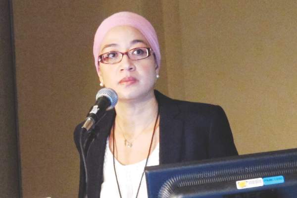
In addition to adding to patient distress, re-excisions diminish cosmetic results because they take additional tissue, and delay the start of adjuvant breast cancer therapy, she noted.
Surgeons typically orient specimens for pathologists by placing two or three sutures in the center of the margin surfaces or, less frequently, by intraoperative marking of the margins with ink. But in transit from the OR to the pathology lab, specimens often flatten or “pancake” under their own weight, making it challenging for the pathologist to identify the originally marked margins.
To test whether suturing or inking is better at identifying the extent of tumors during breast-conserving surgery, the investigators devised a strategy using benign breast tissue removed during prophylactic mastectomy or reduction mammoplasty.
The surgeons first performed a sham lumpectomy within the tissue. They then painted the specimen margins in the OR using a phospholuminescent paint obtained from an art supply store. The paint dries clear and colorless, but glows in different colors under black light. By using the paint, carefully selected so as not to interfere with pathology staining, the investigators were able to blind the pathologists to the actual orientation of the margins.
While the tissue was still in the OR, the surgeons then placed two or three orienting sutures in customary locations, plus a third, “mystery” suture at a location known only to the surgeon.
In the pathology lab, the assistant was asked to look at the sutures and outline in black ink the edges of the margins using the sutures for guidance.
The specimen was then examined under black light to determine the degree of discordance between surgeons and pathologist for identification of margins using the suture and painting methods (primary outcome), and to evaluate the discrepancy in the extent of margin surface areas as identified by the surgeons and the pathologists (secondary outcome).
“We asked breast surgeons ‘what would be a clinically significant discordance rate for you?’ and most of them said between 10% and 20%, so we looked for a 15% discordance rate as being clinically significant between the two margin assessment techniques,” Dr. Arnaout said.
They found that the overall discordance in the location of the mystery suture between surgeons and pathologists was similar whether the samples contained two or three location sutures, with discordance rates of 46% (34 of 75 samples) for two sutures, and 47% (41 of 88 samples) for three sutures.
For the secondary outcome of discordance in the extent of margins, they found that pathologists tended to estimate the mean surface of the anterior and posterior margins to be significantly larger than the surgeons did (P less than .01), while significantly underestimating the superior, inferior, and lateral margins (P less than .01 for superior and inferior, and P = .04 for lateral margins).
Examination under black light showed that often two or three additional margins would be included by the surgeon within what the pathologist had identified as the anterior margin.
“This has implications for the surgeon and for the patient,” Dr. Arnaout said. “For most of us that do lumpectomies that go from skin down to chest wall, if an anterior margin or posterior margin is positive, a lot of times we would fight not to go back because we would say there is no further room for re-excision.”
But if the tumor is within what is actually the superior margin but labeled by the pathologist as an anterior margin, the patient may miss an opportunity for successful re-excision, she explained.
The study was limited by the fact that the sham lumpectomy specimens did not contain skin or muscle, which would have allowed for more accurate margin orientation in an actual operative setting; by lack of compression of specimens with small nonpalpable lesions in containers, which further distorts specimens; and by the use of smaller lumpectomy specimens than normally obtained during cancer surgery, which could have resulted in overestimation of the discordance that might occur when larger specimens are taken, Dr. Arnaout said.
“The conclusion of our study is that specimen margin orientation really should be defined by the surgeon who knows the original shape and orientation of the tissue during surgery, and not to rely on the pathology to reorient based on some sutures placed in the centers of the specimens without defining the extent of the surface area of each of the margins,” she said.
The Canadian Cancer Society Research Institute and the Canadian Surgical Research Fund supported the study. Dr. Arnaout and coauthors reported no conflicts of interest.
BOSTON – With art supplies and “mystery” sutures, a team of investigators has determined that immediate intraoperative inking of lumpectomy specimens appears to be a better method than suture placement for orienting specimens for pathology, a finding that could reduce re-excisions.
“Intraoperative specimen suturing appears to be inaccurate for margin identification, and additional means by the surgeon to improve the accuracy are needed. This could be either immediate inking or routine excision of shave margins, either at the original surgery or when you go back for re-excision, to take all the margins instead of one specific margin,” Dr. Angel Arnaout, a breast surgeon at the University of Ottawa, Ont., said at the annual Society of Surgical Oncology Cancer Symposium.
She presented results of the randomized, blinded SMART (Specimen Margin Assessment Technique) trial comparing suture placement with intraoperative inking within the same surgical specimen.
Positive resection margins during breast-conserving surgery are associated with a twofold increase in the risk of ipsilateral tumor recurrence, and require re-excision or conversion to mastectomy. Re-excisions rates range from 25% to 45%, but in the majority of cases – 53% to 65% – no additional disease is found, Dr. Arnaout said.
In addition to adding to patient distress, re-excisions diminish cosmetic results because they take additional tissue, and delay the start of adjuvant breast cancer therapy, she noted.
Surgeons typically orient specimens for pathologists by placing two or three sutures in the center of the margin surfaces or, less frequently, by intraoperative marking of the margins with ink. But in transit from the OR to the pathology lab, specimens often flatten or “pancake” under their own weight, making it challenging for the pathologist to identify the originally marked margins.
To test whether suturing or inking is better at identifying the extent of tumors during breast-conserving surgery, the investigators devised a strategy using benign breast tissue removed during prophylactic mastectomy or reduction mammoplasty.
The surgeons first performed a sham lumpectomy within the tissue. They then painted the specimen margins in the OR using a phospholuminescent paint obtained from an art supply store. The paint dries clear and colorless, but glows in different colors under black light. By using the paint, carefully selected so as not to interfere with pathology staining, the investigators were able to blind the pathologists to the actual orientation of the margins.
While the tissue was still in the OR, the surgeons then placed two or three orienting sutures in customary locations, plus a third, “mystery” suture at a location known only to the surgeon.
In the pathology lab, the assistant was asked to look at the sutures and outline in black ink the edges of the margins using the sutures for guidance.
The specimen was then examined under black light to determine the degree of discordance between surgeons and pathologist for identification of margins using the suture and painting methods (primary outcome), and to evaluate the discrepancy in the extent of margin surface areas as identified by the surgeons and the pathologists (secondary outcome).
“We asked breast surgeons ‘what would be a clinically significant discordance rate for you?’ and most of them said between 10% and 20%, so we looked for a 15% discordance rate as being clinically significant between the two margin assessment techniques,” Dr. Arnaout said.
They found that the overall discordance in the location of the mystery suture between surgeons and pathologists was similar whether the samples contained two or three location sutures, with discordance rates of 46% (34 of 75 samples) for two sutures, and 47% (41 of 88 samples) for three sutures.
For the secondary outcome of discordance in the extent of margins, they found that pathologists tended to estimate the mean surface of the anterior and posterior margins to be significantly larger than the surgeons did (P less than .01), while significantly underestimating the superior, inferior, and lateral margins (P less than .01 for superior and inferior, and P = .04 for lateral margins).
Examination under black light showed that often two or three additional margins would be included by the surgeon within what the pathologist had identified as the anterior margin.
“This has implications for the surgeon and for the patient,” Dr. Arnaout said. “For most of us that do lumpectomies that go from skin down to chest wall, if an anterior margin or posterior margin is positive, a lot of times we would fight not to go back because we would say there is no further room for re-excision.”
But if the tumor is within what is actually the superior margin but labeled by the pathologist as an anterior margin, the patient may miss an opportunity for successful re-excision, she explained.
The study was limited by the fact that the sham lumpectomy specimens did not contain skin or muscle, which would have allowed for more accurate margin orientation in an actual operative setting; by lack of compression of specimens with small nonpalpable lesions in containers, which further distorts specimens; and by the use of smaller lumpectomy specimens than normally obtained during cancer surgery, which could have resulted in overestimation of the discordance that might occur when larger specimens are taken, Dr. Arnaout said.
“The conclusion of our study is that specimen margin orientation really should be defined by the surgeon who knows the original shape and orientation of the tissue during surgery, and not to rely on the pathology to reorient based on some sutures placed in the centers of the specimens without defining the extent of the surface area of each of the margins,” she said.
The Canadian Cancer Society Research Institute and the Canadian Surgical Research Fund supported the study. Dr. Arnaout and coauthors reported no conflicts of interest.
BOSTON – With art supplies and “mystery” sutures, a team of investigators has determined that immediate intraoperative inking of lumpectomy specimens appears to be a better method than suture placement for orienting specimens for pathology, a finding that could reduce re-excisions.
“Intraoperative specimen suturing appears to be inaccurate for margin identification, and additional means by the surgeon to improve the accuracy are needed. This could be either immediate inking or routine excision of shave margins, either at the original surgery or when you go back for re-excision, to take all the margins instead of one specific margin,” Dr. Angel Arnaout, a breast surgeon at the University of Ottawa, Ont., said at the annual Society of Surgical Oncology Cancer Symposium.
She presented results of the randomized, blinded SMART (Specimen Margin Assessment Technique) trial comparing suture placement with intraoperative inking within the same surgical specimen.
Positive resection margins during breast-conserving surgery are associated with a twofold increase in the risk of ipsilateral tumor recurrence, and require re-excision or conversion to mastectomy. Re-excisions rates range from 25% to 45%, but in the majority of cases – 53% to 65% – no additional disease is found, Dr. Arnaout said.
In addition to adding to patient distress, re-excisions diminish cosmetic results because they take additional tissue, and delay the start of adjuvant breast cancer therapy, she noted.
Surgeons typically orient specimens for pathologists by placing two or three sutures in the center of the margin surfaces or, less frequently, by intraoperative marking of the margins with ink. But in transit from the OR to the pathology lab, specimens often flatten or “pancake” under their own weight, making it challenging for the pathologist to identify the originally marked margins.
To test whether suturing or inking is better at identifying the extent of tumors during breast-conserving surgery, the investigators devised a strategy using benign breast tissue removed during prophylactic mastectomy or reduction mammoplasty.
The surgeons first performed a sham lumpectomy within the tissue. They then painted the specimen margins in the OR using a phospholuminescent paint obtained from an art supply store. The paint dries clear and colorless, but glows in different colors under black light. By using the paint, carefully selected so as not to interfere with pathology staining, the investigators were able to blind the pathologists to the actual orientation of the margins.
While the tissue was still in the OR, the surgeons then placed two or three orienting sutures in customary locations, plus a third, “mystery” suture at a location known only to the surgeon.
In the pathology lab, the assistant was asked to look at the sutures and outline in black ink the edges of the margins using the sutures for guidance.
The specimen was then examined under black light to determine the degree of discordance between surgeons and pathologist for identification of margins using the suture and painting methods (primary outcome), and to evaluate the discrepancy in the extent of margin surface areas as identified by the surgeons and the pathologists (secondary outcome).
“We asked breast surgeons ‘what would be a clinically significant discordance rate for you?’ and most of them said between 10% and 20%, so we looked for a 15% discordance rate as being clinically significant between the two margin assessment techniques,” Dr. Arnaout said.
They found that the overall discordance in the location of the mystery suture between surgeons and pathologists was similar whether the samples contained two or three location sutures, with discordance rates of 46% (34 of 75 samples) for two sutures, and 47% (41 of 88 samples) for three sutures.
For the secondary outcome of discordance in the extent of margins, they found that pathologists tended to estimate the mean surface of the anterior and posterior margins to be significantly larger than the surgeons did (P less than .01), while significantly underestimating the superior, inferior, and lateral margins (P less than .01 for superior and inferior, and P = .04 for lateral margins).
Examination under black light showed that often two or three additional margins would be included by the surgeon within what the pathologist had identified as the anterior margin.
“This has implications for the surgeon and for the patient,” Dr. Arnaout said. “For most of us that do lumpectomies that go from skin down to chest wall, if an anterior margin or posterior margin is positive, a lot of times we would fight not to go back because we would say there is no further room for re-excision.”
But if the tumor is within what is actually the superior margin but labeled by the pathologist as an anterior margin, the patient may miss an opportunity for successful re-excision, she explained.
The study was limited by the fact that the sham lumpectomy specimens did not contain skin or muscle, which would have allowed for more accurate margin orientation in an actual operative setting; by lack of compression of specimens with small nonpalpable lesions in containers, which further distorts specimens; and by the use of smaller lumpectomy specimens than normally obtained during cancer surgery, which could have resulted in overestimation of the discordance that might occur when larger specimens are taken, Dr. Arnaout said.
“The conclusion of our study is that specimen margin orientation really should be defined by the surgeon who knows the original shape and orientation of the tissue during surgery, and not to rely on the pathology to reorient based on some sutures placed in the centers of the specimens without defining the extent of the surface area of each of the margins,” she said.
The Canadian Cancer Society Research Institute and the Canadian Surgical Research Fund supported the study. Dr. Arnaout and coauthors reported no conflicts of interest.
FROM SSO 2016
Key clinical point: Intraoperative suture placement is an inaccurate method for orienting breast tumor specimens for pathology.
Major finding: Discordance in identifying tumor margin surface between surgeons and pathologists was similar whether the samples contained two or three location sutures, with discordance rates of 46% (34 of 75 samples) for two sutures, and 47% (41 of 88 samples) for three sutures.
Data source: Randomized clinical trial of 163 specimens obtained from 49 patients.
Disclosures: The Canadian Cancer Society Research Institute and the Canadian Surgical Research Fund supported the study. Dr. Arnaout and coauthors reported no conflicts of interest.
10-year DCIS recurrence risk dwindles with age
BOSTON – Recurrence rates of cancer following breast-conserving surgery for ductal carcinoma in situ differ significantly with patient age, with younger women having a three-fold higher 10-year recurrence rate than older women.
The lower risk for 10-year recurrence among older women was independent of whether they had received adjuvant radiation therapy, reported Dr. Patricia A. Cronin, a surgical fellow at Memorial Sloan Kettering Cancer Center in New York City.

“Women over 80 are at low risk of recurrence, even in the absence of adjuvant therapies, whilst young age is a highly significant risk factor for any recurrence, in particular invasive recurrence, and therefore age is an important factor that should be weighed when considering other treatment options, including surgical options and adjuvant therapy,” she said at the annual Society of Surgical Oncology Cancer Symposium.
Ductal carcinoma in situ (DCIS) now accounts for more than 20% of all breast cancers. It can be treated with breast conserving surgery (BCS) alone or in conjunction with adjuvant therapy, radiation, and endocrine therapy, or by mastectomy.
“All treatment options have excellent survival but there can be markedly different recurrence rates,” Dr. Cronin said.
She noted that age is recognized as a risk factor for recurrence after BCS for DCIS, an observation confirmed in radiotherapy trials, but added that “the full nature of the relationship between age and recurrence and its interaction with other factors is not clear.”
DCIS through the ages
To get a better handle on the effect of age on recurrence, the investigators looked at data on patients who underwent breast-conserving surgery from 1978 through 2010. The women were stratified by age into six categories, beginning with those under age 40 and continuing at 10-year increments to age 80 and older.
Clinical, pathological, and treatment factors included in the analysis were family history, clinical vs. radiologic presentation, nuclear grade, necrosis, number of excision, margin status, radiotherapy, endocrine therapy, and treatment period.
Outcomes included ipsilateral DCIS recurrence, ipsilateral invasive breast cancer, ipsilateral axillary node recurrence, and distant recurrence in the absence of both ipsilateral recurrence and contralateral breast cancer.
A total of 2,996 women were identified, 363 of whom (12%) had local recurrences, either as DCIS in 192 patients, invasive disease in 160, of unknown status in 11.
An analysis of recurrence rates by age showed decreasing risk with each advance in age category, compared with women under age 40, with women aged 40-49 having a hazard ratio (HR) of 0.68 (P = .05), and women age 80 and over having an HR of 0.34 (P = .01), with 10-year recurrence rates ranging from 27% in the youngest cohort to about 8% in the oldest (log rank P less than .0001 for the overall effect of age on recurrence).
Looking at the individual factors associated with age, women under age 40 had a significantly higher proportion of clinical vs. radiologic presentation, with either a mass, nipple discharge, or Paget disease. Younger women also had less endocrine therapy.
Older women less frequently underwent multiple excisions, and more frequently had close or positive surgical margins, as well as lower rates of adjuvant therapy, and less endocrine therapy than women in the middle-age categories.
In a multivariable analysis for recurrence risk by age controlling for presentation, family history, necrosis, excision number, margin status, adjuvant radiation, endocrine therapy, and treatment period, the association between age and risk remained significant beginning in patients aged 50-59 (HR, 0.46; P = .0005) and continuing to dwindle through the decades to those age 80 and older (HR, 0.21; P = .001).
Age was also significantly associated with recurrence both among who received radiation and those who did not.
Women under age 40 were also found to be at greater risk for invasive recurrences, with 15.8% of all patients in this age group having invasive recurrences, and 11.5% having recurrent DCIS. In contrast, among all women aged 80 and older, 3.2% had invasive recurrences, and 3.4% had recurrent DCIS.
Question of mammography
Following Dr. Cronin’s presentation, Dr. Nicholas Tranakas from Broward Health Medical Center in Fort Lauderdale, Fla., asked whether, since the evidence shows that the majority of younger patients do not have indolent disease, they should be screened with mammography at earlier age than current guidelines recommend.
Dr. Cronin noted that although women under 40 don’t routinely undergo screening, “you can see that most patients under 40 present clinically, and therefore it’s hard in this study to make any recommendations in regards to screening.”
Asked how she counsels younger patients, she noted that although the data on recurrence risk in younger women are provocative, overall survival rates are excellent for both breast-conserving procedures and mastectomy, and the decision about which procedure to choose may rely on the patient’s degree of aversion to risk.
“We don’t automatically recommend mastectomy under 40, but I think this data does make us very concerned about recurrence, and particularly invasive recurrence and its potential influence then on distant disease,” she said.
The study funding source was not reported. The authors reported having no disclosures. Dr. Tranakas reported no disclosures.
BOSTON – Recurrence rates of cancer following breast-conserving surgery for ductal carcinoma in situ differ significantly with patient age, with younger women having a three-fold higher 10-year recurrence rate than older women.
The lower risk for 10-year recurrence among older women was independent of whether they had received adjuvant radiation therapy, reported Dr. Patricia A. Cronin, a surgical fellow at Memorial Sloan Kettering Cancer Center in New York City.
“Women over 80 are at low risk of recurrence, even in the absence of adjuvant therapies, whilst young age is a highly significant risk factor for any recurrence, in particular invasive recurrence, and therefore age is an important factor that should be weighed when considering other treatment options, including surgical options and adjuvant therapy,” she said at the annual Society of Surgical Oncology Cancer Symposium.
Ductal carcinoma in situ (DCIS) now accounts for more than 20% of all breast cancers. It can be treated with breast conserving surgery (BCS) alone or in conjunction with adjuvant therapy, radiation, and endocrine therapy, or by mastectomy.
“All treatment options have excellent survival but there can be markedly different recurrence rates,” Dr. Cronin said.
She noted that age is recognized as a risk factor for recurrence after BCS for DCIS, an observation confirmed in radiotherapy trials, but added that “the full nature of the relationship between age and recurrence and its interaction with other factors is not clear.”
DCIS through the ages
To get a better handle on the effect of age on recurrence, the investigators looked at data on patients who underwent breast-conserving surgery from 1978 through 2010. The women were stratified by age into six categories, beginning with those under age 40 and continuing at 10-year increments to age 80 and older.
Clinical, pathological, and treatment factors included in the analysis were family history, clinical vs. radiologic presentation, nuclear grade, necrosis, number of excision, margin status, radiotherapy, endocrine therapy, and treatment period.
Outcomes included ipsilateral DCIS recurrence, ipsilateral invasive breast cancer, ipsilateral axillary node recurrence, and distant recurrence in the absence of both ipsilateral recurrence and contralateral breast cancer.
A total of 2,996 women were identified, 363 of whom (12%) had local recurrences, either as DCIS in 192 patients, invasive disease in 160, of unknown status in 11.
An analysis of recurrence rates by age showed decreasing risk with each advance in age category, compared with women under age 40, with women aged 40-49 having a hazard ratio (HR) of 0.68 (P = .05), and women age 80 and over having an HR of 0.34 (P = .01), with 10-year recurrence rates ranging from 27% in the youngest cohort to about 8% in the oldest (log rank P less than .0001 for the overall effect of age on recurrence).
Looking at the individual factors associated with age, women under age 40 had a significantly higher proportion of clinical vs. radiologic presentation, with either a mass, nipple discharge, or Paget disease. Younger women also had less endocrine therapy.
Older women less frequently underwent multiple excisions, and more frequently had close or positive surgical margins, as well as lower rates of adjuvant therapy, and less endocrine therapy than women in the middle-age categories.
In a multivariable analysis for recurrence risk by age controlling for presentation, family history, necrosis, excision number, margin status, adjuvant radiation, endocrine therapy, and treatment period, the association between age and risk remained significant beginning in patients aged 50-59 (HR, 0.46; P = .0005) and continuing to dwindle through the decades to those age 80 and older (HR, 0.21; P = .001).
Age was also significantly associated with recurrence both among who received radiation and those who did not.
Women under age 40 were also found to be at greater risk for invasive recurrences, with 15.8% of all patients in this age group having invasive recurrences, and 11.5% having recurrent DCIS. In contrast, among all women aged 80 and older, 3.2% had invasive recurrences, and 3.4% had recurrent DCIS.
Question of mammography
Following Dr. Cronin’s presentation, Dr. Nicholas Tranakas from Broward Health Medical Center in Fort Lauderdale, Fla., asked whether, since the evidence shows that the majority of younger patients do not have indolent disease, they should be screened with mammography at earlier age than current guidelines recommend.
Dr. Cronin noted that although women under 40 don’t routinely undergo screening, “you can see that most patients under 40 present clinically, and therefore it’s hard in this study to make any recommendations in regards to screening.”
Asked how she counsels younger patients, she noted that although the data on recurrence risk in younger women are provocative, overall survival rates are excellent for both breast-conserving procedures and mastectomy, and the decision about which procedure to choose may rely on the patient’s degree of aversion to risk.
“We don’t automatically recommend mastectomy under 40, but I think this data does make us very concerned about recurrence, and particularly invasive recurrence and its potential influence then on distant disease,” she said.
The study funding source was not reported. The authors reported having no disclosures. Dr. Tranakas reported no disclosures.
BOSTON – Recurrence rates of cancer following breast-conserving surgery for ductal carcinoma in situ differ significantly with patient age, with younger women having a three-fold higher 10-year recurrence rate than older women.
The lower risk for 10-year recurrence among older women was independent of whether they had received adjuvant radiation therapy, reported Dr. Patricia A. Cronin, a surgical fellow at Memorial Sloan Kettering Cancer Center in New York City.
“Women over 80 are at low risk of recurrence, even in the absence of adjuvant therapies, whilst young age is a highly significant risk factor for any recurrence, in particular invasive recurrence, and therefore age is an important factor that should be weighed when considering other treatment options, including surgical options and adjuvant therapy,” she said at the annual Society of Surgical Oncology Cancer Symposium.
Ductal carcinoma in situ (DCIS) now accounts for more than 20% of all breast cancers. It can be treated with breast conserving surgery (BCS) alone or in conjunction with adjuvant therapy, radiation, and endocrine therapy, or by mastectomy.
“All treatment options have excellent survival but there can be markedly different recurrence rates,” Dr. Cronin said.
She noted that age is recognized as a risk factor for recurrence after BCS for DCIS, an observation confirmed in radiotherapy trials, but added that “the full nature of the relationship between age and recurrence and its interaction with other factors is not clear.”
DCIS through the ages
To get a better handle on the effect of age on recurrence, the investigators looked at data on patients who underwent breast-conserving surgery from 1978 through 2010. The women were stratified by age into six categories, beginning with those under age 40 and continuing at 10-year increments to age 80 and older.
Clinical, pathological, and treatment factors included in the analysis were family history, clinical vs. radiologic presentation, nuclear grade, necrosis, number of excision, margin status, radiotherapy, endocrine therapy, and treatment period.
Outcomes included ipsilateral DCIS recurrence, ipsilateral invasive breast cancer, ipsilateral axillary node recurrence, and distant recurrence in the absence of both ipsilateral recurrence and contralateral breast cancer.
A total of 2,996 women were identified, 363 of whom (12%) had local recurrences, either as DCIS in 192 patients, invasive disease in 160, of unknown status in 11.
An analysis of recurrence rates by age showed decreasing risk with each advance in age category, compared with women under age 40, with women aged 40-49 having a hazard ratio (HR) of 0.68 (P = .05), and women age 80 and over having an HR of 0.34 (P = .01), with 10-year recurrence rates ranging from 27% in the youngest cohort to about 8% in the oldest (log rank P less than .0001 for the overall effect of age on recurrence).
Looking at the individual factors associated with age, women under age 40 had a significantly higher proportion of clinical vs. radiologic presentation, with either a mass, nipple discharge, or Paget disease. Younger women also had less endocrine therapy.
Older women less frequently underwent multiple excisions, and more frequently had close or positive surgical margins, as well as lower rates of adjuvant therapy, and less endocrine therapy than women in the middle-age categories.
In a multivariable analysis for recurrence risk by age controlling for presentation, family history, necrosis, excision number, margin status, adjuvant radiation, endocrine therapy, and treatment period, the association between age and risk remained significant beginning in patients aged 50-59 (HR, 0.46; P = .0005) and continuing to dwindle through the decades to those age 80 and older (HR, 0.21; P = .001).
Age was also significantly associated with recurrence both among who received radiation and those who did not.
Women under age 40 were also found to be at greater risk for invasive recurrences, with 15.8% of all patients in this age group having invasive recurrences, and 11.5% having recurrent DCIS. In contrast, among all women aged 80 and older, 3.2% had invasive recurrences, and 3.4% had recurrent DCIS.
Question of mammography
Following Dr. Cronin’s presentation, Dr. Nicholas Tranakas from Broward Health Medical Center in Fort Lauderdale, Fla., asked whether, since the evidence shows that the majority of younger patients do not have indolent disease, they should be screened with mammography at earlier age than current guidelines recommend.
Dr. Cronin noted that although women under 40 don’t routinely undergo screening, “you can see that most patients under 40 present clinically, and therefore it’s hard in this study to make any recommendations in regards to screening.”
Asked how she counsels younger patients, she noted that although the data on recurrence risk in younger women are provocative, overall survival rates are excellent for both breast-conserving procedures and mastectomy, and the decision about which procedure to choose may rely on the patient’s degree of aversion to risk.
“We don’t automatically recommend mastectomy under 40, but I think this data does make us very concerned about recurrence, and particularly invasive recurrence and its potential influence then on distant disease,” she said.
The study funding source was not reported. The authors reported having no disclosures. Dr. Tranakas reported no disclosures.
Key clinical point: The risk of 10-year recurrence of ductal carcinoma in situ diminishes significantly with age.
Major finding: The risk for recurrence within 10 years among women younger than age 40 was 27%, compared with approximately 8% for women age 80 and older.
Data source: Retrospective study of 2,996 treated for DCIS from 1978 through 2010.
Disclosures: The study funding source was not reported. The authors reported having no disclosures. Dr. Tranakas reported no disclosures.
Breast density pegged as risk factor for contralateral cancer
BOSTON – Extreme breast density on mammography in women with breast cancer is a strong independent risk factor for in situ or invasive cancer in the contralateral breast, investigators in two studies say.
Among 7,684 women with breast cancer followed by the Breast Cancer Surveillance Consortium, those with extremely dense breasts according to Breast Imaging Reporting and Data System (BI-RADS) criteria had a more than twofold risk for cancer in the contralateral breast, compared with women with fatty breasts, reported Dr. Maureen O’Donnell, a surgeon at Johns Hopkins Hospital in Baltimore.
“Mammographic density may identify women whose lifetime contralateral breast cancer risk is high enough to warrant enhanced surveillance with MRI, and may contribute to individualized contralateral breast cancer risk estimations, and help guide surgical decision making,” she said at the annual Society of Surgical Oncology Cancer Symposium.
The findings of Dr. O’Donnell’s study were supported by a separate, nested case-control study in 680 patients showing that patients with extremely dense or heterogeneously dense breasts had a nearly twofold risk for developing a contralateral breast cancer, compared with those with fatty breasts.
“The hormonal receptor status of the contralateral breast cancer was independent of breast density,” noted Dr. Akshara Raghavendra of the University of Texas MD Anderson Cancer Center in Houston.
Breast density is a known risk factor for primary breast cancer. Breasts with greater than 75% density are associated with a fivefold greater risk for cancers than breasts with no dense tissues, according to the Boyd Classification of Breast Density.
To assess whether mammographic density modifies the risk of contralateral in situ or invasive breast cancer after an initial diagnosis, Dr. O’Donnell and colleagues looked at records on 7,684 patients with a screening mammography and diagnosis of breast cancer from 1998 through 2009. In this group, invasive cancers were diagnosed in 70% of patients, ductal carcinoma in situ (DCIS) in 24%, and mixed DCIS/invasive in 6%.

Of these patients, 1,921 developed cancer in the contralateral breast, with a median time from the diagnosis of the primary cancer of 4.8 years. The distribution of contralateral cancers was similar to that of the primary cancers, with more than two-thirds of patients having invasive tumors.
The patients with contralateral cancers were retrospectively matched with three controls each for year of diagnosis of the primary cancer, race, and follow-up time.
In univariate analysis, factors significantly associated with increased risk for contralateral breast cancer were heterogeneous mammographic density (odds ratio, 1.77; P = .016), extreme density (OR, 2.13; P = .004), age 30-40 at diagnosis (OR, 1.38; P = .005), age younger than 30 at diagnosis (OR, 2.27; P = .009), and estrogen receptor–negative status (OR, 1.23; P = .006).
In multivariate analysis controlling for age and antiestrogen therapy, factors associated with increased risk, compared with fatty breasts, were heterogeneous density (OR, 1.78; P = .015), and extreme density (OR, 2.34; P = .004). Women with breasts with scattered fibroglandular density had a borderline but nonsignificantly increased risk.
In the second study, Dr. Raghavendra and colleagues performed a retrospective nested case-control study in which every patient with breast cancer was matched with two controls by year of diagnosis of the primary cancer and by hormone receptor status of the primary tumor.
In multivariate analysis controlling for body mass index, tumor histology, chemotherapy, endocrine therapy, and radiation, they saw that, compared with women with predominantly fatty breasts, women with dense breasts (extreme and heterogeneous densities combined) had a nearly twofold increased risk for contralateral breast cancer (OR, 1.80; P = .03).
Because hormonal therapy is known to influence breast density, the investigators examined the relationship between hormone receptor subtypes of contralateral breast cancers and density, and found that there was no difference between hormone receptor negative or positive cancers as a function of density.
The study by Dr. O’Donnell and her colleagues was supported by the Breast Cancer Surveillance Consortium and by a grant from the National Cancer Institute. Dr. Raghavendra did not disclose a funding source. Both investigators reported having no conflicts of interest.
BOSTON – Extreme breast density on mammography in women with breast cancer is a strong independent risk factor for in situ or invasive cancer in the contralateral breast, investigators in two studies say.
Among 7,684 women with breast cancer followed by the Breast Cancer Surveillance Consortium, those with extremely dense breasts according to Breast Imaging Reporting and Data System (BI-RADS) criteria had a more than twofold risk for cancer in the contralateral breast, compared with women with fatty breasts, reported Dr. Maureen O’Donnell, a surgeon at Johns Hopkins Hospital in Baltimore.
“Mammographic density may identify women whose lifetime contralateral breast cancer risk is high enough to warrant enhanced surveillance with MRI, and may contribute to individualized contralateral breast cancer risk estimations, and help guide surgical decision making,” she said at the annual Society of Surgical Oncology Cancer Symposium.
The findings of Dr. O’Donnell’s study were supported by a separate, nested case-control study in 680 patients showing that patients with extremely dense or heterogeneously dense breasts had a nearly twofold risk for developing a contralateral breast cancer, compared with those with fatty breasts.
“The hormonal receptor status of the contralateral breast cancer was independent of breast density,” noted Dr. Akshara Raghavendra of the University of Texas MD Anderson Cancer Center in Houston.
Breast density is a known risk factor for primary breast cancer. Breasts with greater than 75% density are associated with a fivefold greater risk for cancers than breasts with no dense tissues, according to the Boyd Classification of Breast Density.
To assess whether mammographic density modifies the risk of contralateral in situ or invasive breast cancer after an initial diagnosis, Dr. O’Donnell and colleagues looked at records on 7,684 patients with a screening mammography and diagnosis of breast cancer from 1998 through 2009. In this group, invasive cancers were diagnosed in 70% of patients, ductal carcinoma in situ (DCIS) in 24%, and mixed DCIS/invasive in 6%.
Of these patients, 1,921 developed cancer in the contralateral breast, with a median time from the diagnosis of the primary cancer of 4.8 years. The distribution of contralateral cancers was similar to that of the primary cancers, with more than two-thirds of patients having invasive tumors.
The patients with contralateral cancers were retrospectively matched with three controls each for year of diagnosis of the primary cancer, race, and follow-up time.
In univariate analysis, factors significantly associated with increased risk for contralateral breast cancer were heterogeneous mammographic density (odds ratio, 1.77; P = .016), extreme density (OR, 2.13; P = .004), age 30-40 at diagnosis (OR, 1.38; P = .005), age younger than 30 at diagnosis (OR, 2.27; P = .009), and estrogen receptor–negative status (OR, 1.23; P = .006).
In multivariate analysis controlling for age and antiestrogen therapy, factors associated with increased risk, compared with fatty breasts, were heterogeneous density (OR, 1.78; P = .015), and extreme density (OR, 2.34; P = .004). Women with breasts with scattered fibroglandular density had a borderline but nonsignificantly increased risk.
In the second study, Dr. Raghavendra and colleagues performed a retrospective nested case-control study in which every patient with breast cancer was matched with two controls by year of diagnosis of the primary cancer and by hormone receptor status of the primary tumor.
In multivariate analysis controlling for body mass index, tumor histology, chemotherapy, endocrine therapy, and radiation, they saw that, compared with women with predominantly fatty breasts, women with dense breasts (extreme and heterogeneous densities combined) had a nearly twofold increased risk for contralateral breast cancer (OR, 1.80; P = .03).
Because hormonal therapy is known to influence breast density, the investigators examined the relationship between hormone receptor subtypes of contralateral breast cancers and density, and found that there was no difference between hormone receptor negative or positive cancers as a function of density.
The study by Dr. O’Donnell and her colleagues was supported by the Breast Cancer Surveillance Consortium and by a grant from the National Cancer Institute. Dr. Raghavendra did not disclose a funding source. Both investigators reported having no conflicts of interest.
BOSTON – Extreme breast density on mammography in women with breast cancer is a strong independent risk factor for in situ or invasive cancer in the contralateral breast, investigators in two studies say.
Among 7,684 women with breast cancer followed by the Breast Cancer Surveillance Consortium, those with extremely dense breasts according to Breast Imaging Reporting and Data System (BI-RADS) criteria had a more than twofold risk for cancer in the contralateral breast, compared with women with fatty breasts, reported Dr. Maureen O’Donnell, a surgeon at Johns Hopkins Hospital in Baltimore.
“Mammographic density may identify women whose lifetime contralateral breast cancer risk is high enough to warrant enhanced surveillance with MRI, and may contribute to individualized contralateral breast cancer risk estimations, and help guide surgical decision making,” she said at the annual Society of Surgical Oncology Cancer Symposium.
The findings of Dr. O’Donnell’s study were supported by a separate, nested case-control study in 680 patients showing that patients with extremely dense or heterogeneously dense breasts had a nearly twofold risk for developing a contralateral breast cancer, compared with those with fatty breasts.
“The hormonal receptor status of the contralateral breast cancer was independent of breast density,” noted Dr. Akshara Raghavendra of the University of Texas MD Anderson Cancer Center in Houston.
Breast density is a known risk factor for primary breast cancer. Breasts with greater than 75% density are associated with a fivefold greater risk for cancers than breasts with no dense tissues, according to the Boyd Classification of Breast Density.
To assess whether mammographic density modifies the risk of contralateral in situ or invasive breast cancer after an initial diagnosis, Dr. O’Donnell and colleagues looked at records on 7,684 patients with a screening mammography and diagnosis of breast cancer from 1998 through 2009. In this group, invasive cancers were diagnosed in 70% of patients, ductal carcinoma in situ (DCIS) in 24%, and mixed DCIS/invasive in 6%.
Of these patients, 1,921 developed cancer in the contralateral breast, with a median time from the diagnosis of the primary cancer of 4.8 years. The distribution of contralateral cancers was similar to that of the primary cancers, with more than two-thirds of patients having invasive tumors.
The patients with contralateral cancers were retrospectively matched with three controls each for year of diagnosis of the primary cancer, race, and follow-up time.
In univariate analysis, factors significantly associated with increased risk for contralateral breast cancer were heterogeneous mammographic density (odds ratio, 1.77; P = .016), extreme density (OR, 2.13; P = .004), age 30-40 at diagnosis (OR, 1.38; P = .005), age younger than 30 at diagnosis (OR, 2.27; P = .009), and estrogen receptor–negative status (OR, 1.23; P = .006).
In multivariate analysis controlling for age and antiestrogen therapy, factors associated with increased risk, compared with fatty breasts, were heterogeneous density (OR, 1.78; P = .015), and extreme density (OR, 2.34; P = .004). Women with breasts with scattered fibroglandular density had a borderline but nonsignificantly increased risk.
In the second study, Dr. Raghavendra and colleagues performed a retrospective nested case-control study in which every patient with breast cancer was matched with two controls by year of diagnosis of the primary cancer and by hormone receptor status of the primary tumor.
In multivariate analysis controlling for body mass index, tumor histology, chemotherapy, endocrine therapy, and radiation, they saw that, compared with women with predominantly fatty breasts, women with dense breasts (extreme and heterogeneous densities combined) had a nearly twofold increased risk for contralateral breast cancer (OR, 1.80; P = .03).
Because hormonal therapy is known to influence breast density, the investigators examined the relationship between hormone receptor subtypes of contralateral breast cancers and density, and found that there was no difference between hormone receptor negative or positive cancers as a function of density.
The study by Dr. O’Donnell and her colleagues was supported by the Breast Cancer Surveillance Consortium and by a grant from the National Cancer Institute. Dr. Raghavendra did not disclose a funding source. Both investigators reported having no conflicts of interest.
FROM SSO 2016
Key clinical point: Breast density is a known risk factor for breast cancer.
Major finding: Women with breast cancer in extremely dense breasts had a more than twofold risk for contralateral breast cancer.
Data source: Two retrospective case-control studies.
Disclosures: The study by Dr. O’Donnell and her colleagues was supported by the Breast Cancer Surveillance Consortium and by a grant from the National Cancer Institute. Dr. Raghavendra did not disclose a funding source. Both investigators reported having no conflicts of interest.
Exploratory analysis provides support for Recurrence Score
AMSTERDAM – A 21-gene expression assessment tool may identify selected breast cancer patients who can safely forgo adjuvant chemotherapy, according to an exploratory analysis of data collected on Recurrence Scores and 5-year disease-free survival in a multicenter German trial primarily designed to compare different chemotherapy regimens.
Recurrence Score joined nodal status, stage, and grade as one of the four prognostic factors that each significantly linked with 5-year disease-free survival in a multivariate-adjusted analysis of 2,642 women with HER2-negative and hormone-receptor positive primary breast cancer, Dr. Oleg Gluz said at the European Breast Cancer Congress. Patients with a low Recurrence Score of 0-11 (on a scale of 0-100) had a 94% rate of disease-free survival during a median 55 months of follow-up when the vast majority of these patients received hormonal therapy only and no chemotherapy, said Dr. Gluz, an oncologist at the Breast Center Niederrhein of Evangelical Hospital Bethesda in Moenchengladbach, Germany. The Recurrence Score is calculated based on results from a RNA-based gene expression test known as Oncotype DX.
A Recurrence Score gene expression profile of 0-11 occurred in 18% of the screened patients, and 86% of these patients accepted the option of no chemotherapy. An additional 30% of the study group had a Recurrence Score of 12-18, and 30% had a score of 19-25, and all these patients with scores of 12-25 collectively had a 94% rate of disease-free survival following chemotherapy during a median 55 months of follow-up. Assigning all these patients with scores of 12-25 likely represents “overtreatment” for patients who also had negative lymph nodes or minimal lymph node involvement, Dr. Gluz said. In the remaining 22% of patients, who had Recurrence Scores of 26 or greater at baseline, the rate of disease-free survival after chemotherapy with a median 55 -month follow-up was 84%, Dr. Gluz reported.

These findings were consistent with an earlier report of disease-free survival after a median 3-year follow-up in the same patients, who were randomized to different chemotherapy alternatives in the PlanB trial, a phase III study primarily designed to assess the relative efficacy and safety of two chemotherapy alternatives in patients with high-risk clinical features including either positive lymph nodes or a high-risk clinical profile despite having negative lymph nodes (J Clin Oncol. 2016 Feb 29. doi: 10.1200/JCO.2015.63.5383). They were also consistent with published 5-year disease-free survival results from a prospective evaluation of the Recurrence Score in more than 10,000 women with node-negative, hormone receptor–positive, HER2-negative breast cancer in the Trial Assigning Individualized Options for Treatment (TAILORx) study (New Engl J Med. 2015 Nov 19;373 [21]:2005-14).
“These results are interesting and provocative, but are not yet practice changing because in these high-risk patients with hormone receptor–positive tumors, relapses can occur several years after treatment so we need to wait for longer follow-up,” commented Dr. Angelo Di Leo, head of medical oncology at Prato (Italy) Hospital. He also cited the need to wait for results from several prospective, randomized trials now in progress that are more directly addressing the role of genetic profiling in predicting patient prognosis and treatment responses.
On Twitter @mitchelzoler
The key question about the genetic signatures of breast cancers obtained using tests like the Recurrence Score is whether gene-expression profiles provide useful information about the efficacy of currently-available treatment options. Before we start using assessment tools like Recurrence Score we need to see their value proven in prospective, randomized trials.

|
| Mitchel L. Zoler/Frontline Medical News Dr. Jonas Bergh |
Our patients face a significant risk from recurrence if we prematurely use analyses like the Recurrence Score to guide treatment decisions. The 5-year median follow-up reported by Dr. Gluz is too short to produce completely reliable results.
I am concerned about the potential for undertreating patients by withholding chemotherapy from patients with a low Recurrence Score who also have one to three positive lymph nodes. We must intelligently balance the appeal of avoiding potentially toxic chemotherapy regimens against the risk for recurrence that patients could face if they don’t receive the chemotherapy regimen they require.
Dr. Jonas Bergh is a professor of oncology-pathology at the Karolinska Institute in Stockholm. He had no relevant disclosures. He made these comments as designated discussant for Dr. Gluz’s report.
The key question about the genetic signatures of breast cancers obtained using tests like the Recurrence Score is whether gene-expression profiles provide useful information about the efficacy of currently-available treatment options. Before we start using assessment tools like Recurrence Score we need to see their value proven in prospective, randomized trials.
|
|
| Mitchel L. Zoler/Frontline Medical News Dr. Jonas Bergh |
Our patients face a significant risk from recurrence if we prematurely use analyses like the Recurrence Score to guide treatment decisions. The 5-year median follow-up reported by Dr. Gluz is too short to produce completely reliable results.
I am concerned about the potential for undertreating patients by withholding chemotherapy from patients with a low Recurrence Score who also have one to three positive lymph nodes. We must intelligently balance the appeal of avoiding potentially toxic chemotherapy regimens against the risk for recurrence that patients could face if they don’t receive the chemotherapy regimen they require.
Dr. Jonas Bergh is a professor of oncology-pathology at the Karolinska Institute in Stockholm. He had no relevant disclosures. He made these comments as designated discussant for Dr. Gluz’s report.
The key question about the genetic signatures of breast cancers obtained using tests like the Recurrence Score is whether gene-expression profiles provide useful information about the efficacy of currently-available treatment options. Before we start using assessment tools like Recurrence Score we need to see their value proven in prospective, randomized trials.
|
|
| Mitchel L. Zoler/Frontline Medical News Dr. Jonas Bergh |
Our patients face a significant risk from recurrence if we prematurely use analyses like the Recurrence Score to guide treatment decisions. The 5-year median follow-up reported by Dr. Gluz is too short to produce completely reliable results.
I am concerned about the potential for undertreating patients by withholding chemotherapy from patients with a low Recurrence Score who also have one to three positive lymph nodes. We must intelligently balance the appeal of avoiding potentially toxic chemotherapy regimens against the risk for recurrence that patients could face if they don’t receive the chemotherapy regimen they require.
Dr. Jonas Bergh is a professor of oncology-pathology at the Karolinska Institute in Stockholm. He had no relevant disclosures. He made these comments as designated discussant for Dr. Gluz’s report.
AMSTERDAM – A 21-gene expression assessment tool may identify selected breast cancer patients who can safely forgo adjuvant chemotherapy, according to an exploratory analysis of data collected on Recurrence Scores and 5-year disease-free survival in a multicenter German trial primarily designed to compare different chemotherapy regimens.
Recurrence Score joined nodal status, stage, and grade as one of the four prognostic factors that each significantly linked with 5-year disease-free survival in a multivariate-adjusted analysis of 2,642 women with HER2-negative and hormone-receptor positive primary breast cancer, Dr. Oleg Gluz said at the European Breast Cancer Congress. Patients with a low Recurrence Score of 0-11 (on a scale of 0-100) had a 94% rate of disease-free survival during a median 55 months of follow-up when the vast majority of these patients received hormonal therapy only and no chemotherapy, said Dr. Gluz, an oncologist at the Breast Center Niederrhein of Evangelical Hospital Bethesda in Moenchengladbach, Germany. The Recurrence Score is calculated based on results from a RNA-based gene expression test known as Oncotype DX.
A Recurrence Score gene expression profile of 0-11 occurred in 18% of the screened patients, and 86% of these patients accepted the option of no chemotherapy. An additional 30% of the study group had a Recurrence Score of 12-18, and 30% had a score of 19-25, and all these patients with scores of 12-25 collectively had a 94% rate of disease-free survival following chemotherapy during a median 55 months of follow-up. Assigning all these patients with scores of 12-25 likely represents “overtreatment” for patients who also had negative lymph nodes or minimal lymph node involvement, Dr. Gluz said. In the remaining 22% of patients, who had Recurrence Scores of 26 or greater at baseline, the rate of disease-free survival after chemotherapy with a median 55 -month follow-up was 84%, Dr. Gluz reported.
These findings were consistent with an earlier report of disease-free survival after a median 3-year follow-up in the same patients, who were randomized to different chemotherapy alternatives in the PlanB trial, a phase III study primarily designed to assess the relative efficacy and safety of two chemotherapy alternatives in patients with high-risk clinical features including either positive lymph nodes or a high-risk clinical profile despite having negative lymph nodes (J Clin Oncol. 2016 Feb 29. doi: 10.1200/JCO.2015.63.5383). They were also consistent with published 5-year disease-free survival results from a prospective evaluation of the Recurrence Score in more than 10,000 women with node-negative, hormone receptor–positive, HER2-negative breast cancer in the Trial Assigning Individualized Options for Treatment (TAILORx) study (New Engl J Med. 2015 Nov 19;373 [21]:2005-14).
“These results are interesting and provocative, but are not yet practice changing because in these high-risk patients with hormone receptor–positive tumors, relapses can occur several years after treatment so we need to wait for longer follow-up,” commented Dr. Angelo Di Leo, head of medical oncology at Prato (Italy) Hospital. He also cited the need to wait for results from several prospective, randomized trials now in progress that are more directly addressing the role of genetic profiling in predicting patient prognosis and treatment responses.
On Twitter @mitchelzoler
AMSTERDAM – A 21-gene expression assessment tool may identify selected breast cancer patients who can safely forgo adjuvant chemotherapy, according to an exploratory analysis of data collected on Recurrence Scores and 5-year disease-free survival in a multicenter German trial primarily designed to compare different chemotherapy regimens.
Recurrence Score joined nodal status, stage, and grade as one of the four prognostic factors that each significantly linked with 5-year disease-free survival in a multivariate-adjusted analysis of 2,642 women with HER2-negative and hormone-receptor positive primary breast cancer, Dr. Oleg Gluz said at the European Breast Cancer Congress. Patients with a low Recurrence Score of 0-11 (on a scale of 0-100) had a 94% rate of disease-free survival during a median 55 months of follow-up when the vast majority of these patients received hormonal therapy only and no chemotherapy, said Dr. Gluz, an oncologist at the Breast Center Niederrhein of Evangelical Hospital Bethesda in Moenchengladbach, Germany. The Recurrence Score is calculated based on results from a RNA-based gene expression test known as Oncotype DX.
A Recurrence Score gene expression profile of 0-11 occurred in 18% of the screened patients, and 86% of these patients accepted the option of no chemotherapy. An additional 30% of the study group had a Recurrence Score of 12-18, and 30% had a score of 19-25, and all these patients with scores of 12-25 collectively had a 94% rate of disease-free survival following chemotherapy during a median 55 months of follow-up. Assigning all these patients with scores of 12-25 likely represents “overtreatment” for patients who also had negative lymph nodes or minimal lymph node involvement, Dr. Gluz said. In the remaining 22% of patients, who had Recurrence Scores of 26 or greater at baseline, the rate of disease-free survival after chemotherapy with a median 55 -month follow-up was 84%, Dr. Gluz reported.
These findings were consistent with an earlier report of disease-free survival after a median 3-year follow-up in the same patients, who were randomized to different chemotherapy alternatives in the PlanB trial, a phase III study primarily designed to assess the relative efficacy and safety of two chemotherapy alternatives in patients with high-risk clinical features including either positive lymph nodes or a high-risk clinical profile despite having negative lymph nodes (J Clin Oncol. 2016 Feb 29. doi: 10.1200/JCO.2015.63.5383). They were also consistent with published 5-year disease-free survival results from a prospective evaluation of the Recurrence Score in more than 10,000 women with node-negative, hormone receptor–positive, HER2-negative breast cancer in the Trial Assigning Individualized Options for Treatment (TAILORx) study (New Engl J Med. 2015 Nov 19;373 [21]:2005-14).
“These results are interesting and provocative, but are not yet practice changing because in these high-risk patients with hormone receptor–positive tumors, relapses can occur several years after treatment so we need to wait for longer follow-up,” commented Dr. Angelo Di Leo, head of medical oncology at Prato (Italy) Hospital. He also cited the need to wait for results from several prospective, randomized trials now in progress that are more directly addressing the role of genetic profiling in predicting patient prognosis and treatment responses.
On Twitter @mitchelzoler
AT EBCC10
Key clinical point: A commercialized assessment of gene expression, the Recurrence Score, identified high-risk breast cancer patients who could forgo chemotherapy and receive hormonal treatment only and still have a good level of 5-year recurrence-free survival.
Major finding: Patients with a Recurrence Score of 0-11 had a 94% 5-year disease-free survival rate while largely avoiding chemotherapy.
Data source: The Plan B study, which included 2,642 German patients with hormone receptor–positive, HER2-negative breast cancer.
Disclosures: The Plan B trial was sponsored by Sanofi-Aventis, Amgen, and Genomic Health, the company that markets the Recurrence Score assay. Dr. Gluz said that he has been a speaker on behalf of Genomic Health and Nanostring. Dr. Di Leo said that he has been a lecturer on behalf of Genomic Health and has been a lecturer or advisor for seven drug companies.
VIDEO: Treat most older women with stage I breast cancer with lumpectomy only
MIAMI – The trend over time to use less invasive surgery for breast cancer – from radical mastectomy to radical modified mastectomy to simplified mastectomy to lumpectomy – should extend now radiation therapy in older women with stage I disease, “and not give it unless it’s absolutely needed,” Dr. Kevin Hughes said at the annual Miami Breast Cancer Conference, held by Physicians’ Education Resource.
In fact, in most instances, these older women should receive lumpectomy without radiation, said Dr. Hughes of Massachusetts General Hospital and Harvard Medical School in Boston.
Three major trials that looked at stage I cancer in women over 50, 65, or 70 years of age reached the same conclusion: that radiation adds little benefit to overall treatment.
Dr. Hughes also said oncologists with genomic information on a specific cancer can also choose to more judiciously order radiation treatment, particularly with luminal A and, possibly, luminal B cancers.
Dr. Hughes had no relevant financial disclosures.
The video associated with this article is no longer available on this site. Please view all of our videos on the MDedge YouTube channel
MIAMI – The trend over time to use less invasive surgery for breast cancer – from radical mastectomy to radical modified mastectomy to simplified mastectomy to lumpectomy – should extend now radiation therapy in older women with stage I disease, “and not give it unless it’s absolutely needed,” Dr. Kevin Hughes said at the annual Miami Breast Cancer Conference, held by Physicians’ Education Resource.
In fact, in most instances, these older women should receive lumpectomy without radiation, said Dr. Hughes of Massachusetts General Hospital and Harvard Medical School in Boston.
Three major trials that looked at stage I cancer in women over 50, 65, or 70 years of age reached the same conclusion: that radiation adds little benefit to overall treatment.
Dr. Hughes also said oncologists with genomic information on a specific cancer can also choose to more judiciously order radiation treatment, particularly with luminal A and, possibly, luminal B cancers.
Dr. Hughes had no relevant financial disclosures.
The video associated with this article is no longer available on this site. Please view all of our videos on the MDedge YouTube channel
MIAMI – The trend over time to use less invasive surgery for breast cancer – from radical mastectomy to radical modified mastectomy to simplified mastectomy to lumpectomy – should extend now radiation therapy in older women with stage I disease, “and not give it unless it’s absolutely needed,” Dr. Kevin Hughes said at the annual Miami Breast Cancer Conference, held by Physicians’ Education Resource.
In fact, in most instances, these older women should receive lumpectomy without radiation, said Dr. Hughes of Massachusetts General Hospital and Harvard Medical School in Boston.
Three major trials that looked at stage I cancer in women over 50, 65, or 70 years of age reached the same conclusion: that radiation adds little benefit to overall treatment.
Dr. Hughes also said oncologists with genomic information on a specific cancer can also choose to more judiciously order radiation treatment, particularly with luminal A and, possibly, luminal B cancers.
Dr. Hughes had no relevant financial disclosures.
The video associated with this article is no longer available on this site. Please view all of our videos on the MDedge YouTube channel
EXPERT ANALYSIS FROM MBCC