User login
Books, text messages increase sun protection behaviors
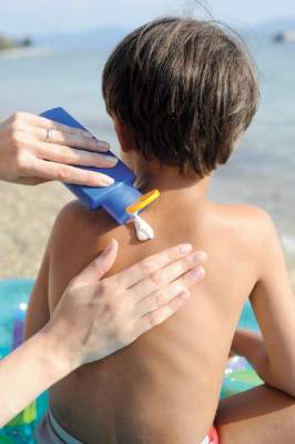
An intervention consisting of text-message reminders, read-along books, and swim shirts achieved significant improvements in sun protection behaviors in children, compared with information about sun protection alone, according to the results of a randomized controlled trial.
The study, published online Feb. 8 in JAMA Pediatrics, enrolled 300 caregiver-child pairs (children aged 2-6 years), randomizing 153 to receive a read-along book emphasizing sun protection behaviors, a swim shirt, and weekly text messages asking about sun protection measures undertaken and 147 to the usual information about sun protection given at a well-child visit.
After 4 weeks, the intervention group showed significantly higher scores for sunscreen use both on sunny and cloudy days, significantly higher scores relating to wearing a shirt on sunny days, and significantly lower increases in skin melanin indices on the sun-protected upper arm, compared with the control group (JAMA Pediatr. 2016 Feb 8. doi: 10.1001/jamapediatrics.2015.4373).
“Pediatricians’ seasonal age-specific sun protection recommendations will be more effective if supported by an effective, easily accessible, multicomponent program that can be reinforced at home,” said Byron K. Ho of Northwestern University, Chicago, and his coauthors.
The study was funded by the Pediatric Sun Protection Foundation. No conflicts of interest were declared.
The choice by the investigators to educate both the caregiver and the child through the use of an active read-aloud book was a pragmatic one because this process engages both caregivers and their children to recruit each other in reinforcing recommended behaviors.
Supplying rather than simply recommending sun-protective clothing as part of the study encourages adherence by eliminating the obstacle of having families purchase the sun-protective clothing themselves, thus removing associated economic barriers.
D. Albert C. Yan and Dr. Leslie Castelo-Soccio are with the section of dermatology at the Children’s Hospital of Philadelphia and from the departments of pediatrics and dermatology at the University of Pennsylvania. These comments are excerpted from an accompanying editorial (JAMA Pediatr. Feb 8. doi: 10.1001/jamapediatrics.2015.4524). Dr. Yan declared consultancies for Galderma, Johnson & Johnson, Pierre Fabre, and Procter & Gamble. No other conflicts of interest were declared.
An intervention consisting of text-message reminders, read-along books, and swim shirts achieved significant improvements in sun protection behaviors in children, compared with information about sun protection alone, according to the results of a randomized controlled trial.
The study, published online Feb. 8 in JAMA Pediatrics, enrolled 300 caregiver-child pairs (children aged 2-6 years), randomizing 153 to receive a read-along book emphasizing sun protection behaviors, a swim shirt, and weekly text messages asking about sun protection measures undertaken and 147 to the usual information about sun protection given at a well-child visit.
After 4 weeks, the intervention group showed significantly higher scores for sunscreen use both on sunny and cloudy days, significantly higher scores relating to wearing a shirt on sunny days, and significantly lower increases in skin melanin indices on the sun-protected upper arm, compared with the control group (JAMA Pediatr. 2016 Feb 8. doi: 10.1001/jamapediatrics.2015.4373).
“Pediatricians’ seasonal age-specific sun protection recommendations will be more effective if supported by an effective, easily accessible, multicomponent program that can be reinforced at home,” said Byron K. Ho of Northwestern University, Chicago, and his coauthors.
The study was funded by the Pediatric Sun Protection Foundation. No conflicts of interest were declared.
FROM JAMA PEDIATRICS
Key clinical point: An intervention of books, text-message reminders, and swim shirts can improve sun protection behaviors in children.
Major finding: A multimodal 4-week intervention aimed at increasing sun protection behavior achieved significant increases in sunscreen use and shirt wearing.
Data source: Randomized controlled study involving 300 caregiver-child pairs.
Disclosures: The study was funded by the Pediatric Sun Protection Foundation. No conflicts of interest were declared.

Who among us has not asked a patient to keep track of a mole?
“Keep an eye on that one, and call me if it changes,” is as much a stock phrase for dermatologists as “Wear your sunscreen.” Yet, how do patients know if a mole changes? I’m quite sure many of my patients wouldn’t notice if I shaved my head and grew a beard, let alone notice if 1 of 30 moles on their back changed color.
Mole Mapper is an iPhone app developed by the department of dermatology at Oregon Health and Science University (OHSU) to solve this problem. The app provides a framework for patients to photo, measure, and track their moles. With clear instructions, an anatomical map, and sophisticated markers, it is a significant aid for motivated patients who want medical-grade photos suitable for tracking.

To standardize the photos, the app prompts you to include a nickel, dime, or quarter in photos with nevi of interest. The user then calibrates the app by pinching onscreen circles overlying the photo such that they correspond exactly to the circumference of the coin and to the mole. Using a coin as a standard, the app then calculates the precise size of the mole regardless of the size of the photo. For example, photos taken 2 feet and 4 feet away both give the same diameter because both photos are calibrated by the dime in each.
The app was developed by a cancer biologist, Dan Webster, Ph.D., to help his wife monitor her moles between dermatology appointments. Interestingly, it was largely developed by a single person, a sign that creating apps is nearly entering into a DIY era. This increases the possibility for useful health care tools to be developed while also increasing the already crushing crowd of apps, few of which are truly useful.
The app’s functionality would not have been possible without the inclusion of Apple’s ResearchKit and Sage Bionetworks’ Bridge Server. ResearchKit provided open-source tools to facilitate informed consent over the phone and the ability to conduct participant surveys, among other activities. Bridge Server enabled the app to encrypt and securely transfer participant data from the phone to firewalled storage. The combination of these two software frameworks is paving the way for an exciting future of integrated technology and biomedical research.
According to Dan Webster, “ResearchKit is a game-changer because it provides an open-source platform for elegant informed consent, measurement tools, and participant data protection. The ability for participants to have so few barriers to contribute to a research study is the truly transformative aspect of ResearchKit, and we have seen unprecedented numbers of research study enrollees as a result.”
But that’s not all. The app is more than just a consumer tool for tracking – ResearchKit allows OHSU researchers to gather data on nevi, track them over time, and learn characteristics associated with melanoma from user-generated outcomes. This could significantly increase our understanding of melanoma and perhaps spawn an artificially intelligent app that learns to diagnose melanoma without human assistance.
Because of the institutional review board’s requirements for their research, users must be 18 years old to participate in the study. The app gracefully walks users through the consent process and even has a knowledge check at the end to ensure that they understood the risks and benefits of participating. The consent process is so streamlined that it ought to be a model for us to consent any patient for any reason.
To be clear, the app does not make diagnoses. It only provides a framework for patients to photograph their moles and track them. It also politely prompts users to rephotograph moles every 30 days so changes can be recorded.
There are apps with similar names, so be sure you have Mole Mapper from Sage Bionetworks. I tried it out to offer my experience here. Taking photos was as simple as any photo on an iPhone. Like any selfie, however, there are azimuth limits to the human arm – you can’t get shots in remote bodily corners easily. Also, placing a coin on yourself is easier said than done, unless you want to use your bubble gum to hold it in place while you take the shot. (I asked for assistance from my wife instead.)
The photos I took were accurate when compared with the measured diameter in real life, but there are still user-dependent adjustments that could lead to large artifacts. Making the measurement circles even slightly smaller or larger around the coin or the mole can lead to more than a millimeter of margins of error. If detecting melanoma requires less than 1-mm error in mole changes, then this could limit its usefulness.
Whether or not it leads to an app that automatically diagnoses melanoma from patient mole selfies, Mole Mapper has value. Any tool that empowers patients to be actively involved in their care and to meticulously monitor their moles will surely help us in keeping them safe.
Dr. Benabio is a partner physician in the department of dermatology of the Southern California Permanente Group in San Diego, and a volunteer clinical assistant professor at the University of California, San Diego. Dr. Benabio is @dermdoc on Twitter. He has no conflicts relating to the topic of this column.
“Keep an eye on that one, and call me if it changes,” is as much a stock phrase for dermatologists as “Wear your sunscreen.” Yet, how do patients know if a mole changes? I’m quite sure many of my patients wouldn’t notice if I shaved my head and grew a beard, let alone notice if 1 of 30 moles on their back changed color.
Mole Mapper is an iPhone app developed by the department of dermatology at Oregon Health and Science University (OHSU) to solve this problem. The app provides a framework for patients to photo, measure, and track their moles. With clear instructions, an anatomical map, and sophisticated markers, it is a significant aid for motivated patients who want medical-grade photos suitable for tracking.
To standardize the photos, the app prompts you to include a nickel, dime, or quarter in photos with nevi of interest. The user then calibrates the app by pinching onscreen circles overlying the photo such that they correspond exactly to the circumference of the coin and to the mole. Using a coin as a standard, the app then calculates the precise size of the mole regardless of the size of the photo. For example, photos taken 2 feet and 4 feet away both give the same diameter because both photos are calibrated by the dime in each.
The app was developed by a cancer biologist, Dan Webster, Ph.D., to help his wife monitor her moles between dermatology appointments. Interestingly, it was largely developed by a single person, a sign that creating apps is nearly entering into a DIY era. This increases the possibility for useful health care tools to be developed while also increasing the already crushing crowd of apps, few of which are truly useful.
The app’s functionality would not have been possible without the inclusion of Apple’s ResearchKit and Sage Bionetworks’ Bridge Server. ResearchKit provided open-source tools to facilitate informed consent over the phone and the ability to conduct participant surveys, among other activities. Bridge Server enabled the app to encrypt and securely transfer participant data from the phone to firewalled storage. The combination of these two software frameworks is paving the way for an exciting future of integrated technology and biomedical research.
According to Dan Webster, “ResearchKit is a game-changer because it provides an open-source platform for elegant informed consent, measurement tools, and participant data protection. The ability for participants to have so few barriers to contribute to a research study is the truly transformative aspect of ResearchKit, and we have seen unprecedented numbers of research study enrollees as a result.”
But that’s not all. The app is more than just a consumer tool for tracking – ResearchKit allows OHSU researchers to gather data on nevi, track them over time, and learn characteristics associated with melanoma from user-generated outcomes. This could significantly increase our understanding of melanoma and perhaps spawn an artificially intelligent app that learns to diagnose melanoma without human assistance.
Because of the institutional review board’s requirements for their research, users must be 18 years old to participate in the study. The app gracefully walks users through the consent process and even has a knowledge check at the end to ensure that they understood the risks and benefits of participating. The consent process is so streamlined that it ought to be a model for us to consent any patient for any reason.
To be clear, the app does not make diagnoses. It only provides a framework for patients to photograph their moles and track them. It also politely prompts users to rephotograph moles every 30 days so changes can be recorded.
There are apps with similar names, so be sure you have Mole Mapper from Sage Bionetworks. I tried it out to offer my experience here. Taking photos was as simple as any photo on an iPhone. Like any selfie, however, there are azimuth limits to the human arm – you can’t get shots in remote bodily corners easily. Also, placing a coin on yourself is easier said than done, unless you want to use your bubble gum to hold it in place while you take the shot. (I asked for assistance from my wife instead.)
The photos I took were accurate when compared with the measured diameter in real life, but there are still user-dependent adjustments that could lead to large artifacts. Making the measurement circles even slightly smaller or larger around the coin or the mole can lead to more than a millimeter of margins of error. If detecting melanoma requires less than 1-mm error in mole changes, then this could limit its usefulness.
Whether or not it leads to an app that automatically diagnoses melanoma from patient mole selfies, Mole Mapper has value. Any tool that empowers patients to be actively involved in their care and to meticulously monitor their moles will surely help us in keeping them safe.
Dr. Benabio is a partner physician in the department of dermatology of the Southern California Permanente Group in San Diego, and a volunteer clinical assistant professor at the University of California, San Diego. Dr. Benabio is @dermdoc on Twitter. He has no conflicts relating to the topic of this column.

Dabrafenib may be useful in patients who discontinue vemurafenib
Treatment with vemurafenib may result in severe skin toxicity, and dabrafenib appears to be useful in patients who discontinue vemurafenib due to such toxicity, according to a retrospective analysis of the cohort.
About a quarter (26%) of 131 melanoma patients treated in real life conditions with the BRAF inhibitor vemurafenib developed grade 3-4 skin toxicity, and 44% (34) of those patients permanently discontinued treatment, corresponding to 11% of the overall cohort, Dr. Lucie Peuvrel of Nantes (France) University Hospital and colleagues reported in Journal of the European Academy of Dermatology and Venereology.
Discontinuations were mainly due to rash and classic adverse skin reactions, including Stevens-Johnson syndrome, drug reaction with eosinophilia, and systemic symptoms, whereas reactions involving only photosensitivity or cutaneous carcinomas rarely resulted in treatment adjustment; 14 of the remaining 19 patients with grade 3-4 toxicity who continued treatment had no dose adjustment, and 5 had a dose reduction.
Clinical or biological abnormalities occurred in 94% of those with grade 3-4 rashes, the investigators noted (J Eur Acad Dermatol Venereol. 2016 Feb;30[2]:250-7).
Study subjects were patients with a mean age of 60 years who were treated with vemurafenib for melanoma between November 2010 and December 2014. Grade 3-4 skin toxicity that emerged within the first 4-8 weeks of treatment was significantly associated with prolonged overall survival; severe skin rashes also were associated with prolonged median overall survival. Of seven patients who switched to dabrafenib due to their skin reaction, only one experienced recurrence of the reaction.
Vemurafenib, which is approved for the treatment of unresectable stage III-IV BRAF mutant melanoma, is commonly associated with skin toxicity, with severe cases occurring only rarely; the impact of severe forms of toxicity on treatment and patient outcomes was unknown, the investigators said.
The current findings reaffirm that vemurafenib is commonly associated with skin toxicity, but rarely with severe cases. Severe skin adverse reactions permanently preclude use of vemurafenib, but dabrafenib – the only other BRAF inhibitor with market authorization for unresectable stage III-IV BRAF mutant melanoma – “seems to be beneficial in case of vemurafenib-induced skin intolerance,” they noted, adding that for other skin toxicities, including photosensitivity and cutaneous carcinoma, treatment adjustment is generally not required, and that treatment resumption at a reduced dose should be considered after skin improvement in patients with rashes.
The finding of increased survival in cases involving severe skin toxicity that emerges within 4-8 weeks of treatment initiation should be confirmed in other studies, Dr. Peuvrel and associates said.
The authors reported having no conflicts of interest.
FROM JOURNAL OF THE EUROPEAN ACADEMY OF DERMATOLOGY AND VENEREOLOGY
Key clinical point: Treatment with vemurafenib may result in severe skin toxicity, and dabrafenib appears to be useful in patients who discontinue vemurafenib due to such toxicity.
Major finding: 26% of patients developed grade 3-4 skin toxicity, and 44% of those permanently discontinued treatment with vemurafenib.
Data source: A retrospective cohort study of 131 patients.
Disclosures: The authors reported having no conflicts of interest.

Organ transplant recipients face increased risk of BCC
Recipients of a solid organ transplant face up to a sixfold increase in the risk of developing a basal cell carcinoma – a risk that seems to increase as time passes.
A pretransplant history of squamous cell carcinoma (SCC) increased this risk to 55 times that seen in the general population, Dr. Britta Krynitz and her colleagues reported (Br J Dermatol. 2015. doi: 10.1111/bjd.14153).

But even when the pretransplant SCC group was removed from the final analysis, the risk of basal cell carcinoma after transplant was five times that of the general population, “indicating that a pretransplant SCC has limited effect on BCC risk overall and that organ transplantation per se is a strong driver of posttransplant BCC risk,” wrote Dr. Krynitz of Karolinska Institute, Stockholm, and her coauthors.
“Our results strongly suggest tumor promoter effects of the immunosuppressive drugs in the pathogenesis of post-transplantation BCC,” the team said. “We speculate that calcineurin inhibitors and also antiproliferative drugs, often used in combination with corticosteroids, play a role.”
The researchers investigated the incidence of both BCC and SCC in a cohort of 4,023 patients who underwent solid organ transplant from 2004 to 2011. Their median age at the time of transplant was 53 years; most (59%) received a kidney. Other organs transplanted were liver (22%), heart and/or lung (15%), and other organs (4%). The median follow-up time was 3.4 years; the longest follow-up was 5.5 years.
Only 17 of patients had a history of melanoma, and 19 patients a history of SCC – less than 1% for each skin cancer. Seven percent (301) of patients had experienced some form of nonskin cancer.
By the end of follow-up, 341 BCCs had developed among 175 patients – an incidence of 6.7%. About half developed more than one BCC.
The researchers compared these patients to a group of almost 200,000 nontransplant patients who had developed BCC. Among these, the median age at BCC appearance was significantly older (71 years); 39% had more than one lesion.
The overall relative risk of BCC was increased sixfold in transplant recipients and was similar between the genders. However, the risk varied according to the type of organ received. Kidney recipients were at the highest risk (relative risk, 7.2), and those who received other organs had a lower risk (heart/lung: RR, 5.8; liver: RR, 2.6).
The risk also appeared to increase over time, the authors noted. From 0 to 2 years, it was 5.8; from 3 to 5 years, it increased to 7.0.
Among men, 54% of lesions appeared in the head/neck area and 35% on the trunk – a similar distribution to that seen in the nontransplant control group. Among women, there were differences between transplant patients and controls: 44% of lesions appeared on patients’ head/neck, compared with 60% in the control group, and 34% appeared on the truck, compared with 24% in the control group.
Histology was similar, as were the proportions of aggressive type II and highly aggressive type III lesions.
A total of 199 SCCs developed among 87 patients during follow-up, a ratio to BCC of 1:1.7. “The low ratio was probably due to the short follow-up in our study,” the authors noted.
The Welander Foundation, the Westerberg Foundation, and the Strategic Research Program in Epidemiology at Karolinska Institute sponsored the study. None of the authors had any financial declarations.
Recipients of a solid organ transplant face up to a sixfold increase in the risk of developing a basal cell carcinoma – a risk that seems to increase as time passes.
A pretransplant history of squamous cell carcinoma (SCC) increased this risk to 55 times that seen in the general population, Dr. Britta Krynitz and her colleagues reported (Br J Dermatol. 2015. doi: 10.1111/bjd.14153).
But even when the pretransplant SCC group was removed from the final analysis, the risk of basal cell carcinoma after transplant was five times that of the general population, “indicating that a pretransplant SCC has limited effect on BCC risk overall and that organ transplantation per se is a strong driver of posttransplant BCC risk,” wrote Dr. Krynitz of Karolinska Institute, Stockholm, and her coauthors.
“Our results strongly suggest tumor promoter effects of the immunosuppressive drugs in the pathogenesis of post-transplantation BCC,” the team said. “We speculate that calcineurin inhibitors and also antiproliferative drugs, often used in combination with corticosteroids, play a role.”
The researchers investigated the incidence of both BCC and SCC in a cohort of 4,023 patients who underwent solid organ transplant from 2004 to 2011. Their median age at the time of transplant was 53 years; most (59%) received a kidney. Other organs transplanted were liver (22%), heart and/or lung (15%), and other organs (4%). The median follow-up time was 3.4 years; the longest follow-up was 5.5 years.
Only 17 of patients had a history of melanoma, and 19 patients a history of SCC – less than 1% for each skin cancer. Seven percent (301) of patients had experienced some form of nonskin cancer.
By the end of follow-up, 341 BCCs had developed among 175 patients – an incidence of 6.7%. About half developed more than one BCC.
The researchers compared these patients to a group of almost 200,000 nontransplant patients who had developed BCC. Among these, the median age at BCC appearance was significantly older (71 years); 39% had more than one lesion.
The overall relative risk of BCC was increased sixfold in transplant recipients and was similar between the genders. However, the risk varied according to the type of organ received. Kidney recipients were at the highest risk (relative risk, 7.2), and those who received other organs had a lower risk (heart/lung: RR, 5.8; liver: RR, 2.6).
The risk also appeared to increase over time, the authors noted. From 0 to 2 years, it was 5.8; from 3 to 5 years, it increased to 7.0.
Among men, 54% of lesions appeared in the head/neck area and 35% on the trunk – a similar distribution to that seen in the nontransplant control group. Among women, there were differences between transplant patients and controls: 44% of lesions appeared on patients’ head/neck, compared with 60% in the control group, and 34% appeared on the truck, compared with 24% in the control group.
Histology was similar, as were the proportions of aggressive type II and highly aggressive type III lesions.
A total of 199 SCCs developed among 87 patients during follow-up, a ratio to BCC of 1:1.7. “The low ratio was probably due to the short follow-up in our study,” the authors noted.
The Welander Foundation, the Westerberg Foundation, and the Strategic Research Program in Epidemiology at Karolinska Institute sponsored the study. None of the authors had any financial declarations.
FROM BRITISH JOURNAL OF DERMATOLOGY
Key clinical point: The risk of a basal cell carcinoma increases after solid organ transplant.
Major finding: Transplant patients have a sixfold increased risk of BCC.
Data source: A retrospective database study of 4,000 transplant patients and almost 200,000 controls.
Disclosures: The Welander Foundation, the Westerberg Foundation, and the Strategic Research Program in Epidemiology at Karolinska Institute sponsored the study. None of the authors had any financial declarations.
Simple points about sunscreen products can address confusion over these products
GRAND CAYMAN – Are your patients noncompliant about sun protection? Do they want to use photoprotection but aren’t sure what is best for them? Perhaps they are confused by the multitude of sunscreen manufacturer’s claims or even some claims by consumer and environmental advocates.
Patients are often confused by how much sun protection factor they actually need and marketing hype often makes things worse, according to Dr. Vincent DeLeoof the department of dermatology at the University of Southern California, Los Angeles. “The use of the words ‘babies, natural, hypoallergenic, or organic’ all mean absolutely nothing when it comes to an SPF’s actual efficacy,” he said at the meeting provided by Global Academy of Medical Education.

The degree of confusion surrounding these products was illustrated in a survey of 114 patients at Chicago dermatology clinic. Of those surveyed, 80% said they purchased a sunscreen product to prevent sunburn (75%) and to prevent skin cancer (66%); having the highest SPF, a sensitive skin formulation, or being water resistant were reasons the products were chosen. But 62% could not identify information explaining its role in cancer prevention, and 77% could not identify how well the product would prevent sunburn. And nearly all participants were unsure how products prevented photoaging (JAMA Dermatol. 2015 Sep;151[9]:1028-30).
Adding to the confusion, Dr. DeLeo said, are warnings issued by some consumer advocacy groups about sunscreens that are not based on actual clinical or animal studies, but from data simulation. For example, he said there are no data to substantiate claims from such groups that oxybenzone has estrogenic effects in humans.
Consumer advocacy groups who say that sunscreens interfere with vitamin D levels may have a point, however, Dr. DeLeo noted, adding that it is time public health officials look into this issue, particularly with regards to revising recommended levels of vitamin D upward for the elderly, dark-skinned persons, and breastfed babies.
Although the Food and Drug Administration does not directly test sunscreens, it issued a 2014 guidance to manufacturers regarding which ingredients can be used and at what concentrations. Based on the FDA’s 2011 final rule on Labeling and Effectiveness Testing of OTC sunscreen products, the highest SPF currently allowed is “50+” without specifying actual numbers above 50, although Dr. DeLeo said this could be revised if industry is able to demonstrate efficacy at higher SPF values.
In an interview, however, he said that this is not likely to happen anytime soon. Citing a recent study of 40 commercially available sunscreens, including top brand names, professional skincare lines, and eponymously labeled products sold at drug store chains, more than half failed to reach the SPF level they purported to have and nine did not provide adequate broad spectrum protection.
“The takeaway is that the FDA is probably correct. Sunscreens should not be labeled higher than 50,” he said.
With the Sunscreen Innovation Act passed in 2014, the FDA is now evaluating data on new sunscreen agents that have proven effective overseas. So far, these agents have not been approved, Dr. DeLeo said in the interview.
With regards to the currently available products, “what patients really need to know is pretty simple,” Dr. DeLeo said during his presentation. An important point is that sunscreens – which work by forming a film on the stratum corneum, preventing the penetration of radiation – are effective only if used properly, he said.

A concept that is helpful for patients to understand is that while an SPF 30 product allows a person to stay in the sun twice as long as an SPF 15 product, with the same amount of protection from burning, it does not block twice as many burn rays, he noted. “When you combine them, it’s not additive, it dilutes” he said. For example, if an SPF 10 product is mixed with an SPF 20 product, the SPF is 15.
According to Dr. DeLeo, other important messages about sunscreen for patients are as follows:
• Use SPF 30 or higher.
• Apply 20-30 minutes before exposure to give the product a chance to create an effective barrier on the skin; and reapply every 2 hours or after going in the water or sweating outside.
• Use lotions or sprays, based on patient preference.
• For pregnant women or parents who don’t want to use chemical sunscreens, inorganic physical screens can be considered, although they will likely be less effective.
• Babies younger than 6 months of age can tolerate very small amounts of sunscreens.
Still, Dr. DeLeo said that the most foolproof form of photoprotection is to stay out of the sun, particularly between 10 a.m. and 4 p.m.
He disclosed being a consultant to LaRoche-Posay and Estée Lauder. Global Academy and this news organization are owned by the same parent company.
On Twitter @whitneymcknight
GRAND CAYMAN – Are your patients noncompliant about sun protection? Do they want to use photoprotection but aren’t sure what is best for them? Perhaps they are confused by the multitude of sunscreen manufacturer’s claims or even some claims by consumer and environmental advocates.
Patients are often confused by how much sun protection factor they actually need and marketing hype often makes things worse, according to Dr. Vincent DeLeoof the department of dermatology at the University of Southern California, Los Angeles. “The use of the words ‘babies, natural, hypoallergenic, or organic’ all mean absolutely nothing when it comes to an SPF’s actual efficacy,” he said at the meeting provided by Global Academy of Medical Education.
The degree of confusion surrounding these products was illustrated in a survey of 114 patients at Chicago dermatology clinic. Of those surveyed, 80% said they purchased a sunscreen product to prevent sunburn (75%) and to prevent skin cancer (66%); having the highest SPF, a sensitive skin formulation, or being water resistant were reasons the products were chosen. But 62% could not identify information explaining its role in cancer prevention, and 77% could not identify how well the product would prevent sunburn. And nearly all participants were unsure how products prevented photoaging (JAMA Dermatol. 2015 Sep;151[9]:1028-30).
Adding to the confusion, Dr. DeLeo said, are warnings issued by some consumer advocacy groups about sunscreens that are not based on actual clinical or animal studies, but from data simulation. For example, he said there are no data to substantiate claims from such groups that oxybenzone has estrogenic effects in humans.
Consumer advocacy groups who say that sunscreens interfere with vitamin D levels may have a point, however, Dr. DeLeo noted, adding that it is time public health officials look into this issue, particularly with regards to revising recommended levels of vitamin D upward for the elderly, dark-skinned persons, and breastfed babies.
Although the Food and Drug Administration does not directly test sunscreens, it issued a 2014 guidance to manufacturers regarding which ingredients can be used and at what concentrations. Based on the FDA’s 2011 final rule on Labeling and Effectiveness Testing of OTC sunscreen products, the highest SPF currently allowed is “50+” without specifying actual numbers above 50, although Dr. DeLeo said this could be revised if industry is able to demonstrate efficacy at higher SPF values.
In an interview, however, he said that this is not likely to happen anytime soon. Citing a recent study of 40 commercially available sunscreens, including top brand names, professional skincare lines, and eponymously labeled products sold at drug store chains, more than half failed to reach the SPF level they purported to have and nine did not provide adequate broad spectrum protection.
“The takeaway is that the FDA is probably correct. Sunscreens should not be labeled higher than 50,” he said.
With the Sunscreen Innovation Act passed in 2014, the FDA is now evaluating data on new sunscreen agents that have proven effective overseas. So far, these agents have not been approved, Dr. DeLeo said in the interview.
With regards to the currently available products, “what patients really need to know is pretty simple,” Dr. DeLeo said during his presentation. An important point is that sunscreens – which work by forming a film on the stratum corneum, preventing the penetration of radiation – are effective only if used properly, he said.
A concept that is helpful for patients to understand is that while an SPF 30 product allows a person to stay in the sun twice as long as an SPF 15 product, with the same amount of protection from burning, it does not block twice as many burn rays, he noted. “When you combine them, it’s not additive, it dilutes” he said. For example, if an SPF 10 product is mixed with an SPF 20 product, the SPF is 15.
According to Dr. DeLeo, other important messages about sunscreen for patients are as follows:
• Use SPF 30 or higher.
• Apply 20-30 minutes before exposure to give the product a chance to create an effective barrier on the skin; and reapply every 2 hours or after going in the water or sweating outside.
• Use lotions or sprays, based on patient preference.
• For pregnant women or parents who don’t want to use chemical sunscreens, inorganic physical screens can be considered, although they will likely be less effective.
• Babies younger than 6 months of age can tolerate very small amounts of sunscreens.
Still, Dr. DeLeo said that the most foolproof form of photoprotection is to stay out of the sun, particularly between 10 a.m. and 4 p.m.
He disclosed being a consultant to LaRoche-Posay and Estée Lauder. Global Academy and this news organization are owned by the same parent company.
On Twitter @whitneymcknight
AT THE CARIBBEAN DERMATOLOGY SYMPOSIUM

Time will tell if medical apps help or compete with dermatologists
GRAND CAYMAN ISLAND – Smartphones and other digital technologies could provide clinicians – and patients – the tools to greatly improve melanoma and other dermatology outcomes, but how might they change actual practice?
“Ten years from now, what is going to distinguish the app world from the dermatology community?” That was the question Dr. Allan C. Halpern, chief of the dermatology service at Memorial Sloan Kettering Cancer Center, New York, posed at the meeting, sponsored by Global Academy for Medical Education.

Many clinicians already use dermatoscopes, which Dr. Halpern said “significantly improves diagnostic accuracy for melanoma by making available to us visual cues that would not be available through routine naked-eye examination.” However, he said, a rapid digital evolution in dermoscopy is already underway, allowing clinicians to use mobile devices that store images and perform short term monitoring of any areas of concern.
“For suspicious lesions, rather than do a biopsy, one of the options is that you can take a dermoscopic image of it, then do so again in another couple of months. If it hasn’t changed, then the data are very convincing that you’re dealing with something other than melanoma,” Dr. Halpern said.
What gives him pause is that there are an increasing number of consumer-based smartphone medical products, including more than 250 for dermatology alone.
“For about 100 dollars or less, patients can purchase their own dermatoscope and attach it to their smartphone” and take images of their own lesions, Dr. Halpern said.
As a result, he believes that the field has reached the “tipping point” for successful teledermatology, where companies can accept and interpret these data.
Because of the exponential growth of mobile medical applications, the Food and Drug Administration issued guidance for their use in 2015, he noted.
Although he said that the FDA is overwhelmed by the task – and that the medical app marketplace is akin to the Wild West – medical apps are here to stay and will continue to develop quickly.
“For one thing, big players are now entering the field,” he said, alluding to a recent Apple initiativeto collect data uploaded by consumers to the Cloud, which can then be parsed by medical researchers. In addition to melanoma, Apple is currently focusing on autism and epilepsy.
“Apple is trying to make the iPhone a conduit for medical research,” Dr. Halpern said.
Over the next 5-10 years, there will be a race to see who can perfect this kind of data analysis. There is even an ongoing competition that pits melanoma researchers around the world against one another to see who can most accurately interpret images uploaded to the archives. The hope, he said, is that it will help dermatologists to develop and agree upon international standards for melanoma diagnosis.
With this kind of standardization, medical apps can help both consumers and practitioners triage care.
“I don’t know when it will happen, but there is no doubt we’re going to reach a point at which these apps are going to be as good as the average dermatologist is today when it comes to looking at a single spot,” Dr. Halpern said.
Aside from some cancer centers’ ability to create three-dimensional images of lesions, another distinguishing technology for dermatologists could be reflectance confocal microscopy.
“As dermatologists, we are trained well enough in pathology that we can use this technology, we will be able to do even better than with dermoscopy alone,” he said. This kind of technology captures images at the cellular level similar to histological examinations, but without the actual biopsy.
In an interview, Dr. Halpern said that while dermatologists have nothing to fear from apps in daily practice, there are some issues to be aware of. “The first is the one we’re most concerned about – automated diagnosis, before we know for certain that the apps work.”
Another issue is that these apps raise awareness among consumers about their individual risk for melanoma, which will “invariably help drive more people in the door,” but there may not be enough clinicians to manage these patients.
The group Dr. Halpern expects will be the most frequent users of personal dermatology technologies initially are “ultra–tech savvy young people motivated to avoid melanoma,” although he expects that over time, this could change as technologies become easier to use.
He disclosed financial relationships with Quintiles, Canfield Scientific, DermTech, SciBase, and Caliber ID.
Global Academy and this news organization are owned by the same parent company.
On Twitter @whitneymcknight
GRAND CAYMAN ISLAND – Smartphones and other digital technologies could provide clinicians – and patients – the tools to greatly improve melanoma and other dermatology outcomes, but how might they change actual practice?
“Ten years from now, what is going to distinguish the app world from the dermatology community?” That was the question Dr. Allan C. Halpern, chief of the dermatology service at Memorial Sloan Kettering Cancer Center, New York, posed at the meeting, sponsored by Global Academy for Medical Education.
Many clinicians already use dermatoscopes, which Dr. Halpern said “significantly improves diagnostic accuracy for melanoma by making available to us visual cues that would not be available through routine naked-eye examination.” However, he said, a rapid digital evolution in dermoscopy is already underway, allowing clinicians to use mobile devices that store images and perform short term monitoring of any areas of concern.
“For suspicious lesions, rather than do a biopsy, one of the options is that you can take a dermoscopic image of it, then do so again in another couple of months. If it hasn’t changed, then the data are very convincing that you’re dealing with something other than melanoma,” Dr. Halpern said.
What gives him pause is that there are an increasing number of consumer-based smartphone medical products, including more than 250 for dermatology alone.
“For about 100 dollars or less, patients can purchase their own dermatoscope and attach it to their smartphone” and take images of their own lesions, Dr. Halpern said.
As a result, he believes that the field has reached the “tipping point” for successful teledermatology, where companies can accept and interpret these data.
Because of the exponential growth of mobile medical applications, the Food and Drug Administration issued guidance for their use in 2015, he noted.
Although he said that the FDA is overwhelmed by the task – and that the medical app marketplace is akin to the Wild West – medical apps are here to stay and will continue to develop quickly.
“For one thing, big players are now entering the field,” he said, alluding to a recent Apple initiativeto collect data uploaded by consumers to the Cloud, which can then be parsed by medical researchers. In addition to melanoma, Apple is currently focusing on autism and epilepsy.
“Apple is trying to make the iPhone a conduit for medical research,” Dr. Halpern said.
Over the next 5-10 years, there will be a race to see who can perfect this kind of data analysis. There is even an ongoing competition that pits melanoma researchers around the world against one another to see who can most accurately interpret images uploaded to the archives. The hope, he said, is that it will help dermatologists to develop and agree upon international standards for melanoma diagnosis.
With this kind of standardization, medical apps can help both consumers and practitioners triage care.
“I don’t know when it will happen, but there is no doubt we’re going to reach a point at which these apps are going to be as good as the average dermatologist is today when it comes to looking at a single spot,” Dr. Halpern said.
Aside from some cancer centers’ ability to create three-dimensional images of lesions, another distinguishing technology for dermatologists could be reflectance confocal microscopy.
“As dermatologists, we are trained well enough in pathology that we can use this technology, we will be able to do even better than with dermoscopy alone,” he said. This kind of technology captures images at the cellular level similar to histological examinations, but without the actual biopsy.
In an interview, Dr. Halpern said that while dermatologists have nothing to fear from apps in daily practice, there are some issues to be aware of. “The first is the one we’re most concerned about – automated diagnosis, before we know for certain that the apps work.”
Another issue is that these apps raise awareness among consumers about their individual risk for melanoma, which will “invariably help drive more people in the door,” but there may not be enough clinicians to manage these patients.
The group Dr. Halpern expects will be the most frequent users of personal dermatology technologies initially are “ultra–tech savvy young people motivated to avoid melanoma,” although he expects that over time, this could change as technologies become easier to use.
He disclosed financial relationships with Quintiles, Canfield Scientific, DermTech, SciBase, and Caliber ID.
Global Academy and this news organization are owned by the same parent company.
On Twitter @whitneymcknight
AT THE CARIBBEAN DERMATOLOGY SYMPOSIUM

MAPK inhibitor combo offers prolonged survival from BRAF+ advanced melanoma
Combination therapy to inhibit the MAP kinase pathway is associated with a median overall survival of more than 2 years for patients with metastatic melanoma positive for the BRAF V600 mutation who have not previously received a BRAF inhibitor.
An analysis of data from a phase I and II trial of the BRAF inhibitor dabrafenib (Tafinlar), 150 mg twice daily, and the MEK inhibitor trametinib (Mekinist), 2 mg once daily, in 78 BRAF inhibitor–naïve patients with metastatic melanoma showed that in a non-randomized cohort (part B) the median overall survival (OS) was 27.4 months. In a cohort randomized to the same doses (part C), the median OS was 25 months, reported Dr. Georgina V. Long and her colleagues from the Melanoma Institute Australia and the University of Sydney in the Journal of Clinical Oncology.
Among 24 patients treated in part B, progression-free survival (PFS) was 18% at 3 years.
“The combination has an acceptable long-term safety profile and is a standard of care for patients with BRAF mutation–positive metastatic melanoma, particularly given the recent publications demonstrating a significant improvement in the PFS and OS in phase III trials of combination versus single-agent BRAF inhibitors,” the investigators wrote (J Clin Oncol. 2016 Jan 25. doi: 10.1200/JCO.2015.62.9345).
The investigators conducted the analysis to determine which patients were the most likely to benefit from combination therapy of the MAP (mitogen-active protein) kinase pathway.
They observed that in the part B, non-randomized cohort, OS was 72% at 1 year, 60% at 2 tears, and 47% at 3 years. In the part C, randomized cohort, OS was 80%, 51%, and 38%, respectively.
Factors associated with better OS in Cox proportional hazards regression models included metastases to fewer than three organs sites, and lower levels of lactate dehydrogenase (LDH) at baseline. Not surprisingly, a complete response to therapy was also associated with better survival. Three-year OS rates were 62% for patients with normal baseline LDH levels, and 63% for patients who had a complete response.
FROM THE JOURNAL OF CLINICAL ONCOLOGY
Key clinical point: Combination MAP kinase pathway inhibition was associated with prolonged survival of BRAF mutation-positive metastatic melanoma.
Major finding: Median overall survival was 27.4 months in a non-randomized cohort, and 25 months in a randomized cohort.
Data source: Analysis of a subset of patients in a phase I and II trial of dabrafenib and trametinib.
Disclosures: The study was supported by GlaxoSmithKline. Dr. Long and many coauthors disclosed consulting and/or research funding from the company.
Anorectal melanoma rates rose significantly in 20-year analysis
Rates of anorectal melanoma rose substantially in the United States from 1992 to 2011, according to a study published in Dermatologic Surgery.
The increases affected men and women, said Dr. Adrienne Callahan of the department of dermatology, University Hospitals Case Medical Center, Cleveland, and her associates. Anorectal carcinoma was significantly more common among Hispanic whites than non-Hispanic whites (P = .02), “suggesting that this population may be targeted for screening interventions,” they added. (Dermatol Surg. 2016;42[1]:94-9). The highest rates were in Hispanic white elderly women.
Anorectal melanoma accounts for 1.3% of all melanomas and 16.5% of mucosal melanomas, and is most common among older women. The prognosis is often poor because the cancer tends to be asymptomatic until its late stages. To study the epidemiology of anorectal melanoma, the researchers analyzed data from the Surveillance, Epidemiology, and End Results 13 (SEER 13) Registries Database for 1992 through 2011.
The SEER database listed 260 cases for the study period. Most involved the rectum, 58% affected adults aged 65 years and older, and almost two-thirds occurred in women – a finding that dovetails with other studies, the researchers said. Notably, the estimated annual change in age-standardized incidence rates increased in both men (5.08%) and women (3.02%), which were statistically significant increases (P less than .05 for both trends).
Anorectal melanoma rates were significantly higher among Hispanic whites, compared with non-Hispanic whites. “Other studies have indicated that Hispanics are underscreened for skin cancer compared to other ethnic groups despite the increasing incidence of melanoma in this population,” said the researchers. “Although this may suggest a role for improved screening among those with Hispanic ethnicity, additional studies must be done to corroborate these results and further elucidate the association of Hispanic ethnicity with anorectal melanoma.”
As far as they know, this is the first study that has analyzed anorectal melanoma incidence in Hispanic whites and non-Hispanic whites, they added.
Ethnicity did not affect survival, which was generally poor. Anorectal melanoma is very rare, so the number of cases was relatively small and rates might have been unstable, the investigators noted. They added that it was not known whether the increases they found were related to improved detection.
The study was funded by the Char and Chuck Fowler Family Foundation, the Dermatology Foundation, and the National Cancer Institute. The researchers had no disclosures.
FROM DERMATOLOGIC SURGERY
Key clinical point: Rates of anorectal melanoma rose substantially in the United States over 2 decades.
Major finding: The age-adjusted incidence rate for anorectal melanoma increased annually by an estimated 5.1% for men and 3.0% for women, over a recent 20-year period.
Data source: The study analyzed data from the Surveillance, Epidemiology, and End Results (SEER) 13 Registries Database between 1992 and 2011.
Disclosures: The study was funded by the Char and Chuck Fowler Family Foundation, the Dermatology Foundation, and the National Cancer Institute. The researchers had no disclosures.
Sirolimus reduced posttransplant skin cancer risk
Sirolimus protects organ-transplant recipients against developing skin cancer, reducing their risk by 40%, according to a retrospective cohort study published in JAMA Dermatology on Jan. 20.
Recipients of solid organs are at three- to fourfold higher risk of developing cancer, compared with the general population, and the most common type they get is nonmelanoma skin cancer. The risk of developing cutaneous squamous cell carcinoma is 65-250 times higher in organ-transplant recipients. Drugs that reduce the growth and proliferation of tumor cells by inhibiting mTOR (mammalian target of rapamycin), including sirolimus, are believed to reduce this cancer risk, said Pritesh S. Karia of the department of dermatology, Brigham and Women’s Hospital and Harvard University, Boston, and his associates (JAMA Dermatol. 2016 Jan 20. doi: 10.1001/jamadermatol.2015.5548).
The investigators reviewed the electronic medical records of 329 patients (mean age, 56 years) who underwent organ transplantation at one of the two medical centers during a 9-year period and who then developed a cancer of any type. The study participants received renal (53.8%), heart (17.6%), lung (16.4%), liver (10.3%), or mixed-organ (1.8%) transplants. The most common index cancers they developed post transplant included cutaneous squamous cell carcinoma (31.9%), basal cell carcinoma (22.5%), and melanoma (2.7%).
Of the 329 patients, 97 (29.5%) then received sirolimus, while 232 (70.5%) did not. During a median follow-up of 38 months, 130 of these patients (39.5%) developed a second posttransplant cancer. The sirolimus-treated group showed a reduction in risk for cancer of any type, compared with the group that did not receive sirolimus (30.9% of 97 vs. 43.1% of 232).
Nearly all (88.5%) of the second posttransplant cancers that developed were skin cancers, and sirolimus reduced the risk of skin cancers by 40%. The 1-year, 3-year, and 5-year rates of skin cancer after an index posttransplant cancer were 9.3%, 20.6%, and 24.7% in the sirolimus group, compared with 17.7%, 31.0%, and 35.8%, respectively, in the untreated group, “thus demonstrating a lower risk for skin cancer with sirolimus treatment,” they said.
“Even for patients who have already had difficulty with skin cancer formation, mTOR inhibition appears to be of benefit. No difference in cancer outcomes was observable between sirolimus-treated and [untreated] groups because poor outcomes were rare,” Mr. Karia and his associates wrote.
These findings suggest that sirolimus chemoprevention should be considered for the subset of organ-transplant recipients who develop post-transplant cancer, they noted. The results also highlight the need for dermatologists and transplant physicians “to be aware of skin cancer history, coordinate regular posttransplant surveillance of skin cancers” in patients with organ transplant recipients, especially those with a history of skin cancer, and to communicate closely “as skin cancers form to consider reduction in immunosuppressive therapy or conversion to an mTOR-based regimen if skin cancer formation is of concern,” they added.
This study was supported by sirolimus manufacturer Novartis Pharmaceuticals. Mr. Karia and his associates reported having no relevant financial disclosures.
FROM JAMA DERMATOLOGY
Key clinical point: Sirolimus protects organ-transplant recipients against skin cancer.
Major finding: The 1-year, 3-year, and 5-year rates of skin cancer after an index posttransplant cancer were 9.3%, 20.6%, and 24.7% in the sirolimus group, compared with 17.7%, 31.0%, and 35.8% in the untreated group.
Data source: A retrospective cohort study of 329 organ-transplant recipients who had already developed one cancer likely related to their immunosuppressive therapy.
Disclosures: This study was supported by sirolimus manufacturer Novartis Pharmaceuticals. Mr. Karia and his associates reported having no relevant financial disclosures.
Encouraging results for MMS in digital melanoma cases
Mohs micrographic surgery (MMS) to remove digital melanomas of less than 2 mm Breslow thickness spared patients from amputation without undermining survival, according to a single-center retrospective study published in Dermatologic Surgery.
The overall recurrence rate was 8%, and there were no recurrences with use of the MART-1 (melanoma antigen recognized by T-cells 1 staining) immunostain, said Dr. Vitaly Terushkin of the Zitelli and Brodland Skin Cancer Center in Pittsburgh and his associates. “The use of MART-1 has revolutionized the value of MMS,” the researchers said, while cautioning that the sample size was too small to draw reliable conclusions about thicker tumors.
Digital melanoma historically eluded diagnosis until it was advanced and was treated with amputation. Amputation and wide local excision remain common, and particularly erode function and balance when the tumor involves the thumb or big toe, the researchers noted. In 35 years at their center, 62 patients underwent MMS for digital melanoma, two-thirds of which involved the nail unit. About 92% of tumors were primary, 8% were recurrent, 53% involved the fingers, and 47% involved the toes. About half of patients were women, and the average age at treatment was about 63 years (Dermatol Surg. 2016;42:83-93).
Nearly 97% of patients avoided amputation over an average of 6.5 years of follow-up, the investigators reported. Five-year and 10-year local recurrence-free survival rates for the primary melanomas were about 92% and 83%, respectively. Of the five local recurrences, three were salvaged with additional MMS and two were treated with an amputation; all five recurrences involved the nail unit. Failing to remove the entire nail unit apparatus probably led to false-negative MMS layers and subsequent recurrence in at least two cases, they pointed out.
This is only the fifth published study to describe the treatment of digital melanoma with MMS, according to Dr. Terushkin and his associates. Although the study lacked a comparison group, it suggested that MART-1 improved outcomes and that most recurrences could be retreated with MMS without amputation or negative effects on survival. These findings will hopefully help inform future melanoma guidelines, said the researchers.
The investigators reported no funding sources and had no disclosures.
FROM DERMATOLOGIC SURGERY